Note: Descriptions are shown in the official language in which they were submitted.
CA 02794303 2015-08-17
1
A DEVICE, A KIT AND A METHOD FOR HEART SUPPORT
Field of the Invention
The present invention relates to an intra-vascular blood circulation enhancing
apparatus, a
system for intra-vascular blood circulation enhancement and a method for
enhancing left ventricular
pump function of a patient. The present invention may specifically be used to
enhance the pump
function of the left ventricle as a permanent measure for treating a heart
failure disease where the heart
function is deficient.
Background of the Invention
Where the heart function is chronically insufficient, there may be a need to
permanently aid
the heart function. Heart failure (HF), more often called Congestive Heart
Failure (CHF), is in general a
condition where the heart, is unable to support the body tissue with its
metabolic demands and to
sustain adequate blood pressure and cardiac output. The term Congestive
relates to a congestion of
blood and fluids in front of the pumping ventricles as a result of
insufficient forward pumping, most often
caused by disease of the left ventricle muscle. A peculiarity of heart cells
is that they do not regenerate
after damage or cell death, thus conditions have a tendency to worsen rather
than heal after heart cell
damage. There are many reasons for heart cell death, the most common cause is
ischemic heart
disease, a condition where the arteries feeding the heart muscle get clogged,
causing myocardial
infarctions (Ml). Viruses may damage the muscle cells, and some diseases, for
instance
cardiomyopathy have unknown reasons. End stage of long standing high blood
pressure may also
cause end stage heart failure. Heart strengthening drugs like digoxin or
treatment with diuretics help for
a while, but are all only treating symptoms. CHF is a progressive untreatable,
disabling and finally a
deadly condition. According to the American Heart Association homepage, there
are in the US at
present more than 5 Million patients living with CHF and 550 000 are added
every year. 40 000 in the
US are in such a bad state that only a heart transplant will keep them alive.
However, due to the limited
number of suitable organs only 2500 transplants are done yearly in the US. One
may extrapolate the
numbers for the rest of the industrialized world.
Total artificial heart, where the whole native heart is excised and replaced
with a mechanical
device was introduced in the 1960's by DeBakey, in the 1980's by among others
Jarvik and recently by
Copeland (CardioWest, Total Artificial Heart). However, these devices are
still based on complex
designs and are very invasive to install in the patient. Failure in operation
of the device is fatal.
There are other techniques supporting only the failing left ventricle, known
as left ventricle
assist devices (LVAD). The most popular LVADs are the Novacor and the
HeartMate devices. Common
CA 02794303 2012-09-21
WO 2011/119100
PCT/SE2011/050337
2
for this devices is the demand for major open heart surgery utilizing
extracorporeal circulation by means
of a Heart- and Lung-machine while stopping (or excising) the heart. These are
bulky devices, a
Novacor weights 1.800 grams, a HeartMate 1.200 grams. There are smaller axial
flow pumps available
nowadays, the HeartMate II, the Jarvik 2000 and the MicroMed DeBakey VAD. In
addition, major open
heart surgery is still necessary to install and connect these devices to the
left ventricle cavity and the
aorta by means of large vascular grafts. The mentioned devices have almost
exclusively been used as a
bridge to a heart transplant due to high frequency of complications, high
mortality and limited durability.
Their use has also been limited because of high prices of up to 150 000 $ only
for the device.
None of the devices for permanent implant described are feasible for minimal
invasive
1 0 catheter based insertion, on the contrary, they all involve major open
heart surgery. There is obviously a
demand for simpler devices, it is the scope of the here presented invention to
omit major cardiac
surgery and to allow implant with catheter technique.
Moreover, health care is permanently searching for improved devices and
methods.
Hence, there is a need of an improved system and/or method for permanently
enhancing or
assisting left ventricular pump function of a heart of a patient. The system
is advantageously not
interfering with the cardiac cycle of the heart.
Hence, an improved system and/or method for permanently enhancing or assisting
left
ventricular pump function of a heart of a patient would be advantageous and in
particular allowing for
increased flexibility, cost-effectiveness, long-term function, and/or patient
friendliness would be
advantageous.
Summary of the Invention
Accordingly, embodiments of the present invention preferably seek to mitigate,
alleviate or
eliminate one or more deficiencies, disadvantages or issues in the art, such
as the above-identified,
singly or in any combination by providing a medical device, a kit, a method,
and a computer-readable
2 5 medium, according to the appended patent claims.
Embodiments of the invention take advantage of an improved understanding of
left ventricular
pump action and the close relationship between the Coronary Sinus (CS), the
Great Cardiac Vein
(GCV) and the Mitral valve (MV). Embodiments of the invention are providing
movement of the CS and
the GCV and thereby the MV along the long axis of the left ventricle (LV)
towards and/or away from the
3 0 heart apex, in synchrony with the cardiac cycle. In some embodiments
energy is provided for this
assisting movement. The here described embodiments of permanent implants do
not take over or
replace the remaining left ventricular pump function, they rather augment,
improve, enhance or support
the remaining natural pump function by means of an at least partly increased
up and/or down movement
of the mitral valve that works as a blood displacement or propulsion piston,
when it is closed during the
35 systole.
The here presented innovation is based on recent understanding of how the left
ventricle
functions and also on utilizing an undiscovered favourable anatomy of the left
heart. Modern catheter
based technology is integrated in embodiments of the here described device,
system and methods.
CA 02794303 2012-09-21
WO 2011/119100
PCT/SE2011/050337
3
Modern imaging of the beating heart has contributed largely to the
understanding of left
ventricle pump action. The pumping force of the left ventricle has before been
understood to be a result
of the heart muscle contracting and squeezing (systole) around the amount of
blood enclosed inside the
left ventricle after closure of the mitral valve, increasing the pressure and
thereby forcing the blood
towards the aortic valve, forcing this to open and ejecting the blood into the
ascending aorta. When the
squeezing is completed, an intermission occurs (diastole), during which a new
portion of blood enters
the left ventricle cavity from the left atrium.
Ultrasound imaging and Magnetic Resonance Imaging (MRI) has revealed that this
previously
taught mode of function is not completely true. Instead, one may describe two
types of pump action, a
1 o long axis and a short axis action. MRI can show that there is a
movement of the atrioventricular mitral
valve (MV) plane downwards along the left ventricle long axis that extends
from the atrium towards the
ventricle's lower end, the apex. The left ventricle muscle cells are pulling
the whole mitral valve plane,
including the mitral valve annulus and part of the left atrial wall (that is
stretching) towards the heart
apex. By pulling the closed mitral valve towards the heart apex, the mitral
valve becomes a piston in a
blood displacement pump.
The downwards movement of the mitral valve is in a healthy human up to
approximately 2
centimetres. The downwards movement accelerates the blood column away from the
left atrium and
towards the aortic valve in a continuous movement. By means of MRI technology
one is able to virtually
mark separate pixels inside the blood column and follow their movement. It is
possible to show that the
2 0 blood column flows more or less continuously from the left atrium to
the ascending aorta without ever
stopping. The blood column is accelerated by the mitral valve piston moving up
and down along the
cardiac long axis, opening every time it takes a new scoop of blood in an
upward movement to the
atrium, and closing just before moving back toward the apex.
The inventor of the present application realized that the location of the
Coronary Sinus (CS)
2 5 and the Great Cardiac Vein (GCV), very close to the mitral valve, can
be utilized for enhancement of the
left ventricular pump function. For instance a downwards movement of the
mitral valve substantially
along the long axis of the left ventricle may be supported. By actively
moving, or supporting a still
existing natural cardiac movement of, the CS and the GCV downwards towards the
apex one
simultaneously can move the mitral valve in the same direction.
3 0 The Coronary Sinus and the Great Cardiac Vein represent the large
veins of the heart. The
arterial blood of the heart passes the capillaries (the smallest vessels of
the heart) and then enters the
venous plexus in the heart tissue wall. Then the venous blood flows together
into veins located on the
heart surface. Distally the cardiac veins are small but unite together into
larger and larger veins before
flowing into the GCV and the CS. All the venous blood from the heart pours
into the CS and then flows
3 5 through the coronary sinus ostium (orifice) into the Right Atrium (RA)
on the right side of the heart.
The major part of the CS and part of the GCV is located on the left atrial
side of the mitral
valve annulus. This is the part of the LA wall that stretches in a healthy
heart when the MV is moving
down towards the apex. The GCV then crosses the MV plane and annulus towards
the LV side and join
CA 02794303 2012-09-21
WO 2011/119100
PCT/SE2011/050337
4
the anterior inter-ventricular vein on the front side of the heart. Thus the
CS and the GCV encircle at
least 2/3 of the MV circumference, substantially in the same plane as the
mitral valve plane, and are
attached or embedded in tissue adjacent to the mitral valve.
Since the ostium of the coronary sinus is on the right side of the heart in
the RA, one has easy
access to the CS, the GCV and their side branches of veins on the heart
surface by puncturing a
peripheral vein, e.g. in the groin on the neck or in an arm. By means of
modern catheter based
technique, embodiments of the here disclosed device may be placed in position
adjacent the mitral
valve without major cardiac surgery. As matter of fact it is possible to place
the device while the patient
is conscious using only local anaesthesia, a common practice for implanting
pacemakers and Infra
1 0 Cardiac Defibrillators (ICD).
According to one aspect of the invention, a medical device is provided for
enhancing intra-
cardiac blood circulation of a heart of a patient by permanently assisting
left ventricular pump action.
The device has at least one first anchor unit implanted in a cardiac vessel of
said heart, e.g. a side
branch of the coronary sinus (CS) or the great cardiac vein (GCV). The first
anchor unit may be an
expandable stent structure for anchoring the anchor unit in the cardiac
vessel, and/or wherein the first
anchor unit has at least one tissue anchoring element, such as a hook or barb.
In embodiments the device has at least one second anchor unit implanted in the
cardiac
vessel, wherein the second anchor unit is located in the CS or the GCV. The
second anchor may serve
in transferring force from a remote force generating unit.
2 0 Thus, the device has a force generating unit that is in communication
with said first and
second anchor units, wherein said force generating unit generates a force in
dependence of a cardiac
cycle of said heart. The anchor units receive said force in such a manner that
an assisted movement of
said cardiac vessel and thus said mitral valve in a mitral valve plane is
provided in a direction to and
from an apex of the heart. However, in a specific embodiment, the second
anchor unit may also have an
2 5 integrated electrical motor instead and the force generating unit is
the motor, the device having a
connecting unit between the motor and the first anchor for the communication,
and wherein the force is
provided by the motor. In turn, the integrated electrical motor is provided
with electrical energy from a
remote energy source by means of an electrical cable.
By means of the applied force, the mitral valve is during systole assisted to
move the mitral
3 0 valve plane along the long axis of the left ventricle (LV) towards an
apex of the heart and/or during
diastole assisted to move the mitral valve plane away from the apex by the
force for assisting the pump
action of the heart. The assisted movement is provided in a controlled manner
to support a natural
movement of the mitral valve. When the mitral valve movement towards the apex
is at least partly
assisted during systole, the (still existing) natural pumping force of the
heart is augmented while ejecting
3 5 blood into the aorta. When the mitral valve movement away from the apex
is at least partly assisted
during diastole, the natural filling of the left ventricle of the heart is
supported. Thus the (still existing)
natural pumping function of the heart is augmented by an improved filling
degree. The force generating
CA 02794303 2012-09-21
WO 2011/119100
PCT/SE2011/050337
unit is operatively connected to a remote energy source to receive energy
therefrom and to controllably
provide the assisting movement in synchrony with the natural heart cycle.
In some embodiments the force generating unit is an actuating unit for
providing the force as a
mechanical force, and wherein the first anchor unit and the actuating unit are
in communication via a
5 connecting unit for transferring the force and providing the movement.
In some embodiments the force is a magnetic unit for providing the force as a
magnetically
induced force. In such embodiments, the two anchors are magnetic, and wherein
the first magnetic
anchor unit and the second magnetic unit in the CS or GCV are in magnetic
communication for
transferring the force and providing the movement. At least one magnetic
anchor unit is an
1 o electromagnet. At least one of the electromagnets is arranged to change
polarity in synchrony with the
cardiac cycle. While the second electromagnet anchor always is located in the
CS or GCV, the first
magnet may be positioned in various locations. In some embodiments the first
magnet is located inside
a side branch of the vein system on the left ventricular wall, e.g. the IAV,
it may also be located in the
left ventricle attached to the LV wall, or in the right ventricle, the right
atrium or the left atrium of the
1 5 heart, or on the left ventricular outer wall of the heart. In other
embodiments the first magnetic anchor
may not be located in, but adjacent to the heart, such as on the pericardium,
the diaphragm, the spine
or thoracic cage, in the pleura or under the skin.
In some embodiments the device has a remote energy source, a control unit, and
a sensor for
measuring physiological parameters related to the cardiac cycle activity
providing a sensor signal. The
2 0 sensor signal is provided to the control unit which controls the force
generating unit to provide the
movement by energy from the remote energy source and based on the sensor
signal. The remote
energy source may have a mechanical section where rotational or linear motion
is generated. The
device further may have an extension unit extending from the mechanical
section, wherein the
mechanical section is the force generating unit and wherein the motion is
transferred in operation of the
2 5 mechanical section to the first and second anchor unit for the movement
of the mitral valve plane via an
extension unit. The remote energy source is controlled by the control unit to
provide electrical energy a)
to one or more electromagnetical anchor units affixed in relation to the
mitral valve, or b) to at least one
force generating unit arranged at or in the heart, to provide the movement of
the mitral valve plane.
In another embodiment, the first anchor unit may be implanted in the GCV or
its continuation,
3 0 more specifically in the anterior interventricular vein (AIV), and the
second anchor unit may be implanted
in the CS. An elongate extension unit connects the first and second anchor
units in a loop shape such
that they are in mechanical communication. Thus, the part of the device that
is located in the CS and the
GCV geometrically forms a loop around 2/3 of the MV, and very close to it. The
extension unit extends
proximally beyond the second anchor unit to a mechanical actuator unit
arranged to rotate the extension
3 5 unit synchronized with the cardiac cycle, wherein the device has
different operative positions upon
rotation of the extension unit, including upon rotation of the extension unit
in a first direction a diastole
operative position where the loop shaped extension unit is flexed towards the
left atrium and the CS,
GCV and MV are moved towards the left atrium, and a second operative position
where upon rotation of
CA 02794303 2012-09-21
WO 2011/119100
PCT/SE2011/050337
6
the extension unit in a second direction, opposite the first direction, where
the loop shaped extension
unit is flexed towards the LV apex and the CS, GCV and MV are moved towards
the LV apex.
In some embodiments the device is a non-powered device. The force generating
unit may be
a resilient unit, and the first anchor unit may include a distal anchor unit.
The distal anchor and a
proximal anchor unit may be arranged in the AIV, CS and GCV. The resilient
unit may be a loop
connecting the distal and proximal anchor units, wherein the resilient unit
has a relaxed position in an
upper MV plane position spring loaded against a MV plane down position, such
that the cardiac muscle
force of the LV brings the loop to the down position, and the resilient unit
assists during the diastole by
assisting the LV diastolic filling by forcing the open MV up against the blood
stream further in the
direction of the LA. In other embodiments, the resilient unit may have a
relaxed position in a lower MV
plane position spring loaded against a MV plane up position, such that the
cardiac relaxation force of the
LV brings the loop to the up position, and the resilient unit assists during
the systole by assisting the LV
systolic contraction by forcing the closed MV down towards the LV apex.
The resilient unit may be initially locked by an integrated bioresorbable
material, such as
PLLA, Polyvinyl or Polylactid, in such a manner that the spring loaded action
is first initiated when the
resorbable material has at least partly been resorbed, such that the device
has a delayed activation
upon implantation.
According to another aspect of the invention, a kit is provided, for
permanently enhancing or
augmenting the left ventricular pump function of a heart. The kit includes an
implantable heart assist
2 0 device according to the first aspect of the invention, and a delivery
system suitable for inserting the
assist device into a patient including a guide wire, a guiding catheter, and
an introducing catheter.
According to another aspect of the invention, there is provided a kit for
permanently enhancing
the left ventricular function of a heart. The kit comprises a left ventricular
enhancement or augmentation
system placed in the coronary sinus and in adjacent tissue able to move the
mitral valve plane, its
2 5 annulus and leaflets along the direction of the long axis of a left
ventricle in synchrony with the
electrocardiogram, an energy source and a delivery system for carrying the
augmentation system to
desired positions in the heart.
The kit may provide a package to a surgeon who is about to introduce an
enhancement
system into a patient. Thus the kit provides both implants that may be used
for permanently treating the
3 0 patient and a delivery system which may be used for inserting the
implants. The enhancing unit may be
mounted in the delivery system for storage, while the energy source may be
packaged separately for
connection during surgery. The kit may further comprise a guide wire for
guiding insertion of the delivery
system to the desired positions through the vascular system of a patient. The
delivery system may also
comprise a guiding catheter which is arranged to be pushed over the guide wire
to the desired position.
3 5 Also an introducing catheter for establishing access to the vascular
system through energy source
pocket is part of the kit. A valve that is prohibiting blood backflow but
still allows a guide wire or a guiding
catheter to pass through is included in the introducing catheter.
CA 02794303 2012-09-21
WO 2011/119100
PCT/SE2011/050337
7
According to yet another aspect of the invention, a method is provided for
permanently
enhancing intra-cardiac blood circulation of a heart of a patient by assisting
left ventricular pump action.
The method includes generating a force in dependence of a cardiac cycle of the
heart by means of a
force generating unit, applying the force to an implant in a cardiac vessel
proximity to and in tissue
connection with a mitral valve of the heart for an assisted movement of the
cardiac vessel and thus the
mitral valve in a mitral valve plane in a direction to and/or from an apex of
the heart.
The assisted movement may include a controlled movement of the mitral valve in
a mitral
valve plane substantially along a long axis of a left ventricle of the heart
by the force.
The aforementioned controlled movement may in some embodiments include moving
the
1 0 mitral valve in the heart in a reciprocating movement during systole
towards an apex of the heart and
during diastole away from the apex for assisting the pump action of the heart.
The generating a force in dependence of a cardiac cycle of the heart may
include detecting
the natural action of the heart, such as by measuring an electrocardiogram, a
blood pressure wave, a
blood flow, or acoustic signals of the heart, and providing energy for
displacement of the mitral valve in
synchrony with the natural heart cycle. Thereby is the natural up and down
movement of a mitral valve
assisted during a heart cycle.
In another embodiment the assisted movement may include a controlled movement
of the
mitral valve in a mitral valve plane substantially along a long axis of a left
ventricle of the heart by the
force and in addition also in a short axis of a left ventricle.
2 0 This additional transversal controlled movement may in some
embodiments include moving
the lateral LV wall in the heart in a reciprocating movement during systole
towards an inter-ventricular
septum of the heart and during diastole away from an inter-ventricular septum
for assisting the pump
action of the heart along the short axis of a LV of a heart.
The generating of a force in dependence of a cardiac cycle of the heart may
include detecting
2 5 the natural action of the heart, such as by measuring an
electrocardiogram, a blood pressure wave, a
blood flow, or acoustic signals of the heart, and providing energy for
displacement of the mitral valve in
synchrony with the natural heart cycle. Thereby is the natural up and down
movement of a mitral valve
assisted during a heart cycle as well as the natural inwards and outwards
movement of the lateral LV
wall relative to an intra-ventricular septum, along the short axis of a LV.
3 0 According to a further aspect of the invention there is provided a
method for permanently
treating failure of a left ventricle in a patient. The method comprises
inserting a left ventricular
enhancement system into the coronary sinus and adjacent veins and tissue and
arranging the
enhancement unit in desired positions such that the enhancement unit may be
connected to energy
source means.
3 5 The method comprises transfer of external energy to the enhancement
unit in the coronary
sinus and the great cardiac vein in order to move the mitral valve up and down
along an axis from the
left atrium towards the left ventricular apex in synchrony with the natural
heart cycle.
CA 02794303 2014-02-24
8
The method includes also insertion of an energy source under the skin. The
method allows
connection of electrical cables or device extensions for transferring power to
the energy source in such
a way that the energy source may be located under the skin but outside a vein.
Further the method involves transfer of electrical energy through the skin
either by cable or
electro-magnetic in order to store electrical energy in a battery under the
skin.
In addition hereto the method comprises the use of computer chips and
algorithms in order to
detect the spontaneous cardiac cycle and guide the enhancing device in
accordance to the heart cycle
by means detecting an electrocardiogram.
A preferable method of placing an energy source would be to do this surgically
through a small
incision in the skin and make a small pocket in the subcutaneous tissue under
the skin. Part of the
method would be to use the same pocket to gain access to a vein by means of
puncturing the introducer
catheter into the vein through the pocket. Still another part of the method
would be to get access to
inside of the left heart by means of puncturing an artery in order to place
anchors. Further it is part of the
method to attach an anchor to the atrial septum in a natural persistent
foramen ovale or to attach it to
the atrial wall by means of hooks. Finally anchors may be attached to the
inside of the ventricles or atria
by means of hooks.
The method may comprise in some embodiments inserting a fist anchor unit of an
implantable
heart assist device according to the first aspect of the invention into the
coronary sinus and/or adjacent
veins and tissue, and arranging the force generating unit in a position remote
of the anchor unit such
2 o that the reciprocal movement of the mitrel valve is provided along an
axis extending from the left atrium
towards the left ventricular apex of the heart.
According to yet a further aspect of the invention a medical procedure is
provided that
includes delivering a medical device adapted to enhance intra-cardiac blood
circulation of a heart of a
patient by assisting left ventricular pump action. The procedure may comprise
providing a medical
2 5 system including the medical device of some embodiments of the first
aspect of the invention that are
supplied with extemal energy, and providing an energy source, as well as
minimally invasively delivering
the medical system in the patient.
The procedure may include providing a delivery system, such as the
aforementioned kit for
minimally invasively delivering the medical device in the patient, and
minimally invasively delivering the
3 o force generating unit of the medical system in the patient by means of
the delivery system, delivering
the energy source, and connecting the energy source and the force generating
unit.
The procedure may comprise using a delivery system that includes an introducer
catheter with
a valve, a guiding catheter and a guide wire, and introducing the introducer
catheter at a puncture site
into the vascular system of the patient, inserting the guide wire into the
vascular system via the
3 5 introducer catheter, navigating through the vasculature and the heart
to a desired site, inserting the
guiding catheter over the guide wire, withdrawing the guide wire, through the
guide catheter delivering a
first anchor unit at a distance from the mitral valve and delivering a second
anchor unit at a mitre{ valve.
CA 02794303 2012-09-21
WO 2011/119100
PCT/SE2011/050337
9
According to a further aspect of the invention, a computer-readable medium
having embodied
thereon a computer program for processing by a computer is provided. The
computer program includes
code segments for controlling a medical device for permanently enhancing intra-
cardiac blood
circulation of a heart of a patient by assisting left ventricular pump action.
A code segment is provided
for controlling a force generating unit to generate a force in dependence of a
cardiac cycle of said heart
for applying said force to an implant in a cardiac vessel proximity to and in
tissue connection with a
mitral valve of said heart for an assisted movement of said cardiac vessel and
thus said mitral valve in a
mitral valve plane in a direction to and/or from an apex of said heart.
Further embodiments of the invention are defined in the dependent claims,
wherein features
for the second and subsequent aspects of the invention are as for the first
aspect mutatis mutandis.
It should be emphasized that the term "comprises/comprising" when used in this
specification
is taken to specify the presence of stated features, integers, steps or
components but does not preclude
the presence or addition of one or more other features, integers, steps,
components or groups thereof.
Brief Description of the Drawings
These and other aspects, features and advantages of which embodiments of the
invention are
capable of will be apparent and elucidated from the following description of
embodiments of the present
invention, reference being made to the following accompanying drawings.
Figs. la and lb are schematic illustrations of the human heart depicting the
cardiac
2 0 anatomical structures that are involved.
Figs. 2a and 2b are schematic illustrations of the anatomy of the cardiac vein
system including
the coronary sinus, the great cardiac vein and the side branches as well as
the level of the mitral valve
plane in relation to the left ventricular axis.
Figs. 3 and 4 are schematic illustrations that explain the normal movement of
the vein system
2 5 of the heart and the mitral valve during a normal cardiac cycle.
Figs. 5-9 are schematic illustrations depict schematic how the here presented
invention may
augment the mitral valve movement utilizing different embodiments.
Figs 10-12 are schematic illustrations that describe different embodiments
utilizing pulling and
pushing forces in order to augment the mitral valve movement.
30 Figs. 1 3-1 6 are schematic illustrations that describe different
embodiments utilizing rotation
forces in order to augment the mitral valve movement.
Fig. 17 is a schematic illustration that shows a remote energy source.
Figs. 18-20 are schematic illustrations that show a delivery system.
Figs. 21-24 are schematic illustrations that explain a method of delivering an
augmentation
35 system.
Fig. 25 is a flowchart of the method.
CA 02794303 2012-09-21
WO 2011/119100
PCT/SE2011/050337
Description of embodiments
Specific embodiments of the invention will now be described with reference to
the
accompanying drawings. This invention may, however, be embodied in many
different forms and should
not be construed as limited to the embodiments set forth herein; rather, these
embodiments are
5 provided so that this disclosure will be thorough and complete, and will
fully convey the scope of the
invention to those skilled in the art. The terminology used in the detailed
description of the
embodiments illustrated in the accompanying drawings is not intended to be
limiting of the invention. In
the drawings, like numbers refer to like elements.
Embodiments of the invention take advantage of new discoveries of left
ventricular pump
1 0 action and the close relationship between the Coronary Sinus (CS), the
Great Cardiac Vein (GCV) and
the Mitral valve (MV). Embodiments are by means of external power able to
provide a movement of the
CS and the GCV and thereby the MV along the long axis of the left ventricle
(LV) towards the heart
apex, in synchrony with the cardiac cycle. The here described permanent
implant does not take over or
replace the remaining left ventricular pump function, it will rather augment
the pump function by means
of an increased up and/or down movement of the mitral valve plane in relation
to the long axis of the left
ventricle.
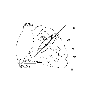
Now turning to the Figures, Figs. 1a, lb, 2a and 2b depict the structures of
the heart 1 of
which at least some are involved in embodiments of the invention. 2 is the
Superior Vena Cava (SVC), 4
is the right atrium (RA), 6 is the CS ostium, 8 is the CS first part, the
remaining part of the CS is behind
2 0 the heart, e.g. depicted in Fig. lb. 10 is the Inferior Vena Cava
(IVC), 12 is the Great Cardiac Vein
(GCV) at the level of the MV annulus 18. 14 is the Left Atrium cavity (LA), 16
is the LA wall, 18 is the
mitral valve annulus, 19 the whole mitral valve, 20 is the anterior leaflet
and 21 is the posterior leaflet of
the mitral valve. 22 is the LV muscular wall, 24 are the papillary muscles, 26
is the apex of the left
ventricle. 28 is the aortic valve, 30 the aorta ascendens, 32 the inter-
ventricular muscular septum, 34
2 5 the left ventricular cavity and 36 the right ventricular cavity. 38 is
the right ventricular muscular wall and
40 is the tricuspid valve.
Fig. lb and 2a show a schematic view of a heart, depicting the vein system,
wherein reference
numeral 42 is the anterior inter-ventricular vein, and 44 are lateral wall
veins, side branches in the
outside wall of the LV, 46 is the posterior descending vein. These side branch
veins are also often
3 0 referred to as the left marginal vein, the posterior veins of the left
ventricle or the middle cardiac vein.
However, they are all side branches of the CS or the GCV whatever they are
called in the literature.
In Fig. 2b. the mitral valve plane 48 is shown in relation to the vein system
and the LV long
axis 49, which is close to perpendicular to the MV valve plane 48.
Fig.3 is a schematic view of the movements in systole of the mitral valve
plane 48 in relation to
3 5 the LV apex 26, the GCV 12 (and CS) the MV anterior 20 and posterior 21
leaflets, the MV annulus 18,
the aortic valve 28, the LA wall 16 and the LA cavity 14 during a normal heart
beat. The large arrow x
shows the direction of the blood flow and the small arrow y illustrates the
direction of movement of the
MV plane 48, the GCV and the CS until the end systole position is reached
("down" position). In the
CA 02794303 2012-09-21
WO 2011/119100
PCT/SE2011/050337
11
cardiac cycle, the following moments are shown in Fig.3: a) is just before
systole, b) during systole and
c) end of systole.
With reference to Fig.4, a schematic view of the movements in diastole is
shown of the mitral
valve plane 48 in relation to the LV apex 26, the GCV 12 (and CS), the MV
anterior 20, and posterior 21
leaflets, the MV annulus 18, the aortic valve 28, the LA wall 16 and the LA
cavity 14 during a normal
heart beat. The large arrow x shows the direction of the blood flow and the
small arrow y the direction of
movement of MV plane 48, the GCV and the CS, until the end diastole position
is reached ("up"
position). In the cardiac cycle, the following moments are shown in Fig. 4: a)
early diastole, b) late
diastole and c) end of diastole, at the end of diastole the mitral valve is
now closed and ready for the
1 0 next move downwards in the following systole.
Fig. 5 is a schematic view of an embodiment of a medical device for cardiac
assist when
inserted in the heart 1. Some embodiments, as the present device, has two
anchor units. A first anchor
unit 50, is located in the CS 8 and/or the GCV 12. The second anchor unit 52
is located remote from the
first anchor unit. The second anchor unit 52 is for instance arranged inside a
side branch of the vein
1 5 system on the LV wall 22. The two anchors 50, 52 are in communication
with each other. They are for
instance, as illustrated, connected by means of a pulling and pushing unit 54
that can move the two
anchors relative to each other. The figure depicts, as in Fig. 3, the
movements in systole of the mitral
valve plane 48 in relation to the LV apex 26, the GCV 12 (and CS) the MV
anterior 20 and posterior 21
leaflets, the MV annulus 18, the aortic valve 28, the LA wall 16 and the LA
cavity 14 during an
2 0 augmented or assisted heart beat. The pulling and pushing unit 54
forces, powered by a power unit (not
shown), such as a remote or external power unit, the two anchors closer to
each other, and is thereby
augmenting the force and extent of the downwards movement of the mitral valve
19. The left ventricular
pump action is assisted. The large arrow (x) show the direction of the blood
flow and the small arrow (y)
the direction of MV plane, the GCV and the CS. In the cardiac cycle, the
following moments are shown
25 in Fig. 5: a) is just before systole, b) during systole and c) end of
systole.
Fig.6 is a schematic view of one embodiment of the invention when inserted in
the heart 1.
The two anchors, 50 is located in the CS 8 or the GCV 12, the other, 52 is
located inside a side branch
of the vein system on the LV wall 22. The two anchors are connected by means
pulling and pushing unit
54 that can move the two anchors relative to each other. The figure depict as
in Fig. 4 the movements
3 0 in diastole of the mitral valve plane 48 in relation to the LV apex 26,
the GCV 12 (and CS) the MV
anterior 20 and posterior 21 leaflets, the MV annulus 18, the aortic valve 28,
the LA wall 16 and the LA
cavity 14 during an augmented heart beat, the pulling and pushing unit 54
forces, powered by means of
an remote or external power unit 84 (not shown) the two anchor units away from
each other. As the
anchors are fixed to the tissue where they are anchored, the tissue structure
is moved with the anchor
35 unit(s). The anchor unit(s) are thereby augmenting the force and extent
of the upwards movement of the
mitral valve 19 towards the LA. Thereby the device is enhancing the diastolic
filling of the LV before the
next heart beat. Hence, even during diastole the cardiac assist is provided.
The large arrow x shows the
direction of the blood flow and the small arrow y the direction of MV plane
48, the GCV and the CS. In
CA 02794303 2012-09-21
WO 2011/119100
PCT/SE2011/050337
12
the cardiac cycle, the following moments are shown in Fig. 6: a) early
diastole, b) late diastole and c)
end of diastole, the mitral valve is now closed and ready for the next move
downwards.
A prototype of the invention was built, using a linear accelerator and a
computer. The
computer allowed action in synchrony with an electrocardiogram. The prototype
was tested in an animal
experiment. The chest of a 60 kilogram pig was opened between the ribs. A rod
from the linear
accelerator was attached to the mitral valve annulus from the outside of the
heart. The heart function
was depressed by means of drugs. After activating the device an increase in
arterial blood pressure and
cardiac output was observed.
Fig.7 is a schematic view of another embodiment of the invention when inserted
in the heart 1.
The device has two anchor units. A first anchor unit 56, is located in the CS
8 and/or the GCV 12. The
second, remote, anchor unit 58, is located inside a side branch of the vein
system on the LV wall 22 or
is attached to the LV outer wall. Here, the two anchors are magnets.
Preferably they are provided in
form of electromagnets, but one or the other magnetic anchoring unit may also
be a traditional
permanent magnet. The electromagnetic magnets are arranged to change polarity,
synchronized with
the heart cycle in order to change between pulling towards each other and
pushing away from each
other. There are no physical connecting units between the magnetic anchoring
units. The anchoring
units are only in magnetic connection. When the anchoring units have different
polarity they move the
two anchors closer to each other and correspondingly when the polarity is
equal they move the two
anchors away from each other. Fig. 7 depicts, as in Fig. 3, the movements in
systole of the mitral valve
2 0 plane 48 in relation to the LV apex 26, the GCV 12 (and CS) the MV
anterior 20 and posterior 21
leaflets, the MV annulus 18, the aortic valve 28, the LA wall 16 and the LA
cavity 14 during an
augmented heart beat. The magnetic anchors 56 and 58 attract each other and
forces by means of
magnetic power the two anchors closer to each other, and is thereby augmenting
the force and extent of
the downwards movement of the mitral valve 19. The large arrow shows the
direction of the blood flow
2 5 and the small arrow the direction of MV plane, the GCV and the CS and
the magnet 56. In the cardiac
cycle, the following moments are shown in Fig. 7: a) is just before systole,
b) during systole and c) end
of systole.
Fig.8 is a schematic view of the same embodiment as in Fig. 7 in diastole. The
first anchor unit
56 is located in the CS 8 and/or the GCV 12. The second anchor unit 58 is
located remote from the first
30 anchor unit 56. Here, the second anchor unit is located inside a side
branch of the vein system on the
LV wall 22. Alternatively, or in addition, it may be attached to the LV outer
wall. The two anchors are
magnets, preferably electromagnets, but one or the other may also be a
traditional permanent magnet.
The electromagnetic magnets may change polarity synchronized with the heart
cycle in order to change
between pulling towards each other and pushing away from each other. There are
no physical
3 5 connecting units. When the anchoring units have different polarity they
move the two anchors closer to
each other and correspondingly when the polarity is equal they move the two
anchors away from each
other. Fig. 8 depicts, as in Fig. 4, the movements in diastole of the mitral
valve plane 48 in relation to the
LV apex 26, the GCV 12 (and CS) the MV anterior 20 and posterior 21 leaflets,
the MV annulus 18, the
CA 02794303 2014-02-24
13
aortic valve 28, the LA wall 16 and the LA cavity 14 during an augmented heart
beat. The magnetic
anchors 56 and 58 now have equal polarity (both negative or both positive)and
push each other away
and thus the two anchors are forced away from each other by means of magnetic
power, and is thereby
augmenting the force and extent of the upwards movement of the mitral valve
19. The large arrow
shows the direction of the blood flow and the small arrow the direction of MV
plane and the magnet 56,
the GCV and the CS. In the cardiac cycle, the following moments are shown in
Fig. 8: a) early diastole,
b) late diastole and c) end of diastole.
In Fig. 9 an alternate positioning of the second magnet anchor unit 58 is
shown. The second
anchor unit 58 can be electromagnetic or classic permanent magnetic. The
second anchor 58 can be
a.o electromagnetic or classic permanent magnetic. When being permanent
magnetic, the first magnetic
anchor 56 is an electromagnetic unit with selectively activateable magnetic
polarity. The second anchor
58 can be placed in different positions in the heart. However, positions
outside the heart are also
possible in certain embodiments. Location 61 indicates a position where the
second anchor 58 is not
attached to or in the heart. One such position is in the pericardium. Another
position is in the pleura or
under the skin. Possible attachment sites include the pericardium, the
diaphragm. The spine or the
thoracic cage (ribs and sternum) are also suitable sites for attachment of the
second anchor 58.
Positions 62, 64, 66, 68 indicate positions for the second magnet anchor 58
relative the heart. Position
62 Is located in the left ventricle and position 64 is located in the right
ventricle. Position 66 is located in
the RA, preferably in the so called atrial septum between the RA and the LA.
One good position is in the
2 o foramen ovale of the atrial septum where often an opening is present to
the LA. in this embodiment, the
second anchor unit may have the shape of a septal occluder and provide both
septal leakage occlusion
and allows for support of the cardiac function. Position 68 indicates a
position in the LA, again a good
attachment site would be the atrial septum, another good position in the LA
would be the LA appendage
(LAA, not shown). In this embodiment, the second anchor unit may have the
shape of an LAA occluder
and provides both LAA occlusion and allows for support of the cardiac
function. These are only
examples and a person skilled in the art may think of multiple variations that
would work equally well for
the purpose.
In Fig. 10a another embodiment is shown where the supporting or assist force
is executed by
means of a mini motor 70 integrated in the CS anchor and/or GCV anchor. MEMS
(micro-electro-
magnetical-systems) technology could be utilized for constructing such a
motor. One or more second
anchor units 72 are arranged in one or more side branches 44, to which the
connecting unit 54 is
attached, respectively.
Permanent magnets in embodiments may be conventional iron magnets.
Altematively, super
magnets, like Neodymium rare earth magnets may be used to improve efficiency
and/or reduce size of
the units of the cardiac assist system, when comprising magnetic elements.
An anchor unit may for instance be provided in form of a stent. The stent
serves as an anchor
in a vessel. Such a stent could be a self expanding stent for instance made of
a shape memory material,
like a shape memory metal like superelastic Nitinol. The mini motor 70 could
then be integrated in the
CA 02794303 2012-09-21
WO 2011/119100
PCT/SE2011/050337
14
stent structure (not shown). The stent could also be a stent made of a
material or having a structure that
has to be expanded by means of a balloon, for instance made of stainless steel
or another metal
suitable for the purpose. Alternatively, or in addition, an anchor unit is
made with hooks that dig into the
tissue made of similar materials, these are only examples and a person skilled
in the art may think of
multiple variations that would work equally well for the purpose when reading
the present description.
Thus, the motor 70 is attached to the vessel structure. This may be made with
stent technology and/or
by means of hooks that a person skilled in the art will find multiple
solutions for. However, common for
all these solutions is that they will be executed by means of catheter based
techniques by means of
puncture of a vessel, preferably a vein, through the skin.
1 o Multiple sets of motors 70, anchors 72 and connecting units 54 may be
implanted
simultaneously and connected to one or more energy sources 84 (not shown) as
is described in Fig.
10b. Electrical power for the mini motors is provided from the remote energy
source 84 by means of
insulated cables 74.
In still another embodiment shown in Figs. lla and 11b, the energy is
mechanically
transferred from the remote energy source 84 to the movement of the MV plane
48. The mechanical
force may be provided through an extended connecting unit 54, like a wire or
elongate flexible rod. The
movement is transferred all the way from a mechanical actuator, e.g. at the
remote energy source, to
the anchor unit 72, through the CS or GCV anchor 76. The anchor unit 76 may
have guiding units 80 for
the connecting unit 54 in order to transfer the mechanical movement from the
anchor 76 into the used
2 0 side branch 44 of the vein system. A guiding sheath 78 may be fixated
in the anchor 76 and in the
energy source 84 in such a way that when pulling in the connecting unit 54
inside by the mechanical
actuator, e.g. at the energy source, relatively to the guiding sheath 78 the
distance between the anchors
72 and 76 will shorten. Correspondingly, when pushing the connecting unit
inside the remote energy
source, the distance between the two anchors 72 and 76 increases. The guiding
unit may also be a
2 5 mechanical unit that transfers a longitudinal (or rotational movement,
see below) into a movement in a
perpendicular direction of the unit 54. Thus the reciprocating up and down
cardiac assist movement of
the MV plane 48 is provided.
Turning to Fig. 11b, an embodiment of the type described with reference to
Fig. 11a is shown,
except that the CS or GCV anchor 82 is designed for more than one anchor in
side branches 44. In this
3 0 manner, advantageous improved efficiency of the cardiac assist device
may be provided. Geometric
distribution of the supporting force may be provided that is advantageous for
the cardiac structures in a
long term use of the device.
The Figs. 12a and 12b show examples of configurations described in Figs. 7, 8
and 9 where
electromagnets are used as anchors. Different combinations of electromagnets
and classical permanent
35 magnets will not be described in separate figures as they would be
apparent for the skilled person when
reading the present application. In Fig. 12a the first anchor is located in a
side branch 44 from the CS or
the GCV and in Fig. 12b in the anterior inter-ventricular vein (AIV).
CA 02794303 2012-09-21
WO 2011/119100
PCT/SE2011/050337
Still another embodiment of the innovation is depicted in the figures 13, 14,
15 and 16. Instead
of pulling and pushing the extension 54, the mechanical force is instead
transferred by means of rotation
of the extension unit 54. Now the distal anchor 73 of the device is not
located in a side branch. Instead,
it is placed in the distal GCV 12 itself or in its continuation, the anterior
inter-ventricular vein 42. This
5 embodiment takes advantage of the fact that the three dimensional shape
of the CS and the GCV
represents a loop from behind the heart, around the left angle of the heart to
its front surface. The loop
is substantially oriented in the mitral valve plane 48, see e.g. Fig. 2b. The
extension unit 54 is an
elongate loop shaped unit, distally ending at the distal anchor unit 73, where
it is attached to the distal
anchor unit 73, see e.g. Figs. 15a-c. Hence, the loop shaped extension unit 54
may be suitably actuated
1 0 to move the CS and/or the GCV in direction to and/or from the LV apex
26. As the MV is connected by
cardiac tissue to the CS and GCV, a movement of the extension unit 54 is
transferred to the MV plane
48.
In Fig. 13 the part of the extension 54 that is located inside the CS and the
GCV, here
numbered with 55 is depicted. The device has different operative positions, as
shown in Figs. 13a-c. In
15 the neutral position, depicted in Fig. 13a, we have a perpendicular view
of the loop that will appear as a
straight line from that angle. Compare also the view in Fig. 15a.
A distal anchor unit 73 is located at the front of the heart. Most preferable
the distal anchor
unit 73 is made of a stent design. A second anchor 75 is arranged proximally
of the distal anchor 73 in
the GCV or preferably in the CS as close to the ostium 6 (Fig 1) as possible.
The second anchor is
2 0 preferably made of a stent design. Additional anchors 77 may be located
for support anywhere between
the distal end anchor 73 and the proximal end anchor 75, see e.g. Fig. 14. The
additional anchors are
preferably made of a stent design.
The extension unit 54 is proximally connected to a mechanical actuator that
controllably
rotates the extension unit 54 synchronized with the cardiac cycle. In the
embodiment, the extension unit
2 5 54 is proximally connected to the remote energy source 84. However,
other arrangements and locations
of the mechanical actuator providing the rotational movement of the elongate
extension unit 54 may be
provided in other embodiments. The mechanical actuator may for instance be
arranged intra-cardiac.
While rotating the extension unit 54 clockwise (seen from the mechanical
actuator, here the
remote energy source 84 end), as shown in position b in Fig. 13, the loop 55
flexes towards the LA 14,
3 0 moving the CS and the GCV also in this direction. Since the CS and the
GCV are so closely related to
the MV, such a backwards movements in relation to the LV apex will augment the
normal upwards
movement of the MV in diastole if the clockwise rotation is done in diastole.
In analogy to this, a counter-clockwise rotation in systole will augment the
downwards
movement of the closed MV (piston) in systole, as depicted in Fig. 13,
position c).
35 In
Fig. 14 it is also illustrated that there in addition may be a retention unit
79 that locks the
extension unit 54 longitudinally to stay at the location of the proximal
anchor unit 75. The retention unit
may be a tube or loops located in the anchors allowing the extension 54 to
rotate, but will prohibit axial
movements in order to prevent dislocation of the extension units 54 and 55.
Extension units 54 and 55
CA 02794303 2014-02-24
16
may be in one integral piece or have different segments that are articulated
(not shown). The number of
segments and articulation may be suitably chosen in order to design stiffness
or flexibility necessary to
accommodate the device in place while still being functional.
Fig. 15 illustrates in more detail the embodiment taking advantage of rotating
a loop in an
anatomical environment. Fig. 15a depicts the neutral position. In Fig. 15b the
extension units 54 and 55
are rotated clockwise. Now the loop of 55, the CS, the GCV and the mitral
valve move up towards the
LA in diastole. In Fig. 15c the extension units 54 and 55 are rotated counter-
clockwise and the loop of
55, the CS, the GCV and the mitral valve moves down towards the LV apex in
systole.
The direction of the MV plane movement, here related to the rotation, is
controlled, e.g. based
on ECG detection, and in synchronisation with the cardiac cycle. A control
unit operatively connected to
implement the control is provided, as described in an example below. The
control unit may be
implemented in the remote energy source unit 84.
Further, in another embodiment, in addition to the rotational movement, a
longitudinal
movement of the extension unit 54 may be added. By pulling the extension unit
54, attached to the distal
anchor 73, relative to the sheath 78, that now is fixed to the proximal anchor
75, the distance between
anchors 73 and 75 may be reduced. This additional transversal controlled
movement may in some
embodiments include moving the lateral LV in the heart in a reciprocating
movement during systole
towards an inter-ventricular septum of the heart and during diastole away from
an inter-ventricular
septum for assisting the pump action of the heart along the short axis of a LV
of a heart. In Fig 15d it is
2 0 illustrated that in diastole the extension unit 54 is moved distally
relative to the sheath 78, in addition to
the clockwise rotation. The length of the connecting extension unit portion
between the proximal and the
distal anchor is thus extended. Thus, the outwards movement of the lateral LV
wall is augmented
relative to the intra-ventricular septum. In Systole on the other hand, as
shown in Fig. 15e, the extension
unit 54 is moved proximally relative to the sheath 78, the distal anchor 73 is
pulled closer to the proximal
2 5 anchor 75, in addition to the counterclockwise rotation. The length of
the connecting extension unit
portion between the proximal and the distal anchor is thus shortened. Thus,
the inwards movement of
the lateral LV wall Is augmented relative to the intra-ventricular septum. The
direction of the LV lateral
wall movement, here related to the pulling and pushing in addition to the
rotation, is controlled, e.g.
based on ECG detection, and in synchronisation with the cardiac cycle. A
control unit operatively
3 o connected to implement the control is provided, as described in an
example below. The control unit may
be implemented in the remote energy source unit 84. The coronary sinus implant
of embodiments may
thus be adjusted during at least a portion of a single cardiac cycle.
Adjustment is made instantaneously
upon actuation. In alternative embodiments, the short axis support actuation
may be made based on
other units and actuating principles, including electric or magnetic
actuators, etc. In addition, the medical
3 5 device may have a plurality of sections which are individually
adjustable in length by an actuating unit,
controlled by said control unit arranged to controllably change said shape of
said sections individually.
For instance, embodiments of the device may comprise anchoring units between
each of said plurality
CA 02794303 2012-09-21
WO 2011/119100
PCT/SE2011/050337
17
of sections, wherein the length of the sections is adjustable e.g. by pulling
together or pushing apart
distal and proximal anchoring units of a section.
In another embodiment the inherent force of a spring is utilized shown in
Figs. 16a and 16b.
Here the extension unit 55 is inserted and detached in the CS and the GCV or
in the AIV. Preferably the
extension 55 in this embodiment has fix attachments to the distal and proximal
anchor units 73, 75. The
cardiac assist device is provided as a resilient unit. In this embodiment, the
cardiac assist device is
provided in a relaxed position in the MV plane up position. The relaxed
position of the unit is spring
loaded against a MV plane down position. The loop 55 of the extension unit 54
has as a default
preferred state the relaxed position. The extension unit thus forces the CS,
the GCV and the mitral valve
1 0 to move up towards the LA, both in diastole and in systole, namely
against the spring load force. The
inherent spring load force is chosen to be less than the MV plane downward
force provided by the LV
muscle. Thus, in systole, the cardiac muscle force of the LV will be stronger
than the inherent spring
force of the extension 55 and bring the loop down towards the LV apex in
systole. Such a device thus
assists during the diastole when it increases the LV diastolic filling by
forcing the open MV up against
the blood stream further in the direction of the LA. On the other hand, the
resilient unit may have a
relaxed position in a lower MV plane position spring loaded against a MV plane
up position, such that
the cardiac relaxation force of the LV brings the loop to the up position, and
the resilient unit assists
during the systole by assisting the LV systolic contraction by forcing the
closed MV down towards the LV
apex.
2 0 Such non-powered devices might be made of Nitinol, a memory shape
metal or stainless steel
or any other suitable material, preferably metal. A control unit or remote
energy unit 84 are omitted in
these particular embodiments. The action may be delayed by integrating
resorbable material, in the
device in order to delay its action and allow the device to grow in before its
action is initiated while the
resorbable material disappears. Such material could be for instance be PLLA,
Polyvinyl or Polylactid or
2 5 other resorbable materials.
Alternatively, or in addition, the cardiac assist system may be provided as a
bistable system.
Here, the diastolic up position and the systolic down position of the MV plane
may be provided as
equilibrium states of the system. Energy is either provided from the external
energy, or from the LV
muscle source to initiate the system to move between the two stable positions.
These embodiments
3 0 may be more energy efficient than others.
In embodiments the cardiac assist device has a control unit and a sensor for
measuring
physiological parameters related to the cardiac cycle activity providing a
sensor signal. The sensor
signal is provided to the control unit which controls the displacement unit to
provide the movement by
energy from an energy source and based on the sensor signal. The cardiac
assist device operation is
3 5 thus controlled in synchronicity with the heart action. The sensor may
be an ECG electrode or in
addition or alternatively be based on detecting other physiological parameters
related to the cardiac
activity, such as a blood pressure wave, blood flow patterns, or acoustic
signals of the cardiac activity.
CA 02794303 2014-02-24
18
A remote energy source 84 as comprised in some embodiments, is shown in Fig
17. It has a
battery section 86 and a computing section 88 containing computer algorithms
and chips. The computer
section 88 has receiving electrodes or surfaces 92 connected, which are able
to detect an
Electrocardiogram (ECG) signal. Based on the ECG signal, the cardiac assist
device operation is in
embodiments controlled in synchronicity with the heart action.
Such synchronicity may in addition or altematively be established by means of
detecting other
physiological parameters related to the cardiac activity. Such parameters
include a blood pressure
wave, blood flow patterns, or acoustic signals of the cardiac activity.
Alternatively, or in addition, the assisted movement of the cardiac assist
device may be
o controlled according to a set sequence of assisted movements of the MV
plane that mimics the natural
cardiac cycle to optimize the cardiac assist function. Frequency, speed, and
duration of different pause
times of the assisted movement may be set in the sequence to mimic a natural
or desired movement.
The different parameters, such as pause time duration of the movement, may
vary over any time
interval, and may be set to vary according to a repeating program. The
sequence may be programmed
is into the computing section/control unit 88 which controls the force
generating unit. The force generating
unit may then provide the assisted movement according to the set sequence.
Energy from an energy
source 84 may thus be controllably provided to the force generating unit
according to the set sequence
for providing the assisted movement.
Alternatively, or in addition, the medical device may be incorporated into an
artificial
2 o pacemaker system controlling or assisting the natural cardiac muscle
function. For instance the assisted
movement of the cardiac assist device may be controlled from a processing unit
of a pacemaker. The
pacemaker including the processing unit may be implanted in a patient. The
pacemaker triggers heart
muscle activity in a per-se known manner, e.g. via leads connected to the
cardiac tissue for artificially
triggering the cardiac activity. Triggering of the assisted movement of the
cardiac assist device may be
2 5 controlled by the electrical triggering of the cardiac activity by the
artificial pacemaker system, which is
already synchronized with the cardiac cycle. Preferably a time delay is
provided from triggering
electrical triggering of the heart muscle activity to the
triggering/activation of the assisted movement of
the cardiac assist device during a heart cycle. The amount of the time delay
may be optimized,
depending on the transfer time of electrically triggering the heart muscle
activity and the resulting pump
3 o function of the heart caused by the controlled heart muscle
contraction.
The remote energy source 84 may have a mechanical section 90, where rotational
or linear
motion may be transferred to extension unit 54. Rotational movement may be
transferred directly from
an electrical motor, or geared down in revolutions by a gear-box. Rotational
energy from an electrical
motor may be converted to linear movement, enabling pulling and pushing force
to a wire connecting
3 5 unit 54 that is extending all the way to the distal anchor position.
Alternatively, or in addition, the
mechanical section 90 may contain other actuators. For instance one or more
strong electromagnets
may be provided in an actuator that alternately are able to provide pulling
and pushing force to wire
connecting unit 54 that is extending all the way 10 the distal anchor
position.
CA 02794303 2012-09-21
WO 2011/119100
PCT/SE2011/050337
19
Further, the pulling and pushing force from the remote energy source 84 may
also be
achieved by means of a linear accelerator in the mechanical section 90.
Alternatively, or in addition, the
mechanical section 90 contains an actuator providing pulling and pushing force
to extension unit 73, e.g.
a wire or elongate flexible rod of carbon fibre, that is extending all the way
to the distal anchor position
by means of electrically alternately cooling and warming a Nitinol actuator as
commercially available
from MIGA Motor Company, Modern Motion, www.migamotors.com. Finally, in other
embodiments, the
remote energy source is without a significant mechanical section, instead
computer chips are
distributing electricity from the battery according to the physiological
cardiac cycle related signal, e.g.
ECG signal, either to electromagnets in one or more of the anchor units of the
implanted cardiac assist
device or to mini-motors or linear actuators in a heart chamber or on the
heart surface.
The remote energy source may have a rechargeable battery that is charged by
means of a
wire 94 penetrating the skin and when charging the battery connected to a
charging device externally
(not shown). Charging might also be done wireless through the skin, e.g. by
means of electromagnetic
coils transferring energy inductively. The skilled person in the art may alter
and design such charging
according to specific requirements and available actual technology.
Figure 18, and the following illustrations refer to explain a delivery system
that is part of a
treatment kit, the medical procedure of using the delivery system to deliver a
cardiac assist device, and
a medical method for therapeutically enhancing the left ventricular function
of a patient permanently.
In some particular embodiments, the remote energy source is located in the
fatty tissue under
the skin, adjacent to a vessel, preferably a large vein. This allows for
convenient access to the heart.
Alternatively, the energy source may be attached to the clavicle (not shown)
in order to prohibit
dislocation of the same when delivering mechanical energy to the cardiac
assist device inside the heart.
A pocket or pouch 104 in subcutaneous tissue may be created close to the
actual access vessel, e.g.
the subclavian vein, see Fig. 18.
In Fig. 18 the heart is shown relative to the great vessels and the skin
surface. An introducer
catheter 100 with a valve (not shown) is penetrating the skin and enters a
large vein, in this case the
subclavian vein 3, however any other large enough vein can be used for access.
Adjacent to the skin
puncture site a pouch 104 may be created under the skin in the fatty tissue in
order to accommodate a
remote energy source 84 (not shown). The energy source may be attached to the
clavicle (not shown) in
order to prohibit dislocation of the same when delivering mechanical energy to
the cardiac assist device
inside the heart. A guide wire 102 is advanced through the introducer catheter
100 to the right atrium 4.
By means of a guiding catheter 106 (first shown in Fig. 21) access to the
coronary sinus is obtained via
the RA and the guide wire is guided to the appropriate side branch of the
coronary sinus vein system. In
addition to the guide catheter, the kit contains delivery catheters where the
different parts are loaded.
Figs. 19 and 20 show examples of delivery systems, however, only depicting the
principle of delivering
the device. Figs. 19 a-c show how a push and pull system is delivered from the
delivery system 98.
In Fig. 19a a delivery system for a cardiac assist device as described above
with reference to
Fig. 10a is shown. The delivery system comprises a delivery catheter 108 that
has a distal anchor 72
CA 02794303 2012-09-21
WO 2011/119100
PCT/SE2011/050337
loaded inside at the tip. A pusher tube 110 that has a smaller outer diameter
than the inner diameter of
the delivery catheter may be advanced axially forward inside the delivery
catheter 108 in order to push
the anchor 72 out of the delivery catheter 108 at the desired site.
Alternatively, the delivery catheter 108
may be retracted over the pusher catheter in order to deliver the device
without any axial movement.
5 The distal anchor unit 72, here shown as a self expanding stent, is
attached to the extension unit 54 and
space is accommodated inside the delivery catheter for the extension unit 54
to be able to extend until
outside the patient, see Fig. 19b. The pusher tube 110, accommodates a lumen
for the guide wire 102
that also is permitted to run through the anchor 72. The distal anchor unit is
released and expands such
that it safely anchors into the surrounding vessel tissue. Thus the distal
anchor is in place, having the
1 0 extension unit 54 extending therefrom.
Once the first anchor is in place, a second delivery catheter 116, shown in
Fig 19c is
advanced over the extension 54 until the guiding unit 80 is aligned with the
side branch in which the
distal anchor 72 is located. When holding the pushing catheter 110 still in
this position and retracting the
deliver catheter 116, the anchor 76 may be exactly released with the guiding
unit facing towards the
1 5 side branch. Another aid in placing the device exactly is an X-ray
marker 112 attached to the catheter in
order to better visualize the exact position of the catheter, e.g. by means of
fluoroscopy.
Figure 20 depict positioning of a device where rotational force is transferred
to the coronary
sinus. This delivery catheter 118 is similar to the one shown in Fig. 19,
except that it may have another
lumen added in order to accommodate an extra guide wire 102. Any additional
figures of the delivery
2 0 systems accommodating other embodiments are not provided, since it
would show variations that are
apparent to the skilled person when reading the present disclosure.
The Figs. 21 ¨ 25 illustrate the method 800 of inserting a cardiac assist
system for permanent
heart function augmentation.
The skin is penetrated and an introducer catheter 100 with a valve (not shown)
is introduced
2 5 into a large vein, e.g. the subclavian vein 3, in step 800. Any other
large enough vein may be used for
access. A guide wire 102 is advanced through the introducer catheter 100 to
the right atrium 4. By
means of a guiding catheter 106 access to the coronary sinus is obtained via
the RA and the guide wire
is guided to the appropriate side branch of the coronary sinus venous system
in step 810. Fig. 21a
illustrates the advancement of a guide wire 102 into a desired side branch 44
by means of the guiding
catheter 106.
In step 820, as illustrated in Fig. 21b, the distal anchor 72 is released by
means of the delivery
catheter 108 in the side branch 44.
In step 830, as shown in Fig. 22, the proximal anchor 76 is positioned at the
opening of the
side branch.
In Fig 23 the positioning of a mini motor 70 by means of the delivery catheter
108 is shown.
Finally, as shown in Figs. 24a and b, the positioning of a rotation device is
depicted. In Fig.
24a, it is shown how the guide wire is advanced into the anterior inter-
ventricular vein 42 by means of
the guide wire 102 and the guide catheter 106. In Fig. 24b both anchors are
depicted in place, showing
CA 02794303 2015-08-17
=
21
the loop 55. An additional guide wire may be accommodated through a separate
lumen 114 (in Fig. 20 c).
In step 840, adjacent to the skin puncture site a pouch 104 is created under
the skin in the fatty
tissue in order to accommodate a remote energy source 84 (not shown). In step
850, the energy source
may be attached to the clavicle (not shown) in order to prohibit dislocation
of the same when delivering
mechanical energy to the cardiac assist device inside the heart.
Once both anchors are securely attached, the extension unit 54 is adjusted in
length and
attached to the remote energy source 84 in step 860, and the system may be
activated in step 870. The
remote energy source has a unit to detect the natural action of a heart, e.g.
based on an electrocardiogram,
a blood pressure wave, acoustic heart activity, or blood flow. The remote
energy source may thus provide
energy for the distance change between the two anchors in synchrony with the
natural heart cycle, thereby
enhancing the natural up and down movement of a mitral valve during a heart
cycle.
A method is provided for permanently enhancing left ventricular pump function
of a heart of a
patient, the method comprising controlled assisted mitral valve plane movement
synchronized with a
cardiac cycle of the heart.
The present invention has been described above with reference to specific
embodiments.
2 o However, other embodiments than the above described are equally
possible within the scope of the
invention. Different method steps or a different order than those described
above may be provided within
the scope of the invention. The different features and steps of the invention
may be combined in other
combinations than those described. Several actuating principles may be
combined with each other in
certain embodiments, e.g. a linear actuator and magnetic driving. The scope of
the invention is only limited
by the appended patent claims.
14824372.1