Note: Descriptions are shown in the official language in which they were submitted.
CA 02795703 2013-12-20
INSERTION DETECTOR FOR A MEDICAL PROBE
FIELD OF THE INVENTION
The present invention relates generally to medical devices that use probes
that come in
close proximity to, or in contact with, a patient. More specifically,
embodiments of the
invention relate to a device for detecting the insertion of the probe into a
body cavity,
BACKGROUND OF THE INVENTION
A general body cavity medical probe may be inserted into the patient's body
cavity for
either measuring vital signs or for providing treatment, There are numerous
type of general
body cavity probes, such as a medical ear thermometer. General body cavity
probes may
contain a functional module to perform the intended medical measurement or
procedure. For a
medical ear thermometer, the functional module may be an infrared ("IR")
temperature sensor.
Temperature of an object, specifically of a living being such as a human or an
animal, can
be measured by thermal conduction or thermal radiation. For thermal
conduction, a
temperature sensing probe is brought into a physical contact with a surface of
the living being.
For thermal radiation, a temperature sensing probe is brought near the surface
of a the living
being and aimed at the area of interest, such as within the open space of a
body cavity.
Naturally emanated electromagnetic radiation in the mid and far infrared
spectral ranges is
detected by an appropriate sensor, whose output signal indicates the surface
temperature of an
object. For both thermal conduction and thermal radiation measurement methods,
the
temperature sensor is positioned inside or coupled to the medical probe.
Medical thermometers that operate by contact, for example, oral or rectal, may
use a
probe cover, for instance a sanitary probe cover. Thermal energy (i.e., heat)
is transmitted
1
CA 02795703 2013-12-20
through the probe cover by thermal conduction, thus at least the portion of
the probe cover
material overlying the thermal sensor should be highly transmissive of thermal
energy. Various
conventional probe covers for contact thermometers are described in, for
example, in U.S.
Patent No. 4,159,766 issued to Kluge.
Medical thermometers that operate by radiation may also use a probe cover,
because the
possibility still exists of contact with the body of the patient. For example,
when measuring the
temperature of a tympanic membrane and the surrounding tissue inside the ear
canal, the probe
is inserted into the ear canal body cavity and may contact the wall of the ear
canal. Before
insertion, a probe cover may be installed onto the probe to envelop its parts
that otherwise
might come in contact with the patient's skin. Such a cover provides sanitary
protection
against contamination of the probe by ear wax and other soiling biological
compounds, and
includes properties that promote accurate temperature measurement by the
detection of infrared
signal. Such properties include a good transparency of the front portion of
the probe cover in at
least the spectral range of interest, low directional distortion of optical
rays, tight
manufacturing tolerances, stability of the optical properties during
installation onto the probe,
long term storage stability, etc. Probe covers for the IR thermometers are
exemplified by U.S.
Patent No. 5,088,834 issued to Howe et al. and U.S. Patent No. 5,163,418
issued to Fraden et
al.
A probe cover may include one or more components such as polyethylene,
polypropylene, and copolymers thereof. Probe cover materials may also possess
relatively low
absorption of electromagnetic energy over a broad spectral range from visible
to the far
infrared.
Figure 1 is an example of a medical device having a probe intended for
insertion into a
body cavity, illustrating a perspective view of the infrared ear thermometer
Model "Braun
4000" produced by Ka.z, Inc., as known in the art. The example of FIG. 1
includes a
thermometer body 1 having a display 2, a power button 3, a probe 7, a probe
cover sensing
switch 8, and a probe cover ejecting ring 5. Before measuring temperature, a
reusable or
disposable probe cover 6 is moved in a direction 12 to be positioned over
probe 7. The probe
cover has a skirt 9 and a groove 10. When installed onto probe 7, groove 10
engages with the
2
CA 02795703 2012-10-05
WO 2011/127063 PCT/US2011/031264
offset 11 that is part of the probe 7. This coupling will hold the probe cover
6 on the probe 7
during use. Skirt 9 actuates switch 8 to generate a signal going to the
internal electronic circuit
indicating a correct installation of the probe cover. If no probe cover is
detected by the switch
8, the internal circuit may either warn the operator or make the theimometer
inoperable to
prevent an erroneous reading.
When a medical probe is used, either with or without a probe cover, it may be
desirable to
detect either a close proximity of the probe to the patient body surface, or
to detect insertion of
the probe into a body cavity, such as an ear canal. A shortcoming of the known
art is that no
medical thermometer has a capability of detecting the probe position relative
to the ear canal.
Therefore, a need exists to provide such a proximity measurement,
SUMMARY OF THE INVENTION
Embodiments of the invention relate generally to an apparatus and method for
the
proximity detection of a medical probe (e.g., a thermometer) to the surface of
a living being.
The embodiments should provide an accurate measurement, with or without the
presence of a
probe cover, by taking into account the detected proximity.
Therefore, as will be apparent from the foregoing description, embodiments of
the
invention include one or more of: a method or device for detection of the
probe cover
installation on a probe; a method or device for detecting proximity between
the medical probe
and a patient body surface; or a method or device to detect the insertion of a
medical probe into
the body cavity of a patient.
One or more embodiments of the invention provide a medical probe for insertion
into a
body cavity of a patient, such that the medical probe includes a probe body
having a sidewall
laterally circumscribing a longitudinal axis and enclosing an inner space, the
sidevyall having a
proximal end and a distal end. Optionally, the distal end may be tapered
relative to the
proximal end. The medical probe also includes a sensor coupled to the probe to
provide a
signal relating to a condition of the body cavity of the patient, and a
proximity sensor coupled
to the probe, the proximity sensor configured to provide a signal indicating
insertion of the
probe into the body cavity. In some embodiments, the sensor may include a
functional sensor
3
CA 02795703 2012-10-05
WO 2011/127063 PCT/US2011/031264
or a temperature sensor, and/or the sidewall may have an elongated shape
adapted for insertion
into an ear canal.
Optionally, the medical probe may be designed such that the proximity sensor
includes
an optical transmitter and an optical receiver positioned such that, when the
medical probe is
positioned for insertion into the body cavity, the optical transmitter is
positioned to transmit an
optical signal toward an opening of the body cavity, including the edge
thereof, and the optical
receiver is positioned to receive the optical signal from the optical
transmitter. The optical
transmitter may be positioned to transmit toward a first position of the
opening of the body
cavity, and the optical receiver may be positioned to receive optical signals
from a second
position of the opening of the body cavity.
As used throughout herein, for signals related at least to the proximity
sensor, signals to
or from the body cavity may include signals to or from portions of the patient
adjacent to the
body cavity, including wall portions and/or edge portions of the cavity.
Optionally, the transmitter may have a first optical axis, and the optical
receiver may
have a second optical axis.
In another embodiment, the proximity sensor may further include a receiving
light
guide disposed within the inner space, the receiving light guide having a
first end coupled to
and protruding through the distal end of the sidewall, and a second end
coupled to the optical
receiver. Optionally, the receiving light guide may protrude through the
distal end of the
sidewall at an angle that is pointed away from the proximal end of the
sidewall.
In another embodiment, the proximity sensor may further include a transmitting
light
guide disposed within the inner space, the transmitting light guide having a
first end coupled to
and protruding through the distal end of the sidewall, and a second end
coupled to the optical
transmitter. Optionally, the transmitting light guide may protrude through the
distal end of the
sidewall at an angle that is pointed away from the proximal end of the
sidewall.
Optionally, the proximity sensor may include a translucent opto-coupler that
protrudes
through the sidewall, the opto-coupler including a first side disposed within
the inner space, the
first side being optically coupled to a light emitter and a light detector;
and the opto-coupler
4
CA 02795703 2012-10-05
WO 2011/127063 PCT/US2011/031264
further including a second side disposed outside the inner space, wherein the
second side
protrudes through the wall of the probe.
In some embodiments, the medical probe may further include an electronic
circuit
coupled to the sensor and to the proximity sensor, the electronic circuit
including a processor
and a memory coupled to the processor, the memory storing software, such that
the software,
when executed by the processor, is configured to execute an algorithm to
process signals from
the sensor and the proximity sensor. The electronic circuit may further
include an output
device coupled to the processor, the output device configured to output a
result of the
algorithm.
The medical probe may further include an ambient temperature sensor
electrically
coupled to the electronic circuit and positioned outside of the inner space,
such that the
software, when executed by the processor, is further configured to execute an
algorithm to
process signals from the sensor, the proximity sensor and the ambient
temperature sensor.
In one or more embodiments, the medical probe may be designed such that at
least one
of the transmitting light guide and receiving light guide comprises a plastic
optical fiber.
Alternatively, at least one of the transmitting light guide and receiving
light guide includes a
glass rod, or a polycarbonate rod. Optionally, at least one of the
transmitting light guide and
receiving light guide may include a rod coated with a coating material,
wherein a refractive
index of the coating material is lower than a refractive index of the rod.
Optionally, the first
end of at least one of the transmitting light guide and receiving light guide
may include a
lensing bulb. Optionally, an optical barrier may be disposed in the inner
space between the
optical transmitter and optical receiver. Optionally, the optical receiver is
disposed within the
inner space.
In one or more embodiments of the invention, the proximity sensor may operate
by use
of ultrasonic signals.
One or more embodiments of the invention provides a method for detecting an
insertion
of a medical probe into a body cavity of a patient, including the steps of:
transmitting, from a
transmitter, a signal toward an edge portion of the body cavity; receiving, at
a receiver, a return
5
signal from an edge portion of the body cavity; and monitoring a flux of the
return signal
for a drop in strength, such that a path from the transmitter to the receiver
is blocked
when the medical probe is inserted into the body cavity such that the flux of
the return
signal decreases when the medical probe is inserted into the body cavity.
In another embodiment of a method for detecting an insertion of a medical
probe,
the medical probe having a longitudinal axis, into a body cavity of a patient
along the
longitudinal axis, the method includes the steps of: transmitting a signal
along a first
direction substantially perpendicular to the longitudinal axis; receiving a
return signal
from a second direction, the second direction substantially parallel to the
first direction;
and monitoring a flux of the return signal for an increase in strength, such
that the flux of
the return signal increases in strength above a predetermined threshold when
the medical
probe is inserted into the body cavity.
In another embodiment of a method of displaying the temperature of a body
cavity of a living being, the method includes the steps of: measuring a base
temperature
of the cavity by use of a temperature sensor; measuring a proximity of the
temperature
sensor to the body cavity; measuring an ambient temperature in an area
adjacent to the
temperature sensor; computing a computed temperature of the body cavity in
accord with
a predetermined function of the base temperature, the proximity, and the
ambient
temperature; and displaying the computed temperature.
Optionally, the method may further detect the presence of a probe cover, and
adjust the computed temperature accordingly.
In one aspect of the invention, there is provided a medical probe for
insertion into
a body cavity of a patient, including: a probe body having a sidewall
laterally
circumscribing a longitudinal axis and enclosing an inner space, the sidewall
having a
proximal end and a distal end; a sensor coupled to the probe body to provide a
signal
relating to a condition of the body cavity of the patient; a proximity sensor
coupled to the
probe body, the proximity sensor configured to utilize an optical signal to
determine
6
CA 2795703 2019-02-11
proximity of the probe to the body cavity or an insertion condition of the
probe whereby
the probe is inserted into the body cavity, wherein the proximity sensor
further includes
an optical transmitter and an optical receiver coupled to a flange portion
located at a
proximal end of the probe, the optical transmitter and optical receiver
positioned such
that, when the medical probe is inserted into the body cavity an optical path
from the
optical transmitter to the optical receiver is at least partially blocked, the
optical
transmitter positioned to transmit an optical signal toward an opening of the
body cavity,
the optical receiver positioned to receive a reflection of the optical signal
along the
optical path, the proximity sensor configured to interpret reduction of the
reflection of the
optical signal relative to the optical signal along the optical path as an
indication of
insertion of the probe into the body cavity.
Still other objects and advantages of the invention will in part be obvious
and will
in part be apparent from the specification. The invention accordingly includes
the
features of construction, combination of elements, and arrangement of parts
which will be
exemplified in the construction hereinafter set forth as well as the methods
of
construction and applying the adhesive discussed herein.
BRIEF DESCRIPTION OF THE DRAWINGS
The above and still further objects, features and advantages of the present
invention will become apparent upon consideration of the following detailed
description
of a specific
6a
CA 2795703 2019-02-11
CA 02795703 2012-10-05
WO 2011/127063 PCT/US2011/031264
embodiment thereof, especially when taken in conjunction with the accompanying
drawings
wherein like reference numerals in the various figures are utilized to
designate like
components, and wherein:
Fig. 1 illustrates a perspective view of an ear thermometer as known in the
art;
Fig. 2 illustrates a cross-sectional view of a probe having an optical
proximity sensor
when the probe is not inserted into the body cavity, according to an
embodiment of the
invention;
Fig. 3 illustrates a cross-sectional view of the probe having a proximity
sensor when the
probe is inserted into the body cavity, according to an embodiment of the
invention;
Fig. 4 illustrates a cross-sectional view of the probe with a light conductive
rod,
according to an embodiment of the invention;
Fig. 5 illustrates a cross-sectional view of the probe with two light
conductive rods,
according to an embodiment of the invention;
Fig. 6 illustrates a cross-sectional view of the probe cover walls situated
away from ear
canal skin, according to an embodiment of the invention;
Fig. 7 illustrates a cross-sectional view of an effect of the probe cover
pressing by the
ear canal wall, according to an embodiment of the invention;
Fig. 8 is a timing diagram of the light flux at the light detector, according
to an
embodiment of the invention;
Fig. 9 illustrates a cross-sectional view of a probe with a single-mode light
pipe,
according to an embodiment of the invention;
Fig. 10 illustrates a cross-sectional view of a probe with a dual-mode light
pipe,
according to an embodiment of the invention; and
Fig. 11 illustrates a simplified block-diagram of an IR thermometer, according
to an
embodiment of the invention.
7
CA 02795703 2012-10-05
WO 2011/127063 PCT/US2011/031264
DETAILED DESCRIPTION
Embodiments of the invention achieve their objectives by adding a proximity
sensor to
a medical probe that may be coupled to a functional module. An example of a
functional
module is a temperature sensor (i.e., thermometer). The proximity sensor may
be a
combination of a light emitter and a light detector. In one embodiment, the
light emitter and
light detector are optically coupled to one another when the probe is
positioned near to, but
outside of the patient body cavity. However, when the probe is inserted into a
body cavity,
such as an ear canal, the optical coupling is modified and sensed by the light
detector. In
another embodiment, the light emitter and light detector are not substantially
optically coupled
to one another when the probe is positioned near to, but outside of the
patient body cavity.
However, when the probe is inserted into a body cavity, such as an ear canal,
the optical
coupling is modified and sensed by the light detector.
An output signal from the proximity sensor may be used by a calculation
algorithm
executed by a microcontroller in the medical device, for instance by adjusting
a calculated and
displayed temperature reading based upon measurements provided by the
temperature sensor,
the proximity sensor, and optionally an ambient air temperature measurement.
For example,
because the IR signal indicative of temperature is different when measured
from the inside or
outside of the ear canal, the temperature that is sent to a user display may
he adjusted to
account for the differing measurement positions as sensed by the proximity
sensor.
Alternatively, the operator may be warned about an incorrect probe position
(e.g., when outside
of the ear canal), or the temperature measuring and displaying process may be
disabled until the
medical probe is in the desired position (e.g., inside the ear canal). A
display of such a warning
may include a light (e.g., a red LED), an icon on an LCD panel, an audible
signal (e.g., a beep
or buzz), a vibration, or any combination thereof.
Some probes intended for insertion into a body cavity employ reusable or
disposable
probe covers. A probe cover for a medical probe is a sanitary envelope that
forms a barrier
between the instrument and the patient. For example, a probe cover may be
coupled to an IR
thermometer that is adapted to take temperature in an ear canal of a human or
animal. Similar
covers are applicable for use with any other body cavity or skin surface of a
human or animal.
8
CA 02795703 2012-10-05
WO 2011/127063 PCT/US2011/031264
Generally, the material for an infrared thermometer probe cover is selected
from the group of
polymers which have significant transparency in the mid and far infrared range
between 3 um
and 15 um. The same material also has a range of light transmission (about 20%
to about 90%)
near and below the wavelength of 1 um, that is in the visible and near-
infrared spectral ranges.
Examples of the polymers are polyethylene, polypropylene, and copolymers of
such. Thus,
installed probe cover presents little attenuation to light over a broad
spectral range.
Figure 2 shows a cross-sectional view of the probe 7 enveloped by the probe
cover 6.
The probe 7 is hollow inside, that is, it has an inner space. A longitudinal
axis 24 is formed
through the center of the probe 7. The probe 7 has a distal end 52 and
proximal end 53. At the
proximal end 53, there is a proximity sensor that includes a light emitter 19
and light detector
21. The emitter and detector preferably operate in a near-infrared spectral
range.
Figure 2 illustrates probe 7 poised for insertion into the body cavity 35, in
particular an
ear canal. Light beam 36 emitted by the emitter 19 propagates along a
direction toward area
37, and subsequently area 38, which are parts of the edge of the body cavity
35. Light beam 4
is reflected from area 37 toward area 38 and subsequently toward light
detector 39 as a light
beam 39. As long as probe cover 6 is substantially transparent to the light
used by the
proximity sensor, emitter 19 and detector 21 may be positioned behind the
skirt 9 without a
substantial loss in light intensity. The light level that is detected by the
detector 21 when the
probe cover 6 is placed over probe 7, with probe 7 being positioned away from
body cavity 35,
is measured and stored as a reference in an electronic circuit (described
later) that may be
connected to the detector 21. Intensity of light detected by detector 21
during insertion of
probe 7 will be compared to the reference level. When the probe 7 is inserted
into the body
cavity 35, it substantially blocks reflection 4 so very little light reaches
area 38. Blockage of
reflection 4 is illustrated in Fig. 3. As a result, intensity of the light
beam 39 is modified, that is
the light is significantly reduced. The lower light intensity is detected by
the detector 21 and
sent to the electronic circuit that compares it with the stored reference. The
circuit interprets
the light reduction as an indication of the probe insertion into the ear
canal.
Fig 4 depicts another embodiment of the optical proximity sensor. It includes
a light
transmitting first rod 17 positioned in the probe 7 inner space 14 and coupled
to the light
9
CA 02795703 2012-10-05
WO 2011/127063 PCT/US2011/031264
detector 21. The rod 17 functions as a light guide, providing a low optical
loss to light detector
21. The distal portion 20 of the probe 7 incorporates the IR sensor 15 that is
connected to the
external circuit by conductors 16, A distal end of the first rod 17 includes a
first bulb 18 that
protrudes through the probe wall 26. The bulb receives light reflected from
the ear canal area
38. A proximal end of the rod 17 is optically coupled to the light detector
21. This
embodiment has a better noise immunity because of a closer proximity between
the first bulb
18 and the skin area 38.
The first rod 17 is fabricated of a material having high transparency in the
wavelength
used by the proximity sensor. Examples of such a material are glass and
polycarbonate.
A further improvement in noise reduction and sensitivity is achieved when the
emitting
part of the optical proximity sensor is also moved toward the distal portion
20 of the probe 7 as
illustrated in Fig. 5. A light transmitting second rod 40 is placed inside the
probe 7. The rod
40 also functions as a light guide. Alternatively, a flexible plastic optical
fiber light pipe may
provide the light guide function rather than rod 40. Rod 40 ends with a second
bulb 41 that
protrudes through the probe wall. Note that bulb 41 and bulb 18 are shaped to
tend to
maximize the flux of light emanated or received to/from areas 37 and 38,
respectively. In other
words, bulb 41 and bulb 18 should have lensing properties. To minimize optical
coupling
between the rods 17 and 40, a light barrier 42 may be positioned in between.
The barrier 42 is
a layer of an opaque material, such as metal, plastic or paper. To reduce
light loss, rods 17 and
40 may be coated with a material having a refractive index lower than that of
the rod material.
For example, if the rods are made of borosilicate glass, the coatings may be
fused silica.
However, no coating should be applied onto the bulbs 18 and 41. The bulbs
should have
smooth slightly convex surfaces. The junctions of rod 40 with bulb 41, and rod
17 with bulb
18, are not limited to the shape shown in the figures, but may be shaped to
reduce optical
losses.
Fig. 6 illustrates the first bulb 18 in contact with the probe cover wall 22,
Note that
light beam 32 passes through the probe cover wall 22 and at the point of
contact 23 enters the
first bulb 18 and further propagates along the rod 17 as the beam 33. Figure 7
illustrates that
when the probe 7 is inserted into the ear canal, the ear canal walls 30
obstruct the entry contact
CA 02795703 2012-10-05
WO 2011/127063 PCT/US2011/031264
23 and the light beam 32 either disappears or becomes very weak.
Figure 8 illustrates an optical flux signature, showing a change in intensity
over time at
detector 21 as a probe is inserted into and removed from an ear canal. Before
the probe cover
is installed and the probe is far away from the patient skin, the detected
light is very small.
Installation of a probe cover provides a weak but detectable coupling between
the emitter 19
and detector 21 causing the light intensity to increase slightly. This
phenomenon may be used
by the electronic circuit as a manifestation of the probe cover installation.
When the probe is
brought into vicinity of the entrance to the ear canal, light is reflected
more greatly from the
skin and reaches its maximum when the probe tip is at the entrance. This is a
manifestation of
the probe being just at the opening of the ear canal and that light magnitude
may be used by the
electronic circuit as a manifestation of the probe being at the entrance of
the ear canal. When
the probe is inserted into the ear canal, the optical obstruction by the ear
canal walls causes the
light intensity dropping to a very low level. This is a manifestation of the
probe insertion.
When the probe is being removed and while passing near the entrance to the ear
canal, the light
magnitude again jumps to the highest level and when the probe is moved away
from the body,
light drops again to a low level. This sequence of modulation of the light
intensity is
interpreted by the electronic circuit as various positions of the probe with
respect to the body
cavity.
It should be clearly understood that there can be a multitude of optical
arrangements for
monitoring a proximity between the probe and the body cavity. One practical
embodiment is
illustrated in Fig. 9 where the light detected is positioned on a circuit
board 60 that is installed
in an empty space 14 inside the probe 7. The light detector 21 is coupled to
the outside of the
probe 7 by a short (2-5 mm) light guide 61 that is fabricated of a clear
material like glass or
polycarbonate. Just as in the above-described embodiments, light intensity at
the light guide 61
depends on its proximity to the ear canal wall 30. The light is partially or
completely dimmed
when the wall, 30, is pressed against the light guide 61 as shown in Fig. 9.
This light guide 61
is called a "single-mode" light guide because it operates in one mode ¨
receiving the incoming
light from emitter 19.
A "dual-mode" mode light guide (opto-coupler) 43 is shown in Fig, 10 where
both the
11
CA 02795703 2012-10-05
WO 2011/127063 PCT/US2011/031264
light emitter 19 and light detector 21 are positioned on a circuit board 60 in
a mutually adjacent
position. They are optically coupled to the opto-coupler 62 at its first side
55 while its second
side 56 protruded through the probe wall 26. This opto-coupler 62 works for
the light going
out and coming in. Obviously, when the probe 7 is away from the patient skin,
a baseline
optical coupling exists between the light emitter 19 and detector 21 and that
baseline shall be
stored in the electronic circuit for future reference. A light modulation in a
dual-mode light
guide 62 is different from a single-mode light modulation. Specifically, for a
dual-mode light
guide (opto-coupler), the light intensity becomes strongest when the probe 7
is inserted into the
ear canal, it is of a medium value when the probe 7 is at the entrance of the
ear canal and drops
down close to the baseline (previously stored in the electronic circuit) when
the probe 7 is
removed away from the patient.
To reduce possible interferences from ambient illumination and lower power
consumption, the light emitter 19 preferably should be used in a pulsing mode,
Then, the
output from detector 21 should be gated to remove a d.c, component that is
associated with the
ambient illumination. These functions are performed by the electronic circuit
and are of a
conventional nature well known in the art.
Regardless of the actual embodiment, the light intensity is generally
modulated by three
external factors: installation of the probe cover, proximity to the ear canal
and insertion into the
ear canal. Obviously, proximity sensors of the above embodiments are not the
only possible
way of detecting insertion of the probe into an ear canal. Other embodiments
of proximity
sensors may be designed by employing physical effects of capacitance,
ultrasonic and other
couplings between the probe and ear canal walls. Since the coupling changes
while the probe
is being inserted into an ear canal, the proximity sensor responds with a
change in the
corresponding signal.
A proximity sensor generates a signal that is used by the electronic circuit
for modifying
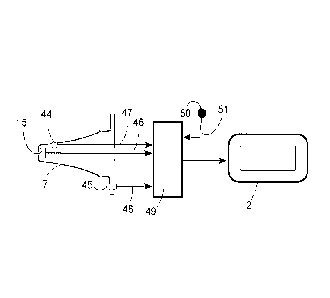
operation of the medical device. Fig. 11 illustrates a simplified block-
diagram of an IR ear
thermometer having a probe 7, electronic circuit 49 and an output device which
is the display 2.
The probe 7 incorporates the IR sensor 15 for measuring a raw patient
temperature, proximity
sensor 44 and probe installation sensor 45. The raw patient temperature may be
used as a base
12
CA 02795703 2012-10-05
WO 2011/127063 PCT/US2011/031264
temperature for further computations. These components are coupled to the
electronic circuit
49 via respective conductors 46, 47, and 48. There is also an ambient
temperature sensor 50
that sends its output signal to the circuit 49 via conductor 51. The ambient
sensor 50 is
positioned outside of the probe 7. The circuit 49 processes all signals
according to the pre-
programmed algorithm and sends a computed temperature number to display 2. The
initial
temperature TB is computed by the circuit 49 from the signals received from
the IR sensor 15
and probe installation sensor 45 (to correct for the probe cover IR
transmission factor). The
signal processing and temperature computation algorithms are well known to a
person of skill
in the art.
If a signal from the proximity sensor 44 indicates that the tip of probe 7
incorporating
the IR sensor 15 is positioned inside the ear canal, the computed temperature
TB is sent to
display 2. However, if a signal from the proximity sensor 44 indicates that
the tip of probe 7 is
positioned at the entrance of the ear canal, the initial temperature TB
represents the exterior skin
rather than the interior of the ear canal and thus should be adjusted to
compensate for a cooling
effect by the ambient temperature. The cooling effect is negligible inside the
ear canal but it is
substantial at the entrance of the ear canal. The ambient temperature is
monitored by use of the
ambient sensor 50 whose signal allows circuit 49 to compute ambient
temperature Ta. The
adjusted temperature Tdmay be calculated according to the following equation:
Td=TB+ k(TB- Ta) , (1)
where k is a constant having a typical value of 0.017. However, the actual
value of k should be
experimentally determined for every practical design. The adjusted temperature
Td is sent to
the display 2.
In another embodiment, a signal from the proximity sensor 44 may be used to
generate
for the operator a warning alarm (by display 2 or by any other visual or
acoustic human
interface) if the probe 7 is not correctly positioned inside the ear canal.
While the invention has been particularly shown and described with reference
to
preferred embodiments thereof, it will be understood by those skilled in the
art as described
herein that various changes in form and details may be made to the disclosed
embodiments
13
without departing from the scope of the invention.
14
CA 2795703 2017-07-10