Note: Descriptions are shown in the official language in which they were submitted.
COMPOSITIONS AND METHODS OF IDENTIFYING TUMOR SPECIFIC
NEOANTIGENS
[0001]
FIELD OF THE INVENTION
[0002] The present invention relates generally to the identification of
tumor specific neoantigens and
the uses of these neoantigens to produce cancer vaccines.
BACKGROUND OF THE INVENTION
[0003] Tumor vaccines are typically composed of tumor antigens and
immunostirnulatory
molecules (e.g. cytokines or TLR ligands) that work together to induce antigen-
specific cytotoxic T
cells (CTLs) that recognize and lyse tumor cells. At this time, almost all
vaccines contain either
shared tumor antigens or whole tumor cell preparations (Gilboa, 1999). The
shared tumor antigens
are immunogenic proteins with selective expression in tumors across many
individuals and are
commonly delivered to patients as synthetic peptides or recombinant proteins
(Boon et al., 2006). In
contrast, whole tumor cell preparations are delivered to patients as
autologous irradiated cells, cell
lysates, cell fusions, heat-shock protein preparations or total mRNA (Parmiani
et al., 2007). Since
whole tumor cells are isolated from the autologous patient, the cells express
patient-specific
tumor antigens as well as shared tumor antigens. Finally, there is a third
class of tumor antigens that
has rarely been used in vaccines due to technical difficulties in identifying
them (Sensi et al. 2006).
This class consists of proteins with tumor-specific mutations that result in
altered amino acid
sequences. Such mutated proteins have the potential to: (a) uniquely mark a
tumor (relative to non-
tumor cells) for recognition and destruction by the immune system (Lennerz et
al., 2005); (b) avoid
central and sometimes peripheral T cell tolerance, and thus be recognized by
more effective, high
avidity T cells receptors (Gotter et al., 2004).
[0004] Thus a need exists for a method of identifying neoepitopes that are
useful as tumor
vaccines.
1
Date recu/Date Received 2020-06-16
CA 02797868 2012-10-29
WO 2011/143656 PCMJS2011/036665
SUMMARY OF THE INVENTION
[0005] The present invention relates in part to the discovery of a method
of identifying
peptides that are capable of elicting a tumor specific T-cell response.
[0006] In one aspect the invention provides methods of identifying a
neoantigen by
identifying a tumor specific mutation in an expressed gene of a subject having
cancer. In some
aspects when the mutation is a point mutation the method further comprises
identifying the
mutant peptide having the mutation. Preferably the mutant peptide binds to a
class I HLA
protein with a greater affinity than a wild ¨type peptide and has an IC50 less
than 500 nm; In
other aspects when the mutation is a splice-site, frameshift, read-through or
gene-fusion
mutation the method further comprise identifying the mutant polypeptide
encoded by the
mutation. Preferably, the mutant polypeptide binds to a class I HLA protein.
[0007] Optionally, the method further includes selecting peptides or
polypeptides that
activate anti-tumor CD8 T cells.
[0008] The mutant peptide or polypeptide preferably binds to a class I HLA
protein with a
greater affinity than a wild ¨type peptide and has an IC50 less than 500 nM.
Preferably, the
peptide or polypeptide has an IC50 less than 250 nM. More preferably, the
peptide or
polypeptide has an IC50 less than 100 nM. Most preferably, the peptide or
polypeptide has an
IC50 less than 50 nM.
[0009] The mutant peptide is about 8-10 amino acids in length. In another
aspect is about 8-
50 amino acids in length. For example, mutant peptide is greater than 10 amino
acids in leghth,
greater than 15 amino acids in length, greater than 20 amino acids in length,
greater than 30
amino acids in length. Preferably the the mutant peptides is about 24-40 amino
acids in length.
[00010] In a further aspect the invention provides methods of inducing a
tumor specific
immune response in a subject by administering one or more peptides or
polypeptides identified
according to the methods of the invention and an adjuvant. The adjuvant is for
example, a TLR-
based adjuvant or a mineral oil based adjuvant. In some aspects the peptide or
polypeptide and
TLR-based adjuvant is emulsified with a mineral oil based adjuvant.
Optionally, the method
further includes administering an anti-immunosuppressive agent such as an an
anti-CTLA-4
antibody, an anti-PD1 antibody an anti-PD-L1 antibody an anti-CD25 antibody or
an inhibitor of
IDO.
[00011] In yet another aspect the invention provides methods of inducing a
tumor specific
immune response in a subject by administering to the subject autologous
dendritic cells or
2
antigen presenting cells that have been pulsed with one or more of the
peptides or polypeptides
identified according to the methods of the inventions. Optionally, the method
further includes
administering an adjuvant such as for example, a TLR-based adjuvant or a
mineral oil based
adjuvant. In some aspects the peptide or polypeptide and TLR-based adjuvant is
emulsified with
a mineral oil based adjuvant. In some embodiments the method further includes
administering an
anti-immunosuppressive agent. Anti-immunosuppressive agents include for
example an anti-
CTLA-4 antibody, an anti-PD1 antibody an anti-PD-Ll antibody an anti-CD25
antibody or an
inhibitor of DO.
[00012] In another aspect the invention provides a method of vaccinating
or treating a subject
for cancer by identifying a plurality of tumor specific mutations in an
expressed gene of the
subject, identifying mutant peptides or polypeptides having the identified
tumor specific
mutations, selecting one or more of the identified mutant peptide or
polypeptides that binds to a
class I HLA protein preferably with a greater affinity than a wild ¨type
peptide and is capable of
activating anti-tumor CD8 T-cells, and administering to the subject the one or
more selected
peptides,polypeptides or autologous dendritic cells or antigen presenting
cells pulsed with the
one or more identified peptides or polypeptides. The mutant peptide is about 8-
10 amino acids in
length. In another aspect is about 8-50 amino acids in length. For example,
mutant peptide is
greater than 10 amino acids in length, greater than 15 amino acids in length,
greater than 20
amino acids in length, greater than 30 amino acids in length. Preferably, the
mutant peptides is
about 24-40 amino acids in length.
[00013] Optionally, the method further includes administering an adjuvant
such as for
example, a TLR-based adjuvant or a mineral oil based adjuvant. In some aspects
the peptide or
polypeptide and TLR-based adjuvant is emulsified with a mineral oil based
adjuvant. In some
embodiments the method further includes administering an anti-
immunosuppressive agent. Anti-
immunosuppressive agents include for example an anti-CTLA-4 antibody, an anti-
PDl antibody
an anti-PD-L1 antibody an anti-CD25 antibody or an inhibitor of IDO.
[00014]
[00015] The subject is a human, dog, cat, or horse. The cancer is breast
cancer, ovarian
cancer, prostate cancer, lung cancer, kidney cancer, gastric cancer, colon
cancer, testicular
cancer, head and neck cancer, pancreatic cancer, brain cancer, melanoma
lymphoma, such as B-
3
Date regu/Date Received 2020-06-16
CA 02797868 2012-10-29
WO 2011/143656 PCT/US2011/036665
cell lumphoma or leukemia, such as cute myelogenous leukemia, chronic
myelogenous
leukemia, chronic lymphocytic leukemia, or T cell lymphocytic leukemia.
[00016] Also included in the invention are pharmaceutical compositions
containing the
peptide or polypeptide identified according the methods of the invention and a
pharmaceutically
acceptable carrier.
[00017] For example, the invention provides a composition containing least
two distinct
SF3B1 peptides wherein each peptide is equal to or less than 50 amino acids in
length and
contains
a leucine at amino acid position 625;
a histidine at amino acid position 626;
a glutamic acid at amino acid position 700;
an aspartic acid at amino acid position 742; or
an arginine at amino acid position 903, when numbered in accordance with wild-
type
SF3B1.
[00018] The invention also provides a composition containing at least two
distinct MYD88
peptides where each peptide is equal to or less than 50 amino acids in length
and contains
a threonine at amino acid position 232; a leucine at amino acid position 258;
or
a proline at amino acid position 265, when numbered in accordance with wild-
type MYD88
[00019] The invention further provides composition containing at least two
distinct TP53
peptides where each peptide is equal to or less than 50 amino acids in length
and contains an
arginine at amino acid position -1 ;an arginine at amino acid position 215; a
serine at amino acid
position 238; a glutamine at amino acid position 248; a phenylalanine at amino
acid position
255; a cysteine at amino acid position 273 or an asparagine at amino acid
position 281, when
numbered in accordance with wild-type TP53.
[00020] The invention further provides composition containing at least two
distinct ATM
peptides wherein each peptide is equal to or less than 50 amino acids in
length and contain
a phenylalanine at amino acid position 1252; an arginine at amino acid
position 2038; a histidine
at amino acid position 2522; or a cysteine at amino acid position 2954, when
numbered in
accordance with wild-type ATM.
[00021] A composition comprising at least two distinct Abl peptides wherein
each peptide is
equal to or less than 50 amino acids in length and contains a valine at amino
acid position 244;
a valine at amino acid position 248;
4
CA 02797868 2012-10-29
WO 2011/143656 PCT/US2011/036665
a glutamic acid at amino acid position 250; an alanine at amino acid position
250; a histidine at
amino acid position 252; an arginine at amino acid position 252; a
phenylalanine at amino acid
position 253; a histidine at amino acid position 253; a lysine at amino acid
position 255; a valine
at amino acid position 255; a glycine at amino acid position 276; an
isoleucine at amino acid
position 315; an asparagine at amino acid position 315; a leucine at amino
acid position 317; a
threonine at amino acid position 343; a threonine at amino acid position 351;
a glycine at amino
acid position 355; a valine at amino acid position 359; an alanine at amino
acid position 359; an
isoleucine at amino acid position 379; a leucine at amino acid position 382; a
methionine at
amino acid position 387; a proline at amino acid position 396; an arginine at
amino acid position
396;a tyrosine at amino acid position 417; or a serine at amino acid position
486, when
numbered in accordance with wild-type ABL.
[00022] Further included in the invention is a composition containing at
least two distinct
FBXW7 peptides where each peptide is equal to or less than 50 amino acids in
length and
contains a leucine at amino acid position 280; a histidine at amino acid
position 465; a cysteine
at amino acid position 505; ora glutamic acid at amino acid position 597, when
numbered in
accordance with wild-type FBXW7.
[00023] In a further a aspect the invention provides a composition
containing at least two
distinct MAPK1 peptides where each peptide is equal to or less than 50 amino
acids in length
and contains an asparagine at amino acid position 162; a glycine at amino acid
position 291; or
a phenylalanine at amino acid position 316, when numbered in accordance with
wild-type
MAPK1.
[00024] The invention also provides a composition conatining at least two
distinct GNB1
peptides wherein each peptide is equal to or less than 50 amino acids in
length and contains a
threonine at amino acid position 180, when numbered in accordance with wild-
type GNB1.
[00025] Also provided by the invention is a method of treating a subject
with an imatinib
resistant tumor to a HLA-A3 positive subject a composition of Bcr-abl peptide
equal to or less
than 50 amino acid in length that contains a lysine at position 255 when
numbered in accordance
with wild-type bcr-abl.
[00026] Further provided by the invention, is method of treating a subject
with an imatinib
resistant tumor comprising administering to the ubject one or more peptides
containing a bcr-abl
mutation where the peptide is equal to or less than 50 amino acid and binds to
a class I HLA
protein with an IC50 less than 500 nm.
[00027] Unless otherwise defined, all technical and scientific terms used
herein have the same
meaning as commonly understood by one of ordinary skill in the art to which
this invention
pertains. Although methods and materials similar or equivalent to those
described herein can be
used in the practice of the present invention, suitable methods and materials
are described below.
In cases of conflict, the present
specification, including definitions, will control. In addition, the
materials, methods, and
examples described herein are illustrative only and are not intended to be
limiting.
[00028] Other features and advantages of the invention will be apparent
from and
encompassed by the following detailed description and claims.
BRIEF DESCRIPTION OF THE DRAWINGS
[00029] Figure 1 shows the balance of specificity and autoimmune toxicity
using 3 classes of
antigens for tumor vaccines. Whole tumor cells may be the the least specific
antigen formulation
for tumor vaccines since the full set of protein antigens expressed in tumor
cells include
thousands of proteins that are also present in other cells of the body.
Overexpressed tumor
antigens are slightly more specific because they have been selected for much
higher and more
selective expression in tumors compared to other cells in the body.
Nevertheless, it is impossible
to test every cell in the body for the expression of these antigens and there
is a substantial risk
that other cells express them. Finally, mutated proteins generate neoepitopes
that are present
only in tumor cells and provide the greatest level of specificity.
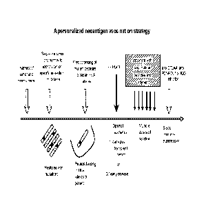
[00030] Figure 2 is a schema for a personalized neoantigen vaccination
strategy that can be
applied to the treatment of any cancer. We also highlight the possibility of
applying this strategy
in two unique scenarios. In the first case, a patient is vaccinated in the
early period following
hematopoietic stem cell transplantation (HSCT) (e.g. as is done for CLL, CML
and other
leukemias). The early post-HSCT period is a unique therapeutic setting as the
immune system is
competent due to reconstitution with HSCT, thus overcoming tumor- or treatment-
induced host
immune defects. Moreover, the abundance of homeostatic cytokines in a
lymphopenia milieu,
such as in the early post-HSCT setting, can contribute to rapid expansion of T
cells. In the
second case, a patient is vaccinated early in the disease course when immune
competence may be
more intact in the early stages of disease, before impairment by exposure to
chemotherapy (e.g.
for solid or hematopoeitic tumors). Since the immune system is likely to be
most active in these
6
CA 2797868 2018-01-30
CA 02797868 2012-10-29
WO 2011/143656 PCT/US2011/036665
two specific situations, we suggest that these are the ideal situations for
applying tumor
vaccination strategies.
[00031] Figure 3 shows a strategy for identifying tumor neoepitopes is
described in 3 steps:
(1) using sequencing technologies, detect gene mutations that are present in
tumor but not
germline DNA of a single patient; (2) using prediction algorithms, predict
whether mutated
peptides have the potential to bind personal HLA allele; these predicted
peptides may optionally
be tested experimentally for binding to appropriate HLA proteins. In addition,
these genes must
also be expressed in tumor cells. (3) generate T cells ex vivo and test
whether they are able to
recognize cells expressing the mutated protein; alternatively, mass
spectrometry can be used to
detect peptides eluted from tumor cell surface HLA proteins. For chronic
lymphocytic leukemia,
our studies to date demonstrate that there are an average of 23 protein-
altering mutations per
patient, 46 predicted binding mutant peptides and 15-25 validated binding
mutant peptides. Of
these, we anticipate that ¨7-12 peptides are expressed and processed in tumor
cells (though this
may differ across tumors and patients).
[00032] Figure 4 shows five classes of mutations generate potential tumor
neoepitopes. New
tumor-specific epitopes can arise as a result of missense, splice-site,
frameshift or read-through
point mutations (red asterisk), or from the fusion of two genes (or within the
same gene). In
particular, splice-site, frameshift, read-through mutations and gene fusions
can each generate
novel stretches of amino acids (in magenta) that are normally not translated,
but now are
expressed and translated as a result of mutation. Missense mutations lead to
peptides with
single amino acid changes.
[00033] Figure 5 shows the frequency of mutations per class in CLL
patients. Our studies
applying next-generation sequencing to a series of 7 CLL tumors reveal that
CLL cells harbor
many mutations, that provide a rich source of possible mutated peptides. We
observe that the
total number of nonsilent gene alterations in CLL ranged from 17-155 per
individual, the
majority of which were somatically altered point mutations (missense). The
tumors of 4 patients
also harbored splice-site mutations; for 3 patients, novel gene fusions were
identified by RNA
sequencing.
[00034] Figure 6 shows data from automated predictions (Step 2A of the
strategy in Figure 3)
of peptide binding (for peptides that harbor a specific missense mutation)
against each of a
patient's 6 HLA (MHC Class I) alleles. Magenta=strong binders;
green=intermediate binders.
7
CA 02797868 2012-10-29
WO 2011/143656 PCT/US2011/036665
[00035] Figure 7 shows methods for conifrming RNA expression of mutated
genes (Step 2B
of the strategy in Figure 3). A. For CLL patient 7, we found that more than
half of the mutated
genes with predicted HLA-binding peptides were expressed at the RNA level. B.
We have also
used RNA pyrosequencing to detect expressed RNAs harboring specific mutations
found in
DNA. C. We can validate novel gene fusions that were seen by DNA sequencing
using PCR-
TOPO cloning of the breakpoint region (depicted is a fusion discovered for
patient 2).
[00036] Figure 8 shows a method and data for experimental validation of HLA-
peptide
binding (Step 2C of the strategy in Figure 3). A. Schema for experimental
validation of peptide
binding to specific HLA alleles. B. Summary of candidate mutated peptides
identified in patients
1 and 2. Shaded cells indicate that analysis is in progress. C. Data for
predicted vs
experimentally verified binding affinity of peptides generated from gene
alterations (missense
mutation or gene fusion) for patient 2. A prediction cutoff of IC50<120nM
(solid vertical line on
left) results in all peptides showing experimental binding to class I HLA.
[00037] Figure 9 shows predicted differential binding of mutated vs
germline (i.e also called
parental, wild type or normal) peptides to HLA alleles. 12 of 25 predicted HLA
binding mutated
peptides of Pt 2 have >2 fold greater binding (cutoff = red dotted line) than
parental peptides.
This further increases the specificity of mutated peptides. Mutated peptides
are specific for two
reasons: first, many of the T cell receptors that recognize a mutated peptide
are not likely to
detect the wild type parental peptide; second, some of the mutated peptides
can bind HLA with
higher affinity than the parental peptide. Since the first property cannot be
computationally
predicted, we will focus on predicting the second property and selecting for
inclusion in vaccines
only those peptides that show higher binding to HLA for mutated relative to
wild type peptides.
[00038] Figure 10 shows T cell reactivity against a candidate personal CLL
neoepitope (Step
3 of the strategy in Figure 3). We observed that T cells isolated from patient
1 post-therapy can
detect a specific mutated TLK2 peptide (peptide #7) (using the Elispot assay).
[00039] Figure 11 shows that BCR-ABL mutations generate many peptides
predicted to bind
HLA-A and HLA-B alleles. By applying the NetMHC prediction algorithm (Nielsen
et al. PLoS
One. 2007, 2(8):e796), we predicted peptides generated from the BCR-ABL
mutations with
potential to bind to 8 common HLA-A and -B alleles. The most common BCR-ABL
mutations
are ordered in decreasing frequency (from left to right), and predicted IC50
of various class I
MHC binding peptides are depicted. In total, we predicted 84 peptides to bind
with good affinity,
defined as an IC50 of less than 1000, across a wide range of HLA alleles. Of
all the predicted
8
CA 02797868 2012-10-29
WO 2011/143656 PCT/US2011/036665
peptides, 24 of 84 (29%) were predicted to be strong binders with an IC50<50.
42 peptides
(50%) were intermediate binders, defined as IC50 between 50 and 500. 18
peptides (21%) were
weak binders defined as IC50 between 500 and 1000.
[00040] Figure 12 shows BCR-ABL peptide harboring the E255K mutation binds HLA
proteins and is associated with specific, polyfunctional T cells present in
CML patients. A.
Experimentally-derived binding scores of E255K-B (and parental peptide) to HLA
A3 and
supertype members. B. In CD8+ T cells expanded from a HLAA3+ E255K+patient
following
HSCT, we detected IFNgamma secretion against the E255K-B (MUT) peptide and A3+
expressing APCs expressing the E255K minigene (MG). This response was
abrogated in the
presence of the class I blocking antibody (w6/32). C. IFNgamma-secreting cells
were also
tetramer+ for the mutated peptide and were (D) polyfunctional, secreting IP10,
TNFalpha and
GM-CSF (based on the Luminex assay).
[00041] Figure 13 shows that patient-derived T cell clones can recognize
tumor-specific
epitopes and kill cells presenting these epitopes. A. Reactivity to the CD8+ T
cell epitope of
CML66 (peptide 66-72C) is restricted by HLA B-4403. B. CML66 mRNA can be
efficiently
nucleofected into CD4OL-expanded B cells. C. CML66-specific CD8+ T cells are
cytotoxic to
CD4OL B cells expressing CML66 by RNA nucleofection or by peptide pulse, but
not control
targets.
[00042] Figure 14 shows significantly mutated genes in CLL. A. The 9 most
significantly
mutated genes among 64 CLL samples. N - total covered territory in base pairs
across 64
sequenced samples. p- and q-values were calculated by comparing the
probability of seeing the
observed constellation of mutations to the background mutation rates
calculated across the
dataset. Red bars ¨ genes not previously known to be mutated in CLL; grey bars
¨ genes in
which mutation in CLL has been previously reported. B. Type (missense, splice-
site, nonsense)
and location of mutations in ATM, SF3B1, TP53, MYD88, FBXW7, DDX3X, M4PK1, and
GNB1
discovered among the 64 CLLs (position and mutation in CLL samples shown above
the gene)
compared to previously reported mutations in literature or in the COSMIC
database (lines show
position of mutations below the gene).
[00043] Figure 15 shows that SF3B1 is expressed in CLL samples (7th column
in graph) and
has higher expression than many control cells, including: PBMC, M: monocyte,
CC: cancer cell
lines (includes K562, Jurkat, IM9, MCF-7, Hela, Ovcar, RPMI, OTM, MCF-CAR,
KM12BM
and MM1S).
9
CA 02797868 2012-10-29
WO 2011/143656 PCT/US2011/036665
[00044] Figure 16 shows that SF3B1 mutations generate peptides that are
predicted to bind to
patient-specific HLA alleles. For example, one peptide that includes the
common SF3B1 K700E
mutation is predicted to bind HLA strongly.
DETAILED DESCRIPTION OF THE INVENTION
[00045] One of the critical barriers to developing curative and tumor-
specific immunotherapy
is the identification and selection of highly restricted tumor antigens to
avoid autoimmunity.
Tumor neoantigens, which arise as a result of genetic change within malignant
cells, represent
the most tumor-specific class of antigens. Neoantigens have rarely been used
in vaccines due to
technical difficulties in identifying them. Our approach to identify tumor-
specific neoepitopes
involves three steps. (1) identification of DNA mutations using whole genome
or whole exome
(i.e. only captured exons) or RNA sequencing of tumor versus matched germline
samples from
each patient; (2) application of validated peptide-MHC binding prediction
algorithms to generate
a set of candidate T cell epitopes that may bind patient HLA alleles and are
based on non-silent
mutations present in tumors; and (3)optional demonstration of antigen-specific
T cells against
mutated peptides or demonstration that a candidate ppeptide is bound to HLA
proteins on the
tumor surface.
[00046] Accordingly, the present invention relates to methods for
identifying and/or detecting
T-cell epitopes of an antigen. Specifically, the invention provides method of
identifying and/or
detecting tumor specific neoantigens that are useful in inducing a tumor
specific immune
response in a subject.
[00047] In particular, the invention provides a method of vaccinating or
treating a subject by
identifying a plurality of tumor specific mutations in the genome of a
subject. Mutant peptides
and polypetideds having the identified mutations and that binds to a class I
HLA protein are
selected. Optionally, these peptide and polypetides binds to a class I HLA
proteins with a greater
affinity than the wild ¨type peptide and/or are capable of activating anti-
tumor CD8 T-cells
These peptides are admistered to the subject. Alternatively, autologous
antigen-presenting cells
that have been pulsed with the peptides are administered.
[00048] The importance of mutated antigens, or neoepitopes, in the immune
control of
tumors has been appreciated in seminal studies showing that: (a) mice and
humans often
mount T cell responses to mutated antigens (Parmiani et al., 2007; Sensi and
Anichini,
2006); (b) mice can be protected from a tumor by immunization with a single
mutated peptide
CA 02797868 2012-10-29
WO 2011/143656 PCT/US2011/036665
that is present in the tumor (Mandelboim et al., 1995); (c) spontaneous or
vaccine-mediated
long-term melanoma survivors mount strong memory cytotoxic T cell (CTL)
responses to
mutated antigens (Huang et al.. 2004; Lennerz et al., 2005; Zhou et al.,
2005a); (d) finallyõ
follicular lymphoma patients show molecular remission when immunized with
patient-
specific mutated immunoglobulin proteins that are present in autologous tumor
cells.
(Baskar et al., 2004). Furthermore, the CTL responses in these patients are
directed toward
the mutated rather than shared regions of the immunoglobulin protein.
Additionally, such
mutated peptides have the potential to: (a) uniquely mark a tumor for
recognition and destruction
by the immune system, thus reducing the risk for autoimmunity; and (b) avoid
central and
peripheral T cell tolerance, allowing the antigen to be recognized by more
effective, high avidity
T cells receptors. (Figure 1)
[00049] Identical mutations in any particular gene are rarely found across
tumors (and are
even at low frequency for the most common driver mutations). Thus, the methods
of the
present invention will comprehensively identify patient-specific tumor
mutations. Using
highly parallel sequencing technologies, HLA-peptide binding prediction tools
and
biochemical assays the methods of the invention will allow: (1) comprehensive
identification
of mutated peptides that are expressed and bind HLA proteins present in a
patient's tumor;
(2) monitoring of the natural immune response of cancer patients to these
identified
neoepitopes; (3) determining whether cytotoxic T cells that recognize these
peptides in the
context of patient HLA proteins can selectively lyse autologous tumor cells ex
vivo. This
strategy addresses several fundamental questions related to how the immune
system of
cancer patients interacts with tumor neoepitopes. These include: which and
what fraction of
tumor neoepitopes are detected by T cells, how many T cell precursors are able
to respond to
neoepitopes, how frequent are neoepitope-specific memory and effector T cells
in
circulation and in the tumor, how much avidity do T cells have for these
epitopes, are
neoepitope-specific T cells functional? The answers to these questions provide
both the
justification and strategy for using tumor neoepitopes in human vaccines.
[00050] The immune system of a human can be classified into two functional
subsystems, i.e.,
the innate and the acquired immune system. The innate immune system is the
first line of
defense against infections, and most potential pathogens are rapidly
neutralized before they can
cause, for example, a noticeable infection. The acquired immune system reacts
to molecular
structures. referred to as antigens, of the intruding organism. There are two
types of acquired
11
CA 02797868 2012-10-29
WO 2011/143656 PCT/US2011/036665
immune reactions, i.e. the humoral immune reaction and the cell-mediated
immune reaction. In
the humoral immune reaction, the antibodies secreted by B cells into bodily
fluids bind to
pathogen-derived antigens, leading to the elimination of the pathogen through
a variety of
mechanisms, e.g. complement-mediated lysis. In the cell-mediated immune
reaction. T-cells
capable of destroying other cells are activated. If, for example, proteins
associated with a disease
are present in a cell, they are, within the cell, fragmented proteolytically
to peptides. Specific cell
proteins then attach themselves to the antigen or peptide formed in this
manner and transport
them to the surface of the cell, where they are presented to the molecular
defense mechanisms, in
particular T-cells, of the body. Cytotoxic T cells recognize these antigens
and kill the cells that
harbor the antigens.
[00051] The molecules which transport and present peptides on the cell
surface are referred to
as proteins of the major histocompatibility complex (MHC). The MHC proteins
are classified
into MHC proteins of class I and of class II. The structures of the proteins
of the two MHC
classes are very similar; however, they differ quite considerably in their
function. Proteins of
MHC class I are present on the surface of almost all cells of the body,
including most tumor
cells. The proteins of MHC class I are loaded with antigens that usually
originate from
endogenous proteins or from pathogens present inside cells, and are then
presented to cytotoxic
T-lymphocytes (CTLs). The MHC proteins of class II are only present on
dendritic cells, B-
lymphocytes, macrophages and other antigen-presenting cells. They present
mainly peptides,
which are processed from external antigen sources, i.e. outside of the cells,
to T-helper (Th)
cells. Most of the peptides bound by the MHC proteins of class I originate
from cytoplasmic
proteins produced in the healthy host organism itself and don't normally
stimulate an immune
reaction. Accordingly, cytotoxic T-lymphocytes which recognize such self-
peptide-presenting
MHC molecules of class I are deleted in the thymus or, after their release
from the thymus, are
deleted or inactivated, i.e. tolerized. MHC molecules are only capable of
stimulating an immune
reaction when they present peptides to non-tolerized cytotoxic T-lymphocytes.
Cytotoxic T-
lymphocytes have, on their surface, both T-cell receptors (TCR) and CD8
molecules. T-Cell
receptors are capable of recognizing and binding peptides complexed with the
molecules of
MHC class I. Each cytotoxic T-lymphocyte expresses a unique T-cell receptor
which is capable
of binding specific MHC/peptide complexes.
[00052] The peptides attach themselves to the molecules of MHC class I by
competitive
affinity binding within the endoplasmic reticulum, before they are presented
on the cell surface.
12
CA 02797868 2012-10-29
WO 2011/143656 PCT/US2011/036665
Here, the affinity of an individual peptide is directly linked to its amino
acid sequence and the
presence of specific binding motifs in defined positions within the amino acid
sequence. If the
sequence of such a peptide is known, it is possible, for example, to
manipulate the immune
system against diseased cells using, for example, peptide vaccines.
[00053] Using computer algorithms, it is possible to predict potential T-
cell epitopes, i.e.
peptide sequences, which are bound by the MHC molecules of class I or class II
in the form of a
peptide-presenting complex and then, in this form, recognized by the T-cell
receptors of T-
lymphocytes. Currently, use is made, in particular, of two programs, namely
SYFPEITHI
(Rammensee et al., Immunogenetics, 50 (1999), 213-219) and HLA_BIND (Parker et
al., J.
Immunol., 152 (1994), 163-175). The peptide sequences determined in this
manner, which
potentially may bind to MHC molecules of class I, then have to be examined in
vitro for their
actual binding capacity.
[00054] The technical object of the present invention is to provide an
improved method for
identifying and screening potential T-cell epitopes present in tumor cells,
which method allows
for simultaneous and rapid examination of a large number of peptide sequences,
for their
capability of binding to specific MHC molecules.
[00055] In the present invention, the technical object on which it is based
is achieved by
providing a method for identifying and/or detecting mutated antigens that are
present in tumors
but not in normal tissue. The method uses massively parallel genomic
sequencing of the entire
coding portion of a cancer patient genome to identify the specific mutated
genes in a tumor. In
order to narrow down the mutant peptides to those with potential to bind more
strongly to HLA
than the wild type peptides and thus confer tumor specificity, well-
established algorithms will be
used to predict peptides that bind any of the 6 unique class I HLA alleles of
each patient and a
predicted IC50 for all 9- or 10-mer peptides with tumor-specific mutant
residues vs. those with
the germline residue will be calculated. Typically, peptides with predicted
IC50<50nM, are
generally considered medium to high affinity binding peptides and will be
selected for testing
their affinity empirically using biochemical assays of HLA-binding. Finally,
it will be
determined whether the human immune system can mount effective immune
responses against
these mutated tumor antigens and thus effectively kill tumor but not normal
cells.
[00056] Definitions
[00057] A "T-cell epitope" is to be understood as meaning a peptide
sequence which can be
bound by the MHC molecules of class I or II in the form of a peptide-
presenting MHC molecule
13
CA 02797868 2012-10-29
WO 2011/143656 PCT/US2011/036665
or MHC complex and then. in this form, be recognized and bound by cytotoxic T-
lymphocytes
or T-helper cells, respectively
[00058] A "receptor" is to be understood as meaning a biological molecule
or a molecule
grouping capable of binding a ligand. A receptor may serve, to transmit
information in a cell, a
cell formation or an organism. The receptor comprises at least one receptor
unit and preferably
two receptor units, where each receptor unit may consist of a protein
molecule, in particular a
glycoprotein molecule. The receptor has a structure which complements that of
a ligand and may
complex the ligand as a binding partner. The information is transmitted in
particular by
conformational changes of the receptor following complexation of the ligand on
the surface of a
cell. According to the invention, a receptor is to be understood as meaning in
particular proteins
of MHC classes I and II capable of forming a receptor/ligand complex with a
ligand, in particular
a peptide or peptide fragment of suitable length.
[00059] A "ligand" is to be understood as meaning a molecule which has a
structure
complementary to that of a receptor and is capable of forming a complex with
this receptor.
According to the invention, a ligand is to be understood as meaning in
particular a peptide or
peptide fragment which has a suitable length and suitable binding motives in
its amino acid
sequence, so that the peptide or peptide fragment is capable of forming a
complex with proteins
of MHC class I or MHC class II.
[00060] A "receptor/ligand complex" is also to be understood as meaning a
"receptor/peptide
complex" or "receptor/peptide fragment complex", in particular a peptide- or
peptide fragment-
presenting MHC molecule of class I or of class II.
[00061] "Proteins or molecules of the major histocompatibility complex
(MHC)", "MHC
molecules", "MHC proteins" or "HLA proteins" are to be understood as meaning,
in particular,
proteins capable of binding peptides resulting from the proteolytic cleavage
of protein antigens
and representing potential T-cell epitopes, transporting them to the cell
surface and presenting
them there to specific cells, in particular cytotoxic T-lymphocytes or T-
helper cells. The major
histocompatibility complex in the genome comprises the genetic region whose
gene products
expressed on the cell surface are important for bindung and presenting
endogenous and/or
foreign antigens and thus for regulating immunological processes. The major
histocompatibility
complex is classified into two gene groups coding for different proteins,
namely molecules of
MHC class I and molecules of MHC class II. The molecules of the two MHC
classes are
specialized for different antigen sources. The molecules of MHC class I
present endogenously
14
CA 02797868 2012-10-29
WO 2011/143656 PCT/US2011/036665
synthesized antigens, for example viral proteins and tumor antigens. The
molecules of MHC
class II present protein antigens originating from exogenous sources, for
example bacterial
products. The cellular biology and the expression patterns of the two MHC
classes are adapted to
these different roles.
[00062] MHC molecules of class I consist of a heavy chain and a light chain
and are capable
of binding a peptide of about 8 to 11 amino acids, but usually 9 or 10 amino
acids, if this peptide
has suitable binding motifs, and presenting it to cytotoxic T-lymphocytes. The
peptide bound by
the MHC molecules of class I originates from an endogenous protein antigen.
The heavy chain of
the MHC molecules of class I is preferably an HLA-A, HLA-B or HLA-C monomer,
and the
light chain is 13-2-microglobulin.
[00063] MHC molecules of class II consist of an la-chain and a I3-chain and
are capable of
binding a peptide of about 15 to 24 amino acids if this peptide has suitable
binding motifs, and
presenting it to T-helper cells. The peptide bound by the MHC molecules of
class II usually
originates from an extracellular of exogenous protein antigen. The a -chain
and the I3-chain are
in particular HLA-DR, HLA-DQ and HLA-DP monomers.
[00064] A "vaccine" is to be understood as meaning a composition for
generating immunity
for the prophylaxis and/or treatment of diseases. Accordingly, vaccines are
medicaments which
comprise antigens and are intended to be used in humans or animals for
generating specific
defense and protective substance by vaccination.
[00065] "Isolated" means that the polynucleotide or polypeptide or
fragment, variant, or
derivative thereof has been essentially removed from other biological
materials with which it is
naturally associated, or essentially free from other biological materials
derived, e.g., from a
recombinant host cell that has been genetically engineered to express the
polypeptide of the
invention.
[00066] "Neoantigen" means a class of tumor antigens which arises from
tumor-specific
mutations in expressed protein.
[00067] "Purified" means that the polynucleotide or polypeptide or
fragment, variant, or
derivative thereof is substantially free of other biological material with
which it is naturally
associated, or free from other biological materials derived, e.g., from a
recombinant host cell that
has been genetically engineered to express the polypeptide of the invention.
That is, e.g., a
purified polypeptide of the present invention is a polypeptide that is at
least about 70-100% pure,
i.e., the polypeptide is present in a composition wherein the polypeptide
constitutes about 70-
100% by weight of the total composition. In some embodiments, the purified
polypeptide of the
present invention is about 75%-99% by weight pure, about 80%-99% by weight
pure, about 90-
99% by weight pure, or about 95% to 99% by weight pure.
[00068] Identification of Tumor Specific Mutations
[00069] The present invention is based, on the identification of certain
mutations (e.g., the
variants or alleles that are present in cancer cells). In particular, these
mutations are present in the
genome of cancer cells of a subject having cancer but not in normal tissue
from the subject.
[00070] Genetic mutations in tumors would be considered useful for the
immunological
targeting of tumors if they lead to changes in the amino acid sequence of a
protein exclusively in
the tumor. Useful mutations include: ( I) non-synonymous mutations leading to
different amino
acids in the protein; (2) read-through mutations in which a stop codon is
modified or deleted,
leading to translation of a longer protein with a novel tumor-specific
sequence at the C-terminus;
(3) splice site mutations that lead to the inclusion of an intron in the
mature mRNA and that, a
unique tumor-specific protein sequence; (4) chromosomal rearrangements that
give rise to a
chimeric protein with tumor-specific sequences at the junction of 2 proteins
(i.e., gene fusion);
(5) frameshift mutations or deletions that lead to a new open reading frame
with a novel tumor-
specific protein sequence.
[00071] Peptides with mutations or mutated polypeptides arising from for
example. splice-
site, frameshify, readthrough, orgene fusion mutations in tumor cells may be
identified by
sequencing DNA, RNA or protein in tumor versus normal cells.
[00072] Also within the scope of the inventions are peptides including
previous identified
tumor specific mutations.
[00073] A variety of methods are available for detecting the presence of a
particular mutation
or allele in an individual's DNA or RNA. Advancements in this field have
provided accurate,
easy, and inexpensive large-scale SNP genotyping. Most recently, for example,
several new
techniques have been described including dynamic allele-specific hybridization
(DASH),
microplate array diagonal gel electrophoresis (MADGE), pyrosequencing,
oligonucleotide-
specific ligation, the TagMan system as well as various DNA "chip"
technologies such as the
Affymetrix SNP chips. These methods require amplification of the target
genetic region,
16
Date Recue/Date Received 2021-04-15
CA 02797868 2012-10-29
WO 2011/143656 PCT/US2011/036665
typically by PCR. Still other newly developed methods, based on the generation
of small signal
molecules by invasive cleavage followed by mass spectrometry or immobilized
padlock probes
and rolling-circle amplification, might eventually eliminate the need for PCR.
Several of the
methods known in the art for detecting specific single nucleotide
polymorphisms are summarized
below. The method of the present invention is understood to include all
available methods.
[00074] PCR based detection means can include multiplex amplification of a
plurality of
markers simultaneously. For example, it is well known in the art to select PCR
primers to
generate PCR products that do not overlap in size and can be analyzed
simultaneously.
Alternatively, it is possible to amplify different markers with primers that
are differentially
labeled and thus can each be differentially detected. Of course, hybridization
based detection
means allow the differential detection of multiple PCR products in a sample.
Other techniques
are known in the art to allow multiplex analyses of a plurality of markers.
[00075] Several methods have been developed to facilitate analysis of
single nucleotide
polymorphisms in genomic DNA or celluar RNA. In one embodiment, the single
base
polymorphism can be detected by using a specialized exonuclease-resistant
nucleotide, as
disclosed, e.g., in Mundy, C. R. (U.S. Pat. No.4,656,127). According to the
method, a primer
complementary to the allelic sequence immediately 3' to the polymorphic site
is permitted to
hybridize to a target molecule obtained from a particular animal or human. If
the polymorphic
site on the target molecule contains a nucleotide that is complementary to the
particular
exonuclease-resistant nucleotide derivative present, then that derivative will
be incorporated onto
the end of the hybridized primer. Such incorporation renders the primer
resistant to exonuclease,
and thereby permits its detection. Since the identity of the exonuclease-
resistant derivative of the
sample is known, a finding that the primer has become resistant to
exonucleases reveals that the
nucleotide present in the polymorphic site of the target molecule was
complementary to that of
the nucleotide derivative used in the reaction. This method has the advantage
that it does not
require the determination of large amounts of extraneous sequence data.
[00076] In another embodiment of the invention, a solution-based method is
used for
determining the identity of the nucleotide of a polymorphic site. Cohen, D. et
al. (French Patent
2,650,840; PCT Appin. No. W091/02087). As in the Mundy method of U.S. Pat. No.
4,656,127,
a primer is employed that is complementary to allelic sequences immediately 3
to a polymorphic
site. The method determines the identity of the nucleotide of that site using
labeled
17
CA 02797868 2012-10-29
WO 2011/143656 PCT/US2011/036665
dideoxynucleotide derivatives, which, if complementary to the nucleotide of
the polymorphic
site will become incorporated onto the terminus of the primer.
[00077] An alternative method, known as Genetic Bit Analysis or GBA is
described by
Goelet, P. et al. (PCT Appin. No. 92/15712). The method of Goelet, P. et al.
uses mixtures of
labeled terminators and a primer that is complementary to the sequence 3 to a
polymorphic site.
The labeled terminator that is incorporated is thus determined by, and
complementary to, the
nucleotide present in the polymorphic site of the target molecule being
evaluated. In contrast to
the method of Cohen et al. (French Patent 2,650,840; PCT Appin. No.
W091/02087) the method
of Goelet, P. et al. is preferably a heterogeneous phase assay, in which the
primer or the target
molecule is immobilized to a solid phase.
[00078] Recently, several primer-guided nucleotide incorporation procedures
for assaying
polymorphic sites in DNA have been described (Komher, J. S. et al., Nucl.
Acids. Res. 17:7779-
7784 (1989); Sokolov, B. P., Nucl. Acids Res. 18:3671 (1990); Syvanen, A.-C..
et al., Genomics
8:684-692 (1990); Kuppuswamy, M. N. et al., Proc. Natl. Acad. Sci. (U.S.A.)
88:1143- 1147
(1991); Prezant, T. R. et al., Hum. Mutat. 1:159-164 (1992); Ugozzoli, L. et
al.. GATA 9:107-
112 (1992); Nyren, P. et al., Anal. Biochem. 208:171-175 (1993)). These
methods differ from
GBA in that they all rely on the incorporation of labeled deoxynucleotides to
discriminate
between bases at a polymorphic site. In such a format, since the signal is
proportional to the
number of deoxynucleotides incorporated, polymorphisms that occur in runs of
the same
nucleotide can result in signals that are proportional to the length of the
run (Syvanen, A.-C., et
al., Amer. J. Hum. Genet. 52:46-59 (1993)).
[00079] A number of initiatives are currently underway to obtain sequence
information
directly from millions of individual molecules of DNA or RNA in parallel. Real-
time single
molecule sequencing-by-synthesis technologies rely on the detection of
fluorescent nucleotides
as they are incorporated into a nascent strand of DNA that is complementary to
the template
being sequenced. In one method, oligonucleotides 30-50 bases in length are
covalently anchored
at the 5' end to glass cover slips. These anchored strands perform two
functions. First, they act as
capture sites for the target template strands if the templates are configured
with capture tails
complementary to the surface-bound oligonucleotides. They also act as primers
for the template
directed primer extension that forms the basis of the sequence reading. The
capture primers
function as a fixed position site for sequence determination using multiple
cycles of synthesis,
detection, and chemical cleavage of the dye-linker to remove the dye. Each
cycle consists of
18
CA 02797868 2012-10-29
WO 2011/143656 PCT/US2011/036665
adding the polymerase/labeled nucleotide mixture, rinsing, imaging and
cleavage of dye. In an
alternative method, polymerase is modified with a fluorescent donor molecule
and immobilized
on a glass slide, while each nucleotide is color-coded with an acceptor
fluorescent moiety
attached to a gamma-phosphate. The system detects the interaction between a
fluorescently-
tagged polymerase and a fluorescently modified nucleotide as the nucleotide
becomes
incorporated into the de novo chain. Other sequencing-by-synthesis
technologies also exist.
[00080] Preferably, any suitable sequencing-by-synthesis platform can be
used to identify
mutations.. As described above, four major sequencing-by-synthesis platforms
are currently
available: the Genome Sequencers from Roche/454 Life Sciences, the 1G Analyzer
from
Illumina/Solexa, the SOLiD system from Applied BioSystems, and the Heliscope
system from
Helicos Biosciences. Sequencing-by-synthesis platforms have also been
described by Pacific
BioSciences and VisiGen Biotechnologies. Each of these platforms can be used
in the methods
of the invention. In some embodiments, a plurality of nucleic acid molecules
being sequenced is
bound to a support (e.g., solid support). To immobilize the nucleic acid on a
support, a capture
sequence/universal priming site can be added at the 3' and/or 5' end of the
template. The nucleic
acids may be bound to the support by hybridizing the capture sequence to a
complementary
sequence covalently attached to the support. The capture sequence (also
referred to as a universal
capture sequence) is a nucleic acid sequence complementary to a sequence
attached to a support
that may dually serve as a universal primer.
[00081] As an alternative to a capture sequence, a member of a coupling
pair (such as, e.g.,
antibody/antigen, receptor/ligand, or the avidin-biotin pair as described in,
e.g., US Patent
Application No. 2006/0252077) may be linked to each fragment to be captured on
a surface
coated with a respective second member of that coupling pair.
[00082] Subsequent to the capture, the sequence may be analyzed, for
example, by single
molecule detection/sequencing, e.g., as described in the Examples and in U.S.
Pat. No.
7,283.337, including template-dependent sequencing-by-synthesis. In sequencing-
by-synthesis,
the surface-bound molecule is exposed to a plurality of labeled nucleotide
triphosphates in the
presence of polymerase. The sequence of the template is determined by the
order of labeled
nucleotides incorporated into the 3' end of the growing chain. This can be
done in real time or
can be done in a step-and-repeat mode. For real-time analysis, different
optical labels to each
nucleotide may be incorporated and multiple lasers may be utilized for
stimulation of
incorporated nucleotides.
19
CA 02797868 2012-10-29
WO 2011/143656 PCT/US2011/036665
[00083] Any cell type or tissue may be utilized to obtain nucleic acid
samples for use in the
diagnostics described herein. In a preferred embodiment, the DNAor RNA sample
is obtained
from a tumor or a bodily fluid, e.g., blood, obtained by known techniques
(e.g. venipuncture) or
saliva. Alternatively, nucleic acid tests can be performed on dry samples
(e.2. hair or skin).
[00084] Alternatively, protein mass spectrometry may be used to identify or
validate the
presence of mutated peptides bound to MHC proteins on tumor cells. Peptides
can be acid-eluted
from tumor cells or from HLA molecules that are immunoprecipitated from tumor,
and then
identified using mass spectrometry.
[00085] Neoantigenic Peptides
[00086] The invention further includes isolated peptides that comprise the
tumor specific
mutations identified by the methods of the invention, peptides that comprise
know tumor specific
mutations, and mutant polypeptides or fragments thereof identified by the
method of the
invention. These peptides and polypeptides are referred to herein as
"neoantigenic peptides" or
"neoantigenic polypeptides". The term "peptide" is used interchangeably with
"mutant peptide"
and "neoantigenic peptide" in the present specification to designate a series
of residues, typically
L-amino acids, connected one to the other, typically by peptide bonds between
the a-amino and
carboxyl groups of adjacent amino acids, Similarly, the term "polypeptide" is
used
interchangeably with "mutant polypeptide" and "neoantigenic polypeptide" in
the present
specification to designate a series of residues, typically L-amino acids,
connected one to the
other, typically by peptide bonds between the a-amino and carboxyl groups of
adjacent amino
acids. The polypeptides or peptides can be a variety of lengths, either in
their neutral (uncharged)
forms or in forms which are salts, and either free of modifications such as gl
ycosylation, side
chain oxidation, or phosphorylation or containing these modifications, subject
to the condition
that the modification not destroy the biological activity of the polypeptides
as herein described.
[00087] In certain embodiments the size of the at least one neoantigenic
peptide molecule may
comprise, but is not limited to, about 5, about 6, about 7, about 8, about 9,
about 10, about 11,
about 12, about 13, about 14, about 15, about 16, about 17, about 18, about
19, about 20, about
21, about 22, about 23, about 24, about 25, about 26, about 27, about 28,
about 29, about 30,
about 31, about 32, about 33, about 34, about 35, about 36, about 37, about
38, about 39, about
40, about 41, about 42, about 43, about 44. about 45, about 46, about 47,
about 48, about 49,
about 50, about 60, about 70, about 80, about 90, about 100, about 110, about
120 or greater
amino molecule residues, and any range derivable therein. In specific
embodiements the
neoantigenic peptide molecules are equal to or less than 50 amino acids.
[00088] In some embodiments the particular neoantigenic peptides and
polypeptides of the
invention are: for MHC Class 113 residues or less in length and usually
consist of between about
8 and about 11 residues, particularly 9 or 10 residues; for MHC Class II, 15-
24 residues.
[00089] A longer peptide may be designed in several ways. In one case, when
HLA-binding
peptides are predicted or known, a longer peptide could consist of either. (1)
individual binding
peptides with an extensions of 2-5 amino acids toward the N- and C-terminus of
each
corresponding gene product; (2) a concatenatation of some or all of the
binding peptides with
extended sequences for each. In another case, when sequencing reveals a long
(>10 residues)
neoepitope sequence present in the tumor (e.g. due to a frameshift, read-
through or intron
inclusion that leads to a novel peptide sequence), a longer peptide would
consist of: (3) the entire
stretch of novel tumor-specific amino acids ¨ thus bypassing the need for
computational
prediction or in vitro testing of peptide binding to HLA proteins. In both
cases, use of a longer
peptide allows endogenous processing by patient cells and may lead to more
effective antigen
presentation and induction of T cell responses.
[00090] The neoantigenic peptides and polypeptides bind an HLA protein. In
some aspect the
neoantigenic peptides and polypeptides binds an HLA protein.with greater
affinity than a wild-
type peptide. The neoantigenic peptide or polypeptide has an IC50 of at least
less than 5000 nM,
at least less than 500 nM, at least less then 250 nM, at least less than 200
nM, at least less than
150 nM, at least less than 100 nM, at least less than 50 nM or less.
[00091]
The neoantigenic peptides and polypeptides does not induce an autoimnriune
response
and/or invoke immunological tolerance when administered to a subject.
[00092] The invention also provides compositions comprising at least two or
more
neoantigenic peptides. In some embodiments the composition contains at least
two distint
peptides. Preferably, the at least two distint peptides are derived from the
same polypeptide. By
distint polypeptides is meant that the peptide vary by lengh, amino acid
sequence or both. The
peptides are derived from any polypeptide know to or have been found to by the
methods of the
invention to contain a tumor specific mutation. Suitable polypeptides from
which the
neoantigenic peptides may be derived can be found for example at the COSMIC
database
COSMIC curates comprehensive information on somatic
mutations in human cancer. The peptide contains the tumor specific mutation.
In some aspects
21
Date Recue/Date Received 2021-04-15
CA 02797868 2012-10-29
WO 2011/143656 PCT/US2011/036665
the tumor specific mutation is a driver mutation for a particular cancer type.
In some aspects, the
peptides are derived from a SF3B1 polypeptide, a MYD88 polypepeptide, a TP53
polypeptide,
an ATM polypeptide, an Abl polypeptide, A FBXW7 polypeptide, a DDX3X
polypeptide, a
MAPK1 polypeptide of a GNB1 polypeptide.
[00093] By a SF3B1 peptide is meant that the peptide contains a portion of
a SF3B1
polypeptide. Preferably, a SF3B I peptide includes either leucine at amino
acid position 625; a
histidine at amino acid position 626; a glutamic acid at amino acid position
700; an aspartic acid
at amino acid position 742; or an arginine at amino acid position 903, when
numbered in
accordance with wild-type SF3B1. A wild type SF3B1 is shown in Table A (SEQ ID
NO:1).
[00094] Table A: Wild Type SF3B1 (SEQ ID NO:1)
makiakthedieaqireiqgkkaaldeaqgvgldstgyydgeiyggsdsr
fagyvtsiaateledddddyssstsligqkkpgyhapvallndipqsteq
ydpfaehrppkiadredeykkhrrtmiisperldpfadggktpdpkmnar
tymdvmreqhltkeereirqq1aekakagelkvvngaaasqppskrkrrw
dqtadqtpgatpkkisswdqaetpghtpsirwdetpgrakgsetpgatpg
skiwdptpshtpagaatpgrgdtpghatpghggatssarknrwdetpkte
rdtpghgsgwaetprtdrggdsigetptpgaskrksrwdetpasqmggst
pvitpgktpigtpamnmatptpghimsmtpeqlqawrwereidernrpls
deeldamfpegykvlpppagyvpirtparkltatptplggmtgfhmqted
rtmksvndqpsgnlpflkpddiqyfdkllvdvdestlspeeqkerkimk1
likikngtppmrkaalrolitdkarefgagplfnqiiplimsptledgerh
llvkvidrilyklddlvrpyvhkilvviepllidedyyarvegreiisnl
akaaglatmistmrpdidnmdeyvrnttarafavvasalgipsllpflka
vckskkswqarhtgikivgqiailmgcailphlrslveiiehglvdeqqk
vrtisalaiaalaeaatpygiesfdsvlkplwkgirghrgkglaaflkai
gyliplmdaeyanyytrevmlilirefqspdeemkkivlkvvkqccgtdg
veanyikteilppilkniwqhrmaldrrnyrqlvdttvelankvgaaeil
srivddlkdeaegyrkmvmetiekimgnlgaadidhkleeqlidgilyaf
qegttedsvmlngfgtvvnalgkrvkpylpqicgtvlwrinnksakvrqq
aadlisrtavvmktcqeeklmghlgvvlyeylgeeypevlgsilgalkai
vnvigmhkmtppikdllprltpilknrhekvqencidlvgriadrgaeyv
sarewmricfellellkahkkairratvntfgyiakaigphdvlatllnn
lkvqergnrvcttvaiaivaetcspftvlpalmneyrvpelnvqngvlks
lsflfeyigemgkdyiyavtplledalmdrdlvhrqtasavvqhmslgvy
gfgcedslnhllnyvwpnvfetsphvigavmgaleglrvaigpormlqyc
lqglfhparkvrdvywkiynsiyigsqdaliahypriynddkntyiryel
dyil
[00095] By a MYD88 peptide is meant that the peptide contains a portion of
a MYD88
polypeptide. Preferably, a MYD88 peptide includes either a threonine at amino
acid position
232; a leucine at amino acid position 258; or a proline at amino acid position
265, when
22
CA 02797868 2012-10-29
WO 2011/143656 PCT/US2011/036665
numbered in accordance with wild-type MYD88 when numbered in accordance with
wild-type
MYD88. A wild type MYD88 is shown in Table B (SEQ ID NO:2).
[00096] Table B: Wild Type MYD88 (SEQ ID NO:2)
mrpdraeapgppamaaggpgagsaapvsstsslplaalnmrvrrrls1f1
nvrtqvaadwtalaeemdfeyleirciletqadptgrfidawqgrpgasvg
rllelltklgrddyllelgpsieedcqkyilkqqqeeaekplqvaavdss
vprtaelagittlddplghmperfdaficycpsdiqfvqemircilecitny
rlklcvsdrdylpgtcvwsiaseliekrcrrmvvvvsddylciskecdfqt
kfals1spgahqkrlipikykamkkefpsilrfitvcdytnpctkswfwt
rlakalslp
[00097] By a TP53 peptide is meant that the peptide contains a portion of a
TP53 polypeptide.
Preferably, a TP53 peptide includes either an arginine at amino acid position
111; an arginine at
amino acid position 215; a serine at amino acid position 238; a glutamine at
amino acid position
248; a phenylalanine at amino acid position 255; a cysteine at amino acid
position 273 or an
aspara2ine at amino acid position 281, when numbered in accordance with wild-
type TP53. A
wild type TP53 is shown in Table C (SEQ 1D NO:3).
[00098] Table C: Wild Type TP53 (SEQ ID NO:3)
meepqsdpsvepplsgetfsdlwkllpennylsplpsqamddlmlspddi
eqwftedpgpdeaprmpeaappvapapaaptpaapapapswp1sssvpsq
ktyggsygfrlgflhsgtaksvtctyspalnkmfcglaktcpvqlwvdst
pppgtrvramaiykqsqhmtevvrrophhercsdsdglappqhlirvegn
lrveylddrntfrhsvvvpyeppevgsdcttihynymcnsscmggmnrrp
iltiitledssgnllgrnsfevrvcacpgrdrrteeenlrkkgephhelp
pgstkralpnntssspqpkkkpldgeyftlqirgrerfemfrelnealel
kdagagkepggsrahsshlkskkgqstsrhkklmfktegpdsd
By an ATM peptide is meant that the peptide contains a portion of a SF3B 1
polypeptide.
Preferably, a ATM peptide includes either a phenylalanine at amino acid
position 1252; an
arginine at amino acid position 2038: a histidine at amino acid position 2522;
or a cysteine at
amino acid position 2954, when numbered in accordance with wild-type ATM.
[00099] A wild type ATM is shown in Table D (SEQ ID NO:4).
[000100] Table D: Wild Type ATM (SEQ ID NO:4)
mslylndlliccrglehdraterkkevekfkrlirdpetikhldrhsdsk
qgkylnwdavfrflqkyigketeclriakpnvsastqasrqkkmqeissl
vkyfikcanrraprlkcqellnyimdtvkdssngaiygadcsnillkdil
svrkywceisqqqwlelfsvyfrlylkpsqdvhrylvariihavtkgccs
qtdglnskfldffskaiqcargeksssglnhilaaltiflktlavnfrir
vcelgdeilptllyiwtqhrindslkeviielfqlqiyihhpkgaktgek
gayestkwrsilynlydllvneishigsrgkyssgfrniavkenlielma
23
CA 02797868 2012-10-29
WO 2011/143656
PCT/US2011/036665
dichqvfnedtrsleisgsytttgressdysvpckrkkielgwevikdhl
qksqndfdlvpwlqiatqliskypaslpncelspllmilsql1pqqrhge
rtpyvlroltevalcqdkrsnlessqksdllklwnkiwcitfrgisseqi
qaenfgllgaiiggslvevdrefwklftgsacrpscpavccltla1ttsi
vpgtvkmgiegnmcevnrsfslkesimkwllfyglegdlenstevppilh
snfphlvlekilvsltmknckaamnffqsvpecehhqkdkeelsfsevee
lflgttfdkmdfltivrecgiekhqssigfsvhqnlkesldrcllglseq
11nnysseitnsetivrcsrllvgvlgcycymgviaeeeaykselfgkak
slmgcagesitlfknktneefrigslrnmmq1ctrc1snctkkspnkias
gfflrlltsklmndiadickslasfikkpfdrgevesmeddtngn1meve
dqssmnlfndypdssysdanepgesgstigainplaeeylskqd11f1dm
lkflolcvttaqtntvsfraadirrkllmlidsstleptkslhlhmylml
ikelpgeeypipmedvielikpisnvcslyrrdqdvcktilnhvihvvkn
lggsnmdsentrdaggqfltvigafwhltkerkyifsvrmalvnc1ktll
eadpyskwailnvmgkdfpvnevftqfladnhhqvrmlaaesinr1fqdt
kgdssrllkalplklqqtafenaylkagegmremshsaenpetldelynr
ksylltliavvlscspicekcialfalcksykenglephlykkvlekvset
fgyrrledfmashldylvlewlnlqdteynlssfpfillnytniedfyrs
cykyliphlvirshfdevksiangiqedwkslltdcfpkilvnilpyfay
egtrdsgmaggretatkvydmlksenllgkgidhlfisnlpeivvellmt
lhepanssasgstdlcdfsgdldpapnpphfpshvikatfayisnchktk
lksileilskspdsyqkillaiceqaaetnnvykkhrilkiyhlfvs111
kdiksglggawafv1rdviytlihyingrpscimdvslrsfslccdllsq
vcqtavtyckdalenhlhvivgtliplvyegvevqkqvldllkylvidnk
dnenlyitiklldpfpdhvvfkdlritqqkikysrgpfslleeinhflsv
svydalpltrleglkdlrrglelhkdqmvdimrasqdnpqdgimvklvvn
ligiskmainhtgekevleavgscigevgpidfstiaighskdasytkal
klfedkelqwtfimltylnntivedcvkyrsaavtc1knilatktghsfw
eiykmttdpmlaylqpfrtsrkkflevprfdkenpfeglddinlwiplse
nhdiwiktltcafldsggtkceilql1kpmcevktdfcgtvlpylihdil
lqdtneswrnllsthvggfftsclrhfscitsrsttpanldsesehffrcc
ldkkscirtmlavvdymrrgkrpssgtifndafwidlnylevakvagscaa
hftallyaeiyadkksmddgekrslafeegsgsttisslsekskeetgis
lqdllleiyrsigepdslygcgggkm1qpitrlrtyeheamwgka1vtyd
letaipsstrgagiigalqn1glchilsvylkgidyenkdwcpeleelhy
qaawrnmqwdhctsyskevegtsyheslynalqs1rdrefstfyeslkya
rykeveemckrsiesvyslyptisrigaigelesigelfsrsvthrgise
vyikwqkhsqllkdsdfsfgepimairtvileilmekemdnsgrecikdi
ltkhlvelsilartfkntqlperaifqikgynsyscgvsewqleeaqvfw
akkegslalsilkqmikkldascaannpslkltyteclrvognwlaetcl
enpavimqtylekavevagnydgessdelrngkmkafislarfsdtqyqr
ienymkssefenkgallkrakeevgllrehkigtnrytvkvqreleldel
alralkedrkrflckavenyincllsgeehdmwvfr1csiwlensgvsev
ngmmkrdgmkiptykflplmyglaarmgtkmmgglgfhevinnlisrism
dhphhtifiiialananrdefltkpevarrsritknvpkgssqldedrte
aanriictirsrrpqmvrsvealcdayiilanldatqwktgrkginipad
qpitkiknledvvvptmeikvdhtgeygnivticisfkaefriaggvn1pk
iidcvgsdgkerrqlvkgrddirgdavmqqvfqmcntllgrntetrkrkl
24
CA 02797868 2012-10-29
WO 2011/143656 PCT/US2011/036665
tictykvvplsgrsgvlewctgtvpigeflvnnedgahkryrpndfsafg
cqkkmmevqkksfeekyevfmdvcqnfgpvfryfcmekf1dpaiwfekr1
aytrsvatssivgyilglgdrhvgnilinegsaelvhidlgvafeggkil
ptpetvpfrltrdivdgmgitgvegvfrrocektmevmrnsgetlltive
vllydplfdwtmnplkalylqqrpedetelhptlnaddgeckrnlsdidg
sfnkvaervlmrlcieklkgveegtvlsvggqvnlliggaidpknlsrlfp
gwkawv
[000101] By an Abl peptide is meant that the peptide contains a portion of an
Abl polypeptide.
Preferably, a Bcr-abl peptide includes a valine at amino acid position 244; a
valine at amino acid
position 248; a glutamic acid at amino acid position 250; an alanine at amino
acid position 250; a
histidine at amino acid position 252; an arginine at amino acid position 252;
a phenylalanine at
amino acid position 253; a histidine at amino acid position 253; a lysine at
amino acid position
255; a valine at amino acid position 255; a glycine at amino acid position
276; an isoleucine at
amino acid position 315; an asparagine at amino acid position 315; a leucine
at amino acid
position 317; a threonine at amino acid position 343; a threonine at amino
acid position 351; a
glycine at amino acid position 355; a valine at amino acid position 359; an
alanine at amino acid
position 359; an isoleucine at amino acid position 379; a leucine at amino
acid position 382; a
methionine at amino acid position 387; a proline at amino acid position 396;
an arginine at amino
acid position 396; a tyrosine at amino acid position 417; or a serine at amino
acid position 486,
when numbered in accordance with wild-type Abl. A wild type Abl is shown in
Table E (SEQ
ID NO:5).
[000102] Table E: Wild Type Abl (SEQ ID NO:5)
MLE ICLKLVGCKSKKGLSSSSSCYLEEALQRPVASDFEPQGL SEAARWNSKENLLAGPSENDPNLFVALY
DFVASGDNTLS I TKGEKLRVLGYNHNGEWCEAQTKNGQGWVP SNYI TPVNSLEKHSTr7YHGPVSRNAAEYL
L SSGINGSFLVRESES SPGQRS I S LRYEGRVYHYRINTAS DGKLYVS SESRFNTLAELVHHHS TVADGL
I
T TLHYPAPKRNKP TVYGVSPNYDKWEMERTD I TMKHKLGGGQYGEVYEGVWKKYS LTVAVKTLKE DTMEV
EEF LKEAAVMKE IKHPNLVQLLGVC TREPPFYI I TEFMTYGNLL DYLRECNRQEVNAVVLLYMATQ I S
SA
MEYLEKKNF I HRDLAARNCLVGENHLVKVADFGL SRLMIGDTYTAHAGAKFP I I:WTAPE SLAYNKF S I
KS
DVWAFGVLLWE IATYGMSPYPG I DL SQVYEL LEKDYRMERPEGCPEKVYELMRACWQWNP S DRP SFAE I
H
QAFETMFQESS I SDEVEKELGRQGVRGAVSTLLQAPELPTKTRT SRRAAEHRDTT DVPEMPHSKGQGE SD
PLDHEPAVSPLLPRKERGPPEGGLNEDERLLPKDKKTNLF SAL I KKKKKTAP TPPKRS S SFREMDGQPER
RGAGEEEGRDISNGALAFTPLDTADPAKSPKPSNGAGVPNGALRESGGSGFRSPHLWKKSS TLTSSRLAT
GEEEGGGSS SKRFLRSCSASCVPHGAKDTETr7RSVTLPRDLQS TGRQFDSS TFGGHKSEKPALPRKRAGEN
RSDQVTRGTVIPPPRINKKNEEAADEVFKDIME S SPGS SPPNLTPKPLRRQVTVAPASGLPHKEEAGKGS
ALGTPAAAEPVTPT SKAGSGAPGGTSKGPAEESRVRRHKHSSESPGRDKGKL SRLKPAPPPPPAASAGKA
GGKPSQSPSQEAAGEAVLGAKTKATSLVDAVNSDAAKPSQPGEGLKKPVLPATPKPQSAKP SGTP I SPAP
VP S TLP SAS SALAGDQPS STAF IPL IS TRVS LRKTRQPPERIASGAI TKGVVL DS TEALCLAI
SRNSEQM
ASH SAVLEAGENL YTFCVSYVD S I QQMRNKFAFREAINKLENNLRE LQ I CPATAGSGPAATQDFSKLL S
S
VKE I SDIVQR
[000103] By a FBXW7 peptide is meant that the peptide contains a portion of a
FBXW7
polypeptide. Preferably, a FBXW7peptide includes either a leucine at amino
acid position 280; a
CA 02797868 2012-10-29
WO 2011/143656 PCT/US2011/036665
histidine at amino acid position 465; a cysteine at amino acid position 505;
or a glutamic acid at
amino acid position 597, when numbered in accordance with wild-type FBXW7. A
wild type
FBXW7 is shown in Table F (SEQ ID N06).
[000104] Table F: Wild Type FBXW7 (SEQ ID NO:6)
mngellsvgskrrrtggslrgnpsssqvdeeqmnrvveeeqqqqlrqqee
ehtarngevvgveprpggqndsqqgqleennnrfisvdedssgnqeeqee
deehagegdeedeeeeemdqesddfdqsddssredehthtnsvtnsssiv
dlpvhqlsspfytkttkmkrkldhgsevrsfslgkkpckvseytsttglv
pcsatpttfgdlraangqgqgrrritsvqpptglgewlkmfqswsgpekl
laldelidsceptqvkhmmqviepqfqrdfisllpkelalyvlsf1epkd
llgaaqtcrywrilaednllwrekckeegideplhikrrkvikpgfihsp
wksayirghridtnwrrgelkspkv1kghddhvitc1qfcgnrivsgsdd
ntlkvwsavtgkclrtivghtggvwssqmrdniiisgstdrtlkvwnaet
gecihtlyghtstvrcmhlhekrvvsgsrdatlrvwdietggclhvlmgh
vaavrovqydgrrvvsgaydfmvkvwdpetetc1ht1qghtnrvyslqfd
gihvvsgsldtsirvwdvetgncihtltghqsltsgmelkdnilvsgnad
stvkiwdiktggcicitlqgpnkhqsavtclqfnknfvitssddgtvklwd
lktgefirnlvtlesggsggvvwrirasntklvcavgsrngteetkl1v1
dfdvdmk
[000105] By a DDX3X peptide is meant that the peptide contains a portion of a
DDX3X
polypeptide. A DDX3X peptide is a peptide that is the result of a missence
mutation at amino
acid position 24; a splice site at amino acid position 342 or a frame shift at
amino acid position
410 when numbered in accordance with wild-type DDX3X. A wild type DDX3X is
shown in
Table G (SEQ ID NO:7).
[000106] Table F: Wild Type DDX3X (SEQ ID NO:7)
mshvavenalgldqgfagldlnssdnqsggstaskgryipphlrnreatk
gfydkdssgwssskdkdayssfgsrsdsrgkssffsdrgsgsrgrfddrg
rsdydgigsrqdrsqfgkfergqnsrwcdksdeddwskplppser1egel
fsggntginfekyddipveatgnncpphiesfsdvemgeiimgnieltry
trptpvqkhaipiikekrdlmacaqtgsgktaafllpilsqiysdgpgea
lramkengrygrrkupislvlaptrelavgiyeearkfsyrsrvrpcvy
yggadiggqirdlergchllvatpgrlvdmmergkigldfckylv1dead
rmldmgfepqirriveqdtmppkgvrhtmmfsatfpkeiqmlardfldey
iflavgrvgstsenitqkvvwveesdkrsflldllnatgkdsltivfvet
kkgadsledflyhegyactsihgdrsqrdreealhqfrsgkspilvatav
aargldisnvkhvinfdlpsdieeyvhrigrtgrvgnlglatsffnerni
nitkdlldllveakqevpswlenmayehhykgssrgrskssrfsggfgar
dyrqssgassssfsssrasssrsgggghgssrgfggggyggfynsdgygg
nynsqgvdwwgn
26
CA 02797868 2012-10-29
WO 2011/143656 PCT/US2011/036665
[000107] By a MAPK1 peptide is meant that the peptide contains a portion of a
MAPK1
polypeptide. Preferably, a MAPK1 peptide includes either an asparagine at
amino acid position
162;a glycine at amino acid position 291; or a phenylalanine at amino acid
position 316, when
numbered in accordance with wild-type MAPK1. A wild type MAPK1 is shown in
Table H
(SEQ ID NO:8).
[000108] Table F: Wild Type MAPK1 (SEQ ID NO:8)
maaaaaagagperrivrgqvfdygprytnlsyigegaygmvcsaydnynkvr
vaikkispfehqtycgrtlreikillrfrheniigindiiraptiecimkd
vyivqdlmetdlykllktqhlsndhicyflygilrglkyihsanylhrdl
kpsn111nttodlkicdtglarvadpdhdhtgflteyvatrwyrapeiml
nskgytksidiwsvgcilaemlsnrpifpgkhyldqlnhilgilgspsqe
dlnciinlkarnyllslphknkvpwnrlfpnadskaldlldkmltfnphk
rievecialahpylegyydpsdepiaeapfkfdmelddlpkeklkelifee
tarfqpgyrs
[000109] By a GNB1 peptide is meant that the peptide contains a portion of a
GNB1
polypeptide. Preferably, a GNB1 peptide includes a threonine at amino acid
position 180, when
numbered in accordance with wild-type GNB1. A wild type GNB1 is shown in Table
I (SEQ ID
NO9).
[000110] Table I: Wild Type GNB1 (SEQ ID NO:9)
mseldqlrcleaeqlknqirdarkacadatlsgitnnidpvgriqmrtrrt
lrghlakiyamhwgtdsrllysasqdgkliiwdsyttnkvhaiplrsswv
mtcayapsgnyvacggldnicsiynlktregnvrysrelaghtgylsccr
flddnclivtssgdttcalwdietgqqtttftghtgdvms1slapdtrlfy
sgacdasaklwdvregmorgtftghesdinaicffpngnafatgsddatc
rlfdlradgelmtyshdniicgitsysfsksgrillagyddfncnvwdal
kadragvlaghdnrysolgvtddgmavatgswdsflkiwn
[000111] Neoantigenic peptides and polypeptides having the desired activity
may be modified
as necessary to provide certain desired attributes, e.g. improved
pharmacological characteristics,
while increasing or at least retaining substantially all of the biological
activity of the unmodified
peptide to bind the desired MHC molecule and activate the appropriate T cell.
For instance, the
neoantigenic peptide and polypeptides may be subject to various changes, such
as substitutions,
either conservative or non-conservative, where such changes might provide for
certain
advantages in their use, such as improved MHC binding. By conservative
substitutions is meant
27
CA 02797868 2012-10-29
WO 2011/143656 PCT/US2011/036665
replacing an amino acid residue with another which is biologically and/or
chemically similar,
e.g., one hydrophobic residue for another, or one polar residue for another.
The substitutions
include combinations such as Gly, Ala; Val, Ile, Leu, Met; Asp, Glu; Asn, Gln;
Ser, Thr; Lys,
Arg; and Phe. Tyr. The effect of single amino acid substitutions may also be
probed using D-
amino acids. Such modifications may be made using well known peptide synthesis
procedures,
as described in e.g.. Merrifield, Science 232:341-347 (1986), Barany &
Merrifield, The Peptides,
Gross & Meienhofer, eds. (N.Y., Academic Press), pp. 1-284 (1979); and Stewart
& Young,
Solid Phase Peptide Synthesis, (Rockford, III., Pierce), 2d Ed. (1984).
[000112] The neoantigenic peptide and polypeptides can also be modified by
extending or
decreasing the compound's amino acid sequence, e.g., by the addition or
deletion of amino acids.
The peptides, polypeptides or analogs can also be modified by altering the
order or composition
of certain residues, it being readily appreciated that certain amino acid
residues essential for
biological activity, e.g., those at critical contact sites or conserved
residues, may generally not be
altered without an adverse effect on biological activity. The non-critical
amino acids need not be
limited to those naturally occurring in proteins, such as L-a-amino acids, or
their D-isomers, but
may include non-natural amino acids as well, such as I3-7-6-amino acids, as
well as many
derivatives of L-a-amino acids.
[000113] Typically, a series of peptides with single amino acid substitutions
are employed to
determine the effect of electrostatic charge, hydrophobicity, etc. on binding.
For instance, a
series of positively charged (e.g., Lys or Arg) or negatively charged (e.g..
Glu) amino acid
substitutions are made along the length of the peptide revealing different
patterns of sensitivity
towards various MHC molecules and T cell receptors. In addition, multiple
substitutions using
small. relatively neutral moieties such as Ala, Gly. Pro, or similar residues
may be employed.
The substitutions may be homo-oligomers or hetero-oligomers. The number and
types of
residues which are substituted or added depend on the spacing necessary
between essential
contact points and certain functional attributes which are sought (e.g.,
hydrophobicity versus
hydrophilicity). Increased binding affinity for an MHC molecule or T cell
receptor may also be
achieved by such substitutions, compared to the affinity of the parent
peptide. In any event, such
substitutions should employ amino acid residues or other molecular fragments
chosen to avoid,
for example, steric and charge interference which might disrupt binding.
[000114] Amino acid substitutions are typically of single residues.
Substitutions, deletions,
insertions or any combination thereof may be combined to arrive at a final
peptide. Substitutional
28
CA 02797868 2012-10-29
WO 2011/143656 PCT/US2011/036665
variants are those in which at least one residue of a peptide has been removed
and a different
residue inserted in its place. Such substitutions generally are made in
accordance with the
following Table when it is desired to finely modulate the characteristics of
the peptide.
Original Residue Exemplary Substitution
Ala Ser
Arg Lys, His
Asn Gln
Asp Glu
Cys Ser
Gin Asn
Glu Asp
Gly Pro
His Lys; Arg
Ile Leu; Val
Leu Ile; Val
Lys Arg; His
Met Leu; Ile
Phe Tyr; Trp
Ser Thr
Thr Ser
Trp Tyr; Phe
Tyr Trp; Phe
Val Ile; Leu
Pro Gly
[000115] Substantial changes in function (e.g., affinity for MHC molecules or
T cell receptors)
are made by selecting substitutions that are less conservative than those in
above Table, i.e.,
selecting residues that differ more significantly in their effect on
maintaining (a) the structure of
the peptide backbone in the area of the substitution, for example as a sheet
or helical
conformation, (b) the charge or hydrophobicity of the molecule at the target
site or (c) the bulk of
the side chain. The substitutions which in general are expected to produce the
greatest changes in
peptide properties will be those in which (a) hydrophilic residue, e.g. seryl,
is substituted for (or
by) a hydrophobic residue, e.g. leucyl, isoleucyl, phenylalanyl, valyl or
alanyl; (b) a residue
haying an electropositive side chain, e.g., lysl, arginyl, or histidyl, is
substituted for (or by) an
electronegative residue, e.g. glutamyl or aspartyl; or (c) a residue having a
bulky side chain, e.g.
phenylalanine, is substituted for (or by) one not having a side chain, e.g.,
glycine.
29
CA 02797868 2012-10-29
WO 2011/143656 PCT/US2011/036665
[000116] The peptides and polypeptides may also comprise isosteres of two or
more residues in
the neoantigenic peptide or polypepeptides. An isostere as defined here is a
sequence of two or
more residues that can be substituted for a second sequence because the steric
conformation of
the first sequence fits a binding site specific for the second sequence. The
term specifically
includes peptide backbone modifications well known to those skilled in the
art. Such
modifications include modifications of the amide nitrogen, the a-carbon, amide
carbonyl,
complete replacement of the amide bond, extensions, deletions or backbone
crosslinks. See,
generally, Spatola, Chemistry and Biochemistry of Amino Acids, Peptides and
Proteins, Vol. VII
(Weinstein ed., 1983).
[000117] Modifications of peptides and polypeptides with various amino acid
mimetics or
unnatural amino acids are particularly useful in increasing the stability of
the peptide and
polypeptide in vivo. Stability can be assayed in a number of ways. For
instance, peptidases and
various biological media, such as human plasma and serum, have been used to
test stability. See,
e.g., Verhoef et al., Eur. J. Drug Metab Pharmacokin. 11:291-302 (1986). Half
life of the peptides
of the present invention is conveniently determined using a 25% human serum
(v/v) assay. The
protocol is generally as follows. Pooled human serum (Type AB, non-heat
inactivated) is
delipidated by centrifugation before use. The serum is then diluted to 25%
with RPMI tissue
culture media and used to test peptide stability. At predetermined time
intervals a small amount
of reaction solution is removed and added to either 6% aqueous trichloracetic
acid or ethanol.
The cloudy reaction sample is cooled (4 C) for 15 minutes and then spun to
pellet the
precipitated serum proteins. The presence of the peptides is then determined
by reversed-phase
HPLC using stability-specific chromatography conditions.
[000118] The peptides and polypeptides may be modified to provide desired
attributes other
than improved serum half life. For instance, the ability of the peptides to
induce CTL activity
can be enhanced by linkage to a sequence which contains at least one epitope
that is capable of
inducing a T helper cell response. Particularly preferred immunogenic
peptides/T helper
conjugates are linked by a spacer molecule. The spacer is typically comprised
of relatively small,
neutral molecules, such as amino acids or amino acid mimetics, which are
substantially
uncharged under physiological conditions. The spacers are typically selected
from, e.g., Ala,
Gly, or other neutral spacers of nonpolar amino acids or neutral polar amino
acids. It will be
understood that the optionally present spacer need not be comprised of the
same residues and
thus may be a hetero- or homo-oligomer. When present, the spacer will usually
be at least one or
CA 02797868 2012-10-29
WO 2011/143656 PCT/US2011/036665
two residues, more usually three to six residues. Alternatively, the peptide
may be linked to the T
helper peptide without a spacer.
[000119] The neoantigenic peptide may be linked to the T helper peptide either
directly or via a
spacer either at the amino or carboxy terminus of the peptide. The amino
terminus of either the
neoantigenic peptide or the T helper peptide may be acylated. Exemplary T
helper peptides
include tetanus toxoid 830-843, influenza 307-319, malaria circumsporozoite
382-398 and 378-
389
[000120] Proteins or peptides may be made by any technique known to those of
skill in the art,
including the expression of proteins, polypeptides or peptides through
standard molecular
biological techniques, the isolation of proteins or peptides from natural
sources, or the chemical
synthesis of proteins or peptides. The nucleotide and protein, polypeptide and
peptide sequences
corresponding to various genes have been previously disclosed, and may be
found at
computerized databases known to those of ordinary skill in the art. One such
database is the
National Center for Biotechnology Information's Genbank and GenPept databases
located at the
National Institutes of Health website. The coding regions for known genes may
be amplified
and/or expressed using the techniques disclosed herein or as would be known to
those of
ordinary skill in the art. Alternatively, various commercial preparations of
proteins, polypeptides
and peptides are known to those of skill in the art.
[000121] In a further aspect of the invention provides a nucleic acid (e.g.
polynucleotide)
encoding a neoantigenic peptide of the invention. The polynucleotide may be
e.g. DNA, cDNA,
PNA, CNA, RNA, either single- and/or double-stranded, or native or stabilized
forms of
polynucleotides, such as e.g. polynucleotides with a phosphorothiate backbone,
or combinations
thereof and it may or may not contain introns so long as it codes for the
peptide. Of course, only
peptides that contain naturally occurring amino acid residues joined by
naturally occurring
peptide bonds are encodable by a polynucleotide. A still further aspect of the
invention provides
an expression vector capable of expressing a polypeptide according to the
invention. Expression
vectors for different cell types are well known in the art and can be selected
without undue
experimentation. Generally, the DNA is inserted into an expression vector,
such as a plasmid, in
proper orientation and correct reading frame for expression. If necessary, the
DNA may be
linked to the appropriate transcriptional and translational regulatory control
nucleotide sequences
recognized by the desired host, although such controls are generally available
in the expression
vector. The vector is then introduced into the host through standard
techniques. Guidance can be
31
CA 02797868 2012-10-29
WO 2011/143656 PCT/US2011/036665
found e.g. in Sambrook et al. (1989) Molecular Cloning, A Laboratory Manual.
Cold Spring
Harbor Laboratory, Cold Spring Harbor, N.Y.
[000122] Vaccine Compositions
[000123] The present invention is directed to an immunogenic composition,
e.2., a vaccine
composition capable of raising a specific T-cell response. The vaccine
composition comprises
mutant peptides and mutant polypeptides corresponding to tumor specific
neoantigens identified
by the methods described herein.
[000124] A person skilled in the art will be able to select preferred
peptides, polypeptide or
combination of therof by testing, for example, the generation of T-cells in
vitro as well as their
efficiency and overall presence, the proliferation, affinity and expansion of
certain T-cells for
certain peptides, and the functionality of the T-cells, e.g. by analyzing the
IFN-y production or
tumor killing by T-cells. Usually, the most efficient peptides are then
combined as a vaccine.
[000125] A suitable vaccine will preferably contain between 1 and 20 peptides,
more
preferably 2, 3, 4, 5, 6, 7, 8,9, 10, 11, 12, 13, 14, 15, 16, 17, 18, 19, or
20 different peptides,
further preferred 6, 7, 8, 9, 10 11, 12, 13. or 14 different peptides, and
most preferably 12, 13 or
14 different peptides.
[000126] In one embodiment of the present invention the different peptides
and/or polypeptides
are selected so that one vaccine composition comprises peptides and/or
polypeptides capable of
associating with different MHC molecules, such as different MHC class I
molecule. Preferably,
one vaccine composition comprises peptides and/or polypeptides capable of
associating with the
most frequently occurring MHC class I molecules. Hence vaccine compositions
according to the
invention comprises different fragments capable of associating with at least 2
preferred, more
preferably at least 3 preferred, even more preferably at least 4 preferred MHC
class I molecules.
[000127] The vaccine composition is capable of raising a specific cytotoxic T-
cells response
and/or a specific helper T-cell response.
[000128] The vaccine composition can further comprise an adjuvant and/or a
carrier. Examples
of useful adjuvants and carriers are given herein below. The peptides and/or
polypeptides in the
composition can be associated with a carrier such as e.g. a protein or an
antigen-presenting cell
such as e.g. a dendritic cell (DC) capable of presenting the peptide to a T-
cell.
[000129] Adjuvants are any substance whose admixture into the vaccine
composition increases
or otherwise modifies the immune response to the mutant peptide. Carriers are
scaffold
structures, for example a polypeptide or a polysaccharide, to which the
neoantigenic peptides, is
32
capable of being associated. Optionally, adjuvants are conjugated covalently
or non-covalently to
the peptides or polypeptides of the invention.
10001301 The ability of an adjuvant to increase the immune response to an
antigen is typically
manifested by a significant increase in immune-mediated reaction, or reduction
in disease
symptoms. For example, an increase in Immoral immunity is typically manifested
by a
significant increase in the titer of antibodies raised to the antigen, and an
increase in 1-cell
activity is typically manifested in increased cell proliferation, or cellular
cytotoxicity, or cytolcine
secretion. An adjuvant may also alter an immune response, for example, by
changing a primarily
humoral or Th response into a primarily cellular, or Th response.
[000131] Suitable adjuvants include, but are not limited to 1018 ISS,
aluminium salts,
Amplivax, AS15, BCG, CP-870,893, CpG7909, CyaA, dSLIM, GM-CSF, IC30, IC31,
hniquimod, linuFact IMP321, IS Patch, ISS, ISCOMATRIX, Juvlmmune, LipoVac,
MF59,
monophosphoryl lipid A, Montanide IMS 1312, Montanide ISA 206, Montanide ISA
50V,
Montanide OK-432, 0M-174, 0M-197-MP-EC, ONTAK, PepTel® vector
system,
PLG microparticles, resiquimod, SRL172, Virosomes and other Virus-like
particles, YF-17D,
VEGF trap, R848, beta-glucan, Pam3Cys, Aquila's QS21 stimulon (Aquila Biotech,
Worcester,
Mass., USA) which is derived from saponin, mycobacterial extracts and
synthetic bacterial cell
wall mimics, and other proprietary adjuvants such as Ribi's Detox. Quil or
Superfos. Adjuvants
such as incomplete Freund's or GM-CSF are preferred. Several immunological
adjuvants (e.g.,
MF59) specific for dendritic cells and their preparation have been described
previously (Dupuis
M, et al., Cell Immunol. 1998; 186(1):18-27; Allison Ac; Dev Biol Stand. 1998;
92:3-11). Also
cytokines may be used. Several cytoldnes have been directly linked to
influencing dendritic cell
migration to lymphoid tissues (e.g., TNF-alpha), accelerating the maturation
of dendritic cells
into efficient antigen-presenting cells for 1-lymphocytes (e.g., GM-CSF, IL-1
and IL-4) (U.S.
Pat. No. 5,849,589) and acting as
immunoadjuvants (e.g., IL-12) (Gabrilovich D I, et al., J Immunother Emphasis
Tumor
Immunol. 1996 (6):414-418).
[000132] CpG immunostimulatory oligonucleotides have also been reported to
enhance the
effects of adjuvants in a vaccine setting. Without being bound by theory, CpG
oligonucleotides
act by activating the innate (non-adaptive) immune system via Toll-like
receptors (TLR), mainly
TLR9. CpG triggered TLR9 activation enhances antigen-specific humoral and
cellular responses
to a wide variety of antigens, including peptide or protein antigens, live or
killed viruses,
33
CA 2797868 2018-01-30
CA 02797868 2012-10-29
WO 2011/143656 PCT/US2011/036665
dendritic cell vaccines, autologous cellular vaccines and polysaccharide
conjugates in both
prophylactic and therapeutic vaccines. More importantly, it enhances dendritic
cell maturation
and differentiation, resulting in enhanced activation of THI cells and strong
cytotoxic
lymphocyte (CTL) generation, even in the absence of CD4 T-cell help. The TH1
bias induced by
TLR9 stimulation is maintained even in the presence of vaccine adjuvants such
as alum or
incomplete Freund's adjuvant (IFA) that normally promote a TH2 bias. CpG
oligonucleotides
show even greater adjuvant activity when formulated or co-administered with
other adjuvants or
in formulations such as microparticles, nano particles, lipid emulsions or
similar formulations,
which are especially necessary for inducing a strong response when the antigen
is relatively
weak. They also accelerate the immune response and enabled the antigen doses
to be reduced by
approximately two orders of magnitude, with comparable antibody responses to
the full-dose
vaccine without CpG in some experiments (Arthur M. Krieg, Nature Reviews, Drug
Discovery,
5, Jun. 2006. 471-484). U.S. Pat. No. 6,406,705 B1 describes the combined use
of CpG
oligonucleotides, non-nucleic acid adjuvants and an antigen to induce an
antigen-specific
immune response. A commercially available CpG TLR9 antagonist is dSLIM (double
Stem
Loop Immunomodulator) by Mologen (Berlin, GERMANY), which is a preferred
component of
the pharmaceutical composition of the present invention. Other TLR binding
molecules such as
RNA binding TLR 7, TLR 8 and/or TLR 9 may also be used.
[000133] Other examples of useful adjuvants include, but are not limited to,
chemically
modified CpGs (e.g. CpR, Idera), Poly(I:C)(e.g. polyi:Cl2U), non-CpG bacterial
DNA or RNA
as well as immunoactive small molecules and antibodies such as
cyclophosphamide, sunitinib,
bevacizumab. celebrex, NCX-4016, sildenafil, tadalafil, vardenafil, sorafinib,
XL-999, CP-
547632, pazopanib, ZD2171, AZD2171, ipilimumab, tremelimumab, and SC58175,
which may
act therapeutically and/or as an adjuvant. The amounts and concentrations of
adjuvants and
additives useful in the context of the present invention can readily be
determined by the skilled
artisan without undue experimentation. Additional adjuvants include colony-
stimulating factors,
such as Granulocyte Macrophage Colony Stimulating Factor (GM-CSF,
sargramostim).
[000134] A vaccine composition according to the present invention may comprise
more than
one different adjuvants. Furthermore, the invention encompasses a therapeutic
composition
comprising any adjuvant substance including any of the above or combinations
thereof. It is also
contemplated that the peptide or polypeptide, and the adjuvant can be
administered separately in
any appropriate sequence.
34
CA 02797868 2012-10-29
WO 2011/143656 PCT/US2011/036665
[000135] A carrier may be present independently of an adjuvant. The function
of a carrier can
for example be to increase the molecular weight of in particular mutant in
order to increase their
activity or immunogenicity, to confer stability, to increase the biological
activity, or to increase
serum half-life. Furthermore, a carrier may aid presenting peptides to T-
cells. The carrier may be
any suitable carrier known to the person skilled in the art, for example a
protein or an antigen
presenting cell. A carrier protein could be but is not limited to keyhole
limpet hemocyanin,
serum proteins such as transferrin, bovine serum albumin, human serum albumin,
thyroglobulin
or ovalbumin, immunoglobulins, or hormones, such as insulin or palmitic acid.
For
immunization of humans, the carrier must be a physiologically acceptable
carrier acceptable to
humans and safe. However, tetanus toxoid and/or diptheria toxoid are suitable
carriers in one
embodiment of the invention. Alternatively, the carrier may be dextrans for
example sepharose.
[000136] Cytotoxic T-cells (CTLs) recognize an antigen in the form of a
peptide bound to an
MHC molecule rather than the intact foreign antigen itself. The MHC molecule
itself is located
at the cell surface of an antigen presenting cell. Thus, an activation of CTLs
is only possible if a
trimeric complex of peptide antigen, MHC molecule, and APC is present.
Correspondingly, it
may enhance the immune response if not only the peptide is used for activation
of CTLs, but if
additionally APCs with the respective MHC molecule are added. Therefore, in
some
embodiments the vaccine composition according to the present invention
additionally contains at
least one antigen presenting cell.
[000137] The antigen-presenting cell (or stimulator cell) typically has an MHC
class I or II
molecule on its surface, and in one embodiment is substantially incapable of
itself loading the
MHC class I or II molecule with the selected antigen. As is described in more
detail below, the
MHC class I or II molecule may readily be loaded with the selected antigen in
vitro.
[000138] Preferably, the antigen presenting cells are dendritic cells.
Suitably, the dendritic cells
are autologous dendritic cells that are pulsed with the neoantigenic peptide.
The peptide may be
any suitable peptide that gives rise to an appropriate T-cell response. T-cell
therapy using
autologous dendritic cells pulsed with peptides from a tumor associated
antigen is disclosed in
Murphy et al. (1996) The Prostate 29, 371-380 and Tjua et al. (1997) The
Prostate 32, 272-278.
[000139] Thus, in one embodiment of the present invention the vaccine
composition containing
at least one antigen presenting cell is pulsed or loaded with one or more
peptides of the present
invention. Alternatively, peripheral blood mononuclear cells (PBMCs) isolated
from a patient
may be loaded with peptides ex vivo and injected back into the patient.
CA 02797868 2012-10-29
WO 2011/143656 PCT/US2011/036665
[000140] As an alternative the antigen presenting cell comprises an expression
construct
encoding a peptide of the present invention. The polynucleotide may be any
suitable
polynucleotide and it is preferred that it is capable of transducing the
dendritic cell, thus resulting
in the presentation of a peptide and induction of immunity.
[000141] Therapeutic Methods
[0001] The invention further provides a method of inducing a tumor specific
immune response
in a subject, vaccinating against a tumor, treating and or alleviating a
symptom of cancer in a
subject by administering the subject a neoantigenic peptide or vaccine
composition of the
invention.
[0002] The subject has been diagnosed with cancer or is at risk of
developing cancer. The
subject has a imatinib resistant tumor. The subject is a human, dog, cat,
horse or any animal in
which a tumor specific immune response is desired. The tumor is any solid
tumor such as breast,
ovarian, prostate, lung, kidney, gastric, colon, testicular, head and neck,
pancreas, brain,
melanoma, and other tumors of tissue organs and hematological tumors, such as
lymphomas and
leukemias, including acute myelogenous leukemia, chronic myelogenous leukemia,
chronic
lymphocytic leukemia, T cell lymphocytic leukemia, and B cell lymphomas.
[000142] The peptide or composition of the invention is administered in an
amount sufficient to
induce a CTL response.
[000143] In specific embodiments, the invention provides methods of treating
an imatinib
resistant tumor by administering to a subject one or more neoantigenic
peptides that contain a
bcr-abl mutation. In some embodiments the subject is HLA-A3. Bcr-abl mutations
include for
example T315I, E255K, M351T, Y253H, Q252H, F317L, F359V, G250E, Y253F, E3556,
E255V. M244V, L248V, G250A, Q252R, D276G,T315N, M343T, F359A, V379I. F382L,
L387M, H396P, H396R, S417Y, F486S.
[000144] The neoantigenic peptide, polypeptide or vaccine composition of the
invention can be
administered alone or in combination with other therapeutic agents. The
therapeutic agent is for
example, a chemotherapeutic agent, radiation, or immunotherapy. Any suitable
therapeutic
treatment for a particular cancer may be administered. Examples of
chemotherapeutic agents
include, but are not limited to, aldesleukin, altretamine, amifostine,
asparaginase, bleomycin,
capecitabine, carboplatin, carmustine, cladribine, cisapride, cisplatin,
cyclophosphamide,
cytarabine, dacarbazine (DTIC), dactinomycin, docetaxel, doxorubicin,
dronabinol, epoetin
alpha, etoposide, filgrastim, fludarabine, fluorouracil, gemcitabine,
granisetron, hydroxyurea,
36
CA 02797868 2012-10-29
WO 2011/143656 PCT/US2011/036665
idarubicin, ifosfamide, interferon alpha, irinotecan, lansoprazole,
levamisole. leucovorin,
megestrol, mesna, methotrexate, metoclopramide, mitomycin, mitotane,
mitoxantrone,
omeprazole, ondansetron, paclitaxel (Taxo110), pilocarpine, prochloroperazine,
rituximab,
tamoxifen, taxol, topotecan hydrochloride, trastuzumab, vinblastine,
vincristine and vinorelbine
tartrate. For prostate cancer treatment, a preferred chemotherapeutic agent
with which anti-
CTLA-4 can be combined is paclitaxel (Taxol ).
[000145] In addition, the subject may be further administered an anti-
immunosuppressive/immunostimul atory agent. For example, the subject is
further administered
an anti-CTLA antibody or anti-PD-1 or anti-PD-Li. Blockade of CTLA-4 or PD-Li
by
antibodies can enhance the immune response to cancerous cells in the patient.
In particular,
CTLA-4 blockade has been shown effective when following a vaccination
protocol.
[000146] The optimum amount of each peptide to be included in the vaccine
composition and
the optimum dosing regimen can be determined by one skilled in the art without
undue
experimentation. For example, the peptide or its variant may be prepared for
intravenous (i.v.)
injection, sub-cutaneous (s.c.) injection, intradermal (i.d.) injection,
intraperitoneal (i.p.)
injection. intramuscular (i.m.) injection. Preferred methods of peptide
injection include s.c., i.d.,
i.p., i.m., and i.v. Preferred methods of DNA injection include i.d., i.m.,
s.c., i.p. and i.v. For
example, doses of between 1 and 500 mg 50 pg and 1.5 mg, preferably 125 pg to
500 rig, of
peptide or DNA may be given and will depend from the respective peptide or
DNA. Doses of
this range were successfully used in previous trials (Brunsvig P F, et al.,
Cancer Immunol
Immunother. 2006; 55(12):1553-1564; M. Staehler, et al., ASCO meeting 2007;
Abstract No
3017). Other methods of administion of the vaccine composition are known to
thoses skilled in
the art.
[000147] The inventive pharmaceutical composition may be compiled so that the
selection,
number and/or amount of peptides present in the composition is/are tissue,
cancer, and/or
patient-specific. For instance, the exact selection of peptides can be guided
by expression
patterns of the parent proteins in a given tissue to avoid side effects. The
selection may be
dependent on the specific type of cancer, the status of the disease, earlier
treatment regimens, the
immune status of the patient, and, of course, the HLA-haplotype of the
patient. Furthermore, the
vaccine according to the invention can contain individualized components,
according to personal
needs of the particular patient. Examples include varying the amounts of
peptides according to
the expression of the related neoantigen in the particular patient, unwanted
side-effects due to
37
CA 02797868 2012-10-29
WO 2011/143656 PCT/US2011/036665
personal allergies or other treatments, and adjustments for secondary
treatments following a first
round or scheme of treatment.
[000148] For a composition to be used as a vaccine for cancer, peptides whose
endogenous
parent proteins are expressed in high amounts in normal tissues will be
avoided or be present in
low amounts in the composition of the invention. On the other hand, if it is
known that the tumor
of a patient expresses high amounts of a certain protein, the respective
pharmaceutical
composition for treatment of this cancer may be present in high amounts and/or
more than one
peptide specific for this particularly protein or pathway of this protein may
be included.
[000149] Pharmaceutical compositions comprising the peptide of the invention
may be
administered to an individual already suffering from cancer. In therapeutic
applications,
compositions are administered to a patient in an amount sufficient to elicit
an effective CTL
response to the tumor antigen and to cure or at least partially arrest
symptoms and/or
complications. An amount adequate to accomplish this is defined as
"therapeutically effective
dose." Amounts effective for this use will depend on, e.g., the peptide
composition, the manner
of administration, the stage and severity of the disease being treated, the
weight and general state
of health of the patient, and the judgment of the prescribing physician, but
generally range for the
initial immunization (that is for therapeutic or prophylactic administration)
from about 1.0 lug to
about 50,000 lag of peptide for a 70 kg patient, followed by boosting dosages
or from about 1.0
lug to about 10,000 lug of peptide pursuant to a boosting regimen over weeks
to months
depending upon the patient's response and condition by measuring specific CTL
activity in the
patient's blood. It must be kept in mind that the peptide and compositions of
the present
invention may generally be employed in serious disease states, that is, life-
threatening or
potentially life threatening situations, especially when the cancer has
metastasized. In such
cases, in view of the minimization of extraneous substances and the relative
nontoxic nature of
the peptide, it is possible and may be felt desirable by the treating
physician to administer
substantial excesses of these peptide compositions.
[000150] For therapeutic use, administration should begin at the detection or
surgical removal
of tumors. This is followed by boosting doses until at least symptoms are
substantially abated
and for a period thereafter.
[000151] The pharmaceutical compositions (e.g., vaccine compositions) for
therapeutic
treatment are intended for parenteral, topical, nasal, oral or local
administration. Preferably. the
38
CA 02797868 2012-10-29
WO 2011/143656 PCT/US2011/036665
pharmaceutical compositions are administered parenterally, e.g.,
intravenously, subcutaneously,
intradermally, or intramuscularly. The composisitions may be administerd at
the site of surgical
exiscion to induce a local immune response to the tumor. The invention
provides compositions
for parenteral administration which comprise a solution of the peptides and
vaccine compositions
are dissolved or suspended in an acceptable carrier, preferably an aqueous
carrier. A variety of
aqueous carriers may be used, e.g., water, buffered water, 0.9% saline, 0.3%
glycine, hyaluronic
acid and the like. These compositions may be sterilized by conventional, well
known sterilization
techniques, or may be sterile filtered. The resulting aqueous solutions may be
packaged for use
as is, or lyophilized, the lyophilized preparation being combined with a
sterile solution prior to
administration. The compositions may contain pharmaceutically acceptable
auxiliary substances
as required to approximate physiological conditions, such as pH adjusting and
buffering agents,
tonicity adjusting agents, wetting agents and the like, for example, sodium
acetate, sodium
lactate, sodium chloride, potassium chloride, calcium chloride, sorbitan
monolaurate,
triethanolamine oleate, etc.
[000152] The concentration of peptides of the invention in the pharmaceutical
formulations
can vary widely, i.e., from less than about 0.1%, usually at or at least about
2% to as much as
20% to 50% or more by weight, and will be selected primarily by fluid volumes,
viscosities, etc.,
in accordance with the particular mode of administration selected.
[000153] The peptide of the invention may also be administered via liposomes,
which target the
peptides to a particular cells tissue, such as lymphoid tissue. Liposomes are
also useful in
increasing the half-life of the peptides. Liposomes include emulsions, foams,
micelles, insoluble
monolayers, liquid crystals, phospholipid dispersions, lamellar layers and the
like. In these
preparations the peptide to be delivered is incorporated as part of a
liposome, alone or in
conjunction with a molecule which binds to, e.g., a receptor prevalent among
lymphoid cells,
such as monoclonal antibodies which bind to the CD45 antigen, or with other
therapeutic or
immunogenic compositions. Thus, liposomes filled with a desired peptide of the
invention can be
directed to the site of lymphoid cells, where the liposomes then deliver the
selected
therapeutic/immunogenic peptide compositions. Liposomes for use in the
invention are formed
from standard vesicle-forming lipids, which generally include neutral and
negatively charged
phospholipids and a sterol, such as cholesterol. The selection of lipids is
generally guided by
consideration of, e.g., liposome size, acid lability and stability of the
liposomes in the blood
stream. A variety of methods are available for preparing liposomes, as
described in, e.g., Szoka
39
CA 02797868 2012-10-29
WO 2011/143656 PCT/US2011/036665
et al., Ann. Rev. Biophys. Bioeng. 9;467 (1980), USAU.S. Patent Nos.
4,235,871.4501728USA
4,501.728, 4,837,028, and 5,019,369.
[000154] For targeting to the immune cells, a ligand to be incorporated into
the liposome can
include, e.g., antibodies or fragments thereof specific for cell surface
determinants of the desired
immune system cells. A liposome suspension containing a peptide may be
administered
intravenously, locally, topically, etc. in a dose which varies according to,
inter alia, the manner
of administration, the peptide being delivered, and the stage of the disease
being treated.
[000155] For solid compositions, conventional or nanoparticle nontoxic solid
carriers may be
used which include, for example, pharmaceutical grades of mannitol, lactose,
starch, magnesium
stearate, sodium saccharin, talcum, cellulose, glucose, sucrose, magnesium
carbonate, and the
like. For oral administration, a pharmaceutically acceptable nontoxic
composition is formed by
incorporating any of the normally employed excipients, such as those carriers
previously listed,
and generally 10-95% of active ingredient, that is, one or more peptides of
the invention, and
more preferably at a concentration of 25%-75%.
[000156] For aerosol administration, the immunogenic peptides are preferably
supplied in
finely divided form along with a surfactant and propellant. Typical
percentages of peptides are
0.01 %-20% by weight, preferably 1%-10%. The surfactant must, of course, be
nontoxic, and
preferably soluble in the propellant. Representative of such agents are the
esters or partial esters
of fatty acids containing from 6 to 22 carbon atoms, such as caproic,
octanoic, lauric, palmitic,
stearic, linoleic, linolenic, olesteric and oleic acids with an aliphatic
polyhydric alcohol or its
cyclic anhydride. Mixed esters, such as mixed or natural glycerides may be
employed. The
surfactant may constitute 0.1%-20% by weight of the composition, preferably
0.25-5%. The
balance of the composition is ordinarily propellant. A carrier can also be
included as desired, as
with, e.g., lecithin for intranasal delivery.
[000157] For therapeutic or immunization purposes, nucleic acids encoding the
peptide of the
invention and optionally one or more of the peptides described herein can also
be administered to
the patient. A number of methods are conveniently used to deliver the nucleic
acids to the
patient. For instance, the nucleic acid can be delivered directly, as "naked
DNA". This approach
is described, for instance, in Wolff et al., Science 247: 1465-1468 (1990) as
well as USAU.S.
Patent Nos. 5580 859 and 5,589,4. The nucleic acids can also be administered
using ballistic
delivery as described, for instance, in U.S. Patent No. 5,204,253. Particles
comprised solely of
CA 02797868 2012-10-29
WO 2011/143656 PCT/US2011/036665
DNA can be administered. Alternatively, DNA can be adhered to particles, such
as gold
particles.
[000158] The nucleic acids can also be delivered complexed to cationic
compounds, such as
cationic lipids. Lipid-mediated gene delivery methods are described, for
instance, in
9618372W0AW0 96/18372; 9324640W0AW0 93/24640; Mannino & Gould-Fogerite ,
BioTechniques 6(7): 682-691 (1988); 5279833USARose U.S. Pat No. 5.279,833;
9106309W0AW0 91/06309; and Feigner et al., Proc. Natl. Acad. Sci. USA 84: 7413-
7414
(1987).
[000159] The peptides and polypeptided of the invention can also be expressed
by attenuated
viral hosts, such as vaccinia or fowlpox. This approach involves the use of
vaccinia virus as a
vector to express nucleotide sequences that encode the peptide of the
invention. Upon
introduction into an acutely or chronically infected host or into a
noninfected host, the
recombinant vaccinia virus expresses the immunogenic peptide, and thereby
elicits a host CTL
response. Vaccinia vectors and methods useful in immunization protocols are
described in, e.g.,
U.S. Patent No. '1.722,848,, Another vector is BCG (Bacille Calmette Guerin).
BCG vectors are
described in Stover et al. (Nature 351:456-460 (1991)). A wide variety of
other vectors useful for
therapeutic administration or immunization of the peptides of the invention,
e.g., Salmonella
typhi vectors and the like, will be apparent to those skilled in the art from
the description herein.
[000160] A preferred means of administering nucleic acids encoding the peptide
of the
invention uses minigene constructs encoding multiple epitopes. To create a DNA
sequence
encoding the selected CTL epitopes (minigene) for expression in human cells,
the amino acid
sequences of the epitopes are reverse translated. A human codon usage table is
used to guide the
codon choice for each amino acid. These epitope-encoding DNA sequences are
directly adjoined,
creating a continuous polypeptide sequence. To optimize expression and/or
immunogenicity,
additional elements can be incorporated into the minigene design. Examples of
amino acid
sequence that could be reverse translated and included in the minigene
sequence include: helper
T lymphocyte, epitopes, a leader (signal) sequence, and an endoplasmic
reticulum retention
signal. In addition, MHC presentation of CTL epitopes may be improved by
including synthetic
(e.g. poly-alanine) or naturally-occurring flanking sequences adjacent to the
CTL epitopes.
[000161] The minigene sequence is converted to DNA by assembling
oligonucleotides that
encode the plus and minus strands of the minigene. Overlapping
oligonucleotides (30-100 bases
long) are synthesized, phosphorylated, purified and annealed under appropriate
conditions using
41
CA 02797868 2012-10-29
WO 2011/143656 PCT/US2011/036665
well known techniques. The ends of the oligonucleotides are joined using T4
DNA ligase. This
synthetic minigene, encoding the CTL epitope polypeptide, can then cloned into
a desired
expression vector.
[000162] Standard regulatory sequences well known to those of skill in the art
are included in
the vector to ensure expression in the target cells. Several vector elements
are required: a
promoter with a down-stream cloning site for minigene insertion; a
polyadenylation signal for
efficient transcription termination; an E. coli origin of replication; and an
E. coli selectable
marker (e.g. ampicillin or kanamycin resistance). Numerous promoters can be
used for this
purpose, e.g., the human cytomegalovirus (hCMV) promoter. See, U.S. Patent
Nos. 5_580.859
and 5,589466 for other suitable promoter sequences.
[000163] Additional vector modifications may be desired to optimize minigene
expression and
immunogenicity. In some cases, introns are required for efficient gene
expression, and one or
more synthetic or naturally-occurring introns could be incorporated into the
transcribed region of
the minigene. The inclusion of mRNA stabilization sequences can also be
considered for
increasing minigene expression. It has recently been proposed that
immunostimulatory
sequences (ISSs or CpGs) play a role in the immunogenicity of DNA' vaccines.
These sequences
could be included in the vector, outside the minigene coding sequence, if
found to enhance
immunogenicity.
[000164] In some embodiments, a bicistronic expression vector, to allow
production of the
minigene-encoded epitopes and a second protein included to enhance or decrease
immunogenicity can be used. Examples of proteins or polypeptides that could
beneficially
enhance the immune response if co-expressed include cytokines (e.g., IL2, ILI
2, GM-CSF),
cytokine-inducing molecules (e.g. LeIF) or costimulatory molecules. Helper
(HTL) epitopes
could be joined to intracellular targeting signals and expressed separately
from the CTL epitopes.
This would allow direction of the HTL epitopes to a cell compartment different
than the CTL
epitopes. If required, this could facilitate more efficient entry of HTL
epitopes into the MHC
class II pathway, thereby improving CTL induction. In contrast to CTL
induction, specifically
decreasing the immune response by co-expression of immunosuppressive molecules
(e.g. TGF-
13) may be beneficial in certain diseases.
[000165] Once an expression vector is selected, the minigene is cloned into
the polylinker
region downstream of the promoter. This plasmid is transformed into an
appropriate E. coli
strain, and DNA is prepared using standard techniques. The orientation and DNA
sequence of
42
CA 02797868 2012-10-29
WO 2011/143656 PCT/US2011/036665
the minigene, as well as all other elements included in the vector, are
confirmed using restriction
mapping and DNA sequence analysis. Bacterial cells harboring the correct
plasmid can be stored
as a master cell bank and a working cell bank.
[000166] Purified plasmid DNA can be prepared for injection using a variety of
formulations.
The simplest of these is reconstitution of lyophilized DNA in sterile
phosphate-buffer saline
(PBS). A variety of methods have been described, and new techniques may become
available. As
noted above, nucleic acids are conveniently formulated with cationic lipids.
In addition,
glycolipids, fusogenic liposomes, peptides and compounds referred to
collectively as protective,
interactive, non-condensing (PINC) could also be complexed to purified plasmid
DNA to
influence variables such as stability, intramuscular dispersion, or
trafficking to specific organs or
cell types.
[000167] Target cell sensitization can be used as a functional assay for
expression and MHC
class I presentation of minigene-encoded CTL epitopes. The plasmid DNA is
introduced into a
mammalian cell line that is suitable as a target for standard CTL chromium
release assays. The
transfection method used will be dependent on the final formulation.
Electroporation can be used
for "naked" DNA, whereas cationic lipids allow direct in vitro transfection. A
plasmid
expressing green fluorescent protein (GFP) can be co-transfected to allow
enrichment of
transfected cells using fluorescence activated cell sorting (FACS). These
cells are then
chromium-51 labeled and used as target cells for epitope-specific CTL lines.
Cytolysis, detected
by 51 Cr release, indicates production of MHC presentation of mini gene-
encoded CTL epitopes.
[000168] In vivo immunogenicity is a second approach for functional testing of
minigene DNA
formulations. Transgenic mice expressing appropriate human MHC molecules are
immunized
with the DNA product. The dose and route of administration are formulation
dependent (e.g. IM
for DNA in PBS, IP for lipid-complexed DNA). Twenty-one days after
immunization,
splenocytes are harvested and restimulated for 1 week in the presence of
peptides encoding each
epitope being tested. These effector cells (CTLs) are assayed for cytolysis of
peptide-loaded,
chromium-51 labeled target cells using standard techniques. Lysis of target
cells sensitized by
MHC loading of peptides corresponding to minigene-encoded epitopes
demonstrates DNA
vaccine function for in vivo induction of CTLs.
[000169] Peptides may be used to elicit CTL ex vivo, as well. The resulting
CTL, can be used
to treat chronic tumors in patients that do not respond to other conventional
forms of therapy, or
will not respond to a peptide vaccine approach of therapy. Ex vivo CTL
responses to a particular
43
CA 02797868 2012-10-29
WO 2011/143656 PCT/US2011/036665
tumor antigen are induced by incubating in tissue culture the patient's CTL
precursor cells
(CTLp) together with a source of antigen-presenting cells (APC) and the
appropriate peptide.
After an appropriate incubation time (typically 1-4 weeks), in which the CTLp
are activated and
mature and expand into effector CTL, the cells are infused back into the
patient, where they will
destroy their specific target cell (i.e., a tumor cell). In order to optimize
the in vitro conditions for
the generation of specific cytotoxic T cells, the culture of stimulator cells
is maintained in an
appropriate serum-free medium.
[000170] Prior to incubation of the stimulator cells with the cells to be
activated, e.g., precursor
CD8+ cells, an amount of antigenic peptide is added to the stimulator cell
culture, of sufficient
quantity to become loaded onto the human Class I molecules to be expressed on
the surface of
the stimulator cells. In the present invention, a sufficient amount of peptide
is an amount that will
allow about 200, and preferably 200 or more, human Class I MHC molecules
loaded with
peptide to be expressed on the surface of each stimulator cell. Preferably,
the stimulator cells are
incubated with >2 g/m1 peptide. For example, the stimular cells are incubates
with > 3, 4, 5, 10,
15, or more itg/m1 peptide.
[000171] Resting or precursor CD8+ cells are then incubated in culture with
the appropriate
stimulator cells for a time period sufficient to activate the CD8+ cells.
Preferably, the CD8+ cells
are activated in an antigen-specific manner. The ratio of resting or precursor
CD8+ (effector)
cells to stimulator cells may vary from individual to individual and may
further depend upon
variables such as the amenability of an individual's lymphocytes to culturing
conditions and the
nature and severity of the disease condition or other condition for which the
within-described
treatment modality is used. Preferably, however, the lymphocyte: stimulator
cell ratio is in the
range of about 30:1 to 300:1. The effector/stimulator culture may be
maintained for as long a
time as is necessary to stimulate a therapeutically useable or effective
number of CD8+ cells.
[000172] The induction of CTL in vitro requires the specific recognition of
peptides that are
bound to allele specific MHC class I molecules on APC. The number of specific
MHC/peptide
complexes per APC is crucial for the stimulation of CTL, particularly in
primary immune
responses. While small amounts of peptide/MHC complexes per cell are
sufficient to render a
cell susceptible to lysis by CTL, or to stimulate a secondary CTL response,
the successful
activation of a CTL precursor (pCTL) during primary response requires a
significantly higher
number of MHC/peptide complexes. Peptide loading of empty major
histocompatability complex
molecules on cells allows the induction of primary cytotoxic T lymphocyte
responses. Peptide
44
CA 02797868 2012-10-29
WO 2011/143656 PCT/US2011/036665
loading of empty major histocompatability complex molecules on cells enables
the induction of
primary cytotoxic T lymphocyte responses.
[000173] Since mutant cell lines do not exist for every human MHC allele, it
is advantageous to
use a technique to remove endogenous MHC-associated peptides from the surface
of APC,
followed by loading the resulting empty MHC molecules with the immunogenic
peptides of
interest. The use of non-transformed (non-tumorigenic), noninfected cells, and
preferably,
autologous cells of patients as APC is desirable for the design of CTL
induction protocols
directed towards development of ex vivo CTL therapies. This application
discloses methods for
stripping the endogenous MHC-associated peptides from the surface of APC
followed by the
loading of desired peptides.
[000174] A stable MHC class I molecule is a trimeric complex formed of the
following
elements: 1) a peptide usually of 8 - 10 residues, 2) a transmembrane heavy
polymorphic protein
chain which bears the peptide-binding site in its al and a2 domains. and 3) a
non-covalently
associated non-polymorphic light chain, 132microg1obuiin. Removing the bound
peptides and/or
dissociating the 132microglobulin from the complex renders the MHC class I
molecules
nonfunctional and unstable, resulting in rapid degradation. All MHC class I
molecules isolated
from PBMCs have endogenous peptides bound to them. Therefore, the first step
is to remove all
endogenous peptides bound to MHC class I molecules on the APC without causing
their
degradation before exogenous peptides can be added to them.
[000175] Two possible ways to free up MHC class I molecules of bound peptides
include
lowering the culture temperature from 37 C to 26 C overnight to
destablize132microglobulin and
stripping the endogenous peptides from the cell using a mild acid treatment.
The methods release
previously bound peptides into the extracellular environment allowing new
exogenous peptides
to bind to the empty class I molecules. The cold-temperature incubation method
enables
exogenous peptides to bind efficiently to the MHC complex, but requires an
overnight incubation
at 26 C which may slow the cell's metabolic rate. It is also likely that cells
not actively
synthesizing MHC molecules (e.g., resting PBMC) would not produce high amounts
of empty
surface MHC molecules by the cold temperature procedure.
[000176] Harsh acid stripping involves extraction of the peptides with
trifluoroacetic acid, pH
2, or acid denaturation of the immunoaffinity purified class 1-peptide
complexes. These methods
are not feasible for CTL induction, since it is important to remove the
endogenous peptides while
preserving APC viability and an optimal metabolic state which is critical for
antigen
CA 02797868 2012-10-29
WO 2011/143656 PCT/US2011/036665
presentation. Mild acid solutions of pH 3 such as glycine or citrate-phosphate
buffers have been
used to identify endogenous peptides and to identify tumor associated T cell
epitopes. The
treatment is especially effective, in that only the MHC class I molecules are
destabilized (and
associated peptides released), while other surface antigens remain intact,
including MHC class II
molecules. Most importantly, treatment of cells with the mild acid solutions
do not affect the
cell's viability or metabolic state. The mild acid treatment is rapid since
the stripping of the
endogenous peptides occurs in two minutes at 4 C and the APC is ready to
perform its function
after the appropriate peptides are loaded. The technique is utilized herein to
make peptide-
specific APCs for the generation of primary antigen-specific CTL. The
resulting APC are
efficient in inducing peptide-specific CD8+ CTL.
[000177] Activated CD8+ cells may be effectively separated from the stimulator
cells using
one of a variety of known methods. For example, monoclonal antibodies specific
for the
stimulator cells, for the peptides loaded onto the stimulator cells, or for
the CD8+ cells (or a
segment thereof) may be utilized to bind their appropriate complementary
ligand. Antibody-
tagged molecules may then be extracted from the stimulator-effector cell
admixture via
appropriate means, e.g., via well-known immunoprecipitation or immunoassay
methods.
[000178] Effective, cytotoxic amounts of the activated CD8+ cells can vary
between in vitro
and in vivo uses, as well as with the amount and type of cells that are the
ultimate target of these
killer cells. The amount will also vary depending on the condition of the
patient and should be
determined via consideration of all appropriate factors by the practitioner.
Preferably, however.
about 1 X 10⁶ to about 1 X 10¹², more preferably about 1 X 10⁸ to about 1 X
10", and even more
preferably, about 1 X 10⁹ to about 1 X 10¹⁰ activated CD8+ cells are utilized
for adult humans,
compared to about 5 X 10⁶ - 5 X 10⁷ cells used in mice.
[000179] Preferably, as discussed above, the activated CD8+ cells are
harvested from the cell
culture prior to administration of the CD8+ cells to the individual being
treated. It is important to
note, however, that unlike other present and proposed treatment modalities,
the present method
uses a cell culture system that is not tumorigenic. Therefore, if complete
separation of stimulator
cells and activated CD8+ cells is not achieved, there is no inherent danger
known to be
associated with the administration of a small number of stimulator cells,
whereas administration
of mammalian tumor-promoting cells may be extremely hazardous.
[000180] Methods of re-introducing cellular components are known in the art
and include
procedures such as those exemplified in U.S. Patent No. 4,844,893 to Honsik.
et al. and U.S.
46
CA 02797868 2012-10-29
WO 2011/143656 PCT/US2011/036665
Patent No. 4,690,915 to Rosenberg. For example, administration of activated
CD8+ cells via
intravenous infusion is appropriate.
[000181] The invention will be further described in the following examples,
which do not limit
the scope of the invention described in the claims.
EXAMPLES
[000182] EXAMPLE 1: A STRATEGY TO IDENTIFY NEOEPITOPES FOR
VACCINATION
[000183] Our approach to identify tumor-specific neoepitopes involves 3 steps.
(1)
Identification of DNA mutations using whole genome or whole exome (i.e. only
captured exons)
sequencing of tumor versus matched germline samples from each patient. Our
preliminary
studies demonstrate that CLL cells contain many distinct genetic changes that
alter amino acid
sequence and could generate potential novel T cell epitopes. (2) Application
of highly validated
peptide-MHC binding prediction algorithms to generate a set of candidate T
cell epitopes based
on non-silent mutations present in tumors. We will confirm expression of
mutated genes as RNA
in CLL samples, and then confirm the peptide-HLA binding predictions using an
experimental
approach to quantify binding of candidate peptides to HLA alleles. (3)
Generation of antigen-
specific T cells against mutated peptides.
[000184] EXAMPLE 2: TUMOR AND NORMAL GENOME SEQUENCING FOR THE
IDENTIFICATION OF MUTATED GENES IN TUMORS OF PATIENTS WITH
CHRONIC LYMPHOCYTIC LEUKEMIA (STEP 1)
[000140] To detect tumor-specific mutations (that are not present in normal
tissues), samples
were collected from tumors and from normal tissues of each patient. For
leukemias, tumors were
purified using magnetic bead isolation Or fluorescence-activated cell sorting
using antibodies
specific to tumor cells, e.g., the tumor cells of patients with chronic
lymphocytic leukemia (CLL)
express the CD5 and CD19 surface markers. Skin fibroblasts were used as a
normal tissue
control. DNA or RNA for sequencing was purified from isolated tumor or normal
tissue cells.
For melanoma, ovarian and other solid tumors (in which there is contamination
with non- tumor
cells). DNA and RNA were isolated from relatively homogeneous short-term
cultures of tumor
cells or from laser-captured tumor. PBMCs were used as normal control cells.
For all samples,
PBMCs were cryopreserved until needed for expansion of mutated peptide-
specific T cells.
Finally, short-term cultures of tumor cells were also cryopreserved for later
use as targets of
expanded T cells. Isolated genomic DNA or RNA was tested for nucleic acid
integrity and purity
47
CA 02797868 2012-10-29
WO 2011/143656 PCT/US2011/036665
prior to sequencing.
[000141] For each sample of DNA, whole genomic DNA was sheared and sequenced,
or
coding exons were captured by complementary oligonucleotides using hybrid
selection and then
sequenced (Gnirke et al., Nat Biotechnol. 2009, 27(2):182-9). DNA and RNA
libraries were
generated and sequenced using Illumina next-generation sequencing instruments.
[000142] Sequencing of 64 patients with chronic lymphocytic leukemia (CLL)
yielded an
average of 23 non-silent mutations that alter protein amino acid sequences
(Figure 3) in the
tumor relative to the germline DNA sequence. These non-silent mutations fall
into 5 distinct
classes with the potential to generate neoepitopes: missense, splice-site,
frame-shift (indel,
insertions and deletions), read-through and gene fusions (Figure 4). The
frequencies of these
mutations vary across individual patients (Figure 5). All these mutations
provide potential
neoepitopes for immunization, with frame-shift, read-through and splice-site
(e.g. with retained
introns) mutations generating longer stretches of novel peptides, missense
mutations leading to
short peptides with single amino acid changes and finally, fusion genes
generating hybrid
peptides with novel junction sequences.
[000143] EXAMPLE 3: IDENTIFICATION OF HLA-BINDING PEPTIDES DERIVED
FROM EXPRESSED PROTEINS HARBORING TUMOR-SPECIFIC MUTATIONS
(STEP 2).
[000144] The next question is whether mutated genes may generate peptides
that can be
presented by patient MHC/HLA proteins. First, several algorithms were used to
predict 30 and
137 HLA-binding peptides with IC50 scores <500 nM from 10 missense mutations
of Patient 1,
and from 53 missense 1 indel and 2 gene fusions of Patient 2. An example for
one missense
mutation in a patient with 6 specific HLA alleles is shown with 2 predicted
binding peptides out
of 54 combinations of 9-mers peptides and HLA alleles (Figure 6). To confirm
that these genes
are expressed in tumors, we measure RNA levels for the mutated genes (using
several
approaches that depend on the mutation class, Figure 7), and found that 98% of
mutated genes
with HLA binding peptides were expressed.
[000145] The HLA binding capacity of all predicted peptides that pass RNA
expression
validation are then experimentally validated by performing competitive binding
assays with test
peptides versus reference peptides known to bind to the HLA allele. (Sidney et
al. Curr Protoc
Immunol. 2001, Chapter 18:Unit 18.3) (Figure 8A). Of the subset that we
submitted for
experimental confirmation of HLA binding, 8 of 17 (47%) predicted peptides
from missense
48
CA 02797868 2012-10-29
WO 2011/143656 PCT/US2011/036665
mutations in Pt 1 were confirmed to have high binding affinities for HLA
alleles (IC50
<500)(Figure 8B). For Pt 2, 25 of 49 predicted peptides were experimentally
confirmed as HLA
binding (Figure 8B). These results suggest that all peptides with predicted
IC50<150nM show
HLA binding experimentally, while a cut-off of <500 nM generates true binding
peptides 40-
50% of the time (Figure 8C). Of note, 12 of the 25 confirmed mutated peptides
of Pt 2 have > 2-
fold better binding affinity than the germline peptide (Figure 9). While such
peptides are
preferable for incorporating in a tumor vaccine to reduce the chance of T
cells cross-reacting
with the germline peptide, peptides that do not show differential binding may
still provide tumor-
specific responses due to differential recognition of mutant vs. germline
peptide by the T cell
receptor.
[000185] EXAMPLE 4: CD8+ T CELL RESPONSES AGAINST MUTATED PEPTIDES
IDENTIFIED BY SEQUENCING CLL PATIENT SAMPLES (STEP 3)
[000186] Based on the predicted or experimentally verified HLA-binding mutated
peptides, we
can now determine whether T cells can be generated to recognize these tumor-
specific mutated
peptides. We thus synthesized peptides with binding scores of less than 1000
nM that are derived
from genes with validated expression in tumor cells. To generate T cells of
desired specificity,
we stimulated T cells of the sequenced patients with peptide-pulsed (either
using an individual
peptide or a peptide pool) autologous APCs (dendritic cells and CD4OL-expanded
autologous B
cells) on a weekly basis, in the presence of IL-2 and IL-7. After 3-4 rounds
of stimulation, the
expanded CD8+ cells were tested on EL1Spot for evidence of reactivity against
the peptide,
based on IFNgamma secretion. Of the 17 candidate peptides of Patient 1 (Figure
10), we have
detected IFNgamma secretion in T cells against autologous DCs pulsed with a
mutated peptide
from the TLK2 gene.
[000187] EXAMPLE 5: MUTATED BCR-ABL GENE BINDS TO PATIENT MHC/HLA
PROTEINS AND CAN ELICIT MUTANT-PEPTIDE-SPECIFIC CD8+ T CELLS
[000152] We performed a more complete study of T cell responses to tumor-
specific mutant
peptides in patients with another type of leukemia, chronic myeloid leukemia
(CML). CML is
defined by the expression of a tumor-specific translocation, the product of
the BCR-ABL gene
fusion. Mutations in BCR-ABL develop in CML patients who develop drug
resistance to front-
line pharmacologic therapy with imatinib mesylate, which targets BCR-ABL.
Potentially, these
mutations may generate neoepitopes that T cells from the host, or an engrafted
normal donor, can
recognize when bound to MHC proteins; these T cells are likely to be minimally
tolerized.
49
CA 02797868 2012-10-29
WO 2011/143656 PCT/US2011/036665
[000153] We considered the 20 most common mutations that evolve in patients
with
resistance to imatinib, and predicted the binding of 9- and 10-mer peptides
tiled around each
mutation. Using either the NetMHC (Nielsen et al. PLoS One. 2007, 2(8):e796)
or IEDB (Vita R
et al. Nucleic Acids Res. 2010, 38:D854-62) predictive algorithms, we
predicted binding of 84
peptides from 20 common mutations to one or more 8 common HLA alleles
(IC50<1000), with
many peptides derived from the three most common mutations. 24 of 84 peptides
were predicted
to be strong binders (IC50<50) (Figure 14), 42 peptides intermediate binders
(50<IC50<500), and
18 peptides weak binders (500<IC50<1000).
[000188] We focused our attention on a mutant peptide generated from the E255K
(E255K-
B255_963) mutation (KVYEGVWKK)(SEQ ID NO: 10) that is predicted to bind with
high affinity
to HLA-A3. (IC50=33.1). Using a competitive MHC binding assay (Figure 8A), we
experimentally confirmed the high binding affinity of E255K¨B for HLA-A3 (IC50-
= 17nM) with
¨10-fold stronger HLA- binding of the mutant peptide compared to the parental
(wildtype)
peptide (Figure 15A). E255K¨B was also experimentally verified to bind other
A3 supertype
family members HLA-A*1101 and HLA¨A*68. We next generated T cell lines against
E255K-
B from a normal HLA-A3+ donor and 2 E255K+/HLA-A3+ CML patients that each
demonstrated greater specificity against the mutated than the parental peptide
(Figure 15B, C).
E255K-B appears to be endogenously processed and presented since T cells
reactive for E255K-
B also responded to HLA-A3+ APCs transfected with a minigene encompassing 227
base pairs
surrounding the E255K mutation. Finally, E255K reactivity in one patient
developed only
following curative allo-HSCT (Figure 15D). These studies demonstrate that
leukemia-driven
genetic alterations can provide novel immunogenic tumor-specific antigen
targets that are
associated with clinical response in vivo. Our approach to identifying
immunogenic T cell
epitopes of mutated BCR-ABL thus illustrates an effective strategy for
applying bioinformatics
tools to discover T cell epitopes from mutated genes.
[000189] EXAMPLE 6: PATIENT T CELL CLONES THAT RECOGNIZE TUMOR EPITOPES CAN
SELECTIVELY KILL CELLS PRESENTING MUTATED EPITOPES.
[000190] Confirmation of target specificity of T cells is best addressed by
characterization of
individual T cell clones. We therefore typically isolate mutated peptide-
specific T cell clones by
limiting dilution of reactive T cell lines and then use standard chromium
release assays to screen
for T cell clones that demonstrate differential killing of mutated vs germline
peptide-pulsed
autologous APCs. Using a standard dilution series for each peptide, we measure
the
CA 02797868 2012-10-29
WO 2011/143656 PCT/US2011/036665
concentration of peptide required for 50% killing. If the ratio of wild type
to mutant peptides
needed for 50% killing is greater than 10-fold, we conclude that there is
differential recognition
of these peptides by T cells, as seen previously for mutated tumor antigens.
We have carried out
this procedure for a CML tumor antigen, CML66. To determine whether CML66-
peptide-
specific T cells recognize processed and presented epitopes, CML66-peptide-
reactive T cells
were incubated with autologous APCs transduces to express the entire CML66
proetin. We
expressed CML66 by nucleofection of either plasmid DNA, or in vitro
transcribed RNA (in
DCs, CD4OL-expanded B cells, or K562 cells with engineered HLA molecules). As
shown in
Figure 12A, stimulated T cells were specific to HLA-B4403 bound CML66-derived
peptide
epitope (peptide 66-72C). Since whole CML66 protein was efficiently expressed
when CD4OL-
expanded B cells were nucleofected with CML66 mRNA (Figure 12B), we were able
to use
these cells (or peptide pulsed cells) as targets in a standard chromium
release assay and found
that the T cells lysed these targets cell effectively (Figure 12C). Comparable
assays, including
lysing of patient-matched tumor cells, are being carried out for each of the
mutated peptide-
specific T cell lines generated from each cancer patient (e.g. using the T
cell lines described in
Examples 6 and 7).
[000191] EXAMPLE 7: MUTATED TUMOR DRIVERS AS POTENTIAL TUMOR
ANTIGENS
[000192] Of 1188 nonsilent mutations across 64 patients, we identified 8
recurrent mutations,
including SF3B1 (16% of CLL patients). TP53 (12.5%), MYD88 (9%), ATM (9%),
FBXW7
(6%), MAPK1 (5%), GNB1 (3%) and M6PR (3%) (Figure 1). These mutations
(especially the
most frequent ones: SF3B1, TP53, MYD88 and ATM) are predicted to be driver
mutations that
are essential for tumor development or progression. These driver genes
represent promising
tumor-specific antigens for inclusion in a vaccine.
[000193] SF3B1 is the most frequently mutated gene in CLL, is mutated at
conserved sites, is
highly expressed in CLL patients (Figure 12), and has not been previously
described. The most
common SF3B1 mutation was K700E (40% of SF3B1 mutations); genotyping of an
additional
89 independent CLL patients uncovered 6 more patient tumors harboring this
mutation. By
applying peptide-HLA binding algorithms to the SF3B1 mutations, we predict
binding of the
mutated peptides to the most common HLA-A2 allele (Figure 13). If a peptide
that harbors the
most common mutation in CLL (SF3B1 K700E) binds the most common class I HLA
allele
51
CA 02797868 2012-10-29
WO 2011/143656 PCT/US2011/036665
(HLA-A2), then this peptide is an excellent candidate for inclusion in a CLL
vaccine for many
CLL patients.
REFERENCES
Albert, T. J., Molla, M. N., Muzny, D. M., Nazareth, L., Wheeler, D., Song,
X., Richmond, T.
A., Middle, C. M., Rodesch, M. J., Packard, C. J., et al. (2007). Direct
selection of human
genomic loci by microarray hybridization. Nat Methods 4, 903-905.
Alyea, E. P., Soiffer, R. J.. Canning, C., Neuberg, D., Schlossman, R.,
Pickett, C., Collins, H.,
Wang, Y., Anderson, K. C., and Ritz, J. (1998). Toxicity and efficacy of
defined doses of
CD4(+) donor lymphocytes for treatment of relapse after allogeneic bone marrow
transplant.
Blood 91, 3671-3680.
Annunziata, C. M., Davis, R. E., Demchenko, Y., Bellamy, W., Gabrea, A., Zhan,
F., Lenz, G.,
Hanamura, I., Wright, G., Xiao, W., et al. (2007). Frequent engagement of the
classical and
alternative NF-kappaB pathways by diverse genetic abnormalities in multiple
myeloma. Cancer
Cell 12, 115-130.
Attia, P., Phan, G. Q., Maker, A. V., Robinson, M. R., Quezado, M. M., Yang,
J. C., Sherry, R.
M., Topalian, S. L., Kammula, U. S., Royal, R. E., et al. (2005). Autoimmunity
correlates with
tumor regression in patients with metastatic melanoma treated with anti-
cytotoxic T-lymphocyte
antigen-4. J Clin Oncol 23, 6043-6053.
Austen, B., Powell, J. E., Alvi, A., Edwards, I., Hooper, L., Starczynski, J.,
Taylor, A. M., Fegan,
C., Moss, P., and Stankovic, T. (2005). Mutations in the ATM gene lead to
impaired overall and
treatment-free survival that is independent of IGVH mutation status in
patients with B-CLL.
Blood 106, 3175-3182.
Balaluishnan. A., Bleeker, F. E., Lamba, S., Rodolfo, M., Daniotti, M.,
Scarpa, A., van Tilborg,
A. A., Leenstra, S., Zanon, C., and Bardelli, A. (2007). Novel somatic and
germline mutations in
cancer candidate genes in glioblastoma, melanoma, and pancreatic carcinoma.
Cancer Res 67,
3545-3550.
Baskar, S., Kobrin, C. B., and Kwak, L. W. (2004). Autologous lymphoma
vaccines induce
human T cell responses against multiple, unique epitopes. J Clin Invest 113,
1498-1510.
52
CA 02797868 2012-10-29
WO 2011/143656 PCT/US2011/036665
Baurain, J. F., Colau, D., van Baren, N., Landry, C., Martelange, V., Vikkula,
M., Boon, T., and
Coulie, P. G. (2000). High frequency of autologous anti-melanoma CTL directed
against an
antigen generated by a point mutation in a new helicase gene. J Immunol 164,
6057-6066.
Beck, K. E., Blansfield, J. A., Tran, K. Q., Feldman, A. L., Hughes, M. S.,
Royal, R. E.,
Kammula, U. S., Topalian, S. L., Sherry, R. M., Kleiner, D., et al. (2006).
Enterocolitis in
patients with cancer after antibody blockade of cytotoxic T-lymphocyte-
associated antigen 4. J
Clin Oncol 24, 2283-2289.
Bellucci, R., Wu, C. J.. Chiaretti, S., Weller, E., Davies, F. E., Alyea, E.
P., Dranoff, G.,
Anderson, K. C., Munshi, N. C., and Ritz, J. (2004). Complete response to
donor lymphocyte
infusion in multiple myeloma is associated with antibody responses to highly
expressed antigens.
Blood 103, 656-663.
Boon, T., Coulie, P. G., Van den Eynde, B. J., and van der Bruggen, P. (2006).
Human T cell
responses against melanoma. Annu Rev Immunol 24, 175-208.
Brandle, D., Brasseur, F., Weynants, P., Boon, T., and Van den Eynde, B.
(1996). A mutated
HLA-A2 molecule recognized by autologous cytotoxic T lymphocytes on a human
renal cell
carcinoma. J Exp Med 183, 2501-2508.
Carpten, J. D., Faber, A. L., Horn, C., Donoho, G. P., Briggs, S. L., Robbins,
C. M., Hostetter,
G., Boguslawski, S., Moses, T. Y., Savage, S., et al. (2007). A transforming
mutation in the
pleckstrin homology domain of AKTI in cancer. Nature 448, 439-444.
Chiari, R., Foury, F., De Plaen, E., Baurain, J. F., Thonnard, J., and Coulie,
P. G. (1999). Two
antigens recognized by autologous cytolytic T lymphocytes on a melanoma result
from a single
point mutation in an essential housekeeping gene. Cancer Res 59, 5785-5792.
De Plaen, E., Lurquin, C., Van Pel, A., Mariame, B., Szikora, J. P., Wolfel,
T., Sibille, C.,
Chomez, P., and Boon, T. (1988). Immunogenic (tum-) variants of mouse tumor
P815: cloning
of the gene of tum- antigen P91A and identification of the tum- mutation. Proc
Natl Acad Sci U
S A 85, 2274-2278.
Dudley, M. E., Wunderlich, J. R., Robbins, P. F., Yang, J. C.. Hwu, P.,
Schwartzentruber, D. J.,
Topalian, S. L., Sherry, R., Restifo, N. P., Hubicki, A. M., et al. (2002).
Cancer regression and
autoimmunity in patients after clonal repopulation with antitumor lymphocytes.
Science 298,
850-854.
53
CA 02797868 2012-10-29
WO 2011/143656 PCT/US2011/036665
Estep, A. L., Palmer, C., McCormick, F., and Rauen, K. A. (2007). Mutation
Analysis of BRAF.
MEK1 and MEK2 in 15 Ovarian Cancer Cell Lines: Implications for Therapy. PLoS
ONE 2,
e1279.
Garcia-Marco, J. A., Caldas, C., Price, C. M., Wiedemann, L. M., Ashworth, A.,
and Catovsky,
D. (1996). Frequent somatic deletion of the 13q12.3 locus encompassing BRCA2
in chronic
lymphocytic leukemia. Blood 88, 1568-1575.
Gilboa, E. (1999). The makings of a tumor rejection antigen. Immunity 11, 263-
270.
Greenman, C., Stephens. P., Smith, R., Dalgliesh, G. L., Hunter, C., Bignell,
G., Davies, H.,
Teague, J., Butler, A., Stevens, C., et al. (2007). Patterns of somatic
mutation in human cancer
genomes. Nature 446, 153- 158.
Gueguen, M., Patard, J. J., Gaugler, B., Brasseur, F., Renauld, J. C., Van
Cangh, P. J., Boon, T.,
and Van den Eynde, B. J. (1998). An antigen recognized by autologous CTLs on a
human
bladder carcinoma. J Immunol 160, 6188-6194.
Herman, J., Jongeneel, V., Kuznetsov, D., and Coulie, P. G. (1999).
Differences in the
recognition by CTL of peptides presented by the HLA-B*4402 and the HLA-B*4403
molecules
which differ by a single amino acid. Tissue Antigens 53, 111-121.
Hocker, T., and Tsao, H. (2007). Ultraviolet radiation and melanoma: a
systematic review and
analysis of reported sequence variants. Hum Mutat 28, 578-588.
Hodi, F. S., Butler, M., Oble, D. A., Seiden, M. V., Haluska, F. G., Kruse,
A., Macrae, S.,
Nelson, M., Canning, C., Lowy, I., et al. (2008). Immunologic and clinical
effects of antibody
blockade of cytotoxic T lymphocyte-associated antigen 4 in previously
vaccinated cancer
patients. Proc Natl Acad Sci U S A 105, 3005- 3010.
Hodi, F. S., Mihm. M. C., Soiffer, R. J., Haluska, F. G., Butler, M., Seiden,
M. V., Davis, T.,
Henry-Spires, R., MacRae, S., Willman, A., et al. (2003). Biologic activity of
cytotoxic T
lymphocyte-associated antigen 4 antibody blockade in previously vaccinated
metastatic
melanoma and ovarian carcinoma patients. Proc Natl Acad Sci U S A 100, 4712-
4717.
Huang, J., El-Gamil, M., Dudley. M. E., Li, Y. F., Rosenberg, S. A., and
Robbins, P. F. (2004). T
cells associated with tumor regression recognize frameshifted products of the
CDKN2A tumor
suppressor gene locus and a mutated HLA class I gene product. J Immunol 172,
6057-6064.
Jocham, D., Richter, A., Hoffmann, L., Iwig, K., Fahlenkamp, D., Zakrzewski,
G., Schmitt, E.,
Dannenberg, T., Lehmacher, W., von Wietersheim. J., and Doehn, C. (2004).
Adjuvant
autologous renal tumour cell vaccine and risk of tumour progression in
patients with renal-cell
54
CA 02797868 2012-10-29
WO 2011/143656 PCT/US2011/036665
carcinoma after radical nephrectomy: phase III, randomised controlled trial.
Lancet 363, 594-
599.
Kanzler, H., Barrat, F. J., Hessel, E. M., and Coffman, R. L. (2007).
Therapeutic targeting of
innate immunity with Toll-like receptor a2onists and antagonists. Nat Med 13,
552-559.
Keats, J. J., Fonseca, R., Chesi, M., Schop, R., Baker, A., Chng, W. J., Van
Wier, S., Tiedemann,
R., Shi, C. X., Sebag. M., et al. (2007). Promiscuous mutations activate the
noncanonical NF-
kappaB pathway in multiple myeloma. Cancer Cell 12, 131-144.
Ladetto, M., Omede, P., Sametti, S., Donovan, J. W., Astolfi, M., Drandi, D.,
Volpato, F.,
Giaccone, L., Giaretta, F., Palumbo, A., et al. (2002). Real-time polymerase
chain reaction in
multiple myeloma: quantitative analysis of tumor contamination of stem cell
harvests. Exp
Hematol 30, 529-536.
Lennerz, V., Fatho, M., Gentilini, C., Frye, R. A.. Lifke, A., Ferel, D.,
Wolfe', C., Huber, C., and
Wolfel, T. (2005). The response of autologous T cells to a human melanoma is
dominated by
mutated neoantigens. Proc Natl Acad Sci U S A 102, 16013-16018.
Lin, H. H., Ray, S., Tongchusak, S., Reinherz, E., and Brusic, V. (2008).
Evaulation of MHC
class I peptide binding prediction servers: applications for vaccine research.
BMC
Bioinformatics (in press).
Maker, A. V., Yang, J. C., Sherry, R. M., Topalian, S. L., Kammula, U. S.,
Royal, R. E., Hughes,
M., Yellin, M. J., Haworth, L. R., Levy, C., et al. (2006). Intrapatient dose
escalation of anti-
CTLA-4 antibody in patients with metastatic melanoma. J 1mmunother (1997) 29,
455-463.
Mandelboim, 0., Vadai, E., Fridkin, M., Katz-Hillel, A., Feldman, M., Berke,
G., and Eisenbach,
L. (1995). Regression of established murine carcinoma metastases following
vaccination with
tumour-associated antigen peptides. Nat Med 1, 1179-1183.
Mandruzzato, S., Brasseur, F., Andry, G., Boon, T., and van der Bruggen, P.
(1997). A CASP-8
mutation recognized by cytolytic T lymphocytes on a human head and neck
carcinoma. J Exp
Med 186, 785-793.
Marijt, W. A., Heemskerk, M. H., Kloosterboer, F. M., Goulmy, E., Kester, M.
G., van der
Hoorn, M. A., van Luxemburg-Heys, S. A., Hoogeboom, M., Mutis, T., Drijfhout,
J. W., et al.
(2003). Hematopoiesis-restricted minor histocompatibility antigens HA-1- or HA-
2-specific T
cells can induce complete remissions of relapsed leukemia. Proc Natl Acad Sci
U S A 100, 2742-
2747.
CA 02797868 2012-10-29
WO 2011/143656 PCT/US2011/036665
Marina 0, Hainz U, Biernacki MA, et al. (2010) Serologic markers of effective
tumor immunity
against chronic lymphocytic leukemia include nonmutated B-cell antigens.
Cancer Res. 70,
1344-1355.
Mullally, A., and Ritz, J. (2007). Beyond HLA: the significance of genomic
variation for
allogeneic hematopoietic stem cell transplantation. Blood 109, 1355-1362.
Ofran, Y., Brusic, V., Soiffer, R., Antin, J. H., and Ritz, J. (2008).
Identification of human minor
histocompatibility antigens (mHa) by combining bioinformatic prediction of
peptide epitopes
with validation of T cell reactivity in patient blood samples after allogeneic
hematopoietic stem
cell transplantation. Biol Bone Marrow Transplant 14, 1.
Parmiani, G., De Filippo, A., Novellino, L.. and Castelli, C. (2007). Unique
human tumor
antigens: immunobiology and use in clinical trials. J Immunol 178, 1975-1979.
Pasmant, E., Laurendeau, I., Heron, D., Vidaud, M., Vidaud. D., and Bieche, I.
(2007).
Characterization of a germ-line deletion, including the entire INK4/ARF locus,
in a melanoma-
neural system tumor family;identification of ANRIL, an antisense noncoding RNA
whose
expression coclusters with ARF. Cancer Res 67, 3963-3969.
Peters, B., Sidney, J., Bourne, P., Bui, H. H., Buus, S., Doh, G., Fleri, W.,
Kronenberg, M.,
Kubo, R., Lund, 0., et al. (2005). The immune epitope database and analysis
resource: from
vision to blueprint. PLoS Biol 3, e91.
Phan, G. Q., Yang, J. C., Sherry, R. M., Hwu, P., Topalian, S. L.,
Schwartzentruber, D. J.,
Restifo, N. P., Haworth, L. R., Seipp, C. A., Freezer, L. J., et al. (2003).
Cancer regression and
autoimmunity induced by cytotoxic T lymphocyte-associated antigen 4 blockade
in patients with
metastatic melanoma. Proc Natl Acad Sci U S A 100, 8372-8377.
Provan, D., Bartlett-Pandite, L., Zwicky, C., Neuberg, D., Maddocks, A.,
Corradini, P.. Soiffer,
R., Ritz, J.. Nadler, L. M., and Gribben, J. G. (1996). Eradication of
polymerase chain reaction-
detectable chronic lymphocytic leukemia cells is associated with improved
outcome after bone
marrow transplantation. Blood 88, 2228-2235.
Reifenberger, J., Knobbe, C. B., Sterzinger, A. A., Blaschke, B., Schulte, K.
W.. Ruzicka, T.. and
Reifenberger, G. (2004). Frequent alterations of Ras signaling pathway genes
in sporadic
malignant melanomas. Int J Cancer 109, 377-384.
Ribas, A., Camacho, L. H., Lopez-Berestein, G., Pavlov, D., Bulanhagui, C. A.,
Millham, R.,
Comin-Anduix, B., Reuben, J. M., Seja, E., Parker, C. A., et al. (2005).
Antitumor activity in
56
CA 02797868 2012-10-29
WO 2011/143656 PCT/US2011/036665
melanoma and anti-self responses in a phase I trial with the anti-cytotoxic T
lymphocyte-
associated antigen 4 monoclonal antibody CP-675,206. J Clin Oncol 23, 8968-
8977.
Robbins, P. F., El-Gamil, M., Li, Y. F., Kawakami, Y., Loftus, D., Appella,
E., and Rosenberg,
S. A. (1996). A mutated beta-catenin gene encodes a melanoma-specific antigen
recognized by
tumor infiltrating lymphocytes. J Exp Med 183, 1185-1192.
Rondon, G., Giralt, S., Huh, Y., Khouri, I., Andersson, B., Andreeff, M.. and
Champlin. R.
(1996). Graft-versus-leukemia effect after allogeneic bone marrow
transplantation for chronic
lymphocytic leukemia. Bone Marrow Transplant 18, 669-672.
Rosenberg, S. A., Yang, J. C., and Restifo, N. P. (2004). Cancer
immunotherapy: moving beyond
current vaccines. Nat Med 10, 909-915.
Rubinfeld, B., Robbins, P., El-Gamil, M., Albert, I., Porfiri, E., and
Polakis, P. (1997).
Stabilization of beta-catenin by genetic defects in melanoma cell lines.
Science 275, 1790-1792.
Sanderson, K., Scotland, R., Lee, P., Liu, D., Groshen, S., Snively, J., Sian,
S., Nichol, G., Davis,
T., Keler, T., et al. (2005). Autoimmunity in a phase I trial of a fully human
anti-cytotoxic T-
lymphocyte antigen-4 monoclonal antibody with multiple melanoma peptides and
Montanide
ISA 51 for patients with resected stages III and IV melanoma. J Clin Oncol 23,
741-750.
Sato, E., Olson, S. H., Ahn, J., Bundy, B., Nishikawa, H., Qian, F.,
Jungbluth, A. A., Frosina, D.,
Gnjatic, S., Ambrosone, C., et al. (2005). Intraepithelial CD8+ tumor-
infiltrating lymphocytes
and a high CD8+/regulatory T cell ratio are associated with favorable
prognosis in ovarian
cancer. Proc Natl Acad Sci U S A 102, 18538- 18543.
Schaffner, C., Stilgenbauer, S., Rappold, G. A., Dohner, H., and Lichter, P.
(1999). Somatic
ATM mutations indicate a pathogenic role of ATM in B-cell chronic lymphocytic
leukemia.
Blood 94, 748-753.
Segal, N. H., Parsons, D. W., Peggs, K. S., Velculescu, V., Kinzler, K. W.,
Vogelstein, B., and
Allison, J. P. (2008). Epitope landscape in breast and colorectal cancer.
Cancer Res 68, 889-892.
Sensi. M., and Anichini, A. (2006). Unique tumor antigens: evidence for immune
control of
genome integrity and immunogenic targets for T cell-mediated patient-specific
immunotherapy.
Clin Cancer Res 12, 5023-5032.
Sjoblom, T., Jones, S., Wood, L. D., Parsons, D. W., Lin, J., Barber, T. D.,
Mandelker, D.,
Leary, R. J., Ptak, J., Silliman, N., et al. (2006). The consensus coding
sequences of human
breast and colorectal cancers. Science 314, 268-274.
57
CA 02797868 2012-10-29
WO 2011/143656 PCT/US2011/036665
Soiffer, R., Hodi, F. S., Haluska, F., Jung, K., Gillessen, S., Singer, S.,
Tanabe, K., Duda, R.,
Mentzer, S., Jaklitsch. M., et al. (2003). Vaccination with irradiated,
autologous melanoma cells
engineered to secrete granulocyte-macrophage colony-stimulating factor by
adenoviral-mediated
gene transfer augments antitumor immunity in patients with metastatic
melanoma. J Clin Oncol
21, 3343-3350.
Soiffer, R., Lynch, T., Mihm, M., Jung, K., Rhuda, C., Schmollinger, J. C.,
Hodi, F. S., Liebster,
L., Lam, P., Mentzer, S., et al. (1998). Vaccination with irradiated
autologous melanoma cells
engineered to secrete human granulocyte-macrophage colony-stimulating factor
generates potent
antitumor immunity in patients with metastatic melanoma. Proc Natl Acad Sci U
S A 95, 13141-
13146.
Srivastava, P. K. (2006). Therapeutic cancer vaccines. Curr Opin Immunol 18,
201-205.
Stankovic, T., Hubank, M., Cronin, D., Stewart, G. S., Fletcher, D., Bignell,
C. R., Alvi, A. J.,
Austen, B., Weston, V. J., Fegan. C., et al. (2004). Microarray analysis
reveals that TP53- and
ATM-mutant B-CLLs share a defect in activating proapoptotic responses after
DNA damage but
are distinguished by major differences in activating prosurvival responses.
Blood 103, 291-300.
Su, Z., Dannull, J., Heiser, A., Yancey, D., Pruitt, S., Madden, J., Coleman,
D., Niedzwiecki, D.,
Gilboa, E., and Vieweg, J. (2003). Immunological and clinical responses in
metastatic renal
cancer patients vaccinated with tumor RNA-transfected dendritic cells. Cancer
Res 63, 2127-
2133.
Thomas, R. K., Baker, A. C., Debiasi, R. M., Winckler, W., Laframboise, T.,
Lin, W. M., Wang,
M., Feng, W., Zander, T., MacConaill, L., et al. (2007). High-throughput
oncogene mutation
profiling in human cancer. Nat Genet 39, 347-351.
Thompson, A. A.. Talley, J. A., Do, H. N., Kagan, H. L., Kunkel, L., Berenson,
J., Cooper, M.
D., Saxon, A., and Wall, R. (1997). Aberrations of the B-cell receptor B29
(CD79b) gene in
chronic lymphocytic leukemia. Blood 90, 1387-1394.
Thornton, P. D., Gruszka-Westwood, A. M., Hamoudi, R. A., Atkinson, S.,
Kaczmarek, P.,
Morilla, R. M., Hilditch, B. L., A'Hern, R., Matutes, E., and Catovsky, D.
(2004).
Characterisation of TP53 abnormalities in chronic lymphocytic leukaemia.
Hematol J 5, 47-54.
Timmerman, J. M., Czerwinski, D. K., Davis, T. A.. Hsu, F. J., Benike, C.,
Hao, Z. M., Taidi, B.,
Rajapaksa, R., Caspar, C. B., Okada, C. Y., et al. (2002). Idiotype-pulsed
dendritic cell
vaccination for B-cell lymphoma: clinical and immune responses in 35 patients.
Blood 99, 1517-
1526.
58
CA 02797868 2012-10-29
WO 2011/143656 PCT/US2011/036665
Toze, C. L., Galal, A., Barnett, M. J., Shepherd, J. D., Conneally, E. A.,
Hogge, D. E., Nantel, S.
H., Nevi11, T. J., Sutherland, H. J., Connors, J. M., et al. (2005).
Myeloablative allografting for
chronic lymphocytic leukemia: evidence for a potent graft-versus-leukemia
effect associated
with graft-versus-host disease. Bone Marrow Transplant 36, 825-830.
Ueda, M., Toji. E., and Noda, S. (2007). Germ line and somatic mutations of
BRAF V599E in
ovarian carcinoma. Int J Gynecol Cancer 17, 794-797.
van der Bruggen, P., Traversari, C., Chomez, P., Lurquin, C., De Plaen, E.,
Van den Eynde, B.,
Knuth. A., and Boon. T. (1991). A gene encoding an antigen recognized by
cytolytic T
lymphocytes on a human melanoma. Science 254, 1643-1647.
Van Pd, A., Georlette, M., and Boon, T. (1979). Tumor cell variants obtained
by mutagenesis of
a Lewis lung carcinoma cell line: immune rejection by syngeneic mice. Proc
Natl Acad Sci U S
A 76, 5282-5285.
Van Trappen. P. 0., Cullup, T., Troke, R., Swann, D., Shepherd, J. H., Jacobs,
I. J., Gayther, S.
A., and Mein. C. A. (2007). Somatic mitochondrial DNA mutations in primary and
metastatic
ovarian cancer. Gynecol Oncol 104, 129-133.
Willmore-Payne, C., Holden, J. A., Tripp, S., and Layfield, L. J. (2005).
Human malignant
melanoma: detection of BRAF- and c-kit-activating mutations by high-resolution
amplicon
melting analysis. Hum Pathol 36, 486-493.
Wolfel, T., Hauer, M., Schneider, J., Serrano, M., Wolfel, C., Klehmann-Hieb,
E., De Plaen, E.,
Hankeln, T., Meyer zum Buschenfelde, K. H., and Beach, D. (1995). A p16INK4a-
insensitive
CDK4 mutant targeted by cytolytic T lymphocytes in a human melanoma. Science
269, 1281-
1284.
Wu, C. J., Biemacki, M., Kutok, J. L., Rogers, S., Chen, L., Yang, X. F.,
Soiffer, R. J., and Ritz,
J. (2005). Graft-versus-leukemia target antigens in chronic myelogenous
leukemia are expressed
on myeloid progenitor cells. Clin Cancer Res 11, 4504-4511.
Wu, C. J., Chillemi, A., Alyea, E. P., Orsini, E., Neuberg, D., Soiffer, R.
J., and Ritz, J. (2000a).
Reconstitution of T-cell receptor repertoire diversity following T-cell
depleted allogeneic bone
marrow transplantation is related to hematopoietic chimerism. Blood 95, 352-
359.
Wu, C. J., and Ritz. J. (2006). Induction of tumor immunity following
allogeneic stem cell
transplantation. Adv Immunol 90, 133-173.
Wu, C. J., Yang, X. F., McLaughlin, S., Neuberg, D., Canning, C., Stein, B.,
Alyea, E. P..
Soiffer, R. J., Dranoff, G., and Ritz, J. (2000b). Detection of a potent
humoral response
59
CA 02797868 2012-10-29
WO 2011/143656 PCT/US2011/036665
associated with immune-induced remission of chronic myelogenous leukemia. J
Clin Invest 106.
705-714.
Wu, R., Hendrix-Lucas, N., Kuick, R., Zhai, Y., Schwartz, D. R., Akyol, A.,
Hanash, S., Misek,
D. E., Katabuchi, H., Williams, B. 0., et al. (2007). Mouse model of human
ovarian
endometrioid adenocarcinoma based on somatic defects in the Wnt/beta-catenin
and PI3K/Pten
signaling pathways. Cancer Cell 11, 321-333.
Yang, X. F., Wu, C. J., McLaughlin, S., Chillemi, A., Wang, K. S., Canning,
C., Alyea, E. P.,
Kantoff, P., Soiffer, R. J., Dranoff, G., and Ritz, J. (2001). CML66, a
broadly immunogenic
tumor antigen, elicits a humoral immune response associated with remission of
chronic
myelogenous leukemia. Proc Natl Acad Sci U S A 98, 7492-7497.
Zhang, L., Conejo-Garcia, J. R., Katsaros, D., Gimotty, P. A., Massobrio, M.,
Regnani, G.,
Makrigiannakis, A., Gray, H., Schlienger, K., Liebman, M. N., et al. (2003).
Intratumoral T cells,
recurrence, and survival in epithelial ovarian cancer. N Engl J Med 348, 203-
213.
Zhang W, Choi J, Zeng W, et al. (2010) Graft-versus-Leukemia Antigen CML66
Elicits
Coordinated B-Cell and T-Cell Immunity after Donor Lymphocyte Infusion. Clin
Cancer Res.
16, 2729-2739.
Zhou, J., Dudley, M. E., Rosenberg, S. A., and Robbins, P. F. (2005a).
Persistence of multiple
tumor-specific T-cell clones is associated with complete tumor regression in a
melanoma patient
receiving adoptive cell transfer therapy. J Immunother (1997) 28, 53-62.
Zhou, X., Jun, D. Y., Thomas, A. M., Huang, X., Huang, L. Q., Mautner, J., Mo,
W., Robbins, P.
F.. Pardo11, D. M., and Jaffee, E. M. (2005b). Diverse CD8+ T-cell responses
to renal cell
carcinoma antigens in patients treated with an autologous granulocyte-
macrophage colony-
stimulating factor gene-transduced renal tumor cell vaccine. Cancer Res 65,
1079-1088.