Note: Descriptions are shown in the official language in which they were submitted.
CA 02798122 2012-10-29
WO 2011/137419 PCT/US2011/034728
1
METHODS USING LIPOPROTEIN-ASSOCIATED PHOSPHOLIPASE A2
IN AN ACUTE CARE SETTING
This patent application claims the benefit of priority from U.S. Provisional
Application Serial No. 61/330,193, filed April 30, 2010, which is hereby
incorporated by
reference in its entirety.
FIELD OF THE INVENTION
This invention relates to methods for using Lipoprotein-associated
Phospholipase
A2 (Lp-PLA2) to care for subjects in an acute care setting. Specifically, Lp-
PLA2 can be
used determine if a subject having a vascular event, such as a stroke or heart
attack, will
benefit from therapy in the acute care setting. Moreover, it relates to
methods of assessing
risk and severity of a stroke by evaluating Lp-PLA2 levels alone or in
combination with
other assessments. In addition the invention relates to methods of using Lp-
PLA2 to assess
the functional outcome in a subject having a vascular event such as a stroke
or heart
attack.
BACKGROUND OF THE INVENTION
Introduction
Lipoprotein-associated Phospholipase A2 (Lp-PLA2) is an enzymatically active
50
kD protein that has been associated with Coronary vascular disease (CVD)
including
coronary heart disease (CHD) and stroke. Lp-PLA2 has been previously
identified and
characterized in the literature by Tew et al. (1996) Arterioscler. Thromb.
Vasc. Biol.
16:591-599, Tjoelker, et al. (1995) Nature 374(6522):549-53), and Caslake et
al. (2000)
Atherosclerosis 150(2): 413-9. In addition, the protein, assays and methods of
use have
been described in the patent literature WO 95/00649-Al: U.S. Patents
5,981,252,
5,968,818, 6,177,257, 7,052,862, 7,045,329, 7,217,535, 7,416,853; WO 00/24910-
Al:
U.S. Patents 5,532,152 ; 5,605,801; 5,641,669; 5,656,431; 5,698,403;
5,977,308; and
5,847,088; WO 04/089184; WO 05/001416: U.S. Patents 7,531,316; WO 05/074604;
WO
05/113797; the contents of which are hereby incorporated by reference in their
entirety.
Lp-PLA2 is expressed by macrophages, with increased expression in
atherosclerotic
lesions (Hakkinin (1999) Arterioscler Thromb Vasc Biol 19(12): 2909-17). Lp-
PLA2
CA 02798122 2012-10-29
WO 2011/137419 PCT/US2011/034728
2
circulates in the blood bound mainly to LDL, co-purifies with LDL, and is
responsible for
>95% of the phospholipase activity associated with LDL (Caslake 2000).
The United States Food and Drug Administration (FDA) has granted clearance for
the PLAC Test (diaDexus, South San Francisco, CA) for the quantitative
determination
of Lp-PLA2 in human plasma or serum, to be used in conjunction with clinical
evaluation
and patient risk assessment as an aid in predicting risk for coronary heart
disease, and
ischemic stroke associated with atherosclerosis.
Various methods for detecting Lp-PLA2 protein have been reported which include
immunoassays (Caslake, 2000)., activity assays (PAF Acetylhydrolase Assay Kit,
Cat#760901 product brochure, Cayman Chemical, Ann Arbor, MI, 12/18/97
(caymanchem
with the extension. com of the world wide web); Azwell/Alfresa Auto PAF-AH kit
available from the Nesco Company, Alfresa, 2-24-3 Sho, Ibaraki, Osaka, Japan
or Karlan
Chemicals, Cottonwood, Arizona, see also Kosaka (2000)), spectrophotometric
assays for
serum platelet activating factor acetylhydrolase activity (Clin Chem Acta 296:
151-161,
WO 00/32808 (to Azwell)). Other published methods to detect Lp-PLA2 include WO
00/032808, WO 03/048172, WO 2005/001416, WO 05/074604, WO 05/113797. The
contents of the published applications are hereby incorporated by reference in
their
entirety.
Stroke
Stroke is a leading cause of death and disability in the world. Worldwide
there are
16 million first time strokes annually and 5.7 million stroke deaths. Eighty-
seven percent
of these deaths occur in low- and middle-income countries. Globally, there are
more than
50 million survivors of stroke and transient ischemic attack (TIA). Of these
survivors, at
least 1 in 5 will have another stroke within 5 years (Strong K (2007) Lancet
Neurol.
6:182-187).
In the United States stroke is the third-leading cause of death with about
150,000
per year. Only heart disease and cancer kill more people.
There are approximately 780,000 strokes per year, of which 600,000 are strokes
occurring in patients for the first time and 180,000 are recurrent strokes.
These attacks
leave a large number of survivors with disabilities. Of the approximately 5- 6
million
stroke survivors in the United States 15%-30% of stroke victims experience
permanent
disability and 20% require institutional care at 3 months after onset. The
total annual cost
CA 02798122 2012-10-29
WO 2011/137419 PCT/US2011/034728
3
of stroke was estimated to be $62.7 billion in 2004 in the United States. See
Heron (2007)
National Vital Statistics Reports. 56(5):1-96 and Rosamond (2008) Circulation.
117:e25-
e146. Accordingly, there is a great need to assess an individual's risk for
stroke and to
provide appropriate care for those who have had a stroke.
Data presented from the Rotterdam Study - Oei et al (European Society of
Cardiology in August 2004) and from the ARIC Study - Ballantyne et al.
(Scientific
Sessions of the American Heart Association (AHA) in November 2004) indicate Lp-
PLA2
is an independent risk factor for stroke. In addition, the ARIC stroke study
indicated that
the measurement of both hsCRP and Lp-PLA2 was particularly useful for stroke
risk
assessment. After adjusting for traditional cardiovascular risk factors,
lipids and hsCRP,
elevated levels of Lp-PLA2 were associated with a doubling of risk for
ischemic stroke.
As in other stroke epidemiological studies, LDL cholesterol (LDL-C) did not
differentiate
stroke cases from controls in ARIC. Interestingly, statins lower risk of
ischemic stroke
(and levels of Lp-PLA2), even though LDL-C is not a reliable predictor of
stroke
(Ballantyne (2005) Arch Intern Med.165:2479-2484).
Several studies have evaluated Lp-PLA2 and stroke in acute settings. Elkind et
al
(Arch Intern Med. 2006;166:2073-2080) evaluated 467 patients with first-ever
ischemic
stroke who were followed for four years to determine whether levels of hs-CRP
and Lp-
PLA2 drawn in the setting of acute stroke (84% drawn within 72 hours of
stroke) predict
risk of stroke recurrence. Levels of Lp-PLA2 and hs-CRP were weakly
correlated. After
multivariate analysis, patients with the highest Lp-PLA2 levels had double the
risk for
recurrent stroke and for the combined outcome of stroke, MI, or vascular
death. Lp-PLA2
identifies stroke patients who require the most aggressive treatment to
prevent a second
event. Cucchiara et al (Stroke. 2009 Jul;40(7):2332-6) conclude that many
patients with
TIA have a high-risk mechanism (large vessel stenosis or cardioembolism) or
will
experience stroke/death within 90 days. The results from their study suggest a
potential
role for measuring Lp-PLA2 for short-term risk stratification of patients with
acute TIA.
A review article by Philip Gorelick (Am J Cardiol. 2008;101 [suppl] :34F-40F)
provides
the first published review of several important prospective epidemiological
studies of Lp-
PLA2 and risk of stroke. He finds that the "Lp-PLA2 immunoassay may prove to
be
especially useful for proper risk classification of persons with stroke or
cardiovascular
diseases who are found to be at moderate risk. It appears useful in overall
cardiovascular
risk classification and may lead to more aggressive therapeutic approaches
with statin
CA 02798122 2012-10-29
WO 2011/137419 PCT/US2011/034728
4
agents for lipid control or with other high-risk patient approaches for
cardiovascular
disease reduction." Dr. Gorlick characterizes the findings of Furie et al.
(Stroke
2007;38:458) in a study evaluating Lp-PLA2 in patients with acute ischemic
stroke stating
"Lp-PLA2 was a significant predictor of risk of early stroke recurrence at 6
month and
remained significant after multivariate adjustment for diabetes, hypertension,
hyperlipidemia, atrial fibrillation, smoking and stroke subtype."
While Lp-PLA2 has previously been shown to be associated with primary and
secondary stroke and useful as a marker to assess risk of stroke, no data have
shown Lp-
PLA2 as a useful marker to select patients who will benefit from therapy in an
acute
setting.
Coronary Heart Disease
Lipoprotein-associated phospholipase A2 (Lp-PLA2) levels have been shown to be
significantly correlated in men with angiographically-proven Coronary Heart
Disease
(CHD) (Caslake 2000) and associated with cardiac events in men with
hypercholesterolemia (Packard (2000) N Engl J Med 343(16): 1148-55).
Coronary heart disease (CHD) is the single most prevalent fatal disease in the
United States. In the year 2003, an estimated 1.1 million Americans are
predicted to have
a new or recurrent coronary attack (see the American Heart Association web
site,
americanheart with the extension.org of the world wide web). Approximately 60%
of
these individuals have no previously known risk factors. It is apparent that
there is a great
need to diagnose individuals at risk of developing CHD, selecting patients
suitable for
therapy and monitoring response to therapies directed at reducing the
individual's risk.
Coronary vascular disease (CVD) encompasses all diseases of the vasculature,
including high blood pressure, coronary heart disease (CHD), stroke,
congenital
cardiovascular defects and congestive heart failure. Studies have shown that
CHD is
responsible for the majority of the CVD. The prevalence of CHD increases
markedly as a
function of age, with men having a higher prevalence than women within most
age groups.
The current standard of care used to identify individuals at risk for heart
disease is
the measurement of a lipid panel, including triglycerides, total cholesterol,
low density
lipoprotein (LDL)-cholesterol, and high density lipoprotein (HDL)-cholesterol
(Adult
Treatment Panel III). Executive Summary of The Third Report of The National
Cholesterol Education Program (NCEP) Expert Panel on Detection, Evaluation,
And
CA 02798122 2012-10-29
WO 2011/137419 PCT/US2011/034728
Treatment of High Blood Cholesterol In Adults (Adult Treatment Panel III).
JAMA (2001)
285(19): 2486-97. According to the recent Adult Treatment Panel III (ATP III)
guidelines (2001), depending on the risk factor score, individuals with LDL-
cholesterol
levels from >100 to <130 mg/dL are recommended to initiate therapeutic
lifestyle
5 changes. Adults with LDL-cholesterol >130 mg/dL are recommended for
intensive
lifestyle therapy and an LDL-cholesterol-lowering drug therapy to achieve an
LDL-
cholesterol goal of <100 mg/dL. Patients with LDL levels >160 mg/dL should be
considered for therapies with lipid-lowering drugs. The American Heart
Association has
estimated that over 100 million adults in the US exceed the optimal level of
total
cholesterol. See the website americanheart with the extension org of the world
wide web.
While research continues to link elevated LDL-cholesterol levels with CHD
risk, it
is well understood that a significant number of individuals with normal LDL-
cholesterol
levels experience a cardiac event, suggesting that other factors not currently
recognized
may be involved (Eaton (1998) J Am Board Fam Pract 11(3): 180-6). In the
search for
new risk factors, significant attention has been focused in recent years on
markers of
inflammation, as a growing body of basic and clinical research emerges
regarding the role
of inflammation in atherogenesis (Lusis (2000) Atherosclerosis. Nature
407(6801): 233-
41; Lindahl (2000) N Engl J Med 343(16): 1139-47). Some of the inflammatory
markers
under investigation include cell adhesion molecules, CD-40 ligand, interleukin
6 and C-
reactive protein (CRP, measured by the high sensitivity method, or hsCRP).
CRP, a non-
specific acute phase inflammatory marker, has recently received significant
attention as a
potential risk indicator for CHD (Ridker (2002) N Engl J Med 347(20): 1557-65;
Blake
(2002)); J Intern Med 252(4): 283-94). CRP, however, is well known to be
responsive to
many sources of inflammation, which justifies further investigations to
identify more
specific markers of arterial involvement.
The pathogenesis of atherosclerosis leading to the formation of unstable
plaque has
been recognized as one of the major causes of CHD (Lusis 2000). Recently, new
understanding of the pathogenesis of atherosclerosis has placed emphasis on
the
inflammatory process as a key contributor to the formation of unstable plaque.
The
instability of the atherosclerotic plaque, rather than the degree of stenosis,
is considered to
be the primary culprit in the majority of myocardial infarctions (MI). This
realization has
led to the investigation of plaque biology and recognition that markers of
inflammation
may be useful as predictors of cardiovascular risk. Among the various
candidate markers
CA 02798122 2012-10-29
WO 2011/137419 PCT/US2011/034728
6
of inflammation, CRP (measured by high sensitivity method, hs-CRP), a non-
specific
acute phase inflammatory marker, has received the most attention as a
predictor of CHD
(Ridker 2002).
Peripheral Vascular Disease and Additional Diseases
Peripheral vascular disease (PVD) is a nearly pandemic condition that has the
potential to cause loss of limb, or even loss of life. PVD manifests as
insufficient tissue
perfusion caused by existing atherosclerosis that may be acutely compounded by
either
emboli or thrombi. Because of the connection between Lp-PLA2, atherosclerosis
and
vascular inflammation, measurement of Lp-PLA2 levels may be useful for
detecting,
diagnosing or monitoring PVD. Recently, Santos et al. reported studies of Lp-
PLA2 and
ankle-brachial index (ABI) a measure of peripheral vascular disease. They
found Lp-
PLA2 was a borderline-significant predictor of lower ABI (p = 0.05) whereas
the other
markers studied, CRP and white blood count (WBC), were not significant (Santos
(2004)
Vasc Med. 9(3):171-6).
Lp-PLA2 has been implicated in several other diseases including respiratory
distress syndrome (Grissom (2003) Crit Care Med. 31(3):770-5), immunoglobulin
A
nephropathy (Yoon (2002) Clin Genet. 62(2):128-34 ), graft patency of
femoropopliteal
bypass (Unno (2002) Surgery 132(l):66-71), oral-inflammation (McManus and
Pinckard
(2000) Crit Rev Oral Biol Med. II (2):240-5 8 ), airway inflammation and
hyperreactivity
(Henderson (2000) J. Immunol. 15; 1 64(6):3360-7), HIV and AIDS (Khovidhunkit
(1999) Metabolism 48(12):1524-31), asthma (Satoh (1999) Am J Respir Crit Care
Med.
159(3):974-9), juvenile rheumatoid arthritis (Tselepis (1999) Arthritis Rheum.
42(2):373-
83), human middle ear effusions (Tsuji (1998) ORL J Otorhinolaryngol Relat
Spec.60(l):25-9), schizophrenia (Bell (1997) Biochem Biophys Res Commun.
29;241(3):630-5 9), necrotizing enterocolitis development (Muguruma,(1997) Adv
Exp
Med Biol. 407:3 79-82), and ischemic bowel necrosis (Furukawa (1993) Pediatr
Res.
34(2):237-41).
The Molecular Basis for Disease
Oxidation of LDL in the endothelial space of the artery is considered a
critical step
in the development of atherosclerosis. Oxidized LDL, unlike native LDL, has
been shown
to be associated with a host of pro-inflammatory and pro-atherogenic
activities, which can
ultimately lead to atherosclerotic plaque formation (Glass (2001) Cell 104(4):
503-16;
CA 02798122 2012-10-29
WO 2011/137419 PCT/US2011/034728
7
Witztum (1994) Lancet 344(8925): 793-5). Increasing evidence from basic
research
suggests that atherosclerosis has an inflammatory component and represents
much more
than simple accumulation of lipids in the vessel wall. The earliest
manifestation of a
lesion is the fatty streak, largely composed of lipid-laden macrophages known
as foam
cells. The precursors of these cells are circulating monocytes. The ensuing
inflammatory
response can further stimulate migration and proliferation of smooth muscle
cells and
monocytes to the site of injury, to form an intermediate lesion. As layers of
macrophages
and smooth muscle cells accumulate, a fibrous plaque is formed, which is
characterized by
a necrotic core composed of cellular debris, lipids, cholesterol, calcium
salts and a fibrous
cap of smooth muscle, collagen and proteoglycans. Gradual growth of this
advanced
lesion may eventually project into the arterial lumen, impeding the flow of
blood. Further
progression of atherosclerosis may lead to plaque rupture and subsequent
thrombus
formation, resulting in acute coronary syndromes such as unstable angina, MI
or sudden
ischemic death (Davies (2000) Heart 83:361-366; Libby (1996) Curr Opin Lipidol
7(5):
330-5).
Lp-PLA2 plays a key role in the process of atherogenesis by hydrolyzing the sn-
2
fatty acid of oxidatively modified LDL, resulting in the formation of
lysophosphatidylcholine and oxidized free fatty acids (Macphee (1999) Biochem
J 338 (Pt
2): 479-87). Both of these oxidized phospholipid products of Lp-PLA2 action
are thought
to contribute to the development and progression of atherosclerosis, by their
ability to
attract monocytes and contribute to foam cell formation, among other pro-
inflammatory
actions (Macphee (2001) Cuff Opin Pharmacol 1(2): 121-5; Macphee (2002) Expert
Opin
Ther Targets 6(3): 309-14).
Clinical Studies
Lp-PLA2 has been previously reported as a potential risk factor for CHD. The
predictive value of plasma levels of Lp-PLA2 for CHD has been reported in a
large,
prospective case-control clinical trial involving 6,595 men with
hypercholesterolemia,
known as the West of Scotland Coronary Prevention Study (WOSCOPS) (Packard
2000).
Lp-PLA2 was measured in 580 CHD cases (defined by non-fatal MI, death from
CHD, or
a revascularization procedure) and 1,160 matched controls. The results
indicated that
plasma levels of Lp-PLA2 were significantly associated with development of CHD
events
by univariate and multivariate analyses, with almost a doubling of the
relative risk for
CA 02798122 2012-10-29
WO 2011/137419 PCT/US2011/034728
8
CHD events for the highest quintile of Lp-PLA2 compared to the lowest
quintile. The
association of Lp-PLA2 with CHD was independent of traditional risk factors
such as
LDL-cholesterol and other variables. This study provided an encouraging
preliminary
indication of the clinical utility of Lp-PLA2 as a risk factor for CHD.
Furthermore, in a study of angiographically proven CHD, Lp-PLA2 was shown to
be significantly associated with the extent of coronary stenosis (Caslake
2000).
In another study, in which only females were examined (n=246, 123 cases and
123
controls), baseline levels of Lp-PLA2 were higher among cases than controls
(p=0.016),
but was not significantly associated with CHD when adjusted for other
cardiovascular risk
factors. In this study, cases included 40% of women with stroke, 51 % non-
fatal
myocardial infarction and 9% fatal CHD (Blake (2001) J Am Coll Cardiol 38(5):
1302-6).
Recently, several large studies have added to the clinical evidence. For
example,
the Atherosclerosis Risk in Communities Study (ARIC) was designed to study,
over a ten
year period, the etiology, risk factors, clinical sequelae, and treatment
alternatives for
atherosclerosis. It was sponsored by the National Institutes of Health (NIH)
and involved
15,792 apparently healthy men and women, aged 45 to 64, in four communities in
the
United States. Ina retrospective study using banked samples, individuals with
LDL <130
mg/dL but elevated levels of Lp-PLA2 (highest tertile) had a 2.08-fold higher
risk of a
coronary event compared to those individuals with low levels of Lp-PLA2
(Ballantyne
(2004) Circulation. 109(7): 837-42).
Monitoring Trends and Determinants in Cardiovascular Diseases Study
(MONICA) was a recent World Health Organization project collecting data from
282,279
apparently healthy men from urban and rural areas in twenty-one countries. In
a
subsequent study using serum samples from a sub-population of the MONICA
subjects,
the association between Lp-PLA2 and coronary events was investigated. In this
sub-
study, 934 men, aged 45 to 64, were followed for 14 years. Mean baseline
levels of Lp-
PLA2 were significantly higher in the cases versus the non-cases (p = 0.01). A
one
standard deviation increase in Lp-PLA2 concentration as measured by an ELISA
was
associated in a univariate analysis with a relative risk of 1.37 (p = 0.0002),
and the risk
association remained statistically significant even after adjusting for other
factors such as
age, diabetes, smoking, blood pressure, lipid levels, BMI and CRP level
(relative risk:
1.21; p < 0.04). In this study, individuals with the highest levels of both Lp-
PLA2 and
CRP had a 1.9-fold greater risk than individuals with low levels of both
markers.
CA 02798122 2012-10-29
WO 2011/137419 PCT/US2011/034728
9
Lp-PLA2 has been cleared by the FDA for predicting risk for coronary heart
disease, and ischemic stroke associated with atherosclerosis and these data
support the
utility of Lp-PLA2 to predict a first ever stroke and is beginning to be
suggested as a
marker to predict a second stroke or vascular event after a first
cerebrovascular event.
Alberts et al showed at the ISC in New Orleans (Stroke. 2008;39(2):642) a meta-
analysis reviewing five published prospective epidemiological studies
confirming the
association of elevated Lp-PLA2 and the risk of stroke (Atherosclerosis Risk
in
Communities (ARIC), 2005, Healthy middle-aged adults; Rotterdam Study, 2005,
Healthy
men and women; Veterans Affairs HDL Intervention Trial (VA-HIT), 2006,
Recurrent CV
events, low LDL and low HDL; Women's Health Initiative Observational Study,
2008,
Postmenopausal women; Malmo Diet and Cancer Study, 2008, 5393 (60% women)
healthy subjects).
In a study evaluating recurrent strokes (Elkind et al, 2006) Lp-PLA2 was
related
with an increased risk of recurrent stroke (adjusted hazard ratio, 2.08; 95%
confidence
interval, 1.04-4.18) and of the combined outcome of recurrent stroke, MI, or
vascular
death (adjusted hazard ratio, 1.86; 95% confidence interval, 1.01-3.42).
However in the
study by Furie presented at the ISC 2007, an association was found for a
recurrent stroke
within the next 6 months after a first stroke 1.014 (1.3-6.6), but not for the
combined
endpoint of stroke, MI or vascular death.
Care in the Acute Setting
The American Heart Association and American Stroke Association strongly urge
people to seek medical attention as soon as possible if they believe they're
having a stroke
or heart attack. The sooner thrombolytic agents or other appropriate treatment
is begun,
the better the chances for recovery. One such thrombolytic agent is tissue
plasminogen
activator (tPA), a clot-busting drug. tPA is approved for use in certain
patients having a
heart attack or stroke. The drug can dissolve blood clots, which cause most
heart attacks
and strokes. tPA is the only drug approved by the U.S. Food and Drug
Administration for
the acute (urgent) treatment of ischemic stroke.
According to the American Heart Association studies have shown that
thrombolytic agents, such as tPA, can reduce the amount of damage to the heart
muscle
and save lives. However, to be effective, they must be given within a few
hours after
symptoms begin. Administering tPA or other clot-dissolving agents is complex
and is
CA 02798122 2012-10-29
WO 2011/137419 PCT/US2011/034728
done through an intravenous (IV) line in the arm by hospital personnel. tPA
has also been
shown to be effective in treating ischemic stroke. This kind of stroke is
caused by blood
clots that block blood flow to the brain.
In 1996 the U.S. Food and Drug Administration (FDA) approved the use of tPA to
5 treat ischemic stroke in the first three hours after the start of symptoms.
This makes it very
important for people who think they're having a stroke to seek help
immediately. If given
promptly, tPA can significantly reduce the effects of stroke and reduce
permanent
disability. tPA can only be given to a person within the first few hours after
the start of
stroke symptoms. The National Institute of Neurological Disorders and Stroke
(NINDS)
10 study suggested that 8 out of 18 stroke patients who receive tPA according
to a strict
protocol will recover by three months after the event without significant
disability. This is
compared to 6 out of 18 stroke patients (one-third) who recover substantially
regardless of
treatment. (N Engl J Med 333:1581-1587, 1995.)
While tPA or other thrombolytics can reduce disability from a heart attack or
stroke, there is also a higher risk of bleeding. Studies vary in predicting
the likelihood of
complications, which include bleeding into the brain, other types of serious
bleeding (e.g.,
gastrointestinal), and death. The NINDS study suggested that bleeding into the
brain
occurred in about 1 out of 18 patients receiving tPA (specifically, 5.8%).
When this
occurred, there was a 45 percent fatality rate. Several studies suggested
treatment with
"clot-dissolving" medications increases the number of patients who die
following a stroke
(JAMA 274(13):1017, 1995; Lancet 346:1509-1514, 1995; JAMA 276(12):961-6,
1996;
NEJM 335(3):145, 1996; Lancet 352:1245-1251, 1998; JAMA 282(21):2019-26.
1999).
Subsequent studies demonstrated that using tPA more liberally than is
recommended in
the NINDS protocol resulted in a higher rate of intracranial hemorrhage (JAMA
283:1151-
1158, 2000; Cerebrovasc Dis 8(suppl 4):48, 1998; Arch Intern Med 162:1994-
2001, 2002;
Cochrane Database Syst Rev. 2000:CD000213; Cochrane Database Syst Rev.
2000:CD000029). Complications are more likely when tPA is used in patients
over 70
years old, those with more severe stroke, or those with glucose over 300
mg/dl.
Due to the severe risks associated with thrombolytics, it is important for
physicians
to weigh the possibility of benefit (e.g. improved function at 3 months)
against the
possibility of harm (severe bleeding or death). Stroke symptoms alone are
insufficient to
definitely diagnose stroke and, in patients with a stroke mimic, tPA use
results only in
potential adverse effects without any possibility of benefit. It is clear
there is a need to
CA 02798122 2012-10-29
WO 2011/137419 PCT/US2011/034728
11
identify patients who are suspected of having a cardiovascular event who will
benefit from
administration of thrombolytics (e.g. tPA).
Lp-PLA2 Inhibitors
Several papers have been published citing the potential of Lp-PLA2 as a
therapeutic target for the treatment of coronary artery disease and
atherosclerosis (Caslake
2000; Macphee 2001; Carpenter (2001) FEBS Lett. 505(3):357-63.; Leach (2001)
Farmaco 56(1-2): 45-50). Evidence that Lp-PLA2 is a therapeutic target for the
treatment
of CHD has been published in many articles describing several genuses of
inhibitors of
Lp-PLA2 and their use. These genuses include but are not limited to:
azetidinone
inhibitors, SB-222657, SB-223777 (MacPhee 1999); reversible 2-(alkylthio)-
pyrimidin-4-
ones (Boyd et al. (2000) Bioorg Med Chem Lett. 10(4):395-8); natural product
derived
inhibitors, SB-253514 and analogues (Pinto (2000); Bioorg Med Chem Lett.
10(17):2015-
7); inhibitors produced by Pseudomonas fluorescens DSM 11579, SB-253514 and
analogues (Thirkettle (2000) et al. J Antibiot (Tokyo). 53(7):664-9; Busby
(2000) J
Antibiot (Tokyo). 53(7):670-6.; Thirkettle (2000) J Antibiot (Tokyo).
53(7):733-5); 2-
(alkylthio)-pyrimidones, orally active 1-((amidolinked)-alkyl)-pyrimidones
(Boyd et al.
(2000) Bioorg Med Chem Lett. 10(22):2557-61); modified pyrimidone 5-
substituent in 1-
((amidolinked)-alkyl)-pyrimidones is highly water soluble (Boyd, et al. (2001)
Bioorg
Med Chem Lett. 2001 11(5):701-4); phenylpiperazineacetamide derivative of
lipophilic
1-substituent in 1-((amidolinked)-alkyl)-pyrimidones (Bloomer (2001) Bioorg
Med
Chem Lett. 11(14):1925-9.); 5-(Pyrazolylmethyl) derivative and 5-
(methoxypyrimidinylmethyl) derivative of 1-(biphenylmethylamidoalkyl)-
pyrimidones
(Boyd et al. (2002) Bioorg Med Chem Lett. 12(1):51-5); cyclopentyl fused
derivative, SB-
480848, of the pyrimidone 5-substituent in clinical candidate SB-435495
(Blackie (2003)
Bioorg Med Chem Lett. 2003 Mar 24;13(6):1067-70). To date, GlaxoSmithKline
(GSK)
has announced positive clinical data for a novel compound, darapladib, that
dramatically
lowers Lp-PLA2 activity. Darapladib and other Lp-PLA2 inhibitors, including
ralapladib,
may represent a new generation of drugs that reduce cardiovascular disease and
death.
Lp-PLA2 and Other Therapeutic Molecules
Winkler recently reported a multicenter, double-blind, randomized study
evaluating the effects of fluvastatin XL versus placebo on the level of Lp-
PLA2 in 89
patients with type 2 diabetes (42 fluvastatin and 47 placebo) (Winkler (2004)
J Clin
CA 02798122 2012-10-29
WO 2011/137419 PCT/US2011/034728
12
Endocrinol Metab. 89(3) 1153-1159). Among these subjects, higher Lp-PLA2
activity
was significantly associated with a history of CAD. The highest quartile in
terms of Lp-
PLA2 activity was at significantly greater risk than the lowest quartile (risk
ratio: 2.09;
95% Cl: 1.02 - 4.29; p = 0.043). Fluvastatin treatment decreased Lp-PLA2
activity by
22.8%. Blankenberg also reported that taking statins lowered the measurable Lp-
PLA2
activity (Blankenberg (2003) J of Lipid Research 44: 1381-1386).
Albert et al reported on the effect of statin therapy on lipoprotein
associated
phospholipase a2 levels. The researchers evaluated the effect of pravastatin
40 mg daily
vs. placebo on Lp-PLA2 levels in a cardiovascular disease free population
derived from
the PRINCE trial. After 12 weeks, Lp-PLA2 levels decreased by 22.1% among
treated
patients (vs. 7.8% among placebo group). Only 6% of the lowering of Lp-PLA2 by
pravastatin could be accounted for by the lowering of LDL-C (Albert (2005)
Atherosclerosis. 182:193-198).
Schaefer et al reported on the effects of atorvastatin versus other statins on
fasting
and postprandial c-reactive protein and Lp-PLA2 in patients with coronary
heart disease
versus control subjects. In this study the impact of various statins at the 40
mg/day dosage
on Lp-PLA2 was compared. The study found that "atorvastatin is more effective
than
fluvastatin, lovastatin, pravastatin, or simvastatin for decreasing not only
low density
lipoprotein cholesterol but also hs-CRP and Lp-PLA2" (Schaefer (2005) Am J
Cardiol.
95:1025-1032).
Saougos et al have reported on the effect of hypolipidemic drugs on Lp-PLA2.
This is the first study to demonstrate that ezetimibe and rosuvastatin both
lower Lp-PLA2
mass. Statin intolerant Type Ila dyslipidemics had an 18% reduction in Lp-PLA2
mass
with ezetimibe 10 mg/day, and Type Ila dyslipidemics had a 29% reduction in Lp-
PLA2
mass with rosuvastatin 10 mg/day. It also showed that fenofibrate 200 mg/day
lowered
Lp-PLA2 mass 32%, a finding similar to fenofibrate's effect on Lp-PLA2 mass in
Type 2
DM (Saogos (2007) Arterioscler Thromb Vase Biol. 27:2236-2243).
Muhlestein et al reported on The Reduction of Lp-PLA2 by statin, fibrate, and
combination therapy among diabetic patients with mixed dyslipidemia. This
study
evaluated the effect of simvastatin 20mg and fenofibrate 160mg on Lp-PLA2 and
CRP in
type 2 diabetic patients with mixed dyslipidemia. Fenofibrate, simvastatin and
the
combination each lowered Lp-PLA2, and the effect was greatest among patients
with
baseline levels greater than the median. In this study, lipid-modifying agents
lowered Lp-
CA 02798122 2012-10-29
WO 2011/137419 PCT/US2011/034728
13
PLA2 by more than 25% (fenofibrate: 27%; simvastatin: 35%) (Muhlestein (2006)
J Am
Coll Cardiol. 48:396-401).
Rosenson et al recently reported on the effects of fenofibrate on Lp-PLA2
levels in
non-diabetic patients with metabolic syndrome. In this study reduction in
small LDL-P
particles was significantly associated with the reduction in Lp-PLA2,
suggesting that
fenofibrate may lower Lp-PLA2 via plaque stabilization mediated by lowering
small LDL-
P (Rosenson (2008) Am Heart J. 155(3):499.e9-16.).
Schmidt et al reported on the effects of eicosapentaenoic acid (EPA) on Lp-
PLA2
levels in patients admitted to elective coronary angiography because of
suspected coronary
artery disease (CAD). The content of the marine n-3 fatty acid,
eicosapentaenoic acid
(EPA) in adipose tissue, a measure of long-term intake of seafood
independently and
inversely correlated with plasma levels of Lp-PLA2(r =-0.18, p < 0.01). The
results
support that Lp-PLA2 may relate to CAD and that intake of marine n-3 fatty
acids might
reduce plasma Lp-PLA2 suggesting another mechanism by which n-3 fatty acids
could
reduce the risk of cardiovascular disease.
Kuvin et al reported on effects of extended-release niacin on lipoprotein
particle
size, distribution, and inflammatory markers in patients with coronary artery
disease. This
study evaluated the effect on Lp-PLA2 of adding niacin to stable coronary
heart disease
patients with well-managed baseline LDL levels of 76 mg/dL. While there was no
significant change in baseline LDL levels after three months, niacin
significantly lowered
Lp-PLA2 by 20% (Kuvin (2006) Am J Cardiol. 98:743-745).
It appears from this study that Lp-PLA2 lowering was independent of LDL (which
did not change) and that there appears to be residual opportunity to lower Lp-
PLA2 in
patients with low achieved LDL cholesterol, consistent with the concept that
low achieved
LDL alone may not assure that plaque has stabilized.
These studies identify therapies which benefit patients who have an increased
risk
of CVD including coronary heart disease and stroke.
All publications and other materials described herein are used to illuminate
the
invention or provide additional details respecting the practice and are hereby
incorporated
by reference in their entirety.
CA 02798122 2012-10-29
WO 2011/137419 PCT/US2011/034728
14
SUMMARY OF THE INVENTION
An aspect of the present invention relates to a method for selecting a
thrombolytic
therapy for a subject comprising the steps of determining the level of
Lipoprotein-
associated Phospholipase A2 (Lp-PLA2) in the subject.
Another aspect of the present invention relates to a method for selecting a
thrombolytic therapy for a subject, who has or is suspected of having coronary
vascular
disease (CVD), comprising the steps of determining the level of Lipoprotein-
associated
Phospholipase A2 (Lp-PLA2) in the subject.
Another aspect of the present invention relates to a method for selecting a
thrombolytic therapy for a subject comprising the steps of determining the
level of
Lipoprotein-associated Phospholipase A2 (Lp-PLA2) in the subject and
determining if the
subject has a proximal vascular lesion or occlusion.
In these methods a low level of Lp-PLA2 indicates a subject likely to benefit
from
thrombolytic therapy while a high level of Lp-PLA2 indicates a subject likely
to benefit
from aggressive thrombolytic therapy, drug combinations and/or interventional
and
surgical approaches.
Another aspect of the present invention relates to a method of selecting a
subject
for therapeutic intervention comprising determining the level of Lipoprotein-
associated
Phospholipase A2 (Lp-PLA2) and the presence of a proximal vascular lesion or
occlusion
in the subject and selecting the subject with a high Lp-PLA2 level and a
proximal vascular
lesion or occlusion for therapeutic intervention.
Another aspect of the present invention relates to a method of selecting a
subject,
who has or is suspected of having coronary vascular disease (CVD), for
therapeutic
intervention comprising determining the level of Lipoprotein-associated
Phospholipase A2
(Lp-PLA2) and the presence of a proximal vascular lesion or occlusion in the
subject and
selecting the subject with a high Lp-PLA2 level and a proximal vascular lesion
or
occlusion for therapeutic intervention.
Another aspect of the present invention relates to a method of assessing the
functional outcome of a subject who has had or is suspected of having a
myocardial
infarction, stroke, TIA or cerebrovascular accident (CVA) comprising
determining the
level of Lp-PLA2 and the presence of a proximal vascular lesion or occlusion
in the
subject wherein the functional outcome of a subject with a high Lp-PLA2 level
and a
proximal vascular lesion or occlusion is functional dependence.
CA 02798122 2012-10-29
WO 2011/137419 PCT/US2011/034728
Yet another aspect of the present invention relates to a method of selecting a
subject for therapeutic intervention comprising assessing a subject for
functional outcome
wherein a subject assessed to have functionally dependent outcome is selected
for
therapeutic intervention.
5 BRIEF DESCRIPTION OF THE FIGURES
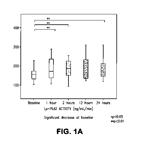
Figures 1A and 1B show the Lp-PLA2 mass and activity temporal profile in acute
stroke.
Figures 2A and 2B show the Lp-PLA2 mass and activity temporal profile from
baseline to the 3rd month.
10 Figures 3A and 3B show the Lp-PLA2 levels in healthy controls (n=135) and
pooled samples from baseline, 1 hour and 24 hours (n=35 in each time) after
baseline.
Figures 4A and 4B show the association of Lp-PLA2 mass and activity levels to
admission NIHSS scores and stroke etiology.
Figures 4C and 4D show the association of Lp-PLA2 mass and activity levels to
15 the location of occlusion and 1-hour recanalization.
Figure 4E and 4F show the association of Lp-PLA2 mass and activity levels to
early neurological status and functional outcome at follow-up (third month).
Figure 5 shows the relationship between location of vessel occlusion and Lp-
PLA2
level and successful 1-hour complete recanalization.
Figure 6 shows the relationship between location of vessel occlusion and Lp-
PLA2
level and third month-functional outcome.
Figure 7 shows levels of Lp-PLA2 mass and activity (boxplots) in TIA cases and
controls.
Figures 8A and 8B show Kaplan-Meier curves demonstrating survival analyses for
the presence of further vascular events or stroke/TIA considering ABCD2 score.
Figures 9A, 9B, 9C and 9D show Kaplan-Meier curves showing survival analyses
for presence of further vascular events or stroke/TIA considering Lp-PLA2
activity
(highest versus the lowest quartile of Lp-PLA2 activity).
Figures 9E, 9F, 9G and 9H show Kaplan-Meier curves showing survival analyses
for presence of further vascular events or stroke/TIA considering Lp-PLA2
activity (cases
above or below an optimal cut-off point of Lp-PLA2 activity).
CA 02798122 2012-10-29
WO 2011/137419 PCT/US2011/034728
16
Figures 1 OA and 1 OB show scatterplots of the correlation between Lp-PLA2
mass
and activity and total cholesterol.
Figures 1 1A and 11B show scatterplots of the correlation between Lp-PLA2 mass
and activity and LDL-cholesterol.
Figure 12 shows Kaplan-Meier curves showing the cumulative survival of any
vascular event during follow-up between lth and a combination of 3rd and 4th
quartiles of
Lp-PLA2 activity.
Figure 13 shows Kaplan-Meier curves show the cumulative survival of any
vascular event during follow-up between groups above and under Lp-PLA2
activity
cutpoint levels.
Figure 14 shows the vascular risk stratification using the combination of Lp-
PLA2
and ABI.
DETAILED DESCRIPTION OF THE INVENTION
Lp-PLA2 can be used to identify patients who will benefit from administration
of
thrombolytics. Lp-PLA2 expression has been shown to be higher in carotid
plaques of
patients with than without cardiac events (Herrmann (2009) Eur Heart J.
30(23):2930-8).
In the event of a plaque rupture and vascular thrombus, high levels of Lp-PLA2
may be
released into circulation from the rupture site. Measuring Lp-PLA2 levels of
individuals
suspected of having a stroke or myocardial infarction (e.g. individuals who
present
symptoms of a stroke or MI) can identify individuals who will benefit from
standard
thrombolytic therapy or and those who may need aggressive therapy including
aggressive
thrombolytic drug dosing, drug combinations and/or interventional and surgical
therapies.
This invention is directed to methods of using Lp-PLA2 levels to select
patients for
therapy, assess risk of cerebrovascular accident (CVA), and assess functional
outcome for
patients.
As used herein, the terms "embodiment" and "aspect" are used interchangeably.
As used herein, the term "coronary vascular disease" or "CVD" means diseases
of
the vasculature, including high blood pressure, coronary heart disease (CHD),
myocardial
infarction, stroke, transient ischemic attack (TIA), cerebrovascular accident
(CVA),
congenital cardiovascular defects and congestive heart failure. Coronary
vascular disease
includes primary and subsequent acute events including myocardial infarction,
stroke, TIA
and CVA.
CA 02798122 2012-10-29
WO 2011/137419 PCT/US2011/034728
17
"Lipoprotein-associated Phospholipase A2", "Lp-PLA2", "LpPLA2", "Lp-PLA2",
"Platelet-activating factor-acetylhydrolase", "PAF-AH", and "LDL-PLA2" are
used
interchangeably herein and within the literature and refer to native Lp-PLA2,
and allelic
variants thereof, as described, for example, in Tew et al. (1996)
Arterioscler. Thromb.
Vase. Biol. 16:591-599, Tjoelker, et al. (1995) Nature 374(6522):549-53),
Caslake et al.
(2000) Atherosclerosis 150(2): 413-9, Genbank RefSeq IDs: NM 005084,
NP_005075,
NM-00 11683 57 and NP-00 1161829 and Genebank Entrez GenelD: 7941 (PLA2G7),
which are hereby incorporated by reference in their entirety. Unless indicated
otherwise,
the terms "Lipoprotein-associated Phospholipase A2", "Lp-PLA2", "LpPLA2", "Lp-
PLA2", "Platelet-activating factor-acetylhydrolase", "PAF-AH", and "LDL-PLA2"
when
used herein refer to the human protein.
As used herein, the term "acute care" means health-care or necessary treatment
of
a disease over a short period of time in which a patient is treated for a
brief but severe
episode of illness, such as CVD, myocardial infarction and stroke. Acute care
is typically
rendered in an emergency department, ambulatory care clinic, or other short-
term stay
facility. An acute care setting or timeframe means within half an hour, 1
hour, 2 hours, 3
hours, 4 hours, 5 hours or 6 hours.
As used herein, the term "functional outcome" means the classification system
that
summarizes the neurological impairments, disabilities, and handicaps that
occur after a
vascular event such as stroke. The functional outcome for stroke encompasses a
broad
range of disabilities and impairments as well as the relationship of
disability and
impairment to independent function. Typically indicators for functional
outcome are
measured at 1 month, 3 months or 6 months after an acute vascular event, such
as stroke.
See Stroke. 1998;29:1274-1280, which is hereby incorporated by reference in
its entirety.
Generally, a functional outcome of functional dependence is a poor outcome
from a
vascular event, such as stroke, in which the subject suffers from impairment,
disability,
handicap or compromised quality of life. The likelihood of a subject having a
functional
outcome of functional dependence after a vascular event, such as a stroke, can
be reduced
by aggressive therapy, including surgery, at the time of the vascular event in
the acute care
setting.
"High" refers to a measure that is greater than normal, greater than a
standard such
as a predetermined measure or a subgroup measure or that is relatively greater
than
another subgroup measure. For example, high Lp-PLA2 refers to a measure of Lp-
PLA2
CA 02798122 2012-10-29
WO 2011/137419 PCT/US2011/034728
18
that is greater than a normal Lp-PLA2 measure. A normal Lp-PLA2 measure may be
determined according to any method available to one skilled in the art. High
Lp-PLA2
may also refer to a measure that is equal to or greater than a predetermined
measure, such
as a predetermined cutoff. High Lp-PLA2 may also refer to a measure of Lp-PLA2
wherein a high Lp-PLA2 subgroup has relatively greater levels of Lp-PLA2 than
another
subgroup. For example, without limitation, according to the present
specification, two
distinct patient subgroups can be created by dividing samples around a
mathematically
determined point, such as, without limitation, a median, thus creating a
subgroup whose
measure is high (ie, higher than the median) and another subgroup whose
measure is low.
Lp-PLA2 can be measured by any method known to one skilled in the art such as,
for
example, without limitation, using the PLAC Test, an Lp-PLA2 activity assay,
an
immunohistochemical (IHC) assay or using any standard method for detecting Lp-
PLA2,
including Lp-PLA2 mass and Lp-PLA2 activity. In some cases, a "high"
expression level
may comprise a range of expression that is very high and a range of expression
that is
"moderately high" where moderately high is a level of expression that is
greater than
normal, but less than "very high". Example ranges for high (including very
high and
moderately high) high Lp-PLA2 expression are provided in the literature cited
herein, the
PLAC Test product specification, in the present application and include >200
ng/mL,
>201 ng/mL, >201.5 ng/mL, >210 ng/mL, >220 ng/mL, >230 ng/mL, >240 ng/mL, >250
ng/mL, >260 ng/mL, >270 ng/mL, >280 ng/mL, >290 ng/mL, >300 ng/mL,
> 100ng/mL/min, >1 lOng/mL/min, > 120ng/mL/min, > 13 0ng/mL/min, >
140ng/mL/min,
>150ng/mL/min, >160ng/mL/min, >170ng/mL/min, >180ng/mL/min, >190ng/mL/min,
and >200ng/mL/min.
"Likely to" (and "unlikely to"), as used herein, refers to an increased (or
decreased) probability that an item, object, thing or person will occur. Thus,
in one
example, a subject that is likely to benefit from treatment with a
thrombolytic agent has an
increased probability of benefiting from treatment with a thrombolytic agent
relative to a
reference subject or group of subjects.
"Long," as used herein, refers to a time measure that is greater than normal,
greater
than a standard such as a predetermined measure or a subgroup measure that is
relatively
longer than another subgroup measure. For example, with respect to a patient's
longevity,
a long time progression refers to time progression that is longer than a
normal time
progression. Whether a time progression is long or not may be determined
according to
CA 02798122 2012-10-29
WO 2011/137419 PCT/US2011/034728
19
any method available to one skilled in the art. In one embodiment, "long"
refers to a time
that is greater than the median time course required for a significant event
to occur in a
disease.
"Low" is a term that refers to a measure that is less than normal, less than a
standard such as a predetermined measure or a subgroup measure that is
relatively less
than another subgroup measure. For example, low Lp-PLA2 means a measure of Lp-
PLA2
that is less than a normal Lp-PLA2 measure in a particular set of samples of
patients. A
normal Lp-PLA2 measure may be determined according to any method available to
one
skilled in the art. Low Lp-PLA2 may also mean a measure that is less than a
predetermined measure, such as a predetermined cutoff. Low Lp-PLA2 may also
mean a
measure wherein a low Lp-PLA2 subgroup is relatively lower than another
subgroup. For
example, without limitation, according to the present specification, two
distinct patient
subgroups can be created by dividing samples around a mathematically
determined point,
such as, without limitation, a median, thus creating a group whose measure is
low (i.e.,
less than the median) with respect to another group whose measure is high
(i.e., greater
than the median). Lp-PLA2 can be measured by any method known to one skilled
in the
art such as, for example, without limitation, using the PLAC Test, an Lp-PLA2
activity
assay, an immunohistochemical (IHC) assay or using any standard method for
detecting
Lp-PLA2, including Lp-PLA2 mass and Lp-PLA2 activity. Example ranges for low
values
of Lp-PLA2 expression are provided in the literature cited herein, the PLAC
Test product
specification, in the present application and include <200 ng/mL, <201 ng/mL,
<201.5
ng/mL, <210 ng/mL, <220 ng/mL, <230 ng/mL, <240 ng/mL, <250 ng/mL, <260 ng/mL,
<270 ng/mL, <280 ng/mL, <290 ng/mL, <300 ng/mL, <100ng/mL/min, <1 IOng/mL/min,
<120ng/mL/min, <130ng/mL/min, <140ng/mL/min, <150ng/mL/min, <160ng/mL/min,
<170ng/mL/min, <180ng/mL/min, <190ng/mL/min, and <200ng/mL/min.
"Overall survival" or "OS" refers to a time as measured from the start of
treatment
to death or censor. Censoring may come from a study end or change in
treatment. Overall
survival can refer to a probability as, for example, a probability when
represented in a
Kaplan-Meier plot of being alive at a particular time, that time being the
time between the
start of the treatment to death or censor.
"Pre-determined cutoff" as used herein, refers to the value of a predetermined
measure on subjects exhibiting certain attributes that allow the best
discrimination
between two or more categories of an attribute. For example, a pre-determined
cutoff that
CA 02798122 2012-10-29
WO 2011/137419 PCT/US2011/034728
allows one to discriminate between two categories such as high Lp-PLA2
expression and
low Lp-PLA2 expression for determining overall survival may be used. Pre-
determined
cutoffs may be used to separate the subjects with values lower than or higher
than the pre-
determined cutoff to optimize the prediction model.
5 "Respond" to treatment, and other forms of this verb, as used herein, refer
to the
reaction of a subject to treatment with an agent. As an example, , a subject
responds to
treatment with an agent if the subject experiences a life expectancy extended
by about 5%,
10%, 20%, 30%, 40%, 50% or more beyond the life expectancy predicted if no
treatment
is administered. In another example, a subject responds to treatment with an
agent if the
10 subject has an overall survival or increased time to progression. Several
methods may be
used to determine if a patient responds to a treatment.
"Sample" or "tissue sample" or "patient sample" or "patient cell or tissue
sample"
or "specimen" each refers to a collection of similar cells obtained from a
tissue of a
subject or patient. The source of the tissue sample may be solid tissue as
from a fresh
15 tissue, frozen and/or preserved organ or tissue or biopsy or aspirate;
blood or any blood
constituents, bodily fluids such as cerebral spinal fluid, amniotic fluid,
peritoneal fluid or
interstitial fluid or cells from anytime in gestation or development of the
subject. The
tissue sample may contain compounds that are not naturally intermixed with the
tissue in
nature such as preservatives, anticoagulants, buffers, fixatives, nutrients,
antibiotics or the
20 like. Cells may be fixed in a conventional manner, such as in an FFPE
manner.
"Short," as used herein, refers to a time measure that is shorter than normal,
shorter
than a standard such as a predetermined measure or a subgroup measure that is
relatively
shorter than another subgroup measure. For example, with respect to a
patient's longevity,
a short time progression refers to time progression that is shorter than a
normal time
progression or shorter than predicted. Whether a time progression is short or
not may be
determined according to any method available to one skilled in the art. In one
embodiment, "short" refers to a time that is less than the median time course
required for a
significant event to occur in a disease.
"Significant event," as used herein, shall refer to an event in a patient's
disease that
is important as determined by one skilled in the art. Examples of significant
events
include, for example, without limitation, primary diagnosis, myocardial
infarction, stroke,
TIA, CVA, death, recurrence, the determination that a patient's disease is
metastatic,
relapse of a patient's disease or the progression of a patient's disease from
any one of the
CA 02798122 2012-10-29
WO 2011/137419 PCT/US2011/034728
21
above noted stages to another. A significant event may be any important event
used to
assess OS, TTP and/or other response criteria, as determined by one skilled in
the art.
As used herein, the terms "subject" and "patient" are used interchangeably. As
used herein, the terms "subject" and "subjects" refer to an animal, preferably
a mammal
including a non-primate (e.g., a cow, pig, horse, donkey, goat, camel, cat,
dog, guinea pig,
rat, mouse or sheep) and a primate (e.g., a monkey, such as a cynomolgus
monkey, gorilla,
chimpanzee or a human).
As used herein, "time course" shall refer to the amount of time between an
initial
event and a subsequent event. For example, with respect to a patient's cancer,
time course
may relate to a patient's disease and may be measured by gauging significant
events in the
course of the disease, wherein the first event may be diagnosis and the
subsequent event
may be a significant event, for example.
"Time to progression" or "TTP" refers to a time as measured from the start of
the
treatment to progression or a significant event or censor. Censoring may come
from a
study end or from a change in treatment. Time to progression can also be
represented as a
probability as, for example, in a Kaplan-Meier plot where time to progression
may
represent the probability of being progression free over a particular time,
that time being
the time between the start of the treatment to progression or censor.
"Treatment," and other forms of this word, including "therapy", refer to the
administration of an agent to impede a disease, such as progression of CVD, to
cause a
reduction in risk for CVD, to extend the expected survival time of the subject
and/or time
to progression of the CVD or the like. Treatment may also refer to any course
which one
skilled, for example, a treating physician, deems expedient.
"Chemotherapeutic agent" means a chemical substance that is used to treat a
condition, particularly cardiovascular disease.
As used herein, the term "metabolic disorder" includes a disorder, disease or
condition which is caused or characterized by an abnormal metabolism (i.e.,
the chemical
changes in living cells by which energy is provided for vital processes and
activities) in a
subject. Metabolic disorders include diseases, disorders, or conditions
associated with
hyperglycemia or aberrant adipose cell (e.g., brown or white adipose cell)
phenotype or
function. Metabolic disorders can detrimentally affect cellular functions such
as cellular
proliferation, growth, differentiation, or migration, cellular regulation of
homeostasis,
inter- or intra-cellular communication; tissue function, such as liver
function, renal
CA 02798122 2012-10-29
WO 2011/137419 PCT/US2011/034728
22
function, or adipocyte function; systemic responses in an organism, such as
hormonal
responses (e.g., insulin response). Examples of metabolic disorders include
obesity,
diabetes, hyperphagia, endocrine abnormalities, triglyceride storage disease,
Bardet-Biedl
syndrome, Lawrence-Moon syndrome, Prader-Labhart-Willi syndrome, anorexia, and
cachexia. Obesity is defined as a body mass index (BMI) of 30 kg/m<sup>2</sup> or
more
(National Institute of Health, Clinical Guidelines on the Identification,
Evaluation, and
Treatment of Overweight and Obesity in Adults (1998)). However, the invention
is also
intended to include a disease, disorder, or condition that is characterized by
a body mass
index (BMI) of 25 kg/m2 or more, 26 kg/m2 or more, 27 kg/m<sup>2</sup> or more, 28
kg/m<sup>2</sup> or more, 29 kg/m<sup>2</sup> or more, 29.5 kg/m<sup>2</sup> or more, or 29.9
kg/m<sup>2</sup>
or more, all of which are typically referred to as overweight (National
Institute of Health,
Clinical Guidelines on the Identification, Evaluation, and Treatment of
Overweight and
Obesity in Adults (1998)).
As used herein, "greater than or equal to" (i.e., > or > =) can in certain
alternative
embodiments mean "greater than" (>). Also, as used herein, "less than or equal
to"
(i.e., < or <=) can in certain alternative embodiments mean "less than" (<).
Agents for reducing the risk of a Coronary Vascular Disorder include those
selected from the group consisting of Lp-PLA2 inhibitors (Leach 2001), anti-
inflammatory
agents, anti-thrombotic agents, anti-platelet agents, fibrinolytic agents,
lipid reducing
agents, niacin, direct thrombin inhibitors, and glycoprotein II b/IIIa
receptor inhibitors and
agents that bind to cellular adhesion molecules and inhibit the ability of
white blood cells
to attach to such molecules (e.g. anti-cellular adhesion molecule antibodies).
Anti-inflammatory agents include Alclofenac; Alclometasone Dipropionate;
Algestone Acetonide; Alpha Arnylase; Amcinafal; Amcinafide; Amfenac Sodium;
Amiprilose Hydrochloride; Anakinra; Anirolac; Anitrazafen; Apazone;
Balsalazide
Disodium; Bendazac; Benoxaprofen; Benzydamine Hydrochloride; Bromelains;
Broperamole; Budesonide; Carprofen; Cicloprofen; Cintazone; Cliprofen;
Clobetasol
Propionate; Clobetasone Butyrate; Clopirac; Cloticasone Propionate;
Cormethasone
Acetate; Cortodoxone; Deflazacort; Desonide; Desoximetasone; Dexamethasone
Dipropionate; Diclofenac Potassium; Diclofenac Sodium; Diflorasone Diacetate;
Diflumidone Sodium; Diflunisal; Difluprednate; Diftalone; Dimethyl Sulfoxide;
Drocinonide; Endrysone; Enlimomab; Enolicam Sodium; Epirizole; Etodolac;
Etofenamate; Felbinac; Fenamole; Fenbufen; Fenclofenac; Fenclorac; Fendosal;
CA 02798122 2012-10-29
WO 2011/137419 PCT/US2011/034728
23
Fenpipalone; Fentiazac; Flazalone; Fluazacort; Flufenamic Acid; Flumizole;
Flunisolide
Acetate; Flunixin; Flunixin Meglumine; Fluocortin Butyl; Fluorometholone
Acetate;
Fluquazone; Flurbiprofen; Fluretofen; Fluticasone Propionate; Furaprofen;
Furobufen;
Halcinonide; Halobetasol Propionate; Halopredone Acetate; Ibufenac; Ibuprofen;
Ibuprofen Aluminum; Ibuprofen Piconol; Ilonidap; Indomethacin; Indomethacin
Sodium;
Indoprofen; Indoxole; Intrazole; Isoflupredone Acetate; Isoxepac; Isoxicam;
Ketoprofen;
Lofemizole Hydrochloride; Lornoxicam; Loteprednol Etabonate; Meclofenamate
Sodium;
Meclofenamic Acid; Meclorisone Dibutyrate; Mefenamic Acid; Mesalamine;
Meseclazone; Methylprednisolone Suleptanate; Morniflumate; Nabumetone;
Naproxen;
Naproxen Sodium; Naproxol; Nimazone; Olsalazine Sodium; Orgotein; Orpanoxin;
Oxaprozin; Oxyphenbutazone; Paranyline Hydrochloride; Pentosan Polysulfate
Sodium;
Phenbutazone Sodium Glycerate; Pirfenidone; Piroxicam; Piroxicam Cinnamate;
Piroxicam Olamine; Pirprofen; Prednazate; Prifelone; Prodolic Acid;
Proquazone;
Proxazole; Proxazole Citrate; Rimexolone; Romazarit; Salcolex; Salnacedin;
Salsalate;
Salycilates; Sanguinarium Chloride; Seclazone; Sermetacin; Sudoxicam;
Sulindac;
Suprofen; Talmetacin; Talniflumate; Talosalate; Tebufelone; Tenidap; Tenidap
Sodium;
Tenoxicam; Tesicam; Tesimide; Tetrydamine; Tiopinac; Tixocortol Pivalate;
Tolmetin;
Tolmetin Sodium; Triclonide; Triflumidate; Zidometacin; Glucocorticoids;
Zomepirac
Sodium.
Anti-thrombotic and/or fibrinolytic agents include Plasminogen (to plasmin via
interactions of prekallikrein, kininogens, Factors XII, XIIIa, plasminogen
proactivator, and
tissue plasminogen activator[TPA]) Streptokinase; Urokinase: Anisoylated
Plasminogen-
Streptokinase Activator Complex; Pro-Urokinase; (Pro-UK); rTPA (alteplase or
activase; r
denotes recombinant), rPro-UK; Abbokinase; Eminase; Sreptase Anagrelide
Hydrochloride; Bivalirudin; Dalteparin Sodium; Danaparoid Sodium; Dazoxiben
Hydrochloride; Efegatran Sulfate; Enoxaparin Sodium; Ifetroban; Ifetroban
Sodium;
Tinzaparin Sodium; retaplase; Trifenagrel; Warfarin; Dextrans.
Anti-platelet agents include Clopridogrel; Sulfinpyrazone; Aspirin;
Dipyridamole;
Clofibrate; Pyridinol Carbamate; PGE; Glucagon; Antiserotonin drugs; Caffeine;
Theophyllin Pentoxifyllin; Ticlopidine; Anagrelide. Lipid reducing agents
include
gemfibrozil, cholystyramine, colestipol, nicotinic acid, probucol lovastatin,
fluvastatin,
simvastatin, atorvastatin, pravastatin, cirivastatin (for statins, see Crouch
2000). Direct
thrombin inhibitors include hirudin, hirugen, hirulog, agatroban, PPACK,
thrombin
CA 02798122 2012-10-29
WO 2011/137419 PCT/US2011/034728
24
aptamers. Glycoprotein IIb/IIIa receptor Inhibitors are both antibodies and
non-
antibodies, and include but are not limited to ReoPro (abcixamab), lamifiban,
tirofiban.
One preferred agent is aspirin.
Markers of systemic inflammation, such as CRP, are well-known to those of
ordinary skill in the art. It is preferred that the markers of systemic
inflammation be
selected from the group consisting of C-reactive protein, cytokines, and
cellular adhesion
molecules. Cytokines are well-known to those of ordinary skill in the art and
include
human interleukins 1-17. Cellular adhesion molecules are well-known to those
of ordinary
skill in the art and include integrins, ICAM-1, ICAM-3, BL-CAM, LFA-2, VCAM-1,
NCAM, and PECAM. The preferred adhesion molecule is soluble intercellular
adhesion
molecule (sICAM-1).
The level of the markers of this invention may be obtained by a variety of
recognized methods. Typically, the level is determined by measuring the level
of the
marker in a body fluid, for example, blood, lymph, saliva, urine and the like.
The
preferred body fluid is blood. The level can be determined by ELISA, or
immunoassays
or other conventional techniques for determining the presence of the marker.
Conventional
methods include sending samples of a patient's body fluid to a commercial
laboratory for
measurement. For the measurement of Lp-PLA2 enzymatic assays may also be used,
see
U. S. Pat. Nos. 5,981,252 or 5,880,273, the contents of which are hereby
incorporated by
reference into the subject application.
The invention also involves comparing the level of marker for the individual
with a
predetermined value. The predetermined value can take a variety of forms. It
can be single
cut-off value, such as a median or mean. It can be established based upon
comparative
groups, such as where the risk in one defined group is double the risk in
another defined
group. It can be a range, for example, where the tested population is divided
equally (or
unequally) into groups, e.g., tertiles, such as-a low-risk group, a medium-
risk group and a
high-risk group, or into quadrants, the lowest quadrant being individuals with
the lowest
risk and the highest quadrant being individuals with the highest risk.
In preferred embodiments the invention provides novel kits or assays which are
specific for, and have appropriate sensitivity with respect to, predetermined
values
selected on the basis of the present invention. The preferred kits, therefore,
would differ
from those presently commercially available, by including, for example,
different cut-offs,
CA 02798122 2012-10-29
WO 2011/137419 PCT/US2011/034728
different sensitivities at particular cut-offs as well as instructions or
other printed material
for characterizing risk based upon the outcome of the assay.
As discussed above the invention provides methods for evaluating the
likelihood
that an individual will benefit from treatment with an agent for reducing risk
of a future
5 cardiovascular disorder. This method has important implications for patient
treatment and
also for clinical development of new therapeutics. Physicians select
therapeutic regimens
for patient treatment based upon the expected net benefit to the patient. The
net benefit is
derived from the risk to benefit ratio. The present invention permits
selection of
individuals who are more likely to benefit by intervention, thereby aiding the
physician in
10 selecting a therapeutic regimen. This might include using drugs with a
higher risk profile
where the likelihood of expected benefit has increased. Likewise, clinical
investigators
desire to select for clinical trials a population with a high likelihood of
obtaining a net
benefit. The present invention can help clinical investigators select such
individuals. It is
expected that clinical investigators now will use the present invention for
determining
15 entry criteria for clinical trials.
An effective amount is a dosage of the therapeutic agent sufficient to provide
a
medically desirable result. The effective amount will vary with the particular
condition
being treated, the age and physical condition of the subject being treated,
the severity of
the condition, the duration of the treatment, the nature of the concurrent
therapy (if any),
20 the specific route of administration and the like factors within the
knowledge and expertise
of the health practitioner. For example, an effective amount can depend upon
the degree to
which an individual has abnormally elevated levels of markers of systemic
information. It
should be understood that the anti-inflammatory agents of the invention are
used to
prevent cardiovascular disorders, that is, they are used prophylactically in
subjects at risk
25 of developing a cardiovascular disorder. Thus, an effective amount is that
amount which
can lower the risk of, slow or perhaps prevent altogether the development of a
cardiovascular disorder. When the agent is one that binds to cellular adhesion
molecules
and inhibits the ability of white blood cells to attach to such molecules,
then the agent may
be used prophylactically or may be used in acute circumstances, for example,
post-
myocardial infarction or post-angioplasty. It will be recognized when the
agent is used in
acute circumstances, it is used to prevent one or more medically undesirable
results that
typically flow from such adverse events. In the case of myocardial infarction,
the agent
can be used to limit injury to the cardiovascular tissue which develops as a
result of the
CA 02798122 2012-10-29
WO 2011/137419 PCT/US2011/034728
26
myocardial infarction and in the case of restenosis the agent can be used in
amounts
effective to inhibit, prevent or slow the reoccurrence of blockage. In either
case, it is an
amount sufficient to inhibit the infiltration of white blood cells and
transmigration of white
blood cells into the damaged tissue, which white blood cells can result in
further damage
and/or complications relating to the injury.
Generally, doses of active compounds would be from about 0.01 mg/kg per day to
1000 mg/kg per day. It is expected that doses ranging from 50-500 mg/kg will
be suitable,
preferably orally and in one or several administrations per day. Lower doses
will result
from other forms of administration, such as intravenous administration. In the
event that a
response in a subject is insufficient at the initial doses applied, higher
doses (or effectively
higher doses by a different, more localized delivery route) may be employed to
the extent
that patient tolerance permits. Multiple doses per day are contemplated to
achieve
appropriate systemic levels of compounds.
When administered, the pharmaceutical preparations of the invention are
applied in
pharmaceutically-acceptable amounts and in pharmaceutically-acceptably
compositions.
Such preparations may routinely contain salt, buffering agents, preservatives,
compatible
carriers, and optionally other therapeutic agents. When used in medicine, the
salts should
be pharmaceutically acceptable, but non-pharmaceutically acceptable salts may
conveniently be used to prepare pharmaceutically-acceptable salts thereof and
are not
excluded from the scope of the invention. Such pharmacologically and
pharmaceutically-
acceptable salts include, but are not limited to, those prepared from the
following acids:
hydrochloric, hydrobromic, sulfuric, nitric, phosphoric, maleic, acetic,
salicylic, citric,
formic, malonic, succinic, and the like. Also, pharmaceutically-acceptable
salts can be
prepared as alkaline metal or alkaline earth salts, such as sodium, potassium
or calcium
salts.
The anti-inflammatory agents, anti-Lp-PLA2 agents or statins may be combined,
optionally, with a pharmaceutically-acceptable carrier. The term
"pharmaceutically-
acceptable carrier" as used herein means one or more compatible solid or
liquid filler,
diluents or encapsulating substances which are suitable for administration
into a human.
The term "carrier" denotes an organic or inorganic ingredient, natural or
synthetic, with
which the active ingredient is combined to facilitate the application. The
components of
the pharmaceutical compositions also are capable of being co-mingled with the
molecules
CA 02798122 2012-10-29
WO 2011/137419 PCT/US2011/034728
27
of the present invention, and with each other, in a manner such that there is
no interaction
which would substantially impair the desired pharmaceutical efficacy.
The pharmaceutical compositions may contain suitable buffering agents,
including:
acetic acid in a salt; citric acid in a salt; boric acid in a salt; and
phosphoric acid in a salt.
The pharmaceutical compositions also may contain, optionally, suitable
preservatives,
such as: benzalkonium chloride; chlorobutanol; parabens and thimerosal.
Compositions suitable for parenteral administration conveniently comprise a
sterile
aqueous preparation of the anti-inflammatory agent, which is preferably
isotonic with the
blood of the recipient. This aqueous preparation may be formulated according
to known
methods using suitable dispersing or wetting agents and suspending agents The
sterile
injectable preparation also may be a sterile injectable solution or suspension
in a non-toxic
parenterally-acceptable diluent or solvent, for example, as a solution in 1,3-
butane diol.
Among the acceptable vehicles and solvents that may be employed are water,
Ringer's
solution, and isotonic sodium chloride solution. In addition, sterile, fixed
oils are
conventionally employed as a solvent or suspending medium. For this purpose
any bland
fixed oil may be employed including synthetic mono- or di-glycerides. In
addition, fatty
acids such as oleic acid may be used in the preparation of injectables.
Carrier formulation
suitable for oral, subcutaneous, intravenous, intramuscular, etc.
administrations can be
found in Remington's Pharmaceutical Sciences, Mack Publishing Co., Easton, Pa.
A variety of administration routes are available. The particular mode selected
will
depend, of course, upon the particular drug selected, the severity of the
condition being
treated and the dosage required for therapeutic efficacy. The methods of the
invention,
generally speaking, may be practiced using any mode of administration that is
medically
acceptable, meaning any mode that produces effective levels of the active
compounds
without causing clinically unacceptable adverse effects. Such modes of
administration
include oral, rectal, topical, nasal, interdermal, or parenteral routes. The
term "parenteral"
includes subcutaneous, intravenous, intramuscular, or infusion. Intravenous or
intramuscular routes are not particularly suitable for long-term therapy and
prophylaxis.
They could, however, be preferred in emergency situations. Oral administration
will be
preferred for prophylactic treatment because of the convenience to the patient
as well as
the dosing schedule.
The pharmaceutical compositions may conveniently be presented in unit dosage
form and may be prepared by any of the methods well-known in the art of
pharmacy. All
CA 02798122 2012-10-29
WO 2011/137419 PCT/US2011/034728
28
methods include the step of bringing the anti-inflammatory agent into
association with a
carrier which constitutes one or more accessory ingredients. In general, the
compositions
are prepared by uniformly and intimately bringing the anti-inflammatory agent
into
association with a liquid carrier, a finely divided solid carrier, or both,
and then, if
necessary, shaping the product.
Compositions suitable for oral administration may be presented as discrete
units,
such as capsules, tablets, lozenges, each containing a predetermined amount of
the anti-
inflammatory agent. Other compositions include suspensions in aqueous liquids
or non-
aqueous liquids such as a syrup, elixir or an emulsion.
Other delivery systems can include time-release, delayed release or sustained
release delivery systems. Such systems can avoid repeated administrations of
the anti-
inflammatory agent, increasing convenience to the subject and the physician.
Many types
of release delivery systems are available and known to those of ordinary skill
in the art.
They include polymer base systems such as poly(lactide-glycolide),
copolyoxalates,
polycaprolactones, polyesteramides, polyorthoesters, polyhydroxybutyric acid,
and
polyanhydrides. Microcapsules of the foregoing polymers containing drugs are
described
in, for example, U.S. Pat. No. 5,075,109. Delivery systems also include non-
polymer
systems that are: lipids including sterols such as cholesterol, cholesterol
esters and fatty
acids or neutral fats such as mono- di- and tri-glycerides; hydrogel release
systems;
peptide based systems; wax coatings; compressed tablets using conventional
binders and
excipients; partially fused implants; and the like. Specific examples include,
but are not
limited to: (a) erosional systems in which the anti-inflammatory agent is
contained in a
form within a matrix such as those described in U.S. Pat. Nos. 4,452,775,
4,667,014,
4,748,034 and 5,239,660 and (b) diffusional systems in which an active
component
permeates at a controlled rate from a polymer such as described in U.S. Pat.
Nos.
3,832,253, and 3,854,480. In addition, pump-based hardware delivery systems
can be
used, some of which are adapted for implantation.
Use of a long-term sustained release implant may be particularly suitable for
treatment of chronic conditions. Long-term release, are used herein, means
that the
implant is constructed and arranged to delivery therapeutic levels of the
active ingredient
for at least 30 days, and preferably 60 days. Long-term sustained release
implants are
well-known to those of ordinary skill in the art and include some of the
release systems
described above.
CA 02798122 2012-10-29
WO 2011/137419 PCT/US2011/034728
29
An aspect of the invention comprises a method for detecting vascular disease
in an
individual comprising utilizing the methods described above to determine the
individual's
Lp-PLA2 activity in a sample wherein increased activity of Lp-PLA2 in the
sample is
indicative of vascular disease. In a preferred embodiment the vascular disease
is selected
from the group consisting of coronary vascular disease (CVD), coronary heart
disease
(CHD), peripheral vascular disease, peripheral arterial disease, high blood
pressure, stroke,
congenital cardiovascular defects and congestive heart failure.
Another aspect of the invention comprises a method for selecting a therapy to
treat
vascular disease for an individual comprising utilizing the methods described
above to
determine the individual's Lp-PLA2 level in a sample wherein increased
activity or mass
of Lp-PLA2 in the sample is indicative of an individual who will benefit from
therapy to
treat vascular disease. In particular, a low level of Lp-PLA2 indicates a
subject likely to
benefit from thrombolytic therapy while a high level of Lp-PLA2 indicates a
subject likely
to benefit from aggressive thrombolytic therapy, drug combinations and/or
interventional
and surgical approaches. In a preferred embodiment the vascular disease is
selected from
the group consisting of coronary vascular disease (CVD), coronary heart
disease (CHD),
peripheral vascular disease, peripheral arterial disease, high blood pressure,
stroke,
secondary stroke, congenital cardiovascular defects and congestive heart
failure. In
another preferred embodiment the therapy is selected from the group consisting
of
thrombolytic, niacin, statins and Lp-PLA2 inhibitors.
A further aspect of the invention comprises a method for monitoring an
individual's response to therapy to treat vascular disease comprising
utilizing the methods
described above to determine the individual's Lp-PLA2 activity in a sample
wherein
decreased activity of Lp-PLA2 in the sample is indicative of an individual who
is
responding favorably to therapy to treat vascular disease. In a preferred
embodiment the
vascular disease is selected from the group consisting of coronary vascular
disease (CVD),
coronary heart disease (CHD), peripheral vascular disease, peripheral arterial
disease, high
blood pressure, stroke, secondary stroke, congenital cardiovascular defects
and congestive
heart failure. In another preferred embodiment the therapy is selected from
the group
consisting of thrombolytic, niacin, statins and Lp-PLA2 inhibitors.
Unless otherwise defined herein, medical, scientific and technical terms used
in
connection with the present invention shall have the meanings that are
commonly
understood by those of ordinary skill in the art. Further, unless otherwise
required by
CA 02798122 2012-10-29
WO 2011/137419 PCT/US2011/034728
context, singular terms shall include pluralities and plural terms shall
include the singular.
Generally, nomenclatures used in connection with, and techniques of, cell and
tissue
culture, molecular biology, immunology, microbiology, genetics and protein and
nucleic
acid chemistry and hybridization described herein are those well known and
commonly
5 used in the art. The methods and techniques of the present invention are
generally
performed according to conventional methods well known in the art and as
described in
various general and more specific references that are cited and discussed
throughout the
present specification unless otherwise indicated. See, e.g., Sambrook et al.,
Molecular
Cloning: A Laboratory Manual, 2d ed., Cold Spring Harbor Laboratory Press
(1989) and
10 Sambrook et al., Molecular Cloning: A Laboratory Manual, 3d ed., Cold
Spring Harbor
Press (2001); Ausubel et al., Current Protocols in Molecular Biology, Greene
Publishing
Associates (1992, and Supplements to 2000); Ausubel et al., Short Protocols in
Molecular
Biology: A Compendium of Methods from Current Protocols in Molecular Biology -
4th
Ed., Wiley & Sons (1999); Harlow and Lane, Antibodies: A Laboratory Manual,
Cold
15 Spring Harbor Laboratory Press (1990); and Harlow and Lane, Using
Antibodies: A
Laboratory Manual, Cold Spring Harbor Laboratory Press (1999).
Methods to measure Lp-PLA2 levels are known in the art. For instance, a
competition assay may be employed wherein an anti- Lp-PLA2 antibody is
attached to a
solid support and an allocated amount of a labeled Lp-PLA2 and a sample of
interest are
20 incubated with the solid support. The amount of labeled Lp-PLA2 attached to
the solid
support can be correlated to the quantity of Lp-PLA2 in the sample. These
assays and
variations therefore comprise a further embodiment of the present invention.
The methods described herein can further be utilized as prognostic assays to
identify subjects having or at risk of developing a disease or disorder
associated with
25 increased or decreased expression levels of Lp-PLA2. The presence of higher
(or lower)
Lp-PLA2 levels as compared to normal human controls is diagnostic for the
human patient
being at risk for developing CVD. The effectiveness of therapeutic agents to
decrease (or
increase) expression or activity of Lp-PLA2 of the invention can also be
monitored by
analyzing levels of expression of the Lp-PLA2 in a human patient in clinical
trials or in
30 vitro screening assays such as in human cells. In this way, the gene
expression pattern can
serve as a marker, indicative of the physiological response of the human
patient or cells, as
the case may be, to the agent being tested.
CA 02798122 2012-10-29
WO 2011/137419 PCT/US2011/034728
31
The methods described herein can further be utilized in an acute care-setting
or
timeframe.
The above tests can be carried out on samples derived from a variety of cells,
bodily fluids and/or tissue extracts such as homogenates or solubilized tissue
obtained
from a patient. Tissue extracts are obtained routinely from tissue biopsy and
autopsy
material. Bodily fluids useful in the present invention include blood, urine,
saliva or any
other bodily secretion or derivative thereof. As used herein "blood" includes
whole blood,
plasma, serum, circulating epithelial cells, constituents, or any derivative
of blood.
In addition to detection in bodily fluids, the proteins and nucleic acids of
Lp-PLA2
are suitable to detection by cell capture technology. Whole cells may be
captured by a
variety of methods for example magnetic separation, U.S. Patents 5,200,084;
5,186,827;
5,108,933; 4,925,788, the disclosures of which are incorporated herein by
reference in
their entireties. Epithelial cells may be captured using such products as
Dynabeads or
CELLectionTM (Dynal Biotech, Oslo, Norway). Alternatively, fractions of blood
may be
captured, e.g., the buffy coat fraction (50mm cells isolated from 5m1 of
blood) containing
epithelial cells. Cells may also be captured using the techniques described in
WO
00/47998, the disclosure of which is incorporated herein by reference in its
entirety. Once
the cells are captured or concentrated, the proteins or nucleic acids are
detected by the
means described in the subject application. Alternatively, nucleic acids may
be captured
directly from blood samples, see U.S. Patents 6,156,504, 5,501,963; or WO
01/42504 , the
disclosures of which are incorporated herein by reference in their entireties.
EXAMPLES
The present invention may be better understood by reference to the following
nonlimiting examples.
Example 1: Introduction and Study Populations
Lp-PLA2 levels were evaluated in well phenotyped stroke cohorts from available
stored samples. Informed consents were received from the patients for the
study of blood
biomarkers to permit prognostic studies.
Lp-PLA2 levels were evaluated in the "acute phase" of a stroke to evaluate
temporal profile of Lp-PLA2 after stroke and to test prognostic value of Lp-
PLA2 in the
acute setting.
CA 02798122 2012-10-29
WO 2011/137419 PCT/US2011/034728
32
Lp-PLA2 levels were also evaluated in the "sub-acute phase" of a stroke to
evaluate the utility of Lp-PLA2 to predict a second stroke (or any vascular
event), predict
a second stroke after transient ischemic attack (TIA), and predict a second
stroke among
specific stroke subtypes (i.e. atherosclerotic stroke due to intracranial
stenosis).
Acute Phase Study
The cohorts evaluated for the acute phase study were (i) 20 patients with
blood
draws at 4 time-points (80 samples) (to evaluate temporal profile of Lp-PLA2
after stroke)
and (ii) 100 patients that had blood drawn within three hours of having a
stroke (to test
prognostic value of Lp-PLA2 in the acute setting). Similar protocols for this
study have
been previously described by Montaner J et al (Stroke. 2006;37(5):1205-10) the
disclosure
of which is herein incorporated by reference in its entirety.
Sub-Acute Phase Study
The cohorts evaluated for the sub-acute phase study were (i) 77 patients who
had
recurrent events after a stroke and 77 patients who did not have recurrent
events after a
stroke (to predict a second stroke), (ii) 135 patients who had a TIA (to
predict a second
stroke after TIA), and (iii) 135 patients who had a stroke that has been
characterized by
subtype (predict a second stroke among specific stroke subtypes). Similar
protocols for
these study have been previously described by Castillo J et al (J Neurol. 2009
Feb;256(2):217-24), Purroy F et al (ActaNeurol Scand. 2007;115(1):60-6), and
Arenillas
JF et al (Stroke. 2003;34(10):2463-8), respectively, the disclosures of which
are herein
incorporated by reference in their entirety.
Measurement of Lp-PLA2
Lp-PLA2 mass was assayed using the PLAC Test (diaDexus, Inc.) and Lp-PLA2
activity
was assayed using a colorimetric activity method (diaDexus, Inc.).
Example 2: Temporal Profile of Lp-PLA2 After Stroke (Acute Phase)
Study protocol and Results
Peripheral blood samples were drawn at baseline (less than 3 hours from stroke
onset) and serially thereafter. Specifically, in a series of 19 patients,
blood samples were
taken serially during the acute phase (baseline, 1 hour after (by the end of
the tPA
treatment), 2 hours after t-PA, and 12 and 24 hours after stroke onset.
Figures 1A and 1B
demonstrate that Lp-PLA2 activity levels were decreased at baseline compared
to later
CA 02798122 2012-10-29
WO 2011/137419 PCT/US2011/034728
33
time-points and Lp-PLA2 mass levels were increased at baseline compared to
later time-
points.
Also, in 15 patients, blood samples were obtained at baseline, 1 hour after
(by the
end oft-PA infusion), 24 hours after stroke onset, by discharge and at the
third month
visit. Figures 2A and 2B demonstrate that Lp-PLA2 mass significantly decreased
between
1 hour after baseline and time of discharge. However, there was no significant
difference
between baseline and discharge or baseline and the 3 month time-point.
Finally, results from baseline, 1 hour and 24 hours in both groups were pooled
(n=35 at each time point) and compared to a pooled control group (n=135).
Figure 3A
demonstrates that Lp-PLA2 levels were significantly decreased at baseline in
subjects
having a stroke when compared to later time-points and controls. Figure 3B
also
demonstrates that Lp-PLA2 mass was significantly increased in subjects with a
stroke at
baseline and later time-points compared to controls.
Example 3: Prognostic Value of Lp-PLA2 in the Acute Setting
Study protocol
Our study protocol included 100 consecutive stroke patients with a documented
arterial occlusion who received thrombolytic treatment within the first 3
hours from
symptoms onset. For the purpose of this study, only 92 patients with a middle
cerebral
artery (MCA) occlusion were analyzed.
A detailed history of vascular risk factors was obtained from each patient and
to
identify potential etiology of cerebral infarction, all patients underwent a
set of diagnostic
tests, including electrocardiogram, chest radiography, carotid
ultrasonography, complete
blood count and biochemistry.
Clinical examinations were performed on admission and at 12, 24 and 48 hours
from symptom onset by means of National Health Institutes Stroke Scale (NIHSS)
score.
Neurological deterioration was defined as the increase of 4 or more points in
NIHSS score
between baseline and any other time point through follow-up. Likewise,
neurological
improvement was defined as the decrease in 4 or more points in that scale
during the
follow-up. Finally, functional outcome was defined according to the modified
Rankin
Scale (mRS) score. Patients scoring 3 or more points were considered to be
functionally
dependent.
CA 02798122 2012-10-29
WO 2011/137419 PCT/US2011/034728
34
To evaluate vessel status, transcranial Doppler measurements were performed
before t-PA administration and serially (1, 2, 6 and 24 hours) thereafter, by
an experienced
neurologist using a Multi-Dop X4 (DWL Elektoniche Systeme GmbH, Sipplingen,
Germany) device, with a hand-held transducer in a range-gated, pulsed-wave
mode at a
frequency of 2 MHz. On admission, the location of the MCA occlusion was
recorded as
proximal or distal. The presence of recanalization on follow-up TCD
examinations was
assessed according to the Thrombolysis in Brain Ischemia (TIBI) flow grading
system.
Complete recanalization was diagnosed as improvement to a stenotic or normal
(TIBI 4 to
5 flow grades) waveforms; partial recanalization was diagnosed as an
improvement in
residual flow signals by at least 1 TIBI flow grade up to TIBI flow grades 2
to 3, and no
recanalization was defined as the absence of improvement of the residual flow
signal from
baseline TCD.
Peripheral blood samples were drawn from each patient at baseline (before tPA
administration and within 3 hours from onset).
Results
Baseline characteristics associated with Lp-PLA2 levels
All the factors associated with the baseline Lp-PLA2 mass and activity were
determined. The results regarding demographics and vascular risk factors are
shown in
Table 1. Dyslipemic patients showed higher Lp-PLA2 activity than non-
dyslipemic
patients (182.2 vs 157.2, p=0.058) and also, patients who were taking statins
had lower
Lp-PLA2 mass concentrations than those patients who were not taking statins by
the time
of the stroke (244.5 versus 283, p=0.17).
Table 1. Demographic characteristics and vascular risk factors associated with
Lp-PLA2.
Lp-PLA2 mass Lp-PLA2 activity
n /mL p n /mL/min p
All 211 (204.2-350.2) - 161.4 (131.3-189.3) -
Gender
- Male 292 (236.5-352.2) 0.56 162.9 (136.1-198) 0.62
- Female 247 (200.5-350) 161.6 (122.3-161.6)
Age 0.53
-:576 277.5 (201.7-347) 0.78 157.6 (130.6-185)
- >76 256 202-390 162.3 (130-197.4)
Tobacco use
- No 259.5 (201.3-350.7) 0.17 162.4 (133.4-197.9) 0.26
- Yes 288 271-367.5 152.9 (125.6-176.4)
Hypertension
- No 294 (236.5-370.5) 0.6 162.3 (130-196.7) 0.99
.99
Yes 264 (202-346) .6 161.6 (131-189.3)
CA 02798122 2012-10-29
WO 2011/137419 PCT/US2011/034728
Diabetes
- No 283 (214-353) 0.1 162 (134-194.5) 0.37
- Yes 237 (202-309.5) 158.4 119.4-195.5
Dyslipemia
- No 275 (209-349) 0 68 157.2 (127-181.7) 0.058
- Yes 279 (198-380) 182.2 157.5-182
Previous
stroke 272 (202-349) 161.6 (130-194.7)
-No
-Yes 331 (215.5-386.5) 0'79 165.7 (150.2-165.7) 0.76
CAD
-No 261.5 (203.7-350.7) 0.64 161.4 (133.4-186.8) 0.85
-Yes 296 (232.5-361.5) 168.4 119.6-214.5
Antiplatelet
-No 247 (201.346) 0.74 159 (130.3-184.2) 0.47
-Yes 291 (236-359) 165.9 (134.6-165.9)
Statins
-No 283 (214-354) 0.17 162.1 (130.3-194.5) 0 89
-Yes 244.5 (198-323.5) 162 (132.1-195)
Also, associations between Lp-PLA2 mass and activity and stroke severity
(measured by NIHSS score), etiology, as well as with the location of occlusion
at baseline
TCD (proximal versus distal occlusions) and the recanalization of the occluded
vessel
5- were studied (Figures 4A through 4F).
Lp-PLA2 activity and NIHSS score on admission showed a weak negative
correlation (r=-0.23, p=0.032), with more severely affected patients showing
the lowest
Lp-PLA2 activity. Regarding etiology, the lowest mass and activity levels were
found
among patients with other determined stroke etiology, such as arterial
dissection, and the
10 highest levels in the patients with an atherothrombotic stroke etiology.
Regarding recanalization, both higher mass and activity levels were found
among
those patients who did not achieve complete recanalization by the end of
thrombolytic
therapy (tPA treatment), and after multivariate analysis (Table 2) adjusted by
age, gender
and vascular risk factors, Lp-PLA2 mass together with the presence of a
proximal
15 occlusion on baseline TCD were the most strongest predictors of occlusion
persistence,
and therefore resistance to thrombolytic treatment. Moreover, when a proximal
occlusion
was combined with Lp-PLA2 mass higher than 201.5 ng/mL, almost none of the
patients
achieved complete recanalization at 1 hour as compared with 50% of those
without
proximal occlusions and Lp-PLA2 mass lower than that level. (see Figure 5; p
for trend
20 <0.001).
Table 2. Significant predictors for absence of complete recanalization at the
end of the tPA
treatment after multivariate analyses.
CA 02798122 2012-10-29
WO 2011/137419 PCT/US2011/034728
36
OR
Proximal occlusion on baseline TCD 6.75 (1.11-40) 0.037
Baseline Lp-PLA2 mass 1.016 (1.004-1.028) 0.011
Baseline Lp-PLA2 activity 1.022 (0.99-1.05) 0.12
Adjusted by age, gender and vascular risk factors.
These results demonstrate that patients with high Lp-PLA2 levels did not
benefit
from thrombolytic therapy as well as those with low Lp-PLA2 levels. Therefore,
patients
with high Lp-PLA2 levels who are having a vascular event, or are suspected of
having a
vascular event, will benefit from more aggressive drug dosing (e.g.
thrombolytic therapy)
and drug combinations as well as interventional and surgical approaches.
Further, for patients with high Lp-PLA2 and a proximal occlusion, almost none
benefit from thrombolytic therapy. Therefore, patients with a proximal
occlusion and high
Lp-PLA2 levels who are having a vascular event, or are suspected of having a
vascular
event, will benefit from more aggressive drug dosing (e.g. thrombolytic
therapy) and drug
combinations as well as interventional and surgical approaches.
Finally, neurological status during the acute phase of stroke and functional
status at
third month were explored and the results are shown in Figures 4E and 4F. No
relation
was found between Lp-PLA2 mass or activity and neurological status during the
first 48
hours. Moreover, neither Lp-PLA2 mass or activity themselves were related with
functional outcome at the third month.
Lp-PLA2 levels add significant prognostic information when combined with the
presence of a proximal occlusion. Lp-PLA2 levels and the presence/absence of a
proximal
occlusion predict a patient's 3 month functional outcome. Patients with high
Lp-PLA2
levels (mass or activity) and a proximal occlusion are more likely to be
functionally
dependent at three months after a vascular event (Figure 6). Patients who are
more likely
to be functionally dependent may benefit from additional or more aggressive
therapies
including thrombolytics (e.g. tPA) and surgical/PCI intervention.
Example 4: Lp-PLA2 Predicts a Second Stroke after TIA (or the combined end-
point
of recurrent non-fatal stroke, non-fatal myocardial infarction and vascular
death) -
Subacute Phase
Study protocol
In this case, we prospectively studied 166 consecutive patients with transient
neurologic deficit attended by the neurologist in the emergency department.
TIA was
defined as a reversible episode of neurologic deficit of ischemic origin that
resolved
CA 02798122 2012-10-29
WO 2011/137419 PCT/US2011/034728
37
completely within 24 hours. A total of 11 clinical episodes were attributable
to causes
other than brain ischemia and were excluded for this study.
Demographics and classical vascular risk factors were recorded, as well as the
clinical characteristics. Clinical symptoms and neurological signs at the
examination were
assessed and the vascular territory involved in each episode was recorded as
carotid,
vertebrobasilar or undetermined territory depending on the presence and
combination of
the above described findings. Finally, TIA duration and number of clinical
episodes were
recorded. TIA was categorized as single or a cluster of TIAs (when repeated
TIAs
occurred within the first week of the index event).
Blood samples for Lp-PLA2 determinations were drawn at the emergency
department and always within the first 24 hours after the symptom onset.
Other examinations during admission included medical history, physical
examination, routine blood biochemistry, electrocardiogram (ECG), chest X-ray,
transthoracic echocardiography and Holter ECG when indicated, cervical carotid
ultrasound and transcranial Doppler (TCD) ultrasonography; and CT scan. TCD
recordings were performed on admission, within the first 24 h after symptom
onset, with
the use of a Multi-Dop-X/TCD device (DWL Elektronische Systeme GmbH;
Compumedics Germany GmbH, Lindau, Germany). Intracranial stenoses were
diagnosed
if the mean blood flow velocity at a circumscribed insonation depth was >80
cm/s, with
side-to-side differences >30 cm/s and signs of disturbed flow. Baseline
cervical internal
carotid artery (ICA) atherosclerosis was categorized by Eco Doppler as
follows: absent;
mild, if one or both ICAs had <50% stenoses; moderate, when any of the ICA
presented
<70% stenoses; and severe, if any ICA had >70% stenoses or there was a history
of
carotid surgery. Patients were classified as having large-artery occlusive
disease if
moderate or severe stenoses were detected by cervical and cranial
ultrasonographic
studies.
Once all the diagnostic tests had been performed, transient ischemic attacks
were
classified etiologically according to the Trial of ORG 10172(2) as due to
large-artery
occlusive disease (atherothrombotic), small-vessel disease, cardioembolic,
uncommon or
undetermined cause. Patients were followed up for 12 months and clinical
interviews were
performed at the seventh day, 1 month and every 3 months during the follow-up.
End
point events included further stroke or TIA, and the combined end-point of
stroke,
myocardial infarction or vascular death.
CA 02798122 2012-10-29
WO 2011/137419 PCT/US2011/034728
38
Statistical Analyses
After the examination of the distribution was analyzed, Lp-PLA2 mass and
activity
were categorized by quartiles for further analysis. Cumulative event-free
rates for the time
to a stroke or any vascular event were estimated by the Kaplan-Meier product
limit
method, and patients with Lp-PLA2 mass and activity highest and lowest
quartile were
compared by the log-rank test.
Time to recurrent stroke or the combined end-point was analyzed with censoring
at
the time to either non-vascular death or last follow up.
Also, a receiver operating characteristic (ROC) curve was performed to
identify an
optimal cut-off point of LP-PLA2 activity that best discriminate between the
presence or
absence of a new recurrent event.
Cox proportional hazard models were constructed to estimate hazard ratios (HR)
and 95% confidence intervals (95% Cl) of the potential role of Lp-PLA2 as
predictor of
recurrent stroke, and the combined end-point of recurrent stroke, myocardial
infarction, or
vascular death after adjustment for age and classical vascular risk factors at
the first week
and the first month. P < 0.05 was considered significant. Finally, because
this was a post
hoc analysis of a previously assembled cohort, power was not formally
calculated
prospectively.
Results
Baseline Characteristics and Lp-PLA2 Levels
Among the 166 TIA patients studied, 70% of them were first-ever stroke
patients,
whereas the remaining ones have had one or more previous strokes before the
index event.
The distribution of demographic factors, classical vascular risk factors and
comorbid
vascular diseases is given in Table 3. Regarding etiology, most of the TIA was
of
undetermined etiology after appropriate diagnostic tests were performed and
affected the
anterior circulation.
Table 3. Study participants characteristics
Characteristic Value
Demographic characteristics
Age, mean SD, 74(66-81)
Gender, male 86(52%)
Risk factors
Hypertension
88(53%
Diabetes mellitus 41(25%)
H erli emia 44 27%
CA 02798122 2012-10-29
WO 2011/137419 PCT/US2011/034728
39
Current smoking
Alcohol 7(4%)
Coronary artery disease 25(15%)
Peripheral artery disease 10(6%)
Atrial fibrillation 38(23%)
Previous stroke 48(30%)
Stroke etiology
= Atherothrombotic 38 (23%)
= Cardioembolic 50 (30%)
= Small vessel occlusion 7 (4%)
= Undetermined 71(43%)
Previous treatments
Anti platelet treatment 53(32%)
Lipid-lowering treatment 18(11%)
Oral anticoagulation 18(11%)
Data is expressed as mean SD or n (%) when appropriate
Lp-PLA2 mass and activity were not normally distributed in our population and
both were significantly higher in TIA cases than in healthy controls ([347
(273 to 414)] vs
[199 (167 to 243)], p<0.001 for Lp-PLA2 mass; [187 (151 to 228)] versus [160
(130 to
195)], p<0.001 for Lp-PLA2 activity), as it is shown in Figure 7.
Among baseline characteristics and past medical history, several factors were
found to be associated with Lp-PLA2 mass and/or activity (Table 4). Patients
with past
medical history of hyperlipemia had lower Lp-PLA2 mass and activity than
patients
without it and patients who were taking antiplatelets before the TIA also had
lower Lp-
PLA2 activity. There were no differences in the Lp-PLA2 level when other risk
factors or
previous treatments were considered.
Table 4. Demographic characteristics & vascular risk factors according to Lp-
PLA2 levels.
Risk Factor Lp-PLA2 Mass P value Lp-PLA2 activity P value
n /mL n /mL/min
Overall (n=166) 347 (273-414) - 187 (151-228) -
Age
= <74 years (n=) 390 (289-482) 0.35 205 (158-237) 0.54
= >_74 years (n=) 350 (264-416) 196 (167-240)
Gender
= Male (n=86) 352 (274-407) 0.71 193 (161-232) 0.067
= Female (n=80) 341 (272-433) 176 (142-221)
Hypertension
= No (n=78) 353 (275-419) 0.57 187 (162-225) 0.85
= Yes n=88 347 (266-434) 187 (143-235)
Diabetes mellitus
= No (n=125) 354 (277-434) 0.47 188 (160-228) 0.78
= Yes (n=41) 333 (264-392) 186 (145-236)
Hyperlipemia
= No (n=122) 360 (277-446) 0.03 197 (277-446) 0.007
= Yes n=44 316 (238-364) 167 (149-197)
CA 02798122 2012-10-29
WO 2011/137419 PCT/US2011/034728
Smoking
= No (n=141) 344 (269-411) 0.67 187 (150-229) 0.87
= Yes (n=25) 378 (294-448) 194 (158-215)
Atrial fibrillation
= No (n=128) 354 (268-419) 0.8 184 (150-230) 0.26
= Yes (n=38) 338 (276-432) 202 (161-228)
Antiplatelet
= No (n=113) 381 (277-482) 0.18 213 (173-241) 0.039
= Yes (n=53) 347 (259-398) 180 (148-233)
Coronary artery disease
= No (n=141) 354 (276-435) 0.095 191 (152-230) 0.19
= Yes (n=25) 340 (264-361) 174 (149-206)
Stroke etiology
= Atherothrombotic (n=38) 389 (333-454) 0.7 226 (175-298) 0.11
= Cardioembolic (n=50) 339 (268-410) 197 (150-226)
= Small Vessel Occlusion (n=7) 372 (282-529) 179 (120-248)
= Undetermined (n=71) 359 (266-456) 187 (155-235)
Data are expressed as median (interquartile range).
The presence of a documented large artery stenosis either at the intracranial
circulation or intracranial plus extracranial vessels was associated with
higher Lp-PLA2
5 activity.
Follow-up and Outcome Events
During follow-up, the presence of a new vascular events (coronary artery
disease,
stroke or TIA and peripheral vascular disease) was assessed early after the
index TIA
(during the first week) and later on at the first month and after 1-year
follow-up. New
10 vascular events were classified and recorded by an experienced neurologist
as part of the
outpatient clinic evaluation and by phone interviews. Table 5 shows the rate
of new events
according to the time of their verification. All vascular events during the
first week were
stroke/TIA.
15 Table 5. Rate of new vascular events depending on the time they were
assessed.
Follow-up Any vascular event Stroke or TIA
1 year 41 (25%) 33 (19.9%)
1 month 23 (13.9%) 21(12.7%)
1 week 9 (5.4%) 9 (5.4%)
Univariate analyses were performed to identify all the factors associated with
the
presence of stroke/TIA or any vascular event both within the first week and
the first
month. After ultrasonographic study, the atherothrombotic stroke etiology (i.e
detection of
CA 02798122 2012-10-29
WO 2011/137419 PCT/US2011/034728
41
an intracranial or extracranial stenosis responsible for the index event with
no other cause
for stroke) was the most important factor associated with the presence of a
recurrent event
(p<0.01 for all outcomes). Other baseline associated factors which showed
p<O.1 in the
univariate analyses were the past medical history of peripheral artery disease
for any
vascular event within the first week, and the past medical history of coronary
artery
disease, dislipemia and peripheral artery disease, when the event occurred
within the first
month.
Outcome Events and ABCD 2
A subgroup of 96 patients with available clinical information for the index
TIA
event, were also classified according to the ABCD2 score to stratify the risk
of further
events. No significant differences were observed in the ABCD2 groups (low,
moderate
and intermediate risk) for the first week, and the obtained results regarding
the first month
are shown in Figures 8A and 8B. Patients classified as having high risk showed
the
maximum number of events, although not reaching statistical significance.
Moreover, a
considerable overlap in the low and moderate risk groups was observed
regarding the
number of events.
Outcome events and Lp-PLA2 mass and activity
The relationship between Lp-PLA2 mass and activity with the presence or any
further vascular event or stroke/TIA was analyzed by means of survival
analyses. Patients
at the lowest quartile of LP-PLA2 mass and activity versus those at the
highest quartile
were compared using the log-rank test. Figures 9A through 9D show the Kaplan-
Meier
curves regarding Lp-PLA2 activity quartiles. No significant differences were
found in the
rate of new events considering Lp-PLA2 mass quartiles (data not shown).
Finally, a receiver operating characteristic (ROC) curve identified an Lp-PLA2
activity of 207 ng/mL/min as the optimal cut-off point to discriminate the
presence of a
new (first week) stroke or TIA, with a 78% sensibility, 66% specificity (area
under the
curve equal to 0.71).The same cut-off point was used for all other outcomes.
Figures 9E
through 9H show Kaplan-Meier curves for the first week and month Stroke/TIA or
any
vascular event comparing groups above or below the 207 ng/mL/min cut-off
point.
Finally, Cox regression models were performed to identify the potential
predictors
for the appearance of an early (first week) vascular event or late (1 month)
stroke/TIA or
vascular events and the results are shown in Table 6.
CA 02798122 2012-10-29
WO 2011/137419 PCT/US2011/034728
42
Table 6. Cox regression analyses to identify potential predictors of early and
late recurrent
events.
HR (Cl 95%) p
Early Stroke/TIA or any vascular event
Atherothrombotic stroke etiology 9.3 (1.81-48.2) 0.008
Late (1 month) Stroke/TIA
Dislipemia 3.68 (1.04-7.07) 0.008
Atherothrombotic etiology 3.28 (1.32-8.15) 0.011
Lp-PLA2 activity>207 ng/mL/min 2.7 (1.04-7.07) 0.042
Late (1-month) vascular event
Atherothrombotic stroke etiology 3.33 (1.39-7.94) 0.007
Coronary artery disease 3.38 (1.36-8.41) 0.009
All models are adjusted by age, gender, vascular risk factors and factors
showing p<0.1 in the univariate
analyses.
Example 5: Lp-PAL2 Predict a Second Stroke or the Combined End-Point -
Subacute Phase
The combined End-Point is recurrent non-fatal stroke, non-fatal myocardial
infarction and vascular death in specific stroke subtype (atherosclerotic
stroke due to
intracranial stenoses).
Patients and Methods
Study Participants
Between June 2001 and January 2004, 196 consecutive patients with TIA or
ischemic stroke admitted to our Stroke Unit showing intracranial stenoses
potentially
responsible for the cerebral ischemic event on TCD recordings were evaluated.
Diagnostic protocol examinations during admission included medical history,
physical examination, routine blood biochemistry and blood count, EKG, chest x-
ray,
thyroid function, immunologic study, transthoracic echocardiography and EKG
Holter
when indicated, cranial MRI or CT scan including angiographic sequences, and
cervical
carotid ultrasound.
Included in this study were 75 consecutive patients in whom an angiographic
confirmation of intracranial stenoses could be achieved by MR-angiography or
CT-
angiography. The same cohort has been described in detail in previous studies
by our
CA 02798122 2012-10-29
WO 2011/137419 PCT/US2011/034728
43
group (Arenillas JF (2008) Stroke 39: 1456-1463; Juan F (2009) Cerebrovasc Dis
28:95-
102).
Reasons for excluding the remaining candidates belong to the following
categories:
(1) presence of other potential causes of cerebral ischemia (n = 46), (2) non-
atherosclerotic
origin of intracranial stenoses (n = 23), (3) existence of inflammatory
conditions (n = 20)
and (4) impossibility of performing follow-up due to stroke-related death or
severe
disability (n = 10), lack of an adequate acoustic window (n = 21) or denial of
informed
consent (n = 1). At the inclusion visit, which took place at least 3 months
after the
qualifying event, informed consent and blood samples were obtained from all 75
patients
with symptomatic intracranial atherostenoses.
Blood samples were drawn always after overnight fast. Acute infections,
surgery or
trauma during the previous 3 months and incident neoplasm or inflammatory
conditions
were ruled out by a careful medical history and physical examination prior to
sampling.
135 samples obtained from healthy volunteers and patients relatives were used
as
controls for this study.
This study was approved by the local ethics committee.
Clinical Variables and Long-Term Follow- Up
Cigarette smoking and medical history of hypertension, hypercholesterolemia
and
type 2 diabetes mellitus were recorded at the inclusion visit. Stroke severity
was assessed
using the maximum National Institutes of Health Stroke Scale score during
admission.
Functional status at day 90 was assessed by means of the modified Rankin scale
score
(mRS). Secondary prevention therapies were established following the
recommendations
of the American Heart Association guidelines available during the study
period.
Antithrombotic treatment was indicated in an individualized manner following
the criteria
of the stroke team responsible for each patient. The use of acenocumarol,
aspirin,
clopidogrel, statins, angiotensin-converting enzyme inhibitors and angiotensin
receptor
blockers was registered.
After inclusion, clinical visits were conducted every 6 months by a stroke
neurologist (J.F.A.) who remained unaware of the biochemical data of the
patients
throughout the study period. The following major vascular events were
considered as
predefined clinical end points: acute ischemic stroke; TIA diagnosed by a
stroke
neurologist; acute myocardial infarction or angina requiring hospitalization,
and vascular
death.
CA 02798122 2012-10-29
WO 2011/137419 PCT/US2011/034728
44
Ultrasound Protocol
TCD recordings were performed using a Multi-Dop-X/TCD (DWL Elektronische
Systeme GmbH, Germany) device, with a hand-held transducer in a range-gated,
pulsed-
wave mode at a frequency of 2 MHz. We used a standard method of insonation
through
the temporal, occipital, and orbital windows without compression testing, as
previously
described. According to validated criteria, intracranial stenoses were
diagnosed if the
mean blood flow velocity at a circumscribed insonation depth was >80 cm/s,
with side-to-
side differences of >30 cm/s and signs of disturbed flow. TCD examinations
were carried
out on admission and repeated at the inclusion visit to confirm the
persistence of stenoses.
Baseline cervical ICA atherosclerosis was categorized as absent; mild, if one
or both ICAs
had a mild <50% stenosis; moderate, when any of the ICAs presented a moderate
<70%
stenosis; and severe, if any ICA had a severe asymptomatic stenosis.
Lp-PLA2 Mass and Activity Determinations
Total cholesterol high-density lipoprotein (HDL)-cholesterol, and low density
lipoprotein (LDL)-cholesterol levels were determined by automatic enzymatic
methods in
serum samples.
Lp-PLA2 mass and activity were determined in EDTA-plasma samples by means
of the PLAC test at an automated Olympus analyzer and by a colorimetric
activity method
(diaDexus). All samples were run in duplicates.
Statistical Analysis
Analyses were performed with the SPSS statistical package (Chicago, Ill.,
USA),
version 15Ø Statistical significance for intergroup differences was assessed
by the X2 test
or Fisher's exact test for categorical variables and by the Mann-Whitney U
test for
continuous variables.
To prevent overmodeling of the data and false-positive results, only clinical
recurrence was considered an end point of the study.
Univariate analyses were performed to detect variables associated with Lp-PLA2
mass and activity as well as the occurrence of further ischemic events.
A Cox proportional hazards multivariate analysis was used to identify
predictors of
further ischemic events during follow-up, in which age, sex, current smoking,
hypertension, diabetes, hypercholesterolemia and variables showing p values
<0.05 on
CA 02798122 2012-10-29
WO 2011/137419 PCT/US2011/034728
univariate testing were included. Results were expressed as adjusted hazard
ratios and
corresponding 95% confidence intervals.
Results
Baseline Clinical Variables
5 Baseline characteristics and vascular risk factors of the study population
are shown
in Table 7. The study sample consisted of 55 men (73%) and 20 women (27%).
Mean age was 66.2 8.3 years. The qualifying event attributable to a
symptomatic
intracranial atherostenosis was an ischemic stroke in 54 patients (72%) and a
TIA in
the remaining 21 (28%). The symptomatic lesion was located in the intracranial
internal
10 carotid artery in 17 patients (23%), in the middle cerebral artery in 25
(33%), in the
anterior cerebral artery in 2 (3%), in the posterior cerebral artery in 9
(12%) and in the
vertebrobasilar system in 14 (19%). In 8 patients with multiple stenoses
presenting with a
TIA, it was not possible to determine which intracranial stenosis had been
symptomatic.
Very early recurrence during admission was observed in 26 patients; ischemic
strokes
15 were preceded by a TIA in 12 patients, and 14 patients presented with
repeated TIAs.
For stroke patients, the median National Institutes of Health Stroke Scale
score on
admission was 2 (interquartile range 0-4). All studied subjects remained free
of ischemic
events during the period between hospital discharge and the inclusion visit.
Regarding
secondary prevention therapies, 58 patients (77%) received antiplatelet
agents, 17 (23%)
20 received oral anticoagulants and 53 (71%) were treated with statins
throughout the follow-
up period. Intracranial stenoses were confirmed by MRA in 66 patients (88%)
and by CTA
in 9 (12%). Besides the 75 symptomatic stenoses, a total of 165 coexistent
asymptomatic
stenoses were detected. The median number of intracranial stenoses per patient
was 3
(interquartile range 2-4).
25 Table 7. Baseline characteristics of the study sample (n=75).
Characteristic Value
Demographic characteristics
Age, years 66.2 8.3
Gender, male 55(73%
Risk factors and comorbid
vascular diseases
Current smoking 35 (47%)
Hypertension 60 (80%)
Diabetes mellitus 40 (53%)
Hypercholesterolemia 55 (73%)
Coronary heart disease 13 (17.5%)
Peripheral arterial disease 15(20%)
CA 02798122 2012-10-29
WO 2011/137419 PCT/US2011/034728
46
>2 vascular risk factors 36 (48%)
Qualifying event
= Stroke 54 (72%)
= TIA 21 (28%)
Location of symptomatic
intracranial stenosis
= Intracranial ICA 17 (23%)
= MCA 25 (33%)
= ACA 2 (3%)
= PCA 9 (12%)
= VB 14 (19%)
= Undetermined 8 (10%)
Asymptomatic extracranial
= ICA >30% stenosis 28 (37%)
90-day mRS score 0 or 1 62 (83%)
Antithrombotic treatment
= Anticoagulants 17 (23%)
= Aspirin 26 (35%)
= Clopidogrel 43 (57%)
Statins 53 (71%)
ACEI 25 (33%)
ARB 10 (13%)
Results are expressed as means f standard deviation, n (percentage) or medians
(interquartile range) as
appropriate. ACA = Anterior cerebral artery; ACEI = angiotensin converting
enzyme inhibitors; ARB =
angiotensin receptor blocker; ICA = internal carotid artery; MCA = middle
cerebral artery; mRS = modified
Rankin scale; PCA = posterior cerebral artery; VB = intracranial vertebral and
basilar arteries.
Lp-PLA2 mass and activity
Lp-PLA2 mass level was significantly higher in patients with symptomatic
intracranial stenosis than in controls subjects (312.87 vs. 221.24 ng/ml,
p<0.001).
Regarding activity, it was significantly lower in cases than in controls
(152.11 vs. 168.58,
p=0.008). Decreased activity in cases could be related with the fact that most
of the
patients were under statins and antiplatelet treatments, which might have
affected Lp-
PLA2 activity measured three months after the ischemic event.
Table 8 shows univariate analyses regarding Lp-PLA2 mass and activity. In
summary, mass was found higher in patients with an abnormal ankle-brachial
index
(ABI<0.9) and lower in patients under statins or clopidogrel treatments.
Regarding Lp-PLA2 activity, men had higher activity than women and patients
with an abnormal ABI and with multiple or bilateral stenoses had also higher
activity than
patients with a single or unilateral stenosis.
Finally, patients with the lowest LDL-cholesterol levels (less than 100
mgrs/dL) or
under statins had lower Lp-PLA2 activity than those without them.
Table 8. Demographic characteristics and vascular risk factors according to Lp-
PLA2
levels in patients with symptomatic intracranial stenosis.
CA 02798122 2012-10-29
WO 2011/137419 PCT/US2011/034728
47
Variable Lp-PLA2 mass P value Lp-PLA2 P value
(ng/ml) activity
Mean SD (ng/ml/min)
Mean SD
Cases vs. controls
= Cases 312.87 99.67 <0.001 152.11 36.58 0.008
= Controls 221.24 111.83 168.58 51.33
Gender
= Men 312.24 99.65 0.951 158.09 31.91 0.02
= Women 314.55 102.29 135.97 43.89
Age
<=66 years 315.89 108.41 0.953 146.05 40.77 0.167
= >66 years 310.00 92.00. 157.86 31.60
Current smoking
= No 303.59 90.39 0.562 147.41 40.20 0.246
= Yes 323.20 109.49 157.35 31.84
HTA
= No 311.53 117.73 0.732 157.25 34.92 0.546
= Yes 313.20 95.69 150.81 37.17
DM
= No 307.14 105.74 0.398 147.53 32.70 0.324
= Yes 317.72 95.29 156.01 39.58
DL
= No 327.89 113.55 0.569 157.24 44.72 0.482
= Yes 307.67 95.00 150.34 33.62
Basal LDL <100mg/dL
= No 314.07 94.46 0.605 162.66 34.28 0.002
= Yes 311.10 108.50 136.64 34.78
Coronary artery disease
= No 317.11 98.26 0.498 152.23 36.11 0.743
= Yes 298.67 110.54 156.02 38.57
CV risk factors
. 0-2 304.26 101.02 0.317 149.31 38.20 0.501
= >2 321.94 98.82 155.08 35.09
TIA
= Single 295.14 72.04 0.97 161.86 41.43 0.19
= Repeated. 325.79 133.58 136.90 38.94
Stroke preceded by TIA/
repeated TIAs 307.34 90.15 155.29 39.82
= No 336.32 115.37 0.457 142.35 35.31 0.212
= Yes
Abnormal ABI (<0.9)
= No 286.05 112.41 0.032 135.85 36.93 0.013
= Yes. 327.08 93.07 159.37 35.08
Carotidean lesion
= No 317.98 100.63 0.612 151.62 34.25 0.884
= Yes 304.46 99.32 152.92 40.78
Intracraneal stenosis by TCD.
= Unilateral. 304.26 126.06 0.172 139.82 37.02 0.051
= Bilateral 316.74 86.38 157.66 35.36
(basilar=bilateral).
CA 02798122 2012-10-29
WO 2011/137419 PCT/US2011/034728
48
Intracraneal stenosis by TCD.
= Single 291.64 122.99 0.176 131.96 27.40 0.047
= Multiple. 316.57 95.72 155.63 37.01
DTC progresion
= No 311.65 99.78 0.841 150.50 35.36 0.598
= Yes 315.24 101.46 155.28 39.43
Carotid aterosclerosis progresion
by Doppler
= No 317.56 101.20 0.147 151.68 37.28 0.710
= Yes 248.00 39.41 158.04 27.39
N of lesions in DWI
= Single 311.21 99.03 0.183 152.77 38.67 0.681
= Multiple 353.18 103.84 157.51 35.44
Leucoaraiosis
= No 315.67 92.49 0.281 154.43 35.24 0.537
= Yes. 347.00 98.66 160.83 37.31
Chronic lacunar infarcts in RM
= No 320.65 100.97 0.578 159.58 37.84 0.780
= Yes. 333.77 99.31 156.70 36.54
Microhemorrages in RM
= No 322.86 93.34 0.664 157.45 34.72 0.569
= Yes. 311.53 87.61 151.17 38.07
Secondary prevention until basal
extraction:
Statins
= No 333.44 90.65 0.034 161.16 35.72 0.049
= Yes 295.37 104.68 144.42 35.97
IECAs
= No 318.22 101.46 0.544 153.05 35.83 0.746
= Yes 301.00 96.72 150.04 38.94
Preventive treatment.
= Antiplaquelets 300.68 99.10 0.082 152.18 34.05 0.981
= Dicumarinics. 338.25 98.03 151.97 42.16
Aspirin
= No 310.20 99.94 0.648 151.20 38.54 0.758
= Yes 318.42 101.12 154.02 32.82
Triflusal
= No 317.59 101.87 0.195 153.41 36.64 0.309
= Yes 259.33 47.38 137.46 35.70
Clopidogrel
= No 343.51 96.45 0.001 156.62 39.03 0.240
= Yes 274.79 91.30 146.51 33.03
Basal episode:
= TIA 326.30 111.08 0.647 152.70 40.26 0.926
= Established infarct. 308.96 96.33 151.79 35.86
Also, several significant correlations were found between Lp-PLA2 mass and
activity and total cholesterol. Correl. coef--0.225, p=0.054 for mass and
Correl.
coef.=0.294, p=0.011, for activity, see Figures 1OA and lOB. LDL-cholesterol
(Correl.
coe=0.372, p=0.001 for mass and Correl. coef--0.278, p=0.016, for activity,
see Figures
11A and 11B.
CA 02798122 2012-10-29
WO 2011/137419 PCT/US2011/034728
49
Example 6: Lp-PAL2 Predicts of New Major Vascular Events - Subacute Phase
During a median follow-up time of 23 months (interquartile range 17-29), 18
patients (24%) suffered a major ischemic event, categorized as follows: 10
ischemic
strokes, 3 TIAs and 5 myocardial infarctions. Of these major ischemic events,
9 (50%)
occurred within the first 5 months after inclusion and only 4 took place after
the first year
of follow-up.
All recurrent cerebral ischemic events were attributable to intracranial
atherostenoses, located as follows: in the intracranial internal carotid
artery in 5 patients,
in the middle cerebral artery in 5, in the basilar artery in 2 and in the
posterior cerebral
artery in 1. Three patients died during follow-up, 2 due to fatal strokes and
1 due to
cancer.
Several factors were significantly associated with the pre-specified combined
end-
point (stroke/TIA, MI/angina or vascular death) in the survival analyses, such
as the past
medical history of coronary artery disease (p=0.046), the number of stenoses
detected by
TCD (p=0.007), progression of intracranial stenoses detected by TCD over time
(p<0.001), the presence of an abnormal ankle-brachial index (ABI<0.9)
(p=0.002) and an
increased Lp-PLA2 activity.
Regarding Lp-PLA2 activity across quartiles, we observed a significantly
higher
rate of events in the patients at the highest quartiles (3rd and 4Th),
compared with the
patients at that the lowest quartile (Figure 12).
Also, a ROC curve identified 153.36 nmol/mL/min as an optimal cut-off point
(sensitivity 0.72 and specificity 0.59) to discriminate between the patients
who
experienced a recurrent vascular event and the patients who did not. Figure 13
shows the
Kaplan-Meier curve considering the vascular events in the groups with Lp-PLA2
above
and below this cut-off point.
Finally, in order to find potential predictors of recurrent vascular events, a
multivariate analysis (Cox Regression) was performed including baseline
clinical variables
and Lp-PLA2 activity. Among all them, Lp-PLA2 activity higher than 153.36
nmol/mL/min was the strongest predictor of vascular recurrence (HR 2.89 (1.029-
8.096),
p=0.044).
Therefore, measurement of Lp-PLA2 activity might be potentially useful in the
daily clinics evaluation of vascular recurrence risk, especially in patients
without other
available instrumental data (such as TCD or ABI).
CA 02798122 2012-10-29
WO 2011/137419 PCT/US2011/034728
We have also shown that Lp-PLA2 activity is not related to DTC progression.
Therefore, Lp-PLA2 activity might be associated with vascular recurrence by
mechanisms
other than atherosclerosis progression (i.e. plaque instability).
Another clinical scenario could include the results of other complementary
tests,
5 such as ABI, which as it was shown before is also associated with vascular
recurrence.
Then, we explored how the combination between Lp-PLA2 activity and ABI may
improve
the risk stratification, and the results are shown in Figure 14.
High levels of Lp-PLA2 activity increase the risk of vascular recurrence among
patients with an abnormal ABI (<0.9) although the same does not occur in
patients with
10 normal ABI.
All publications and other materials described herein are used to illuminate
the invention
or provide additional details respecting the practice and are incorporated by
reference in
their entirety.