Note: Descriptions are shown in the official language in which they were submitted.
METHOD OF ENGRAFTING CELLS FROM SOLID TISSUES
100011
Field of the Invention
100021 The present invention is directed generally to the field of tissue
engrafting. More
specifically, the invention concerns compositions and methods for the
engraftment of cells.
Background of the Invention
[00031 Current methodologies of cell transplant therapies introduce donor
cells into hosts
via a vascular route, a strategy modeled after hematopoietic therapies.
However,
hematopoietic cell therapies are relatively easily performed as these cells
have evolved to be
in suspension and have inherent features that support their horning to
specific target tissues.
Thus, the many thousands of studies on transplantation of hematopoietic cell
subpopulations
have little relevance to the transplantation of cells from solid organs, such
as skin or internal
organs (e.g., liver, lung, heart). Indeed, when cells from solid organs are
transplanted via a
vascular route, there effects are muted due to inefficient engraftment, poor
survival of the
cells, and propensity for formation of life-threatening emboli. Hence, the
diseases of most
solid organs have yet to be treated as successfully as they might be if
alternate approaches for
transplantation were tried.
[0004) The present invention is therefore directed to methods of transplanting
cells from
solid organs by grafting protocols using available diverse strategies.
Summary of the Invention
100051 In one embodiment of the present invention, a method of engrafting
tissue of an
internal organ in a subject having the internal organ in diseased or
dysfunctional condition is
provided. The method comprises: (a) obtaining isolated cells of the internal
organ from a
donor; (b) embedding the cells in biomaterials comprised of extracellular
matrix components,
optionally admixing a nutrient medium and/or signaling molecules (growth
factors,
cytokines, hormones), and c) introducing the cells into the target organ,
wherein the mixture
CA 2798458 2019-12-03
CA 02798458 2012-11-05
WO 2011/140428 PCMJS2011/035498
of cells and biomaterials gels or solidifies in place in the internal organ or
on its surface or
both in vivo. The internal organs may be liver, biliary tree, pancreas, lung,
intestine, thyroid,
prostate, breast, uterus, or heart. Suitable signaling molecules are growth
factors and
cytokines and may include, for example, epidermal growth factor (EGF),
hepatocyte growth
factor (HGF), stromal cell-derived growth factor (SGF), retinoids (e.g.,
vitamin A), fibroblast
growth factor (FGF, e.g., FGF2, FGF10), vascular endothelial cell growth
factor (VEGF),
insulin like growth factor I (IGF-I), insulin-like growth factor II (IGF-II),
oncostatin M,
leukemia inhibitory factor (LIF), transferrin, insulin, glucocorticoids,
(e.g., hydrocortisone),
growth hormone, any of the pituitary hormones (e.g., follicle stimulating
hormone (FSH)),
estrogens, androgens, and thyroid hormones (e.g., T3 or T4).
[0006] For treatment of a diseased or dysfunctional organs, the donor of cells
may be one
other than the recipient (allograft) or may also be the subject (autologous)
having the internal
organ in diseased or dysfunctional condition, provided that the normal cells
are obtained from
a portion of the internal organ that is not diseased or dysfunctional. For
establishing a model
system to study a disease, the donor cells can be ones that have the disease
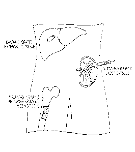
and that are
transplanted onto/into normal tissue in an experimental host.
[0007] The cells may comprise stem cells, mature cells, angioblasts,
endothelia.
mesenchymal stem cells (from any source), stellate cells, fibroblasts or
mixtures of these, In
addition, the biomaterials may comprise collagens, adhesion molecules
(laminins,
fibronectins, nidogen), elastins, proteoglycans, hyaluronans (HAs),
glycosaminoglycan
chains, chitosan, alginate, and synthetic, biodegradable and biocompatible
polymers.
Hyaluronans are one of the preferred materials.
[0008] The isolated cells of the internal organ may be solidified ex vivo
within the
biomaterials prior to introducing the cells into the hosts, or in the
alternative, injected as a
fluid substance and allowed to solidify in vivo. Preferably, the cells are
introduced at or near
the diseased or dysfunctional tissue, and may be introduced via injection,
biodegradable
covering, or sponge.
[0009] In another embodiment of the present invention, a method of repairing
tissue of an
internal organ in a subject suffering from the internal organ in a diseased or
dysfunctional
condition is provided. The method comprises (a) obtaining normal cells of the
internal organ
from a donor; (b) combining the cells with one or more biomaterials; (c)
optionally
combining the cell suspension with signaling molecules (growth factors,
cytokines),
2
CA 02798458 2012-11-05
WO 2011/140428 PCMJS2011/035498
additional cells, or combinations thereof; and (d) introducing the mixture (b)
into the subject,
wherein the mixture becomes insoluble and forms a graft onto or into the
internal organ in
vivo.
[0010] In yet another embodiment of the present invention, a method of
localizing cells of
an internal organ onto a surface, into an interior portion, or both of a
target internal organ is
provided, the method comprising introducing a preparation comprising cells of
an internal
organ and a solution of one or more hydrogel-forming precursors, in the
presence of an
effective amount of a cross-linker, onto a surface, into an interior portion,
or both of a target
internal organ in vivo, which preparation forms a hydrogel comprising cells of
an internal
organ on a surface, in an interior portion, or both of a target internal
organ. The mixture my
further comprise nutrient medium, extracellular matrix molecules, and
signaling molecules.
The solidified mixture, such as a hydrogel, provides a graft into a target
internal organ either
on its surface, in an interior portion, or both.
[0011] The cells may be localized for a period of at least twelve hours, at
least twenty-four
hours, at least about forty-eight hours, or at least about 72 hours into/onto
the target internal
organ, which may be liver, pancreas, biliary tree, lung, thyroid, intestine,
breast, prostate,
uterus, bone, or kidney. In treatment of patients, the donor cells of the
internal organ should
not be diseased cells (e.g., tumor or cancer cells). However, diseased cells
might be
considered in a graft when trying to establish an experimental model system of
a disease.
[0012] The biomaterials that can form hydrogels, or a parallel insoluble
complex, can
comprise glycosaminoglycans, proteoglycans, collagensõ laminins, nidogen,
hyaluronans, a
thiol-modified sodium hyaluronate, denatured forms thereof (e.g., gelatin), or
combinations
thereof. A trigger for solidification can be any factor eliciting cross-
linking of the matrix
components or gelation of those that can gel. The cross-linker may comprise
polyethylene
glycol diacrylate or a disulfide-containing derivative thereof Preferably, the
insoluble
complex of cells and biomaterials possesses a viscosity ranging from about 0.1
to about 100
kPa, preferably about 1 to about 10 kPa, more preferably about 2 to about 4
kPa, and most
preferably a stiffness from about 11 to about 3500 Pa.
[0013] In still yet another embodiment of the present invention, a method of
cryopreserving
cells is provided, comprising: (a) obtaining isolated cells; (b) combining the
cells with gel-
forming biomaterials and, optionally, one or more of isotonic basal medium,
signaling
molecules (cytokines, growth factors, hormones), and extracellular matrix
components (e.g.
3
CA 02798458 2012-11-05
WO 2011/140428 PCMJS2011/035498
hyaluronans); and freezing the cell mixture so as to be stored in a -90 C or -
180 C freezer.
The isotonic medium can be CS10 (biolife) or an equivalent isotonic
cryopreservation buffer.
The signaling molecules can be Suitable signaling molecules are growth factors
and
cytokines and include, for example, epidermal growth factor (EGF), hepatocyte
growth factor
(HGF), stromal cell-derived growth factor (SGF), retinoids (e.g., vitamin A),
fibroblast
growth factor (FGF, e.g., FGF2, FGF10), vascular endothelial cell growth
factor (VEGF),
insulin like growth factor I (IGF-I), insulin-like growth factor II (IGF-II),
oncostatin M,
leukemia inhibitory factor (LIF), transferrin, insulin, glucocorticoids,
(e.g., hydrocortisone),
growth hormone, any of the pituitary hormones (e.g., follicle stimulating
hormone (FSH)),
estrogens, androgens, and thyroid hormones (e.g., T3 or T4). The extracellular
matrix
components can be glycosaminoglcyanas, hyaluronans, collagens, adhesion
molecules
(laminins, fibronectins), proteoglycans, chitosan, alginate, and synthetic,
biodegradable and
biocompatible polymers, or combinations thereof.
[0014] For cryopreservation of the mixtures of cells and biomaterials,
mixtures, they may
be further combined with a (i) cryoprotectant selected from the group
consisting of dimethyl
sulfoxide(DMS0), glycerol, ethelyene glycol, ethylenediolethalenediol, 1,2-
propaendiol, 2,-3
butenediol, formamide, N-methylformamide, 3-methoxy-1,2-propanediol by
themselves, and
combinations thereof and/or (ii) an additive selected from the group
consisting of sugara,
glycine, alanine, polyvinylpyrrolidone, pyruvate, an apoptosis inhibitor,
calcium,
lactobionate, raffinose, dexamethasone, reduced sodium ions, choline,
antioxidants,
hormones, or combination thereof. The sugar may be trehalose, fructose,
glucose, or a
combination thereof and the antioxidants may be vitamin E, vitamin A, beta-
carotene, or a
combination thereof.
Brief Description of the Figures
[0015] Figure 1 is a schematic of methods according to the invention of
grafting cells to
various target tissues. These methods include, implantable grafts, injectable
grafts, and grafts
that can be attached onto the surface of a target organ ("bandaid grafts").
[0016] Figure 2 provides rheological measurements on hyaluronans prepared with
Kubota's Medium (KM-HAs). a) The shear modulus G*1of KM-HAs, a measurement of
mechanical gel stiffness, remains constant while viscoelastic damping G' '/G'
a
measurement of deformation response delay upon external forcing, is negligible
within the
0.1 Hz ¨ 10 Hz forcing frequency range for each of the formulations tested;
error bars: 95%
CA 02798458 2012-11-05
WO 2011/140428
PCT/US2011/035498
confidence interval of measurements at each frequency tested. b) KM-HAs
exhibit shear
thinning, i.e. decrease in viscosity with increasing forcing frequencies,
across experimental
0.6 1/s ¨60 1/s shear rate range [0.1 Hz ¨ 10 Hz forcing frequency]; upper and
lower limits:
power law model-based 95% confidence interval (Cox-Merz rule assumption, R2 >
0.993 for
all formulations in the 0.3 1/s ¨ 30 1/s shear rate range [0.05 Hz ¨ 5 Hz
forcing frequency]).
Rheological measurements performed only on lettered formulations shown in
Table 3.
[0017] Figure 3 shows size, morphology and proliferation data of human hepatic
stem cells
(hHpSCs) in KM-HAs. Colonies of hHpSCs acquire three-dimensional
configurations and
exhibit a) spheroid-like agglomeration (bottom left) or folding (middle, top
right) upon
seeding in KM-HAs [image frame: 900 pm x 1200 m]. Confocal microscopy on
histological sections of hHpSC-seeded KM-HAs reveals mixed cell morphology
phenotypes
after 1 week of culture, with cell sizes of b) about 7 pm, or c) up to 10-15
pm amongst
parenchymal cells [cell nuclei in blue from DAPI counterstaining, EpCAM in red
for both b)
and c), green for either b) CD44, ore) CDH1; image frames b) and c): 150 pm x
150 pm;
white highlight in b) and c): 15 pm x 15 pm]. d) Viability of hHpSCs in KM-
HAs, measured
by AlamarBlue metabolic reduction, reveals functional recovery and
proliferation in KM-HA
hydrogels with 1.6% CMHA-S and 0.4% PEGDA (formulation E, Table 3) throughout
1
week of culture; AlamarBlue reduction measurements after 24-hr incubation,
normalized
with respect to measurements at 2-3 days post-seeding; data reported as mean
standard
error.
[0018] Figure 4 provides protein expression of differentiation markers in KM-
HA-seeded
hHpSCs after 1 week of culture. Colonies of hHpSCs exhibit differential levels
of expression
for differentiation markers in hHpSCs at the translational level depending on
KM-HAs
properties. Metabolic secretion rates of human AFP correlate mRNA expression
levels
across KM-HA formulations. NCAM expression is positive in all KM-HAs, while
CD44
expression is richest in KM-HAs with CMHA-S contents of 1.2% or less (lettered
formulations A, B, C, D: Table 3). CDH1 expression is positive for KM-HA
hydrogels with
IG*1 <200 Pa and negative for G* > 200 Pa. Data for human AFP secretion rate
reported as
mean standard error. Immunohistochemical staining for EpCAM, NCAM, CD44 and
CDH1 performed on 15 ¨20 !_tm sections (-2 to 3 hHpSCs thick; hHpSC diameter:
5-7 pm)
and imaged by fluorescence microscopy [image frames: 100 pm x 100 m]. KM-HA
formulations ordered with respect to increasing stiffness (1G*1 = 25 Pa for A,
1G*1 = 73 Pa
CA 02798458 2012-11-05
WO 2011/140428
PCT11JS2011/035498
for B, = 140 Pa
for F, G* ¨ 165 Pa for C, G*1 = 220 Pa for D, and G*1 = 520 Pa for
F).
[0019] Figure 5 provides gene expression levels by qRT-PCR for hepatic
progenitor
markers in KM-HA-grown hFlpSCs after 1 week of culture. Comparisons between
the
mRNA expression levels of markers for hHpSCs and their immediate descendents
hHBs
(hepatic-specific AFP, EpCAM, NCAM, CD44 and CDH1) show that KM-HA-grown
hHpSCs acquire early hHB characteristics at the transcriptional level in
passive culture for 1
week. The expression ranges in hHpSCs and freshly isolated hHBs for CD44 are
comparable; the expression levels for the remaining markers are statistically
distinct, with
approximately 2-fold decrease in EpCAM, 3-fold decrease in CDH1, NCAM
silencing and
AFP enrichment upon hHpSCs differentiation into hHBs. In all KM-HAs, mean
expression
levels of seeded hHpSCs for AFP, NCAM and CDH1 shifted outside the hHpSC range
towards the hHB range, while EpCAM expression is enriched throughout, after 1
week of
culture. KM-HA formulations ordered with respect to increasing stiffness (1G*1
= 25 Pa for
A, G* = 73 Pa for B, 1G* = 140 Pa for E, 1G* = 165 Pa for C, G* = 220 Pa for
D, and
1G*1 = 520 Pa for F). Expression levels (mean standard error) were
normalized with
respect to GAPDH. Measurements in lettered KM-HA formulations (Table 3)
compared to
hHpSC colonies (green) and freshly isolated hHBs (red) for significance
(Student's t-test).
[0020] Figure 6 is a schematic of one embodiment of the disclosed
cryopreservation and
thawing methods.
[0021] Figure 7 shows the results from in vivo real time imaging of
luminescent signal
produced by luciferin-producing cells both grafted with hyaluronans versus
injected as a cell
suspension.
[0022] Figure 8 provides serum human albumin at day 7 post-transplantation in
grafted
versus cell suspension in both healthy and CC14 liver injury models.
[0023] Figure 9 shows gene expression of hepatic stem cell phenotype markers.
Expression levels are normalized to GAPDH expression, and fold changes are
normalized to
initial expression in colonies. * denotes p<0.05% significance between
experimental
condition and initial colony expression. ** denotes p<0.05% significance
between
experimental condition and initial colony expression as well as significant
expression
between the two experimental conditions.
6
CA 02798458 2012-11-05
WO 2011/140428 PCT11JS2011/035498
[0024] Figure 10 provides data from functional assays of hepatic function over
time. A)
albumin, B) Transfen-in, and C) Urea in three-dimensional hyaluronan culture
over time for
levels are normalized per cell.
[0025] Figure 11 provides data from mechanical characterization of KM-HAs. a)
Stiffness
of KM-HAs is controllable and depends on CMHA-S and PEGDA contents. The
average
shear modulus 1G*1 increases with increasing CMHA-S and PEGDA contents
following a
power-law behavior, thus providing direct control of the final mechanical
properties of KM-
HAs during the initial hydrogel mixing; rheological measurements performed
only on lettered
formulations shown in Table 3. Error bars: 1 standard deviation for
measurements in the
0.05 Hz ¨ 5 Hz forcing frequency. b) Diffusion in KM-HAs. Measurements of
diffusivity
within KM-HAs by FRAP (70 kDa fluorescein-labeled dextran) do not differ
significantly
from Kubota's medium alone; diffusivity measurements performed on all
formulations shown
in Table 3. Error bars: 95% confidence interval of measurements.
[0026] Figure 12 shows the secretion of human AFP, albumin and urea by hHpSCs
seeded
into KM-HAs. Colonies of hHpSCs in KM-HAs exhibit some hepatic function with
increasing concentrations of human AFP and albumin found in culture media (KM)
and
equilibration of urea synthesis by day 7 post-seeding. The metabolic secretion
rates of
human AFP, human albumin and urea are distinctive by day 7 post-seeding
amongst KM-HA
formulations, with minimum rates for AFP, albumin and decreased urea synthesis
in KM-
HAs with 1.6% CMHA-S and 0.4% PEGDA (formulation E, Table 3). Left column:
metabolite concentration in culture media collected daily after 24-hr
incubation for each
lettered formulation (Table 3). Right column: metabolite mass secretion rate
per hHpSC
colony in culture media after 24-hr incubation; total metabolite mass in media
is normalized
to number of functional hHpSC colonies at each interval as calculated by
viability assay with
AlamarBlue reduction (approximate number of colonies seeded per sample: 12).
All data
reported as mean standard error.
[0027] Figure 13 shows that controlled rate freezing program minimizes liquid-
ice phase
entropy preventing internal ice damage and allows for repeatable freezing. A)
Graph shows
chamber temperature in relation to sample temperature (10% DMSO). B) freezing
program
rates used for Cryomed 1010 system.
[0028] Figure 14 provides both (A) Cell Viability % of cryopreserved fetal
hepatic cells
post-thaw and (B) Colony counts after 3 weeks of culture for each condition,
normalized to
7
CA 02798458 2012-11-05
WO 2011/140428 PCT11JS2011/035498
fresh samples. Results are reported as mean standard error of the mean. KM=
Kubotas
Medium with 10% DMSO and10% FBS. CS10¨cryostor, CS10+sup=cryostor10 with KM
supplements. 0.05% and 0.10% refer to the HA% supplemented in each sample.
[0029] Figure 15 shows the relative mRNA expression normalized to GAPDH
expression.
Mean standard error of the mean. Significance *p>0.05 to Fresh samples. KM=
Kubotas
Medium with 10% DMSO and 10% FBS. CS10=cryostor, CS10+sup¨cryostor10 with KM
supplements. 0.05% and 0.10% refer to the HA% supplemented in each sample.
Detailed Description of the Invention
[0030] At present, cell transplantation involving cells derived from solid
organs is
performed typically via a vascular route, and the results routinely provide
overwhelming
evidence of inefficient engraftment, typically on the order of about 20-30%
for mature cells
and less than 5% for stem cells. The differential engraftment is due to their
sizes which in
liver are small for stem cells (typically under 10 pm) and larger for the
mature cells (typically
>18 pm). Our studies have confilmed this observation. In one study, for
example, human
hepatic stem cells (hl-IpSCs) and hepatoblasts (hHBs) were injected into
immunocompromised mice by injecting the cells into the spleen. Since the
spleen connects
directly with the liver, the cells flowed into the liver where they were
expected to engraft.
However, most of the cells died prior to engraftment or lodged in tissues
other than the
intended target (ectopic sites).
[0031] Even when cells properly did reach their destination, conversion of the
cells to fully
functional ones was hindered by lack of vascularization, lack of growth (when
transplanting
mature cells), and highly immunogenic properties of the cells if mature cells
were used and
necessitating long-term immunosuppression. Other hurdles include the sourcing
of clinical
grade, high-quality cells and the need to use freshly isolated cells due to
difficulties with
cryopreservation.
[0032] In addition to the inefficiencies and difficulties noted,
transplantation of cells from
solid organs via a vascular route is dangerous. The cells from solid organs
have surface
molecules (cell adhesion molecules, tight junction proteins) that make the
cells bind to each
other rapidly and enhance aggregation. This clumping phenomenon can result in
life-
threatening pulmonary emboli.
[0033] To address some of these hurdles and concerns, the present invention is
directed to
grafting technologies that involve the delivery of transplanted cells as an
aggregate on or in
8
scaffolds that can be localized to the diseased tissue to promote necessary
proliferation and
engraftment. Thus, the invention takes into account not only the cell type to
be transplanted,
but also the cell type in combination with the appropriate biomaterials and
grafting method
for the most efficient and successful transplant therapies. Grafting
technologies of the
present invention are translatable to therapeutic uses in patients and provide
alternative
treatments for regenerative medicine to reconstitute diseased or dysfunctional
tissue.
Cell Sourcing
[0034] According to the invention, desired cell populations may be obtained
directed from
a donor having "normal," "healthy" tissue and/or cells, meaning any tissue
and/or cells that
is/are not afflicted with disease or dysfunction. Of course, such a cell
population may be
obtained from a person suffering from an organ with disease or dysfunction,
albeit from a
portion of the organ that is not in such a condition. The cells may be sourced
from any
appropriate mammalian tissue, regardless of age, including fetal, neonatal,
pediatric, and
adult tissue. If experimental models of a disease state are to be established,
then one can
utilized diseased cells in the grafts that are to be transplanted into an
appropriate experimental
host.
[0035] More specifically, cells may be sourced for different therapies from
"lineage-
staged" populations based on the therapeutic need. For example, later-stage
"mature" cells
may be preferred in cases where there is a need for rapid acquisition of
functions offered only
by the late lineage cells, or if the recipient has a lineage-dependent virus
that preferentially
infects the stem cells and/or progenitors such as occurs with hepatitis C or
papilloma virus.
In any event, "progenitor" cells may be used to establish any of the lineage
stages of their
respective tissue(s).
[0036] For a discussion of lineage-staged liver cell populations and method of
their
isolation, see US patent application nos. 11/560,049 and 12/213100.
Briefly, there are at least eight
maturational lineage stages that are intrahepatie. Below are given those
stages and brief
statements about them:
[0037] Lineage Stage 1: Human hepatic stem cells (hHpSCs) are multipotent
cells, located
within the ductal plates of fetal and neonatal livers and the canals of Hering
in pediatric and
adult livers. These cells usually range from 7-10 m in diameter and have high
nucleus to
cytoplasmic ratios. They are tolerant of ischemia, can be found in cadaveric
livers for more
9
Date Recue/Date Received 2020-07-02
CA 02798458 2012-11-05
WO 2011/140428 PCT11JS2011/035498
than 48 hours after systolic death, and form colonies of hHpSCs capable of
differentiation to
mature cells. These cells constitute approximately 0.5-2% of the parenchyma of
livers of all
age donors.
[0038] Lineage Stage 2: The hepatoblasts (hHBs) are the immediate descendents
of the
hHpSCs and are the liver's probable transit amplifying cells. They are located
just outside
the stem cell niche proper. These cells are larger (10-12 p.m) with higher
amounts of
cytoplasm and are found in vivo throughout the parenchyma in fetal and
neonatal livers and
near the ends of or adjacent to the canals of Hering in pediatric and adult
livers. With age,
the hepatoblasts decline in numbers to <0.01% of the parenchymal cells in
postnatal livers.
This population of cells has been shown to expand during regenerative
processes especially
those associated with certain diseases such as cirrhosis. Hepatoblasts mature
into either
hepatocytes (H) or cholangiocytes, also called biliary epithelia (B):
[0039] Lineage Stage 3E1 and 3B: Committed (unipotent) hepatocytic (3f1) and
cholangiocyte progenitors¨biliary progenitors (3B) are found within the liver.
These
unipotent precursors give rise to only one adult cell type, and no longer
express some of the
stem cell genes (e.g., low or no levels of expression for CD133/1, Hedgehog
proteins
(Sonic/Indian) but express genes typical for cells in the fetal tissues.
[0040] Lineage Stage 4 H and 4B: Periportal adult parenchymal cells comprise
relatively
small hepatocytes (4H) and intrahepatic biliary epithelia (4B). The
hepatocytes are diploid,
are approximately 18 p.m in diameter, and express multiple factors/enzymes
associated with
gluconeogenesis such as PEPCK, connexins 26 and 32.
[0041] The cholangiocytes of this stage (4B) are diploid, are approximately 6-
7 m in
diameter, line a portion of the canals of Hering, and express various genes
including
aquaporins 1 and 4, MDR1, secretin receptor, but not CL7FIC03- exchanger or
somatostatin
receptor.
[0042] Lineage Stage 5 H and 513: Cells of this stage comprise relatively
larger
hepatocytes (5H) and cholangiocytes (5B), both diploid. The size of the
hepatocytes is
approximately 22-25 jim in diameter, and they are found in the midacinar zone.
The
midacinar hepatocytes express high levels of albumin and tyrosine
aminotransferase (TAT);
especially characteristic is that they express transferrin as a protein (by
contrast, lineage
stages 1-4 express it only as mRNA.
CA 02798458 2012-11-05
WO 2011/140428
PCT11JS2011/035498
[0043] Lineage stage 5B cholangiocytes are approximately 14 p.m in diameter,
located
within the intralobular ducts, and express CFTR, Secretin receptor,
somatostatin receptor,
MDR1 and MDR3, and the CL/HCO3- exchanger.
[0044] Lineage Stage 6H: The diploid pericentral hepatocytes of stage 6 can
form
colonies in culture, but have limited capacity to expand and essentially no
capacity to be
subcultured. The percentage of these declines with age (in parallel with an
increase in the
percentage of tetraploid pericentral cells). In addition to albumin, TAT, and
transferrin, they
express also strongly a number of the P450s such as P450-3A, glutamine
synthetase (GT),
heparin proteoglycans, and the genes associated with urea formation.
100451 Lineage Stage 711: This stage comprisese tetraploid pericentral
parenchymal cells
that are no longer able to undergo complete cell division. They can undergo
DNA synthesis
but with limited capacity for cytokinesis. They are much larger cells (>30 m
in diameter) and
express high levels of the genes that become apparent in lineage stages 5-6.
[0046] Lineage Stage 8: Apoptotic Cells: express various markers of apoptosis
and
demonstrate DNA fragmentation.
[0047] In addition to the cells required to provide the "functions" per se of
a diseased or
dysfunctional internal organ, the graft preferably includes additional
cellular components that
preferably mimic the categories of cells comprising the epithelial-mesenchymal
cell
relationship, the cellular foundation of all tissues. Epithelial-mesenchymal
cell relationships
are distinct at every maturational lineage stage. Epithelial stem cells are
partnered with
mesenchymal stem cells and their maturation is coordinate with each other as
they mature to
all the various adult cell types within a tissue. The interactions between the
two are mediated
by paracrine signals that comprise soluble signals (e.g., growth factors) and
extracellular
matrix components.
[0048] In livers, for example, the hepatic stem cells (HpSCs) give rise to
hcpatocytes and to
cholangiocytes. The mesenchymal partners for the HpSCs are angioblasts. There
is evidence
to indicate that angioblasts give rise to both endothelial cell precursors and
to hepatic stellate
cell precursors, the mesenchymal cell partners for intrahepatic lineage stage
2 parenchyma,
the hepatoblasts (Bs). The endothelial cell precursors mature in subsequent
lineage stages
to be endothelia that become the mesenchymal partners for the lineage stages
of hepatocytes.
The stellate cell precursors cells give rise to stellate cells, and then to
stromal cells, and then
to myofibroblasts, the mesenchymal cellspartners for cholangiocytes.
11
CA 02798458 2012-11-05
WO 2011/140428 PCMJS2011/035498
[0049] The formation of the liver, called hepatogenesis, is regulated through
signals from
the angioblasts in the embryonic mesenchyme associated with the heart. During
the initial
stages of liver development, fibroblast growth factors (FGFs) are secreted
from pre-cardiac
mesoderm while bone morphogenetic proteins (BMPs) are delivered from the
mesenchyme.
These newly specified hepatic cells then break away and migrate into the
surrounding
mesenchyme and interact with precursors to both endothelia and stroma. The
mesenchymal
cells remain in contact with hepatic cells throughout development.
[0050] Human hepatic stem cells (hHpSCs) require contact with mesenchymal
cells for
survival. They will self-replicate, that is remain as hHpSCs when on feeders
of angioblasts.
They lineage restrict to hepatoblasts, if cultured on feeders of hepatic
stellate cells. They
mature into adult hepatocytes if cultured on mature endothelia and to
cholangiocytes if on
mature stroma (e.g. mature stellate cells or myofibroblasts). The control of
the fate of the
stem cells by the feeders has been shown to be due to the exact combinations
of paracrine
signals produced in each of the epithelial-mesenchymal relationships in the
lineages.
[0051] According to one embodiment of the invention, the isolated cell
populations are
combined with known paracrine signals (discussed below) and "native"
epithelial-
mesenchymal partners, as needed, to optimize the graft. Thus, the grafts will
comprise the
epithelial stem cells, the hepatic stem cells, mixed together with their
native mesenchymal
partners, angioblasts. For a transit amplifying cell niche graft, hepatoblasts
can be partnered
with hepatic stellate cells and endothelial cell precusors. In some grafts one
can make a mix
of the two sets: hepatic stem cells, hepatoblasts, angioblasts, endothelial
cell precursors,
hepatic stellate cell precursors cells to optimize the establishment of the
liver cells in the host
tissue. The microenvironment of the graft into which the cells are seeded will
be comprised
of the paracrine signals, matrix and soluble signals, that are produced at the
relevant lineage
stages used for the graft.
[0052] Grafts can also he tailored to manage a disease state. For example, to
minimize
effects of lineage dependent viruses (e.g., certain hepatitis viruses) that
infect early lineage
stages and then mature coordinately with the host cells, one can prepare
grafts of later lineage
stage (e.g., hepatocytes and their native partners, sinusoidal endothelial
cells) that are non-
permissive for viral infection. Grafts can be used also to establish a disease
model by using
diseased cells in a graft that is transplanted into/onto a target organ in an
experimental animal
model.
12
(00531 An example of a stem cell graft, using liver cell therapies as a model,
would
comprise the hepatic stem cells, angioblasts and hepatic stellate cell
precursors. In contrast, a
graft of "mature" liver cells would comprise hepatocytes, mature endothelial
cells and
pericytes, which are the mature stellate cells. For a discussion of the
epithelial-mesenchymal
cell relationship of livers, see US patent application no. 11/753,326.
(00541 The issue of vascularization is important for all grafts, and therefore
should be
implanted in location conducive to vascularization (e.g., liver). For most
disease conditions,
stem cell grafts are preferred, given their expansion potential, their ability
to mature into all
of the adult cell types, their tolerance for ischemia, enabling their sourcing
from cadaveric
tissue, and their minimal, if any, immunogenicity.
Graftina Materials
100551 The use of gel-forming biomaterials according to the invention provides
a scaffold
for cell support and signals that assist in the success of the grafting and
regenerative
processes. As tissue of solid organs in an organism undergo constant
remodeling, dissociated
cells tend to reform their native structures under appropriate environmental
conditions. The
cells may be combined with one or more of a nutrient medium (e.g., RPM 1640),
signaling
molecules (e.g., insulin, transferrin, VEGF) and one or more extracellular
matrix components
(e.g., hyaluronans, collagens, nidogen, proteoglycans).
100561 In all tissues, the paracrine signaling comprises both soluble (myriad
growth factors
and hormones) and insoluble (extracellular matrix (ECM) signals). Synergistic
effects
between the soluble and (insoluble) matrix factors can dictate growth and di
fferentiative
responses by the transplanted cells. The matrix components are the primary
determinants of
attachment, survival, cell shape (as well as the organization of the
cytoskeleton), and
stabilization of requisite cell surface receptors that prime the cells for
responses to specific
extracellular signals.
100571 The ECM is known to regulate cell morphology, growth and cellular gene
expression. Tissue-specific chemistries similar to that in vivo may be
achieved ex vivo by
using purified ECM components. Many of these are available commercially and
are
conducive to cell behavior mimicking that in vivo.
100581 Suitable matrix components include collagens, adhesion molecules (e.g.,
cell
adhesion molecules (CAMs), tight junctions (cadherins), basal adhesion
molecules (laminins,
13
CA 2798458 2019-12-03
fibronectins), gap junction proteins (connexins), elastins, and sulfated
carbohydrates that
form proteoglycans (PCs) and glycosaminoglycans (GAGs). Each of these
categories defines
a genus of molecules. For example, there are at least 25 collagen types
present, each one
encoded by distinct genes and with unique regulation and functions. Additional
biomaterials
include inorganic, natural materials like chitosan and alginate as well as
many synthetic,
biodegradable and biocompatible polymers. These materials are often
"solidified" (e.g.,
made into a gel or an insoluble material) through methods including thermal
gelation, photo
cross-linking, or chemical cross-linking or exposure to microenvironment
(e.g., high salt) that
elicit insolubility of the materials. With each method, however, it is
necessary to account for
cell damage (e.g., from excessive temperature ranges, UV exposure). For a more
detailed
discussion of biomaterials, specifically the use of hyaluronan hydrogels, see
US patent
application no. 12/073,420.
100591 The particular selection of which matrix components may be guided by
gradients in
vivo, for example, that transition from components found in association with
the stem cell
compartment to that found in association with the late lineage stage cells.
The graft
biomaterials preferably mimic the matrix chemistry of the particular lineage
stages desired
for the graft. The efficacy of the chosen mix of matrix Components may be
assayed in ex vivo
studies using purified matrix components and soluble signals, many of which
are available
commercially, according to good manufacturing practice (GM?) protocol. The
biomaterials
selected for the graft preferably elicit the appropriate growth and
differentiation responses
required by the cells for a successful transplantation.
100601 Concerning the liver organ, the matrix chemistry associated with liver
parenchymal
cells, and outside of the stem cell and transit amplifying cell niches, is
present in the Space of
Disse, the area located between the parenchyma and the endothelia or other
forms of
mesenchymal cells. In addition to a change in cell maturity within the
different zones of the
liver, a change in matrix chemistries is also observed. The matrix chemistry
periportally in
zone 1 is similar to that found in fetal livers and consists of type III and
type IV collagens,
hyaluronans (HA), laminins, and forms of chondroitin sulfate protcoglycans.
This zone
transitions to a different matrix chemistry in the pericentral zone 3,
containing type I
collagen, fibronectin, and unique forms of heparin and heparan sulfate
proteoglycans.
100611 The stem cell niche of the liver has been characterized partially and
found to
comprise hyaluronans, laminin forms (e.g., laminin 5) that bind to alpha 6-
beta 4 integrin ,
14
CA 2798458 2019-12-03
CA 02798458 2012-11-05
WO 2011/140428
PCMJS2011/035498
type III collagen and unique fotors of minimally sulfated chondroitin sulfate
proteoglycans
(CS-PGs) There are limited amounts of type IV collagen and no type I collagen
in this niche.
[0062] This niche matrix chemistry transitions to that associated with the
transit amplifying
cell compartment and is comprised of type IV collagen, forms of laminin that
bind to other
integrins (41) , and forms of GAGs and PGs that include forms of CS-PGs with
higher
sulfation, dermatan sulfate-PGs, and to specific forms of heparan sulfate-PGs
(HS-PGS).
100631 The transit amplifying cell compartment transitions to yet later
lineage stages, and
with each successive stage, the matrix chemistry becomes more stable (e.g.,
more highly
stable collagens), turns over less, and contains more highly sulfated forms of
GAGs and PGs.
The most mature cells are associated with forms of heparin-PGs (HP-PGs),
meaning that
myriad proteins (e.g., growth factors and hormones, coagulation proteins,
various enzymes)
can bind to the matrix and be held stably there via binding to the discrete
and specific
sulfation patterns in the GAGs. Thus, the matrix chemistry transitions from
its start point in
the stem cell niche having labile matrix chemistry associated with high
turnover and minimal
sulfation (and therefore minimal binding of signals in a stable fashion near
to the cells) to
stable matrix chemistries with increasing amounts of sulfation (and therefore
higher and
higher levels of signal binding and held near to the cells).
[0064] Hence, the present invention takes into consideration that the
chemistry of the
matrix molecules changes with maturational stages, with host age, and with
disease states.
Grafting with the appropriate materials should optimize engraftment of
transplanted cells in a
tissue, prevent dispersal of the cells to ectopic sites, minimize embolization
problems, and
enhance the ability of the cells to integrate within the tissue as rapidly as
possible. Moreover,
the factors within the graft can also be chosen to minimize immunogenicity
problems.
100651 In the case of human livers, cells may be cultured under serum free
conditions.
Human hepatic stem cell or hepatoblasts (hHpSC or hHB) can be grafted by
themselves, or in
combination with angioblastsiendothelial cell precursors and stellate cell
precursors cells.
Cells can be suspended in thiolated and chemically-modified HA (CMHA-S, or
Glycosil,
Glycosan BioSystems, Salt Lake City, UT) containing medium (HA-M) and in KM
(Kubota's Medium) and loaded into one of the syringes of a set of paired
syringes. The other
syringe may be loaded with a cross-linker, e.g., poly(ethylene glycol)
diacrylate or PEGDA,
prepared in KM (or with the conditions required to elicit insolubility of the
biomaterials).
The two syringes are coupled by a needle that flares into two luer lock
connections. Thus,
CA 02798458 2012-11-05
WO 2011/140428 PCMJS2011/035498
the cells in hydrogel and the cross-linker can emerge through one needle to
allow for rapid
cross-linking of the CMHA-S into a gel upon injection (or insolubility of the
biomaterials by
alternate means).
[0066] The cell suspension in CMHA-S and crosslinker can be either directly
injected or
grafted to the liver using the omentum tissue to form a pouch. Alternatively,
the cells may be
encapsulated in Glycosil without the use of a PEGDA crosslinker by allowing
the suspension
to stand overnight in air, leading to disulfide bond crosslinking to a soft,
viscous hydrogel. In
addition, other thiol-modified macromonomers, e.g., gelatin-DTPH, heparin-
DTPH,
chondroitin sulfate-DTPH, may be added to give a covalent network mimicking
the matrix
chemistry of particular niches in vivo. In another manifestation, polypeptides
containing
cysteine or thiol residues can be coupled to the PEGDA prior to adding the
PEGDA to the
Glycosil, allowing specific polypeptide signals to be incorporated into the
hydrogel.
Alternatively, any polypeptide, growth factor or matrix component such as an
isoform of a
collagen, laminin, vitronectin, fibronectin, etc., may be added to the
Glycosil and cell
solution prior to crosslinking, allowing passive capture of important
polypeptide components
in the hydrogel.
[0067] Hyaluronans: Hyaluronans (HAs) are members of one of the 6 large
glycosaminoglycan (GAG) families of carbohydrates, all being polymers of a
uronic acid and
an aminosugar [1-3]. The other families comprise the chondroitin sulfates (CS,
[glucuronic
acid-galactosamine]x), dermatan sulfates (DS, more highly sulfated [glucuronic
acid-
galactosamine]x), heparan sulfates (HS, [glucuronic acid-glucosamine]x),
heparins (HP, more
highly sulfated [gluronic acid-glucosamine]x) and keratan sulfates (KS,
[galactose-N-
acetylglucosamine]x).
[0068] HAs are composed of a disaccharide unit of glucosamine and gluronic
acid linked
by [31-4, f31-3 bonds. Biologically, the polymeric glycan is composed of
linear repeats of a
few hundreds to as many as 20.000 or more of disaccharide units. The HAs have
molecular
masses typically ranging from 100,000 Da in serum to as much as 2,000,000 in
synovial
fluid, to as much as 8,000,000 in umbilical cords and the vitreous.. Because
of its high
negative charge density, HA attracts positive ions, drawing in water. This
hydration allows
HA to support very compressive loads. HAs are located in all tissues and body
fluids, and
most abundant in soft connective tissue, and the natural water carrying
capacity lends itself to
speculation to other roles including influences of tissue form and function.
It is found in
extracellular matrix, on the cell surface and inside the cell.
16
CA 02798458 2012-11-05
WO 2011/140428 PCT11JS2011/035498
[00691 Native forms of HA chemistry are diverse. The most common variable is
the chain
length. Some are high molecular weight due to having long carbohydrate chains
(e.g. those
in the coxcomb of gallinaceous birds and in umbilical cords) and others are
low molecular
weight due to having short chains (e.g. from bacterial cultures). The chain
length of HAs
plays a key role in the biological functions elicited. A low molecular weight
HA (below
3.5x10⁴ kDa) may induce the cytokine activity that is associated with matrix
turnover and is
shown to be related to inflammation in tissues. A high molecular weight (above
2 X 10⁵kDa)
may inhibit cell proliferation. Small HA fragments, between 1-4 kDa, have been
shown to
increase angiogenesis.
[0070] Native forms of HA have been modified to introduce desired properties
(e.g.,
modification of the HAs to have thiol groups allowing the thiol to be used for
binding of
other matrix components or hormones or for novel forms of cross-linking).
Also, there are
forms of cross-linking that occur in nature (e.g., regulated by oxygen) and
yet others that
have been introduced artificially by treatment of native and modified HAs with
certain
reagents (e.g., akylating agents) or, as noted above, establishment of
modified HAs that make
them permissive to unique fowls of cross-linking (e.g., disulfide bridge
formation in the
thiol-modified HAs).
[0071] According to the invention, thiol-modified HAs and in situ
polymerizable
techniques used for them are preferred. These techniques involve disulfide
crosslinking of
thiolated carboxymethylated HA, known as CMHA-S or Glycosil. For in vivo
studies, HA
with lower molecular weight, e.g., 70-250 kDa, can be used, since the
crosslinking, either
disulfide or PEGDA, creates a hydrogel of very high molecular size. A thiol-
reactive linker,
polyethylene glycol diacrylate (PEGDA) crosslinker, is suitable for both cell
encapsulation
and in vivo injections. This combined Glycosil-PEGDA material crosslinks
through a
covalent reaction and in a matter of minutes, is biocompatible and allows for
cell growth and
proliferation.
[0072] The hydrogel material, Glycosil, takes into account the gel properties
conducive to
tissue engineering of stem cells in vivo. Glycosil is part of the semi-
synthetic extracellular
matrix (sECM) technology available from Glycosan Biosciences in Salt Lake
City, UT. A
variety of products in the Extracel and HyStem trademarked lines are
commercially available.
These materials are biocompatible, biodegradable, and non-immunogenic.
17
CA 02798458 2012-11-05
WO 2011/140428
PCT11JS2011/035498
[0073] Furthermore, Glycosil and Extralink can be easily combined with other
ECM
materials for tissue engineering applications. HA can be obtained from many
commercial
sources, with a preference for bacterial fermentation using either
Streptomyces strains (e.g.,
Genzyme, LifeCore, NovaMatrix, and others) or bacterial-fermentation process
using
Bacillus subtilis as the host in an ISO 9001:2000 process (unique to
Novozymes).
[0074] The ideal ratios of the cell populations should replicate those found
in vivo and in
cell suspensions of the tissue. A mix of cells allows for maturation of
progenitor cells and/or
maintenance of the adult cell types concomitant with the development of
requisite
vascularization. In this way, a composite microenvironment using hyaluronans
as a base for
a complex containing multiple matrix components and soluble signaling factors
and designed
to mimic specific microenvironmental niches comprised of specific sets of
paracrine signals
produced by an epithelial cell and a mesenchymal cell at a specific
maturational lineage stage
is achieved. The following are examples:
Table 1: REPRESENTATIVE NICHE GRAFTS FOR PROGENITORS
STEM CELL NICHE GRAFT TRANSIT
AMPLIFYING CELL
NICHE GRAFT
Cellular components hHpSCs, angioblasts flepatoblasts, hepatic
stellate cell precursors,
endothelial cell
precursors(or
mesenchymal stem cell)
Base Medium Kubota's Medium (optimal for endodennal Kubota's Medium
or a
progenitors) or a medium tailored for the medium optimal for the
specific category of stem cells specific endodermal
progenitors tailored for the
transit amplifying cells
Additional soluble LIF, VEGF EGF, HGF, VEGF
factors
[tailored for liver]
Base Scaffold HA or chemically-modified HA as an sECM HA or chemically-
modified HA as an sECM
Other Matrix Type Ill collagen, embryonic forms of laminin Type IV
collagen, any of a
Components (e.g. laminin 5 that binds to a6/B4 integrin); number of
forms of
chondroitin sulfate-PG (novel form found in laminins, CS-PGs, HS-
niches) PGs
18
CA 02798458 2012-11-05
WO 2011/140428 PCT11JS2011/035498
Table 2: REPRESENTATIVE NICHE CRAFTS FOR MATURE PARENCHYMAL
CELLS
Lineage stage 4 Hepatocytes Lineage stage 5,
Intrahepatic
Cholangiocyte
Cellular components Periportal IIepatocytes (-- 18 urn in diameter);
Intrahepatic cholangiocyte¨
periportal endothelial cells (-15 gm in diameter);
stromal cells
Base Medium Kubota's Medium (or a medium tailored for Kubota's
Medium( or one
adult hepatocytes): addition of copper (10E-10 tailored for adult
M), calcium (0.6 mM), EGF (10 ng/ml) cholangiocytes): addition
of copper (10E-10 M),
calcium (0.6 mM), EGF
(10 ngim1),
Additional soluble VEGF (10 ng/ml), EGF (10 ng/ml), T3 (10F,- PDGF (10
ng/ml), EGF
factors 9M), Glucocorticoids (10E-8M) (10 ng/ml). HGE (10
[tailored for liver] ng/ml), T3 (10E-9M),
Glucocorticoids (10E-8M)
Base Scaffold HA or chemically-modified HA as an ECM HA or chemically-
modified HA as an ECM
Other Matrix Type III collagen, embryonic form of laminin Type IV
collagen, any of a
Components (e g. laminin 5 that binds to a6/134 integrin); number
of forms of
chondroitin sulfate-PG (novel form found in laminin that bind to a/ 1,
niches) CS-PGs, HS-PGs
100751 The microenvironment of a stem cell niche in the liver consists of the
paracrine
signals between the hepatic stem cell and angioblasts. It is comprised of
hyaluronans, type
III collagen, specific forms of laminin (e.g., laminin 5), a unique form of
chondroitin sulfate
proteoglycan (CS-PG) that has almost no sulfation and a soluble signal/medium
composition
close to or exactly that of "Kubota's Medium", a medium developed for hepatic
progenitors.
No other factors are strictly required, though effects can be observed by
supplementation with
stem cell factor, leukemia inhibitory factor (LIF), and/or certain
interleukins (e.g., IL 6, IL11
and TGF-131). The stem cell niche form of CS-PG is not yet available
100761 The transit amplifying cell microenvironment in the liver is
morphologically
between that of the hepatoblasts and hepatic stellate cells. The components of
this
19
CA 02798458 2012-11-05
WO 2011/140428 PCMJS2011/035498
microenvironment include hyaluronans, type IV collagen, specific forms of
laminins that
bind to 131 integrin, more sulfated CS-PGs, forms of heparan sulfate-
proteoglycans (HS-PGs),
and soluble signals that include Kubota's Medium supplemented further with
epidermal
growth factor (EGF), hepatocyte growth factor (HGF), stromal cell-derived
growth factor
(SGF), and retinoids (e.g., vitamin A).
Grafting Methods
[0077] Depending on the tissue type, an appropriate grafting method may be
selected. For
tissues where grafts would replace a diseased or missing tissue (bone, for
example), an
implantable graft is suitable. Then, depending on the chosen method,
appropriate
biomaterials may be chosen to compliment the method. Different methods will be
required.
For example, in the bone example, a solid matrix allows cells to be seeded
with necessary
growth factors into the matrix, cultured, and then implanted into the patient.
Figure 1.
[0078] Injectable grafts have an advantage in that they can fill any deficit
shape or space
(e.g., damaged organs or tissues). According to this method, cells are co-
cultured and
injected in a cell suspension embedded in gelable biomaterials, which
solidifies in situ using
various crosslinking methods. The mixture may be directly injected into the
host tissue or
organ (e.g., liver); injected under the organ capsule, any membrane enveloping
an organ or
tissue; injected into a pouch formed by folding over a part of the omentum
onto itself and
gluing it to form a pouch; or forming a pouch by using surgical glue to affix
another material
(e.g. spider silk) to the surface of the organ and injecting the mixture into
it..
[0079] Direct injection can consist of injection under a liver's Glisson
capsule and into the
parenchyma at multiple sites, but as few as possible to avoid hydrostatic
pressure from the
hydrogel that may cause damage to the liver tissue. Injection of the hepatic
stem cell niche
grafts into the livers is done using a double barreled syringe as described
hereinabove.
Briefly, the cells-matrix-medium mixture is loaded into one side of the
syringe with
connecting needle to the other syringe containing the cross-linker PEGDA. The
mixture can
be injected through a 25 gauge needle directly into the livers and instantly
cross-linked to
form a hydrogel. The use of CMHA-S with PEGDA at pH 7.4 allows cell
encapsulation as
well as injection in vivo, since the crosslinking reaction occurs within a few
minutes or up to
10-20 min time frame depending on the concentration of the cross-linker.
[0080] Inorganic, natural materials like chitosan, alginate, hylauronic acid,
fibrin, gelatin,
as well as many synthetic polymers can suffice as biomaterials for injections.
These
CA 02798458 2012-11-05
WO 2011/140428 PCMJS2011/035498
materials are often solidified through methods including thermal gelation,
photo cross-
linking, or chemical cross-linking. The cell suspension may also be
supplemented with
soluble signals or specific matrix components. Since these grafts can be
relatively easily
injected into a target area, there is no (or minimal) need for invasive
surgery, which reduces
cost, patient discomfort, risk of infection, and scar formation. CMHA may also
be used for
injectable material for tissue engineering due to its long-lasting effect
while maintaining
biocompatibility. Cross-linking methods also maintain the material
biocompatibility, and its
presence in extensive areas of regenerative or stem/progenitor niches make it
an attractive
injectable material.
[0081] In some embodiments, a graft may be designed for placement onto the
surface of an
organ or tissue, in which case the graft would be held in place with a
biocompatible and
biodegradable covering ("band aid"). For some abdominal organs, this covering
could be
from autologous tissues. For example, grafting of liver cells (e.g., hepatic
progenitors) onto
the surface of livers can by done by using the host omentum to form an
injection pouch. The
omentum is lifted from its location within the abdominal cavity and glued onto
the liver using
surgical glue (e.g., fibrin glue, dermabond) to form a pouch for the
transplant material. The
double barreled syringe can again be used to inject the matrix material within
the pouch on
the exterior of the liver.
[0082] As well, a graft may be formed within the omentum pouch, independent of
the target
tissue. For example, instead of grafting a transplant into or onto the target
tissue, one can use
the grafting method for ectopic sites. The graft could be established within
an omentum
pouch, which pouch would be formed by fibrin glue (or equivalent). This
approach may be
especially suitable for liver grafts when the host liver is too scarred or has
some other
parameter that would block success of a graft into the tissue itself. Another
example is of
endocrine cells (e.g., islets) that have a primary requirement to be able to
access the vascular
supply. A graft of endocrine cells such as islets could be made into an
omentum pouch.
[0083] The present inventors have learned that the stiffness, viscoelastic
properties and
viscosity of KM-HA hydrogels can depend on CMHA-S and PEGDA content. KM-HA
hydrogels, for example, maintain a constant stiffness across a broad forcing
frequency range
while exhibiting perfectly elastic behavior (Figure 2a) and shear thinning, in
which their
viscosity decreases with increasing forcing frequency (Figure 2b). These KM-HA
hydrogels
can yield shear moduli ranging from 11 to 3500 Pa with different PEGDA and
CMHA-S
21
CA 02798458 2012-11-05
WO 2011/140428 PCMJS2011/035498
concentrations when mixed in buffered distilled water, but these values can be
modulated by
using diverse basal medium like Kubota's medium (Figure 2 and Figure 11).
[0084] The mechanical properties of the ECM into which the cells to be
transplanted are
seeded can have profound effects on signaling, transport, and on the ability
of the cells to
respond to mechanical forces using mechanisms collectively known as
mechanotransduction.
For example, human hepatic progenitors, such a hepatic stem cells, can
differentiate when
seeded in mechanically rigid grafts, such as within stiff HA hydrogels .having
a yield shear
moduli ranging from 11 to 3500 Pa with different PEGDA and CMHA-S
concentrations
when mixed in buffered distilled water (Figure 2).
[0085] Hepatic stem cell colonies have distinct metabolic activities in
accordance with the
composition of KM-HA hydrogel hosting them. Absolute secretion is comparable
across
KM-HA formulations for indicators of hepatic function (AFP, albumin and urea)
throughout
culture; however, absolute secretion coupled with metabolic efficiency depicts
a selection
process that depends on the HA content. (Figure 12). In this process,
secretion rates increase
under metabolic duress for KM-HA hydrogels with CMHA-S contents lower than
1.2%; in
contrast, secretion rates are comparatively poor in KM-HA hydrogels with more
CMHA-S
(1.6%) and higher metabolic function ¨ or even increased viability, as in
formulation E
(Figure 3d). Because hHpSCs and hHBs exhibit different metabolic capabilities,
KM-HA
hydrogels can select for expansion or differentiation of hepatic progenitors.
[0086] Expression analysis of differentiation markers in hepatic progenitors
confirms that
differentiation takes place within KM-HA hydrogels, as evidenced by an
increased overall
gene expression of EpCAM beyond established levels for hHpSC colonies on
plastic plates
(Figure 5), as well as heterogeneous NCAM expression across colonies towards
the outer
boundaries and on the apical surface of external cells. (Figure 4). CD44 was
found
expressed on both hHpSCs and hHBs at the mRNA expression level. (Figure 5).
Unlike
NCAM, greater CD44 expression was observed in KM-HA hydrogels over CMHA-S
contents of 1.2% or less (Figure 4).
[0087] mRNA expression levels depend on the stiffness of KM-HA hydrogels
(Figure 5),
that this dependency on stiffness defines two regimes (one at low graft
rigidities where gene
expression decreases with increasing stiffness, and one of gene expression
recovery at high
graft rigidities with1G*1> 200 Pa). The effect is even more drastic for E-
cadherin: protein
expression is absent past the bifurcation around G*1= 200 Pa despite strong
mRNA
22
expression levels that match those of softer hydrogels, in which there is
protein expression of
E-cadherin. (Figure 4). The cells that are directly exposed to external
mechanical forces are
thus thought able to communicate the signal to adjacent cells at the external
surface of the
colony.
[0088] In this way, by showing that translational control of E-cadherin
expression depends
on environmental stiffness, signaling mechanisms in hHpSCs with their ability
to collectively
adapt to the stiffness of their substrate can be linked. Thus, gene-to-protein
conversion
processes in hHpSCs are subject to stiffness-dependent bifurcation criteria.
[0089] Changes in gene expression for hHpSC colonies cultured in KM-HA
hydrogels
suggest gradual differentiation within these 3D environments. Most notably,
differentiation
in the present culture model can occur in the absence of biochemical
supplementation. These
results indicated hHpSCs embedded in various KM-HA hydrogels exhibit
differentiation to
an intermediate hHB lineage within 1 week of static culture.
Crvonreservation
100901 In another embodiment of the present invention HA gels may be used with
conventional cryopreservation methods to yield superior preservation and
viability upon
thawing. An overview of the process is shown in Figure 6. Without being held
to or bound
by theory, it is believed that inclusion of HA improves preservation by
stimulating adhesion
mechanisms (e.g., expression of Integrin [31) that facilitate culturing the
cells and
preservation of functions post-thawing. Preferably the HA concentration ranges
from 0.01 to
I weight percent, and more preferably from 0.5 to 0.10%.
[0091] Unless otherwise defined, all technical and scientific terms used
herein have the
same meaning as commonly understood by one of ordinary skill in the art to
which this
invention belongs.
In case of conflict, the
present specification, including definitions, will control. In addition, the
materials, methods,
and examples are illustrative only and not intended to be limiting.
100921 The invention now will be described in particularity with the following
illustrative
examples; however, the scope of the present invention is not intended to be,
and shall not be,
limited to the exemplified embodiments below.
Example 1
23
CA 2798458 2019-12-03
CA 02798458 2012-11-05
WO 2011/140428 PCT11JS2011/035498
[0093] Mouse hepatic progenitor cells were isolated from a host C57/BL6 mouse
(4-5
weeks) according to reported protocols. For the "grafting" studies, a GFP
reporter was
introduced into the hepatic progenitor cells. The cells were then mixed with
hyaluronan
(HA) hydrogels and the HA crosslinked with Poly (Ethylene Glycol)-Diacrylate
(PEG-DA)
prior to introduction into a subject mouse. For introduction/transplantation,
mice were
anesthetized with ketamine (90-120mg/kg) and xylazine (10mg/kg), and their
abdomens were
opened. The cells, with or without HA, were then slowly injected into the
front liver lobe.
The incision site was closed and animals were given 0.1.mg/kg buprenorphine
every 12 hrs
for 48 hrs. After 48 hrs, animals were euthanized, and tissue was removed,
fixed, and
sectioned for histology.
100941 To determine cell localization within the murine models, "control"
hepatic
progenitor cells were infected for 4 hrs at 37 C with a luciferase-expressing
adenoviral
vector at 50 POI. Survival surgery was performed as described above, and cells
(1-1.5E6)
were injected directly into the liver lobe with or without HA. Just prior to
imaging, mice
were injected subcutaneously with luciferin, producing a luminescent signal by
the
transplanted cells. Using an IVIS Kinetic optical imager, the localization of
cells within the
mice was determined.
Results
[00951 At 24 hrs, "control" cells injected without HA grafting were found both
in the liver
and lung. At 72 hrs, however, most cells could not be located with only a few
identifiable
cells remaining in the liver. The grafted cells according to the invention, by
contrast, were
observed as a group of cells successfully integrated into the liver at both 24
and 72 hrs, and
remained present even after two weeks. Cells transplanted via this stem cell
niche graft were
also seen to localize almost exclusively to liver tissue and were not found in
other tissues by
assays on randomized histological samples (Figure 7).
Example 2
10096] Human hepatic progenitor cells were isolated from fetal liver tissue
(16-20 weeks)
according to reported protocols. A luciferase-expressing adenoviral vector was
introduced
into the hepatic progenitor cells. The cells were then mixed with thiol-
modified
carboxymethyl HA (CMHA-S) and in the presence of the crosslinker Poly
(Ethylene Glycol)-
Diacrylate (PEG-DA) prior to introduction into a subject mouse. More
specifically, the
hydrogel was constructed by dissolving HA dry reagents in KM to give a 2.0%
solution
24
CA 02798458 2012-11-05
WO 2011/140428 PCMJS2011/035498
(weight/volume) and the crosslinker was dissolved in KM to give a 4.0%
weight/volume
solution. Samples were then allowed to incubate in a 37 C water bath to
completely
dissolve. Collagen III and laminin were prepared at a concentration of 1.0
mg/ml and blended
with crosslinker/hydrogels in a 1:4 ratio.
[0097] For introduction/transplantation, mice were anesthetized with ketamine
(90-
120mg/kg) and xylazine (10mg/kg), and their abdomens were opened. The cells,
with or
without HA, were then slowly injected into the front liver lobe. The incision
site was closed
and animals were given 0.1.mg/kg buprenorphine every 12 hrs for 48 hrs. For
liver injury
models, a one-time dose of carbon tetrachloride (CC14) was administered IP at
0.6 ul/g. After
48 hrs, animals were euthanized, and tissue was removed, fixed, and sectioned
for histology.
[0098] To determine cell localization within the murine models, "control"
hepatic
progenitor cells were infected for 4 hrs at 37 C with a luciferase-expressing
adenoviral
vector at 50 POI. Survival surgery was performed as described above, and cells
(1-1.5E6)
were injected directly into the liver lobe with or without HA. Just prior to
imaging, mice
were injected IP with luciferin K salt (150 mg/kg), producing a luminescent
signal by the
transplanted cells. Using an IVIS Kinetic optical imager, the localization of
cells within the
mice was determined 10-15 minutes thereafter. (Figure 7).
[0099] Concentration levels of secreted human albumin in mouse serum at day 7
was
assessed to determine the function of the transplanted human hepatic
progenitor cells.
Albumin production was measured by ELISA with horseradish peroxidase-
conjugated
fiuoroprobes and colorimetric absorbance at 450 nm. (Figure 8). At day 7,
tissue samples
were removed from mice and fixed 2 days in 4% PFA and stored in 70% ethanol. 5
gm thick
sections were stained for histological examination.
Results
[0100] At day 7, blood samples were taken and tissues were removed and fixed
for
histology. A slight increase in serum albumin was observed in the injury model
versus
healthy model, and the HA-grafting methods also showed an increase when
compared to the
results from cell suspensions lacking HA (Figure 8).
[0101] Tissue from the CC14-treated mice were stained for human albumin. Cells
transplanted via grafting methods using HA were found grouped and maintained
large cell
masses of transplanted cells within the host cells. Cells transplanted via
cell suspension,
however, resulted in small aggregates dispersed throughout the liver.
CA 02798458 2012-11-05
WO 2011/140428 PCMJS2011/035498
Example 3
[0102] Human pancreatic progenitor cells are isolated from pancreatic tissue.
A luciferase-
expressing adenoviral vector is introduced into the progenitor cells. The
cells are then mixed
with thiol-modified carboxymethyl HA (CMHA-S) and in the presence of the
erosslinker
Poly (Ethylene Glycol)-Diacrylate (PEG-DA) as described in Example 2.
[0103] For introduction/transplantation, mice are anesthetized with ketamine
(90-
120mg/kg) and xylazinc (10mg/kg), and their abdomens are opened. The cells,
with or
without HA, are then slowly injected into the pancreas. The incision site is
closed and
animals are given 0.1.mg/kg buprenorphine every 12 hrs for 48 hrs. After 48
hrs, animals are
euthanized, and tissue is removed, fixed, and sectioned for histology.
[0104] To determine cell localization within the murine models, "control"
progenitor cells
are infected for 4 hrs at 37 C with a luciferase-expressing adenoviral vector
at 50 POI.
Survival surgery is performed as described above, and cells (1-1.5E6) are
injected directly
into the pancreas with or without HA. Just prior to imaging, mice are injected
IP with
lueiferin K salt (150 mg/kg), producing a luminescent signal by the
transplanted cells. Using
an IVIS Kinetic optical imager, the localization of cells within the mice is
determined 10-15
minutes thereafter.
Results
[0105] At 24 hrs, "control" cells injected without HA grafting are found in
the pancreas
among other organs. At 72 hrs, however, most cells can not be located with
only a few
identifiable cells remaining in the pancreas. The grafted cells according to
the invention, by
contrast, are observed as a group of cells successfully integrated into the
pancreas at both 24
and 72 hrs, and remain present even after two weeks.
Example 4
[0106] Studies were performed to assess the viability and function of hepatic
stem cells
seeded in hydrogels. Viability was assessed in cultures using Molecular Probes
Calcein AM
live cell viability kit (Molecular Probes, Eugene Oregon). Membrane-permcant
calcein AM
was cleaved by esterases in live cells to yield cytoplasmic green
fluorescence. Concentration
levels of secreted, albumin, transferrin, and urea in culture media were
measured during 1
week of culture. Briefly, media supernatant was collected and stored frozen at
-20 C until
analyzed. Albumin production was measured by ELISA using human albumin EL1SA
quantitation sets. Urea production was analyzed using blood urea nitrogen
colorimetric
26
CA 02798458 2012-11-05
WO 2011/140428 PCMJS2011/035498
reagents. All assays were measured individually with a cytofluor Spectramax
250 multi-well
plate reader.
Results
[0107] The results are provided in Figures 9 and 10. After 3 weeks of culture,
cells were
analyzed for genetic expression. Levels of mRNA expression were normalized to
GAPDH.
All measurements are expressed as fold changes compared to initial hepatic
stem cell
colonies prior to three-dimensional culture in hyaluronan hydrogels. In both
experimental
hyaluronan culture conditions (HA and HA+collagen III+laminin), there is a
significant
increase in EpCAM (7.72+1.42, 9.04+1.82) and Albumin (5.57+0.73, 4.84 0.84)
when
compared to initial colony expression. There was also a significant decrease
in the
hepatoblast differentiating marker AFP in both conditions (0.55 0.11, 0.17
0.03). In
addition, the HA+CIII+Lam condition showed a significant decrease in AFP
expression when
compared to the basic HA culture.
Example 5
101081 The effects of mechanical properties of HA hydrogels with diverse
concentrations of
HA and PEGDA on embedded hHpSCs cultured in a serum-free medium were assessed.
The
formulations used are summarized in Table 3 below:
Final contents PEGDA initial solution content (1 part)
(4:1 apportionment) 2.0% (w/v) 4.0% (w/v) ___ 6.0% (w/v)
8.0% (w/v)
Formulation A Formulation B
1.0% CMHA-S 0.8%
(w/v) CMHA-S 0.8% (w/v)
CMHA-S 0.8% (w/v)
CMHA-S 0.8% (w/v)
(w/v) PEGDA 0.8% (w/v) PEGDA 1.2% (w/v)
PEGDA 0.4% (w/v)
PEGDA 1.6% (w/v)
CMHA-S _____________________________________________________________________
initial Formulation C Formulation D
1.5% CMHA-S 1.2% (w/v)
CMHA-S 1.2% (w/v)
solution CMHA-S 1.2%
(w/v) CMHA-S 1.2% (w/0
(w/v) PEGDA 0.4% (w/v)
PEGDA 1.6% (w/v)
content PEGDA 0.8% (w/v) PEGDA 1.2% (w/v)
(4 parts)
Formulation E Formulation F
2.0% CMHA-S 1.6%
(w/v) CMHA-S 1.6% (w/v)
CMHA-S 1.6% (w/v)
CMHA-S 1.6% (w/v)
(w/v) PEGDA 0.8% (w/v) PEGDA 1.2%
(vv/v)
PEGDA 0.4% (w/v)
PEGDA 1.6% (w/v)
[0109] Final KM-HA hydrogel composition for each formulation was achieved by
mixing
the thiol-modified carboxymethyl HA (CMHA-S) and poly(ethylene glycol)-bis-
acrylate
(PEGDA) solutions at a 4:1 ratio. Specific concentrations of CMHA-S and PEGDA
dry
reagents were mixed separately in KM at pH 7.4 at a specific concentration of
CMHA-S and
of PEGDA, and were warmed for 30 minutes at 37 C to enhance dissolution of
dry reagents.
Maximum hydrogel cross-linking occurred without additional media for 1 hour
under sterile
27
CA 02798458 2012-11-05
WO 2011/140428 PCMJS2011/035498
conditions in an incubator at 5% CO2/air mix and 37 C. Afterwards, hydrogels
were supplied
with 2.5 ml of HK media and incubated overnight prior to testing.
[0110] For diffusivity assays, hydrogel formulations were homogenized by
vortexing and
plated at ¨ 1 mm thickness. The hydrogels were incubated without additional
media for 1
hour under sterile conditions in an incubator at 5% CO2/air mix and 37 C to
allow maximum
cross-linking after mixing. Samples were then supplemented with equal volumes
of
additional KM supplied with 2.5 mg/ml (0.036 mM) fluorescein-conjugated 70-kDa
Dextran
molecules, allowed to diffuse into samples during overnight incubation prior
to testing.
[0111] Diffusion coefficients of HA hydrogels were measured using a
fluorescence
recovery after photobleaching (FRAP) system. "In-well" testing was performed
on samples
after equilibration to room temperature for imaging purposes without prior
aspiration of D70-
supplemented KM. A total of 5 individual 30-second photobleaching spots (13.5-
mW
458/488 nm excitation Argon laser, bleached geometry: 35-urn diameter circle)
were tested
per sample, and a single unidirectional scan pre-bleaching image, a single
unidirectional scan
image immediately after the end of photobleaching, and 28 unidirectional scan
time-series
images at 4.0-second delay intervals afterwards (256 x 256 pixels frame size,
0.9 um/pixel
resolution) were acquired for post-processing through a single channel (LP 505
nm, green
emission channel).
Results
[0112] Stiffness, viscoelastic properties and viscosity of KM-HA hydrogels
depend on
CMHA-S and PEGDA content. KM-HA hydrogels maintained a constant stiffness
across a
broad forcing frequency range while exhibiting a perfectly elastic behavior
and exhibited
shear thinning, as their viscosity decreased with increasing forcing
frequency. The content of
CMHA-S and PEGDA controlled the mechanical properties of KM-HA hydrogels
(Figure
11a). In contrast, the diffusion properties of KM-HA hydrogels are optimal
because they are
comparable to that of Kubota's medium alone (Figure 11b).
[0113] Hepatic stem cell colonies were mixed with KM-HA hydrogels and began to
abandon their flat configurations in favor of agglomeration to spheroid-like
structures or
folding into complex 3D structures, both signs of differentiation. After 1
week of culture, cell
morphology became diverse and some cells enlarged to about 15 urn in size,
which is
characteristic of hHBs. Immunostaining with antibodies for cell surface
markers for hHpSCs
and hHBs like EpCAM, CD44 and CDH1 confirmed differentiation.
28
CA 02798458 2012-11-05
WO 2011/140428 PCMJS2011/035498
[0114] Throughout culture, hHpSCs in all tested compositions for KM-HA
hydrogels
secreted AFP and albumin at increasing concentrations, while urea synthesis
equilibrated to
comparable levels in all KM-HA hydrogels by day 7 (Figure 12). After 1 week of
culture,
levels of mRNA expression of EpCAM in hHpSC colony cells seeded within KM-HA
hydrogels were significantly higher than those of 2D-grown hHpSC colonies or
freshly
isolated hHBs. Levels of mRNA expression of NCAM, AFP and E-cadherin (CDH1)
for
hHpSCs in KM-HA hydrogels were also significantly different from those of 2D-
grown
hHpSC colonies. (Figure 5).
[0115] Quantitative measurements of gene expression of differentiation markers
for
hHpSCs (NCAM, AFP, CDH1) and markers common to hHpSCs and hHBs (CD44,
EpCAM) exhibit a gradual decrease with increasing KM-HA hydrogel stiffness for
G* <
200 Pa and recovery afterwards (Figure 5). The cells from all hydrogel
formulations
expressed EpCAM, NCAM and CD44 protein; however, CD44 appeared enriched in KM-
HA
formulations with 1.2% CMHA-S or less, whereas NCAM remained rich in all KM-HA
hydrogels. (Figure 4).
Example 6
[0116] The effects of HA to improve preservation of adhesion mechanisms that
could
facilitate culturing the cells and preservation of functions post-thawing was
assessed. Freshly
isolated hHpSCs and hepatoblasts were isolated from fetal livers and
cryopreserved in one of
a number of different cryopreservation buffers, with or without
supplementation of 0.5 or
0.10% hyaluronans (HA). More specifically, samples were frozen at 2x 10⁶ cells
/ml in
cryopreservation solution comprising either culture medium supplemented with
10% DMSO
or CryoStorTM-CS10 (Biolife Solutions), and with 0,0.05, or 0.10% HA hydrogel
by
weight. The cells were allowed to equilibrate in the cryopreservation solution
for 10 min at
4 C, before being frozen in uncrosslinked HA in a controlled manner as shown
in Figure 13.
[0117] Upon thawing, the cells were plated onto tissue culture plates coated
with collagen
III at 1 ug/cm2 to facilitate stem cell attachment.
Results
[0118] All of the buffers tested yielded high viabilities (80-90%) on thawing
(Figure 14).
However, supplementation with HA showed considerable improvement in the
ability of the
preserved cells to attach to tissue culture surface(s) and to be cultured.
Best results observed
were for cells cryopreserved in CS 10 isotonic medium supplemented with small
amounts of
29
CA 02798458 2012-11-05
WO 2011/140428
PCMJS2011/035498
hyaluronans (0.05 or 0.10%). The findings reveal improved methods in
cryopreservation of
freshly isolated human hepatic progenitors under serum-free conditions,
offering more
efficient methods for stem cell banking in both research and potential therapy
applications.
[0119] The expression of cell-cell and cell-matrix adhesion factors was
determined. A
summary of the genetic expression profiles of cell adhesion molecules in
cryopreserved
samples can be seen in Figure 15. The highest expression of Integrin131 was
seen in samples
frozen in CS 10 +0.05% HA (0.130 0.028, n=28). This is significantly different
when
compared to expression seen in fresh samples (0.069 0.007, n= 24, p<0.01). As,
well, CDH-
1 (E-cadherin) expression in cells frozen in CS10+0.1%HA (0.049 0.006, n=20)
and
CS10+0.05%HA (0.064 0.003, n--16) showed significant increases in expression
when
compared to fresh samples (0.037 .005, n=36, p<0.05).
[0120] While the invention has been described in connection with specific
embodiments
thereof, it will be understood that it is capable of further modifications and
this application is
intended to cover any variations, uses, or alterations of the invention
following. In general,
the principles of the invention and including such departures from the present
disclosure as
come within known or customary practice within the art to which the invention
pertains and
as may be applied to the essential features hereinbefore set forth and as
follows in the scope
of the appended claims.