Note: Descriptions are shown in the official language in which they were submitted.
CA 02798982 2016-11-16
IDENTIFICATION AND/OR CHARACTERIZATION OF A MICROBIAL
AGENT USING TAXONOMIC HIERARCHICAL CLASSIFICATION
BACKGROUND
[0001] This invention relates to the field of methods for automatically
characterizing and/or identifying a microbial agent present in a sample, such
as blood
or other biological sample. stored in a specimen container. As an example, the
methods of this disclosure provides information as to Gram type (positive or
negative), morphology, species or other relevant clinical information of the
microbial
agent rapidly and automatically.
[0002] Instruments currently exist on the market in the U.S. that detect the
growth and therefore the presence of a microorganism in a blood sample. One
such
instrument is the BacT/ALERT 3D instrument of the present assignee bioMerieux,
Inc. The instrument receives a blood culture bottle containing a blood sample,
e.g.,
from a human patient. The instrument incubates the bottle. Periodically during
incubation an optical detection unit in the incubator analyzes a colorimetric
sensor
incorporated into the bottle to detect whether microbial growth has occurred
within
the bottle. The optical detection unit, specimen containers and sensors are
described
in the patent literature, see U.S. patents 4,945,060; 5,094,955; 5,162,229;
5,164,796;
5,217,876; 5,795,773; and 5,856,175.
Other prior art of interest relating generally to the
detection of microorganisms in a biological sample includes the following
patents:
U.S. 5,770,394, U.S. 5,518,923; U.S. 5,498,543, U.S. 5,432,061, U.S.
5,371,016, U.S.
5,397,709, U.S. 5,344,417, U.S. 5,374,264, U.S. 6,709,857; and U.S. 7,211,430.
[0003] In detection instruments such as the BacT/ALERT 3D and similar
instruments, once the blood culture bottle has been tested positive for
microorganism
presence, it is difficult to obtain a high level of characterization of the
microbial
agent, or identification of the species of the microbial agent, due to the
interference of
blood components and artifacts of the disposable system (e.g., bottle)
containing the
sample. Therefore, current methods use a bottle or other suitable disposable
container and a related instrument for natural growth and detection of a
microorganism in the sample, as described above. Once the instrument indicates
that
the bottle is positive for presence of a microbial agent, according to current
methods
the "positive" bottle is manually retrieved from the instrument and a portion
of the
1
CA 02798982 2016-11-16
sample is manually removed from the bottle and cultured on an agar plate.
There are
instruments in the art that automate the streaking of a sample medium on a
culture
plate and incubating the plate. One such instrument is described in U.S.
Patent
6,617,146. After streaking, the plate is manually placed in an incubator and
periodically inspected for growth of a subculture of the microorganism. After
the
subculture has grown sufficiently, a sample of the culture is taken from the
plate and
placed in a test tube. The test tube is then introduced into yet another
instrument for
identification testing via a disposable test sample card having a multitude of
individual wells. The disposable test cards are known in the patent
literature, see e.g.,
U.S. Patents 4,118,280, 3,963,355, 4,018,65; 4,116,775 and 4,038,151,
5,609,828,
5.746.980. 5,766.553, 5,843,380, 5,869,005, 5,916,812, 5,932,177, 5,951,952,
and
6,045,758.
[0004] The test sample card is then processed in an analytical instrument
known in the art as the VITEK 2 instrument of the assignee. The VITEK 2
instrument incubates and periodically reads the wells of the test sample card
with a
reader unit. Growth of the sample in one or more of the wells of the cards
results in
identification of the microbial agent. The VI ILK 2 instrument is described in
the
patent literature, see e.g., U.S. Patents 5,762,873 and 6,086,824.
[0005] This entire process from the time of introducing the sample into the
blood collection bottle to culture, detection of microorganism presence, and
then
identification of the microorganism by the VI ILK 2 instrument typically takes
2-5
days. The identification steps alone, occurring after positive bottle
detection,
typically occupy 1-3 of these days.
[0006] Substantial, and potentially life saving, clinical benefits for a
patient
are possible if the time it takes for detection and identification of a
microbial agent in
a blood sample and reporting the results to a clinician could be reduced from
the
current 2-5 days to less than one day. This document discloses a method for
rapid
identification and/or characterization of a microbial agent in a biological
sample such
as a blood sample using a taxonomical hierarchical classification method.
2
CA 02798982 2012-11-08
WO 2011/149447
PCT/US2010/034929
SUMMARY
[0007] In a first aspect, a method is disclosed for identification and/or
characterization of a microbial agent present in a sample. The method includes
the
steps of obtaining intrinsic fluorescence values over a range of emission
wavelengths
from the microbial agent. The fluorescence values are obtained at a plurality
of
excitation wavelengths. The intrinsic fluorescence measurements are subject to
a
transformation operation, thereby minimizing strain to strain variations in
intrinsic
fluorescence measurements within an organism group.
Examples of the
transformation operations include a natural logarithm transformation and a
first
derivative operation. With the aid of a programmed computer, the method
includes a
step of performing a multi-level classification algorithm coded as a set of
processing
instructions operating on the transformed intrinsic fluorescence measurements.
The
multiple levels corresponding to different levels in a taxonomic hierarchy for
microbial agents suspected of being in the sample.
[0008] In one embodiment, the multi-level classification algorithm proceeds
monotonically in an order from a higher level in the taxonomic hierarchy to a
lower
level in the taxonomic hierarchy. For example, the multi-level classification
algorithm first classifies the microbial agent by Gram class, then family, and
then
species.
[0009] In one embodiment, the multi-level classification algorithm includes,
for each level in the algorithm, steps of: (a) performing a distance
calculation on
transformed fluorescence values and an inverse of a covariance matrix for a
pre-
defined set of excitation/emission pairs; (b) performing a classification
interpretation
using the results of the distance calculation and a minimum distance threshold
and a
low discrimination threshold; and (c) generating a classification result. The
pre-
defined set of excitation/emission pairs are obtained from intrinsic
fluorescence
measurements from known microbial agents across a range of excitation and
emission
values, with the pre-defined set of excitation/emission pairs selected for
their ability
to distinguish between different microbial agents.
[0010] In another aspect, a method is disclosed for identification and/or
characterization of a microbial agent present in a sample, comprising the
steps of:
experimentally obtaining intrinsic fluorescence measurements from known
microbial
agents across a range of excitation and emission values and selecting from
such
measurements a set of excitation/emission pairs for their ability to
distinguish
3
CA 02798982 2012-11-08
WO 2011/149447
PCT/US2010/034929
between different microbial agents; obtaining intrinsic fluorescence
measurements
from an unknown microbial agent at the set of excitation/emission pairs;
transforming
the intrinsic fluorescence measurements from an unknown microbial agent
thereby
minimizing strain to strain variations in intrinsic fluorescence measurements
within an
organism group; and identifying and/or characterizing the unknown microbial
agent
using the transformed intrinsic fluorescence measurements and the
experimentally
obtained intrinsic fluorescence measurements from known microbial agents with
the
aid of a programmed computer executing a classification algorithm.
[0011] In a preferred embodiment, the classification algorithm comprises a
multi-level classification algorithm coded as a set of processing instructions
operating
on the transformed intrinsic fluorescence measurements, the multiple levels
corresponding to different levels in a taxonomic hierarchy for microbial
agents
suspected of being in the sample.
[0012] The methods are applicable to microbial agents and samples generally.
In one possible implementation, the samples are samples of human or animal
blood
and the microbial agents are agents (e.g., bacteria) present in the blood.
[0013] The taxonomic hierarchical classification method can be used with
different analytical data besides intrinsic fluorescence data. Generalizing
the
disclosure, a method for rapid identification and/or characterization of a
microbial
agent present in a sample is disclosed, comprising the steps of: obtaining
analytic test
data of the microbial agent (e.g., mass spectrometry or Raman scattering
data);
transforming the analytic test data, thereby minimizing strain to strain
variations in
analytic test data within an organism group; and with the aid of a programmed
computer, performing a multi-level classification algorithm coded as a set of
processing instructions operating on the transformed analytic test data, the
multiple
levels corresponding to different levels in a taxonomic hierarchy for
microbial agents
suspected of being in the sample.
[0014] In still another aspect, a method for identification and/or
characterization of a microbial agent present in a sample is disclosed,
comprising the
steps of: experimentally obtaining analytical test data from known microbial
agents
and selecting from such test data a subset of the test data for its ability to
distinguish
between different microbial agents; obtaining analytical test data from an
unknown
microbial agent associated with the subset of analytical test data;
transforming the
analytical test data from the unknown microbial agent thereby minimizing
strain to
4
CA 02798982 2016-11-16
strain variations in intrinsic fluorescence measurements within an organism
group; and identifying
and/or characterizing the unknown microbial agent using the transformed
analytical test data and the
experimentally obtained analytical test data from known microbial agents with
the aid of a
programmed computer executing a classification algorithm.
In another aspect it is provided, a method for rapid identification and/or
characterization of a
microbial agent present in a sample, comprising the steps of:
obtaining fluorescence values over a range of emission wavelengths from the
microbial
agent, the range of emission wavelengths obtained at a plurality of excitation
wavelengths;
transforming the fluorescence measurements, thereby minimizing strain to
strain variations
in fluorescence measurements within an organism group, wherein transforming
comprises computing
a natural logarithm of the fluorescence values across a given excitation
wavelength and calculating a
first derivative of the natural logarithm values; and
with the aid of a programmed computer, performing a multi-level classification
algorithm
coded as a set of processing instructions operating on the transformed
fluorescence measurements,
the multiple levels corresponding to different levels in a taxonomic hierarchy
for microbial agents
suspected of being in the sample.
In another aspect it is provided a measurement apparatus for rapid
identification and/or
characterization of a microbial agent present in a sample, comprising:
means for obtaining fluorescence values over a range of emission wavelengths
from the
microbial agent, the range of emission wavelengths obtained at a plurality of
excitation wavelengths;
and
a computer, programmed to transform the fluorescence measurements, thereby
minimizing
strain to strain variations in fluorescence measurements within an organism
group, wherein
transforming comprises computing a natural logarithm of the fluorescence
values across a given
excitation wavelength and calculating a first derivative of the natural
logarithm values; and
programmed to perform a multi-level classification algorithm coded as a set of
processing
instructions operating on the transformed
fluorescence measurements, the multiple levels
corresponding to different levels in a taxonomic hierarchy for microbial
agents suspected of being in
the sample.
In yet another aspect it is provided, a method for identification and/or
characterization of a microbial
agent present in a sample, comprising the steps of:
experimentally obtaining intrinsic fluorescence measurements from known
microbial agents
across a range of excitation and emission values and selecting from such
measurements a set of
excitation/emission pairs for their ability to distinguish between different
microbial agents;
obtaining intrinsic fluorescence measurements from an unknown microbial agent
at the set
of excitation/emission pairs;
transforming the intrinsic fluorescence measurements from an unknown microbial
agent
thereby minimizing strain to strain variations in intrinsic fluorescence
measurements within an
organism group, wherein transforming comprises computing a natural logarithm
of the fluorescence
values across a given excitation wavelength and calculating a first derivative
of the natural logarithm
5
CA 02798982 2016-11-16
values; and
identifying and/or characterizing the unknown microbial agent using the
transformed
intrinsic fluorescence measurements and the experimentally obtained intrinsic
fluorescence
measurements from known microbial agents with the aid of a programmed computer
executing a
classification algorithm, wherein the classification algorithm comprises a
multi-level classification
algorithm coded as a set of processing instructions operating on the
transformed intrinsic
fluorescence measurements, the multiple levels corresponding to different
levels in a taxonomic
hierarchy for microbial agents suspected of being in the sample.
In yet another aspect it is provided a measurement apparatus for
identification and/or
characterization of a microbial agent present in a sample, comprising:
a memory for storing experimentally obtained intrinsic fluorescence
measurements from
known microbial agents across a range of excitation and emission values, the
stored measurements
being a set of excitation/emission pairs selected for their ability to
distinguish between different
microbial agents;
means for obtaining intrinsic fluorescence measurements from an unknown
microbial agent
at the set of excitation/emission pairs; and
a computer, programmed to transform the intrinsic fluorescence measurements
from an
unknown microbial agent thereby minimizing strain to strain variations in
intrinsic fluorescence
measurements within an organism group, wherein transforming comprises
computing a natural
logarithm of the fluorescence values across a given excitation wavelength and
calculating a first
derivative of the natural logarithm values; and
programmed to identify and/or characterize the unknown microbial agent using
the
transformed intrinsic fluorescence measurements and the experimentally
obtained intrinsic
fluorescence measurements from known microbial agents using a classification
algorithm, wherein
the classification algorithm comprises a multi-level classification algorithm
coded as a set of
processing instructions operating on the transformed intrinsic fluorescence
measurements, the
multiple levels corresponding to different levels in a taxonomic hierarchy for
microbial agents
suspected of being in the sample.
BRIEF DESCRIPTION OF THE DRAWINGS
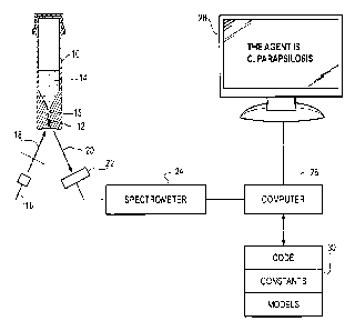
[0015] Figure 1 is a schematic illustration of a measurement apparatus in
which the methods
of this disclosure may be used.
[0016] Figures 2A-2C are a flow chart showing a sequence of processing
instructions which
perform identification and/or characterization of the concentrated microbial
agent using intrinsic
fluorescence measurements.
[0017] Figures 3-8 are plots of intrinsic fluorescence (IF) measurements, and
transforms
thereof which illustrate the benefit of the pre-processing instructions of
Figure 2A in terms of
minimizing strain-to-strain variations within an organism group.
[0018] Figures 9 and 10 are plots of the first derivative of natural logarithm
transforms of IF
measurements showing the discrimination potential between a subset of species
for excitation
wavelengths of 315 and 415 nm.
5a
CA 02798982 2016-11-16
DETAII ;ED DESCRIPTION
[0019] Methods are described herein for identification and or characterization
of a microbial agent. In preferred embodiments, the identification and/or
characterization is performed on a concentrated microbial agent which has been
isolated from other components in a sample. The method can be performed within
while the concentrated microbial agent is stored in a disposable device used
for
separation and concentration of the microbial agent; alternatively it can be
performed
after the microbial agent has been removed from the disposable device.
Examples of
methods, instruments, and devices for separation and concentration of a
microbial
agent in a sample, e.g., blood, are described in the co-pending application
serial
no. 12/800,388, entitled "System for rapid identification and/or
characterization of a microbial agent in a sample, e.g., blood", attorney
docket no. 09-
271-B. Such methods, instruments and
devices are not particularly important to the methods of this disclosure and
therefore a
detailed description is not provided so as to not obfuscate the present
disclosure.
[0020] One representative example of a detection arrangement and disposable
device will be described now in conjunction with Figure 1. Figure 1 is a
schematic
illustration of a measurement apparatus in which the methods of this
disclosure may
be used. The apparatus includes a disposable separation and concentration
device 10
into which a sample 14 containing an unknown microbial agent is placed. The
microbial agent is concentrated into an pellet-like mass 12 using optional
selective
lysis of non-microbial agent components in the sample, (e.g., blood cells) a
density
cushion present in the device 10 and centrifugation. The density gradient and
centrifugation concentrate the microbial agent in the bottom of a capillary
tube 15
present in the device 10.
[0021] The measurement apparatus includes a light source 16 emitting light 18
at an excitation wavelength to stimulate production of intrinsic fluorescence
from the
microbial agent 12. Emission radiation 20 is directed onto a sensor array 22
which is
optionally coupled to a spectrometer 24. Fluorescence emission data in a band
of
wavelengths are sent to a computer 26. The computer is coupled to a memory 30
storing program code (including code executing the sequence of processing
instructions shown in Figures 2A-2C), constants used in the modules, and
models
comprising a list of expected microbial agents and experimentally obtained
fluorescence data in particular excitation and emission pairs which are
discriminatory
6
CA 02798982 2016-11-16
between microorganisms in the manner described below. The computer 26
processes
the data with the aid of the information and code stored in memory 30 and
generates a
classification result which is displayed on an attached workstation display 28
or other
suitable output device, the details of which are not important.
100221 The separation, concentration and interrogation methods are described
in further detail in the following applications,
U.S. Serial No. 12/89,929, entitled "Methods for the isolation
and identification of microorganisms", filed October 30, 2009; US Serial No.
12/589,969, entitled "Separation device for use in the separation,
identification and/or
characterization of microorganisms", filed October 30, 2009; US Serial No.
12/589,952. entitled "Method for separation, identification and/or
characterization of
microorganisms using spectroscopy", filed October 30, 2009; US Serial No.
12/589,936, entitled "Method for separation, identification and/or
characterization of
microorganisms using mass spectrometry", filed October 30, 2009; U.S Serial
No.
12/589,985, entitled "Method for separation and characterization of
microorganisms
using identifier agents", filed October 30, 2009; US Serial No. 12/589,968,
entitled
"Method for detection, identification and/or characterization of
microorganisms in a
sealed container", filed October 30, 2009; US Serial No. 12/589,976, entitled
"Method
for separation, identification and/or characterization of microorganisms using
Raman
spectroscopy", filed October 30, 2009. the present inventive methods are not
limited
to these techniques.
[0023] Once the microorganism or other microbial agent present in the sample
has been isolated and/or pelleted in the separation device 10, the isolated
sample or
pellet is interrogated (e.g., spectroscopically, using intrinsic fluorescence
measurements) as described below to characterize and/or identify the
microorganisms
in the sample or pellet. The interrogation can take place in a non-invasive
manner,
that is, the pellet can be interrogated while it remains in the device 10 used
to separate
and concentrate the microbial agent. The ability to identify the
microorganisms in a
non-invasive manner, optionally coupled with keeping the device 10 sealed
throughout the separation and characterization/identification process and
automating
the procedure avoids the constant handling of contaminated and/or infectious
samples
and greatly increases the safety of the entire process. Furthermore, the
ability to
characterize and/or identify microorganisms by direct interrogation without
further
processing of the sample or pellet 12 (e.g., re-suspension, plating, and
growth of
7
CA 02798982 2012-11-08
WO 2011/149447
PCT/US2010/034929
colonies), greatly increases the speed with which
identification/characterization can
be made.
[0024] In one embodiment, optical spectroscopic methods can be used to
analyze one or more intrinsic properties of the microorganisms, e.g., a
property
present within the microorganism in the absence of additional agents, such as
stains,
dyes, binding agents, etc. In other embodiments, the optical spectroscopic
methods
can be used to analyze one or more extrinsic properties of the microorganisms,
e.g., a
property that can only be detected with the aid of additional agents. The
interrogation
in preferred forms is carried out using fluorescence spectroscopy. For
example, front
face fluorescence (where the exciting and emitted light enters and leaves the
same
optical surface, and if the sample is generally optically thick, the
excitation light
penetrates a very short distance into the sample (see, e.g., Eisinger, J., and
J. Flores,
"Front-face fluorometry of liquid samples," Anal. Biochem. 94:15 (1983)) can
be used
for identification of microorganisms in pellets.
[0025] Typically, the light source 16, or excitation source, results in the
excitation of the sample, followed by measurement of the emission of
fluorescence 20
of the sample at predetermined time points or continuously. Similarly, the
reflected
light from interaction of the excitation source with the sample may be
measured to
provide pertinent data for identification and/or characterization. The
emission from
the sample may be measured by any suitable means of spectral discrimination,
most
preferably employing a spectrometer 24.
[0026] In a presently preferred embodiment, control measurements (e.g.,
fluorescence spectra) are taken for known microorganisms and data stored in
the
memory 30, thus allowing for correlation of measured test data with
characterization
of the microorganisms of interest using various mathematical methods known to
those
skilled in the art. The measured test data from known microorganisms is stored
in
machine-readable memory 30, e.g., within an instrument implementing the method
or
within an associated data processing device, such as connected workstation.
These
methods may be used to classify unknown microorganisms of interest in the
sample
being tested into relevant groups (e.g., species) based on existing
nomenclature,
and/or into naturally occurring groups based on the organism's metabolism,
pathogenicity and/or virulence in designing the system for monitoring,
detecting
and/or characterizing the organism as described previously.
8
CA 02798982 2012-11-08
WO 2011/149447
PCT/US2010/034929
[0027] The sample illumination source (See Figure 1), or excitation source
16, may be selected from any number of suitable light sources as known to
those
skilled in the art. Any portion of the electromagnetic spectrum that produces
usable
data can be used. Light sources capable of emission in the ultraviolet,
visible and/or
near-infrared spectra, as well as other portions of the electromagnetic
spectrum, can
be utilized and are known to those skilled in the art. For example, light
sources may
be continuum lamps such as a deuterium or xenon arc lamp for generation of
ultraviolet light and/or a tungsten halogen lamp for generation of
visible/near-infrared
excitation. These light sources provide a broad emission range and the
spectral
bandwidth for specific excitation wavelengths may be reduced using optical
interference filters, prisms and/or optical gratings, as are well known in the
art.
[0028] Alternatively, a plurality of narrowband light sources, such as light
emitting diodes and/or lasers, may be spatially and/or temporally multiplexed
to
provide a multi-wavelength excitation source. For example, light emitting
diodes are
available from 240 nm to in excess of 900 nm and the sources have a spectral
bandwidth of 20-40 nm (full width at half maximum). Lasers are available in
discrete
wavelengths from the ultraviolet to the near-infrared and can be employed
using
multiplexing methods well known to those skilled in the art.
[0029] The spectral selectivity of any of the light sources may be improved by
using spectral discrimination means such as a scanning monochromator. Other
methods of discrimination may be utilized, as known to those of skill in the
art, such
as an acousto-optic tunable filter, liquid crystal tunable filter, an array of
optical
interference filters, prism spectrograph, etc., and in any combination. A
consideration
in selecting the spectral discriminator takes into the account the range of
tunability as
well as the level of selectivity. By way of illustration, for example, a
discriminator
might utilize the wavelength range of 300 ¨ 800 nm with a selectivity of 10
nm.
These parameters generally determine the optimum technology necessary to
achieve
the tunability range as well as the selectivity.
[0030] Illumination from the light source 16 results in the excitation of the
sample, followed by measurement of the emission of fluorescence of the sample
at
predetermined time points or continuously. Similarly, the reflected light from
interaction of the excitation source with the sample may be measured to
provide
pertinent data for detection and/or characterization.
9
CA 02798982 2012-11-08
WO 2011/149447
PCT/US2010/034929
[0031] The emission from the sample may be measured by any suitable means
of spectral discrimination, most preferably employing a spectrometer 24. The
spectrometer may be a scanning monochromator that detects specific emission
wavelengths whereby the output from the monochromator is detected by a
photomultiplier tube and/or the spectrometer may be configured as an imaging
spectrograph whereby the output is detected by an imaging detector array such
as a
charge-coupled device (CCD) detector array. In one embodiment, a discriminator
allows the observation of the fluorescence and/or scattering signal by a
photodetection
means (such as a photomultiplier tube, avalanche photodiode, CCD detector
array,
and/or electron multiplying charge coupled device (EMCCD) detector array).
[0032] The spectroscopic technique is used to obtain measurements that are
preferably provided as Excitation-Emission Matrix (EEM) measurements. As used
herein, EEM is defined as the luminescent spectral emission intensity of
fluorescent
substances as a function of both excitation and emission wavelength, and
includes a
full spectrum or a subset thereof, where a subset may contain a single or
multiple
excitation/emission pairs(s). Additionally, a cross section of the EEM with a
fixed
excitation wavelength may be used to show the emission spectra for a specific
excitation wavelength, and a cross section of the EEM with a fixed emission
wavelength may be used to show the excitation spectra for a sample. In one
embodiment, multiple EEMs are measured at more than one specific excitation-
emission wavelength pair, e.g., at least at 2, 3, 4, 5, 6, 7, 8, 9, 10, or
more specific
excitation-emission wavelength pairs.
[0033] It has been found that a front-face fluorescence spectroscopy provides
an advantage in measuring the fluorescence and/or reflectance properties of
highly
scattering and highly quenching samples. In one embodiment, the front-face
method
may be particularly useful. For example, front-face fluorescence may be
particularly
useful in highly absorbent samples because the excitation and emission beam
does not
need to travel through the bulk of the sample, and thus, may be less affected
by the
interfering components that may be contained therein (e.g., blood cells and
microbiological culture media). The optical surface of the separation device
1904
may be illuminated at such an angle as to provide acceptable results as known
to those
skilled in the art, (e.g., Eisinger, J., and J. Flores, "Front-face
fluorometry of liquid
samples," Anal. Biochem. 94:15-21 (1983)). In one embodiment, the system is
designed such that the spectroscopic system measures diffuse reflected light
at a
CA 02798982 2012-11-08
WO 2011/149447
PCT/US2010/034929
minimum of one fixed angle in addition to measuring emitted fluorescence at a
minimum of one fixed angle.
[0034] In some embodiments, characterization and/or identification of the
microorganisms in the isolated sample or pellet need not involve
identification of an
exact species. Characterization encompasses the broad categorization or
classification
of biological particles as well as the actual identification of a single
species.
Classification of microorganism from an isolated sample or pellet may comprise
determination of phenotypic and/or morphologic characteristics for the
microorganism. For example, characterization of the biological particles may
be
accomplished based on observable differences, such as, composition, shape,
size,
clustering and/or metabolism. In some embodiments, classification of the
biological
particles of interest may require no prior knowledge of the characteristics of
a given
biological particle but only requires consistent correlations with empiric
measurements thus making this method more general and readily adaptable than
methods based on specific binding events or metabolic reactions. As used
herein
"identification" means determining to which family, genus, species, and/or
strain a
previously unknown microorganism belongs to. For example, identifying a
previously unknown microorganism to the family, genus, species, and/or strain
level.
[0035] In some instances, characterization encompasses classification models
which provide sufficient useful information for action to be taken. As used
herein, the
preferred classification models comprise grouping into one or more of the
following:
(1) Gram Groups; (2) Clinical Gram Groups; (3) Therapeutic Groups; (4)
Functional
Groups; and (5) Natural Intrinsic Fluorescence Groups.
[0036] (1) Gram Groups:
Within the Gram Groups classification,
microorganisms may be placed into one of three broad classification categories
based
on their Gram staining reaction and overall size, said groups selected from
one or
more of the following: (a) Gram positive microorganisms that stain dark blue
with
Gram staining; (b) Gram negative microorganisms that stain red with Gram
staining;
and (c) yeast cells that stain dark blue with Gram staining, but are very
large rounded
cells that are distinguished from bacteria by their morphological
characteristics and
size.
[0037] (2) Clinical Gram Groups: The Gram Groups may be further divided
into several sub-categories representing distinguishing morphological
features. These
sub-categories comprise all the relevant clinical information reported by an
11
CA 02798982 2012-11-08
WO 2011/149447
PCT/US2010/034929
experienced laboratory technologist, and thus provide a higher level of
identification
than a positive or negative Gram reaction. This particular classification is
very
helpful because it eliminates concerns about relying on the quality of a Gram
stain
and/or the skill level of the technician reading the smear by providing the
equivalent
clinically relevant information with an automated system. More specifically,
subcategories of microorganisms based on this classification model may be
selected
from one or more of the following: (a) cocci, which are small rounded cells;
(b)
diplococci, which are two small rounded cells joined together; (c) rods, which
are
rectangular shape; and (d) bacilli, which are rod shaped. Examples of these
sub-
categories that can be ascertained by additional morphological information
include: (i)
Gram positive cocci; (ii) Gram positive cocci in chains; (iii) Gram positive
cocci in
clusters (i.e., "grape-like" clusters); (iv) Gram positive diplococci; (v)
Gram positive
rods; (vi) Gram positive rods with endospores; (vii) Gram negative rods;
(viii) Gram
negative coccobacilli; (ix) Gram negative diplococci; (x) yeast; and (xi)
filamentous
fungi.
[0038] (3) Therapeutic Groups: The therapeutic groups comprise multiple
microbial species that, when isolated from particular specimen types, are
treated with
the same class of antibiotics or mixture of antibiotics (e.g., as described in
"Sanford
Guide to Antimicrobial Therapy 2008"). In many cases, identity to the species
level is
not required by the clinician to enable a change from initial empiric therapy
to a more
targeted therapy because more than one species can be treated with the same
choice of
antibiotic(s). This classification level correctly places these "same-
treatment"
microorganisms into single therapeutic categories. Examples of this
characterization
level include the ability to distinguish highly resistant Enterobacteriacae
(EB) species
from sensitive EB species (Enterobacter spp. from E. coli), or fluconazole-
resistant
Candida species (C. glabrata and C. kruzei) from sensitive Candida species (C.
albicans and C. parapsilosis), and so on.
[0039] (4) Functional Groups: According to the invention, microorganisms
may also be placed into several groups based upon a mixture of metabolic,
virulence
and/or phenotypic characteristics. Non-fermentative organisms may be clearly
distinguished from fermentative ones. Furthermore, microorganism species that
produce hemolysins may be grouped separately from non-hemolytic species. In
some
cases, these groups represent broader categories than genus level (e.g.,
coliforms,
Gram negative non-fermentative rods), some at the genus level (e.g.,
Enterococcus,
12
CA 02798982 2012-11-08
WO 2011/149447
PCT/US2010/034929
Candida), and some with closer to species-level discrimination (e.g.,
coagulase-
negative staphylococci, alpha-hemolytic streptococci, beta-hemolytic
streptococci,
coagulase-positive staphylococci, i.e., S. aureus).
[0040] (5) Natural Intrinsic Fluorescence ("IF") Groups: Microorganisms
may also be placed into categories based on their natural tendency to group
together
by their innate and/or intrinsic fluorescence characteristics. Some of these
groups
may be common to Therapeutic and Functional Group categories. These groupings
may comprise individual species, such as E. faecalis, S. pyo genes, or P.
aeruginosa
that have characteristic IF signatures and/or may contain small groups of
organisms
with relatively conserved IF signatures such as the K. pneumoniae- K. oxytoca
or E.
aerogenes-E. cloacae groups.
[0041] In addition to measuring intrinsic properties of microorganisms (such
as intrinsic fluorescence) for identification purposes, the methods may use
additional
identifier agents to aid in the separation and/or identification process.
Agents that
bind to specific microorganisms, such as affinity ligands, can be used to
separate
microorganisms, to identify a class or species of microorganism (e.g., through
binding
to a unique surface protein or receptor) and/or to identify a characteristic
of the
microorganism (e.g., antibiotic resistance). Useful identifier agents include,
without
limitation, monoclonal and polyclonal antibodies and fragments thereof (e.g.,
anti-Eap
for S. aureus identification), nucleic acid probes, antibiotics (e.g.,
penicillin,
vancomycin, polymyxin B), aptamers, peptide mimetics, phage-derived binding
proteins, lectins, host innate immunity biomarkers (acute phase proteins, LPS-
binding
protein, CD14, mannose binding lectin, Toll-like receptors), host defense
peptides
(e.g., defensins, cathelicidins, proteogrins, magainins), bacterocins (e.g.,
lantibiotics,
such as nisin, mersacidin, epidermin, gallidermin, and plantaricin C, and
class II
peptides), bacteriophages, and dyes selective for nucleic acids, lipids,
carbohydrates,
polysaccharides, capsules/slime or proteins, or any combination thereof If the
agent
does not itself give out a detectable signal, the agent can be labeled to
provide a
detectable signal, such as by conjugating the agent to a marker (e.g., visible
or
fluorescent). Markers include, without limitation, fluorescent, luminescent,
phosphorescent, radioactive, and/or colorimetric compounds. The agent can be
added
to the microorganisms at any step in the methods of the invention, e.g., when
the
sample is obtained, during lysis, and/or during separation. In some
embodiments, the
presence of the agent in the pellet can be determined during interrogation of
the pellet.
13
CA 02798982 2012-11-08
WO 2011/149447
PCT/US2010/034929
Other useful identifier agents include substrates for microbial enzymes,
chelating
agents, photosensitizing agent, quenching agent, reducing agent, oxidizing
agent,
buffer, acid, base, solvent, fixative, detergents, surfactants, disinfectants
(eg. alcohols,
bleach, hydrogen peroxide) and toxic compounds (eg. sodium azide, potassium
cyanide) and metabolic inhibitors such as cyclohexamide, etc. Similarly, many
fluorescent compounds for measuring microbial cell viability, metabolism
and/or
membrane potential may be used as an identifier agent in the present
invention. As
would be readily appreciated by one of skill in the art, the sensitivity of a
particular
microorganism to any compound affecting its physical state or metabolism, such
as an
antibiotic, could be rapidly ascertained by adding the compound to the sample,
lysis
buffer, density cushion or any mixture thereof
[0042] An embodiment of a method for performing identification and/or
characterization of microbial agents in samples using intrinsic fluorescence
and a
hierarchical taxonomic classification process will now be described in
conjunction
with Figures 2-10. Basically, the method can be embodied as a sequence of
processing instructions stored in memory and executed using a conventional
data
processor or computer 26. The processing instructions execute an algorithm
shown
in Figures 2A-2C which is designed to provide the identification of a blood
culture
isolate (concentrated pellet) given an intrinsic fluorescence (IF) scan of the
isolate
from a predefined set of emission wavelengths. The algorithm can be adapted
for
other types of analytical test data (e.g., Raman scattering or mass
spectrometry).
[0043] In preferred embodiments, the method is encoded as software
instructions implementing a multi-level identification algorithm.
Traditional
classification algorithms that take input data and determine the
identification of a
microorganism use a single classification model. Given data from an intrinsic
fluorescence scan at a predefined set of wavelengths of an unknown organism,
the
multi-leveled identification algorithm classifies the organism following the
branches
of a taxonomic hierarchy ¨ Gram class, family, and species. A unique feature
is the
use of separate classification models at each identification step from
highest, Gram
class, to lowest, species. Additionally, the approach incorporates the use of
parallel
classification models to evaluate consistency between results. Thus, the
probability of
accurate identification and/or characterization is maximized, and generation
of
incorrect identification or characterization results is minimized.
14
CA 02798982 2012-11-08
WO 2011/149447 PCT/US2010/034929
[0044] The identification method includes a set of data pre-processing steps
(shown as blocks 5102, 5104 and 5106 of Figure 2A, and a set of analysis steps
(the
remaining blocks 5108, 5110, etc. in Figures 2B, 2C). The method determines
the
identification of the organism at multiple levels of the taxonomic hierarchy.
The pre-
processing steps are designed to acquire IF scan data and perform data
transformations that minimize variation between different strains of a
microbial agent
within a given organism group or species. The data analysis steps implement a
multi-
level identification using parallel classification models, as will be
understood from the
following discussion.
[0045] As noted above, preferred embodiments provide an organism
identification at the Gram, family, and species levels. Organisms commonly
found in
blood cultures that can be identified by the algorithm include, but not
necessarily
limited to, those listed in Table 1. Obviously, for different applications
(e.g., food,
water, environmental samples, etc.) the organisms may differ from those listed
in
Table 1, however the methodology is the same.
(a) Table 1: Intrinsic Fluorescence Algorithm Identification
Organism List
Article II. Gram Class Family Species
C. freundii
E. aerogenes
E. cloacae Complex
E. coli
K. oxytoca
K. pneumoniae
Enterobacteriaceae
M. morganii
P. mirabilis
Gram-negative P. stuartii
P. vulgaris
S. enteritidis
S. marcescens
Moraxellaceae A. baumanii
Neisseriaceae N. meningitidis
Pasteurellaceae H. influenzae
Pseudonomadaceae P. aeruginosa
Xanthomonadaceae S. maltophilia
E. faecalis
Enterococcaceae
E. faecium
Listeriaceae L . mono cytogenes
S. aureus
Gram-positive
S. capitis
Staphylo co cc aceae S. epidermidis
S. hominis
S. lugdunensis
CA 02798982 2012-11-08
WO 2011/149447 PCT/US2010/034929
S. warneri
S. agalactiae
S. bovis
Strepto co c cace ae S. mitis / S. oralis
S. pneumoniae
S. pyogenes
C. albicans
C. glabrata
Yeast Ascomycetes C. krusei
C. parapsilosis
C. tropicalis
[0046] The processing steps or modules shown in Figures 2A-C will now be
described in detail.
Pre-processing
[0047] Step 5102: Obtain a fluorescence value, nj, for each excitation value,
i
= 1,2, ..., x , and each emission, j = 1,2, ..., y , combination. The ratio,
emission
value/excitation value, must fall within the interval (1.05, 1.95).
[0048] Step 5104: For each fluorescence value, nii, calculate the natural
logarithm value, ln (n).
[0049] Step 5106: Calculate the 1st derivative of the natural log transform
value (from step 5104) for each emission value, j = 2, 3, ..., y-1, across a
given
excitation wavelength, i.
[0050] It is advantageous to transform the raw fluorescence data to minimize
strain-to-strain variation within each organism group, using both steps 5104
and 5106.
Additionally, the transformation process tends to create similar variance
across
organism groups. Figures 3, 4 and 5 illustrate by way of example the effects
of
performing the described pre-processing for multiple strains of Staphylococcus
aureus
evaluated across the emission range at excitation 315. In Figure
3, each line
represents the fluorescence signal from a single strain. The line 5202
indicates the
mean fluorescence signal at each emission value. Figure 4 shows the strain-to-
strain
variation in the fluorescence signal after application of the natural
logarithm
transformation (step 5104); note that the curve for all of the strains are
close together.
Figure 5 shows the strain-to-strain variation at excitation of 315 nm after
calculation
of the first derivative of the natural logarithm transform (step 5106). Again,
note that
16
CA 02798982 2012-11-08
WO 2011/149447
PCT/US2010/034929
the curve for all the strains are very close together, particularly at the
emission range
of 400-610 nm.
[0051] As another example, Figure 6 shows the strain-to-strain variation in
the
fluorescence signal at excitation of 415 nm for Candida parapsilosis, prior to
performing the transformation steps. Note the wide variation in emission in
the range
of 400-650 nm. Strain-to-strain variation for this organism at excitation of
415 nm
after performing the natural logarithm transformation is shown in Figure 7.
Strain-to-
strain variation after performing the first derivative transformation is shown
in Figure
8. Note that in Figure 8 the strain-to-strain variation is much reduced.
Analysis
[0052] Step 5108: The
first level of classification in the analysis after
performing the pre-processing steps is gram classification 5108. At this step,
the
processing includes two branches, one represented by steps 5110 and 5112 and
another represented by steps 5114 and 5116. Figure 2A is not meant to imply
that the
branches could not be performed sequentially; the branches could be performed
either
sequentially or in parallel.
[0053] Step 5110: Gram Classification Distance Calculation. Using the 1st
derivative transforms for a predefined set of excitation / emission pairs,
calculate the
distance,
da Rm may E-1 (m ma)] 1/2
for each Gram class defined in the model
where
- a = 1, 2, 3, represents the Gram classes defined in the model
- m represent the vector of calculated values of the et derivative, mu , of
the natural
log transform for each excitation / emission pair i, j
- ma represent the vector of mean values ma( from a distribution for each
class a at
excitation / emission pair i, j
- t represent the transpose of the vector
- (m ¨ ma) represent the vector of differences mu ¨ ma(u) for each
excitation /
emission pair i, j
17
CA 02798982 2012-11-08
WO 2011/149447
PCT/US2010/034929
- E-/ represents the inverse of the covariance matrix for the predefined
set of
excitation / emission pair. The set of excitation and emission pairs are
experimentally determined from fluorescence measurements (with preprocessing
performed) of known microorganisms (see Figures 9 and 10 and the discussion
below).
[0054] The term "model" is used to refer to a set of known microbial agents
for which IF measurements (including transforms) at the predetermined
excitation
wavelengths have been previously obtained and for which a specimen is a
candidate
for classification, e.g., the agents listed in Table 1.
[0055] Step 5112: Gram Classification Interpretation.
- Let ug represent the maximum distance threshold
- If all distances, di, d2, and d3, are greater than ug, the classification
result is
Unknown
- Else, determine the value of dmm, the minimum value of di, d2, and d3
- Let wg represent the low discrimination threshold factor
- If more than one distance, di, d2, and d3, is less than (d..* wq), the
classification
result is Low Discrimination between the Gram classes having distances less
than
(dm.,* wq)
- If only one distance, di, d2, and d3, is less than (411.* wq), the
classification result
is the corresponding Gram class.
[0056] Step 5114: All Families Classification Distance Calculation
Using the 1st derivative transforms for a predefined set of excitation /
emission
pairs, calculate the distance,
da Rm may E-1 (m ma)] v2
for each organism family defined in the model
where
- a = 1, 2, ...,k, represents all of the organism families defined in the
model
- E-/ represents the inverse of the covariance matrix for the predefined set
of
excitation / emission pairs (same remark as above, the set of excitation and
emission pairs are experimentally determined)
- m represent the vector of calculated values of the 1st derivative, mu ,
of the natural
log transform for each excitation / emission pair i, j
18
CA 02798982 2012-11-08
WO 2011/149447
PCT/US2010/034929
- ma represent the vector of mean values mum from a distribution for each
class a at
excitation / emission pair i, j
- t represent the transpose of the vector
- (m ¨ ma) represent the vector of differences mu ¨ ma(u) for each
excitation /
emission pair i, j
[0057] Step 5116: All Families Classification Interpretation
- Let uf represent the maximum distance threshold
- If all
distances, d1, d2, da, are greater than uf, the classification result is
Unknown
- Else, determine
the value of dim., the minimum value of d1, d2, da
- Let wf represent the low discrimination threshold factor
- If more than one distance, d1, d2, da, is less than (dmin wf), the
classification
result is Low Discrimination between the organism families having distances
less
than (dm,.. wf)
- If only one distance, d1, d2, da, is less than (dmq, wq), the
classification result
is the corresponding family.
[0058] Step 5118: Pooling gram and all families classification interpretations
for final gram classification result.
[0059] If the Gram classification is a single choice and the all families
classification is a single choice, the pooled classification result is the
indicated Gram
class if the family classification falls under the taxonomic hierarchy of the
Gram
class.
[0060] If the Gram classification is a single choice and the all families
classification is a single choice, the pooled classification result is Unknown
if the
family classification does not fall under the taxonomic hierarchy of the Gram
class.
[0061] If the Gram classification is a single choice and the all families
classification is a low discrimination, the pooled classification is the
indicated Gram
class if the family associated with the shortest distance falls under the
taxonomic
hierarchy of the Gram class.
[0062] If the Gram classification is a single choice and the all families
classification is a low discrimination, the pooled classification is Unknown
if the
family associated with the shortest distance does not fall under the taxonomic
hierarchy of the Gram class.
19
CA 02798982 2012-11-08
WO 2011/149447
PCT/US2010/034929
[0063] If the Gram classification is a low discrimination and the all families
classification is a single choice, the pooled classification result is the
Gram class that
corresponds to the Gram class under which the family resides on the taxonomic
hierarchy.
[0064] If the Gram classification is a low discrimination and the all families
classification is a single choice, the pooled classification result is Unknown
if none of
the Gram classes correspond to the Gram class under which the family resides
on the
taxonomic hierarchy.
[0065] If the Gram classification and the all families classification are both
Unknown, the pooled classification result is Unknown.
[0066] The processing then proceeds to step 5120, Gram Family
Classification, a second, lower, level of classification in a taxonomic
hierarchy. This
step consists of sub-steps 5122, 5124 and 5126.
[0067] Step 5122: Gram family classification distance calculation.
Using the 1st derivative estimates for a predefined set of excitation /
emission
pair that are specific to the Gram classification result, calculate the
distance,
= [(m ¨ma) E-1 ¨ ma)] 1/2
for each organism family defined in the model,
where
- a = 1, 2, ..., k, represents the number of organism families defined in
the model
- E-/ represents the inverse of the covariance matrix for the predefined
set of
excitation / emission pairs (same remark as before regarding the pairs)
- m represents the vector of calculated values of the et derivative, mu ,
of the
natural log transform for each excitation / emission pair i, j
- ma represent the vector of mean values ma( from a distribution for each
class a at
excitation / emission pair i, j
- t represent the transpose of the vector
- (m ¨ ma) represent the vector of differences mu ¨ ma(u) for each
excitation /
emission pair i, j
[0068] Step 5124: Gram Family Classification Interpretation
Let ut represent the maximum distance threshold
If all distances, d1, d2, da, are greater than ut, the classification
result is Unknown
CA 02798982 2012-11-08
WO 2011/149447
PCT/US2010/034929
Else, determine the value of dn., the minimum value of di, d2, da
Let wt represent the low discrimination threshold factor
If more than one distance, di, d2, da,
is less than (dm., wt.), the classification result
is Low Discrimination between the organism families having distances less than
(dm,.
wit)
If only one distance, di, d2, d.,
is less than (dmin wt.), the classification result is the
corresponding family.
[0069] Step 5126 Gram Family Classification Result.
[0070] If the Gram family classification result is Unknown, the test organism
classification is finalized at the Gram level.
[0071] If the Gram family classification result is Low Discrimination, the
test
organism classification is finalized as the Gram and families included in the
low
discrimination.
[0072] If the Gram family classification result a single family, the IF data
from
the test organism are further analyzed to determine if a species level
identification can
be determined.
[0073] Step 5128 Gram family Species Classification. The
processing
instructions proceed to a gram family species classification level, a third
and even
lower level of classification in a taxonomic hierarchy, consisting of sub-
steps 5130,
5132, and 5134.
[0074] Step 5130 Gram family species classification distance calculation.
[0075] Using the et derivative estimates for a predefined set of excitation /
emission pair that are specific to the Gram family classification result,
calculate the
distance,
da Rm mot E-1 on ma)] 1/2
for each organism species defined in the model,
where
- a = 1, 2, ..., k, represents the number of organism species defined in the
model
- E-/ represents the inverse of the covariance matrix for the predefined
set of
excitation / emission pairs (same remark as before)
- m represents the vector of calculated values of the 1st derivative, mu ,
of the
natural log transform for each excitation / emission pair i, j
21
CA 02798982 2012-11-08
WO 2011/149447
PCT/US2010/034929
- ma represent the vector of mean values mum from a distribution for each
class a at
excitation / emission pair i, j
- t represent the transpose of the vector
- (m ¨ ma) represent the vector of differences mu ¨ ma(u) for each
excitation /
emission pair i, j
[0076] Step 5132 Gram family species classification interpretation.
- Let us represent the maximum distance threshold.
- If all distances, d1, d2, da, are greater than
ut, the classification result is
Unknown.
- Else, determine the value of dim., the minimum value of dt, d2,
- Let ws represent the low discrimination threshold factor.
- If more than one distance, d1, d2, da, is less than (dm., ws), the
classification
result is Low Discrimination between the organism species having distances
less
than (dm,t, ws)
- If only one distance, d1, d2, da, is less than (dm., wt), the
classification result is
the corresponding species.
[0077] Step 5134 Gram family species classification result.
[0078] If the Gram family species classification result is Unknown, the test
organism classification is finalized at the Gram and family level.
[0079] If the Gram family species classification result is Low Discrimination,
the test organism classification is finalized as the Gram, family, and species
included
in the low discrimination.
[0080] If the Gram family species classification result a single species, the
test
organism classification is finalized at the Gram, family, and species level.
[0081] At step 5136, the results determined at steps 5134, 5118, and 5126 are
returned and reported to the user, e.g., on a user interface for the
identification
instrument, transmitted to an attached workstation, returned to another
software
module, or otherwise generated for the user.
[0082] In regards to organism identification (step 5134), discrimination
between species is possible only if the values of the first derivative (of the
natural
logarithm transform of the emission value) are unique for each species in the
model at
some portion of the emission range for at least one excitation wavelength.
Figures 9
and 10 illustrate the discrimination potential between a subset of species for
excitation
22
CA 02798982 2012-11-08
WO 2011/149447
PCT/US2010/034929
wavelengths 315 nm (Figure 9) and 415 nm (Figure 10). Referring to Figure 9,
it is
apparent that several of the species can be discriminated from the others
based on the
first derivative at excitation wavelength 315. The mathematical model uses the
first
derivative values (of natural log transform) for emissions where visual
differences
exist as inputs to discriminate between species. Using selected sections of
values
across the emission range the following species can be clearly discriminated
from the
others: E. coli, H. influenzae, P. aeruginosa, and S. pneumoniae. In addition,
S.
aureus and S. epidermidis can be discriminated from other species but not each
other.
The sections of values across the emission range at a given excitation
wavelength are
the predefined pairs in the inverse matrices E-/ in the distance calculations
in the
processing steps described above. These pairs may for example be excitation at
315
nm and the range of emission values indicated by the circles shown in Figure
9, i.e.,
(315/300-450), (315, 485-500), (315/570-580).
[0083] Referring to Figure 10, it is apparent that the emissions at excitation
wavelength 415 nm has the ability to discriminate between species. Using
selected
sections of values across the emission range C. parasilopsis and P. aurginosa
can be
clearly discriminated from the other species. It is also of interest to note
the
difference between first derivative values for S. aureus and S. epidermidis
that occurs
around emission 450 nm. When the information from the selected sections of
values
across the emission range for wavelengths 315 and 415 (Figures 9 and 10) is
combined, all of the species in the model can be discriminated from each other
at a
high rate (> 97% reliability).
[0084] To enhance fluorescence signals, microorganisms could either be
coated with gold and/or silver nanoparticles prior to
centrifugation/concentration,
and/or the inner optical surface could be pre-coated with metal colloids of
particular
size and shape (refs: Lakowicz, Anal. Biochem. 337:171 (2005) for
fluorescence;
Efrima et al., J. Phys. Chem. B. (Letter) 102:5947 (1998) for SERS). In
another
embodiment, the nanoparticles are present in a density cushion present in the
separation device prior to centrifugation and associate with microorganisms as
the
microorganisms pass through the density cushion.
[0085] The taxonomic hierarchical classification method explained above in
the context of Figures 2-10 is applicable to other data sets obtained from
interrogation
of a microbial agent. For example, the classification method would be equally
useful
23
CA 02798982 2016-11-16
in the case of that Raman scattering data or mass spectrometry data is
obtained from a
concentrated microbial agent instead of intrinsic fluorescence data. In the
case of
Raman scattering, data is obtained from known microbial agents and such data
is
analyzed (typically after transform steps are performed) to determined subsets
of the
data that are discriminatory between Gram, family and species and the results,
i.e.,
discriminatory subsets stored. Similarly, data from an unknown microbial agent
is
subject to a transformation steps to minimize strain-to-strain variation
between
species; the transformation may be natural logarithm, first derivative, or
other
transform, the selection and details of the transformation will be within the
ability of
persons skilled in the art based upon examination of the data for known
microbial
agents. The processing of Figures 2A-2DC (hierarchical classification at the
Gram,
Family and Species level) then proceeds. Alternatives to the minimum distance
calculation used for classification, such as the well-known K-Nearest Neighbor
classification algorithm, may be used for classification of the test sample at
each
hierarchical level. It will also be apparent that additional pre-processing
steps may be
required which are not shown in the flow chart of Figures 2A-2C, that are
unique to
the analytic test method, such as background subtraction or normalization, but
these
steps are known in the art and therefore a detailed description is not
necessary.
[0086] Generalizing the foregoing, we have described a method for rapid
identification and/or characterization of a microbial agent present in a
sample,
comprising the steps of:
obtaining analytic test data of the microbial agent; transforming the analytic
test data,
thereby minimizing strain to strain variations in analytic test data within an
organism
group; and with the aid of a programmed computer, performing a multi-level
classification algorithm coded as a set of processing instructions operating
on the
transformed analytic test data, the multiple levels corresponding to different
levels in
a taxonomic hierarchy for microbial agents suspected of being in the sample.
In
some embodiments the analytic test data (e.g., Raman scattering) is performed
while
the microbial agent is concentrated within a test device in which the agent
was
separated and concentrated, as shown in Figure 1; in other embodiments the
concentrated agent is removed from the test device and subject to analysis is
a
separate instrument, e.g., mass spectrometer. Further examples of analytical
methods
which may be used are disclosed in U.S. Patent 6,780,602.
24
CA 02798982 2012-11-08
WO 2011/149447
PCT/US2010/034929
[0087] Variation from the specifics from the disclosed embodiments are of
course possible without departure from the scope of the invention. All
questions
concerning scope are to be answered by reference to the appended claims.
CA 02798982 2016-11-16
IDENTIFICATION AND/OR CHARACTERIZATION OF A
MICROBIAL AGENT USING TAXONOMIC HIERARCHICAL
CLASSIFICATION
CROSS-REFERENCE TO RELATED APPLICATIONS
[0088] This application is related to the following US patent applications:
[0089] US Serial No. 12/589,929, entitled "Methods for the isolation and
identification of microorganisms", filed October 30, 2009.
[0090] US Serial No. 12/589,969, entitled "Separation device for use in the
separation, identification and/or characterization of microorganisms", filed
October
30, 2009.
[0091] US Serial No. 12/589,952, entitled "Method for separation,
identification and/or characterization of microorganisms using spectroscopy",
filed
October 30, 2009.
[0092] US Serial No. 12/589,936, entitled "Method for separation,
identification and/or characterization of microorganisms using mass
spectrometry",
filed October 30, 2009.
[0093] US Serial No. 12/589,985, entitled "Method for separation and
characterization of microorganisms using identifier agents", filed October 30,
2009.
[0094] US Serial No. 12/589,968, entitled "Method for detection,
identification and/or characterization of microorganisms in a sealed
container", filed
October 30, 2009.
[0095] US Serial No. 12/589,976, entitled "Method for separation,
identification and/or characterization of microorganisms using Raman
spectroscopy",
filed October 30, 2009.
[0096] This application is also related to the following application filed on
the
same date as this application:
[0097] Attorney Docket no. 09-271-B, entitled "System for rapid
identification and/or characterization of a microbial agent in a sample," U.S.
serial no.
12/800,388, filed May 14, 2010.
[0098] Attorney Docket no. 09-271-A, entitled "Methods for rapid
identification and/or characterization of a microbial agent in a sample," U.S.
serial no.
12/800,387, filed May 14, 2010.
26