Note: Descriptions are shown in the official language in which they were submitted.
11-
WO 2011/146139 PCT/US2011/000911
1
ULTRASONIC TRANSDUCER ASSEMBLY
FIELD OF THE INVENTION
This invention relates to an ultrasonic transducer assembly. The invention is
particularly useful in medical diagnostic and therapeutic applications.
BACKGROUND OF THE INVENTION
Ultrasound is widely used in modem medicine for diagnostics and minimally
invasive
treatment in such fields as obstetrics, cardiology, endocrinology,
gastroenterology, neurology,
ophthalmology, urology, osteoporosis, and clinical diagnostics. Ultrasound
diagnostics uses
low-power ultrasonic scanners for investigation and visualization of inner
organs, tissue
layers and structures, for determination of blood flow direction and velocity,
for measurement
of density and other parameters of tissues, and for detection of cancer and
other tumors. In
diagnostics, acoustic lenses have been traditionally used in pulse mode to
manipulate the
wave front propagation delays. In therapeutic applications, continuous
ultrasound waves
with an average acoustic intensity of up to several watts per centimeter
square at the
transducer surface are typically used to focus ultrasound. The focused
ultrasonic waves
produce highly localized and intense acoustic fields, up to several hundreds
of watts in power
density, and enable controlled, deep-reaching and localized treatment of
malignant tissues,
with few secondary effects for surrounding health tissues. It is beneficial to
control
ultrasonic energy deposition for quickly overheating target focal tissue while
minimizing the
impact on surrounding non-targeted tissues. The mastery of focusing determines
the success
of therapy and requires an understanding of the vibration condition of the
radiating surface
and thermal and mechanical constraints. Because acoustic focusing is an
interference
phenomenon, the phase of individual ultrasound rays becomes a controlling
factor in a
continuous therapy mode. In a diagnostic imaging mode, focusing limits the
beam width and
constrains the acoustic energy content of the beam to a smaller cross
sectional area, hence
improving imaging sensitivity. In this mode, the beam is typically focused
using a fixed lens
that just bends acoustic rays and preserves the pressure-time waveform of
incoming signals.
Imaging lenses are used in pulsed mode where their function relies primarily
on determining
and manipulating the wave front propagation delays. For therapeutics, the mode
of operation
is typically continuous wave, in which case the phase becomes an important
lens design
factor as opposed to wave front propagation delay. Traditional convex or
concave lenses
(Folds, Focusing properties of solid ultrasound cylindrical lenses, 53, 3, pp
826-834, 1973)
that converge light rays towards the lens principal axis offer a simple method
to focus low
power acoustic energy in both therapy and imaging. However, high acoustic
absorption in
11-
WO 2011/146139 PCT/US2011/000911
2
thicker regions of the lenses and excessive heat build up result in a poor
lens longevity and
large focusing aberration when attempts are made to focus high power acoustic
energy in a
continuous regime. Hence, thin focusing lenses with discrete phase shifts are
both
permissible and beneficiary in therapy, greatly reducing overall lens depth
profile and
allowing different designs, including zone plate Fresnel (Hadimioglu et al,
1993), multilevel
(Chan et al, Finite element analysis of multilevel acoustic Fresnel lenses,
Vol 43, 4, 1996),
field conjugate (Lalonde and Hunt, Variable frequency field conjugate lenses
for ultrasound
hyperthermia, 42, 5, 825-831, 1995) and other designs (Rosenberg, High
intensity ultrasound,
Moscow, pp 69-91, 1949; Tarnoczy, Sound focusing lenses and waveguides,
Ultrasonics,
115-127, 1965).
Discrete phase acoustic focusing lenses in combination with flat transducers
or arrays
offer an elegant and cost effective solution for hyperthermia treatment of
cancer and tumors,
where the tissue is heated using ultrasound to temperatures of 43 - 45 C for
several minutes.
It is well known that tumor cells become much more susceptible to radiotherapy
and
chemotherapy under elevated temperature. In physiotherapy lens focused
ultrasound may be
used to increase the elasticity of sinews and scars, improve the mobility of
joints, provide
analgesic effects, alter blood flow, and produce muscular spasms. High
intensity ultrasound
(10 - 2000 W/cm2) is used for tissue ablation, cutting, fractionation
(histotripsy) and for
arresting internal bleeding (hemostasis). Historically, piezoelectric and
magnetostrictive
transducers are widely used to transform generate a high intensity ultrasound
field.
In therapeutic applications the precision targeting of deep tissues is
important.
Desired therapeutic effect must be confined to a small spot within the body
where
temperature elevation is sufficient to create a localized tissue impact
without affecting
surrounding tissue and organs. This technique is used to selectively destroy
the unwanted
tissue within the body without perturbing adjacent tissues. Typically, heating
the tissue to
60 C - 80 C results in tissue necrosis, a process commonly termed as thermal
ablation. In
most cases, the high intensity focused ultrasound is used in thermal ablation
procedures.
Ultrasound focusing can be achieved by having concave focused transducers
producing
convergent beams of predetermined geometry and/or by manipulating the driving
electrical
signals (phase and amplitude) of multiple active transducers (Cathignol, 2002,
High Intensity
Piezoelectric Sources for Medical Applications: Technical Aspects, Nonlinear
Acoustics at
the Beginning of the 21st Century, 1, 371-378.). Single focused elements are
more
economical but require mechanical steering and suffer a loss of acoustic
efficiency due to
heating and presence of parasitic surface waves (Kluiwstra et al., 1997,
Design Strategies for
11-
WO 2011/146139 PCT/US2011/000911
3
Therapeutic Ultrasound Phased Arrays, SPIE International Medical Imaging
Symposium,
Chapelon et al. Transducers for therapeutic ultrasound, Ultrasound in Med. &
Biol., Vol. 26,
No. 1, pp. 153-159, 2000).
Ultrasound systems use relatively small, low-power transducers for diagnostic
visualization and large high-power transducers for therapy. Typically, the
radiation surfaces
of the two types of transducers coincide and often form a surface of
revolution of a conic
section: circle, ellipse or parabola. Transducers with large radiating
surfaces are used to
generate sufficient acoustic power and are expensive to manufacture.
Additionally, the
applicability of large concave transducers is limited to an open field
clinical cases, where the
size of the transducer does not matter, as opposed to the most intra-luminal
or intra-cavity
applications, where access is limited and the dimensional requirements counter
acoustic
power and sensitivity requirements.
SUMMARY OF THE INVENTION
The present invention aims to provide an improved focused ultrasound
transducer
assembly. The transducer of the present invention provides an alternative for
ultrasound
focusing at different depths in a subject for ultrasound scanning and therapy.
The present invention in part aims to provide an ultrasound transducer with a
substantially flat radiating shape and an interchangeable disposable focusing
lens to provide
an alternative for ultrasound focusing at different depths in tissue for
ultrasound visualization
and therapy.
This invention is directed in principal part to an apparatus and method for
applying
sonic energy within the body of the living subject. More particularly, this
invention is
directed in principal part to a probe for applying ultrasound energy within
the body of a
subject and that includes a probe body having a proximal end and a distal end
that is adapted
for insertion into the body of a subject. The probe further includes an
ultrasound transducer
disposed proximate to the distal end of the probe body and a device for moving
one portion
of the transducer relative to the probe body while the distal end of the probe
is disposed
within the body of the subject. The ultrasound transducer typically includes a
set of
piezoelectric elements having an essentially flat front radiating surface. The
probe further
includes an interchangeable lens for focusing an ultrasound wave. The lens is
disposed in the
front of the piezoelectric elements parallel to their radiating surface and is
movable relative to
the piezoelectric elements to focus ultrasound energy at different locations.
A set of
piezoelectric elements has an arrangement of electrodes enabling its use for
diagnostic
investigations and therapeutic applications.
11-
WO 2011/146139 PCT/US2011/000911
4
One aspect of the present invention provides a substantially flat set of
ultrasonic
transducers conveniently sized for passage into and/or through body cavities
and lumens and
optimized for acoustic power efficiency to effectively visualize and/or treat
internal organs or
regions of the body. One form of such transducer includes one or a plurality
of discrete
transducers elements mounted in a layered structure with a substrate or
backing layer and
with cooling produced by channeling water through one or more gaps between the
layers of
the transducer assembly, the gaps being of predefined size to maximize the
forward acoustic
power. A further aspect of present invention provides a disposable lens
attachable to such a
transducer in order to focus ultrasound at a single spot or multiple spots for
therapeutic and
diagnostic applications. Such a disposable lens can be manufactured at a low
cost in a variety
of focusing configurations. It shall provide doctors with an additional set of
reliable tools to
deliver configurable ultrasound energy focusing based on a patient's anatomy.
One form of
the lens variation can be interchangeable Fresnel lenses of substantially
similar dimensions
designed to focus at different tissue depths. The depth of focus can be
controlled by a
mechanical exchange of different focal length lenses or by adjusting the
transducer operating
frequency. In the latter case, the Fresnel lens changes its depth of focus
depending on the
frequency thus offering an elegant way of controlling energy deposition at
different depths
when treating large tissue volumes using a single fixed lens and a set of high-
power
transducers capable of operating at a range, or with a discrete set, of
frequencies. This option
is particularly attractive because it does not require any device constituent
components
exchanges and can be fully controlled electronically. Fig. 24 shows relative
intensity profiles
created by the 8-zones Fresnel at a set of frequencies. The lens was designed
to focus 4 MHz
waves at 40 mm depth. Clearly, the use of 5 MHz frequency moves the focal zone
deeper,
outward by about 10 mm, while focal spot is brought to a shallower depth at 3
MHz. This
invention further contemplates moving the transducer relative to a lens or
both relative to a
probe in order to achieve large volume tissue impact.
As yet another alternative, a field conjugate lens (Lalonde and Hunt, Variable
frequency field conjugate lenses for ultrasound hyperthermia, 42, 5, 825-831,
1995) for
simultaneous focusing of an acoustic field in multiple locations can provide a
volume
distributed focal pattern that can enable stationary ablation of large tissue
volumes.
The present invention contemplates that one or more imaging transducer
elements and
one or more therapeutic transducer elements are integral parts of a transducer
assembly. The
imaging and therapeutic transducer elements are either adjacent to and joined
to one another
or located in close proximity. The device may further comprise a probe casing,
a lens, and a
11-
WO 2011/146139 PCT/US2011/000911
holder. The lens and the transducer module may be mounted to the holder inside
the probe
casing.
In accordance with a feature of the present invention, a lens and a therapy
transducer
are mounted to a holder assembly with the lens inserted in front of the
transducer to thereby
5 create a desirable focal pattern (spot, multiple spots, line, or spatially
distributed pattern) in
accordance with a diagnosis of a diseased organ to be treated with therapeutic
ultrasound.
The lens according to this aspect of invention is made of material such as
polystyrene,
polyethylene, parylene, nylon, or acrylic or combinations thereof, that has a
sound speed
higher than that of water, or Flourinert liquid, contained in a thin wall mold
or low absorption
moldable silicone rubbers, such as in RTV-615 family, offering a lens design
with sound
speed lower than that of water. The lens may be disposable and has a potential
to be geometry
compliant to a desired shape and form, if made out of flexible material such
as silicone.
Another aspect of this invention includes a lens movable relative to the
transducer to
thereby vary the location of a focal zone relative to the transducer. The
movability of the lens
facilitates the application of ultrasonic waveform energy to an extended
surgical target
region. The lens may be movable in parallel to a planar transducer element,
which facilitates
the targeting of a planar tissue structure.
The lens may constitute a thin sheet not exceeding several ultrasound
wavelengths in
thickness and a few times larger than the transducer to expose different
sections of the sheet
when it is moved over an active area of the transducer. The sheet my contain a
continuously
varying imprinted lens pattern or a plurality of discretely varying imprinted
lens patterns that
provide for different focal zones, for example, varying in focal depth, thus
enabling simple
mechanism to have a device with variable focal length. A Fresnel lens larger
than the
transducer may enable shifting of the focal pattern from one location to
another.
Alternatively, separate lens patterns can be imprinted on a sheet to enable
focusing at
different distances and/or angles and produce spatially distributed multiple
focal spot patterns
required for an effective and fast ablation procedure.
An ultrasonic transducer device in accordance with the present invention
comprises at
least one high-intensity ultrasound transducer element made of a piezoelectric
ceramic
material, an acoustic focusing lens, and a holder assembly. The lens and the
module are
mounted to the holder assembly so that the lens is spaced a predetermined
distance from the
transducer element. A liquid layer having a thickness of the predetermined
distance is
provided between the lens and the transducer element.
11-
WO 2011/146139 PCT/US2011/000911
6
The flat transducer sandwiched between two lenses with different focal depth
mounted on a holder or fixed parallel to said transducer through a water gap
constitute an
enabling arrangement to achieve tissue ablation at different depth. The part
of the acoustic
energy emanated by the transducer toward the tissue, propagate through a lens
and is focused
at a depth fully defined by the lens design. The other part of the energy is
radiated away from
the tissue and blocked by the holder or scattered inside a water cooled probe.
Pursuant to another feature of the present invention, this device further
comprises a
solid backing member disposed on a side of the transducer element opposite the
lens. The
backing member is spaced by an additional predetermined distance from the
transducer
element. A liquid layer having a thickness of the additional predetermined
distance is
provided between the transducer element and the backing member.
Pursuant to a supplemental feature of the present invention, this device may
also
comprise at least one imaging transducer element made of a piezoelectric
polymeric material,
the imaging transducer element being bonded to either the high-intensity
ultrasound
transducer element or the lens. The imaging transducer element may be bonded
to a front or
rear major surface of the high-intensity ultrasound transducer element or
disposed inside a
recess therein.
The lens and the transducer element may be mounted to the holder assembly so
that
the lens is movable relative to the transducer element to thereby enable one
to vary the
location of a focal locus relative to the holder assembly (and concomitantly
relative to the
patient, with the probe or holder assembly being held stationary relative to
the patient).
Where the transducer element has a planar form, the lens may be shiftable in a
plane oriented
substantially parallel to the transducer element, thereby enabling a
relocating of the focal
locus in a plane parallel to the transducer element. Where the lens is
rotatable about an axis,
the focal locus may be repositioned along a cylindrical locus.
Pursuant to an additional feature of the present invention, the device further
comprises
at least one metal member operatively mounted to the holder assembly laterally
of the lens so
as to block transmission of ultrasonic vibrations along pathways laterally
displaced relative to
the lens. Where the lens is movable relative to the transducer, the metal
member(s) are
stationary with respect to the lens and move therewith relative to the
transducer.
An ultrasonic diagnostic and treatment probe in accordance with another
feature of
the present invention comprises a casing provided at a distal end with a
sidewall having a
window, a transducer holder disposed inside the probe, at least one high-
intensity or high-
power therapeutic transducer element made of a piezoelectric ceramic and
mounted to the
11-
WO 2011/146139 PCT/US2011/000911
7
holder so as to be juxtaposable to the window, and at least one imaging
transducer element
disposed in a region about the window.
It is to be understood that at least the therapeutic transducer element is
disposed in a
liquid-filled bladder (bolus) which in turn is disposed mainly inside the
casing (but
potentially extends out through the window in the casing). The liquid-filled
bladder enables
efficient transmission of ultrasonic pressure waves between target tissues of
a patient, on the
one hand, and the therapeutic transducer element and possibly the imaging
transducer
element, on the other hand.
Where the holder is provided with a plurality of faces (for instance, where
the holder
is in part a right rectangular prism), the holder may be rotatably mounted in
the casing so that
different ones of the faces may be alternately positioned adjacent to and
facing the window.
In that case, the high-intensity or high-power therapeutic transducer element
may be provided
on a first one of the faces, and the imaging transducer element on a second
one of the faces.
Accordingly, the mode of operation of the probe may be changed from therapy to
diagnostic
examination and vice versa in part by rotating the holder to juxtapose the
appropriate
transducer element to the window.
The faces of the probe holder are oriented at a non-zero angle relative to one
another.
Where the holder includes a right rectangular prism, the therapeutic
transducer element and
the imaging transducer element may be disposed in faces that are parallel, or
alternatively
perpendicular, to one another.
Alternatively, where the probe casing and the holder each exhibit a
longitudinal axis
oriented coaxially or in parallel to one another, the high-intensity or high-
power therapeutic
transducer element and the imaging transducer element may be disposed along a
common
side of the holder. In that event, the holder is longitudinally reciprocatable
relative to the
casing so that high-intensity or high-power therapeutic transducer element and
the imaging
transducer element are alternatively disposable adjacent the window in the
casing.
In another alterative configuration, rather than being provided on a holder
inside the
casing (and inside the bolus), the imaging transducer element is provided on
the casing in
juxtaposition to the window. Thus, one or more imaging transducer elements may
be
disposed on a distal and/or proximal side of the window, or alternatively
along a web
intermediate of the window (bisecting the window into two openings).
The imaging transducer element is preferably made of a piezoelectric polymeric
material such as polyvinylidene fluoride (PVDF). Further materials are
discussed hereinafter.
As indicated, an acoustic Fresnel lens may be mounted at least indirectly to
the casing
11-
WO 2011/146139 PCT/US2011/000911
8
adjacent to the window for focusing ultrasonic waves from the therapeutic
transducer onto a
focal locus such as a line or point.
An ultrasonic diagnostic and treatment probe in accordance with yet another
feature
of the present invention comprises a casing provided at a distal end with a
sidewall having a
window, at least one high-intensity or high-power therapeutic transducer
element made of a
piezoelectric ceramic and disposed inside the casing in juxtaposition to the
window, and an
acoustic focusing lens mounted at least indirectly to the casing adjacent to
the window.
The lens may be mounted to the casing so that the lens is movable relative to
the
transducer element, thereby varying the location of a focal locus relative to
the casing. For
instance, the lens may be shiftable parallel to a longitudinal axis of the
casing, thereby
enabling a relocating of the focal locus in a plane parallel to the transducer
element.
Alternatively or additionally, the lens may be rotatable about an axis
parallel to a longitudinal
axis of the casing, thereby enabling a relocating of the focal locus along a
cylinder.
The transducer element may be planar or cylindrical, and the lens may be
cylindrical
or spherical.
Pursuant to the above-described embodiments of the present invention, the
invention
provides in part a multifocal dual mode ultrasonic transducer for use in a
medical therapy and
imaging apparatus.
The multifocal ultrasonic transducers of the present invention may be used in
a
diagnostic mode, applying ultrasonic energy within a body of living subject
for visualization
of body internal organs, and alternately in a therapeutic mode, implementing
thermal
ablation, hyperthermia, transfection and/or drug delivery. An imaging
transducer element as
used in the present invention may be made of polymeric piezoelectric
materials. Suitable
polymeric materials for imaging transducer elements include polyvinylidene
fluoride
(PVDF), and copolymers of PVDF such as trifluoroethylene (TrFE) with a
piezoelectric
voltage constant g33 > 100x10-3 Vm/N. Piezoceramic materials suitable for
therapy
transducer elements include modifications of BaTiO3, Pb(Ti,Zr)03 (PZT) and
PbNb2O6
ceramics with a high piezoelectric strain constant, d33 > 200x10"12 m/V.
Pursuant to an additional feature of the present invention, the device further
comprises
at least one flat transducer assembly element axially symmetrically mounted to
the rotatable
holder assembly and enclosed between the focusing lenses on both sides so as
to focus
ultrasound energy on one side and block transmission of ultrasonic vibrations
on the other
side by means of probe holder that permits energy propagation to the tissue
along the
11-
WO 2011/146139 PCT/US2011/000911
9
predefined pathways. The focal depth of such assembly can be easily change by
rotating the
transducer - lens assembly 180 degrees inside the holder assembly.
Yet another feature of phase discrete lenses is the ability to change the
focal depth
with operating frequency. It can be utilized to produce ablation patterns at
different depth and
enhance treatment of large tissue volumes. For example, the lens designed to
operate at 4.0
MHz at 40 mm depth will focus at a deeper depth when operated at frequency
exceeding 4.0
MHz. Alternatively a lens can be constructed of the slow materials, such as,
for example the
Flourinert liquid, and will focus deeper at higher frequencies, thus being
especially attractive
for the high resolution imaging applications, which can selectively utilize
different
frequencies for visualization and targeting of organs located at different
depths. For small
variation of operating frequency f from the lens design frequency fo the
focusing depth of a
lens can be expressed as F = Fo fo If , where Fp is the focal depth at the
design frequency
fo . A combination of Fresnel lens and multiple transducer set, each of which
coincides with
an area of a single Fresnel zone, provides an ability to perform multiwave
imaging and
improve an imaging resolution for deep seated organs. The higher frequency
signals coming
from deeper depth will be focused by a lens to the respective array receiving
elements and
processed. This is especially attractive for the monitoring of the cavitation
and tissue erosion
processes accompanied by an emission of broad spectrum and higher frequency
harmonics
indicative of lesion formation and location in application of high intensity
focused
ultrasound.
BRIEF DESCRIPTION OF THE DRAWINGS
FIG. 1 is a schematic cross-sectional view of a dual mode transducer assembly
in
accordance with the present invention, showing a backing layer.
FIG. 2 is a schematic cross-sectional view of yet a further dual mode
transducer
assembly in accordance with the present invention, showing a backing layer.
FIG. 3 is a schematic cross-sectional view of another dual mode transducer
assembly
in accordance with the present invention, showing a backing layer.
FIG. 4 is a circuit diagram incorporating a dual mode transducer, in
accordance with
the present invention.
FIG. 5 is a schematic cross-sectional view of another dual mode transducer
assembly
in accordance with the present invention.
FIG. 6 is a schematic cross-sectional view of an alternate dual mode
transducer
assembly in accordance with the present invention.
11-
WO 2011/146139 PCT/US2011/000911
FIG. 7 is a is a schematic cross-sectional view of a transducer assembly or
device
having a Fresnel lens in accordance with the present invention, showing a
holder for the
transducer and lens assembly.
FIG. 8 is a schematic cross-sectional view of a transducer assembly with a
relatively
5 shiftable Fresnel lens, in accordance with the present invention.
FIG. 9 is a schematic side elevational view of a dual mode transducer assembly
with a
rotatable holder, in accordance with the present invention.
FIG. 10 is a schematic perspective view of another dual mode transducer
assembly
with a rotatable holder, in accordance with the present invention.
10 FIG. 11 is a schematic perspective view of a dual mode transducer assembly
with a
reciprocatable holder, in accordance with the present invention.
FIG. 12 is a schematic perspective or isometric view of a dual mode transducer
assembly with an imaging transducer element disposed on a casing, in
accordance with the
present invention.
FIG. 13 is a schematic perspective or isometric view of a dual mode transducer
assembly with two imaging transducer elements stationary relative to a casing,
in accordance
with the present invention.
FIG. 14 is a schematic partial perspective or isometric view of a transducer
assembly
with a tiltable and longitudinally positionable spherical Fresnel lens, in
accordance with the
present invention.
FIG. 15 is a schematic transverse cross-sectional view of the transducer
assembly of
FIG. 22.
FIG. 16 is a schematic transverse cross-sectional view similar to FIG. 23,
showing an
alternative transducer assembly.
FIG. 17 is a schematic partial perspective view of a cylindrical Fresnel lens
included
in the transducer assembly of FIG. 16.
FIG. 18 is a schematic perspective view of an ultrasound transducer assembly
in
accordance with the present invention, including an ultrasound transducer and
a Fresnel lens
with a focal length gradient along one major dimension.
FIGS. 19A-19C are a series of diagrams showing variation in a focal length as
a
function of relative position of the transducer and Fresnel lens of FIG. 18.
FIG. 20 is schematic cross-sectional view of another ultrasound transducer
assembly
in accordance with the present invention, including a transducer element and a
Fresnel lens
having a plurality of discrete sections of different focal lengths.
11-
WO 2011/146139 PCT/US2011/000911
11
FIG. 21 is a schematic perspective view of yet a further ultrasound transducer
assembly in accordance with the present invention, including a transducer
element and a
generally cylindrical Fresnel lens element having a focal length that varies
in a continuous
gradient around a circumference of the lens.
FIG. 22 is a graph of focusing effectiveness as a function of distance from a
2-zone 4
MHz Fresnel lens as a function of three acoustic frequencies.
FIG. 23 is a graph of focusing effectiveness as a function of distance from a
4-zone 4
MHz Fresnel lens as a function of three acoustic frequencies.
FIG. 24 is a graph of focusing effectiveness as a function of distance from an
8-zone 4
MHz Fresnel lens as a function of three acoustic frequencies.
FIG. 25 is a perspective view of a flat-pack HIFU head assembly in accordance
with
the present invention.
FIG. 26 is an end elevational view of the HIFU head assembly of FIG. 25.
FIG. 27 is a longitudinal cross-sectional view taken along line XXVII-XXVII in
FIG.
26.
FIG. 28 is a schematic cross-sectional view of a piezoelectric transducer
showing
vibration modes at the top and metal supports or electrodes at the bottom for
damping modes
of vibration.
FIG. 29 is a schematic perspective view of a cylindrical transducer and
associated
cylindrical lens in accordance with the present invention.
FIG. 30 is a schematic end view of the transducer and lens of FIG. 29, showing
an
associated focal locus.
FIG. 31 is a diagram showing wavefronts of two different frequencies directed
to
respective focal points by a Fresnel lens.
FIG. 32 is a schematic perspective view of another flat-pack HIFU head
assembly in
accordance with the present invention.
FIG. 33 is a cross-sectional view of the assembly of FIG. 32.
FIG. 34 is a schematic cross-sectional view of another transducer assembly in
accordance with the present invention, showing an imaging transducer disposed
at an inactive
position relative to a focusing lens.
FIG. 35 is a schematic cross-sectional view of the transducer assembly of FIG.
34,
showing the imaging transducer disposed at an active location aligned with a
central region of
the focusing lens.
11-
WO 2011/146139 PCT/US2011/000911
12
FIG. 36 is a pair of graphs, the first graph showing power transmission
through a
Fresnel lens as a function of radius, the second graph showing phase shift as
a function of
radius.
DETAILED DESCRIPTION
As shown in FIG. 1, a dual mode ultrasound transducer assembly or device 148
may
comprise a single piezoelectric ceramic transducer element 150 that serves in
part as a
substrate to one or more piezoelectric polymeric transducer elements 152
bonded to a major
face 154 of the ceramic transducer element 150 on a front side thereof,
opposite a backing
layer 156. Ceramic transducer element 150 functions in a therapy mode of
operation to
generate high-intensity ultrasonic mechanical vibrations that are transmitted
to a desired
surgical site inside an organ of a patient. Likewise, polymeric transducer
element or elements
152 function in a diagnostic mode of operation to detect incoming ultrasonic
pressure waves
that are processed to generate image data as to tissue and organ structures of
the patient
primarily in a region closely about the target surgical site. An acoustic lens
157 may be
provided on the front side of transducer 148 (which has a planar front
radiating face),
opposite backing 156 for focusing at least the therapeutic ultrasonic pressure
waves at a focal
point (spherical lens) or along a focal line (cylindrical lens). In that case,
a single imaging
transducer element 150 is provided, which is located in alignment with a
center region of lens
157. Lens 157 may be a concave lens, a convex lens, a Fresnel lens, a Fresnel
multilevel
lens, or a Field Conjugate lens.
As shown in FIG. 2, in a modification of ultrasound transducer assembly 148 of
FIG.
9, a dual mode ultrasound transducer assembly or device 158 has a single
piezoelectric
ceramic transducer element 160 serving in part as a substrate to one or more
piezoelectric
polymeric transducer elements 162 that are bonded to a major face 164 of the
ceramic
transducer element 150 on a rear side thereof, facing a backing layer 166. The
one or more
polymeric transducer elements 162 extend into respective recesses 168 formed
in backing
layer 166. Ceramic transducer element 160 and polymeric transducer element or
elements
162 function in alternate operating modes as discussed above. As above, an
acoustic lens
167 may be provided on the front side of transducer 158 (which takes a planar
form having a
planar front radiating face), opposite backing 166 for focusing at least the
therapeutic
ultrasonic pressure waves at a focal point (spherical lens) or along a focal
line (cylindrical
lens). In that case, a single imaging transducer element 162 is provided,
which is located in
alignment with a center region of the lens. Lens 167 may be a concave lens, a
convex lens, a
Fresnel lens, a Fresnel multilevel lens, or a Field Conjugate lens.
11-
WO 2011/146139 PCT/US2011/000911
13
Backing layers 156 and 166 serve in part to reflect ultrasonic pressure waves.
Ceramic transducer elements 150 and 160 are spaced from backing layers 156 and
166,
respectively, by liquid layers 170 and 172 (typically water or saline) of a
thickness selected to
facilitate ultrasonic pressure wave transmission, as discussed hereinafter.
Likewise, lenses
157 and 167 are spaced from ceramic transducer elements 150 and 160,
respectively, by
liquid layers 174 and 176 of a thickness selected to facilitate ultrasonic
pressure wave
transmission.
As shown in FIG. 3, in another modification of dual mode ultrasound transducer
148
of FIG. 9, a dual mode ultrasound transducer assembly or device 178 has a
single
piezoelectric ceramic transducer element 180 that serves in part as a
substrate to a
piezoelectric polymeric transducer elements 182 disposed inside a hole 183 in
the ceramic
transducer element 180 on a front side thereof, facing away from a backing
layer 186. An
epoxy or solid metal plug 188 is also disposed in hole 183, on a rear side,
facing backing
layer 186. As discussed above with respect to ultrasound transducer 148 of
FIG. 9, ceramic
transducer element 180 and polymeric transducer element or elements 182
function in a
therapeutic and an imaging operating mode, respectively. As above, an acoustic
lens 190
may be provided on the front side of transducer 180 (which takes a planar
form), opposite
backing 186 for focusing at least the therapeutic ultrasonic pressure waves at
a focal point
(spherical lens) or along a focal line (cylindrical lens). Lens 190 may be a
concave lens, a
convex lens, a Fresnel lens, a Fresnel multilevel lens, or a Field Conjugate
lens.
FIG. 4 is a circuit diagram applicable to any of the dual mode piezocomposite
transducers described herein. As shown in FIG. 12, one or more piezoelectric
ceramic
transducer elements 192 and one or more piezoelectric PVDF transducer elements
194 are
connected in parallel to a source of high-intensity alternating voltage 196
and to a filter 198
having an output extending to an analog-to-digital converter 200 and from
thence to an
ultrasonic signal processor 202.
A relatively low driving voltage applied by source 196 to ceramic transducer
elements
192 in a therapy mode does not engage PVDF transducer elements 194. PVDF
transducer
elements 194 have a substantially higher electrical impedance than the
impedance of ceramic
transducer elements 192 so that the total electrical impedance of the parallel
circuit of FIG.
12 quite similar to that of ceramic, so that the presence of PVDF elements 194
in the circuit
consequently has little effect on electrical power transfer and produced
acoustic power. In an
imaging mode, the low acoustic impedance of the PVDF transducer elements 194
provide
broad band signals in response to received echoes due to the higher
sensitivity of PVDF
11-
WO 2011/146139 PCT/US2011/000911
14
material relative to ceramic, while ceramic transducer elements 192 reflect
most of the
incoming acoustic energy due to high impedance contrast in an absence of
acoustic matching
layers.
Ceramic transducer elements 192 and polymeric transducer elements 194 can
share
the same electrodes or be connected to different electrodes. The number of
individual
therapeutic ceramic transducer elements 192 and imaging polymeric elements
transducer
elements 194 depends on the application.
If a PVDF transducer element 194 is used to send and receive acoustic signals
as it is
done in a standard pulse-echo imaging systems, then there is a need to couple
that PVDF
transducer to both a high-voltage excitation pulse generator (not separately
shown) and the
sensitive receiving electronics, i.e., ultrasonic signal processor 202. A
transmit-receive (T/R)
switching circuit (not shown) that would close during the application of a
higher voltage
signal but open while the probe is receiving acoustic echoes can be used.
Alternatively, one
may use a circuit designed to send acoustic signals using one or more
piezoceramic
transducer elements 192 and receive echoes with PVDF transducer elements 194.
This is
feasible, because of close packed interpenetrant nature of piezocomposite
transducers
disclosed herein and consequent negligible differences in beam directivity
between ceramic
and polymer elements.
FIG. 5 depicts a dual mode transducer assembly 204 including a piezoceramic
therapy
transducer element 206 and an acoustic lens 208 spaced from one another by a
liquid layer
210. Lens 208 is a Fresnel lens is provided in a central region with a
piezoelectric polymeric
imaging transducer element 212. Transducer element 212 occupies a through hole
214 in the
lens. A backing layer 213 is paced by a liquid layer 215 from a back side of
ceramic
transducer element 206.
FIG. 6 shows a modification 216 of the dual mode transducer assembly of FIG.
13.
Dual mode transducer assembly 216 includes a piezoceramic therapy transducer
element 218
and an acoustic lens 220 spaced from one another by a liquid layer 222. Lens
220 is
provided in a central region with a piezoelectric polymeric imaging transducer
element 224.
Transducer element 224 is disposed in a recess 226 on a rear side of lens 220,
facing ceramic
transducer element 218 and a backing layer 219. Lens 220 may be a concave
lens, a convex
lens, a Fresnel lens, a Fresnel multilevel lens, or a Field Conjugate lens.
Backings 156, 166, 186, and backing layers (not illustrated) in dual mode
transducer
assemblies 204 and 216 of FIGS. 5 and 6 may be made of such a material as
brass or SiC.
Ceramic transducer elements 150, 160, 180, 206, and 218, as well as backings
156, 166, 186,
11-
WO 2011/146139 PCT/US2011/000911
and backing layers (not illustrated) in dual mode transducer assemblies 204
and 216 of FIGS.
5 and 6, are mounted to respective casings or holder members, as discussed
below with
reference to FIG. 7. Accordingly, it is to be understood that all transducers
disclosed herein
are typically provided as integrated parts of ultrasound probes, mounted
inside liquid-filled
5 bladders or boluses that in turn are disposed at least in part inside rigid
probe casings.
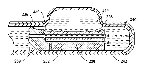
As illustrated in FIG. 7, an ultrasound transducer assembly 228 includes a
planar
piezoceramic therapy transducer element 230, a backing 232, and an acoustic
lens 234 (e.g., a
concave lens, a convex lens, a Fresnel lens, a Fresnel multilevel lens, or a
Field Conjugate
lens) that are connected to one or more mounting members 236, 238 and disposed
inside a
10 flexible bladder 240 that is in turn disposed inside a casing 242 provided
with a window 244.
Casing 242 and the contents thereof comprise a probe for high-intensity
focused ultrasound
(HIFU) surgical therapy.
Lens 234 is spaced from transducer element 230 by a distance dl equal to
(2n+1)X/4
where n is a non-negative integer and X is the wavelength of the ultrasonic
pressure waves for
15 therapeutic applications. Transducer element 230 is spaced from backing 232
by a distance
d2 equal to n)12 where again n is a non-negative integer and ? is the
wavelength of the
ultrasonic pressure waves for therapeutic applications.
As depicted in FIG. 8, an ultrasound transducer assembly 246 at least for use
in a
therapy mode comprises a planar piezoceramic transducer element 248, a backing
layer 250,
and an acoustic lens 252 (a concave lens, a convex lens, a Fresnel lens, a
Fresnel multilevel
lens, or a Field Conjugate lens) aligned with one another and spaced by liquid-
filled gaps 254
and 256 of thickness d1 and d2, respectively. Two metal plates 258 and 260,
which serve to
block ultrasonic wave transmission, are connected to lens 252 on opposing
sides thereof.
Lens 252, together with metal blockers 258 and 260, is longitudinally
shiftable alternately in
opposite directions, as indicated by double headed arrow 262, relative to
transducer element
248 for enabling a user to move a focal zone of the ceramic transducer. Lens
252 may be a
cylindrical lens, in which case the focal zone or locus (set of points) is a
line. Moving the
lens 252 relative to the transducer 248 (and probe casing, not shown) shifts
the focal locus
along a plane parallel to the transducer and thus parallel to an organ surface
against which the
therapy probe lies. Metal blockers 258, 260 prevent ultrasonic pressure waves
from radiating
into the patient except towards the focal locus defined in part by lens 252.
Any of the lenses
disclosed herein may be movably mounted relative to the respective ceramic
transducer
element to facilitate application of focused high-intensity ultrasound to an
extended target
site.
11-
WO 2011/146139 PCT/US2011/000911
16
FIG. 9 depicts a dual mode transducer assembly 264 with a rotatable holder
266, as
indicated by an arrow 268. Holder 266 includes a head 270 in the form of a
right rectangular
prism. Head 270 is provided on one face 272 with at least one high-power
ceramic
transducer element 274 that is either planar or shaped for focusing. In the
case of a planar
transducer element 274, an acoustic lens (not shown) is provided for focusing
the planar
ultrasonic waves from transducer onto a focal locus such as a point (spherical
lens) or a line
(cylindrical lens). Another face 276 of head 270 carries at least one planar
or focally shaped
piezoelectric polymeric (e.g., PVDF) transducer element 278. In the case of a
planar
transducer element 278, an acoustic lens (not shown) may be provided for
focusing the planar
ultrasonic waves from a focal locus onto transducer element 278. Where the
ultrasonic
pressure waves generated by ceramic transducer element 274 for therapy have a
frequency
that is substantially different than the frequency of pressure waves for
imaging, two different
lenses may be provided. The lens may be shiftably mounted to a probe casing
(not shown)
for alternate use during therapy and imaging operations.
As in the case of other transducer devices described above, the dual mode
transducer
assembly 264 of FIG. 9 is typically incorporated into an ultrasound probe
including a casing
and bolus. More particularly, holder 266 is disposed inside a liquid-filled
bladed or bolus
(not shown) that in turn is disposed inside a probe casing (not shown) in
juxtaposition to a
window in the probe casing. As discussed above, transducer elements 274 and
278 are used
in alternation in therapeutic and imaging operating modes, respectively.
Holder 266 is
rotated in order to juxtapose transducer element 274 to the casing window
during a period of
therapy application and subsequently to juxtapose transducer element(s) 278 to
the casing
window during an imaging interval.
FIG. 10 depicts a modification 280 of the dual mode transducer assembly 264 of
FIG.
9 and uses the same reference numerals to designate the same parts. In
transducer assembly
280, ceramic transducer element 274 and polymeric transducer element 278 are
located in
adjacent faces 272 and 282, rather than opposite faces 272 and 276 as in
transducer assembly
264 of FIG. 9. Accordingly, the operation is slightly altered inasmuch as
holder 266 need be
rotated only 90 rather than 180 to change from therapy mode to imaging mode
and vice
versa.
As shown in FIG. 11, a dual mode transducer assembly 284 has a longitudinally
reciprocatable holder 286, as indicated by a double-headed arrow 288. Holder
286 includes a
head 290 in the form of a right rectangular prism or plate. Head 290 is
provided on one face
292 with both a planar or shaped high-power ceramic transducer element 294 and
a planar or
11-
WO 2011/146139 PCT/US2011/000911
17
shaped piezoelectric polymeric (e.g., PVDF) transducer element 296. In the
case that
transducer elements 294 and/or 296 are planar, one or more acoustic lenses
(not shown) may
be provided for focusing purposes. Again, holder 286 is disposed inside a
liquid-filled bladed
or bolus (not shown) that in turn is disposed inside a probe casing (not
shown) in
juxtaposition to a window 298 in the probe casing. Transducer elements 294 and
296 are
alternately juxtaposed to the casing window 298 to implement therapeutic and
imaging
operating modes, respectively, by shifting holder 286 in a distal or proximal
direction as
appropriate.
FIG. 12 shows a dual mode transducer assembly 300 including a probe casing 302
provided at a distal end with a window 304 in a sidewall (not separately
designated) and
further including a piezoceramic transducer element 306 in a holder 308
disposed inside the
casing. A piezoelectric polymeric imaging transducer element 310 is disposed
in window
304 and located along an arcuate bridge (not separately designated) so as to
bifurcate the
window. A bolus or bladder member 312 is provided outside of casing 302 and
may be
pressurized to expand from a partially inflated storage configuration (not
shown) to a fully
inflated use configuration as shown.
FIG. 13 illustrates a dual mode transducer assembly 314 including a probe
casing 316
provided in a sidewall (not separately designated) at a distal end with a
window 318. A
planar or focally shaped piezoceramic transducer element 320 is disposed on a
holder 322
inside casing 316. Two piezoelectric polymeric imaging transducer elements 324
and 326,
disposed along distal and proximal sides of window 318, are configured for
scanning tissues
at a focal zone 327. A bolus or bladder member 328 is provided outside of
casing 316 and
may be pressurized to expand from a partially inflated storage configuration
(not shown) to a
more expanded use configuration as indicated.
Pursuant to FIGS. 14 and 15, an ultrasound transducer assembly 330 includes a
probe
casing 332 provided in a sidewall (not separately designated) at a distal end
with a window
334. A piezoceramic transducer element 336 is disposed on a holder 338 inside
casing 332.
Also disposed on a holder 340 inside casing 332 is a spherical acoustic
Fresnel lens 342. As
indicated by an arrow 344 in FIG. 15, holder 340 and Fresnel lens 342 are
rotatable about a
longitudinal axis 346, whereby a focal point of the lens moves along an arc.
In addition,
holder 340 and lens 242 may be longitudinally reciprocatable, as indicated by
a double-
headed arrow 348, so that the focal point of the lens may be moved distally
and proximally.
A bolus or liquid-filled bladder (not shown) is provided about the casing 332.
11-
WO 2011/146139 PCT/US2011/000911
18
Lens 342 may be flanked by metal plates (not shown) for limiting ultrasound
irradiation.
As depicted in FIGS. 16 and 17, an ultrasound transducer assembly 350 includes
a
high-power therapy transducer 352 in a parabolic or cylindrical configuration,
on a holder
354 inside a probe casing 356. Transducer element 352 is disposed so that its
axis extends in
a distal-proximal direction, parallel to a longitudinal axis 358 of the probe
casing. Ultrasound
transducer assembly or probe 350 further includes a cylindrical acoustic
Fresnel lens 360 that
is oriented with its axis transverse to the axis of transducer element 352,
thereby producing a
focal zone or locus that is a point. As indicated, lens 360 may be rotatable
and optionally
longitudinally shiftable, for shifting the location of the focal point
relative to the probe and
accordingly relative to a patient.
Polymeric piezoelectric materials suitable for imaging transducer elements
152, 162,
182, 212, 194, 224, 278, 296, 310, 324, and 326 include polyvinylidene
fluoride (PVDF), and
copolymers of PVDF such as trifluoroethylene (TrFE) with a piezoelectric
voltage constant
933 > 100x10"3 Vm/N. Piezoceramic materials suitable for therapy transducer
elements 150,
160, 180, 206, 192, 218, 230, 248, 274, 294, 306, 320, and 336 include
modifications of
BaTi03i Pb(Ti,Zr)03 (PZT) and PbNb2O6 ceramics with a high piezoelectric
strain constant,
d33 > 200x10"12 m/V.
Imaging transducer elements as used herein are derived from an appreciation of
the
properties of polyvinylidene fluoride (PVDF). That polymer is a semi-
crystalline,
thermoplastic fluoroplastic. It has received a considerable research attention
in past decades
that stems from the discovery of its piezoelectric and pyroelectric properties
and its
subsequent application as an electret and piezoelectric transducer. With its
low acoustic
impedance of 3.5 MRyals and high voltage constant PVDF makes an ideal
ultrasound
receiver and shows definite advantages over ceramic counterparts. As a
transmitter of
acoustic power, the PVDF transducer is quite poor, but its enhanced
sensitivity on reception
provides a send-receive factor comparable to that of ceramic. The table below
summarized
common applications and lists relevant piezoelectric properties for typical
piezoelectric
ceramic, quartz and PVDF.
11-
WO 2011/146139 PCT/US2011/000911
19
Piezoelectric material properties (Gallentree, 1983, Review of Transducer
Applications o
olyvinylidene Fluoride, Piezoelectricity, Key Paper in Physics, 189-194; Kino,
1987, Acoustic
Waves: Devices, Imaging, and Analog Signal Processing, Prentice Hall,
Englewood Cliffs, NJ
Appendix B; Mason, 1966, Physical Acoustics: Principles and Methods, edit
Rosenberg, Mir,
oMoscow)
Applications Curie Q.. d335 933,
C m/V m/N
1012 103
Navy Type I STM, nanopositioning, medical 328 500 89 5
(PZT4) therapeutics.
Navy Type II low and level sensing 365 15 374 5
(PZT5A) and medical Doppler transducers
Navy Type Ultrasonic cleaners, cell 300 1000 25 5
III disruption, phacoemulsification,
PZT8) and high power ultrasonics
Navy Type VI Medical diagnostics, industrial 193 65 593 0
PZTSH) T, STM/AFM, and nano-
Positioning
VDF Insulation (Kynar ), key boards, 100 13 0 10
onar hydrophones, pulse-echo
ultrasonic transducers
Quartz crystal clock oscillator, mass- 5000 50
icrobalance, and thin-film
ickness monitoring
A typical PVDF transducer does not require cumbersome acoustic matching
layers,
inherent in ceramic transducers, and is relatively easy to produce in a
variety of forms and
may be press fit into a curved shape.
Polymeric imaging transducer elements 152, 162, 182, 212, 224, 278,296 310,
324,
and 326 are operatively connected to ultrasound image processor 202 or other
appropriate
waveform processing and digital image generation apparatus, as well known in
the art.
Ceramic therapy transducers 150, 160, 180, 206, 192, 218, 230, 248, 274, 294,
306, 320, and
336 may operate in part to generate outgoing scanning waveforms. Where there
are moving
parts, such as lenses moving relative to therapy transducers, the motion may
be implemented
via electric motors, stepper motors, linear motors, etc., and the motion may
be monitored by
feedback sensors such as encoders, voltage dividers, etc.
Ceramic transducer elements 150, 160, 180, 206, 192, 218, 230, 248, 274, 294,
306,
320, and 336 function in a therapy mode of operation of the respective
transducer assembly
or device to generate high-power ultrasonic pressure waves, in response to a
suitable
11-
WO 2011/146139 PCT/US2011/000911
energizing signal, that are transmitted into a patient for implementing or
assisting in a
surgical operation such as thermal ablation, hyperthermia, transfection and/or
drug delivery.
Polymeric transducer elements 152, 162, 182, 212, 224, 278,296 310, 324, and
326 function
in a diagnostic or scanning mode of operation of the respective transducer
assembly or device
5 to detect incoming ultrasonic pressure waves that are reflected from
internal tissue structures
of a patient in response to a suitable scanning wave. As discussed above with
reference to
FIG. 4, the therapeutic ceramic transducer elements and the diagnostic
polymeric transducer
element may be connected in parallel in the same circuit.
Thus, the ultrasound transducer devices described herein are provided with
electrical
10 contacts (not shown) enabling a connection of the respective ceramic
transducer elements
150, 160, 180, 206, 192, 218, 230, 248, 274, 294, 306, 320, and 336 in
operative circuits for
generating, for example, high-intensity focused ultrasound and enabling a
connection of the
respective polymeric transducer elements 152, 162, 182, 212, 224, 278,296 310,
324, and 326
in operative circuits for scanning organic tissues to generate ultrasonic scan
data for analysis
15 and processing into images.
FIG. 18 depicts a Fresnel lens 402 that is reciprocatable along a given
direction, as
indicated by a double-headed arrow 404, in parallel to a planar front
radiating face 406 of a
flat transducer element 408. Lens 402 is spaced from front face 406 of
transducer element
408 by a gap 409 of a thickness (2n+1)A./4 where n is a non-negative integer
and ? is the
20 wavelength of the ultrasonic pressure waves for therapeutic applications.
Transducer element
408 is spaced from a backing (not shown) by a distance na./2 where again n is
a non-negative
integer and ), is the wavelength of the ultrasonic pressure waves for
therapeutic applications.
Lens 402 is configured to have a focal length that varies in a continuous
gradient from
a maximum focal length f, at one end 410 of the lens to a minimum length f2 at
an opposite
end 412 of the lens. As depicted in FIG. 19A, a focal zone 414 is disposed at
a maximum
distance D1 (approximately length f1) from lens 402 when transducer element
408 is aligned
with the first end 410 of the lens. When transducer element 408 is aligned
with the opposite
end 412 of lens 408, ultrasound waves converge at a focal zone 416 located at
a minimum
distance D2 (approximately length f2) from lens 402, as shown in FIG. 19C.
When transducer
element 408 is aligned with a middle region of lens 408 as shown in FIG. 19B,
ultrasonic
pressure waves generated in a subject converge at a focal zone 418 disposed at
an
intermediate distance D3 from lens 402. Accordingly, by moving lens 402 in the
direction of
arrow 404 one focuses destructive ultrasound energy at target regions or focal
zones 414,
416, 418 located at different depths DI, D2, D3 in the patient and at
different laterally
11-
WO 2011/146139 PCT/US2011/000911
21
staggered positions along a skin or internal surface. As indicated in FIG.
19C, generation of
ultrasound energy by transducer element 408 while lens is moving from right to
left relative
to the transducer can produce a continuous elongate region 420 of
therapeutically damaged
tissue.
FIG. 20 shows a transducer element 422 ensconced in a backing layer 424 and
spaced
from a Fresnel lens 426 by a liquid-filled gap 428 of thickness (2n+1)714
where n is a non-
negative integer and A. is the wavelength of the ultrasonic pressure waves for
therapeutic
applications. Lens 426 comprises a plurality of adjacent sections 430, 432,
434 each of a
respective focal length si, s2, s3. Focal lengths Si, 52i s3 are shown to vary
in a monotonically
decreasing sequence. However, any arrangement of any practicable number of
sections of
different focal lengths may be made.
The distance (generally sl, s2, s3) of a target tissue mass or focal zone 438,
440, 442
from lens 426 varies in accordance with which lens section 430, 432, 434 is in
alignment with
transducer element 422. In addition, limited lateral motion of lens 426 (see
arrow 444)
relative to transducer 422 while any given lens section 430, 432, 434 remains
in alignment
with transducer element 422 will shift the respective focal zone 438, 440, 442
laterally in
parallel to lens 426 and transducer element 422 (assuming planar
configurations of both).
As illustrated in FIG. 21, an ultrasound transducer assembly 446 includes a
transducer
element 448 on a holder (not separately illustrated) disposed inside a
generally cylindrical or
tubular Fresnel lens 450 which has a focal length that varies in a continuous
gradient (or,
alternatively, in discrete steps) around the circumference of the lens. Thus
rotating lens 450
relative to transducer element 448, as indicated by an arrow 454, enables one
to target a tissue
mass at a controllably variable depth or distance from assembly 446. Shifting
lens 450
longitudinally (arrow 452) relative to transducer element 448 enables one to
vary the position
of the focal zone or target tissue region in the direction of arrow 452.
The depth of focus can be controlled by adjusting the transducer operating
frequency.
In the latter case, the Fresnel lens changes its depth of focus depending on
the frequency thus
offering an elegant way of controlling energy deposition at different depths
when treating
large tissue volumes using a single fixed lens and a set of high-power
transducers capable of
operating at a range, or with a discrete set, of frequencies. FIGS. 23-24
shows relative
intensity profiles created by a 2-zone Fresnel lens, a 4-zone Fresnel lens and
an 8-zone
Fresnel lens at a set of three frequencies. The 8-zone lens of FIG. 24 was
designed to focus 4
MHz waves at 40 mm depth. Clearly, the use of a 5 MHz frequency moves the
focal zone
deeper, outward by about 10 mm, while the focal spot is brought to a shallower
depth at an
11-
WO 2011/146139 PCT/US2011/000911
22
operating frequency of 3 MHz. The transducer is moved relative to a lens or
both are moved
relative to a probe in order to achieve large volume tissue impact.
FIGS. 25-27 are a particular configuration of a probe head of a HIFU treatment
device
showing a housing 456, a Fresnel lens 458, a rectangular piezoelectric
transducer 460, a
reflector 462, and mill-max spring-loaded pins 464.
FIG. 28 shows a planar piezoelectric transducer element 466 affixed at
opposite ends
468 and 470 to a housing or frame (not shown) and provided with three metal
supports 472-
474, optionally in the form of electrodes. Supports 472 and 472 are positioned
at the nodes
of vibration mode 1, while support 473 is positioned at the node of vibration
mode O.FIGS.
29 and 30 depict a HIFU transducer assembly or device including a cylindrical
transducer
element 476 operating in wall thickness mode and an essentially cylindrical
Fresnel lens 478
having an azimuthally variable focal length, the transducer element being
located inside the
lens. FIG. 30 shows a variable-depth focal zone 480 about the lens 478.
FIG. 31 shows a lens L constructed to focus planar acoustic waves of frequency
f1 at a
focal point F1. The line 'P constitutes a construction line. The solid arcs
with the center of
origin F1 are the phase fronts spaced apart by one wavelength ?1 from each
other. The first
solid circle is tangent to the line 'P not shown. The intersection of solid
circles with line 'P
marks the location of the respective Fresnel zones. At a higher frequency f2
the acoustic
wavelength becomes smaller: X2 < xt. If lens L design is fixed, passing a
higher frequency
waves through lens L is similar to having the phase circles spaced apart by a
smaller distance
X2i shown by dashed arcs. In order to focus the dashed arcs, which correspond
to frequency f2
> fl, must intersect the line 'P at the same points as solid arcs, which
correspond to the
original lens frequency fl. Clearly, on average dashed arcs can intersect line
'P at the same
points if their center of origin F2 is located farther away from the line 'P
than F1. Using this
geometrical construction and neglecting terms of the second order in
wavelength an
approximate formulae that related the focal depth of a lens and operating
frequency is:
F = F, fj If 2. This equation predicts the focal distances for the relatively
small number of the
Fresnel zones and for the cases where wavelength is much smaller than focal
distances F. For
example, going from 4 MHz to 5 MHz would result in a shift of the focal spot
from 35 mm to
approximately 43 mm, in good agreement with FIG. 22 field simulation results.
Thus, the
higher frequency will focus deeper and, respectively, lower frequency will
focus at shallower
depth than original frequency.
By constructing a lens made of relatively soft silicone, like RTV rubber, one
can achieve the limited field transformation effects without changing
frequency of
11-
WO 2011/146139 PCT/US2011/000911
23
transducers. For example, simulation shows that 30% stretch in one direction
results in a field
blurring and slight depth decrease. This effect can be used to control the
volume of ultrasonic
energy deposited by a transducer and focused by deformable lens. There is a
potential to
ablate larger tissue volume with a field that is less focused, yet has
sufficient intensity.
Stretching the lens is a simple and controllable process that will enable
blurring of the focal
intensity zone over larger volume, which can be beneficial for large tumor
ablations.
FIGS. 32 and 33 show a flat circular configuration for a probe head of a HIFU
treatment device, including a housing 482, a Fresnel lens 484, a rectangular
piezoelectric
transducer 486, a reflector 488, mill-max spring-loaded pins 490, and a center
electrode 492.
Another aspect of the present invention, depicted in FIGS. 34 and 35, includes
an
ultrasound imaging transducer or transducer array 502 movable relative to a
therapeutic
transducer element or array 504 to thereby obtain an image of a region exposed
to high power
ultrasound generated by the therapeutic transducer element or array and ensure
a controlled
and safe therapy process. The movability of imaging transducer or transducer
array 502, as
represented by double headed arrow 506 in FIG. 35, facilitates the application
of the high
power ultrasonic energy to an extended surgical target region.
As shown in FIGS. 34 and 35, therapeutic transducer element or array 504 takes
a
planar form and a Fresnel focusing lens 508 is held by a casing or frame 516
in position
parallel to element or array 504, separated by small water gap 510. The water
or other
suitable liquid in gap 510 facilitates the cooling of therapeutic transducer
element or array
504 and serves as a pathway for the introduction of imaging transducer or
probe 502.
Imaging transducer or array 502 may constitute a thin plate not exceeding in
thickness
the width of gap 510 between therapy transducer 504 and lens 508 and having
transverse
dimensions comparable to a first or innermost or central Fresnel zone 512 of
the lens.
Fresnel zone 512 is the thinnest part of lens 508 and enables efficient and
lossless
transmission and reception of ultrasound by imaging transducer 502, when that
transducer
element or array is positioned in alignment with the central or innermost
Fresnel zone 512 as
depicted in FIG. 35.
Imaging transducer 502 may contain several layers of acoustical matching
layers,
active piezo-materials, bonding and backing layers, constituting a stacked
design, or made of
piezo-composite material, which can contain a single or plurality of
discretely imprinted
electrodes that provide for a single element probe or imaging phased array
configuration, thus
enabling imaging at variable focal depths.
11-
WO 2011/146139 PCT/US2011/000911
24
The middle section of Fresnel lens zone 512 is thinner than an outermost
section 514
that has the minimum thickness: d >_ 1 , where c,, and cm are the sound speed
in
f 1 _ 1
C,, Cm
water and lens material, respectively, and f is the frequency. Thus innermost
or central
Fresnel section or zone 512 enables most of the transmission.
For example a 4 MHz lens with a nominal focal depth of 45 mm has a first or
innermost Fresnel zone of about 11 mm in diameter. As shown in FIG. 36, the
relative power
transmission (solid line, left axis) as a function of radius varies from 100%
in the middle to
less than 70% in the outer section, assuming first order polystyrene lens. At
the same time,
the outer section or zone produces a larger phase shift (dashed line, right
axis) for the
propagating ultrasound waves, which is important for focusing, while middle
section
introduces minimal phase shift to propagating waves. Thus, it is feasible to
replace the middle
or innermost section with an opening of about 6 mm diameter, which is
sufficient to provide
an imaging window for a movable imaging transducer. Alternatively, an imaging
transducer
made of low ultrasound absorption piezo-polymer material can be an integral
part of a
movable and variable focal distances lens, as disclosed above, to enable
simultaneous
focusing and imaging at different distances, which is required for a
controllable, effective and
safe ultrasound ablation performed under ultrasound imaging guidance.