Note: Descriptions are shown in the official language in which they were submitted.
CA 02800460 2012-10-17
WO 2010/129302 PCT/US2010/032616
1
LATERAL-FLOW IMMUNO-CHROMATOGRAPHIC ASSAY DEVICES
BACKGROUND
1. Field of the Invention.
[0001] This invention generally relates to lateral-flow immuno-chromatographic
assay
devices.
2. Related Art.
[0002] Various types of lateral-flow immuno-chromatographic assay devices have
been
developed for carrying out diagnostic assays on blood samples. Lateral-flow
immuno-
chromatographic assay devices typically include an arrangement of layers of
materials in a
housing having openings for sample introduction and for reading of assay
results.
[0003] There is a continuing need for lateral-flow immuno-chromatographic
assay devices
having structures and modes of utilization facilitating improved assay
performance
capabilities.
SUMMARY
[0004] In an example of an implementation, a device is provided that includes
a migration
membrane, a conjugate pad on the migration membrane, a plasma separation
membrane on the
conjugate pad, and a pre-filter on the plasma separation membrane. The
migration membrane
has a test line configured for loading onto the test line of one or a
plurality of capture
antibodies having specific binding affinity for an assay target. The migration
membrane is
configured for allowing lateral flow of blood plasma or serum across the
migration membrane
to the test line. The conjugate pad is configured for loading onto the
conjugate pad of one or a
plurality of detection antibodies having specific binding affinity for an
assay target. The
plasma separation membrane is configured for allowing passage of blood plasma
or serum
through the plasma separation membrane and for trapping erythrocytes. The pre-
filter is
configured for loading of an assay sample including erythrocytes and either or
both of blood
plasma and blood serum onto the pre-filter. Further, the pre-filter is
configured for allowing
passage of blood plasma or serum through the pre-filter, and configured for
causing lateral
flow of blood plasma or serum within the pre-filter.
[0005] As another example of an implementation, a method is provided. The
method
includes providing a lateral-flow immuno-chromatographic assay device
including a migration
CA 02800460 2012-10-17
WO 2010/129302 PCT/US2010/032616
2
membrane, a conjugate pad being on the migration membrane, a plasma separation
membrane
being on the conjugate pad, and a pre-filter being on the plasma separation
membrane. In the
lateral-flow immuno-chromatographic assay device, the conjugate pad is loaded
with one or a
plurality of detection antibodies having specific binding affinity for an
assay target; and the
migration membrane has a test line loaded with one or a plurality of capture
antibodies having
specific binding affinity for the assay target. The method further includes
carrying out a
diagnostic assay cycle. In the diagnostic assay cycle, an assay sample
including erythrocytes
and either or both of blood plasma and blood serum is loaded onto the pre-
filter. Also in the
cycle, blood plasma or serum is caused to laterally flow within the pre-filter
and allowed to
pass through the pre-filter to the plasma separation membrane. The diagnostic
assay cycle
further includes causing erythrocytes to be trapped in the plasma separation
membrane; and
allowing blood plasma or serum to pass through the plasma separation membrane
to the
conjugate pad. Additionally, the cycle includes allowing blood plasma or serum
to pass
through the conjugate pad onto the migration membrane and allowing blood
plasma or serum
to laterally flow across the migration membrane to the test line.
[0006] Other devices, methods, features and advantages of the invention will
be or will
become apparent to one with skill in the art upon examination of the following
figures and
detailed description. It is intended that all such additional devices,
methods, features and
advantages be included within this description, be within the scope of the
invention, and be
protected by the accompanying claims.
BRIEF DESCRIPTION OF THE FIGURES
[0007] The invention can be better understood with reference to the following
figures. The
components in the figures are not necessarily to scale, emphasis instead being
placed upon
illustrating the principles of the invention. Moreover, in the figures, like
reference numerals
designate corresponding parts throughout the different views.
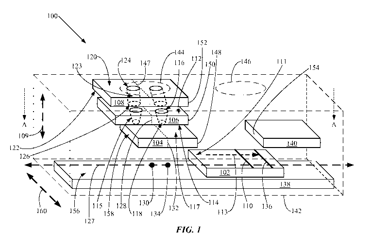
[0008] FIG. 1 is an exploded perspective view illustrating an example of an
implementation of a lateral-flow immuno-chromatographic assay device.
[0009] FIG. 2 is a cross-sectional view, taken along line A-A, of the lateral-
flow immuno-
chromatographic assay device shown in FIG. 1.
[0010] FIG. 3 is a flow diagram illustrating an example of an implementation
of a method.
[0011] FIG. 4 is a photograph showing the lateral-flow immuno-chromatographic
assay
devices utilized to carry out Examples A, B and C.
CA 02800460 2012-10-17
WO 2010/129302 PCT/US2010/032616
3
[0012] FIG. 5 is a photograph showing the lateral-flow immuno-chromatographic
assay
devices utilized to carry out Examples D, E, F and G.
[0013] FIG. 6 is a photograph showing the lateral-flow immuno-chromatographic
assay
devices utilized to carry out Examples H, I, J and K.
[0014] FIG. 7 is a photograph showing the lateral-flow immuno-chromatographic
assay
devices utilized to carry out Examples L, M, N and O.
[0015] FIG. 8 is a photograph showing the lateral-flow immuno-chromatographic
assay
devices utilized to carry out Examples P, Q, R and S.
[0016] FIG. 9 is a photograph showing the lateral-flow immuno-chromatographic
assay
devices utilized to carry out Examples T, U, V and W.
[0017] FIG. 10 is a photograph showing the lateral-flow immuno-chromatographic
assay
devices utilized to carry out Examples X, Y, Z and AA.
[0018] FIG. 11 is a photograph showing the lateral-flow immuno-chromatographic
assay
devices utilized to carry out Examples AB, AC, AD and AE.
[0019] FIG. 12 is a photograph showing the lateral-flow immuno-chromatographic
assay
devices utilized to carry out Examples AF, AG, AH and Al.
[0020] FIG. 13 is a photograph showing the lateral-flow immuno-chromatographic
assay
devices utilized to carry out Examples AJ and AK.
[0021] FIG. 14 is a photograph showing the lateral-flow immuno-chromatographic
assay
devices utilized to carry out Examples AL and AM.
[0022] FIG. 15 is a photograph showing the lateral-flow immuno-chromatographic
assay
devices utilized to carry out Examples AN and AO.
[0023] FIG. 16 is a photograph showing the lateral-flow immuno-chromatographic
assay
device utilized to carry out Example AP.
[0024] FIG. 17 is a photograph showing the lateral-flow immuno-chromatographic
assay
devices utilized to carry out Examples AQ and AR.
DETAILED DESCRIPTION
[0025] A lateral-flow immuno-chromatographic assay device may function, for
example,
by carrying a diagnostic assay sample to a conjugate pad loaded with one or a
plurality of
detection antibodies having specific binding affinity for an assay target. The
diagnostic assay
sample may then be laterally carried across a migration membrane to a test
line loaded with
one or a plurality of capture antibodies also having specific binding affinity
for the assay
CA 02800460 2012-10-17
WO 2010/129302 PCT/US2010/032616
4
target. If a sufficient concentration of the assay target was present in the
diagnostic assay
sample, then a detectable quantity of the detection antibodies may
specifically bind with the
assay target at the conjugate pad and may then laterally flow along with the
assay sample to the
test line. The capture antibodies at the test line may then also specifically
bind there with the
assay target, generating a visible mark constituting a positive test result of
the diagnostic assay.
For example, the detection antibodies may be tagged with a colored marker such
as colloidal
gold. Where the assay sample carries a sufficient quantity of the detection
antibodies to the
test line and then a sufficient quantity of the capture antibodies bind the
assay target carrying
the bound detection antibodies at the test line, a visibly colored mark may be
formed. Where
the detection antibody marker is colloidal gold, for example, a mark having a
reddish, pinkish,
or brownish hue may be formed.
[0026] The diagnostic assay sample may include red blood cells, also being
referred to
herein as "erythrocytes." For example, the diagnostic assay sample may include
whole blood,
or may otherwise include erythrocytes and either or both of blood plasma and
blood serum.
Blood plasma or blood serum may, for example, be separated from erythrocytes
before
carrying out a lateral-flow immuno-chromatographic diagnostic assay to reduce
or
substantially eliminate the reddish color caused by hemoglobin. However, if
erythrocytes in an
assay sample become ruptured before or during performance of a lateral-flow
immuno-
chromatographic assay, hemoglobin released from the ruptured erythrocytes may
stain the
blood plasma or serum. If blood plasma or serum stained by hemoglobin reaches
the test line
in a lateral-flow immuno-chromatographic assay, then a visibly colored mark
may be formed
by the hemoglobin at the test line, generating a false positive assay result.
These false positive
results may be avoided by removing erythrocytes from an assay sample before
carrying out a
lateral-flow immuno-chromatographic diagnostic assay. For example, whole blood
may be
centrifuged to remove erythrocytes, so that the assay sample to be tested is
in the form of blood
plasma. Further, blood coagulation factors may also be removed, so that the
assay sample
tested is blood serum. However, in some cases, utilization of an assay sample
including
erythrocytes as well as either or both blood plasma and blood serum may be
needed. For
example, a medical professional may need to carry out a lateral-flow immuno-
chromatographic
diagnostic assay where equipment such as a centrifuge for removal of
erythrocytes from whole
blood is not available, or where rapid test performance is needed, such that
waiting for
preparation of a blood plasma or serum sample becomes an unacceptable delay.
For example,
a medical professional may need to carry out a lateral-flow immuno-
chromatographic
CA 02800460 2012-10-17
WO 2010/129302 PCT/US2010/032616
diagnostic assay at a location away from clinical facilities, or where a
patient suffers from a
critical, life-threatening condition.
[0027] As another example, a layman lacking both the skill and equipment
needed to
prepare a blood plasma or serum sample may need to himself carry out a lateral-
flow immuno-
5 chromatographic assay using whole blood. Further, such a layman may also
lack both the
equipment and skill needed to collect venous blood from a patient. Moreover, a
layman may
be reluctant to collect more than a single drop of the patient's capillary
blood by a fingertip
lance, especially if the layman needs to collect and then perform a diagnostic
assay on his own
blood. Additionally, such a layman may need to self-administer a lateral-flow
immuno-
chromatographic assay while suffering from a life-threatening condition at a
location distant
from professional medical personnel.
[0028] Hence, a lateral-flow immuno-chromatographic assay device capable of
utilization
with a small assay sample including erythrocytes and either or both of blood
plasma and blood
serum, such as a minimal sample of whole blood, may be useful in a variety of
circumstances.
Such a lateral-flow immuno-chromatographic assay device may, for example, need
to
effectively separate erythrocytes from blood plasma or serum so that false
positive results due
to ruptured erythrocytes are avoided. Further, for example, such a lateral-
flow immuno-
chromatographic assay device may need to effectively deliver as much of a
small sample of
blood plasma or serum as possible to the test line, so that a qualitative
assay test result may be
generated utilizing a small assay sample, such as a single drop of whole
blood.
[0029] A lateral-flow immuno-chromatographic assay device may include a plasma
separation membrane configured for allowing passage of blood plasma or serum
through the
plasma separation membrane and for trapping erythrocytes. In so trapping
erythrocytes, the
plasma separation membrane may become partially blocked and then impede the
flow of blood
plasma or serum through the plasma separation membrane toward the test line,
detracting from
a capability of the lateral-flow immuno-chromatographic assay device for
utilization with an
assay sample having a minimal volume, such as a single drop of blood. Hence,
lateral-flow
immuno-chromatographic assay devices are needed that may be capable of
utilization with an
assay sample of minimal volume, such as a single drop, including erythrocytes
and either or
both of blood plasma and blood serum. Such lateral-flow immuno-chromatographic
assay
devices, and methods for carrying out lateral-flow immuno-chromatographic
assays, are
provided herein.
CA 02800460 2012-10-17
WO 2010/129302 PCT/US2010/032616
6
[0030] A lateral-flow immuno-chromatographic assay device is provided herein
that
includes a migration membrane, a conjugate pad being on the migration
membrane, a plasma
separation membrane being on the conjugate pad, and a pre-filter being on the
plasma
separation membrane. The migration membrane has a test line configured for
loading onto the
test line of one or a plurality of capture antibodies having specific binding
affinity for an assay
target. The migration membrane is configured for allowing lateral flow of
blood plasma or
serum across the migration membrane to the test line. The conjugate pad is
configured for
loading onto the conjugate pad of one or a plurality of detection antibodies
having specific
binding affinity for an assay target. The plasma separation membrane is
configured for
allowing passage of blood plasma or serum through the plasma separation
membrane and for
trapping erythrocytes. The pre-filter is configured for loading of an assay
sample including
erythrocytes and either or both of blood plasma and blood serum onto the pre-
filter. The pre-
filter is also configured for allowing passage of blood plasma or serum
through the pre-filter.
Further, the pre-filter is configured for causing lateral flow of blood plasma
or serum within
the pre-filter. The pre-filter may additionally be configured for causing
selective passage of
blood plasma or serum through the pre-filter and for trapping erythrocytes.
The device may be
configured for being capable of utilizing a single drop of whole blood, such
as hanging drop
for example, as the assay sample.
[0031] In examples, the one or plurality of detection antibodies and the one
or plurality of
capture antibodies may have specific binding affinity for a cardiac Troponin-I
epitope. The
plurality of detection antibodies may include cardiac Troponin-I antibody
clone 19C7 together
with either or both of cardiac Troponin-I antibody clones 4C2 and M155. The
plurality of
capture antibodies may include both cardiac Troponin-I antibody clones MF4 and
16A11. As
another example, the lateral-flow immuno-chromatographic assay device may
include first and
second detection antibodies and third and fourth capture antibodies, each of
the first, second,
third and fourth antibodies having specific binding affinity for substantially
different cardiac
Troponin-I epitopes. Throughout this specification, the term "substantially
different" as
applied to two epitopes of an assay target means that the two epitopes are
sufficiently different
to allow two antibodies to bind the target simultaneously through binding to
the two epitopes.
[0032] The following conventions apply regarding terminology utilized
throughout this
specification. A "layer" of a material is any component of a lateral-flow
immuno-
chromatographic assay device that is bonded or attached to, formed or
deposited on, or
otherwise provided on any other layer or on or in the housing of the lateral-
flow immuno-
CA 02800460 2012-10-17
WO 2010/129302 PCT/US2010/032616
7
chromatographic assay device. A layer may include, as examples, a mat,
surface, film, foil,
region, body, or substrate. When one layer or material is referred to as being
"on", "over", or
"loaded onto" another layer or housing, then all or a portion of the layer or
material may be
directly on and in contact with all or a portion of the other layer or
housing, or alternatively,
intervening layers may also be present such that all or portions of the one
layer and of another
layer that is "on" or "over" the one layer or housing are not mutually in
direct contact. When a
layer is stated as being "directly on" another layer or the housing, then no
intervening layer is
present unless otherwise indicated. When a layer is stated as being "between"
two other
layers, then one or more additional intervening layers may also be present
between the two
other layers. When one layer is referred to as being "on" (or "over") another
layer, then the
one layer may cover the entire surface of the other layer, or may cover only a
portion of the
other layer. When a material is stated as being "loaded onto" a layer or a
surface of a layer, the
material may remain on a surface of the layer, or may also penetrate through
the surface into
the layer, or may penetrate into and pass through the layer. Terms such as
"formed on",
"disposed on", "loaded onto" or "deposited on" are not intended to introduce
any limitations
relating to specific methods for fabricating a layer except as otherwise
designated.
[0033] FIG. 1 is an exploded perspective view illustrating an example of an
implementation of a lateral-flow immuno-chromatographic assay device 100. FIG.
2 is a
cross-sectional view, taken along line A-A, of the lateral-flow immuno-
chromatographic assay
device 100 shown in FIG. 1. The lateral-flow immuno-chromatographic assay
device 100
includes a migration membrane 102, a conjugate pad 104 being on the migration
membrane
102, a plasma separation membrane 106 being on the conjugate pad 104, and a
pre-filter 108
being on the plasma separation membrane 106. These components of the lateral-
flow immuno-
chromatographic assay device 100 are exploded in FIG. 1 in the directions of
an arrow 109.
The migration membrane 102 has a test line 110 configured for loading onto the
test line 110
of one or a plurality of capture antibodies (not shown) having specific
binding affinity for an
assay target (not shown). So configuring the test line 110 may include forming
the migration
membrane 102 with a surface 111 selected as suitable for loading and binding
the one or
plurality of capture antibodies onto the surface 111 at the test line 110. The
migration
membrane 102 is also configured for allowing lateral flow of blood plasma or
serum (not
shown) across the migration membrane 102 to the test line 110. So configuring
the migration
membrane 102 may include arranging the conjugate pad 104, the migration
membrane 102,
CA 02800460 2012-10-17
WO 2010/129302 PCT/US2010/032616
8
and the test line 110 to form a pathway in the direction of an arrow 113
suitable to allow lateral
flow of blood plasma or serum across the migration membrane 102 to the test
line 110.
[0034] Throughout this specification, the term "blood plasma" means the
components of
whole blood from which the solid cellular components, including erythrocytes,
leukocytes and
thrombocytes, have been removed. Throughout this specification, the term
"blood serum"
means the components of whole blood from which the coagulants and the solid
cellular
components have been removed. Throughout this specification, all references to
"blood
plasma" are deemed to designate, except where expressly stated or clear from
the context
otherwise: "blood plasma" and "blood serum" together in a mixture, as well as
either "blood
plasma" alone or "blood serum" alone. Throughout this specification, all
references to "blood
plasma or serum" are deemed to collectively designate and include, except
where expressly
stated otherwise: "blood plasma", "blood serum", and "blood plasma and blood
serum".
[0035] Throughout this specification, the term "lateral flow" as applied to
flow of blood
plasma or serum across the migration membrane 102 means that the lateral-flow
immuno-
chromatographic assay device 100 is configured to allow flow of blood plasma
or serum from
the conjugate pad 104 to the test line 110. However, the orientation of the
lateral-flow
immuno-chromatographic assay device 100 shown in FIGS. 1-2 is for purposes of
illustration
and does not indicate a horizontal positioning or any other specific
positioning of the lateral-
flow immuno-chromatographic assay device 100 relative to gravity during
utilization or
otherwise.
[0036] The conjugate pad 104 is on the migration membrane 102, being
configured for
loading onto the conjugate pad 104 of one or a plurality of detection
antibodies (not shown)
having specific binding affinity for an assay target. So configuring the
conjugate pad 104 may
include forming the conjugate pad 104 with a surface 117 selected as suitable
for loading the
one or plurality of detection antibodies onto the surface 117. The one or
plurality of detection
antibodies may, for example, penetrate into the surface 117 and soak into the
conjugate pad
104. The detection antibodies may include a visibly colored tagging agent,
such as colloidal
gold particles or blue latex microspheres, as examples. Colloidal gold
particles having an
average diameter of about 40 nanometers (nm) that may be utilized, and
services of tagging
antibodies with such particles, are commercially available from Arista
Biologicals, having a
business address at 1101 Hamilton Street, Allentown, Pennsylvania 18101 USA.
The plasma
separation membrane 106 is configured for allowing passage of blood plasma or
serum through
the plasma separation membrane 106 and for trapping erythrocytes (not shown).
The plasma
CA 02800460 2012-10-17
WO 2010/129302 PCT/US2010/032616
9
separation membrane 106 may further be configured for causing selective
passage of blood
plasma or serum through the plasma separation membrane 106.
[0037] The pre-filter 108 is configured for loading of an assay sample (not
shown)
including erythrocytes and either or both of blood plasma and blood serum onto
the pre-filter
108. As an example, the assay sample may include whole blood. In further
examples, the
assay sample may include whole blood together with either or both of blood
plasma and blood
serum. In additional examples, the assay sample may include either or both of
blood plasma
and blood serum, while not including blood cells. Although the pre-filter 108
is configured for
loading of an assay sample that includes erythrocytes, it is understood
throughout this
specification that the lateral-flow immuno-chromatographic assay devices that
are disclosed
herein may be utilized for carrying out an immuno-chromatographic assay on an
assay sample
that does not include erythrocytes but that includes either or both of blood
plasma and blood
serum. For example, the assay sample to be utilized may be a sample of blood
plasma or of
blood serum.
[0038] Configuring the pre-filter 108 for loading of an assay sample including
erythrocytes
and either or both of blood plasma and blood serum onto the pre-filter may
include, for
example, forming the pre-filter 108 with a first surface 120 selected as
suitable for loading
such an assay sample onto the surface 120. The pre-filter 108 is also
configured for allowing
passage of blood plasma or serum through the pre-filter 108 and configured for
causing lateral
flow of blood plasma or serum within the pre-filter 108. Configuring the pre-
filter 108 for
allowing such passage of blood plasma or serum and for causing such lateral
flow may include,
for example, selecting a pre-filter 108 having a fibrous structure, or having
a structure
including pores forming pathways communicating between the first surface 120
and a second
surface 122.
[0039] In an example, the plasma separation membrane 106 may have a first
surface 112
facing toward the pre-filter 108 and a second surface 114 facing toward the
conjugate pad 104.
Configuring the plasma separation membrane 106 for allowing passage of blood
plasma or
serum through the plasma separation membrane 106 and for trapping erythrocytes
may include
providing the plasma separation membrane 106 with a fibrous structure, or
having a structure
including pores forming pathways communicating between the first surface 112
and the second
surface 114. In another example, the plasma separation membrane 106 may
include a plurality
of passageways 115 each communicating with both of the first and second
surfaces 112, 114,
and wherein a plurality of the passageways 115 each has a first opening 116 at
the first surface
CA 02800460 2012-10-17
WO 2010/129302 PCT/US2010/032616
112 and a second opening 118 at the second surface 114, the second opening 118
being smaller
than the first opening 116. Each of a plurality of the passageways 115 may
have a
frustoconical shape, the plurality of passageways 115 being laterally spaced
apart from each
other within the plasma separation membrane 106. The frustoconical shape of
each of the
5 plurality of passageways 115 may be configured for trapping and immobilizing
erythrocytes.
[0040] The pre-filter 108 may, for example, have a random structure configured
for
allowing omni-directional passage of an assay sample, such as an assay sample
including
whole blood, or otherwise including erythrocytes and either or both of blood
plasma and blood
serum, or an assay sample including either or both of blood plasma and serum,
through the pre-
10 filter 108. The pre-filter 108 may have a random fibrous structure. The pre-
filter 108 may be
configured for causing selective passage of blood plasma or serum through the
pre-filter 108
and for trapping erythrocytes. The pre-filter 108 may be configured for
trapping at least a
minimum proportion of the erythrocytes from an assay sample sufficient to
significantly reduce
a tendency of the plasma separation membrane 106 to become blocked by
erythrocytes,
thereby improving the plasma separation membrane's performance in trapping
erythrocytes
and in allowing blood plasma or serum to flow through the plasma separation
membrane 106.
For example, the pre-filter 108 may be configured for trapping at least about
10%, or at least
about 30%, of a quantity of erythrocytes from an assay sample. The pre-filter
108 may be
configured for causing substantial lateral flow of an assay sample, such as an
assay sample
including whole blood, or otherwise including erythrocytes and either or both
of blood plasma
and blood serum, or an assay sample including either or both of blood plasma
and serum,
within the pre-filter 108. Throughout this specification, the term
"substantial lateral flow" as
applied to flow of blood plasma or serum through the pre-filter 108 means that
the blood
plasma or serum exits from the pre-filter 108 through a portion of the second
surface 122
having an area at least about 5% larger than an area on the first surface 120
of the pre-filter 108
through which the blood plasma or serum enters the pre-filter 108. Throughout
this
specification, the term "substantial lateral flow" as applied to flow of an
assay sample
including erythrocytes through the pre-filter 108 means that the assay sample
exits from the
pre-filter 108 through a portion of the second surface 122 having an area at
least about 5%
larger than an area on the first surface 120 of the pre-filter 108 through
which the assay sample
enters the pre-filter 108.
[0041] The pre-filter 108 may, for example, have an exposed first surface 120
and a second
surface 122 facing toward the plasma separation membrane 106. The pre-filter
108 may have a
CA 02800460 2012-10-17
WO 2010/129302 PCT/US2010/032616
11
structure including a plurality of passageways 123 having first openings 124
communicating
with the first surface 120 and second openings 126 communicating with the
second surface
122. The pre-filter 108 may be configured for causing an assay sample, such as
an assay
sample including whole blood, or otherwise including erythrocytes and either
or both of blood
plasma and blood serum, or an assay sample including either or both of blood
plasma and
serum, to flow out of a larger quantity of second openings 126 than a quantity
of first openings
124 through which the assay sample enters the pre-filter 108. The pre-filter
108 may cooperate
with the plasma separation membrane 106 to convey a greater portion of the
assay sample,
such as an assay sample including whole blood, or otherwise including
erythrocytes and either
or both of blood plasma and blood serum, to the conjugate pad 104 than the
plasma separation
membrane 106 would be capable of so conveying without the pre-filter 108. In
this regard, the
pre-filter 108 may cause the assay sample to laterally spread over an enlarged
portion of the
first surface 112 of the plasma separation membrane 106. The assay sample,
such as an assay
sample including whole blood, or otherwise including erythrocytes and either
or both of blood
plasma and blood serum, then passes into an enlarged portion of the plasma
separation
membrane 106, allowing blood plasma or serum to flow through the plasma
separation
membrane 106 and allowing erythrocytes to be trapped in a larger quantity of
the passageways
115. As trapped erythrocytes are spread over a larger quantity of the
passageways 115,
blockage to flow of blood plasma or serum through the plasma separation
membrane 106 is
accordingly reduced. Further, the blood plasma or serum may then likewise flow
onto an
enlarged portion of the conjugate pad 104, allowing the detection antibodies
to make contact
with the blood plasma or serum over an enlarged area. As another example, the
pre-filter 108
may have an asymmetric structure wherein an average spacing between the second
openings
126 is larger than an average spacing between the first openings 124.
[0042] The pre-filter 108 and the plasma separation membrane 106 may, for
example, be
collectively configured for trapping at least about 90% of a quantity of
erythrocytes from an
assay sample. The lateral-flow immuno-chromatographic assay device 100 may be
configured
for conveying a substantial portion of the blood plasma or serum from an assay
sample to the
migration membrane 102. It is understood throughout this specification that
the term
"substantial portion" means that at least about 60% by volume of the blood
plasma or serum
from an assay sample is conveyed to the migration membrane 102. For example,
the lateral-
flow immuno-chromatographic assay device 100 may be configured for conveying
between
about 60% by volume and about 80% by volume of the blood plasma or serum from
an assay
CA 02800460 2012-10-17
WO 2010/129302 PCT/US2010/032616
12
sample to the migration membrane 102. The lateral-flow immuno-chromatographic
assay
device 100 may be configured for being capable of utilizing a single drop of
whole blood, such
as hanging drop for example, as the assay sample. A drop of blood may have a
volume within
a range of between about 20 microliters ( l) and about 65 l. A hanging drop
of blood may
have a volume within a range of between about 60 gl and about 65 l. The blood
plasma or
serum in a hanging drop of whole blood may have a volume within a range of
between about
32 gl and about 42 l. Where an assay sample includes either or both of blood
serum and
blood plasma, but does not include erythrocytes, an assay sample volume within
that range, i.e.
between about 30 gl and about 42 l, may for example be utilized.
[0043] The lateral-flow immuno-chromatographic assay device 100 may be capable
of
carrying out a diagnostic assay on a small sample of whole blood, such as a
single drop of
whole blood, without a need to separate the blood plasma or serum from
erythrocytes before
loading the whole blood sample onto the lateral-flow immuno-chromatographic
assay device
100. For example, centrifugation of whole blood before loading a drop of whole
blood onto
the pre-filter 108 may not be needed. Accordingly, the lateral-flow immuno-
chromatographic
assay device 100 may facilitate carrying out a diagnostic assay under
circumstances where
external cellular component-separating equipment for pre-treatment of an assay
sample, such
as a centrifuge, may be unavailable. For example, the lateral-flow immuno-
chromatographic
assay device 100 may be suitable for utilization "in the field", away from any
hospital or clinic,
as a stand-alone portable diagnostic assay device. The capability of utilizing
the lateral-flow
immuno-chromatographic assay device 100 to carry out a diagnostic assay on a
single drop of
whole blood also facilitates utilization of the lateral-flow immuno-
chromatographic assay
device 100 by a layman, who may be able to self-draw the small needed sample
of whole blood
from a capillary by a routine finger prick with a lance, and to then himself
carry out the
diagnostic assay. Further for example, a layman who suspects that he or she
has had or is
having a heart attack may be able to successively self-administer a plurality
of diagnostic
assays utilizing a plurality of lateral-flow immuno-chromatographic assay
devices 100, in order
to monitor his or her own physical condition over a period of time as well as
to provide
ongoing status information to a remotely-located cardiologist.
[0044] The migration membrane 102 may have a longitudinal axis 127. The plasma
separation membrane 106 may have a midpoint 128 tangentially located over a
first point 130
along the longitudinal axis 127. The conjugate pad 104 may have a midpoint 132
tangentially
located over a second point 134 along the longitudinal axis 127, and wherein
the second point
CA 02800460 2012-10-17
WO 2010/129302 PCT/US2010/032616
13
134 is nearer to the test line 110 than is the first point 130. This relative
orientation of the
midpoints 130, 134 may serve to bias flow of blood plasma or serum toward the
migration
membrane 102 as the blood plasma or serum passes from the plasma separation
membrane 106
to the conjugate pad 104 and then onto the migration membrane 102. The
migration
membrane 102 may also include a control line 136, on which a control test may
be carried out.
For example, antibodies selected as capable of binding the detection
antibodies may be loaded
at the control line 136 to verify proper assay functionality, including flow
of the detection
antibodies together with blood plasma or serum from the conjugate pad 104 to
the test line 110.
The lateral-flow immuno-chromatographic assay device 100 may further include a
substrate
138, an absorption pad 140, and a housing 142. The substrate 138 may include
an adhesive
layer (not shown) for securing the migration membrane 102, conjugate pad 104,
and absorption
pad 140 on the substrate 138. The housing 142 may include an opening 144 for
assay sample
introduction and an opening 146 for reading of assay results. In an example,
the opening 144
may be centered over a mid-point 147 of the pre-filter 108. During utilization
of the lateral-
flow immuno-chromatographic assay device 100 to carry out an assay, this
orientation of the
opening 144 may enable an assay sample to spread out in all directions over
the first surface
120 of the pre-filter 108, to improve flow of blood plasma or serum.
[0045] When the lateral-flow immuno-chromatographic assay device 100 is
utilized to
carry out a diagnostic assay, an assay sample loaded onto the pre-filter 108
flows through the
pre-filter 108 and then through the plasma separation membrane 106.
Erythrocytes in the
assay sample are trapped in the plasma separation membrane 106; and may also
be trapped on
or in the pre-filter 108. Leukocytes and thrombocytes, if present in the assay
sample, may be
trapped in the plasma separation membrane 106; and may also be trapped on or
in the pre-filter
108. Blood plasma or serum then passes through the conjugate pad 104. The
detection
antibodies loaded onto the conjugate pad 104 then specifically bind with
target antigen if
present in the blood plasma or serum, and the blood plasma or serum then
carries the bound
detection antibodies laterally across the migration membrane 102 to the test
line 110. The
visibly colored agent, bound to detection antibodies specifically bound to the
target antigen, is
accordingly carried to the test line 110. The capture antibodies, which may be
bound to the
migration membrane 102 at the test line 110, then specifically bind with the
target antigen if
present in the blood plasma or serum, effectively binding the visibly colored
agent to the test
line 110. If the target antigen was present at a detectable concentration in
the assay sample,
then an accumulation of the visibly colored agent so bound may form a visible
mark at the test
CA 02800460 2012-10-17
WO 2010/129302 PCT/US2010/032616
14
line 110, being a positive qualitative assay result indicating the presence of
the target antigen in
the assay sample. Where the visibly colored agent is colloidal gold, the
visible mark so formed
at the test line 110 may have, as examples, a reddish, pinkish or brownish
colored appearance.
[0046] As the diagnostic assay takes place, a visibly colored leading edge
(not shown)
formed by the visibly colored agent bound to detection antibodies specifically
bound to the
target antigen is carried to the test line 110. The migration membrane 102
may, for example,
be configured for causing the leading edge of the visibly colored agent to be
conveyed across
the migration membrane 102 at a controlled speed within a range of between
about 2.5 minutes
per 3 centimeters (min/3cm) and about 3.75 min/3cm. The migration membrane 102
may be
impregnated with a membrane blocking buffer at a concentration selected for
causing the
leading edge of the visibly colored agent to be conveyed at the controlled
speed. The
migration membrane 102 may have an average pore diameter selected for causing
the leading
edge of the visibly colored agent to be conveyed at the controlled speed. For
example, a
migration membrane 102 including pores having an average pore diameter of
between about 1
micrometer (gm) and about 250 m may be suitable for causing the leading edge
of the visibly
colored agent to be conveyed at the controlled speed. The selected migration
membrane 102
may be electrically uncharged in furtherance of maintaining the controlled
speed.
[0047] The physical dimensions of the lateral-flow immuno-chromatographic
assay device
100 may be selected, for example, by establishing the flow rates of blood
plasma or serum
through the pre-filter 108, the plasma separation membrane 106, and the
conjugate pad 104;
and the flow rate of blood plasma or serum across the migration membrane 102
to the test line
110. These flow rates may then be utilized to define physical dimensions for
the lateral-flow
immuno-chromatographic assay device 100 such that a diagnostic assay may be
carried out
over and completed after a moderate period of time. It is understood
throughout this
specification that a "moderate" period of time is a time period of less than
about 20 minutes.
As an example, a moderate period of time may be a time period within a range
of between
about 7 minutes and about 20 minutes; or a time period within a range of
between about 10
minutes and about 15 minutes. An excessively short assay completion time
period may lead to
inaccurate assay test results, for example because the blood plasma or serum
may migrate too
rapidly past the test line 110. In that case, the capture antibodies may not
adequately bind with
the assay target in the blood plasma or serum. An excessively long assay
completion time
period detracts from the usefulness of the lateral-flow immuno-chromatographic
assay device
100, and may lead to false positive results. For example, such an excessively
long assay
CA 02800460 2012-10-17
WO 2010/129302 PCT/US2010/032616
completion time period may enable sufficient hemoglobin from ruptured
erythrocytes in an
assay sample to reach the test line 110 and accumulate there to generate a
visible line to mimic
the presence of detection antibody-bound target antigen. However, the lateral-
flow immuno-
chromatographic assay device 100 may also enable an accurate qualitative assay
result to be
5 observed over an extended time period, continuing after completion of a
diagnostic assay, such
extended time period being longer than a moderate period of time. For example,
an accurate
qualitative assay result may remain visible upon inspection of the lateral-
flow immuno-
chromatographic assay device 100 at any point over an extended time period
within a range of
between about 7 minutes and about 90 minutes following initiation of a
diagnostic assay.
10 Suitable physical dimensions for the lateral-flow immuno-chromatographic
assay device 100
may further be selected according to a particular end-use application, such
that a particular
diagnostic assay may be effectively carried out. For example, the physical
dimensions of the
lateral-flow immuno-chromatographic assay device 100 may additionally take
into
consideration the flow rates of detection antibody-tagged target antigen
through the conjugate
15 pad 104 and across the migration membrane 102 to the test line 110.
[0048] As an example, the migration membrane 102 may have a length within a
range of
between about 22.0 millimeters (mm) and about 30.0 mm; or of about 25.0 mm. It
is
understood throughout this specification that all length dimensions of
components in examples
of the lateral-flow immuno-chromatographic assay device 100 are defined in
directions of the
arrow 127. It is further understood throughout this specification that all
dimensions of
components in examples of the lateral-flow immuno-chromatographic assay device
100,
including lengths, widths, heights, relative proportions between dimensions,
and any other
dimensions, are examples for purposes of illustration; and that lateral-flow
immuno-
chromatographic assay devices 100 having other dimensions and proportions may
be
fabricated and utilized. The conjugate pad 104 may have a length within a
range of between
about 9.0 mm and about 12.0 mm; or of about 10.0 mm. The conjugate pad 104 may
overlap
with the migration membrane 102 over a length within a range of between about
0.5 mm and
about 3.5 mm; or of about 2.0 mm. The test line 110 may be spaced apart from a
trailing edge
148 of the conjugate pad 104 by a distance along the migration membrane 102
having a length
within a range of between about 7.0 mm and about 12.0 mm; or of about 9.0 mm.
The control
line 136 may be spaced apart from the trailing edge 148 of the conjugate pad
104 by a distance
along the migration membrane 102 having a length within a range of between
about 12.0 mm
and about 20.0 mm; or of about 17.0 mm. The plasma separation membrane 106 may
have a
CA 02800460 2012-10-17
WO 2010/129302 PCT/US2010/032616
16
length within a range of between about 11.8 mm and about 15.0 mm; or of about
13.0 mm.
The pre-filter 108 may have a length within a range of between about 12.5 mm
and about 15.0
mm; or of about 14.0 mm. A trailing edge 150 of the plasma separation membrane
106 and a
trailing edge 152 of the pre-filter 108 may, for example, be mutually aligned
together along the
longitudinal axis 127 slightly farther away than the trailing edge 148 from
the test line 110.
The absorption pad 140 may have a length within a range of between about 19.0
mm and about
22.0 mm; or of about 20.0 mm. A leading edge 154 of the absorption pad 140 may
overlap
with the migration membrane 102 over a length within a range of between about
0.5 mm and
about 2.0 mm; or of about 1.0 mm. The trailing edge 148 of the conjugate pad
104 may be
spaced apart from the leading edge 154 of the absorption pad 140 by a distance
along the
migration membrane 102 having a length within a range of between about 21.0 mm
and about
24.0 mm; or of about 22.0 mm. The substrate 138 may have a length within a
range of
between about 61.0 mm and about 63 mm; or of about 62.5 mm. The migration
membrane
102 may be located along the length of the substrate 138 such that a portion
of the substrate
138 defines a dead space 156. The dead space 156 may have a length within a
range of
between about 8.5 mm and about 11.5 mm; or of about 10.0 mm, extending away
from a
leading edge 158 of the conjugate pad 104. The dead space 156 may serve to
orient the
midpoint 147 of the pre-filter 108 in a position approximately centered along
the longitudinal
axis 127 relative to the opening 144 in the housing 142.
[0049] The lateral-flow immuno-chromatographic assay device 100 may have a
width
defined by directions of an arrow 160, and a height defined by directions of
the arrow 109.
The lateral-flow immuno-chromatographic assay device 100 may have a width 160
within a
range of between about 8.0 mm and about 8.3 mm; or of about 8.2 mm. The
lateral-flow
immuno-chromatographic assay device 100 may have a height in directions of the
arrow 109
within a range of between about 1.5 mm and about 1.8 mm; or of about 1.7 mm.
[0050] The pre-filter 108 may, for example, have a selected thickness within a
range of
between about 355 gm and about 508 gm. The plasma separation membrane 106 may,
for
example, have a selected thickness within a range of between about 310 gm and
about 350 gm,
or of about 330 gm. The conjugate pad 104 may, for example, have a selected
thickness
within a range of between about 355 gm and about 508 gm. The migration
membrane 102
may, for example, have a selected thickness within a range of between about
165 gm and about
205 gm. The absorption pad 140 may, for example, have a selected thickness
within a range of
between about 304 gm and about 370 gm. The substrate 138 may, for example,
have a
CA 02800460 2012-10-17
WO 2010/129302 PCT/US2010/032616
17
selected thickness including an adhesive layer, within a range of between
about 550 m and
about 650 m
[0051] The pre-filter 108 is formed of a material having a structure suited
for causing
blood plasma or serum to laterally flow within the pre-filter 108, and also
for allowing blood
plasma or serum to pass through the pre-filter 108. The pre-filter 108 may
have a random
structure that is both porous to flow of blood plasma or serum and that causes
such lateral flow
to occur. The random structure may be a fibrous random structure. Such a
material may also
have a porosity suitably sized or a fiber density suitable for trapping some
of the erythrocytes
present in an assay sample. The pre-filter 108 may, for example, be formed of
a cellulosic
glass fiber material. In further examples, the pre-filter 108 may be formed of
borosilicate glass
fiber with a polyvinyl alcohol binder, having the grade designation "SMCON64"
or
"SMCON75", both of which are commercially available from the Pall Corporation,
having a
business address at 2200 Northern Blvd., East Hills, New York 11548 USA; -ww.
pall.com.
The entirety of the Pall Corporation's 3-page "Conjugate Pads" product data
sheet including
the SMCON64 and SMCON75 materials is hereby incorporated herein by reference.
[0052] The plasma separation membrane 106 is formed of a material having a
structure
suited for allowing blood plasma or serum to pass through the plasma
separation membrane
106, and for trapping erythrocytes. For example, the plasma separation
membrane 106 may be
formed of an asymmetric membrane material having large pores 116 at the first
surface 112
and smaller pores 118 at the second surface 114. As an example, the large
pores 116 may have
diameters of about 220 m, and the small pores 118 may have diameters of about
2.5 m.
Erythrocytes may then be trapped in the large pores 116, while blood plasma or
serum flows
out of the plasma separation membrane 106 through the smaller pores 118. In an
example, the
plasma separation membrane 106 may be an asymmetric membrane material formed
of a
polysulfone and having the trade name "VividTM Plasma Separation Membrane"
which is
commercially available from the Pall Corporation. The entirety of the Pall
Corporation's 6-
page product data sheet for the VividTM Plasma Separation Membrane is hereby
incorporated
herein by reference.
[0053] The conjugate pad 104 is formed of a material having a structure suited
for loading
onto the conjugate pad 104 of one or a plurality of detection antibodies
having specific binding
affinity for an assay target. For example, the material may have a structure
suited for allowing
or causing the one or plurality of detection antibodies to penetrate into and
soak the conjugate
pad 104. The conjugate pad 104 may have a random structure that is porous to
flow of blood
CA 02800460 2012-10-17
WO 2010/129302 PCT/US2010/032616
18
plasma or serum. The random structure may be a fibrous random structure. Such
a material
may also have a porosity suitably sized or a fiber density suitable for
trapping any remaining
erythrocytes from an assay sample. The conjugate pad 104 may, for example, be
formed of a
cellulosic glass fiber material. In further examples, the conjugate pad 104
may be formed of
borosilicate glass fibers with a polyvinyl alcohol binder, having the grade
designation
"SMCON64" or "SMCON75", both of which are commercially available from the Pall
Corporation. Further, for example, the conjugate pad 104 may be formed of a
fibrous material
having the grade designation "FUSION 5TM", commercially available from Whatman
Inc.,
having a business address at Building 1, 800 Centennial Avenue, Piscataway,
New Jersey
08854 USA; w w _w akman _com. The entirety of Whatman Inc.'s 2-page product
data sheet
for the Fusion 5TM material is hereby incorporated herein by reference. As
another example, a
binder- and surfactant-free hydrophilic fibrous material formed of
hydroxylated polyester,
having a basis weight of about 101 grams per square meter (g/m2), a hold-up
volume of about
39 microliters per square centimeter (gl/cm), a water wicking rate of about 44
seconds per 3
centimeters (44 sec/3 cm), and an absorption capacity of about 38 tUcm2, may
be utilized.
[0054] The materials from which each of the pre-filter 108, the plasma
separation
membrane 106, and the conjugate pad 104 are formed, may take the form of
sheets, flat discs,
or webs, as examples. The materials may have a high water wicking rate to
facilitate flow of
blood plasma or serum into and through the pre-filter 108, the plasma
separation membrane
106, and the conjugate pad 104. The materials from which each of the pre-
filter 108, the
plasma separation membrane 106, and the conjugate pad 104 are formed may
further be
selected to minimize binding of a target antigen to such material; and may be
treated with a
membrane blocking buffer to inhibit protein binding. The assay sample may be
treated with an
anti-coagulant such as a heparin salt, a citrate such as sodium citrate, or an
ethylene diamine
tetra-acetic acid (EDTA) salt before loading onto the pre-filter 108.
Alternatively, the material
from which the pre-filter 108 is formed may be treated with an anti-coagulant.
In addition, the
materials from which the pre-filter 108, the plasma separation membrane 106,
and the
conjugate pad 104 are formed may be treated with a surfactant suitable for
causing such
materials to be hydrophilic.
[0055] The migration membrane 102 is formed of a material having a structure
suited to
allow lateral flow of blood plasma or serum across the migration membrane 102
to the test line
110. As an example, the migration membrane 102 may be formed of a material
selected as
suitable to allow such lateral flow to occur over a selected period of time.
Further, for
CA 02800460 2012-10-17
WO 2010/129302 PCT/US2010/032616
19
example, the structure of the material utilized in forming the migration
membrane 102 may
actively cause the lateral flow of the blood plasma or serum to occur, such as
by capillary
action or wicking. The migration membrane 102 may be formed of a material
having a
structure including a generally uniform wicking rate, thickness, tensile
strength, and protein
binding level. The migration membrane 102 may have a generally uniform surface
111 with
minimal scratches, dust and other irregularities. The migration membrane 102
may be a layer
of material formed on a backing (not shown), where the backing has sufficient
tensile strength
to maintain the shape and integrity of the migration membrane 102 during
fabrication and use
of the lateral-flow immuno-chromatographic assay device 100. For example, the
migration
membrane 102 may include a nitrocellulose layer formed on a polyester backing.
Further, the
migration membrane 102 may be formed on such a backing without an adhesive, to
avoid
leaching of the adhesive into the blood plasma or serum when an assay is
carried out. For
example, the migration membrane 102 may be formed of a material selected as
having a tensile
strength of at least about 12 Newtons. Tensile strength may be measured on a
sample of
material for forming the migration membrane 102 having a width of 15 mm and a
length of
1,000 mm, using the testing protocol in DIN 53 112, part 1; or utilizing ASTM
D 828
"Standard Test Method for Tensile Properties of Paper and Paperboard Using
Constant-Rate-
of-Elongation Apparatus." In an example, the migration membrane 102 may be a
polyester-
backed nitrocellulose membrane material having a wicking rate within a range
of between
about 150 sec/4 cm and about 225 sec/4 cm, a tensile strength of at least
about 12 Newtons,
and a protein binding (bovine serum albumin) rate within a range of between
about 45
micrograms per square centimeter ( g/cm) and about 59 gg/cm2, sold under trade
name
"VividTM 170 Lateral Flow Nitrocellulose Membrane" by the Pall Corporation.
The entirety of
the Pall Corporation's 4-page product data sheet for the VividTM 170 Lateral
Flow
Nitrocellulose Membrane is hereby incorporated herein by reference. The
migration
membrane 102 may be impregnated with a membrane blocking buffer. The migration
membrane 102 may be treated with a surfactant suitable for causing the
migration membrane
102 to be hydrophilic.
[0056] The substrate 138 may be formed of a material having a structure suited
for
physically supporting the pre-filter 108, the plasma separation membrane 106,
the conjugate
pad 104, the migration membrane 102, and the absorption pad 140, and for
maintaining such
components of the lateral-flow immuno-chromatographic assay device 100 in
position within
the housing 142. For example, the substrate 138 may be formed of a rigid sheet
material such
CA 02800460 2012-10-17
WO 2010/129302 PCT/US2010/032616
as a high impact polystyrene sheet having a thickness of about 500 m and
including an
adhesive layer. Such a material suitable for forming the substrate 138, having
the grade
designation "L-H50" and including an acrylic adhesive layer, is commercially
available from
Advanced Microdevices Pvt. Ltd., having a business address at 20-21 Industrial
Area, Ambala
5 Cantt 133 006, INDIA; ww.mdimembrane.com. The entirety of the "Lateral Flow
Test"
section of the Advanced Microdevices Pvt. Ltd. product catalog, pages 2-5,
including data
sheet information for the L-H50 material, is hereby incorporated herein by
reference.
[0057] The absorption pad 140 may be formed of a material having a structure
suited to be
highly absorbent, generating a wicking action facilitating the lateral flow of
the blood plasma
10 or serum across the migration membrane 102 to the test line 110. For
example, the absorption
pad 140 may be formed of a cellulosic fibrous material. Such a cellulosic
material suitable for
forming the absorption pad 140, having a nominal pore size of 3 m, a
thickness of about
330.2 m, and a basis weight of about 186.3 g/m2 is commercially available
under the grade
designation "BSP113PK Cellulose Absorbent 113" from the Pall Corporation. The
entirety of
15 the Pall Corporation's "Cellulose Absorbent Papers" 2-page product data
sheet including
information regarding BSP113PK Cellulose Absorbent 113 is hereby incorporated
herein by
reference.
[0058] The housing 142 may be formed of a material suited for fabricating a
rigid
protective container for the lateral-flow immuno-chromatographic assay device
100. For
20 example, an organic polymeric material may be utilized.
[0059] In an example of a lateral-flow immuno-chromatographic assay device
100, the one
or plurality of detection antibodies on the conjugate pad 104 may have
specific binding affinity
for a cardiac Troponin-I epitope. The plurality of detection antibodies may
include cardiac
Troponin-I antibody clone 19C7 together with either or both of cardiac
Troponin-I antibody
clones 4C2 and M155. Further, for example, the lateral-flow immuno-
chromatographic assay
device 100 may include one or a plurality of capture antibodies on the test
line 110 having
specific binding affinity for a cardiac Troponin-I epitope. The plurality of
capture antibodies
may include both cardiac Troponin-I antibody clone MF4 and cardiac Troponin-I
antibody
clone 16A11. The detection and capture antibodies may, as further examples,
have specific
binding affinity for cardiac Troponin-I in its free form, or fragmented forms,
or phosphorylated
forms, or in forms partially-digested by proteases, or as part of a complex
with either or both of
Troponin-T and Troponin-C, such as a cardiac Troponin-ITC complex. The
detection and
capture antibodies may have specific binding affinity for human cardiac
Troponin-I epitopes.
CA 02800460 2012-10-17
WO 2010/129302 PCT/US2010/032616
21
A threshold of sensitivity for a lateral-flow immuno-chromatographic assay
device 100 utilized
for qualitatively detecting cardiac Troponin-I in an assay sample including
whole blood may
be, for example, about 0.001 microgram/milliliter ( g/ml) equivalent to about
1.0 nanogram
per milliliter (ng/ml), of cardiac Troponin-I in free form, or fragmented
forms, or
phosphorylated forms, or in forms partially-digested by proteases, or as part
of a complex with
either or both of Troponin-T and Troponin-C, such as a cardiac Troponin-ITC
complex. As
another example, the lateral-flow immuno-chromatographic assay device 100 may
include first
and second detection antibodies on the conjugate pad 104, wherein the lateral-
flow immuno-
chromatographic assay device 100 may include third and fourth capture
antibodies on the test
line 110, and wherein each of the first, second, third and fourth antibodies
has specific binding
affinity for a substantially different cardiac Troponin-I epitope. As
examples, the first
antibody may be cardiac Troponin-I antibody clone 19C7, the second antibody
may be selected
from cardiac Troponin-I antibody clones 4C2 and M155, the third antibody may
be cardiac
Troponin-I antibody clone MF4, and the fourth antibody may be cardiac Troponin-
I antibody
clone 16A11.
[0060] Suitable cardiac Troponin-I antibody clones, including clones 19C7,
4C2, M155,
MF4 and 16A11, are commercially available under the grade designation 4T21
from HyTest
Ltd., having a business address at Intelligate, Joukahaisenkatu 6, 20520
Turku, Finland
(wcvw.h ~test.fi); and from Abeam plc, having a business address at 332
Cambridge Science
Park, Cambridge CB4 OWN, England (www.abcam.com). Further cardiac Troponin-I
antibodies that may be utilized are disclosed in "Markers of Cardiovascular
Diseases and
Metabolic Syndrome - II Troponin-specific Antibodies," (pp. 14-22, 2009),
published by
HyTest Ltd. and downloaded from
htt ://www.h -test.fi/%data sheets/NJarkers%20ofli%20(-
'ardiovascular'YE)20Di_seases%20and%
20Metabolic%20Syndrome.pdf, the entirety of which hereby is incorporated
herein by
reference. The Troponin-I detection and capture antibodies as obtained may be
further diluted
by their buffers, such as phosphate buffered saline (PBS) with sodium azide.
Skeletal
Troponin-I in its free form, fragmented forms, or as part of a complex with
either or both of
Troponin-T and Troponin-C may be utilized as a negative control. The detection
and capture
antibodies selected for utilization in a cardiac Troponin-I assay may be
screened to rule out
cross-reactivity with skeletal Troponin-I, by carrying out trials utilizing
assay samples
including skeletal Troponin-I. Cardiac Troponin-I in its free form, fragmented
forms, or as
part of a complex with either or both of Troponin-T and Troponin-C may be
included in assay
CA 02800460 2012-10-17
WO 2010/129302 PCT/US2010/032616
22
samples utilized in trials carried out as positive controls to verify the
sensitivity and specificity
of binding activity of the selected cardiac Troponin-I detection and capture
antibodies.
Suitable human cardiac Troponin-ITC complex is commercially available under
the grade
designation "8T62" from HyTest Ltd., chosen by AACC cTnI Standardization
Subcommittee
for international reference material. Suitable human cardiac Troponin-I is
commercially
available under the grade designation "8T53" from HyTest Ltd. Troponin-I -
free blood serum
may be utilized as another negative control. Suitable Troponin-I - free blood
serum, purified
by immunoaffinity chromatography, is commercially available under the grade
designation
"8TFS" from HyTest Ltd.
[0061] An assay sample to be tested may be pre-treated with an anti-coagulant
such as a
heparin salt, a citrate such as sodium citrate, or an EDTA salt before loading
onto the pre-filter
108, to prevent coagulation of the assay sample while a diagnostic assay is
being carried out.
For example, an anti-coagulant - coated pipette or tube may be utilized.
Alternatively, the
material from which the pre-filter 108 is formed may be treated with an anti-
coagulant. The
lateral-flow immuno-chromatographic assay device 100 may be utilized, as
examples, to
qualitatively detect cardiac Troponin-I at a concentration within a range of
between about 1
ng/ml and at least about 2,000 ng/ml.
[0062] Figure 3 is a flow diagram illustrating an example of an implementation
of a
method 300. The method 300 starts at step 305, and then step 310 includes
providing a lateral-
flow immuno-chromatographic assay device 100 including a migration membrane
102, a
conjugate pad 104 being on the migration membrane 102, a plasma separation
membrane 106
being on the conjugate pad 104, and a pre-filter 108 being on the plasma
separation membrane
106; wherein the conjugate pad 104 is loaded with one or a plurality of
detection antibodies
having specific binding affinity for an assay target; and wherein the
migration membrane 102
has a test line 110 loaded with one or a plurality of capture antibodies
having specific binding
affinity for the assay target. Step 310 may further include providing a
lateral-flow immuno-
chromatographic assay device 100 having any of the further features discussed
earlier in
connection with FIGS. 1-2. Step 315 includes loading an assay sample including
erythrocytes
and either or both of blood plasma and blood serum onto the pre-filter 108.
The assay sample
may include, in addition to erythrocytes, either blood plasma or blood serum
alone, or blood
plasma and blood serum together. Step 320 includes causing blood plasma or
serum to
laterally flow within the pre-filter 108 and allowing blood plasma or serum to
pass through the
pre-filter 108 to the plasma separation membrane 106. Step 325 includes
causing erythrocytes
CA 02800460 2012-10-17
WO 2010/129302 PCT/US2010/032616
23
to be trapped in the plasma separation membrane 106 and allowing blood plasma
or serum to
pass through the plasma separation membrane 106 and to flow to the conjugate
pad 104. Step
330 includes allowing blood plasma or serum to pass through the conjugate pad
104 and to
then flow onto the migration membrane 102 and to laterally flow across the
migration
membrane 102 to the test line 110. Steps 315, 320, 325 and 330 collectively
define a
diagnostic assay cycle 335. The method 300 may then end at step 340.
[0063] In an example, step 315 may include loading a chase buffer onto the pre-
filter 108
after loading the assay sample onto the pre-filter 108, and step 330 may
include causing the
chase buffer to enhance lateral flow of blood plasma or serum across the
migration membrane
102 to the test line 110. As an example, the chase buffer may include bovine
serum albumin
(BSA). Further, for example, a buffered saline solution including a nonionic
detergent and a
preservative such as sodium azide may be utilized as the chase buffer. As an
example, the
chase buffer may include, at a pH of 7.2: 10 mM 4-(2-hydroxyethyl)-l-
piperazine
ethanesulfonic acid (HEPES), 135 mM NaCl, 1% w/v BSA, and 50 milliliters per
liter (mL/L)
Tween 20. As another example, the chase buffer may include, at a pH of 7.8:
0.5%
poly(ethylene glycol), 0.5% BSA, 0.1% Tween 20, and 0.1% MgC12 in Tris-
buffered saline.
As an additional example, the chase buffer may include 0.15M NaCl and 0.015M
sodium
citrate, supplemented with 1.4% Triton X- 100 and 0.1% sodium dodecyl
sulphate.
[0064] Step 320 may further include causing the blood plasma or serum to
selectively pass
through the pre-filter 108, and causing erythrocytes to be trapped on or in
the pre-filter 108.
Causing blood plasma or serum to laterally flow across the migration membrane
102 to the test
line 110 at step 330 may include causing the leading edge of a visibly colored
agent bound to
the detection antibodies to be conveyed across the migration membrane 102 at a
controlled
speed, such as a controlled speed within a range of between about 2.5 min/3 cm
and about 3.75
min/3 cm.
[0065] As an example, step 310 may include fabricating or obtaining a lateral-
flow
immuno-chromatographic assay device 100 that either has not yet been loaded
with the
detection antibodies or has not yet been loaded with the capture antibodies,
or has not been
loaded with the detection and capture antibodies, and then providing and
loading such
antibodies onto the lateral-flow immuno-chromatographic assay device 100. In
another
example, step 310 may include providing one or a plurality of detection
antibodies having, and
may include providing one or a plurality of capture antibodies having,
specific binding affinity
for a cardiac Troponin-I epitope. The plurality of detection antibodies may
include cardiac
CA 02800460 2012-10-17
WO 2010/129302 PCT/US2010/032616
24
Troponin-I antibody clone 19C7 together with either or both of cardiac
Troponin-I antibody
clones 4C2 and M155. The plurality of capture antibodies may include cardiac
Troponin-I
antibody clones MF4 and 16A11.
[0066] In another example, providing the lateral-flow immuno-chromatographic
assay
device 100 at step 310 may include loading first and second detection
antibodies onto the
conjugate pad 104 and loading third and fourth capture antibodies onto the
test line 110;
wherein each of the first, second, third and fourth antibodies has specific
binding affinity for a
substantially different cardiac Troponin-I epitope. For example, the first
antibody may be
cardiac Troponin-I antibody 19C7, the second antibody may be selected from
cardiac
Troponin-I antibody clones 4C2 and M155, the third antibody may be cardiac
Troponin-I
antibody clone MF4, and the fourth antibody may be cardiac Troponin-I antibody
clone
16A11.
[0067] Step 315 of the method 300 may include utilizing an assay sample that
includes
whole blood. The assay sample may be a single drop of whole blood, such as a
hanging drop.
The method 300 may include collecting the whole blood from a human patient
suspected of
recently having suffered from or suspected of currently suffering from a
myocardial infarction,
also referred to as a heart attack.
[0068] In another example, the method 300 may include collecting another assay
sample
including whole blood or otherwise including erythrocytes together with either
or both of
blood plasma and serum from the same human patient, repeating step 310 to
provide another
lateral-flow immuno-chromatographic assay device 100, and carrying out another
diagnostic
assay cycle 335 utilizing the other assay sample and the other lateral-flow
immuno-
chromatographic assay device 100.
[0069] EXAMPLES
[0070] In each of the following Examples, a lateral-flow immuno-
chromatographic assay
device 100 was fabricated. Each such device 100 included a substrate 138, a
migration
membrane 102 on the substrate 138, a conjugate pad 104 on the migration
membrane 102, a
plasma separation membrane 106 on the conjugate pad 104, and a pre-filter 108
on the plasma
separation membrane 106. The pre-filters 108 and conjugate pads 104 in each of
the lateral-
flow immuno-chromatographic assay devices 100 were formed of a material
including
borosilicate glass fibers with a polyvinyl alcohol binder, having the grade
designation
SMCON64, obtained from the Pall Corporation. The plasma separation membranes
106 in
each of the devices 100 were formed of a polysulfone material having the trade
name "VividTM
CA 02800460 2012-10-17
WO 2010/129302 PCT/US2010/032616
Plasma Separation Membrane", also obtained from the Pall Corporation. The
migration
membranes 102 in each of the devices 100 were formed of a polyester-backed
nitrocellulose
membrane material sold under trade name "VividTM 170 Lateral Flow
Nitrocellulose
Membrane", obtained from the Pall Corporation. Each migration membrane 102
included a
5 defined test line 110 loaded with 1.4 l of a dispersion including equal
parts of mouse-derived
cardiac Troponin-I antibody clones MF4 and 16A11 at concentrations of 0.9
mg/ml each, in 20
millimolar (mM) sodium phosphate buffer containing 2.5% v/v (2.5 milliliters
per 100
milliliters) isopropanol. The migration membrane 102 was impregnated with a
membrane
blocking buffer dispersion containing 0.01M Na2HPO4, 0.5% w/v (grams per 100
milliliters)
10 BSA, 0.1% w/v Tween 20, 0.5% v/v polyvinylpyrrolidone, 0.06M sucrose, and
0.05M NaCl.
The substrate 138 was formed of L-H50, obtained from Advanced Microdevices
Pvt. Ltd.
Each conjugate pad 104 was loaded with 48 l of a dispersion including equal
parts of 40nm
colloidal gold-conjugated (at 10 g/ml) mouse-derived cardiac Troponin-I
antibody clones
19C7 and 4C2 each at a concentration of 3 optical density units, in a dilution
vehicle at pH 7.3
15 containing 5% w/v sucrose, 1.25% w/v trehalose, 0.01M disodium tetraborate
decahydrate, 1%
v/v polyvinyl acetate, 0.2% w/v Tween 20, and 0.2% w/v Triton-X 100. Each
migration
membrane 102 also included a defined internal control line 136 loaded with 1.4
l of a
dispersion including goat anti-mouse antibodies in sodium phosphate buffer, to
verify proper
assay functionality. Each of the devices 100 also included an absorption pad
140 formed of a
20 cellulosic fibrous material. The above-identified components for each
lateral-flow immuno-
chromatographic assay device 100 were assembled together to form a structure
as shown in
FIGS. 1-2, and secured within a housing 142 also as shown in FIGS. 1-2. These
components
of the lateral-flow immuno-chromatographic assay devices 100 were held in
position together
by being moderately compressed between upper and lower halves of the housing
142.
25 [0071] Each of the lateral-flow immuno-chromatographic assay devices 100
was then
utilized to carry out a qualitative assay for the detection of human cardiac
Troponin-ITC
complex. Each assay sample was prepared by drawing a sample of whole blood
having the
volume of a hanging drop, supplemented with human cardiac Troponin-ITC complex
at the
concentrations in ng/ml indicated in Table 1, from a lithium-heparin-salt -
containing tube.
Each assay sample was loaded through an opening in the housing 142 onto the
pre-filter 108,
immediately followed by addition of 100 l of a chase buffer. The human
cardiac Troponin-
ITC complex was obtained from HyTest Ltd., having the grade designation 8T62.
CA 02800460 2012-10-17
WO 2010/129302 PCT/US2010/032616
26
Photographs of the lateral-flow immuno-chromatographic assay devices 100
showing their test
lines 110 and control lines 136 were taken at the elapsed time periods
indicated in Table 1
following the loading of the hanging drops of blood. The results are
summarized in Table 1
and shown in FIGS. 4-17. In each photograph among FIGS. 4-17, the test lines
110 and
control lines 136 of all of the lateral-flow immuno-chromatographic assay
devices 100 shown
in the photograph are mutually oriented in the same direction. The control
lines 136 are to the
left of the test lines 110 in Examples K, S, and AI; and the control line 136
is above the test
line 110 in Example AK. Vivid control lines as shown in FIGS. 4-17 confirmed
that the
detection antibodies were carried to the test lines 110 in each of the
examples.
[0072] Example B, using an assay sample having the volume of a hanging drop of
whole
blood including human cardiac Troponin-ITC complex at a concentration of 1
ng/ml, showed a
faint positive test line after an elapsed test time period of about 15
minutes. Example C, using
an assay sample having the volume of a hanging drop of whole blood including
human cardiac
Troponin-ITC complex at a concentration of 5 ng/ml, showed a clearly visible
positive test line
after the same elapsed test time period. Example A, serving as a negative
control and carried
out over the same elapsed test time period using an assay sample having the
volume of a
hanging drop of human whole blood and not supplemented with any added cardiac
Troponin-
ITC complex, did not yield a falsely-positive test line.
[0073] Example E, using an assay sample having the volume of a hanging drop of
whole
blood including human cardiac Troponin-ITC complex at a concentration of 1
ng/ml, showed a
faint positive test line after an elapsed test time period of about 10
minutes. Examples I, M, Q,
U, Y, AC and AG show that the faint positive test line remained visible after
total elapsed test
time periods of 11, 12, 15, 20, 25, 35 and 70 minutes, respectively. Each of
Examples F, J, N,
R, V, Z, AD and AH was carried out using an assay sample having the volume of
a hanging
drop of whole blood including human cardiac Troponin-ITC complex at a
concentration of 5
ng/ml, yielding clearly visible positive test lines. Each of Examples G, K, 0,
S, W, AA, AE
and Al was carried out using an assay sample having the volume of a hanging
drop of whole
blood including human cardiac Troponin-ITC complex at a concentration of 25
ng/ml, yielding
vivid positive test lines. None of Examples D, H, L, P, T, X, AB or AF, each
serving as a
negative control and carried out using an assay sample having the volume of a
hanging drop of
human whole blood and not supplemented with any added cardiac Troponin-ITC
complex,
yielded any falsely-positive test lines.
CA 02800460 2012-10-17
WO 2010/129302 PCT/US2010/032616
27
[0074] Examples AJ through AO were carried out utilizing human whole blood
having a
concentration of hemolyzed red blood cells sufficient to yield blood plasma
having a visibly
reddish color. Example AJ, serving as a negative control and carried out using
an assay sample
having the volume of a hanging drop of human whole blood and not supplemented
with any
added cardiac Troponin-ITC complex, did not yield a falsely-positive test line
after an elapsed
test time period of about 15 minutes. Examples AK and AL, each using an assay
sample
having the volume of a hanging drop of whole blood including human cardiac
Troponin-ITC
complex at a concentration of 1 ng/ml serving as a positive control, showed a
faint positive test
line after an elapsed test time period of about 15 minutes. Examples AM and
AN, each using
an assay sample having the volume of a hanging drop of whole blood including
human cardiac
Troponin-ITC complex at a concentration of 5 ng/ml, showed a clearly visible
positive test line
after an elapsed test time period of about 15 minutes. Example AO, using an
assay sample
having the volume of a hanging drop of whole blood including human cardiac
Troponin-ITC
complex at a concentration of 25 ng/ml, showed a vivid positive test line
after an elapsed test
time period of about 15 minutes.
[0075] Example AP, using an assay sample having the volume of a hanging drop
of whole
blood including human cardiac Troponin-ITC complex at a concentration of 1,000
ng/ml
(equivalent to 1 gg/ml), showed a very vivid positive test line after an
elapsed test time period
of about 2 minutes.
[0076] In each of Examples AQ and AR, an assay sample was prepared by drawing
a
sample of whole blood, having the volume of a hanging drop supplemented with
human
skeletal Troponin-I instead of human cardiac Troponin ITC complex. The
concentrations of
human skeletal Troponin-I utilized in Examples AQ and AR were 25 ng/ml and
1,000 ng/ml,
respectively. Neither of Examples AQ and AR yielded a falsely-positive test
line after an
elapsed test time period of about 15 minutes.
CA 02800460 2012-10-17
WO 2010/129302 PCT/US2010/032616
28
TABLE 1
. . . . . . . . . . . . . . . . . . . . . . . . . . . . . . . . . . . . . . .
. . . . . . . . . . . . . . . . . . . . . . . . . . . . . . . . . . . . . . .
. . . . . . . . . . . . . . . . . . . . . . . . . . . . . . . . . . . . . . .
. . . . . . . . . . . . . . . . . . . . . . . . . . . . . . . . . . . . . . .
. . . . . . . . . . . . . . . . . . . . . . . . . . . . . . . . . . . . . . .
. . . . . . . . . . . . . . . . . . . . . . . . . . . . . . . . . . . . . . .
. . . . . . . . .
Example Fig. No. ITC Elapsed Qualitative Assay Result
ng/ml Minutes
. . . . . . . . . . . . . . . . . . . . . . . . . . . . . . . . . . . . . . .
. . . . . . . . . . . . . . . . . . . . . . . . . . . . . . . . . . . . . . .
. . . . . . . . . . . . . . . . . . . . . . . . . . . . . . . . . . . . . . .
. . . . . . . . . . . . . . . . . . . . . . . . . . . . . . . . . . . . . . .
. . . . . . . . . . . . . . . . . . . . . . . . . . . . . . . . . . . . . . .
. . . . . . . . . . . . . . . . . . . . . . . . . . . . . . . . . . . . . . .
. . . . . . . . .
A 4 0 15 Negative result at test line
B 4 1 15 Faint positive test line
C 4 5 15 Clearly visible positive test line
D 5 0 10 Negative result at test line
E 5 1 10 Faint positive test line
F 5 5 10 Clearly visible positive test line
G 5 25 10 Vivid positive test line
H 6 0 11 Negative result at test line
I 6 1 11 Faint positive test line
J 6 5 11 Clearly visible positive test line
K 6 25 11 Vivid positive test line
L 7 0 12 Negative result at test line
M 7 1 12 Faint positive test line
N 7 5 12 Clearly visible positive test line
0 7 25 12 Vivid positive test line
P 8 0 15 Negative result at test line
Q 8 1 15 Faint positive test line
R 8 5 15 Clearly visible positive test line
S 8 25 15 Vivid positive test line
T 9 0 20 Negative result at test line
U 9 1 20 Faint positive test line
V 9 5 20 Clearly visible positive test line
W 9 25 20 Vivid positive test line
X 10 0 25 Negative result at test line
Y 10 1 25 Faint positive test line
Z 10 5 25 Clearly visible positive test line
AA 10 25 25 Vivid positive test line
AB 11 0 35 Negative result at test line
AC 11 1 35 Faint positive test line
. . . . . . . . . . . . . . . . . . . . . . . . . . . . . . . . . . . . . . .
. . . . . . . . . . . . . . . . . . . . . . . . . . . . . . . . . . . . . . .
. . . . . . . . . . . . . . . . . . . . . . . . . . . . . . . . . . . . . . .
. . . . . . . . . . . . . . . . . . . . . . . . . . . . . . . . . . . . . . .
. . . . . . . . . . . . . . . . . . . . . . . . . . . . . . . . . . . . . . .
. . . . . . . . . . . . . . . . . . . . . . . . . . . . . . . . . . . . . . .
. . . . . . . . . .
CA 02800460 2012-10-17
WO 2010/129302 PCT/US2010/032616
29
. . . . . . . . . . . . . . . . . . . . . . . . . . . . . . . . . . . . . . .
. . . . . . . . . . . . . . . . . . . . . . . . . . . . . . . . . . . . . . .
. . . . . . . . . . . . . . . . . . . . . . . . . . . . . . . . . . . . . . .
. . . . . . . . . . . . . . . . . . . . . . . . . . . . . . . . . . . . . . .
. . . . . . . . . . . . . . . . . . . . . . . . . . . . . . . . . . . . . . .
. . . . . . . . . . . . . . . . . . . . . . . . . . . . . . . . . . . . . . .
. . . . . . . . .
Example Fig. No. ITC Elapsed Qualitative Assay Result
ng/ml Minutes
. . . . . . . . . . . . . . . . . . . . . . . . . . . . . . . . . . . . . . .
. . . . . . . . . . . . . . . . . . . . . . . . . . . . . . . . . . . . . . .
. . . . . . . . . . . . . . . . . . . . . . . . . . . . . . . . . . . . . . .
. . . . . . . . . . . . . . . . . . . . . . . . . . . . . . . . . . . . . . .
. . . . . . . . . . . . . . . . . . . . . . . . . . . . . . . . . . . . . . .
. . . . . . . . . . . . . . . . . . . . . . . . . . . . . . . . . . . . . . .
. . . . . . . . . .
AD 11 5 35 Clearly visible positive test line
AE 11 25 35 Vivid positive test line
AF 12 0 70 Negative result at test line
AG 12 1 70 Faint positive test line
AH 12 5 70 Clearly visible positive test line
Al 12 25 70 Vivid positive test line
AJ 13 0 15 Negative result at test line
AK 13 1 15 Faint positive test line
AL 14 1 15 Faint positive test line
AM 14 5 15 Clearly visible positive test line
AN 15 5 15 Clearly visible positive test line
AO 15 25 15 Vivid positive test line
AP 16 1,000 2 Very vivid positive test line
AQ 17 0 15 Negative result at test line
AR 17 0 15 Negative result at test line
. . . . . . . . . . . . . . . . . . . . . . . . . . . . . . . . . . . . . . .
. . . . . . . . . . . . . . . . . . . . . . . . . . . . . . . . . . . . . . .
. . . . . . . . . . . . . . . . . . . . . . . . . . . . . . . . . . . . . . .
. . . . . . . . . . . . . . . . . . . . . . . . . . . . . . . . . . . . . . .
. . . . . . . . . . . . . . . . . . . . . . . . . . . . . . . . . . . . . . .
. . . . . . . . . . . . . . . . . . . . . . . . . . . . . . . . . . . . . . .
. . . . . . . . . .
[0077] It is understood that the method 300 may include fabricating,
obtaining, or
otherwise providing a lateral-flow immuno-chromatographic assay device 100
having any of
the features included in the examples of lateral-flow immuno-chromatographic
assay devices
100 discussed in this specification. It is further understood that the
discussion herein of the
lateral-flow immuno-chromatographic assay devices 100 illustrates suitable
variations of the
method 300. Likewise, it is understood that the discussion of the method 300
herein illustrates
suitable variations of the lateral-flow immuno-chromatographic assay devices
100.
Accordingly, the entire discussion of the lateral-flow immuno-chromatographic
assay devices
100 is deemed incorporated into the discussion of the method 300. In addition,
the entire
discussion of the method 300 is deemed incorporated into the discussion of the
lateral-flow
immuno-chromatographic assay devices 100.
[0078] The lateral-flow immuno-chromatographic assay devices 100 may be
utilized in
carrying out qualitative sandwich immunoassays for detection of target
antigens that may be
CA 02800460 2012-10-17
WO 2010/129302 PCT/US2010/032616
found in an assay sample including whole blood, or otherwise including
erythrocytes and
either or both of blood plasma and blood serum; or an assay sample including
either or both of
blood plasma and blood serum and not including erythrocytes. Examples of such
target
antigens include proteins, viruses, bacteria, microbes, drugs of abuse, and
other normal and
5 abnormal constituents of human and non-human blood, blood plasma or serum.
[0079] While the foregoing description refers in some instances to the lateral-
flow
immuno-chromatographic assay devices 100, it is appreciated that the subject
matter is not
limited to these devices, or to the devices discussed in the specification.
Devices having other
configurations consistent with the foregoing teachings may be fabricated.
Further, it is
10 understood that the method 300 may include additional steps and
modifications of the
indicated steps. It will be understood that the foregoing description of
numerous examples has
been presented for purposes of illustration and description. This description
is not exhaustive
and does not limit the claimed invention to the precise forms disclosed.
Modifications and
variations are possible in light of the above description or may be acquired
from practicing the
15 invention. The claims and their equivalents define the scope of the
invention.