Note: Descriptions are shown in the official language in which they were submitted.
CA 02801223 2012-11-28
1
WO 2011/152694 PCT/KR2011/004131
Description
Title of Invention: FUSION PROTEIN HAVING FACTOR VII
ACTIVITY
Technical Field
[1] The present invention relates to a fusion protein having factor VII
(FVII) activity;
and, more particularly, to a fusion protein comprising FVII and transfenin and
having
0.7 or more of FVII specific activity compared to the unfused natural type
FVII, a
DNA coding therefor, a recombinant vector comprising the DNA, and a host cell
comprising the recombinant vector.
[2]
Background Art
[3] A variety of hemorrhagic disorders are caused by the lack of blood
coagulation
factors. The most common disorders are hemophilia A and B caused by
deficiencies or
abnormality of blood coagulation factors VIII and IX, respectively.
[4] Hemophilia A is a genetic bleeding disorder caused by an X-linked
recessive trait of
defective factor VIII gene. A concentrate of a plasma-derived or recombinant
factor
VIII has been used for the treatment of hemophilia A. Hemophilia B is caused
by a de-
ficiency or dysfunction of factor IX, which is treated by using a concentrate
of plasma-
derived or recombinant factor IX. However, the emergence of alloantibodies
against
the replacement factors remains as a serious medical problem in the treatment
of
hemophilia A and B. Antibodies against factor VIII are generated in up to 30%
of
patients with hemophilia A. Although antibodies against factor IX are less
produced,
they are less sensitive to an immune tolerance induction therapy, leading to
more
serious results.
[5] Blood coagulation is initiated by the formation of a complex between
tissue factor
exposed to circulating blood after a vessel wall damage and an activated form
of factor
VII (FVIIa). Such complex activates factor IX and factor X and the resultant
factor Xa
produces the limited amount of thrombin. In a positive feedback loop, thrombin
activates a variety of factors (such as factor VIII, factor V, factor XI,
etc.) of blood co-
agulation cascade, and the activated factors constitute a factor Xase complex
or a pro-
thrombinase complex. These complexes further amplify their own generation and
the
production of thrombin. This sufficient amount of thrombin called 'thrombin
burst'
converts fibrinogen at bleeding sites to fibrin, thereby achieving complete
hemostasis.
However, in case of hemophilia patients having a high concentration of
neutralizing
antibodies against factor VIII or factor IX, no sufficient hemostasis is
attained since the
factor Xase complexes mentioned above can't be produced. FVIIa has been used
as a
2
WO 2011/152694 PCT/KR2011/004131
major therapeutics for the patients who have neutralizing antibodies against
factor VIII
or factor IX, because it can activate factor X, even in the absence of factor
VIII and
factor IX, thereby ultimately producing a sufficient amount of thrombin to
achieve the
desired therapeutic effects.
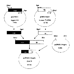
[6] FVII is a single-chain glycoprotein consisting of 406 amino acids, has
a molecular
weight of 50kDa, and is secreted into blood stream as a zymogen. FVII consists
of four
distinct domains, i.e., an amino terminal-carboxyglutamic acid (Gla) domain,
two
epidermal growth factor (EGF)-like domains and a serine protease domain (Hagen
FS
et al., Proc. Natl. Acad. Sci. USA, 83(8):2412-2416, 1986). FVII is converted
to its
activated form, FVIIa, by forming two polypeptide chains linked by a disulfide
bond,
i.e., N-terminal light chain (24 kDa) and C-terminal heavy chain (28 kDa)
through the
proteolysis of a single peptide bond located at Arg152-fle153. FVII is present
at a con-
centration of 500 ng/mL in plasma, and 1% (i.e., 5 ng/mL) of FVII is present
as FVIIa.
[7] Meanwhile, it has been reported that the half-life of FV11 in plasma is
approximately
4 hours (3-6 hours), while that of FVIIa is about 2.5 hours. Due to the short
half-life,
FVIIa is required to be administered via multiple intravenous injections or
continuous
injection. However, this would limit the therapeutic uses of FVIIa in terms of
high
treatment expenses and making the patient's discomfort.
[8] To overcome these problems, methods have been provided for preparing
fusion
proteins comprising FVII and a fusion partner linked thereto, but the
resulting proteins
had the problem of losing their biological activities, even though the short
in-vivo half-
life was somewhat improved compared to the unfused protein.
[9] Accordingly, there are needs for providing and securing a FVII fusion
protein which
has an improved in-vivo half-life while retaining the biological activity of
the natural
type FVII.
[10]
Disclosure of Invention
Technical Problem
[11] It is an object of the present invention to provide a fusion protein
having the bi-
ological activity of natural type FVII.
[12] It is other object of the present invention to provide a gene coding
for the fusion
protein.
[13] It is a further object of the present invention to provide a
recombinant vector
comprising the gene.
[14] It is a still further object of the present invention to provide a
host cell comprising the
recombinant vector.
[15]
CA 02801223 2012-11-28
3
Solution to Problem
[16] In accordance with one aspect of the present invention, there is
provided a fusion
protein comprising factor VII (FYI!) and transferrin, wherein the transferrin
is linked
to the C-terminus of the FVII.
[16a] According to a particular aspect, the invention relates to a fusion
protein comprising
factor VII (FYI!) and transferrin, wherein said transferrin is linked to the C-
terminus
of said FVII ,wherein the fusion protein further comprises a linker between
the FVII
and the transferrin, and wherein the linker comprises a protease cleavage
site, which is
capable of being cleaved by a protease selected from the group consisting of
thrombin,
factor Xa, factor IXa, and factor VIIa.
[17] In accordance with other aspect of the present invention, there is
provided a DNA
coding for the fusion protein.
[18] In accordance with a further aspect of the present invention, there is
provided a
recombinant vector comprising the DNA.
[19] In accordance with a still further aspect of the present invention,
there is provided a
host cell comprising the recombinant vector.
[20]
Advantageous Effects of Invention
[21] The fusion protein according to the present invention has an improved
in-vivo
half-life compared to the natural type FVII while retaining a high biological
activity of
FVII, and thus can be effectively employed in the therapy using FVII.
[22]
Brief Description of Drawings
[23] The above and other objects and features of the present invention will
become
apparent from the following description of the invention, when taken in
conjunction with the
accompanying drawings, which respectively show:
[24]
[25] Fig. 1 is a schematic diagram showing a cloning procedure for
constructing FVII-Tf
expression vector from a vector comprising a cDNA coding for FVII sequence and
a vector
comprising a cDNA coding for transferrin (TO sequence;
[26] Fig. 2 is a schematic diagram showing a procedure for constructing
FVII-GS1
(linker)-Tf expression vector by overlapping PCR;
[27] Fig. 3 is a schematic diagram showing a construction procedure of FVII-
GS
linker-Tf expression vectors comprising GS3, GS5, GS7, GS9, GS11, GS13, GS15
or GS-1-T
as a linker;
CA 2801223 2018-08-01
3a
[28] Fig. 4
displays Western blot results of VII-Tf, FVII-0S3-Tf,
FVII -GS5-Tf, FVI 1 FV1I-GS9-If, FVII-GS11-Tf, FVII-GS13-Tf,
FV1I-GS1-I-If and FVII-Helix-Tf fusion proteins of the present invention, FVII-
albumin
fusion protein (FVII-Alb) and FV11 (NovoSevenTm);
[29] Fig. 5 is a graph showing specific activities of FVII-Tf, FVII-GS1-Tf,
FVII-GS3-Tf,
FVII-GS5-Tf, FVII-GS7-Tf, FVIl-GS9-Tf, FVII-GS11-Tf, FVII-GS13-Tf, FVII-GS 15-
If,
FVII-GS1-T-Tf and FVII-Helix-Tf fusion proteins of the present invention, and
FVII-albumin fusion protein (FVII-Alb);
CA 2801223 2017-08-04
4
WO 2011/152694 PCT/KR2011/004131
[30] Fig. 6 presents the structure of a linker and restriction recognition
sequences at both
termini in FVII-GS1-T-Tf fusion protein; and
[31] Fig. 7 shows western blot results of purified FVII-Tf, FVII-GS1-Tf,
FVII-GS1-T-Tf,
FVII-GS3-Tf, and FVII-GS15-Tf fusion proteins of the present invention,
NovoSeven
TM and FVII.
[32]
Best Mode for Carrying out the Invention
[33] The present invention provides a fusion protein comprising factor VII
(FVII) and
transferrin.
[34] The FVTI and transferrin of the fusion protein of the present
invention may be
derived from any mammal, preferably a human. More preferably, FVII and
transferrin
used in the present invention may have not less than 95% of sequence
homologies with
those of natural type of the proteins found in human blood, respectively. Most
preferably, FVII has the amino acid sequence of SEQ ID NO: 1 and transferrin
has the
amino aicd sequence of SEQ ID NO: 2.
[35] In addition, FVII or transferrin used in the fusion protein of the
present invention
may be a functional equivalent or a functional derivative of natural type
thereof which
has a substantially equivalent functional activity. Exemplary functional
equivalents
include mutants induced by deletion, insertion or non-conservative or
conservative
substitution of any amino acid residues, or a combination thereof in amino
acid
sequences represented by SEQ ID NOs: 1 and 2, respectively, in which such
changes
do not substantially alter active sites or domains offering biological
activities to FVII.
[36] In some cases, the fusion protein of the present invention may be
modified, e.g., via
phosphorylation, sulfation, acrylation, glycosylation, methylation,
farnesylation,
acetylation, amidation, and others, for the improvement or reduction of its
physical or
chemical properties, and such functional derivatives also fall within the
scope of the
present invention so long as the biological activity of FVII is substantially
retained.
[37]
[38] In the fusion protein of the present invention, transferrin is
preferably linked to the
C-terminus of FVII. The fusion protein in the order of FVII-transferrin is
superior to a
fusion protein in the order of tranferrin-FVII, probably due to the exposure
of the N-
terminus of the FVII (see Table 3).
[39] The fusion protein of the present invention may further comprise
recognition
sequence(s) for a restriction enzyme between FVII and transferrin in order to
facilitate
the insertion of a linker as described below. The restriction recognition
sequence may
be any restriction recognition sequence known to one of ordinary skill in the
art, and
AgeI recognition sequence (A/CCGGT) may be preferably used. In other words,
fusion
CA 02801223 2012-11-28
5
WO 2011/152694 PCT/KR2011/004131
proteins, in which the restriction recognition sequence is linked to the C-
terminus of
FVII and transferrin is linked to the restriction recognition sequence, are
included
within the scope of the present invention.
[40]
[41] The present invention provides a fusion protein comprising a linker
between FVII
and transferrin.
[42] The linker may have 1 to 100 amino acids, preferably 1 to 75 amino
acids, more
preferably 5 to 25 amino acids, and it may be any peptides which can
functionally
separate FVII and transferrin. The linker may have a stable secondary
structure such as
a helix or be originated from IgG hinge region. Preferably, the linker may
rotate freely
in aqueous solution and does not have a fixed structure, and, therefore, it
would be
non-immunogenic and would increase FVII activities of fusion proteins by
minimizing
the potential interference between two fusion partners. As an example, such
linker may
be a helix linker represented by the amino acid sequence of SEQ ID NO: 11.
Further,
such flexible linker may contain glycine (G) and serine (S) in a repeated or
random
pattern. For example, the linker comprises (GGGGS)N (wherein N is an integer
ranging
from 1 to 20), and preferably has any one selected from the group consisting
of the
amino acid sequences of SEQ ID NOs: 3 to 11 (see Table 1). In addition, any
amino
acid sequences having not less than 80% of homologies with the linker,
preferably
having not less than 85% of homologies may be also used in the fusion protein
of the
present invention.
[43]
[44] Furthermore, the linker may also include protease cleavage site(s)
which is
recognized by protease(s) abundant in an injured tissue. The cleavage site may
be
cleaved by a protease selected from the group consisting of thrombin, factor
Xa, factor
IXa, and factor VIIa. The fusion protein having such protease cleavage site is
cleaved
at the working site to produce each protein, i.e., FVII and transferrin, and
the resulting
proteins function as individual proteins. Preferably, the linker has the amino
acid
sequence of SEQ ID NO: 12 (see Table 1).
[45]
[46] The linker may be inserted into a fusion protein more easily via the
restriction
enzyme recognition sequence which is located between FVII and transferrin. Ac-
cordingly, the restriction enzyme recognition sequence may be present at any
one end
or both ends of the linker, and be in turn translated into amino acids encoded
by the
sequence. For example, when AgeI restriction enzyme recognition sequence is
used,
Thr may be present at the N-terminus of the linker and Thr-Gly may be present
at the
C-terminus of the linker. That is, when a linker (GGGGS)3 is used, The
recognition
sequence and linker may be present in a form of --T(GGGGS)3TG--. The amino
acids
CA 02801223 2012-11-28
6
WO 2011/152694 PCT/KR2011/004131
translated at the N- and C-termini of the linker may vary depending on the
restriction
enzyme recognition sequence employed, but the presence thereof does not
influence
the activities of the fusion proteins (see Table 5).
[47]
[48] The fusion protein of the present invention exhibits not less than 0.7
of FVII specific
activity compared to the unfused natural type FVII.
[49] In an aspect of the present invention, the fusion protein, which
comprises FVII rep-
resented by the amino acid sequence of SEQ ID NO: 1 and transferrin
represented by
the amino acid sequence of SEQ ID NO: 2, has about 0.82 to about 0.92 of FVII
specific activity compared to the unfused natural type FVII (see Tables 2 and
3).
[50] In addition, the fusion protein, which comprises FVII represented by
the amino acid
sequence of SEQ ID NO: 1, the linker represented by the amino acid sequence of
SEQ
ID NO: 3 and transferrin represented by the amino acid sequence of SEQ ID NO:
2,
has about 0.97 of FVII specific activity compared to the unfused natural type
FV11 (see
Table 2).
[51] The fusion proteins according to the present invention, in which other
linkers are
inserted between FVII and transferrin, have also about 0.74 to about 1 of FVII
specific
activities compared to the unfused natural type FVII (see Table 2).
[52] Furthermore, the fusion protein of the present invention has a half-
life 3-4 times
longer than that of FVII with no transferrin linked thereto (see Table 6).
[53]
[54] The present invention also provides a DNA coding for the fusion
protein.
[55] The DNA coding for the fusion protein may be subjected to various
changes and
modifications due to the codon's degeneracy or considering codons preferred in
the
organism to express the fusion protein, unless the amino acid sequence of the
fusion
protein is substantially altered, and the modified DNAs are also included in
the scope
of the present invention. In the present invention, the DNA coding for the
fusion
protein may be preferably represented by any one of the nucleotide sequences
of SEQ
ID NOs: 13 to 24. The DNA coding for the fusion protein of the present
invention may
be provided by a vector for expressing the DNA.
[56]
[57] The present invention provides a recombinant vector comprising the DNA
coding for
the fusion protein.
[58] The term "vector" used herein refers to a means for introducing a DNA
coding for
said fusion protein into a host cell and expressing the fusion protein
therein. The vector
may include all conventional vectors such as plasmid vectors, cosmid vectors,
bacte-
riophage vectors, virus vectors, and others, preferably a plasmid vector.
[59] A suitable expression vector contains expression regulatory elements
such as
CA 02801223 2012-11-28
7
WO 2011/152694 PCT/KR2011/004131
promoters, initiation codons, termination codons, polyadenylation signals and
enhancers, as well as signal sequences or leader sequences for membrane
targeting or
secretion, and it may be diversely prepared according to the purposes. The
initiation
codon and termination codon should be sure to work in an organism where the
gene
construct is administered, and be in-frame with the coding sequence. Further,
the ex-
pression vector contains a selective marker for selecting a host cell
containing the
vector, and an origin of replication if the expression vector is reproducible.
The vector
may self-replicate or be integrated into the DNA of a host cell.
[60] Specifically, the recombinant expression vector according to the
present invention
may be prepared by inserting a DNA coding for the fusion protein sequence into
pcDNA3.1-hygro vector.
[61]
[62] Further, the present invention provides a host cell which produces the
fusion protein
by transformation with said recombinant expression vector.
[63] Since the expression levels and modifications of the proteins vary
depending on the
type of host cells, it is preferred to choose a host cell most suitable for
the purpose.
Examples of host cells include mammal cells, e.g., Chinese hamster ovary (CHO)
cells, human embryonic kidney cells (HEK293), hamster kidney cells (BHK 21),
human liver cancer cells (Hep G2), and others, but not limited thereto.
[64] In order to transform a host cell with the recombinant expression
vector according to
the present invention, any method known to those of ordinary skill in the art
may be
employed, and the example of such method includes, but not limited to, electro-
poration, protoplast fusion, calcium phosphate (CaPO4) precipitation, calcium
chloride
(CaCl2) precipitation, and others.
[65]
Mode for the Invention
[66] The following Examples are given for the purpose of illustration only,
and are not
intended to limit the scope of the present invention.
[67]
[68] Example 1: Preparation of factor VII (FYI!) plasmid vector
(pcDNA3.1-hygro-FVII)
[69]
[70] Total RNA purified from Hep G2 cells (KCLB No. 88065) was used as a
template
for reverse transcription. Complementary DNA (cDNA) transcript was amplified
by
PCR using FVII gene specific primers, FVII-F and FVII-R (SEQ ID NOs: 25 and
26)
to obtain open reading frame of human FVII gene. The PCR was performed by
treating
50 [11_, of reaction solution (0.5 4, of cDNA, 0.4 [IIVI (10 pmoWL) of primers
of SEQ
CA 02801223 2012-11-28
8
WO 2011/152694 PCT/KR2011/004131
ID NOs: 25 and 26, 0.2 mM of dNTP, 5 unit of Taq DNA polymerase and water)
under
the following condition: 1 cycle of denaturation at 94 C for 5 min, 35 cycles
of ampli-
fication at 94 C for 1 min, at 60 C for 1 min, and at 72 C for 2.5 min, and 1
cycle of
final extension at 72 C for 5 min. The purified PCR product was cloned into
pGEM-T
easy vector (Promega, Cat #: A1360). Positive clones were selected by
restriction
digestion using EcoRI and NcoI. The Selected clones were further verified by
DNA se-
quencing. To transfer ORF of FVII (FVII-ORF) to an expression vector, FVII-ORF
cleaved with Nod was blunted by T4 DNA polymerase and ligated with
pcDNA3.1-hygro vector (Invitrogen) digested with HindIII1XbaI and blunted. The
ligated vector was confirmed by restriction digestion with ApaI, Xbal, EcoRI,
Ncol, Psi'
I and DNA sequencing. This vector was designated 'pcDNA3.1-hygro-FVII'.
[71]
[72] Example 2: Construction of FVII-Tf expression vector
(pcDNA3.1-hygro-FVII-Tf)
[73]
[74] FVII cDNA prepared in Example 1 was fused to human transferrin (Tf)
cDNA in
order to express as a single zymogen in an animal cell. Human tranfenin cDNA
was
purchased from Origene (Cat #: SC322130) and was verified whether it is equal
to the
sequence of GenBank accession #: NM_001063.2. Primers used in the fusion were
designed to remove termination codon of FVII and signal peptide of
transferrin. In
order to facilitate the insertion of the varying sizes of linkers between FVII
and Tf, Age
I site (ACCGGT), which will be translated to threonine (Thr) and glycine
(Gly), was
added to the linking primers. The resulting fusion protein would have the
following
structure: (leader peptide)-(mature FVII)-(Thr-Gly)-(mature Tf) (in which the
leader
peptide consists of a signal peptide (prepeptide) not present in mature FVII
and a
propeptide to be cleaved by a processing enzyme, which is composed of 38 amino
acids and corresponds to amino acids ranging from positions 1 to 38 in the
amino acid
sequence of SEQ ID NO:1). cDNAs of FVII and Tf were amplified by using primers
FVII-S1, FVII-AS1, Tf-S1 and Tf-AS1 (SEQ ID NOs: 27 to 30) and the vector as
described in Example 1 was used. The primers of SEQ ID NOs: 27 and 30 contains
NheI and Xhol sites, respectively.
[75] A cloning strategy for linking of FVII cDNA and Tf cDNA is depicted in
Fig. 1.
First, FVII cDNA was amplified from pcDNA3.1-hygro-FVII vector by PCR. The
PCR was performed by treating 50 4 of reaction solution (1 4 of vector
template, 1
[IL of a primer set, FVII-S1 and FVII-AS1(10 [IM), 10 [iL of 5x Phusion HF
buffer,
200 [iM of dNTP, 0.5 [iL of Phusion DNA polymerase (FINNZYMES, #F-5305, 2
units/4) and 35.5 4 of water) under the following condition: 1 cycle of
denaturation
at 98 C for 30 sec, 30 cycles of amplification at 98 C for 10 sec, at 60 C for
45 sec. and
CA 02801223 2012-11-28
9
WO 2011/152694 PCT/KR2011/004131
at 72 C for 30 sec, and 1 cycle of final extension at 72 C for 7 min.
[76] Next, Tf was amplified using transferrin cDNA as a template. The above
mentioned
PCR procedure was repeated except for using a primer set (Tf-S1: 10 [iM; Tf-
AS1: 10
[77] The amplified FVII and Tf cDNA were joined by a series of restriction
and ligation.
Each DNA amplified by PCR was digested with AgeI and XhoI, or with Nhel. The
digested DNAs were purified and ligated at 1:1 molar ratio. The ligated DNA
was
subcloned into pcDNA3.1-hygro vector (Invitrogen) digested with NhellXhol. The
size
and sequence of insert was further verified by DNA sequencing.
[78]
[79] Example 3: Construction of FVII-GS linker-Tf expression vector
[80]
[81] A peptide consisting of 5 amino acids comprising glycine and serine
was used as a
basic linker unit. The basic linker unit comprises four glycines and one
serine with
following sequence: 'GGGGS'. The basic linker unit (hereinafter, referred to
"GS-X
linker" in which X is the repeat number of the basic GS linker unit) was
utilized to
constitute the longer GS linkers. In this Example, the linkers ranging from GS-
1 to GS-
15 were constructed.
[82]
[83] 1) Construction of FVII-GS-1 linker-Tf expression vector
[84]
[85] A set of primers, GS-FV-AS1 and GS-Tf-S1 (SEQ ID NOs: 31 and 32),
containing
the basic GS linker unit was synthesized and inserted between FVII and Tf by
overlapping PCR (see Fig. 2).
[86] The GS-1 linker was linked to FVII by PCR using a set of primers FVII-
Sl and GS-
FV-ASI (SEQ ID NOs: 27 and 31) and Phusion DNA polymerase (FINNZYMES,
#F-5305). The PCR was performed by treating 50 4 of reaction solution (1 4 of
pcDNA3.1-hygro-FVII-Tf vector, 1 4 of FVII-Sl (10 pmole/14), 1 4 of GS-
FV-AS1 (10 pmole/4), 1 4 of 10 mM dNTP. 10 4 of 5x Phusion HF buffer, 35.5
4 of water and 0.5 4 of Phusion DNA polymerase (2 unit/4)) under the following
condition: 1 cycle of denaturation at 98 C for 30 sec, 35 cycles of
amplification at 98 C
for 10 sec, at 64 C for 30 sec, and at 72 C for 45 sec, and 1 cycle of final
extension at
72 C for 7 min. Meanwhile, to connect the GS-1 linker to Tf, the above PCR
procedure
was repeated except for using a set of primers GS-Tf-S1 and Tf-AS1 (SEQ ID
NOs: 32
and 30). The amplified PCR products were utilized as overlapping PCR
templates. The
overlapping PCR was performed by treating the reaction solution (1 4 of
amplified
PCR products, 1 4 of FVII-S1 (10 pmole/4, SEQ ID NO: 27), 1 4 of antisense
primer (Tf-AS1 10 pmole/4, SEQ ID NO: 30), 10 4 of 5x Phusion HF buffer, 1 4
CA 02801223 2012-11-28
10
WO 2011/152694 PCT/KR2011/004131
of 10 mM dNTP, 34.5 tL of water, 0.5 [IL, of Phusion DNA polymerase (2 units/
L))
under the following condition: 1 cycle of denaturation at 98 C for 1 min, 45
cycles of
amplification at 98 C for 10 sec, at 66/68 C for 30 sec, and at 72 C for 45
sec, and 1
cycle of final extension at 72 C for 7 min. The amplified overlapping PCR
product was
cloned into pcDNA3.1-hygro-lacZ digested with Nile' and XhoI.
[87]
[88] 2) Construction of FVII-GS-3 linker-Tf expression vector
[89]
[90] A primer set, GS3-S and GS3-AS (SEQ ID NOs: 33 and 34), containing GS-
3 and
AgeI site was synthesized. To make a GS-3 double stranded linker, the primers
were
annealed by heating a mixture (5 [th of GS3-S (100 pmole4iL), 5 [IL, of GS3-AS
(100
pmole/[kL), 2 !IL of 10X annealing buffer (100 mM Tris-Cl [pH 8.01, 1 M NaCl,
10
mM EDTA) and 8 [IL, of water) at 98 C for 10 min and cooling at 25 C for 1
hour. The
annealed linker was digested with Agel and pcDNA3.1-hygro-FV11-Tf vector
prepared
in Example 2 was also digested with AgeI. The digested vector was treated with
1 tL
of CIP (Calf intestinal phosphatase: NEB, #M0290S) at 37 C for 1 hour and
subjected
to gel extraction procedure (QIAGEN, #28704), followed by ligation at a molar
ratio
of 1:3 (vector: insert) using T4 DNA ligase (TAKARA, #2011A).
[91]
[92] 3) Construction of FVII-GS-5 linker-Tf to FVII-GS-15 linker-Tf
expression vectors
[93]
[94] In order to construct a fusion protein expression vector containing GS-
5 linker, a new
strategy was implemented. FVII-Tf fusion vectors containing extended linkers
were
constructed by following two steps.
[95] First step is adding a synthesized double stranded (ds) G52 linker to
the previously
obtained linker. After assuring the extension of linker, the linker was cut
out and
inserted into between FVII and Tf genes in pcDNA3.1-hygro-FVII-Tf vector. For
example, to extend the GS-3 linker to GS-5 linker, the synthesized dsGS-2
linker unit
of SEQ ID NO: 35 was digested with Bg111 and ligated with
pcDNA3.1-hygro-FVII-GS3-Tf vector treated with BamHI and StuI. Next, after
confirming the extension of the linker by BamH1 and AgeI digestion, the
extended
linker was cut out with AgeI and subcloned into pcDNA3.1-hygro-FVII-Tf vector
treated with AgeI and CIP. FVII-Tf fusion expression vectors containing GS-7,
GS-9,
GS-11, GS-13 and GS-15 linkers were constructed by the same strategy (see Fig.
3).
[96]
[97] Example 4: Construction of FVII-Tf expression vector
(pcDNA3.1-hygro-FVII-GS1-T-Tf) containing a linker comprising thrombin
cleavage site
CA 02801223 2012-11-28
11
WO 2011/152694 PCT/KR2011/004131
[98]
[99] A linker containing thrombin cleavage site was prepared by adjoining a
GS-1 unit to
both ends of thrombin recognition sequence (hereinafter, referred to "GS1-T
linker").
dsGS1-T linker of SEQ ID NO: 36 (sense) was designed and synthesized to
contain
Agel sites at both ends. The dsGS1-T linker was digested with Agel and
purified using
PCR purification kit (Qiagen, cat #: 28104). The purified linker was ligated
into
pcDNA3.1-hygro-FVII-Tf vector treated with CIP/AgeI.
[100]
[101] Example 5: Construction of FVII-Tf expression vector
(pcDNA3.1-hygro-FVII-Helix-Tf) containing a helix linker
[102]
[103] A helix linker DNA was prepared by the method disclosed in U.S Laid-
open Pub-
lication No. 2009/0170163. Agel site was added to the both ends of the
prepared helix
linker DNA by using primers, Helix linker S and Helix linker AS (SEQ ID NOs:
37
and 38). The primers Helix linker S and Helix linker AS were annealed and
digested
with Agel, followed by insertion into pcDNA3.1-hygro-FVII-Tf vector treated
with
Agel and CIP. The constructed vector was confirmed by DNA sequencing.
[104]
[105] Comparative Example 1: Construction of FVII-albumin fusion expression
vector (FVII-Alb)
[106]
[107] The FVII-albumin fusion protein disclosed in EP Patent No. 1816201
was con-
structed. Human albumin cDNA was obtained by RT-PCR using human liver mRNA
(Clontech) as a template and albumin gene-specific primers, Albumin-S and
Albumin-
AS (SEQ ID NOs: 39 and 40). RT-PCR was performed by using AccuScript High
Fideleity RT-PCR system kit (Cat# 600180) according to the manufacturer's
manual.
First, 10 4 of reverse transcription reaction solution (1 4 of 10X Reverse
Tran-
scriptase buffer, 0.6 4 of oligo-dT primer, 1 4 of dNTP, 0.4 4 of water, 5 4
of
human liver mRNA (10 ng/4)) was kept at 65 C for 5 min and at a room
temperature
for 5 min, and was subjected to a reaction with 1 4 of 100 mM DTT and 1 4 of
Reverse Transcriptase at 42 C for 1 hour. A human albumin sequence was
obtained by
PCR using the synthesized cDNA as a template and primers Albumin-S and Albumin-
AS. PCR was performed by treating 50 4 of a reaction solution (1 4 of cDNA, 10
4 of 5x Phusion HF buffer, 1 4 of primers Albumin-S and Albumin-AS, re-
spectively, 1 4 of 10 mM dNTP, 0.5 4 of Phusion DNA polymerase (FINNZYMES,
#F-530S; 2 units/4) and 35.5 4 of water) under the following condition: 1
cycle of
denaturation at 98 C for 1 mM, 30 cycles of amplification at 98 C for 10 sec,
at 62 C
for 30 sec, and at 72 C for 60 sec, and 1 cycle of final extension at 72 C for
7 mM. The
CA 02801223 2012-11-28
12
WO 2011/152694 PCT/KR2011/004131
synthesized oligonucleotides of SEQ ID NOs: 41 and 42 were annealed to the GS
linker [SS(GGS)9G5] (SEQ ID NO: 45) disclosed in EP Patent No. 1816201. To
connect the FVII cDNA prepared in Example 1 to the above linker, FVII
termination
codon in pcDNA3.1-hygro-FVII vector was replaced with no' site by PCR-based mu-
tagenesis using primers mut FVII(XhoI)-S and mut FVII(XhoI)-AS (SEQ ID NOs: 43
and 44). Using XhollApaI sites, the GS linker ISS(GGS)9GS1 was fused to 3' end
of
FVII cDNA in pcDNA3.1-hygro-FVII vector. Finally, the human albumin cDNA
digested with BamHI was inserted into pcDNA3.1-hygro-FVII-GS-linker vector.
The
prepared pcDNA3.1-hygro-FVII-GS-linker-albumin expression vector was verified
by
DNA sequencing.
[1081
[109] The characteristics of expression vectors constructed in Examples 2
to 5 and Com-
parative Example 1 were shown in Table 1.
[110] Table 1
CA 02801223 2012-11-28
13
WO 2011/152694 PCT/KR2011/004131
[Table 1]
FVII fusion C-terminu Linker sequence (SEQ ID NO) N-terminus of Number
protein s of FVII fusion partner of amino
acids in
linker
FVII-Tf APFP VPDKTV 0
FVII-GS1-Tf APFP GGGGS (SEQ ID NO: 3) VPDKTV 5
FVII-GS3-Tf APFP (GGGGS)3 (SEQ ID NO: 4) VPDKTV 15
FVII-GS5-Tf APFP (GGGGS)5 (SEQ ID NO: 5) VPDKTV 25
FVII-GS7-Tf APFP (GGGGS)7 (SEQ ID NO: 6) VPDKTV 35
FVII-GS9-Tf APFP (GGGGS)9 (SEQ ID NO: 7) VPDKTV 45
FVII-GS11-Tf APFP (GGGGS)ii (SEQ ID NO: 8) VPDKTV 55
FVII-GS13-Tf APFP (GGGGS)13 (SEQ ID NO: 9) VPDKTV 65
FVII-GS15-Tf APFP (GGGGS)15 (SEQ ID NO: 10) VPDKTV 75
FVII-GS1-T-T APFP GGGGSLVPRGSGGGS (SEQ VPDKTV 15
ID NO: 12)
FVII-Helix-Tf APFP GA(EAAAK)4A (SEQ ID NO: VPDKTV 23
11)
FVII-Alb APFP SS(GGS)9G5 (SEQ ID NO: 45) DAHK 31
* For FVII-Tf, Thr-Gly derived from Agel is present.* For the linkers of SEQ
ID
NOs: 4 to 12, Thr derived from Agel is present at the N-terminus, and Thr-Gly
derived from Agel is present at the C-terminus.
[111]
[112] Experimental Example 1: Measurement of specific activities of FVII-
fusion
proteins
[113]
[114] The FVII-fusion proteins constructed in Example 2 to 5 and
Comparative Example 1
were expressed in a CHO cell (CHO(VK2)) which stably expresses VKORCI (vitamin
K epoxide reductase complex subunit 1).
[115] The expression vectors constructed in Example 2 to 5 and Comparative
Example 1
were purified by using Endo-free plasmid maxi kit (Qiagen, #27104). li-
galactosidase
was used as an internal control for transfection. CHO (VK2) cells were seeded
at a
density of 1.5x10⁶ cells/well in 6-well plates. The cells were incubated in a-
MEM
(Lonza, #12-169F) supplemented with 10% FBS (Lonza, #14-501F), 1X HT
CA 02801223 2012-11-28
14
WO 2011/152694 PCT/KR2011/004131
(Invitrogen,#11067-030), 4 mM L-glutamine (Lonza, #17-605E) and 200 [ig/mL of
hy-
gromycin (Invitrogen, #10687-010) for 24 hours, and then transfected using
lipo-
fectamine 2000 (Invitrogen) according to the manufacturer's manual. Four hours
after
transfection, the medium was replaced with serum-free medium (OptiMEM), and 5
[ig/
mL of vitamin K was supplemented. After 48 hours of incubation, the culture
medium
was sampled and stored at -70 C.
[1161 FVII-fusion proteins expressed were analyzed for their chromogenic
activities and
antigen amounts by using COATEST factor VII assay kit (Chrmogenix, #821900-63)
and FVII ELISA kit (Cedarlene Lab, #CL20030K), respectively. The assays were
performed according to the manufacturer's manual. Standard human plasma
normalized against WHO standard was used as a control FVII in both assays. The
ex-
pression of protein was assessed via western blot analysis. Equal amounts of
FVII
fusion proteins were loaded based on the ELISA results. The expressed FVII-
fusion
proteins were found to have the expected sizes without detectable
fragmentation (see
Fig. 4).
[117] Meanwhile, the specific activities of FVII-transferrin fusion
proteins were 0.74 to 1,
which were higher compared to that of FVII-albumin fusion protein (0.52) (see
Table
2). The FVII-transferrin fusion proteins containing linkers also retained not
less than
70% of FVII activities. There was no relationship between the linker lengths
and the
specific activities, but FVII fusion proteins with shorter GS linkers showed
somewhat
higher specific activities than fusion proteins with longer ones. In
particular, FVII-
GS1-Tf and FVII-GS1-T-Tf fusion proteins showed comparable specific activities
(see
Fig. 5).
[118] Table 2
CA 02801223 2012-11-28
15
WO 2011/152694 PCT/KR2011/004131
[Table 2]
FVII fusion Antigen (%) Activity (%) Specific activity
protein (activity/antigen)
FVII-Tf 53.2 5.0 43.9 0.3 0.82
FVII-GS1-Tf 53.4 3.1 52.0 0.5 0.97
FVII-G53-Tf 61.9 8.0 57.7 0.2 0.93
FVII-G55-Tf 69.3 5.6 55.9 1.4 0.81
FVII-G57-Tf 70.9 8.2 59.3 1.1 0.84
FVII-G59-Tf 64.2 8.6 47.5 0.7 0.74
FVII-GS11-Tf 59.1 3.9 45.3 0.9 0.77
FVII-G513-Tf 59.7 5.1 49.1 0.8 0.82
FVII-GS15-Tf 59.2 6.0 50.2 0.5 0.85
FVII-GS1-T-Tf 70.8 8.7 71.0 2.6 1.00
FVII-Helix-Tf 89.0 5.7 78.9 2.2 0.89
FVII-Alb 106.6 5.4 54.9 3.3 0.52
[119]
[120] Example 6: Characterization of FVII fusion proteins according to the
direction
of Tf fusion
[1211
[122] In this Example, a fusion protein in which human transferrin (TO is
linked to N-
terminus of FVII was prepared and compared with a fusion protein in which
transferrin
is linked to the C-terminus of FVII, in order to examine the change of
characteristics
according to the direction of fusion in fusion proteins. Detailed procedure is
as follows.
[123]
[124] <6-1> Construction of Tf-FVII and Tf-GS1-T-FVII expression vectors
[125]
[126] Two fusion proteins with Tf linked to N-terminus of FVII were
designed as follows:
(1) (leader peptide of Tf)-(mature Tf)-(Thr-Gly)-(mature FVII); and (2)
(leader peptide
of Tf)-(mature TO-(Thr)-(GS1-T; SEQ ID NO: 12)-(Thr-Gly)-(mature FVII).
[127] First, in order to obtain a Tf gene sequence containing a leader
peptide, a forward
primer (Nhe-Tf: SEQ ID NO: 46) was designed to contain Nhel site for the
purpose of
cloning and a reverse primer (Tf-Age: SEQ ID NO: 47) was designed to contain
Agel
site for the purpose of removing the termination codon of transferrin and
cloning. For
cloning of mature FVII with leader peptide removed, a forward primer (Age-
FVII:
CA 02801223 2012-11-28
16
WO 2011/152694 PCT/KR2011/004131
SEQ ID NO: 48) was designed to contain AgeI site and a reverse primer was
designed
to contain Xhol site.
[128] For Tf gene, cDNA purchased from Origene (Cat #: SC322130) as in
Example 2 was
used as a PCR template. The PCR was performed by treating 50 [IL of a reaction
solution (1 [IL of vector template, 2 [IL of primers Mie-Tf and Tf-AgeI (10
[1M), 10 [IL
of 5x Phusion HF buffer, 1 [IL of 10 mM dNTP, 0.5 [IL of Phusion DNA
polymerase
(FINNZYMES, #F-530S, 2 units/[1L) and 33.5 [IL of water) under the following
condition: 1 cycle of denaturation at 98 C for 30 sec, 25 cycles of
amplification at 98 C
for 10 sec, at 70 C for 30 sec, and at 72 C for 36 sec, and 1 cycle of final
extension at
72 C for 10 min. FVII was amplified by PCR using pcDNA3.1-hygro-FVII-GS1-T-Tf
vector as a template as in Example 4. The PCR conditions were same with the
above
Tf PCR conditions, except for using primers Age-FVII (10 [iM) and VII-Xho (10
[1M).
[129] The amplified Tf gene was inserted into pcDNA3.1-hygro-FVII-GS1-T-Tf
vector by
using NhellAgel to obtain pcDNA3.1-hygro-Tf-Tf vector. The pcDNA3.1-hygro-Tf-
Tf
vector and the FVII PCR product were digested with Age llXhol and ligated to
construct an expression vector containing pcDNA3.1-hygro-Tf-FVII fusion
protein.
pcDNA3.1-hygro-Tf-GS1-T-FVII expression vector was constructed by inserting
the
dsGS1-T sequence synthesized in Example 4 via Agel. The constructed expression
vectors were confirmed by restriction mapping and DNA sequencing.
[130]
[131] <6-2> Expression of fusion proteins and characterization
[132]
[133] In order to characterize fusion proteins with Tf linked to the N-
terminus of FVII, the
expression vectors thereof, i.e., pcDNA3.1-hygro-FVII-Tf,
pcDNA3.1-hygro-FVII-GS1-Tf-VII, pcDNA3.1-hygro-Tf-FVII and
pcDNA3.1 -hygro-Tf-GS1-T-FVII, were transiently expressed in CHO cells.
[134] The constructed four plasmid DNAs were isolated using Endo-free maxi
prep kit
(Qiagen). On one day before transfection, CHO (DG44) cells cultured in T75
flasks
were isolated by trypsin, and seeded at a density of 1.5x10⁶ cells/well in 6-
well plates.
After 24 hours, the cells were transfected according to the manufacturer's
manual.
Four hours after transfection, the medium in each well was removed and
replaced with
2 mL of a growth medium supplemented with 5 [tg/mL of vitamin K. After
transfection, the 6-well plate was incubated in a 37 C, 5% CO2 incubator, and
after 48
hours, the medium was harvested. The harvested supernatant was transferred to
1.5 mL
tubes and stored -70 C for chromogenic assay and ELISA of FVII. The plate with
the
medium removed was washed with 2 mL of HBSS per a well and lysed with 250 [IL
of
a lysis solution (Tropix, #ABX210LM, 1 mM DTT addition), followed by being
stored
at -70 C for P-galactosidase assay.
CA 02801223 2012-11-28
17
WO 2011/152694 PCT/KR2011/004131
[135] The chromogenic assay and FVII ELISA were performed as in
Experimental
Example 1. Samples for analysis were prepared by thawing the frozen-stored
media
following the transfection just before the experiment and obtaining the
supernatant via
centrifugation. Standard human plasma (Dade Behring, # ORKL13, Lot#503216F)
was
used as a standard in the assay.
[136]
[137] The measurement results are shown in Table 3. For Tf-FVII and Tf-GS1-
T-Tf with
Tf linked to N-terminus of FVII, no activities were measured unlike FVII-Tf
and FVII-
GS1-T-Tf with Tf linked to the C-terminus of FVII. Further, low amounts of
fusion
proteins with Tf linked to N-terminus were detected in FVII ELISA. However,
the
fusion proteins showed similar detection sensitivities, irrespective of fusion
directions,
in western blot results using polyclonal antibodies of Tf. The results
indicate that,
when Tf is linked to N-terminus of FVII, fusion proteins with no FVII
activities were
generated, even though translations of amino acids were normally conducted.
[138] Table 3
[Table 3]
Fusion protein FVII activity (%) FVII antigen (%) Specific activity
(activity/antigen)
FVII-Tf 33.8 0.73 37.0 2.17 0.92
Tf-FVII not detected 8.0 1.51
FVII-GS-1-T-Tf 46.6 0.29 43.0 4.75 1.08
Tf-GS- 1-T- Tf not detected 13.5 1.01
[139]
[140] Example 7: Characterization of fusion proteins according to the
deletion of re-
striction enzyme recognition sequence used in the fusion
[141]
[1421 In Example 2, restriction enzyme (A gel) recognition sequences were
used to facilitate
the insertion of various linkers between FVII and Tf. As a consequence, some
fusion
proteins became to have Thr and Gly which are encoded by above restriction
enzyme,
at both ends of the linkers. In this Example, it was examined whether the
properties of
fusion proteins are altered or not by the presence of the restriction enzyme
(A gel)
recognition sequence.
[143] The restriction enzyme recognition sequences were deleted from FVII-
GS1-T-Tf
fusion protein containing GS1-T linker, by PCR-based site-directed mutagenesis
using
mutagenic primers. As shown in Fig. 6, this experiment was designed to delete
"Thr" at
the N-terminus and "Thr-Gly" at the C-terminus and primers used in the
experiment
CA 02801223 2012-11-28
18
WO 2011/152694
PCT/KR2011/004131
are listed in Table 4.
[144] Table 4
[Table 4]
Primer Sequence (5'¨>3') SEQ ID NO
TG del-S CAG CGG AGG CGG TTC AGT CCC TGA TAA 50
AAC TG
TG del-AS CAG TTT TAT CAG GGA CTG AAC CGC CTC CGC 51
TG
T del-S CGA GCC CCA TTT CCC GGT GGA GGC GGA TC 52
T del-AS GAT CCG CCT CCA CCG GGA AAT GGG GCT CG 53
[145]
[1461 <7-1> Deletion of Thr-Gly
[147]
[148] PCR-based mutagenesis was conducted. The PCR was performed by
treating a
reaction solution (1 0_, of pcDNA3.1-hygro-FVII-GS1-T-Tf vector, 0.2 [tI_, of
sense
primer (TG del-S 10 IM), 0.2 tL of antisense primer (TG del-AS 10 uM), 1 0_,
of 10
mM dNTP, 4 0_, of 5X PCR buffer, 14 0_, of water, and 0.2 [IL of Phusion DNA
polymerase (FINNZYMES, #F-5305)) under the following condition: 1 cycle of de-
naturation at 98 C for 30 sec, 18 cycles of amplification at 98 C for 10 sec,
at 58 C for
30 sec, and at 72 C for 3 min, and 1 cycle of final extension at 72 C for 7
min. In order
to remove the original template DNA, the amplified PCR product was treated
with 1
0_, of Dpnl (NEB, #R0176S) and incubated at 37 C for 1 hour. 50 0_, of HIT
competent cell (DH5a, RH617) was transformed using 10 0_, of the DpriI-treated
DNA
and incubated in at an LB+amp (10 mg/mL) solid medium overnight. Four clones
thus
obtained were analyzed by DNA sequencing and two clones were verified as
mutants.
[149]
[150] <7-2> Deletion of Thr
[151]
[1521 In order
to delete Thr, the similar method as in Example <7-1> was conducted by
using different primers. Briefly, PCR-based mutagenesis was performed by
using, as a
template, 1 uL of plasmid DNA of the clones in which the mutation was
confirmed in
Example <7-1> and 1 [IL of sense primer (T del-S; 10 pmole) and 1 [IL of antis
ense
primer (T del-AS; 10 pmole), under the same condition. By DNA sequencing of
four
clones which were selected, three clones were confirmed to have mutations. The
secured expression vector was named "pcDNA3.1-hygro-FVII-GS1-T-Tf(M3)".
[1531
CA 02801223 2012-11-28
19
WO 2011/152694 PCT/KR2011/004131
[154] <7-3> Characterization of fusion proteins with restriction enzyme
recognition
sequences deleted
[155]
[156] CHO cells were transfected with FVII-GS1-T-Tf and FVII-GS1-T-Tf(M3)
ex-
pression vectors and FVII-Alb expression vector as a control, and the
supernatants of
the media were obtained. The obtained supernatants were subjected to FVII
chromogenic assay (Chromogenix) and FVII ELISA (cedarlane) to verify the
change
in the ratio of activity/antigen. As shown in Table 5, the antigen amounts and
activities
of FVII-GSI-T-Tf and FVII-GSI-T-Tf(M3) fusion proteins were almost equal each
other, and the ratios (specific activities) also did not vary. In addition,
the specific ac-
tivities were confirmed to be significantly higher than that of FVII-Alb
fusion protein.
[157] Table 5
[Table 5]
FVII antigen (%) FVII activity (%) Specific activity
(activity/antigen)
FVII-GS1-T-Tf 34.1 2.1 39.6 2.2 1.16
FVII-GS1-T-Tf(M3) 33.5 4.7 38.0 0.7 1.14
FVII-Alb 50.1 1.2 26.3 0.8 0.53
[158]
[159] Example 8: Measurement of half-life of fusion proteins
[160]
[161] In order to examine the increase of half-life in the fusion proteins
according to the
present invention, FVII-Tf. FVII-GS1-Tf, FVII-G53-Tf, FVII-GS15-Tf and FVII-
GS1-T-Tf were used as experimental groups, and a wild type FVII and
commercially
available FVIIa (NovoSeven ; Novo Nordisk) were used as control groups.
[162]
[163] <8-1> Sample preparation
[164]
[165] 1) Securing expression medium
[166]
[167] Wild type FVII protein and five Tf-fused FVII fusion proteins were
expressed in
FreeStyleTM CHO-S cell line (Invitrogen, Cat. no. R800-07). The CHO-S cells
were
cultured in suspension in a spinner flask with freestyle CHO expression medium
sup-
plemented with 8 mM L-glu (GIBCO, L-glutamine 200 mM (100X), Cat. No.
25030-081). The cultured cells were seeded at a density of 4x10⁵ cells/mL
before 24
hours to transfection, and were transfected when the density becomes
lx106cells/mL.
CA 02801223 2012-11-28
20
WO 2011/152694 PCT/KR2011/004131
The DNAs used in the transfection were prepared by using Endo-free maxi prep
kit
(QIAGEN, Cat. No.12362) or Endo-free plasmid mega prep kit (QIAGEN, 12381),
and the transfection was conducted in reference to the transfection protocol
of
FreeStyle MAX Reagent (Invitrogen, Cat. No. 16447-100). 500 [Ig of DNA was
added
to 8 mL of OptiPRO SFM (Invitrogen, Cat. No. 12309-019) and mixed. To another
tube, 8 mL of OptiPRO SFM (Invitrogen, Cat. No. 12309-019) and 500 [iL of
FreeStyle Max Reagent was added, and then the above two mixture was slowly
mixed
and stored at room temperature for 10 min. After 10 mM, FreeStyleTM CHO-S
cells
were transfected with the mixture. The transfected cells were cultured in a 37
C, 5%
CO2 incubator for 3-5 days, and then the supernatant was obtained.
[168]
[169] 2) Purification of expression medium
[170]
[171] The medium obtained by a spinner flask culture was filtered through
0.22 [1m of filter
(Corning) to remove remaining cells and debris. The filtered medium was
concentrated
10-fold by ultrafiltration using tangential-flow membrane (satorious, 30KDa).
The
concentrated medium was applied to an XK16/20 column (GE healthcare) charged
with Ceramic Hydroxyapatite (BIO-RAD, 157-0040) resin. The Ceramic Hydrox-
yapatite column was equilibrated with more than 10 column volume of
equilibration
buffer (25 mM imidazole, 0.02% Tween 80 and 150 mM NaCl, pH 6.5). After the
con-
centrated medium was loaded, the column was washed with the equilibration
buffer
and wash buffer-1 (25 mM imidazole, 0.02% Tween 80, 100 mM sodium phosphate,
pH 6.3) and wash buffer-2 (25 mM imidazole, 0.02% Tween 80, 100 mM sodium
phosphate, 1M NaCl, pH 6.3). After washing, the fusion protein captured to the
column was eluted with an elution buffer (25 mM imidazole, 0.02% Tween 80, 500
mM sodium phosphate, pH 6.3). The eluted protein was analyzed by FVII-
chromogenic assay, FVII ELISA assay and SDS-PAGE/western blot.
[172]
[173] <8-2> Western blot assay
[174]
[175] FVII and FVII/Tf fusion proteins partially purified via two-step
columns were
confirmed to have 45% or more of purities by SDS-PAGE/Coomassie Blue staining.
The presence of FVII-derived fragments in the purified proteins was assessed
by
western blot, since the fragmented FVIIs in purified fusion proteins might
have shorter
half-life than intact FVII fusion protein and mislead the determination of
half-life of
each FVII fusion protein. NovoSeven (Novo Nordisk, 1.2 mg/vial, 60 KIU) and
the
purified samples were prepared at 0.1 IU (FVII activity)/10 [IL, and then SDS-
PAGE
was conducted by using NuPage 4-12% bis-Tri gel (Invitrogen). After the
completion
CA 02801223 2012-11-28
21
WO 2011/152694 PCT/KR2011/004131
of electrophoresis, the gel was transferred to a PVDF membrane and the
membrane
was blocked at room temperature for 1 hour by adding 10 mL of blocking buffer
(25
mM Tris, 150 mM NaCl (pH 7.2), 5% skin milk and 0.1% Tween 80). The blocking
solution was decanted, and 10 mL (5% skim milk in PBS-T) of anti-FVII antibody
(Cat. No. F8146, Sigma) or mouse anti-transferrin antibody (sc52256, santa
cruz) was
added at a ratio of 1:5000 and 1:500, and incubated for 1 hour in a rocking
shaker. The
membrane was washed four times with a washing solution (25 mM Tris, 150 mM
NaCl, pH 7.2) and incubated for 1 hour in 10 mL (5% skim milk in PBS-T) of
solution
in which goat anti-mouse IgG-HRP antibody (Cat. No. G21040, Invitrogen) as a
secondary antibody has been added at a ratio of 1:50,000. After the membrane
was
washed four times with a wash solution (25 mM Tris, 150 mM NaCl, pH 7.2), 2 ml
of
Super-signal west Femto mix (Thermo) was added onto it for 5 min. After the
completion of the reaction, the film was developed.
[176] The western blot results were shown in Fig. 7. As shown in Fig. 7, no
FVII-derived
fragments were detected in the purified proteins. No fragmented transferrins
were
detected on the blot probed by anti-transferrin antibody.
[177]
[178] <8-3> Measurement of half-life
[179]
[180] The half-lives of the fusion protein having no linker, the fusion
proteins having four
linkers (GS1, GS1-T, GS3 and GS15), and a wild type FVII expressed and
purified
under the same condition and a commercially available NovoSeven as controls
were
measured and compared each other in rats. The quantitative analysis of FVII
amount in
samples to be administered and samples collected from animal experiment was
conducted by human FVII ELISA (Cedarlane, Paired Antibodies for ELISA factor
VII,
#CL20030K), according to the manufacturer's instruction. The concentrations of
samples to be administered were determined by averaging the values from three
different dilutions of a sample. Administration dilution solution (NaC1 3
mg/mL, CaCl2
dihydrate 1.5 mg/mL, glycylglycine 1.3 mg/mL, polysorbate 80 0.1 mg/mL and
mannitol 30 mg/mL, pH 5.5) was used as a diluent. After FVII ELISA
quantification,
each protein was diluted with the administration dilution solution, and the
diluted
sample was intravenously administered to rats (250-300 g of Sprague Dawley,
three
rats per group) via tail vein at 150 IU/kg based on the weights of rats
measured on the
day of experiment. The bloods were taken at total eleven time points, i.e., 0
min, 5
min, 15 min, 30 min, 60 min, 1.5 hour, 2 hour, 4 hour, 6 hour, 8 hour and 24
hour after
administration of the drug. 225 uL of the blood and 25 uL of 3.2% sodium
citrate were
mixed and centrifuged at 4 C and 13,000 rpm for 1 min, followed by storing the
su-
pernatant at -70 C. Rat plasma was analyzed by dilution of 1/50 or 1/100 with
a wash
CA 02801223 2012-11-28
22
WO 2011/152694 PCT/KR2011/004131
buffer used in FVII ELISA kit (cedarlane). Regression curve was obtained by
plotting
logarithm of human FVII antigen concentration versus the time points of
sampling.
The half-life of each FVII was determined by calculating from the formula
'half-life =
1n2 / slope of regression curve'. As shown in Table 6, the fusion proteins of
the present
invention showed 3-4 folds of improved half-life compared to wild type FVII.
[181] Table 6
[Table 6]
Type Half-life (min)
FVII-GS1-Tf 254.2 19.1
FVfl-GS3-Tf 227.4 23.5
FVII-GS1-T-Tf 235.4 27.4
FV11-GS15-Tf 257.0 23.9
FVII-Tf 277.0 24.5
NovoSeven 80.3 27.4
Natural type FVII 59.6 2.9
[182]
CA 02801223 2012-11-28