Note: Descriptions are shown in the official language in which they were submitted.
CA 02801489 2012-12-03
WO 2011/159627 PCT/US2011/040223
IMAGE-GUIDED DOCKING FOR
OPHTHALMIC SURGICAL SYSTEMS
Adam Juhasz and Kostadin Vardin
TECHNICAL FIELD
[0001] This patent document relates to systems and techniques for surgical
applications,
including ophthalmic surgery. In more detail, the patent document relates to
systems and
methods for docking ophthalmic surgical systems to a surgical eye with high
precision.
BACKGROUND
[0002] A variety of advanced surgical laser systems have been developed over
the years
for ophthalmic surgery, targeting portions of the cornea, the lens, the retina
and other
structures of the eye. Some of these surgical systems increase the precision
of the surgical
procedure by creating a well-controlled connection between the ophthalmic
surgical
apparatus and the ophthalmic target, typically a region or a structure of the
eye. In some
cases this connection is established by lowering a docking module or unit onto
the eye.
Certain systems also employ an additional fixation step, such as the
application of suction to
strengthen the connection. In typical surgical laser systems the precision and
control of the
ophthalmic surgery is substantially impacted by the precision of these docking
and fixation
steps and hence improving the precision of the docking procedure can improve
the precision
of the entire ophthalmic surgical procedure.
SUMMARY
[0003] This patent document discloses examples and implementations of systems
and
techniques for guiding an ophthalmic surgical system to create a well-
controlled connection
with an ophthalmic target, such as a human eye.
[0004] For example, a docking method for an ophthalmic system may include the
steps of
aligning a docking unit of the ophthalmic system and an eye; generating an
image of an
internal structure of the eye by an imaging system; improving an alignment of
the docking
unit with the internal structure of the eye in relation to the generated
image; and docking the
docking unit to the eye.
1
CA 02801489 2012-12-03
WO 2011/159627 PCT/US2011/040223
[0005] The aligning the docking unit step may include using a first imaging
system to
align a target pattern of the ophthalmic system in relation to a feature of
the eye.
[0006] The first imaging system can be one of a microscope or a video
microscope; the
target pattern of the ophthalmic system can include at least one of a center
of a contact lens, a
center of the docking unit, a docking circle, or a docking cross-hair; and the
feature of the eye
may be a center of a region of an iris, a pupil, a cornea, a limbus, or a
lens; or a circular
formation related to a region of the iris, the pupil, the cornea, the limbus
or the lens.
[0007] The generating an image step may include generating an image with a
second
imaging system, wherein the second imaging system is one of an optical
coherence
tomographic imaging system and an imaging system configured to image the
internal
structure of the eye.
[0008] The improving an alignment step may include extracting position
information
regarding the internal structure of the eye from the generated image; and
adjusting a position
of at least one of the eye or the docking unit in relation to the extracted
position information.
[0009] The improving an alignment step may include extracting orientation
information
regarding the internal structure of the eye from the generated image; and
adjusting an
orientation of at least one of the eye or the docking unit in relation to the
extracted orientation
information.
[0010] The generating the image step may include computing scanning data by a
processor corresponding to a scanning pattern; storing the scanning data in a
data buffer;
transferring the scanning data by the data buffer to an output module;
outputting scanning
signals by the output module to one or more scanners based on the scanning
data; and
scanning an imaging beam with the one or more scanners according to the
scanning signals.
[0011] The computing the scanning data step may include implementing a
scanning
pattern that includes at least one of a linear pattern, a circular pattern, an
oval pattern, a loop
pattern, an arc pattern, a raster pattern, an x-y pattern, a crosshair
pattern, a star pattern, a
spiral pattern, and a pattern with outlying points.
[0012] The computing the scanning data step may include inserting
synchronizing signals
into the scanning data by the processor.
2
CA 02801489 2012-12-03
WO 2011/159627 PCT/US2011/040223
[0013] The computing the scanning data step may include computing homing data
corresponding to a homing pattern connecting a starting point of the scanning
pattern to a
previously set point.
[0014] The storing the scanning data step may include storing the scanning
data in a
processor memory; and transferring the stored scanning data from the processor
memory to
the data buffer partially under the control of a dedicated memory controller.
[0015] The dedicated memory controller may include a direct memory access
engine; and
the data buffer may include a first-in-first-out memory.
[0016] The transferring the scanning data step may include outputting the
scanning data
by the data buffer to the output module in a fast data transfer mode.
[0017] The transferring the scanning data step may include outputting the
scanning data
from the data buffer without sending the scanning data through at least one of
a bus
connecting the dedicated memory controller and the processor, the processor
memory, or the
processor.
[0018] The transferring the scanning data step may include outputting the
scanning data
in parallel with the processor performing at least one of processing an image,
computing
scanning data corresponding to a scanning pattern, or performing a control
function.
[0019] The transferring the scanning data step may include receiving the
scanning data by
the output module without an interrupt by another system agent, thereby
keeping a jitter of
the scanning data below 40 microseconds.
[0020] The outputting the scanning signals step may include converting the
scanning data
into analog scanning signals by the output module, wherein the output module
includes a
digital - analog converter.
[0021] The scanning an imaging beam step may include receiving the outputted
scanning
signals by a scanning controller and an imaging synchronizer, wherein the
scanning signals
comprise synchronizing signals; repeatedly adjusting the one or more scanners
by the
scanning controller according to the scanning signals to scan the imaging
beam; and
repeatedly synchronizing an imaging camera by the imaging synchronizer
according to the
synchronizing signals.
3
CA 02801489 2012-12-03
WO 2011/159627 PCT/US2011/040223
[0022] The scanning controller may include at least one galvo-controller; and
the imaging
synchronizer may include at least one ophthalmic coherence imaging camera
controller.
[0023] In some implementations an integration time of an image recording
device can be
a limiting factor of an operating speed of an imaging system.
[0024] The outputting the scanning signals step may include outputting the
scanning
signals at a rate within one of the following ranges: 1 Hz - 1 MHz, 100 Hz - 1
MHz, or 1
kHz - 100 kHz.
[0025] The outputting the scanning signals step may include adjusting an
output rate of
the output of the scanning signals.
[0026] The improving the alignment step may include providing a verbal command
to a
patient to move his eye, moving the patient's head, moving a surgical bed the
patient is
resting on, moving the patient's eye, moving the docking unit via moving a
gantry or an
articulated arm, and using a gripper to move the eye, based on the image of
the internal
structure of the eye.
[0027] The improving the alignment step may include adjusting at least one of
a fixation
beam or a directing light to improve the alignment of the eye and the docking
unit; and
directing the patient to follow the fixation beam or the directing light with
his eye.
[0028] The improving the alignment step may include starting the improving the
alignment step before the docking unit makes contact with the eye, after the
docking unit
makes contact with the eye but before an application of a partial vacuum to
the docking unit,
or after an application of a partial vacuum.
[0029] The docking step may include sensing a distance between a reference
point of the
docking unit and an outer layer of the eye; and lowering the docking unit
according to the
sensed distance.
[0030] In some implementations the reference point can be adjustable.
[0031] The docking step may include bringing the docking unit into physical
contact with
the eye; and applying suction through a portion of the docking unit after the
docking unit
makes physical contact with the eye.
4
CA 02801489 2012-12-03
WO 2011/159627 PCT/US2011/040223
[0032] In some implementations an imaging controller for an ophthalmic system
may
include a processor that computes scanning data for a scanning pattern; a
local memory
controller that partially manages a transfer of the computed scanning data
from the processor
to a data buffer, wherein the data buffer is configured to store the scanning
data and to output
the scanning data; and an output digital-analog converter, coupled to the data
buffer that
converts selected scanning data to analog scanning signals and outputs the
scanning signals.
[0033] The local memory controller may include a direct memory access engine.
[0034] The data buffer may include a first-in-first-out memory that outputs
the stored
scanning data in a fast data transfer mode.
[0035] The imaging controller may further include a processor memory; and a
bus,
coupled to the processor, the local memory controller and the processor
memory, wherein the
processor is configured to output the computed scanning data to the processor
memory
through the bus; and the local memory controller is configured to transfer the
scanning data
from the processor memory to the data buffer through the bus.
[0036] In some implementations the data buffer is configured to output the
scanning data
without sending the scanning data through at least one of the bus, the
processor memory, or
the processor.
[0037] In some implementations the processor is configured to perform at least
one of
processing an image and computing scanning data, while the data buffer outputs
the scanning
data.
[0038] In some implementations the output digital-analog converter is coupled
to the data
buffer so that the scanning data, outputted by the data buffer is received
without an interrupt
by another system agent, thereby keeping a jitter of the scanning data below
40
microseconds.
[0039] In some implementations the output digital-analog converter is
configured to
output the scanning signals to x and y scanning controllers to scan an imaging
beam; and
synchronizing signals to an imaging camera to record a returned imaging beam
synchronously with the scanning.
[0040] In some implementations a method of controlling an ophthalmic imaging
may
include computing scanning control data by a processor; storing the scanning
control data
5
CA 02801489 2012-12-03
WO 2011/159627 PCT/US2011/040223
into a data buffer partially under the control of a memory controller;
transferring the scanning
control data from the data buffer to a signal converter through a dedicated
channel; and
sending scanning signals to a scanning controller by an output module, wherein
the scanning
signals are converted from the scanning control data by the signal converter.
[0041] The storing the scanning control data step may include storing the
computed
scanning control data in a processor memory; and moving the scanning control
data from the
processor memory to the data buffer.
[0042] The transferring the scanning control data step may include
transferring the
scanning data from the data buffer without sending the scanning data through
at least one of a
bus connecting the local memory controller and the processor, the processor
memory, or the
processor.
[0043] The transferring the scanning control data step may include
transferring the
scanning data in parallel with the processor performing at least one of
processing an image;
and computing scanning data corresponding to a scanning pattern.
[0044] The transferring the scanning control data step may include
transferring the
scanning data without an interrupt by another system agent, thereby keeping a
jitter of the
scanning data below 40 microseconds.
[0045] The local memory controller may include a direct memory access engine;
and the
data buffer may be a first-in-first-out memory.
6
CA 02801489 2012-12-03
WO 2011/159627 PCT/US2011/040223
BRIEF DESCRIPTION OF THE DRAWINGS
[0046] FIG. 1 illustrates the human eye.
[0047] FIG. 2 illustrates an ophthalmic surgical system.
[0048] FIG. 3 illustrates a docking method.
[0049] FIGS. 4A-B illustrate an aligning step.
[0050] FIG. 5 illustrates the tilt and displacement of a lens relative to the
docking unit.
[0051] FIGS. 6A-B illustrate a tilted and displaced lens and its image.
[0052] FIG. 7 illustrates an improvement of the alignment between the lens and
the
docking unit.
[0053] FIGS. 8A-B illustrate the alignment of the docking unit with the lens
after the
alignment-improving step, and the corresponding image.
[0054] FIG. 9 illustrates a docking method guided by an imaging method.
[0055] FIG. 10 illustrates an image-guided docking system.
[0056] FIG. 11 illustrates blocks of the image-guided docking system in
detail.
[0057] FIG. 12 illustrates the steps of a control method of the image-guided
docking
method.
7
CA 02801489 2012-12-03
WO 2011/159627 PCT/US2011/040223
DETAILED DESCRIPTION
[0058] Many ophthalmic surgical systems include a docking unit, or patient
interface,
that makes contact with a surgical eye and keeps it effectively immobile
relative to an
objective of the surgical system during an ophthalmic procedure. The precision
of the
ophthalmic procedure can be increased by increasing the precision of the
alignment of the
docking unit with the target of the surgery.
[0059] In corneal procedures, where the surgical target - the cornea - is
unobstructed and
visible, aligning the patient interface with the target can be performed by
the surgeon in a
relatively straightforward manner.
[0060] However, cataract surgeries pose harder challenges for the alignment
and docking
of the patient interface for several reasons. These challenges include that
the targeted lens is
located inside the eye and thus it is less visible for, or partially
obstructed from the surgeon.
[0061] Also, patients often have difficulties aligning their surgical eye with
the optical
axis of the ophthalmic surgical system even if given guidance and verbal
instructions by the
surgeon, as e.g. often the patients are given muscle relaxants or are under
heavy sedation.
[0062] Further, internal eye structures, such as the lens, are often held by
their soft
support muscles off-center and tilted relative to the visible structures of
the eye, such as the
pupil. Therefore, even if a surgeon manages to align the pupil with the
optical axis of the
surgical system, the lens inside the eye may be still displaced and tilted.
[0063] Moreover, as the docking unit is lowered to the eye, it exerts pressure
on the eye,
possibly resulting in additional displacement and tilting of the lens. This
problem can be
exacerbated even further by applying suction to dock the patient interface.
[0064] Implementations and embodiments in this patent document provide docking
procedures and systems for increasing the precision of the docking procedure
of ophthalmic
surgeries by imaging techniques.
[0065] FIG. 1 illustrates a human eye 1 in some detail. The eye 1 includes a
cornea 2
that receives and refracts the incoming light, an iris 3, a pupil 4 that
provides an opening for
the light to enter the inner eye and a lens 5 that focuses the light on the
retina 6. As stated
above, the lens 5 is often not aligned with the pupil 2, and its soft
supporting ciliary muscle-
8
CA 02801489 2012-12-03
WO 2011/159627 PCT/US2011/040223
system can allow additional displacement and tilt when the eye 1 is pressured
by the docking
unit, exacerbating the problem of misalignment with the docking unit.
[0066] Implementations and embodiments in this patent document provide docking
procedures and systems for increasing the precision of the docking procedure
of ophthalmic
surgeries by imaging techniques.
[0067] FIG. 2 illustrates an ophthalmic laser surgical system 50. The surgical
system 50
can include a surgical laser engine 51 that generates the surgical laser beam.
The surgical
laser beam can be scanned across the surgical target region by a laser x-y-z
scanner 52. The
surgical laser beam can be coupled into the main system optical path by a beam
splitter 53-1,
redirecting it to an objective 54. The objective 54 can be part of or may
contain a delivery
tip, distal end, or lens cone.
[0068] In some implementations, parts of the laser x-y-z scanner 52, such as a
z scanner
block, can be located after the beam splitter 53-1 in the optical path. The z
scanner block can
be a separate unit, or may include more than one block, or can be part of the
objective 54.
Each of the x, y, and z scanners may contain more than one functional unit.
For example,
multiple mirrors can be used to perform the scanning in the x direction or the
y direction, or
multiple and separate lens groups can be used for an optimized z scanning.
[0069] A docking unit 55 can be removably appended to the objective 54 to make
contact
with the eye 1 to increase the precision of the targeting of the surgical
laser beam into the
surgical target region in the eye. The docking unit may be integrated into one
piece or may
contain more than one piece. A first part of a multi-piece docking unit can be
first attached to
the surgical eye, whereas a second part of the docking unit can be first
attached to the
objective 54, or a delivery tip. Subsequently, the first and second parts of
the docking unit
can be locked together. The docking unit 55 may be referred to as a patient
interface,
application tip, docking tip, lens cone, or applanation device, and may
contain a contact lens
or applanation lens which may make a contact with the eye or can be disposed
close to the
eye.
[0070] The surgical and docking procedures can be assisted by various imaging
systems.
In some surgical systems 50, a first imaging system, such as an ophthalmic
surgical stereo
microscope or video microscope 56, can be provided to image the surgical
target region for
9
CA 02801489 2012-12-03
WO 2011/159627 PCT/US2011/040223
the surgeon. The (ophthalmic or video) microscope 56 may make use of an
observational or
imaging light.
[0071] The imaging light may share part of the main optical path of the
surgical system
50, or can be projected directly to the target region. In a shared-path
implementation, the
observational light can be generated close to the microscope 56, subsequently
guided to the
eye and returned from the eye, entering the main optical path or optical train
of the surgical
system 50 through the beam splitter 53-1. In a non-shared-path implementation,
the imaging
light can be generated close to and outside the objective 54 and directly
projected onto
portions of the eye. In this embodiment only the returned portion of the
imaging light may be
guided through the main optical pathway of the system to the microscope 56.
[0072] Some implementations may include a second imaging system in the
surgical
system 50 to provide imaging data about the inner structures of the eye and
the target region.
Using the images from the first and second imaging systems in synergy can
provide enhanced
guidance for the ophthalmic procedure in general and improve the accuracy of
the docking of
the patient interface in particular.
[0073] In some surgical systems 50 the second imaging system can be an optical
coherence tomography (OCT) imaging system 57. The OCT imaging system 57 can be
a
time-domain, a swept-source or a spectrometer based OCT imaging system, among
others.
The OCT imaging system 57 can include an OCT imaging unit 58 that creates an
OCT
imaging beam, guides the OCT imaging beam toward the eye and processes the OCT
imaging beam returned from the eye. The OCT imaging system 57 can also include
an OCT
x-y scanner 59 that scans the OCT imaging beam across the target region in the
x-y plane
which can be e.g. perpendicular to the optical axis.
[0074] In general, the notation "x-y-z" is used in a broad sense throughout
this document:
it can refer to scanning in three directions which make substantial angles
with each other.
These angles, however, may not be necessarily right angles. Also, the scanning
may be
performed along either straight or curved lines, on flat or curved surfaces in
a grid, raster,
concentric, spiral, or any other pattern. In some implementations the OCT
imaging beam
may be scanned by the surgical laser x-y-z scanner 52. In others, only some of
the scanning
functionalities of the surgical laser beam and the OCT imaging beam are
performed by a
shared scanner block, such as the x-y scanning functionality. Some OCT
systems, such as
time domain OCT systems require a z scanning of the beam, whereas others, such
as
CA 02801489 2012-12-03
WO 2011/159627 PCT/US2011/040223
spectrometer based OCT systems, do not require z scanning as they capture
image data from
all depth at essentially the same time.
[0075] The OCT imaging beam can be coupled into the main optical path of the
surgical
system 50 through a beam splitter 53-2, and directed into the target region by
the objective 54
and docking unit 55. In some implementations, part or all of the z scanning
functionality can
be performed by a z scanner disposed in the shared optical path, after the
beam splitter 53-2.
The z scanner can be even part of the objective 54.
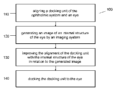
[0076] FIG. 3 illustrates a docking method 100 for the ophthalmic laser
surgical system
50, where the docking method 100 may include:
[0077] An aligning step 110 for aligning the docking unit 55 of the ophthalmic
system 50
and the eye;
[0078] An imaging step 120 for generating an image of an internal structure of
the eye by
an imaging system;
[0079] An alignment-improving step 130 for improving the alignment of the
docking unit
55 with the internal structure of the eye in relation to the generated image;
and
[0080] A docking step 140 for docking the docking unit 55 to the eye.
[0081] These steps are described in detail below.
[0082] The aligning step 110 may include using the first imaging system to
align a target
pattern of the ophthalmic laser surgical system 50 with a feature of the eye.
This aligning
step 110 can be performed e.g. in relation to lowering the docking unit 55
towards the eye.
The first imaging system may be the ophthalmic surgical microscope or video
microscope 56.
[0083] The target pattern of the ophthalmic laser surgical system 50 can
include at least
one of a mark of a center of a contact lens, of a center of the docking unit
55, or of an optical
axis of the objective 54, the docking unit 55 or the contact lens. In other
implementations, it
can include a docking circle, a docking cross-hair, or any other docking
target pattern, as well
as a combination of the above patterns. This target pattern can be formed in
the optics of an
ophthalmic surgical microscope 56, or can be electronically generated and
displayed on a
display or screen of a video microscope 56.
11
CA 02801489 2012-12-03
WO 2011/159627 PCT/US2011/040223
[0084] The feature of the eye can be a center of a region of the cornea 2, the
iris 3, the
pupil 4, a limbus, a sclera, or the lens 5; or a circular formation related to
a region of the
cornea 2, the iris 3, the pupil 4, the limbus, the sclera, or the lens 5.
[0085] FIGS. 4A-B show an illustrative example of the aligning step 110. In
FIG. 4A,
the video microscope 56 displays the eye 1 as seen through the objective 54 of
the laser
surgical system 50, and a variable radius target pattern circle 111, centered
at the shared
optical axis of the objective 54 and docking unit 55. As the surgeon lowers
the docking unit
55 towards the eye, in a pattern adjusting step 112 he may adjust the variable
radius of the
target pattern circle 111 to be essentially equal to the radius of the inner
circular edge 4A of
the patient's pupil 4, as indicated by the arrows 112-1 and 112-2. In
addition, in a pattern
moving step 113, the surgeon may also adjust or move the docking unit 55 in
the x-y plane,
as shown by the arrow 113, to align the target pattern circle 111 with the
inner circular edge
4A of the pupil 4 before, during or after the radius adjustment.
[0086] The radius of the target pattern circle 111 can be chosen to be
somewhat different
from the radius of the inner circular edge 4A of the pupil 4 as long as the
radius enables the
surgeon to align the target pattern circle 111 with the pupil 4 with a desired
precision. In
other embodiments, any other target pattern can be used, including arcs, cross-
hairs, and
raster patterns, as listed above.
[0087] FIG. 4B illustrates that the adjusting of the variable radius of the
target pattern
circle 111 in step 112 and the moving of the docking unit 55 in the x-y plane
in step 113 may
be repeatedly and iteratively performed until the target pattern circle 111
essentially coincides
with the inner circular edge 4A of the pupil 4. Doing so aligns the shared
optical axis of the
objective 54 and the docking unit 55 with the axis or center of the pupil 4.
[0088] During this aligning step 110 the docking unit 55 may get lowered
toward the eye,
possibly even getting into physical contact with the eye during an adjustment
of the z
directional position of the docking unit 55. However, in either case the
docking unit 55 still
can remain movable relative to the eye, allowing the surgeon to carry out the
aligning step
110, possibly iteratively. Even at the end of aligning step 110 the docking
unit may remain
movably connected to the eye to allow for a possible subsequent aligning step.
12
CA 02801489 2012-12-03
WO 2011/159627 PCT/US2011/040223
[0089] In some implementations, the aligning step 110 may not involve a target
pattern.
In these cases the alignment of the docking unit 55 may be guided primarily by
the visual
assessment of the surgeon.
[0090] Embodiments of this aligning step 110 align the docking unit 55 and the
eye to a
certain precision. If the docking unit is docked to the eye after aligning
step 110, an
ophthalmic procedure can be performed with a certain precision. For some
procedures this
precision may be sufficient, but others may benefit from a higher precision.
[0091] FIG. 5 illustrates such a situation. Even after an optical axis 202 of
a docking unit
200 is aligned with the pupil 4 of the eye in the aligning step 110, the lens
5 of the eye may
remain displaced and tilted relative to the optical axis 202, as the lens 5
may not be aligned
with the pupil 4 for one of the reasons outlined above. Here, the docking unit
200 can be an
embodiment of the docking unit 55.
[0092] In FIG. 5, even after an optical axis 12 of the pupil 4 and the eye has
been aligned
with the optical axis 202 of the docking unit 200 in the aligning step 110, a
center 14 of the
lens 5 is still offset by A from the shared optical axis 12/202 of the pupil 4
and the docking
unit 200, and a symmetry axis 16 of the lens 5 still makes an angle a with the
shared optical
axis 12/202.
[0093] Here, the body or housing 204 of the docking unit 200, sometimes called
patient
interface, lens cone, or application tip, may contain a contact lens,
applanation lens or
applanation plate 206 and a skirt or flexible seal 208, which makes contact
with the outer
eye-surface, typically with the cornea, limbus, or sclera. The docking unit
200 can be affixed
to an embodiment of the objective, delivery tip, or distal end 210 or 54,
which may include
several lenses, the ultimate lens being distal lens 212.
[0094] FIGS. 6A-B illustrate the imaging step 120 in some detail.
[0095] FIG. 6A illustrates that in the aligning step 110 the docking unit 55
or 200 can be
properly aligned and centered with the pupil 4 using the video microscope 56,
as evidenced
by the target pattern circle 111 overlapping with the inner circular edge 4A
of the pupil 4, and
its center 118 (denoted by a circle) being at the center of the pupil 4.
However, the lens 5,
shown with a dotted line as its outer perimeter is hidden from the view of the
video
microscope 56, can be off-center with respect to the pupil 4. This is
indicated also by the
center 14 of the lens, denoted by an x, being off the center 118 of the target
pattern 111,
13
CA 02801489 2012-12-03
WO 2011/159627 PCT/US2011/040223
denoted by the circle. Furthermore, the axis 16 of the lens 5 can be tilted
relative to the
shared axis 202/12 of the docking unit 200 and pupil 4.
[0096] Therefore, even after the aligning step 110, the target pattern circle
111 may not
be well-aligned with the lens 5, and thus the precision of cataract procedures
centered with
the target pattern circle 111 may not be optimal. This non-optimal precision
can be improved
by performing the imaging step 120.
[0097] FIGS. 6A and B illustrate that in a typical case, the imaging step 120
can include
a linear scan 121 across the center 118 of the target pattern circle 111 which
coincides with
the center of the pupil 4. This linear scan 121 generates a y-z image 122 that
includes an
image 2c of a corneal segment and images 5a and 5p of segments of the anterior
and posterior
lens capsule, respectively. The images of the lens segments 5a and 5p appear
tilted and off
center relative to the optical axis 202 in the y-z image 122, even if the
corneal segment image
2c appears centered, since the lens 5 can be tilted and off-center relative to
the cornea and
pupil. Therefore, providing the images of the lens segments 5a and 5p may help
the surgeon
to improve the alignment of the docking unit 200 with the tilted and off-
center lens 5.
[0098] In other implementations, the imaging step 120 can involve generating
an image
with a line scan along a linear pattern, an arc, a crosshair pattern, a star
pattern, a circular
pattern, an oval pattern, a loop pattern, a spiral pattern, a concentric multi-
circle pattern, a
shifted multi-circle pattern, a line pattern, and with a two dimensional scan
along an x-y,
raster or grid scanning pattern and a pattern with outlying points.
[0099] The imaging step 120 can involve generating an image with an embodiment
of the
optical coherence tomographic (OCT) imaging system 57, as described in detail
above and
below. The imaging step 120 can be also performed with another imaging system,
capable of
imaging an internal structure of the eye.
[00100] FIG. 7 illustrates that the alignment of the docking unit 200 with the
lens 5 can be
improved by the alignment-improving step 130, based on the imaging step 120.
[00101] In one aspect, the alignment-improving step 130 can include extracting
position
information regarding the lens 5 from the generated image 122, and adjusting a
position of at
least one of the eye 1 or the docking unit 200 in relation to the extracted
position information.
In some implementations, other internal eye-structures can be targeted, such
as the nucleus of
the lens, or a retinal structure.
14
CA 02801489 2012-12-03
WO 2011/159627 PCT/US2011/040223
[00102] In an implementation, the surgeon can analyze the y-z image 122,
generated by
the imaging step 120, and determine the offset A of the lens center 14 from
the optical axis
202 of the docking unit 200. Based on this determination, the surgeon can
shift either the
eye, or the docking unit, or both, to overcome this A offset, as indicated by
arrow 130a. This
adjustment-improving step 130 can reduce or even eliminate the offset A
between the lens
center 14 and the optical axis 202. Typically, this shift 130a can offset the
optical axis 202 of
the docking unit 200 from the optical axis 12 of the lens 5.
[00103] The shift 130a may be performed iteratively because in the first try
the surgeon
may not have determined the offset A precisely. To remedy this, in some
implementations
the alignment-improving step 130 may be followed by a repeated imaging step
120' to
determine how the offset A' was changed by the shift 130a. This repeated
imaging step 120'
can be followed by a repeated alignment-improving step 130' based on the
updated image
122' generated by the repeated imaging step 120', and so on. In efficient
implementations,
the offset A is reduced step-by-step. In other implementations, even if A
increases during a
step, subsequent steps reduce it eventually.
[00104] The shift 130a can be performed by giving a verbal command to the
patient to
move his/her eye, or by physically moving the patient's head, or the surgical
bed the patient
is resting on, or by manually moving the patient's eye, or by moving a
fixation light of a
fixation light source, or by moving a directing light on a directing light
display, in either case
directing the patient to follow the light with his eye, or by moving the
docking unit 200 in an
x-y plane via moving a gantry or an articulated arm. In implementations using
two piece
docking units, the piece which was attached to the eye, such as a gripper, can
be used to
move or rotate the eye. The fixation or directing light can be directed either
into the surgical
eye or into the non-surgical eye. These adjustments can be performed manually
by the
surgeon, or by operating one or more electric actuators, or by a computer. In
some cases,
more than one of the above types of shifts can be performed jointly.
[00105] FIG. 7 also illustrates that in other implementations the alignment-
improving step
130 may include extracting orientation information regarding the lens 5 or
another targeted
internal structure of the eye from the generated image 122, and adjusting an
orientation of at
least one of the eye 1 or the docking unit 200 in relation to the extracted
orientation
information.
CA 02801489 2012-12-03
WO 2011/159627 PCT/US2011/040223
[00106] In an implementation, the surgeon can analyze the y-z image 122,
generated by
the imaging step 120, and determine the angle a between the optical axis 16 of
the lens 5 and
the optical axis 202 of the docking unit 200. Based on this determination, the
surgeon can
rotate either the eye, or the docking unit, or shift the docking unit, or
adjust an optical path of
the laser beam in the laser surgical system 50 to overcome this a
misalignment. The option
of rotating the eye is indicated by arrow 130b. This alignment-improving step
130 can
reduce or even eliminate the angle a between the optical axis 16 of the lens 5
and the optical
axis 202 of the docking unit 200. This alignment-improvement is typically
achieved by
introducing an angle between the optical axis 12 of the eye and the optical
axis 202 of the
docking unit 200, as indicated by the dotted line.
[00107] The rotation 130b may be performed iteratively because in the first
try the surgeon
may not have determined the angle a precisely. To remedy this, in some
implementations the
alignment-improving step 130 may be followed by a repeated imaging step 120'
to determine
the angle a' after the rotation 130b from a repeated image 122', followed by a
repeated
alignment-improving step 130' based on the image 122' generated by the
repeated imaging
step 120' and so on. In efficient implementations, the angle a is reduced step-
by-step. In
other implementations, even if a increases during a step, subsequent steps
eventually reduce
it.
[00108] The rotating step 130b can be performed by giving a verbal command to
the
patient to rotate his/her eye, or by manually rotating the patient's head, or
by physically
rotating the patient's eye, or by moving a fixation light of a fixation light
source, or a
directing light displayed on a display, in either case directing the patient
to follow the light
with his eye, or by moving or rotating the docking unit 200 in the x-y plane
via moving a
gantry or an articulated arm. The fixation or directing light can be directed
either into the
surgical eye or into the non-surgical eye. In implementations using two piece
docking units,
the piece which was attached to the eye, such as a gripper, can be used to
move or rotate the
eye. These adjustments can be performed manually by the surgeon, or by
operating one or
more electric actuators, or by a computer. In some case, more than one of the
above types of
shifts can be performed jointly.
[00109] FIGS. 8A-B illustrate an outcome of the imaging step 120 and alignment-
improving step 130.
16
CA 02801489 2012-12-03
WO 2011/159627 PCT/US2011/040223
[00110] FIG. 8A illustrates that after a successful alignment-improving step
130, a shifted
target pattern circle 111' may have become concentric with the lens 5 instead
of the pupil 4.
Correspondingly, the shifted linear scanning line 121', across the shifted
center 118' of the
target pattern circle 111', can now go through the center 14 of the lens 5
instead of the center
of the pupil 4.
[00111] Some implementations may display both the first target pattern circle
111
concentric with the pupil 4, as well as a second target pattern 111' which is
shifted by the
alignment-improving step 130 to be concentric with the lens 5.
[00112] FIG. 8B illustrates that after an efficient alignment-improving step
130, a
repeated imaging step 120' may record a cross-sectional y-z image 122' showing
that the
center 14 of the lens now lies on the optical axis 202 of the docking unit
200. Further, the
images of the anterior and posterior capsule segments 5a' and 5p' after the
relative rotation
and shift of the eye and the docking unit 200, are close to symmetric,
indicating that the
optical axis 16 of the lens is approximately aligned with the optical axis 202
of the docking
unit 200.
[00113] Achieving alignment of the docking unit 55/200 with the hard-to-see,
displaced
and tilted lens 5 instead of the visible pupil 4 with such an improved
precision is one of the
benefits of the image-guided docking method 100.
[00114] FIG. 9 illustrates that an implementation of a related image-guided
docking
method 300 may include the steps of:
[00115] A video imaging step 310, for generating a video microscope image of a
portion
of the eye;
[00116] A centering step 320, for centering a docking tip based on the video
microscope
image;
[00117] An OCT imaging step 330 for generating an OCT image of a portion of
the eye;
[00118] A distancing step 340 for determining a distance of the docking tip
from the
cornea based on the OCT image;
[00119] A moving step 350 for using the determined distance to move the
docking tip
towards the cornea of the eye;
17
CA 02801489 2012-12-03
WO 2011/159627 PCT/US2011/040223
[00120] A determining step 360 for determining a position or an orientation of
a lens of
the eye based on the OCT image;
[00121] An aligning step 370 for aligning the docking tip with a lens of the
eye by
instructing the patient with verbal commands, or adjusting a directing light
or moving a
gantry; and
[00122] A docking step 380 for applying suction to dock the docking tip.
[00123] Several of the steps 3 10-3 80 of the method 300 can proceed
analogously with the
corresponding steps 110-140 of the method 100. In addition, the distance-
determining step
340 can include determining the distance between the cornea 2 of the eye and
the docking tip,
which can be the docking unit 55 or 200, or any other patient interface. In
this step 340, the
distance from the docking tip can be based on a reference point. This
reference point can be
located in the optical system of the surgical laser system 50, for example in
the objective 54.
This reference point can be movable, and may be adjusted or offset based on
various
considerations.
[00124] FIG. 10 illustrates an OCT imaging system 457 to illustrate the
details of the
imaging step in greater detail. The OCT imaging system 457 can include an OCT
imaging
unit 458 and an OCT x-y scanner 459.
[00125] The principles of the operation of OCT imaging systems are well known
and
documented. The OCT system 457 can be a (a) time domain, a (b) swept source or
a (c)
spectrometer based OCT. The (a) and (b) types of OCT imaging systems use a
narrow band
OCT light source 410 and scan the beam's focal point in the z-direction, thus
they provide
imaging information corresponding to different z-depths sequentially in time.
The (a) type
time domain OCT systems move a reference mirror, whereas the (b) type swept
source OCT
systems sweep the wavelength of the laser beam.
[00126] The (c) type spectrometer based OCT systems utilize a broad band OCT
imaging
light source 410 and capture images from a range of z-depths essentially
simultaneously, or
in parallel, corresponding to the different wavelengths within the broad band
of an OCT
imaging light source. Because of this parallel imaging aspect, spectrometer-
based OCT
systems can be substantially faster that sequential OCT systems. The (b) and
(c) type OCT
systems are sometimes referred to as frequency domain OCT systems.
18
CA 02801489 2012-12-03
WO 2011/159627 PCT/US2011/040223
[00127] All types of OCT imaging units 458 can include an OCT light source
410, an OCT
reference mirror 413 and a beam splitter 417. Among the sequential OCT
systems, for the (a)
type time domain OCT, the OCT light source 410 can be a narrow band laser and
the
reference mirror 413 movable for z-scanning. For the (b) type swept source
OCT, the
reference mirror need not be movable as the wavelength of the light source 410
is varied. For
(c) parallel OCT systems, the OCT light source 410 can emit a broadband
imaging light.
[00128] The OCT imaging beam can be guided by the OCT beam x-y scanner 459,
directed to the eye via an objective 454 and a docking unit 455. The OCT x-y
scanner 459
can scan the OCT imaging beam in the eye in the x and y directions. In
sequential OCT
systems the beam is z scanned by moving either the reference mirror 413 or by
sweeping the
wavelength of the OCT light source 410. In parallel OCT systems, no z-scanning
is
performed, as the different wavelengths carry the imaging information
corresponding to
different z depths essentially simultaneously.
[00129] In all these system, the OCT imaging beam returned from the eye can be
unified
with the reference beam returning from the OCT reference mirror 413 at the
beam splitter
417. This unified beam carries the imaging information in a complex
interference pattern
that is recorded by an OCT camera 420.
[00130] For sequential OCT systems this OCT camera 420 can be simple, e.g.
including a
photodetector. For parallel OCT systems the OCT imaging unit 458 may include a
spectrometer, such as a prism or a grating (not shown explicitly) that
resolves the broad band
imaging light into its different wavelength components, and deflects the
different wavelength
components to different spatial angles. In some parallel OCT systems the OCT
camera 420
may include a linear array of CCD detectors to capture these diverging rays
with different
wavelength, each carrying interference information, specific for its own
wavelength. In
others, a two dimensional CCD array can be used. The amplitude of the resolved
diverging
rays can be recorded in the individual pixels of the CCD array of the OCT
camera 420. Some
high resolution OCT cameras 420 can involve hundreds or even thousands of
pixels.
[00131] The imaging process can by controlled by an imaging sync block 470,
which may
get its sync signal from a later-specified output unit. The image data from
the OCT camera
420 can be forwarded to an OCT analyzer 480, synchronized by the imaging sync
block 470.
In parallel OCT systems the OCT analyzer 480 may include a processor to
perform a Fast
Fourier Transform (FFT). The FFT converts the interference information of
different
19
CA 02801489 2012-12-03
WO 2011/159627 PCT/US2011/040223
wavelength components into image information corresponding to different z-
depths. After
the FFT, the transformed OCT image data represent image information
corresponding to a
range of z-depths. This transformed OCT image data may be forwarded to a
processor 430,
which can generate an OCT image and output the generated OCT image towards a
display
490.
[00132] Next, an OCT scanning-beam-controller system will be described that
solves the
difficulties of the operation of some existing OCT scanning-beam-controllers
which are
described next.
[00133] In some OCT imaging systems the processor 430 can multitask and
perform more
than one function in an interleaved, parallel or overlapping manner. To carry
out these
functions, the processor may perform an "interrupt" by switching from e.g. the
task of
scanning the beam to another task and back. Such interrupts, however short,
can cause
problems, since during the time when the scanning is stopped or frozen by the
interrupt, the
laser beam may remain pointed to the same position. This scanning-freeze can
disrupt the
timing of the x-y scan, introducing an error and noise into the coordinates of
the imaged
location. This timing error in the outputted scanning data can lead to delays
that may reach
50, 100 or more microseconds: a phenomenon sometimes called jitter. Further,
the extended
exposure to the laser beam can cause damage to the sensitive eye tissue.
[00134] In addition, since the processor typically communicates with
input/output agents
through a system bus, this output mode only provides slow data transfer rates,
since several
agents may access the bus simultaneously, all demanding a fraction of its
cycle time. Further,
to manage these competing demands, a portion of the cycle of the system bus is
typically
taken up by control signals. And if an OCT imaging system is designed to avoid
this
scanning-freeze by the processor outputting the scanning data to an output
unit in a single-
task mode, e.g. through a dedicated link, then the processor cannot perform
other functions
during this outputting step, such as computing the next scanning pattern. All
these designs
and constraints slow down the performance of such systems considerably.
[00135] Implementations of the presently described OCT scanning-beam-
controller can
overcome these difficulties by employing an efficient design. The OCT scanning-
beam-
controller can include the processor 430 and an analog input-output board 435.
The
processor 430 can compute scanning data for a scanning pattern. This scanning
data can
include e.g. a sequence of x-y coordinates where the OCT imaging beam will be
directed in
CA 02801489 2012-12-03
WO 2011/159627 PCT/US2011/040223
the target region in the course of scanning. For sequential, z-scanning OCT
systems, the
scanning data can include x-y-z coordinates. As described above, the OCT
scanning pattern
can be a wide variety of patterns, including lines, arcs, loops, circles,
spirals, raster and grid
patterns.
[00136] The processor 430 can compute the scanning data, as well as perform
its other
described functions in connection to a storage medium that stores a computer
code or
instruction set to facilitate these functions of the processor.
[00137] The analog input-output board 435 can include a local or dedicated
memory
controller 440, also referred to as a direct memory access engine 440, or DMA
engine 440.
The DMA engine/memory controller 440 can manage a transfer of the computed
scanning
data, indirectly or directly, from the processor 430 toward a data buffer 450.
The data buffer
450, coupled to the local memory controller 440 can store the scanning data
and output the
scanning data towards an output digital-analog converter 460, or output DAC
460. The
output DAC 460 can be coupled to the data buffer 450 and can (i) convert
selected outputted
scanning data to analog scanning signals, and (ii) output the scanning signals
towards the
OCT beam x-y (or x-y-z) scanner 459.
[00138] FIG. 11 illustrates an implementation of the OCT scanning beam-
controller. The
processor 430' can be coupled to a bus 432, such as a PCI bus 432. The OCT
scanning-
beam-controller can also include a processor memory 433. The processor 430'
can output the
computed scanning data to the processor memory 433. The dedicated DMA engine
440' can
transfer the scanning data from the processor memory 433 to the data buffer
450' which can
be e.g. a first-in-first-out (FIFO) memory. The FIFO buffer memory 450' can
store the
scanning data and output the stored scanning data to the output DAC 460' when
prompted.
In some implementations, the processor can output the scanning data to the
analog input-
output board 435 through a dedicated memory bus or local bus instead of a PCI
bus 432. In
other implementations, there can be even a direct connection between the
processor and the
DMA engine 440'.
[00139] In relation to the above described problems with other systems,
embodiments of
the present OCT scanning-beam-controller offer a fast scanning operation as
(i) the FIFO
memory 450' can output the stored scanning data in an uninterrupted manner;
(ii) the output
mode can be a fast data transfer mode, such as a burst mode; and (iii) the
output can be
21
CA 02801489 2012-12-03
WO 2011/159627 PCT/US2011/040223
performed without sending the scanning data through the shared bus 432, the
processor
memory 433, or the processor 430'.
[00140] For all these reasons, the outputting of the scanning data will not be
interrupted by
competing tasks, or slowed down by the slow data transfer characterizing the
shared bus 432.
[00141] Further, since the FIFO memory 450' drives the outputting of the
scanning data,
the processor 430' is free to perform other functions in parallel with the
data output, such as
processing an image, or computing new scanning data corresponding to a
scanning pattern, or
performing a control function.
[00142] In addition, the output of the scanning data by the data buffer 450'
to the output
DAC 460' is not slowed down by an interrupt by the processor 430 or another
system agent
either since the output proceeds from the data buffer 450' through a dedicated
channel on the
analog input-output board 435 instead of the shared bus 432. Such
implementations can
reduce the jitter considerably, such as keeping it below 50, 40, or even 20
microseconds.
[00143] In some implementations, the output DAC 460' can convert the received
digital
scanning data into analog scanning signals and output the scanning signals to
x and y galvo-
controllers 56a and 56b, or some other types of scanning-controllers that
control x and y
galvo mirrors, or redirector elements, to scan the OCT imaging beam according
to the
scanning pattern, coded in the scanning data. Some implementations may have an
integrated
x-y galvo-controller that controls a mirror capable of rotating around two
axes.
[00144] The output DAC 460' can also output synchronizing signals to the
imaging sync
block 470' coupled to the OCT imaging camera 420 to record the returned OCT
imaging
beam synchronously with the scanning of the OCT imaging beam. The
synchronizing signals
can be based on synchronizing data, inserted by the processor 430' into the
scanning data.
[00145] In addition, the imaging step 120 can include computing homing data
corresponding to a homing pattern connecting an ending point of a first
imaging step to a
starting point of a subsequent second imaging step. This step can be useful in
implementations where the first imaging step ends by simply stopping the
output of the
scanning data, thus leaving the scanning x and y galvos 56a-b in a non-
standard position and
the imaging beam pointed to a non-standard target point. This non-standard
point is typically
different from the starting point of the subsequent second imaging step, thus
necessitating the
"homing" of the x and y galvos 56a-b by computing and outputting homing data,
so that the
22
CA 02801489 2012-12-03
WO 2011/159627 PCT/US2011/040223
imaging beam can start the subsequent second imaging step from a well-defined
starting
point.
[00146] As an example, the first imaging step may include scanning the x and y
coordinates of the imaging beam along a first circle of a first radius. If the
second imaging
step includes scanning along a second circle of a second radius, then the
first imaging step
can be followed by computing homing data that define a path from the endpoint
of the first
circular scan with the first radius to the starting point of the second
circular scan with the
second radius.
[00147] Such implementations can avoid moving the imaging beam back to a
standard
point, e.g. to a center, origin, or otherwise unbiased point, thus saving
additional time and
further accelerating the scanning operation.
[00148] The computing of the homing data can be also useful in implementations
where at
the end of the first imaging step the x and y galvos 56a and 56b are returned
to a neutral
position, as it facilitates the computing of the starting position of a second
imaging step in
relation to the neutral position.
[00149] In some implementations, the speed of the output of the output DAC
460/460' can
be so fast that an operating speed of the imaging system 457 can be limited by
an integration
time of the OCT camera 420.
[00150] In some implementations, the output DAC 460/460' can output the
scanning
signals at a rate within one of the following ranges: 1 Hz - 1 MHz, 100 Hz - 1
MHz, or 1
kHz - 100 kHz.
[00151] In some implementations, the rate of output for the scanning signals
can be
adjustable according to the requirements of the imaging task and pattern.
[00152] Once the imaging step 120 is completed, the alignment-improving step
130 can
include providing a verbal command to a patient based on the image of the
internal structure
of the eye, such as the lens 5.
[00153] The alignment-improving step 130 can also include providing a fixation
light
beam, asking the patient to look at the fixation light, and adjusting the
fixation light based on
the image provided by the imaging step 120. The fixation light can be provided
into the
surgical eye, through the main optical pathway of the laser surgical system
50, or through a
23
CA 02801489 2012-12-03
WO 2011/159627 PCT/US2011/040223
separate fixation light system. In some cases the fixation light can be
provided to the non-
surgical eye.
[00154] The alignment-improving step 130 can be started (i) before the docking
unit
55/200 makes contact with the eye; (ii) after the docking unit 55/200 makes
contact with the
eye but before an application of a vacuum; or (iii) after an application of a
partial vacuum in
relation to the docking unit 55/200 that still allows some degree of alignment
modification.
[00155] The partial vacuum, or suction, can be applied, for example, through a
suction
ring or suction skirt, which can be part of the docking unit 55/200. The
suction can be
applied after the eye was brought into physical contact with the eye.
[00156] The docking method 100 can be performed as part of a surgical process
or a
diagnostic process. In other implementations, the docking method 100 can be
part of an
imaging procedure, which is not part of a surgical or a diagnostic procedure,
such as an
identification process.
[00157] The steps 110-140 can involve program codes or instruction sets that
are stored in
the imaging system 57. The code can be stored e.g. in a dedicated memory or in
a memory
that is part of another functional block. The aligning step 110 can involve a
code stored in a
memory related to the video microscope 56. The imaging step 120 can involve
storing the
scanning patterns or scanning data generated by the processor 430 in a
dedicated or integrated
memory, or storing scanning data in the data buffer 450. The alignment-
improving step 130
can include using a memory unit for storing the generated image to help
improving the
alignment of the docking unit 55 with the lens of the eye 1 in relation to the
generated image.
The docking step 140 can also use a stored program to guide and control the
docking unit 200
docking with the eye.
[00158] FIG. 12 illustrates that an implementation of a fast imaging method
500 can
include:
[00159] A step 510 of computing scanning control data by the processor
430/430';
[00160] A step 520 of storing the scanning control data into the processor
memory 433 by
the processor 430;
[00161] A step 530 of setting up the dedicated memory controller 440/440' for
a scanning
operation by defining operation parameters, such as a scanning output rate;
24
CA 02801489 2012-12-03
WO 2011/159627 PCT/US2011/040223
[00162] A step 540 of transferring scanning control data from the processor
memory 433
to the data buffer 450/450' at least partially under the control of the
dedicated memory
controller 440/440';
[00163] A step 550 of notifying the processor 430/430' by the dedicated memory
controller/DMA engine 440/440' that the transfer of the scanning control data
has been
completed;
[00164] A step 560 of instructing the dedicated memory controller 440/440' by
the
processor 430/430' to start fast output of the scanning control data;
[00165] A step 570 of transferring the scanning control data from the data
buffer
450/450'to the output DAC 460/460' at least partially under the control of the
dedicated
memory controller 440/440', the output DAC 460/460' converting the digital
scanning
control data to analog scanning control signals, and the output DAC 460/460'
outputting the
analog scanning control signals to the x and y scanners 56a and 56b, and to
the sync block
470;
[00166] A step 580 of notifying the processor 430/430' by the dedicated memory
controller 440/440' that the output process is complete.
[00167] In the step 570, the transferring the scanning control data from the
data buffer
450/450' can be performed in a fast transfer mode, such as a burst mode, or a
page mode, or
any similarly fast transfer modes.
[00168] In the step 570, the transferring of the scanning control data from
the data buffer
450/450' can be performed without sending the scanning control data through
the bus 432
that connects the local memory controller 440, the processor 430, and the
processor memory
433.
[00169] In the step 570, the transferring step can also include transferring
the scanning
control data in parallel with the processor 430 processing an image or
computing scanning
data corresponding to a scanning pattern.
[00170] In the step 570, the transferring step can also include transferring
the scanning
data without an interrupt by another system agent, thereby keeping a jitter of
the scanning
data below 50, 40, or 20 microseconds.
CA 02801489 2012-12-03
WO 2011/159627 PCT/US2011/040223
[00171] In an implementation 600 of the above method 500, the above steps can
be
organized into the following steps:
[00172] A step 610 of computing scanning control data by a processor can
include the step
510;
[00173] A step 620 of storing the scanning control data into a data buffer
partially by a
local memory controller can include the steps 520, 530, 540, and 550;
[00174] A step 630 of transferring the scanning control data from the data
buffer in a fast
transfer mode to a converter-output module can include the steps 560 and
elements of the
step 570; and
[00175] A step 640 of outputting scanning signals to scanning controllers, the
scanning
signals converted from the scanning control data by the converter-output
module can include
elements of the step 570.
[00176] While this specification contains many specifics, these should not be
construed as
limitations on the scope of any invention or of what may be claimed, but
rather as
descriptions of features specific to particular embodiments. Certain features
that are
described in this specification in the context of separate embodiments can
also be
implemented in combination in a single embodiment. Conversely, various
features that are
described in the context of a single embodiment can also be implemented in
multiple
embodiments separately or in any suitable subcombination. Moreover, although
features may
be described above as acting in certain combinations and even initially
claimed as such, one
or more features from a claimed combination can in some cases be excised from
the
combination, and the claimed combination may be directed to a subcombination
or variation
of a subcombination.
26