Note: Descriptions are shown in the official language in which they were submitted.
CA 02801774 2012-12-05
WO 2011/156281 PCT/US2011/039301
-1-
DEVICE FOR VIRAL INACTIVATION OF LIQUID MEDIA
RELATED APPLICATION
This application claims the benefit of U. S. Provisional Application No.
61/352,276, filed on June 7, 2010. The entire teachings of the above
application are
incorporated herein by reference.
BACKGROUND OF THE INVENTION
Viral mitigation throughout all phases of biopharmaceutical manufacturing
processes is an increasingly strict requirement established by international
or
national regulatory bodies in order to prevent viral contaminants in the
application.
of biopharmaceuticals for therapeutic or non-therapeutic purposes. Several
methods
have been employed to inactivate and/or remove large or small, enveloped or
non-
enveloped viral particles from biopharmaceutical product compositions.
Examples
of such methods include filtration (e.g., 20 nm filtration, Q membrane
chromatography, depth filter technology), heat (e.g., high temperature short
time
(HTST) pasteurization), chemical (e.g., addition of solvents - detergents or
chemical
agents), or radiation (e.g., ultraviolet or gamma-ray irradiation). These
methods
have been used primarily downstream in the biopharmaceutical manufacturing
process due to their low throughput and/or high cost. Viral inactivation of
cell
culture media input into a biopharmaceutical manufacturing process, where up
to
20,000 L or more are processed per day, would be prohibitive in terms of time
and
cost with existing methods. Some methods, such as ultraviolet C (UVC)
irradiation,
are challenging to apply to biopharmaceutical manufacturing processes,
because,
unlike in, for example, water treatment, over-exposure of the media
(particularly
media containing serum) can be detrimental, and therefore the radiation dose
needs
to be delivered uniformly to the media and controlled to within a relatively
narrow
range. An additional challenge for ultraviolet irradiation of media,
particularly
media containing serum, is that the UV transmittance in the UVC range (e.g.,
at 254
CA 02801774 2012-12-05
WO 2011/156281 PCT/US2011/039301
-2-
nm) of the media is substantially lower than the UV transmittance of, for
example,
water, in this wavelength range. In addition, it is desirable that devices
used in high
throughput biopharmaceutical manufacturing contain components that are
amenable
to cleaning and sterilization (e.g., clean-in-place (CIP) and steam-in-place
(SIP)
procedures). Therefore, there is a need for methods and apparatuses which
enable
high throughput viral inactivation of low transmittance liquid media for
biopharmaceutical and other applications.
SUMMARY OF THE INVENTION
The invention generally is directed to methods and apparatuses which enable
high throughput viral inactivation of liquid media.
In one embodiment, an apparatus capable of viral inactivation of a high
absorbance liquid media includes at least one coaxial cylinder constructed of
an
outer cylinder (with dimensions of length, inner diameter, and outer diameter)
and
an inner cylinder coaxial with the outer cylinder. The apparatus further
includes a
media inlet, at least one emitter of type C ultraviolet radiation, and a media
outlet.
The inner cylinder has a length substantially equal to the length of the outer
cylinder
and an outer diameter adapted to form a gap between the outer diameter of the
inner
cylinder and the inner diameter of the outer cylinder. The liquid media flows
in a
substantially cyclonic flow path along the gap. The media inlet is connected
to the
outer cylinder at, or proximal to, an end of the outer cylinder. The inlet is
configured to flow the media along the substantially cyclonic flow path along
the
gap. The at least one emitter of type C ultraviolet radiation is placed inside
the inner
cylinder so as to emit the type C ultraviolet radiation towards the media to
be treated
with the type C ultraviolet radiation and thereby inactivate viruses in the
media. The
outlet is connected to the outer cylinder at, or proximal to, an end of the
outer
cylinder opposite the inlet. In a further embodiment, the media to be treated
is a cell
culture media. In yet a further embodiment, the media to be treated contains
serum.
In another embodiment, a method of inactivating viruses in a high
absorbance liquid media includes introducing the liquid media into at least
one
coaxial cylinder that includes a gap along the length of the cylinder between
the
outer diameter of an inner cylinder and the inner diameter of an outer
cylinder. The
CA 02801774 2012-12-05
WO 2011/156281 PCT/US2011/039301
-3-
media is introduced through an inlet configured to flow the liquid media along
a
substantially cyclonic flow path along the gap. The method further includes
irradiating the media with at least one emitter of type C ultraviolet
radiation placed
inside the inner cylinder so as to emit the type C ultraviolet radiation
towards the
liquid media to thereby inactivate viruses in the media. The method then
includes
flowing the media through a media outlet connected to the outer cylinder
proximal
to an end of the outer cylinder opposite the inlet. In a further embodiment,
the
media to be treated is a cell culture media. In yet a further embodiment, the
media
to be treated contains serum.
This invention has many advantages, such as high throughput viral
inactivation of liquid media for biopharmaceutical processes, and amenability
to
cleaning procedures (e.g., clean-in-place (CIP) and steam-in-place (SIP)).
Another
advantage of the apparatuses of the invention is the flexibility in the
configurations,
which enable them to be fitted to restricted or customized space requirements.
BRIEF DESCRIPTION OF THE DRAWINGS
The foregoing will be apparent from the following more particular
description of example embodiments of the invention, as illustrated in the
accompanying drawings in which like reference characters refer to the same
parts
throughout the different views. The drawings are not necessarily to scale,
emphasis
instead being placed upon illustrating embodiments of the present invention.
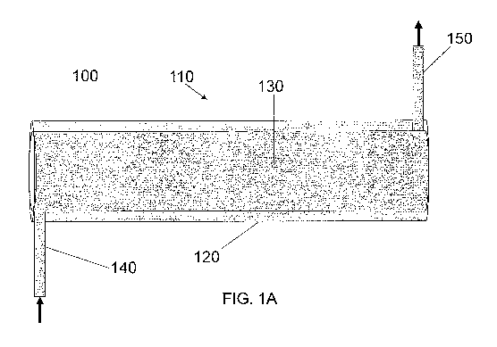
FIG. 1A is a schematic illustration of a perspective view of an apparatus for
viral inactivation of liquid media according to this invention that includes
one
coaxial cylinder.
FIG. 1B is a schematic illustration of cross sections of the inlet and coaxial
cylinder of the apparatus shown in FIG. IA.
FIG. 1 C is a schematic illustration of a side view of the inlet and coaxial
cylinder of the apparatus shown in FIG. IA.
FIG. 1D is a schematic illustration of a perspective view of an apparatus for
viral activation of liquid media with a tangential inlet and outlet, both with
rectangular cross sections and rounded corners, according to this invention.
CA 02801774 2012-12-05
WO 2011/156281 PCT/US2011/039301
-4-
FIG. 1E is a schematic illustration of a cyclonic flow path according to this
invention.
FIG. 1 F is a schematic illustration of a specific example of an apparatus for
viral inactivation of liquid media according to this invention that includes
two
coaxial cylinders and a housing around each coaxial cylinder.
FIG. 2 is a schematic illustration of a cross section of a coaxial cylinder
with
multiple emitters of type C ultraviolet radiation.
FIG. 3 is a schematic illustration of static mixing elements inside the gap
along a coaxial cylinder.
FIG. 4 is a schematic illustration of a side view of an apparatus for viral
inactivation of cell culture media according to this invention with two
vertically
stacked coaxial cylinders.
FIG. 5A is a schematic illustration of stacking two rows of coaxial cylinders
between an input manifold and an output manifold according to this invention.
FIG. 5B is a schematic illustration of stacking four rows of coaxial cylinders
between an input manifold and an output manifold according to this invention.
FIG. 6 is a schematic illustration of horizontal stacking two rows of coaxial
cylinders between an input manifold and an output manifold according to this
invention.
FIG. 7A is a schematic illustration of four coaxial cylinders stacked
horizontally and vertically according to this invention.
FIG. 7B is a schematic illustration of flow of liquid media through the
apparatus shown in FIG. 7A.
FIG. 8 is a schematic illustration of a plan view of an apparatus for viral
inactivation of cell culture media according to this invention; W = wash/flush
valve,
F = flow control valve, D = dose meter.
FIG. 9 is a schematic illustration of the workflow for model development.
FIG. 10 is a graph of the radiation intensity at 254 nm as a function of
radial
distance from the quartz sleeve for water, serum-free cell culture media, and
serum-
containing (10 %vol) cell culture media.
FIG. 11 is a schematic illustration of the apparatus described in Example 2.
CA 02801774 2012-12-05
WO 2011/156281 PCT/US2011/039301
-5-
FIG. 12 is a schematic illustration of cyclonic flow paths in the apparatus
described in Example 2.
FIG. 13 is a graph of frequency (% of cell culture media exposed) as a
function of UV dosage for serum-free cell culture media in the apparatus
described
in Example 2 (5 mm gap, 3.81pm (1 gpm)) and Example 3 (3 mm gap, 4.75 lpm
(1.25 gpm) and 9.5 Ipm (2.5 gpm)).
FIG. 14 is a graph of % particles (cumulative dosage) as a function of UV
dosage for serum-free cell culture media in the apparatus described in Example
2 (5
mm gap, 3.81pm (1 gpm)) and Example 3 (3 mm gap, 4.75 lpm (1.25 gpm) and 9.5
Ipm (2.5 gpm)).
FIG. 15 is a graph of frequency (% of cell culture media exposed) as a
function of UV dosage for serum-containing cell culture media in the apparatus
described in Example 2 (5 mm gap, 1.91pm (0.5 gpm)) and Example 3 (3 mm gap,
2.41pm (0.631 gpm) and 3.81pm (1 gpm)).
FIG. 16 is a graph of % particles (cumulative dosage) as a function of UV
dosage for serum-containing cell culture media in the apparatus described in
Example 2 (5 mm gap, 1.91pm (0.5 gpm)) and Example 3 (3 mm gap, 2.41pm
(0.631 gpm) and 3.81pm (1 gpm)).
FIG. 17 is a schematic illustration of the UVC treatment setup described in
Example 4.
FIGS. 18A and 18B are graphs of fluorescence distributions for controlled
samples before (FIG. 18A) and after (FIG. 18B) normalization.
FIG. 19 is a graph of fluorescence distributions at various uniform dose
levels obtained from collimated beam calibration experiments.
FIGS. 20A and 20B are graphs of means of fluorescence distributions
obtained for various UV fluencies from the collimated beam calibration
experiments
in water.
FIG. 21 is a graph of fluorescence distributions of samples exposed to UV
light in the UVC reactor at three different flow rates. .
FIGS. 22A, 22B, and 22C are pictorial illustrations of definitions of I3j,
[i,t,
and a;.
CA 02801774 2012-12-05
WO 2011/156281 PCT/US2011/039301
-6-
FIG. 23 is a graph of two test fluorescence distributions obtained by
mathematically mixing calibration samples in proportions given in Table 2.
FIG. 24 is a graph of a comparison of the actual and predicted UV dose
distributions for the two test cases.
FIGS. 25A, 25B, and 25C are graphs of UV dose distributions in cell culture
media through the UVC reactor as a function of fluorescence distribution for:
FIG.
25A-1 flow rate = 2.75 lpm, predicted mean UV dose = 91 mJ/cm2; FIG. 25A-2
experimental mean UV dose = 82 mJ/cm2; FIG. 25B-1 flow rate = 4.75 lpm,
predicted mean UV dose = 53 mJ/cm2; FIG. 25B-2 experimental mean UV dose =
52 mJ/cm2; FIG. 25C-1 flow rate = 7.61pm, predicted mean UV dose = 35 mJ/cm2;
FIG. 25C-2 experimental mean UV dose = 50 mJ/cm2.
FIGS. 26A, 26B, and 26C are graphs of UV dose distributions of 10% DBS
in cell culture media through the UVC reactor as a function of fluorescence
distribution for: FIG. 26A-1 flow rate = 2.21pm, predicted mean UV dose = 89
mJ/cm2; FIG. 26A-2 experimental mean UV dose = 47 mJ/cm2; FIG. 26B-1 flow
rate = 3.8 lpm, predicted mean UV dose = 53 mJ/cm2; FIG. 26B-2 experimental
mean UV dose = 36 mJ/cm2; FIG. 26C-1 flow rate = 61pm, predicted mean UV
dose = 34 mJ/cm2; FIG. 26C-2 experimental mean UV dose = 28 mJ/cm2.
FIG. 27 is a graph of UVC absorbance measurements of Vitamin C solution
in water.
FIGS. 28A and 28B are graphs of UV dose distributions of 0.1 g/L
(absorbance of 4.7 absorbance units) Vitamin C solution through the UVC
reactor as
a function of fluorescence distribution for: FIG. 28A-1 flow rate = 2.2 lpm,
predicted mean UV dose = 104 mJ/cm2; FIG. 28A-2 experimental mean UV dose =
81 mJ/cm2; FIG. 28B-1 flow rate = 3.8 lpm, predicted mean UV dose = 61 mJ/cm2;
FIG. 28B-2 experimental mean UV dose = 63 mJ/cm2.
FIGS. 29A, 29B, and 29C are graphs of UV dose distributions of 0.04 g/L
(absorbance of 1.94 absorbance units) Vitamin C solution through the UVC
reactor
as a function of fluorescence distribution for: FIG. 29A-1 flow rate = 2.75
lpm,
predicted mean UV dose = 91 mJ/cm2; FIG. 29A-2 experimental mean UV dose =
81 mJ/cm2; FIG. 29B-1 flow rate = 4.75 lpm, predicted mean UV dose = 53
mJ/cm2;
FIG. 29B-2 experimental mean UV dose = 60 mJ/cm2; FIG. 29C-1 flow rate = 7.6
CA 02801774 2012-12-05
WO 2011/156281 PCT/US2011/039301
-7-
1pm, predicted mean UV dose = 35 mJ/cm2; FIG. 29C-2 experimental mean UV
dose = 47 mJ/cm2.
DETAILED DESCRIPTION OF THE INVENTION
The invention generally is directed high throughput treatment of a liquid. In
a particular aspect, the invention is directed to methods and apparatuses
which
enable high throughput viral inactivation of liquid media. As used herein, a
liquid
media includes any liquid or solution in which it is desirable to remove a
viral
contamination or prevent a potential contamination, including but not limited
to
buffers, ingestible fluids, injectable solutions, biological fluids, serum,
media,
bioprocessing solutions, animal-component containing solutions, and
therapeutics
for human or veterinary use. In one embodiment, a bioprocessing solution is a
cell
culture media, a conditioned media, a chromatography solution (such as a wash
or
elution buffer), or a formulation solution. In one embodiment, the liquid
media is
cell culture media (e.g., serum-containing cell culture media or serum-free
cell
culture media). In another embodiment, the liquid media is a liquid containing
at
least one therapeutic protein, such as a monoclonal antibody, a recombinant
protein,
or an enzyme.
As used herein, "high throughput" treatment of a liquid means treatment at a
flow rate in a range of between about 0.5 liters per minute (lpm) and about 50
liters
per minute, or between about 0.5 liters per minute and about 5 liters per
minute, or
between about 5 liters per minute and about 10 liters per minute, or between
about
10 liters per minute and about 50 liters per minute. In particular
embodiments, a
high flow rate is about 1 liter per minute, or 2 liters per minute, or 3
liters per
minute, or 4 liters per minute, or 5 liters per minute, or 10 liters per
minute, or 20
liters per minute, or 30 liters per minute, or 40 liters per minute, or 50
liters per
minute.
Viruses include enveloped viruses, such as, for example, HIV, BIV, Bovine
leukemia, Hepatitis C, Hepatitis B, Hepatitis G, Herpesvirus, Cache valley
virus,
Poxviruse, Influenza virus, Parainfluenza virus, Alphavirus, Bornavirus,
Vesicular
stomatitis virus, Voronavirus, PRRSV, LDHEV, BVDV, and Flavivirus, and non-
enveloped viruses, such as, for example, Hepatitis A, Hepatitis E, Parvovirus,
CA 02801774 2012-12-05
WO 2011/156281 PCT/US2011/039301
-8-
Calicivirus, Vesivirus, Astrovirus, Picornavirus, Enterovirus, Rhinovirus,
Kobuvirus, Teschovirus, Circovirus, Adenovirus, Reovirus, and Rotavirus. In
one
embodiment of the invention the apparatus or method of the invention is used
for
inactivation of an enveloped virus. In another embodiment of the invention the
apparatus or method of the invention is capable of inactivation of a non-
enveloped
virus. Viral inactivation means the apparatus and methods of this invention
are
capable of at least a 2 log reduction, preferably at least a 3 log reduction,
more
preferably at least a 4 log reduction, most preferably at least a 5 log
reduction or
more, in the concentration of viruses, compared to the concentration of
viruses in an
untreated control media. One of ordinary skill in the art will appreciate that
measuring viral reduction may be based upon common practices in the art, such
as
providing an untreated control that has been spiked with a measured amount of
known virus and comparing the untreated control to the level attained
following
treatment with the apparatus or methods of the invention. See Wang, J.,
Mauser, A.,
Chao, S.-F., Remington, K., Treckmann, R., Kaiser, K., Pifat, D., and Hotta,
J.,
Virus inactivation and protein recovery in a novel ultraviolet-C reactor, Vox
Sanguinis 86: 230-238 (2004); Chevrefils, G., Ing, B., Caron, E., Wright, H.,
Sakamoto, G., Payment, P., Benoit, B., and Cairns, W., UVDose Required to
Achieve Incremental Log Inactivation of Bacteria, Protozoa and Viruses, IUVA
News 8(1): 38-45 (2006).
In one embodiment, shown in FIG. 1A, an apparatus 100 for viral
inactivation of a liquid, such as a cell culture media, includes one coaxial
cylinder
110 constructed of an outer cylinder 120 (with dimensions of length, inner
diameter,
and outer diameter) and an inner cylinder 130 coaxial with the outer cylinder.
The
lengths of the outer cylinder 120 and the inner cylinder 130 can vary
according to
the use. Without limitation, in certain embodiments, the length of the outer
cylinder
120 can be in a range of between about 25 cm and about 100 cm, or between
about
cm and about 90 cm, or between about 45 cm and about 80 cm, or between about
55 cm and about 70 cm. The apparatus further includes a liquid media inlet
140, at
30 least one emitter of type C ultraviolet radiation 145 (shown in FIG. IB in
the cross
section of the coaxial cylinder 110) inside the inner cylinder 130, and a
liquid media
outlet 150. The inner cylinder 130 has a length substantially equal to the
length of
CA 02801774 2012-12-05
WO 2011/156281 PCT/US2011/039301
-9-
the outer cylinder 120 and, as shown in FIG. I B, an outer diameter 120
adapted to
form a gap 160 between the outer diameter of the inner cylinder 130 and the
inner
diameter of the outer cylinder 120. The gap 160 can be in a range of between
about
1 mm and about 5 mm. In particular aspects, the gap is about 1 mm, about 2 mm,
about 3 mm, about 4 mm, or about 5 mm. The liquid media flows in a
substantially
cyclonic flow path, shown in FIG. IE, along all, or a substantial portion of,
the gap
160. The cyclonic flow path can include secondary swirling flow in the plane
of a
cross section along the gap 160 perpendicular to the axis of the coaxial
cylinder 110.
The gap can also optionally include static mixing elements 165 such as
baffles or flow deflectors, as shown in FIG. 3. As described herein, the
liquid can
flow through the gap 160 at a high throughput. Without limitation, in certain
embodiments, the flow rate of the liquid media along the gap 160 can be in a
range
of between about 0.51pm and about 50 lpm, or between about 5 lpm and about 40
1pm, or between about 10 lpm and about 301pm.
Turning back to FIG. IA, the liquid media inlet 140 is connected to the outer
cylinder 120 preferably at, or proximal to, an end of the outer cylinder 120.
The
inlet 140 is configured to flow the liquid media in a substantially cyclonic
flow path
along the gap 160. As shown in FIG. 1 B, the inlet 140 is located such that a
center
line 170 along the inlet 140 intersects a radius 180 of the outer cylinder 120
perpendicular to the center line 170 along the inlet 140 at a location 185 at,
or
proximal to, the outer diameter of the outer cylinder 120. In one aspect, the
inlet
140 is tangential to the outer cylinder 120 and/or the inner cylinder 130, as
shown in
FIG. 1 B.
The tangential connection of the inlet 140 to the outer cylinder 120 creates
or
enhances the cyclonic flow along the gap 160. As one of skill in the art will
appreciate, other means can be used to enhance or maintain the cyclonic flow
along
the gap. For example, another feature that enhances the cyclonic flow is
minimizing
the space, shown in FIG. 1 C, between the connection of the inlet 140 and that
end of
the outer cylinder 120 (i.e., the end of the outer cylinder at which the inlet
is
located). FIG. IF is an illustration of a specific example of an apparatus 100
that
includes two coaxial cylinders 110 and a housing 105 around each coaxial
cylinder
CA 02801774 2012-12-05
WO 2011/156281 PCT/US2011/039301
-10-
110. The housing 105 includes O-ring seals 115 between the inner cylinder 120
and
the outer cylinder 130.
The center line 170 along the inlet 140 forms a radial angle r, shown as 90
in FIG. 1 B, with the radius 180 of the outer cylinder 120. The radial angle r
can be
in a range of between about 90 and about 150 , or between about 100 and
about
140 , or between about 110 and about 130 .
As shown in FIG. 1 C, a line parallel to the center line 170 along the inlet
140
forms an axial angle a, shown as 90 in FIG. 1 C, with the axis 190 of the
outer
cylinder 120. The axial angle a can be in a range of between about 30 and
about
90 , or between about 40 and about 80 , or between about 50 and about 70 .
The inlet 140 can have a variety of shapes, such as a rectangular, square,
elliptical, or circular cross section, as shown in the inset in FIG. 1B. An
inlet 140
with a rectangular cross section is also shown in FIG. 1D. The inlet 140 with
a
rectangular or square cross section can also include rounded corners, as shown
for a
rectangular cross section in FIG. 1D.
As shown in FIG. 1B, the at least one emitter 145 of type C ultraviolet
radiation is placed inside the inner cylinder 130 so as to emit the type C
ultraviolet
radiation towards the liquid media to be treated with the type C ultraviolet
radiation
and thereby inactivate viruses in the liquid media, such as a cell culture
media. The
at least one emitter 145 can have a diameter in a range of between about 1.6
cm and
about 2.54 cm. In particular aspects, the at least one emitter 145 can have a
diameter
of 1.6 cm, 1.7 cm, 1.8 cm, 1.9 cm, 2.0 cm, 2.1 cm, 2.2 cm, 2.3 cm, 2.4cm, 2.5
cm,
and 2.54 cm. Multiple emitters 145 can be placed inside the inner cylinder
130. In
particular aspects, from 1 emitter to 8 emitters, such as 2 emitters, 3
emitters, 4
emitters, 5 emitters, 6 emitters, or 7 emitters can be placed inside the inner
cylinder
130. As shown in FIG. 2, 7 emitters 145 are evenly distributed inside the
inner
cylinder 130. The at least one emitter 145 of type C ultraviolet (UVC)
radiation can
be, for example, a low pressure UVC lamp or a medium pressure UVC lamp, both
of
which are commercially available. See e.g., UV lamps by Heraeus Noblelight
LLC,
Duluth, GA. The at least one emitter 145 emits radiation of a wavelength in a
range
of between about 200 nm and about 280 nm (the UVC range or type C), or between
about 210 nm and about 270 nm, or between about 220 run and about 260 nm, or
CA 02801774 2012-12-05
WO 2011/156281 PCT/US2011/039301
-11-
between about 220 nm and about 270 nm, or between about 245 nm and about 260
nm. In one aspect, the lamp is monochromatic (within the UVC range) with a
wavelength of about 254 nm. The at least one emitter 145 can have a lamp power
in
a range of between about 80 W and about 200 W. In particular aspects, the at
least
one emitter 145 can have a lamp power of about 80 W, or about 90 W, or about
100
W, or about 110 W, or about 120 W, or about 130 W, or about 140 W, or about
150
W, or about 160 W, or about 170 W, or about 180 W, or about 190 W, or about
200
W.
As one of ordinary skill in the art will appreciate, penetration of UVC
radiation drops exponentially with distance according to the Beer-Lambert law.
As
shown in FIG. 10, the UVC transmittance of serum-free cell culture media was
about 0.1% at 254 nm (UVC absorbance of about 3 absorbance units), while the
UVC transmittance of 10 vol% serum-containing media was about 0.001% at 254
nm (UVC absorbance of about 5 absorbance units), as compared to a UVC
transmittance of 70% at 254 nm (UVC absorbance of about 0.15 absorbance units)
for water. Typical UVC absorbance of serum-free cell culture media can be in a
range of between about 1.5 and about 2.5 absorbance units. Typical UVC
absorbance of serum-containing cell culture media can be in a range of between
about 2.5 and about 5.5 absorbance units, depending on the serum
concentration. As
used herein, a low transmittance (i. e., high absorbance) liquid is a liquid
with a UVC
transmittance at about 254 nm in a range of between about 1% and about 1E-38%
(UVC absorbance in a range of between about 2 and about 40 absorbance units),
such as a transmittance at about 254 nm in a range of between about 1 % and
about
1E-5% (UVC absorbance in a range of between about 2 and about 7 absorbance
units), or a transmittance in a range of between about 1% and about lE-8% (UVC
absorbance in a range of between about 2 and about 10 absorbance units), or a
transmittance in a range of between about 1% and about 1E-13% (UVC absorbance
in a range of between about 2 and about 15 absorbance units, or a
transmittance in a
range of between about 1% and about 1E-18% (UVC absorbance in a range of
between about 2 and about 20 absorbance units), or a transmittance in a range
of
between about 1% and about 1 E-23% (UVC absorbance in a range of between about
2 and about 25 absorbance units), or a transmittance in a range of between
about 1%
CA 02801774 2012-12-05
WO 2011/156281 PCT/US2011/039301
-12-
and about 1E-28% (UVC absorbance in a range of between about 2 and about 30
absorbance units), or a transmittance in a range of between about I% and about
1 E-
33% (UVC absorbance in a range of between about 2 and about 35 absorbance
units).
Turning back to FIG. IA, the outlet 150 is connected to the outer cylinder
120 preferably at, or proximal to, an end of the outer cylinder 120 opposite
the inlet
140. The outlet 150 can be configured, in a configuration similar to that of
inlet 140,
to create or maintain the cyclonic flow of the liquid media (such as a cell
culture
media) upon exit. Such a configuration is particularly useful when the
apparatus of
the invention includes multiple coaxial cylinders 110, such as shown in FIGS.
4-7.
The outer cylinder 120 and inner cylinder 130 can be made from a variety of
materials. In one aspect, the outer cylinder 120 is made of a metal or
material
suitable for biopharmaceutical processing, such as stainless steel, typically
316L
grade. In another aspect, the inner cylinder 130 is made of a material that is
substantially transparent to the UVC radiation, such as fluoropolymer and/or
quartz.
Optionally, the inner cylinder 130 and outer cylinder 120 can be molded in a
variety
of shapes to facilitate the cyclonic flow of the liquid. For example, the
inner
cylinder 130 (e.g., made of fluoropolymer) can be molded to maintain or
enhance
the cyclonic flow of the liquid while increasing radial mixing through
secondary
turbulent vortices or eddies, by providing a shape along the gap 160, or by
providing
static mixing elements, rough surfaces, or ridges along the gap 160. An inner
cylinder 130 made of fluoropolymer could also be disposable, for ease of
maintenance of the apparatus 100. Examples of fluoropolymer materials that
meet
Class VI specifications, and are therefore suitable for pharmaceutical
applications,
include, but are not limited to polytetrafluoroethylene (PTFE), fluoroethylene-
propylene (FEP), and perfluoralkoxy (PFA). Saint-Gobain Performance Plastics,
Akron, OR
In particular aspects, the apparatus comprises two or more coaxial cylinders.
In these embodiments, the apparatus comprises a connector between each of the
coaxial cylinders. For example, turning to FIG. 4, the apparatus 100 for viral
inactivation of liquid, (e.g., cell culture media) includes a connector 195
between a
first coaxial cylinder 110 and a second coaxial cylinder 110. The connector
195 can
CA 02801774 2012-12-05
WO 2011/156281 PCT/US2011/039301
- 13 -
be configured, in a configuration similar to that of inlet 140 described
herein, to
create or maintain the cyclonic flow of the liquid media upon exit from the
first
coaxial cylinder 110.
As will be appreciated by those of ordinary skill in the art, the apparatus
100
can comprise multiple coaxial cylinders, depending on the particular use
(e.g.,
amount and type of liquid to be treated, etc.). As shown in FIGS. 5A and 5B,
such
embodiments can further include one or more manifolds. In one aspect, the
apparatus 100 comprises at least one inlet manifold 115, and at least one
outlet
manifold 125 to enable stacking of multiple coaxial cylinders 110. The coaxial
cylinders 110 can be stacked vertically, as shown in FIG. 4, or horizontally,
as
shown in FIG. 6, or as a mixture of vertical and horizontal stacking, as
illustrated in
FIG. 7A, and the corresponding flow diagram shown in FIG. 7B. One advantage of
the apparatus comprising multiple coaxial cylinders is the ability of the
apparatus to
fit into restricted spaces or into customized space requirements. The stacking
arrangements shown in FIGS. 5A, 513, and 6 can include additional valves (not
shown) to shut-off specific coaxial cylinders 110 on a manifold.
In one embodiment, stacking of the coaxial cylinders 110 enable the
footprint of the apparatus to be less than or equal to about 5 feet by 5 feet
by 5 feet.
In another embodiment, stacking of the coaxial cylinders 110 enable the volume
of
the apparatus to be less than or equal to about 125 cubic feet. In another
embodiment, the stacking of coaxial cylinders (either vertically,
horizontally, or
mixed) enables the apparatus to be fitted around existing manufacturing or
other
equipment, while still providing a high throughput of liquid media to be
treated.
Scaling to higher flow rates can involve connecting many units in parallel.
For
example, in one embodiment, connecting just 10 units in parallel, wherein, in
this
example, each unit includes 2 coaxial cylinders with 3 mm gaps and operating
at 10
lpm, can provide a flow rate 1001pm for serum-free cell culture media
treatment
with a pressure drop of 2.5 pounds per square inch (psi), without accounting
for
additional inlet elbow losses. In one embodiment, the apparatus 100 is rated
for a
pressure of less than or equal to about 50 psi, in order to enable using the
apparatus
in process streams that employ pressure downstream of the apparatus, such as
for
filtration (typically employing up to about 30 psi). In another embodiment,
the
CA 02801774 2012-12-05
WO 2011/156281 PCT/US2011/039301
-14-
apparatus 100 is rated for a pressure in a range of between about 25 psi and
about 50
psi. With a pressure drop from flow of less than or equal to 5 psi, treatment
of
serum-containing cell culture media at 100 lpm can be accomplished with 25
coaxial
cylinders in parallel, which can fit comfortably in a 5'x5'x5' footprint, and
yet have
a throughput in a range of between about 2500 liters per hour and about 6000
liters
per hour. In particular embodiments, the throughput range can be about 2200
liters
per hour, about 2400 liters per hour, about 2500 liters per hour, about 2600
liters per
hour, about 2800 liters per hour, about 3000 liters per hour, about 3200
liters per
hour, about 3400 liters per hour, about 3600 liters per hour, about 3800
liters per
hour, about 4000 liters per hour, about 4200 liters per hour, about 4400
liters per
hour, about 4600 liters per hour, about 4800 liters per hour, about 5000
liters per
hour, about 5200 liters per hour, about 5400 liters per hour, about 5600
liters per
hour, about 5800 liters per hour, or about 6000 liters per hour.
As will be appreciated by those of ordinary skill in the art, the apparatus
can
further comprise a variety of optional components. Turning to FIG. 8, in
another
embodiment, an apparatus 200 for viral inactivation of liquid, (e.g., cell
culture
media) includes apparatus 100, and, optionally, a pump 210 for pumping the
liquid
media, (e.g., cell culture media) through the apparatus 100. Alternatively,
the head
pressure from a liquid holding tank 215 can be used to flow liquid media,
(e.g., cell
culture media) through the apparatus 100. The apparatus 200 can further
optionally
include a monitor 220 (marked with a D in FIG. 9) which indicates dosage of
radiation to which the liquid media, (e.g., cell culture media) has been
exposed, and
further optionally include one or more shut-off valves 240.
The dose of radiation can be in a range of between about 5 mJ/cm2 and about
100 mJ/cm2, or between about 10 mJ/cm2 and about 90 mJ/cm2, or between about
20
mJ/cm2 and about 80 mJ/cm2, or between about 30 mJ/cm2 and about 70 mJ/cm2, or
between about 40 mJ/cm2 and about 60 mJ/cm2. In one aspect, the minimum dose
of
radiation to achieve the desired at least 4 log reduction in concentration of
non-
enveloped virus is about 20 mJ/cm2. In another aspect, the minimum dose of
radiation to achieve the desired at least 6 log reduction in concentration of
virus is
about 30 mJ/cm2. In yet another aspect, the minimum dose of radiation to
achieve at
least a 15 log reduction (theoretical basis) in concentration of virus is
about 50
CA 02801774 2012-12-05
WO 2011/156281 PCT/US2011/039301
-15-
mJ/cm2. The apparatus 200 can further optionally include a liquid media flow
rate
control valve 230 (marked with an F in FIG. 9) that can regulate and
optionally turn
off the liquid media flow if needed, as described below. The media flow rate
can be
in a range of between about 0.5 liters per minute and about 50 liters per
minute. The
apparatus 200 can also optionally include a shut-off valve 240 upstream of the
apparatus 100 to turn off the flow of media, and a flushing system 250 (marked
with
a W in FIG. 9) to flush out liquid media (e.g., cell culture media) that has
been over-
exposed or under-exposed to radiation. When the flushing system 250 is
operating,
the flow control valve 230 is closed, and the media is sent to disposal or
another
holding tank through the shutoff valve 240 downstream of apparatus 100. One of
skill in the art will appreciate that the optional elements of apparatus 200
can be
configured in a variety of ways.
Depending on the liquid to be treated, a person of ordinary skill in the art
will appreciate that the gap dimension, length of the coaxial cylinder, and
flow rate
of the liquid can be adjusted to get the desired viral inactivation treatment.
In a
specific embodiment, with a 3 mm gap and an inlet and a connector tangential
to the
outer cylinder, and two coaxial cylinders in series, serum-free or serum-
containing
cell culture media can be exposed to a minimum dosage of radiation in a range
of
between about 20 and about 30 mJ/cm2, with about 90% of cell culture media
being
exposed to a dosage of radiation of less than about 80 to about 100 mJ/cm2,
with an
average dosage of radiation in a range of between about 50 and about 60
mJ/cm2, for
a flow rate in a range of between about 3 and about 5 liters per minute, and
for a cell
culture media having an ultraviolet absorbance in a range of between about 2
and
about 5 absorbance units, with 1 to 2 coaxial cylinders including 1 lamp per
cylinder.
The apparatus can be used for a variety of purposes, such as for any liquid
treatment at high throughput, e.g., in the water or food industries (treatment
of
beverages, for example). In one embodiment, cell culture media can be treated
with
the methods and apparatus of the invention.
Cell culture media, as well as supplements thereto, are well known in the art.
A large variety of cell culture media are commercially available from a
variety of
suppliers, such as, e.g., Life Technologies, Inc. (Carlsbad, CA), Sigma-
Aldrich (St.
CA 02801774 2012-12-05
WO 2011/156281 PCT/US2011/039301
-16-
Louis, MO), Thermo Fisher Scientific (Waltham, MA), Becton Dickinson & Co.
(Franklin Lakes, NJ). Cell culture media are available for cultures of
prokaryotic
cells, eukaryotic cells, and archeal cells. For example, cell culture media is
available
for bacteria, insect cells, archeal cells, plant cells, yeast, mammalian
cells, stem
cells, neuronal cells and other cell types. Cell culture media may comprise
components which are each chemically defined (such as in a chemically-defined
medium) or may include one or more components which are less defined, such as
extracts from plant, animal or mineral sources. As is well known in the art,
cell
culture medium may be supplemented with one or more nutrients, such sugars,
salts,
vitamins, buffers, extracts, chemicals, or other nutrients which assist in the
cell
growth, production or stabilization of the culture. As is also well known in
the art,
cell culture medium may be supplemented with serum. For example, animal serum
is well known for use in cell culture. For example, commonly used animal sera
for
mammalian cell culture includes but is not limited to Donor Bovine Serum
(DBS),
Fetal Bovine Serum (FBS), or Calf Serum.
In certain embodiments, the methods and apparatuses of the invention are
designed to deliver UVC doses to a cell culture medium (with or without
supplementation) which dose is capable of killing virus. In certain
embodiments,
the methods and apparatuses of the invention are designed to deliver UVC doses
to a
cell culture medium (with or without supplementation) which dose is capable of
mitigating the risk of the presences of an infectious agent, such as a virus.
In a particular aspect, a method of inactivating viruses in cell culture media
includes introducing cell culture media into at least one coaxial cylinder
that
includes a gap along the length of the cylinder between the outer diameter of
an
inner cylinder and the inner diameter of an outer cylinder. In another
embodiment,
the apparatus includes multiple coaxial cylinders. In yet another embodiment,
the
apparatus includes a manifold of coaxial cylinders. The media is introduced
through
an inlet configured to flow the cell culture media along a substantially
cyclonic flow
path along the gap. The method further includes irradiating the cell culture
media
with at least one emitter of type C ultraviolet radiation placed inside the
inner
cylinder so as to emit the type C ultraviolet radiation towards the cell
culture media
to thereby inactivate viruses in the cell culture media. The method then
includes
CA 02801774 2012-12-05
WO 2011/156281 PCT/US2011/039301
-17-
flowing the cell culture media through a cell culture media outlet connected
to the
outer cylinder at, or proximal to, an end of the outer cylinder opposite the
inlet. In
one embodiment, the cell culture media contains serum, in a range of between
about
2 vol% and about 12 vol%. In particular aspects, the cell culture media
contains 4
vol% serum, 6 vol% serum, 8 vol% serum, or 10 vol% serum. In another
embodiment, the cell culture media is serum-free. In yet other embodiments,
the
cell culture media is animal-derived-component-free (ADC-free), or chemically
defined. In another embodiment, the cell culture media is cell culture media
intended for fed-batch.
In another aspect, the method of inactivating viruses can be applied
downstream in a biopharmaceutical process to liquid media that comprises at
least
one therapeutic protein, such as a monoclonal antibody, an enzyme, a fusion
protein,
or a recombinant protein. In certain embodiments, the liquid media is a
chromatography liquid, such as a solution containing a wash, elution or resin.
In
other embodiments, the liquid media is a formulated or reconstituted solution
of a
therapeutic protein.
Example 1 - Apparatus with 5 mm gap
The design of the apparatus 100 shown in FIG. 4 employs computer
simulation based on first principles of fluid dynamics and radiation modeling.
The
workflow of model development is shown in FIG. 9. First, a detailed 3D
geometric
model of the apparatus was constructed. The apparatus 100 consists of two UV
treatment chambers 110 which contains the UV lamp (tube lamp) in its core
surrounded by a quartz tube that separates the lamp from the fluid side. The
liquid
media is flowed along the gap between the quartz tube and the outer cylinder
wall.
Computational fluid dynamics (CFD) was used as a tool in solving for the flow
distribution in this model of an apparatus for viral inactivation of liquid
media. Fluent,
Inc., (Lebanon, NH). CFD involves solving first-principles-based flow
equations
numerically at each control volume (the reactor geometry is discretized into
millions of
control volumes) using the finite volume method. S. V. Patankar, Numerical
Heat
Transfer and Fluid Flow, Hemisphere, Washington, DC, 1980.
CA 02801774 2012-12-05
WO 2011/156281 PCT/US2011/039301
- 18-
The flow solution provided information on predicted flow patterns and
allowed for visualization of predicted velocities, pressure drop, and
turbulence
quantities. Subsequent to the flow-solution, a radiation model called the
discrete
ordinates (DO) model was used to predict the radiation distribution in the
computer
modeled apparatus. At each control volume in the computer model, the DO model
accounted for the incident radiation and took into account absorption, in-
scattering
and out-scattering based on the absorbance of the potential fluid to be
treated, to
calculate the resulting radiation leaving this fluid element or control
volume. Just
like the flow-solution, this computer analysis was conducted on each of the
control
volumes throughout the computer modeled apparatus, thus providing a
distribution
of the predicted UV irradiation in the apparatus. For boundary conditions of
the
radiation model, the wall surfaces were assumed to be "diffuse," reflecting
radiation in
all directions.
The incident radiation was calculated based on the UV lamp wattage and
efficiency and the surface area of the lamp. Typical lamp efficiencies are in
a range of
between about 30% and about 40%. Furthermore, a 10% radiation loss was assumed
from the lamp surface to the quartz outer surface, and thus the incident
intensity (lo)
was calculated and assumed to be uniformly distributed across the entire
length of the
quartz tube in all the following calculations.
As a final step, virtual tracer particles (simulating virus particles, protein
particles, fluid packets) were released from the inlet surface. As many as
4000 particles
were tracked through the coaxial cylinder 110 based on hydrodynamic forces
exerted
on them by the fluid. The particles were small enough (assumed size of 1 m)
that they
traveled with the flow and provided a good indicator of mixing and exposure to
UV
dosage along the way.
For each particle, the UV dose was recorded cumulatively as it continued its
journey through the coaxial cylinder 110 to the outlet 150 of the apparatus
100. The
UV dose was calculated as:
UVC Dose (J/m2) = Incident Radiation (W/m2) * Exposure Time (sec) (1)
The result of Eq. 1 was a UVC dose distribution based on the number of
particles
tracked, which was used as a statistic measure to determine the average
dosage,
CA 02801774 2012-12-05
WO 2011/156281 PCT/US2011/039301
-19-
variance, and the minimum dosage for a specified flow-rate, absorbance of
media, and
geometric design of the coaxial cylinder 110.
All the computer simulations were performed using commercial CFD software
- ANSYS FLUENTTM version 6.3.26 on a HP xw8600-Intel Xeon x5450 workstation
running Windows XP x64. The analysis used established modeling practices such
as
high resolution finite volume mesh (more than 1x10⁶ control volumes), second
order
numerical discretization for increased accuracy, and achieving deep
convergence
(1 x10⁻⁴) of the residuals.
In Examples 2 and 3, predicted results from computer modeling of Example 1
are described. As will be appreciated by those of skill in the art, Lagrangian
actinometry is a method of using fluorescent microspheres to confirm the UV
dose
distributions predicted by computer modeling. Anderson, W.A., Zhang, L.,
Andrews,
S.A., Bolton, J.R. A technique for direct measurement of UVfluence
distribution,
Proceedings of the Water Quality Technology Conference; American Water Works
Association: Philadelphia, PA, 2003. In this approach, fluorescent particles
are
released upstream from the apparatus, and the particles undergo a chemical
reaction
when irradiated with UV light. At the outflow of the apparatus, these
particles are
analyzed with flow cytometry for their degree of fluorescence, which
corresponds to
the UV dose exposure distribution, thus providing confirmation to the
mathematical
models described herein.
Example 2 - 5 mm Gap with Tangential Inlet and Connector
A design iteration, shown in FIG. 11, employs a gap of 5 mm, a tangential
inlet 140, and a tangential connector 195 to eliminate dead spots and enhance
mixing. Tangential inlets and connectors create a cyclonic flow path that
results in
better mixing, thus potentially narrowing the UV dose distribution. The
predicted
flow distribution, shown in FIG. 12, shows the cyclonic flow path, although,
given
the length of the tube, the initially tight swirl in the flow eventually tries
to
straighten out. The predicted UV dose distribution of this design, labeled as
"5
mm," is shown in FIG. 13. The predicted cumulative distributions results,
shown in
FIG. 14, provide a direct indicator of variance, since the change in slope of
the
CA 02801774 2012-12-05
WO 2011/156281 PCT/US2011/039301
-20-
cumulative distribution is a direct indicator of variance. The variance for
this design
is around 31.7%.
Example 3 - 3 mm Gap with Tangential Inlet and Connector
This design involves a fluid gap 160 of 3 mm instead of 5 mm in addition to
the tangential inlet 140 and tangential connector 195. The predicted results
from this
design are shown in FIGS. 13 and 14, labeled as "3 mm." The predicted variance
for serum-free media shows a predicted substantial reduction to 21.26% which
provides a very narrow UV dose distribution. The predicted cumulative
distribution
with a steep slope, shown in FIG. 14, predicts a strong indicator of the
design
improvement feature. For example, for a flow-rate of 2.5 gallons per minute
(2.5
gpm or 9.5 lpm) of serum-free cell culture media, the average dose is 57.8
mJ/cm2
(milli Joules per square centimeter) with a minimum dosage of 30 mJ/cm2. As
shown in FIG. 14, 90% of the serum-free cell culture media will be exposed to
a
maximum dosage of 75 mJ/cm2, and 100% of serum-free cell culture media will be
exposed to a dose of less than 100 mJ/cm2.
For serum-containing (10 vol%) cell culture media, shown in FIGS. 15 and
16, predictions are similar, with a narrower UV dose distribution. In
particular, for
serum-containing cell culture media, the distribution predicts long tailing
while the
minimum dosage remains typically below 20 mJ/cm2. With the design for a 3 mm
fluid gap and tangential inlets, the dosage distribution is predicted to be
substantially
narrowed with the minimum dosage at 30 mJ/cm2 and, as shown in FIG. 16, 90% of
serum-containing cell culture media getting an exposure of less than 85 mJ/cm2
for a
flow rate of lgpm (3.8 lpm), with an average dosage of 58 mJ/cm2.
It is predicted that the apparatus with a liquid gap of 3 mm and tangential
inlet and connectors, will provide a sufficiently narrow UVC dose distribution
for
both serum-free cell culture media and serum-containing cell culture media.
The
pressure drop within this unit with a parallel configuration is predicted to
be well
within the limit of 5 psi even for high flow rates. The predicted pressure
drop at the
higher flow rate of 1001pm is 2.5 psi, while the predicted pressure drop of
the lower
flow rates provide a predicted pressure drop of less than 2.5 psi. The design
is also
scalable to accomplish lower pressure drops if desired, by reducing the number
of
CA 02801774 2012-12-05
WO 2011/156281 PCT/US2011/039301
-21-
coaxial cylinders in series (e.g., from two to one) as necessary. In another
aspect,
the design is also scalable to accomplish treatment of concentrated batch-fed
media
with much lower transmittance (absorbance of about 40 absorbance units) by
reducing the gap to 1 mm and employing multiple units operating at 0.5 liters
per
minute.
Example 4 - UVC Treatment Apparatus with 3 mm Gap and Tangential Inlet,
Connector, and Outlet
A lab scale prototype of a UVC reactor, capable of handling high absorbance
fluids such as cell culture media at high flow rates to ensure viral
inactivation while
not exceeding high dosage that may cause media degradation, was built and
tested.
The testing method was based on a method developed by Bohrerova et al, for UV
reactor validation through the use of fluorescent microspheres. See Bohrerova,
Z.,
Bohrer, G., Mohanraj, S.M, Ducoste, J., and Linden, K.G., Experimental
measurements of Fluence distribution in a UV reactor using Fluorescent
microspheres, Environ. Sci. Technol. 39: 8925 - 8930 (2005). In this method, a
distribution of UV dose exposure was obtained through the correlation of
fluorescence of microspheres to UV fluence values.
Materials and Methods
Fluorescent microspheres sensitive to UVC exposure (254 nm) were
obtained from PolyMicrospheres, Division of Vasmo, Inc., Indianapolis, IN,
which
came in a 10 mL solution that was 1wt% solids, which equated to approximately
4.44x10⁹ particles/mL. They had a mean diameter of approximately 1.6 m. These
microspheres undergo photobleaching when exposed to UVC radiation proportional
to the UV fluence. This dependence enabled utilization of these microspheres
in
measuring UV dose distributions.
UVC Treatment of Fluorescent Microspheres in Media
Media Preparation
Two days before the experiment, cell culture media (UVC absorbance of
about 1.95 absorbance units) and 10% donor bovine serum (DBS) containing cell
CA 02801774 2012-12-05
WO 2011/156281 PCT/US2011/039301
-22-
culture media (UVC absorbance of about 5.3 absorbance units) were taken out of
the
cold room and allowed to equilibrate at room temperature. To prepare the
microsphere spiking solution, 500 mL solutions of cell culture media were
mixed
with 2 mL microsphere solution, achieving a concentration of about 1.8x10⁷
microspheres/mL. The spiking solution bottles were covered with aluminum foil
to
prevent pre-experiment exposure.
UVC Prototype Testing
The UVC prototype was built based on the design shown in FIG. 4 with a
fluid gap of 3 mm, and an inner and outer cylinder length of about 30" (76
cm). The
design consisted of two treatment chambers (1 lamp per chamber) with a
tangential
inlet and outlet and a tangential connector to maintain and regenerate the
swirling
cyclonic flow into the chambers. The lamps were low pressure monochromatic
lamps at 254 nm with wattage rating of 85 W. However, the UVC efficiency
rating
was only about 32.9%. The lamps were approximately 29" (73.5 cm) in length.
The
radiation flux at the quartz surface was estimated as approximately 400 W/m2
based
on the quartz surface area and accounting for losses.
The UVC reactor was set up as per the schematic shown in FIG. 17. The
system was flushed with de-ionized water before the media bag was connected.
To reach target mean UV fluences of 40, 58, and 100 mJ/cm2, the necessary
flow rates were estimated based on extrapolation of prior CFD predictions as
shown
in Table 1. A diluted solution of microspheres was spiked into the flow before
the
media entered the UVC reactor to achieve a concentration of about 1x10⁵
microspheres/mL. These flow rates are also summarized in Table 1.
The UVC reactor had a volume of approximately 650 mL. To achieve a
consistent outlet concentration of microspheres, a 99% washout was targeted
once
microsphere spiking began. Table 1 includes the calculated washout times for
each
flow rate. Washout times were calculated assuming a well mixed reactor.
CA 02801774 2012-12-05
WO 2011/156281 PCT/US2011/039301
-23-
Table 1: Calculated volumetric flow rates to meet target UV fluences,
microsphere spiking flow rates, and washout times for concentration
equilibration
Target # of Flow Microsphere Washout
UV Lamps Rate Spiking Flow Time (s)
Fluence (L/min) Rate (mL/min)
(mJ/cm2)
Cell Culture 40 1 7.6 42.9 30
Media (serum- 58 1 4.75 26.8 40
free) 100 1 2.75 15.5 70
Serum 40 2 6.0 33.9 30
Containing 58 2 3.8 21.5 50
Cell Culture 100 2 2.2 12.4 90
Media (10%
DBS in media)
For all cell culture media runs, flow was directed to the waste tank until the
appropriate washout time had been reached, as per Table 1. Flow was then
redirected to the irradiated media bottles for three minutes, switching
bottles every
minute. The system was flushed with de-ionized water between each run.
Control runs for each media type were conducted with both UV lamps turned
off, using the same pump settings as those used for a target fluence of 100
mJ/cm2.
For cell culture media treatment, one of the two UV lamps was disconnected.
For treating the DBS containing media, both lamps were used to achieve target
doses. The lamp was turned on for 10 minutes before treatment with de-ionized
water running through the UVC unit to prevent overheating. Flow rates were run
as
per Table 1.
Sampling
After each treatment run, 1 L was poured from each effluent bottle into a 1 L
round bottle and the rest was discarded. Round bottles were covered with
aluminum
foil to prevent further microsphere exposure and stored in the cold room.
On Day 1 of sampling, three 200 L samples were taken from each cell
culture media effluent bottle into a 96-well plate for flow cytometry (FC)
analysis.
CA 02801774 2012-12-05
WO 2011/156281 PCT/US2011/039301
-24-
One sample of each untreated media (no microspheres) was also included for
comparison purposes.
Day 2 of sampling occurred four days after Day 1 and included two 96-well
plates. A sample from each spiking solution was also included, as well as a
single
sample from each effluent bottle tested Day 1. Plate 2 included a single
sample from
each effluent bottle of cell culture media.
Bench-Scale Calibration
In order to determine a functional relationship between UV fluence and
fluorescence, a bench-scale calibration experiment used a quasi-collimated
beam
apparatus and a solution of microspheres in water for uniform doses ranging
from 10
- 120 mJ/cm2. Samples were run in triplicate in a randomized order, and
withdrawn
from a stock solution.
The Petri factor and the UV transmittance of the stock microsphere solution
were determined prior to sample irradiation. See Bolton, J. R., and Linden, K.
G.,
Standardization of Methods for Fluence (UV Dose) Determination in Bench-Scale
UV Experiments, J. Environ. Eng., 129(3), 209 (2003). Irradiance measurements
were taken immediately before and after sample exposure. Samples were mixed
using a micro magnetic stir bar in a 60 x 15 mm Petri dish with a total volume
of
0.32 mL.
Flow Cytometry
The fluorescence change in the microspheres was detected using the BDTM
Biosciences special order flow cytometer LSR II, equipped with four lasers. BD
Biosciences, San Jose, CA. The 488 nm blue laser and the 351 run UV laser were
used for excitation of the microspheres. The scattered light was detected with
the
blue laser and the microspheres' emitted fluorescence light was collected with
the
UV laser using a band pass filter 407/30nm. The data was processed and
analyzed
using the BDTM Biosciences DiVaTM software and/or F1owJoTM (Tree Star, Inc.,
Ashland OR) data analysis software and also exported to MATLAB for further
processing and analysis.
CA 02801774 2012-12-05
WO 2011/156281 PCT/US2011/039301
- 25 -
CFD Predictions of UVC Dose
New calculations were performed utilizing the CFD techniques described
above at the actual experimental flow-rates and assuming an incident radiation
flux
from the quartz surface of approximately 400 W/m2 to obtain predicted UVC
doses.
CFD calculations for Vitamin C model solution with absorbance of about 4.7
absorbance units (as described further below) were also conducted to compare
with
experimental data.
Data Analysis
Preprocessing
The raw data obtained from flow cytometry was normalized to account for
the day to day variability of fluorescence measurements. The means of the
fluorescence distribution for respective control samples (0 mJ/cm2 UV dose)
were
chosen as the normalization factor. FIGS. 18A and 18B show the fluorescence
distribution for water and cell culture media before (FIG. 18A) and after
(FIG. 18B)
normalization.
Transforming Fluorescence Distributions into UV Dose Distributions
Fluorescence measurements of samples obtained from calibration
experiments showed the distribution of fluorescence for a population of
microspheres irradiated at a single UV dose. FIG. 19 shows the fluorescence
distributions for samples irradiated at various UV dose levels. Without
wishing to
be bound by any particular theory, this variation in fluorescence levels is
likely
attributed to inherent heterogeneity within the microsphere population as well
as to
characteristics of the flow cytometry equipment.
As shown in FIGS. 20A and 20B, based on the literature and scientific
predictions, the mean fluorescence of the water calibration data for each UV
dose
was correlated with a linear fit. The curvature in the fit was primarily at
low doses
between 0 - 20 mJ/cm2. With the predicted fit shown in FIGS. 20A and 20B, the
CA 02801774 2012-12-05
WO 2011/156281 PCT/US2011/039301
-26-
lower dose results would be over-predicted while higher dose results would be
under-predicted with a RMSE of about 12 mJ/cm2.
The calibration data was obtained in water, because collimated beam
experiments require a uniform dose to particles. In applying the calibration
curve to
cell culture media or model solutions, such as Vitamin C solutions, the
fluorescence
data was scaled based on fluorescence readings of microspheres in cell culture
media control (UV dose = 0) with respect to corresponding fluorescence
readings
from water control (UV dose = 0), to account for the calibration data being
collected
with water, but being applied to cell culture media.
Typical fluorescence measurements of samples obtained from UVC reactors
for cell culture media at various flow rates are shown in FIG. 21. The goal of
the
data analysis method was to estimate the dose distribution delivered by the
UVC
reactor given the fluorescence distribution from flow cytometry measurements.
This
estimation was performed using a method developed by Blatchley et al., Dyed
microspheres for quantification of UV dose distributions: photochemical
reactor
characterization by Lagrangian actinometry, J. Env. Eng. 132: 1390-1403.
(2006).
These authors, at page 1396, proposed a hypothesis that:
the [fluorescence] distribution in a sample containing
microspheres that had been subjected to a distribution of doses was
attributable to:
1. UV dose distribution;
2. Measurement errors associated with flow cytometry; and
3. Population heterogeneity among the dyed microspheres.
Moreover, it was assumed that these sources of errors were
independent, and therefore their effects were additive.
In mathematical terms, it was hypothesized that the
[fluorescence] distribution measured in a sample collected from a
continuous-flow UV reactor could be represented as the
convolution of the [fluorescence] distribution attributable to each
individual dose and the dose distribution.
CA 02801774 2012-12-05
WO 2011/156281 PCT/US2011/039301
-27-
Mathematical Formulations following Blatchley et al.
The following definitions were used during the analysis of data from
Example 4, and illustrated pictorially in FIGS. 22A, 22B, and 22C.
i = index for counting dose (D) increments (bin width = 5 mJ/cm2; i =
1,2,..m)
j = index for founding fluorescence (F1) increments (bin width = 0.02; j =
1,2,..n)
a, = fraction of particles in a sample that receive dose D;
P> = fraction of particles in a sample that emit florescence Fly
F1, = fraction of particles receiving dose D; that emit fluorescence Fly
Mathematically, the hypothesis can be represented by following equation -
M
,8j = +a1F.i,1 +...+amFj,m -Yairi,, (2)
i=0
This equation states that /3j, the fraction of particles emitting Fly is a
linear
combination of fractions emitting Fly due to exposure to various UV doses
ranging
from ao to a,,, , Equation 2, when written for various values of j, forms a
set of linear
equations which can be represented in vector form as follows-
N60 FO 'O F0 1 .. .. ro m ao
N1 F1 0 r-,,, r1 m al
= x (3)
Nn rn 0 17, 1 .. .. rn m am
[6] _ [F]x [a] (4)
The objective of the deconvolution was to yield an estimate of the dose
distribution,
which was represented by the vector [a]. For each operating condition, the
vector [/3]
was the fluorescence distribution (histogram) from the flow cytometry analysis
of a
sample from the UVC reactor.
CA 02801774 2012-12-05
WO 2011/156281 PCT/US2011/039301
-28-
Approach to Solving Blatchley et al. Equations
The matrix [F] was calculated by application of the interpolation algorithm
using
Weibull distribution to the data from the flow cytometry analysis of samples
exposed to uniform UV doses during calibration experiments.
To solve the system of equations, it was also assumed that the UV dose
distributions (values of a,) follow log normal distribution, based on the
expected
shape of the UVC dose distribution.
Equation 4 was solved in an iterative fashion as follows:
1) Two parameters of log normal distribution, mean and standard deviation a,
were assumed to generate an initial guess for UV dose distribution, [a]g
2) Fluorescence distribution, [/3]g, was calculated for given [a]g using
equation 4.
3) Calculated [,8]9 values were compared with experimentally obtained
[/]values.
n 2
Total error was calculated as
4) Value of mean and standard deviation of [a ]g was optimized to minimize the
total error. Step 1-3 were repeated until the error was less than a tolerance
criteria.
Steps 1-4 were implemented using a MATLAB code (MathWorks, Natick, MA).
Verification of Data Analysis Technique
The data analysis technique described in the previous section was verified
using a mathematical convolution experiment. Test UV distributions were
mathematically constructed from the fluorescence measurements for the
calibration
samples. Fluorescence distribution measurements for calibration samples were
mathematically mixed in pre-determined proportions to generate new convoluted
fluorescence distributions. Two test distributions were generated by mixing
calibration samples in proportions shown in Table 2. The resulting
fluorescence
distribution is shown in FIG. 23.
CA 02801774 2012-12-05
WO 2011/156281 PCT/US2011/039301
-29-
Table 2. Mixing proportions for two test distributions.
Test dist Test dist
UV dose 1 2
mJ/cm multiplier multiplier
0 0 0
0 0
0 1
1 3
4 4
60 10 6
80 4 3
100 1 2
120 0 1
The mathematical technique described in the previous section was applied
for calculating UV dose distribution from the fluorescence distributions shown
in
5 FIG. 23. Results are shown in FIG. 24. The actual UV dose distributions
corresponding to the mixing proportions listed in Table 2 were plotted on the
same
plot with predicted UV dose distributions. A very good agreement was observed
between the actual and the predicted values. FIG. 24 confirmed that the
mathematical technique used in this work was capable of predicting UV dose
10 distributions from fluorescence distribution data.
Results
The UV dose distributions of cell culture media through the device estimated
from Fl distribution data are shown in FIGS. 25A, 25B, and 25C. The graph for
15 each flow rate shows three different curves obtained from samples collected
in three
different jars, representing different time points in the processes. Minimal
variation
from jar to jar indicated steady operation of the UVC reactor. UV dose
distribution
predicted by CFD simulation was also plotted on the same plot. The CFD
calculations for the exact experimental conditions were used to compare to the
20 experimental results.
At a high flow-rate of 7.6 liters per minute (LPM), it is believed that there
were pumping problems with maintaining uniform flow during the experiment, and
therefore the third sample did not receive enough microspheres for flow
cytometry
analysis, as shown in FIG. 25C-1. The results were also over-predicted at the
high
CA 02801774 2012-12-05
WO 2011/156281 PCT/US2011/039301
-30-
flow-rate, which was consistent with the quality of the fit shown in FIGS. 20A
and
20B.
The UV dose distributions of 10% DBS in cell culture media through the
device estimated from Fl distribution data and compared with model (CFD) as
shown in FIGS. 26A, 26B, and 26C. The observation from serum containing media
was consistent under prediction of expected UV dose distribution including the
mean values.
Since the physics of flow and UVC radiation was not expected to change in
the range of flow-rates and absorbances studied, the observation that all the
experimental UV mean doses were consistently lower than predicted was
potentially
due to interaction of microspheres with serum components in the 10% DBS media,
causing a shielding effect and confounding the results. A model solution that
has
similar absorbance as 10% DBS cell culture media was used to resolve this
potential
discrepancy in the results. Vitamin C solution in water was identified as a
potential
candidate for such a model solution. Vitamin C (Ascorbic acid) solutions at 3
different concentrations were tested using a UV-Spectrophotometer (Agilent
8453)
to measure absorbance at 254 nm. The results, as shown in FIG. 27, confirmed
that
Vitamin C solution can be used as a model fluid to mimic the absorbance of 10%
DBS cell culture media, while maintaining a water-like viscosity and density.
A stock solution of 10 g/L of Vitamin C solution was used to make a 0.1 g/L
Vitamin C solution which had a UVC absorbance of about 4.7 absorbance units.
Absorbance values were measured prior to and after the experiment with UV
irradiation to confirm that the absorbance of the solution did not change due
to UV
exposure. The experimental procedure employed with the model solution was
identical to that of conducting the cell culture irradiation experiments.
The results from 0.1 g/L Vitamin C solution were compared with CFD
predictions for absorbance values of 4.7 and shown in FIGS. 28A and 28B. With
the Vitamin C model solution, the model results agreed well with experimental
results confirming that the device functions well even with high absorbance
fluids,
such as serum-containing cell culture media.
The experiments were also repeated with Vitamin C solution to mimic
serum-free cell culture media (absorbance of 1.95 absorbance units). A
CA 02801774 2012-12-05
WO 2011/156281 PCT/US2011/039301
-31-
concentration of 0.04 g/L of Vitamin C solution provided a UVC absorbance of
1.94
absorbance units, and this was confirmed by sampling the solution prior to UV
treatment. A sample of the irradiated solution was also taken for UVC
absorbance
measurement to confirm that the absorbance of the solution did not change due
to
irradiation. The experimental procedure employed with the model solution was
identical to that of conducting the cell culture irradiation experiments. The
results
from the 0.04 g/L Vitamin C solution are compared with CFD predictions and
shown in FIGS. 29A, 29B, and 29C.
The Vitamin C model solution produced similar results to serum-free cell
culture media, as demonstrated by comparing FIGS. 25A, 25B, and 25C with FIGS.
29A, 29B, and 29C, providing confirmation of the approach taken with Vitamin C
as
a model solution. The variability observed was within experimental variability
and
quality of the fit with water calibration data and the approximations used to
extend
the water calibration dataset to high absorbance fluids such as cell culture
media.
Conclusions
A lab scale prototype built based on the design shown in FIG. 4 was tested
with the use of fluorescent microspheres to measure UVC dose distributions
with
cell culture media. The unit had a 3 mm flow gap with tangential inlet, outlet
and
also a tangential connector for 2 treatment chambers. Results showed that
experimentally measured UV dose distributions matched closely with CFD model
predictions. Serum-containing cell culture media results under-predicted the
dose,
which may be due to interactions of serum components with fluorescent
microspheres confounding the results. The experiments were repeated with
vitamin
C solution in water as a model fluid for serum-containing cell culture media,
since
this provided similarly high absorbance values. The results showed good
agreement
with CFD predictions, demonstrating that the prototype was able to deliver UV
doses capable of viral inactivation in high absorbance liquids. The model
fluid with
Vitamin C as a valid approach was verified by creating model Vitamin C
solutions
for serum-free cell culture media, and the results confirmed good agreement
with the
predictions.
CA 02801774 2012-12-05
WO 2011/156281 PCT/US2011/039301
-32-
In summary, it was demonstrated that the apparatus of the invention, as
illustrated by the UVC prototype unit, was capable of producing narrow UVC
dose
distributions, as predicted, for high absorbance fluids such as cell culture
media with
or without serum. Accordingly, the apparatus of the invention delivers UVC
doses
required to kill a variety of viruses.
The relevant teachings of all patents, published applications and references
cited herein are incorporated by reference in their entirety.
While this invention has been particularly shown and described with
references to example embodiments thereof, it will be understood by those
skilled in
the art that various changes in form and details may be made therein without
departing from the scope of the invention encompassed by the appended claims.