Note: Descriptions are shown in the official language in which they were submitted.
CA 02802925 2013-01-18
ENDOSCOPE AND OPTICAL FIBER ASSEMBLY
BACKGROUND OF THE INVENTION
Field of the Invention
The present invention relates generally to endoscopes, and in particular the
configuration
of a handle used for a multifunction endoscope.
Description of Related Art
An endoscope is an illuminated medical device used look inside the body and
examine
organs. An endo scope can be rigid or flexible. Endoseopes designed for
particular procedures
often have specialized names, such as cystoscope (urethra, bladder),
nephroscope (kidney),
bronchoscope (bronchi), laryngoscope (larynx), otoscope (ear), arthroseope
(joint) and
laparoscope (abdomen). In addition to being used for viewing and examination,
endoscopes are
often used with various types of medical instruments for diagnostic and
therapeutic procedures.
An example of these medical instruments includes a medical laser device using
fiber optics to
deliver the laser energy to, typically, the distal end of the endoscope. Other
medical instruments
that can be used with endoscopes include grasping, cutting, tissue sampling
and suturing medical
instruments as well as medical instruments designed to provide energy other
than laser energy
such as RF and ultrasonic energy.
Endoscopic removal of tissue by means of lasers has been realized in
procedures such as
photoselective vaporization of prostate (PVP) for the treatment of lower
urinary tract symptoms
(LUTS) due to benign prostatic hyperplasia (BPH). Lasers in the visible and
invisible spectral
range have been utilized for endoscopic procedure of tissue removal. Tissue
removal is typically
carried out under endoscopic visuali7nti on of the operating field through a
telescope. Laser light
is guided to the operating field by an optical light guide (laser fiber). To
steer the light guide to
the target tissue an endoscope is often utilized. In some implementations the
telescope can be
embodied in the endoscope as a fixed or modular component. Also, such
procedures rely on a
supply of irrigating fluid to the operating region, to remove debris, cool
tissue and otherwise
cooperate with the activity.
Performing a surgical laser procedure through an endoscope creates several
challenges.
Vaporization of tissue in a body cavity filled with an irrigant can create
vapor bubbles and tissue
particles that get released into the inigant and that can obscure the view of
the surgeon.
Controlling the surgical effect the laser has on tissue requires the surgeon
to position the
laser fiber with high precision. The surgeon has to consider the
characteristics of the laser beam
such as its divergence coming out of the laser fiber and control the distance
between laser fiber
CA 02802925 2013-01-18
and tissue to achieve the desired effect. In some instances the laser effect
can change its nature
dependent on the distance between laser fiber and tissue. In some cases
vaporization will occur
when the fiber is close to tissue but coagulation without vaporization will
occur when the fiber is
farther away from tissue.
The surgeon has to control the position of the distal tip of the laser fiber
relative the distal
tip of the endoscope to avoid damage to the endoscope by unintentional
exposure of the
endoscope to laser light.
Thus, in some high power laser applications, it is possible to damage an
endoscope by
inadvertently directing laser radiation into the structure. In addition, it is
necessary to provide for
O an effective irrigation flow in such systems. Finally, is desirable to
provide a structural design,
which is comfortable to hold and utilize by surgeons. An endoscope is
described herein that
. allows surgeons to safely and effectively perform laser surgery,
including transurethral laser
vaporization of prostate tissue.
SUMMARY
An endoscope and an optical fiber assembly adapted for use with endoscopes are
described. An endoscope described herein is adapted for insertion within a
body lumen, and
comprises an external cannula having a proximal end and a distal end, and a
plurality of tubes
within the external cannula. Tubes in the plurality of tubes have respective
ends at or near the
distal end of the external cannula and are adapted for receiving a telescope
and optical fiber and
for providing inflow and outflow of irrigant. A telescope is included having a
field of view
directed into the working region at or near the distal end. The distal end of
the external cannula
has a bottom side and a top side, arranged to define a "hooded region" with an
opening facing the
bottom side opening a working region within the body lumen and with an
extended, blunt tip
serving to allow insertion into the body lumen, and provide an open volume
within the body
lumen to receive the optical fiber tip and allow direct visualization in the
working region during
operation.
An optical fiber assembly is described that fits within the endoscope, having
a fiber end
element, such as a side firing tip, with an emission surface through which
radiation from the
optical fiber is directed into the working region. The endoscope and optical
fiber assembly
cooperate to maintain the tip of the fiber spaced away from the tissue in the
working region by a
desired amount to assist management of the energy density delivered to the
working region,
while preventing the laser energy from being directed away from the working
region, particularly
to prevent directing laser energy onto the endoscope. A guide element at or
near the distal end of
the external cannula is adapted to movably support the optical fiber in a
position spaced away
from the inner wall of the external cannulL Also, the guide element limits
lateral movement of
2
CA 02802925 2013-01-18
the tip without preventing longitudinal movement of the tip within the working
region. Also, the
guide element does not prevent rotational movement of the tip over at least
the predetermined
arc.
An irrigant nozzle element at the end of one of the plurality of tubes at or
near the distal
end of the external cannula is provided. The irrigant nozzle element having a
crescent shaped
opening in an implementation described herein at the distal end below the
guide element for the
optical fiber, is arranged to direct inflowing irrigant over the emission
surface of the tip as the tip
is moved over a predetermined distance longitudinally into the working region,
and as the tip is
moved rotationally through the predetermined arc. Also, an irrigant back flow
port is included at
the distal end which withdraws irrigant from the working region in cooperation
with the irrigant
nozzle element so that the flow of irrigant over the emission surface of the
tip is maintained.
An optical fiber assembly adapted to work with the endoscope includes an
optical fiber as
mentioned above having a tip at or near the distal end of the optical fiber.
The tip has an
emission surface through which radiation from the optical fiber is directed. A
fiber port cap,
such as a resilient sealing element, is included. The cap has a fiber
receiving opening and is
adapted to cooperate with and at least partially close a corresponding fiber
port on the proximal
end of the endoscope. A travel limiter is coupled to the optical fiber at a
predetermined distance
from the tip of the optical fiber between the fiber port cap and the tip. The
travel limiter is
adapted to cooperate with a corresponding element in the endoscope, such as a
cylindrical
bearing surface with a stop element extending into the cylinder, to limit
rotational movement of
the optical fiber relative to the endoscope to a predetermined arc, and to
limit longitudinal
movement of the optical fiber relative to the endoscope. In embodiments
described herein, the
fiber port cap is adapted to act as a stop, in cooperation with the travel
limiter and the cylindrical
bearing surface in the fiber port, for longitudinal movement of the optical
fiber, preventing
movement of the emission surface that would withdraw it into the endoscope.
A fiber coupler is mounted at the proximal end of the optical fiber assembly
in
embodiments described herein, adapted to couple the optical fiber to a laser
system. Also, a
handle or knob is secured on the optical fiber assembly, preferably a
predetermined distance
away from the travel limiter with the fiber port in between, which is adapted
to be gripped for the
purposes of manipulating the optical fiber for longitudinal and rotational
movement during use.
Other aspects and advantages of the technology described herein are set forth
in the
drawings, the detailed description and the claims which follow.
3
CA 02802925 2013-01-18
BRIEF DESCRIPTION OF THE DRAWINGS
Figure 1 is a simplified overall view of a multifunction endoscope including a
handle
made according to the invention;
Figure IA is a simplified side view of a portion of laser fiber showing a
rotation limiting
element, a coupler and a fiber manipulator mounted thereto;
Figure 2 is an enlarged view of the handle of Fig. 1;
Figures 3-6 illustrate four different handle holding techniques accommodated
by the
handle of Figs. 1 and 2;
Figure 7 is a side view of the distal end of the endoscope positioned within a
urethra near
prostate tissue.
Figure 8 is a prospective view of the opening at the distal end of the
endoscope, looking
from the working region into the opening.
Figure 9 is a diagram of a representative fiber end element for a side firing
optical fiber.
Figures 10A and 10B illustrate a first configuration of the walls of the
external cannula
and internal structure for an embodiment of the endoscope, and a corresponding
inflow and
outflow pattern.
Figures 11A and 11B illustrate a second configuration of the walls of the
external cannula
and internal structure for an embodiment of the endoscope, and a corresponding
inflow and
outflow pattern.
Figures 12A and 12B illustrate a first configuration of the walls of the
external cannula
and internal structure for an embodiment of the endoscope, and a corresponding
inflow and
outflow pattern.
Figure 13 is a side cross-sectional view of the distal end of the external
cannula in one
embodiment.
Figure 14 is a side cross-sectional view of the distal end of the internal
structures adapted
for use with the external cannula shown in Figure 13.
Figure 15 illustrates cooperation of a travel limiter and a pin within a fiber
lumen on. the
endoscope.
Figure 16 illustrates the structure of a travel limiter for use with a fiber
adapted for the
endoscope described herein.
Figure 17 is an end view of the fiber lumen with a travel limiter cam
positioned inside in
a vertical position
Figure 18 is an end view of the fiber lumen with a travel limiter cam
positioned in a
rotated position.
4
CA 02802925 2013-01-18
Figure 19 is a cross-sectional view of an embodiment of a fiber port cap for
sealing a
fiber port on an endoscope as described herein, adapted to be placed on a
optical fiber like that of
Figure 1A.
DETAILED DESCRIPTION
A detailed description of embodiments of the present invention is provided
with reference
to the Figs. 1-19.
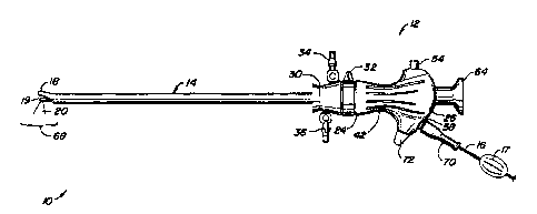
Fig. 1 illustrates a multifunction endoscope 10, such as a transurethral
cystoscope,
including a handle 12 with an external cannula 14 extending distally from the
handle 12. In this
embodiment the multifunction endoscope 10 is designed for use with a medical
laser device of
i0 the type including an optical fiber 16 having a fiber end member 19
which extends into a cavity
formed by a hood structure (described in more detail below) on the distal tip
18 of external
cannula 14. The optical fiber 16 has a knob 17 attached near the handle 12
that is adapted to be
used by a surgeon to manipulate the position of the fiber end member,
rotationally and
longitudinally. External cannula 14 has a number of passageways or lumens
formed by an
internal structure, not shown, extending generally from handle 12 to distal
tip 18 to
accommodate, in this disclosed embodiment, optical fiber 16 and fiber end
member 19, a
telescope type of visualization device typically coupled to a display monitor
(not shown), an
inflow irrigation pathway, and an outflow or suction pathway. The endoscope 10
in the
illustrated embodiment includes an internal cannula, which receives the fiber
16 in a manner,
which allows easy movement of the fiber 16 at least over a range of motion
that allows
manipulation of the fiber end member 19 longitudinally and rotationally within
a working field
by a surgeon grasping the knob 17. Other embodiments are adapted for
manipulation of the fiber
end member 19 by a mechanical system under computer control with active
feedback based on
the video images of the procedure, with or without real time user input.
Individual lumens may be used for a single purpose, such as delivery of
irrigation liquid,
or for two or more purposes, such as housing the telescope and an optical
fiber.
As suggested in Fig. 1, a laser beam 20 is directed laterally in the
illustrated embodiment
from laser end element 19 in a side-firing fashion. Optical fiber 16 could
also have an end
element adapted for forward firing. A telescope provides visualization in the
general direction of
laser beam 20 with appropriately angled optical elements at its distal end. In
addition, other
types of medical instruments may be used as a part of endoscope 10 instead of,
or in addition to,
a medical laser device.
Figure lA is a simplified diagram of an optical fiber assembly adapted for use
with the
endoscopes as described herein. The fiber 1-6 is connected to a coupler 71
adapted to connect the
fiber to the output of a laser system. The fiber end element 19 in the
illustrate embodiment
5
CA 02802925 2013-01-18
comprises a fused quartz cap which captures air between a beveled end 21 of
the fiber 19. The
air/fiber interface provided by the beveled end 21 causes essentially total
internal reflection of the
beam 20 in the side firing direction. At a predetermined distance from the
fiber end element 19,
a travel limiter 76, in the form of a cam in this embodiment, is attached to
the fiber 16. The
travel limiter 76 is adapted to cooperate with a corresponding element within
the endoscope, as
described in more detail below, to prevent the surgeon from withdrawing the
fiber end element
19 into the cannula so that the beam 20 does not damsge the cannula, and to
prevent the surgeon
from rotating the fiber end element 19 toward the hood structure on the distal
tip 18 of the
external cannula, so that the beam 20 does not damage the hood structure on
the distal tip 18. In
to addition, the fiber is threaded through a fiber port cap 70, which is
adapted to couple with a fiber
port on the endoscope 10, as illustrated in Figure 1, which secures the travel
limiter 76 within the
endoscope, and provides a seal on the cannula within which the fiber 16 is
received without
interfering with movement of the fiber within the predetermined ranges of
longitudinal and
rotational motion. The fiber port cap is preferably a flexible material having
an elastic opening
adapted to receive the fiber, and a lip adapted to fit over a corresponding
ridge on the fiber port,
at least partially closing, and in a preferred embodiment substantially
sealing, the fiber port to
prevent fluid leakage while allowing for longitudinal and rotational movement
of the fiber.
In a preferred embodiment the longitudinal motion of optical fiber 16 is
directed axially
along axis 28 (illustrated in Figure 2) and also rotationally about its own
axis to permit laser
beam 20 to be directed proximally and distally as optical fiber 16 moves
generally along axis 28
as well as being swept side to side as optical fiber 16 rotates about its own
axis. Distal tip 18 of
external cannula 14 is beveled to permit this range of movement of laser beam
20 while
providing for proper viewing of working region 68.
Figure 2 shows more detail of handle 12. Handle 12 includes a body cover
portion or
cover 22 having a distal end 24 and a proximal end 26 and defining a central
axis 28. The axial
distance between distal and proximal ends 24, 26 is preferably about 8 to 15
cm, and typically
about 9 to 12 cm. This size range is chosen primarily to accommodate different
hand grasping
techniques, such as shown in Figs. 3-6 for users with a range of sizes of
hands and styles of use
for the endoscope. Handle 12 also includes a supplemental body cover 30
positioned distally of
body cover 22 with a coupler 32 therebetween adapted for covering the proximal
end of the
external cannula 14 and various fittings used for connecting the internal
structures to the external
cannula 14. The external cannula 14 is connected with irrigation inflow and
outflow fittings 34,
36, and secured by bayonet mount 32 to internal structures (not shown), which
are adapted to
receive the telescope 64 and the fiber 16. Handle 12 has a number of ports
opening into the
interior of the handle. For example, inflow and outflow fittings 34, 36 extend
from supplemental
6
CA 02802925 2013-01-18
body cover 30 and provide access to inflow and outflow ports 38, 40 which open
into an inflow
irrigation pathway defined by internal structures and an outflow or suction
pathway extending
along external cannula 14. Inflow fitting 34 may be connected to a source of
an appropriate
irrigation liquid, such as saline fed by a gravity feed structure or by a
pump, while outflow fitting
36 may be connected to an appropriate suction source.
Body cover 22 has a smoothly tapering outer surface 48 that tapers radially
inwardly from
distal and proximal ends 24, 26 towards a central or waist portion 42. The
circumference of
proximal end of 26 is larger than the circumference of distal end 24, which is
larger than the
circumference of waist portion 42. Outer surface 48 has a generally circular,
slightly oval cross-
sectional shape along axis 28 with a diameter in a range of about 1.5 to 2 cm,
for example. Outer
surface 48 may have other, preferably smoothly curving shapes, such as oval
and egg-shaped, at
various positions along axis 28 or along the entire length of axis 28.
Handle 12 also has first and second body cover extensions 44,46 extending
radially
outwardly from the outer surface 48 of body cover 22 adapted to comfortably
shield the
surgeon's hand from fittings for the telescope and the fiber 16. Extensions
44,46 are positioned
between proximal end 26 and waist portion 42. Extensions 44,46 have smoothly
curving,
distally-facing outer surfaces 50, 52 to provide a smooth transition between
outer surface of 48 of
body cover 22 and extensions 44,46. As seen in Fig. 2, first body cover
extension 44 extends
generally directly radially outwardly while the second body cover extension 46
extends both
radially outwardly and distally. An illumination fitting 54 extends from first
body cover
extension 44 and opens into an illumination port 56, discussed below. An
optical fiber fitting 58
extends from second body cover extension 46 and opens into an optical fiber
port 60. Optical
fiber 16 passes through fitting 58, through port 60, and through an
appropriate passageway in
handle 12 for entry into and through an appropriate lumen within external
manila 14. A valve
handle 72 is mounted flush with second body cover extension 46 with a smooth
or otherwise
comfortable surface transition. The valve handle 72 is turned to control a
stop cock within the
handle 12, to seal off port 60 when desired, typically when laser fiber 16 is
removed from handle
12.
As shown in Figure 2, smoothly tapering outer surface 48 is provided with a
number of
grooves 62 to facilitate grasping by the user. The same or other types of
embossing or debossing
may also be provided for outer surface 48 as well as outer surfaces 50,52 to
promote a good grip
of handle 12. One or more of outer =faces 48,50 and 52 may be provided with a
mat or other
suitable surface texture. In the preferred embodiment body cover 22 is of a
stiff polymer
material or metal. In alternative embodiments, the entire body cover 22,
portions of body cover
7
CA 02802925 2013-01-18
22 and/or a skin on the body cover 22 may comprise a resilient or otherwise
yieldable elastomer
material.
Endoscope 10 also includes a telescope 64 extending through a telescope port
65 at
proximal end of 26 and aligned with axis 28. Telescope 64 includes a camera
fitting 66 to permit
images of the working region 68 in the vicinity of laser beam 20 captured by
the telescope at
distal tip 18 to be recorded and/or monitored during use. Illumination port 56
is coupled to the
interior of telescope 64 so the light from the illumination source passes
distally along the
telescope to illuminate working region 68.
A simple endoscope used for examination of an organ may have only two ports,
one for
to the light source and one for the optical image. However, endoscopes used
for medical
procedures such as ablation of tissue using laser energy will typically have
many more ports and
therefore make the design of the proximal portion of the endoscope more
complicated. The
increased complexity includes the presence of tubes, lines, wires and other
things extending from
the proximal portion of the endoscope. One aspect of the endoscope is based on
the recognition
that different individuals using the same endoscope will often hold and
manipulate the endoscope
by its proximal portion in different ways. This is particularly true for
multifunction endoscopes
used for both of viewing and for treatment, at least in part because of the
increased complexity of
the procedure and the number of things extending from the proximal portion, as
well as the
personal preferences of the operator.
The handle described herein for a multifunction endoscope comprises a body
cover, a first
body cover extension and a second body cover extension. The body cover
comprises distal and
proximal ends with an axis extending therebetween with a waist between the
distal and proximal
ends. The distal and proximal ends and the waist have distal and proximal
circumferences and a
waist circumference, respectively. The proximal circumference is larger than
the waist
circumference and the distal circumference is larger than the waist
circumference. The body
cover also comprises an outer surface, the outer surface tapering from the
distal end to the waist
and from the proximal end to the waist. The first body cover extension extends
in a first radial
direction from the outer surface of the body cover between the proximal end
and the waist. The
second body cover extension extends in a second radial direction from the
outer surface of the
body cover between the proximal end and the waist.
In some embodiments the proximal circumference is larger than the distal
circumference.
The outer surface is preferably a smoothly tapering outer surface. The body
cover may comprise
a plurality of ports at a proximal portion thereof. The handle may also
comprise a second body
cover positioned distally of the distal end of the body cover. The second body
cover may
comprise additional ports therein.
CA 02802925 2013-01-18
Figs. 3-6 illustrate four typical ways a surgeon can comfortably and securely
hold or
grasp handle 12 of endoscope 10 by grasping body cover 22 with one hand while
leaving the
other hand free to manipulate laser fiber 16 using fiber manipulator knob 70
to adjust both the
axial and rotary positions of laser beam 20. The shape of handle 12 may
accommodate other
grasping techniques. The different grasping techniques can be based upon
different personal
preferences as well as the particular procedure being accomplished. For
example, an operator
may find the grasping technique of Fig. 6 to be most satisfactory when
initially introducing the
endoscope 10 to the target site to provide the most sensitivity to this
procedure. The provision of
the smaller circumference waist portion 42 provides an exceptionally secure
grasping surface
between the user's thumb and opposed fingers. The grasping techniques of Figs.
3 and 4 provide
extremely stable and secure positioning of handle 12 due to the provision of
the smaller
circumference waist portion 42 and the larger circumference proximal end 26,
as well as first and
second body cover extensions 44,46 with their smoothly tapering, forward
facing outer surfaces
50, 52. The grasping technique of Fig. 5 may be chosen by some users when, for
example,
manipulating laser fiber 16 extending from optical fiber fitting 58. In all
cases, the smoothly
tapering surfaces from the larger circumference distal and proximal ends 24,
26 to the smaller
circumference waist portion 42 provide a comfortable and a secure gripping
surface for the user.
Figure 7 illustrates the distal end 18 of the endoscope positioned within a
urethra adjacent
prostate tissue. The fiber end element 19 directs radiation 20 into the
prostate tissue to cause
vaporization or other effects in the tissue. The distal end 18 includes a hood
structure 101 with a
blunt distal face 102 adapted to be inserted into the urethra. The hood
structure 101 acts as an
obturator, which prevents constriction of the urethra onto the fiber end
element 19, and defines
an open area between the top surface 103 and the working region 68 on the
prostate tissue. The
internal structure (not shown) within the external cannula at the distal end
18 includes a guide
element that is adapted to movably support the optical fiber in a position so
that the emission face
of the end element 19 is spaced away from the working region 68 on the
prostate tissue within
the open area defined by the hood structure 101. In addition, the external
cannula includes a
nozzle for directing inflowing irrigant, and regions for suction of out
flowing irrigant, which
together define an irrigation pathway represented by arrows 104. The
irrigation pathway 104
flows across the emission face of the fiber end element 19 as the fiber end
element 19 is moved
within the open area, maintaining irrigation flow during the delivery of
radiation to facilitate
clear visualization through the telescope and to maintain the emission face of
the fiber end
element 19 clear of debris.
Figure 8 provides a prospective of the distal end of the endoscope outer
cannula, taken
from the direction of the working region 68 where the fiber end element 19
extends outwardly
9
CA 02802925 2013-01-18
between the working region and the telescope face 108, partially blocking the
telescope face 108
in this view. As illustrated in Figure 8, the distal face 102 of the endoscope
represents the end of
a hood structure. An opening on the end of the external cannula is defined by
the distal face 102,
and side walls, which slope away from the end. The inner cannula 110 includes
a first lumen
having an upper ridge 112, which receives the telescope so that the telescope
face 108,
protruding slightly from the upper ridge 112 in this view, faces the working
region 68. The inner
cannula 110 also supports the fiber end element 19. Thus the upper ridge 112
has a radius, which
matches that of the telescope, and the lower ridge 113 as a radius, which
matches that of a
bearing surface on the fiber end element 19. An irrigant inflow channel is
defined by a second
lumen which is bonded to the first lumen by welding or otherwise, and having
crescent shaped
opening 106 which acts as an irrigant nozzle directing irrigation flow
outwardly over the fiber
end element 19. In the illustrated embodiment, a tube 111 is attached to the
outside surface of
the upper ridge 112 of the first lumen acting as a spacer between the inner
cannula that defines
the first and second lumens, and of the inside wall at the top of the outer
cannula. An opening
107 established by tube 111 between the inner cannula and the external cannula
provides an
irrigation outflow channel which is coupled to a suction source tending to
cause the irrigant
which is forced through the crescent shaped opening 106 of the irrigant inflow
channel to flow
outwardly and an upwardly across the fiber end element 19.
Figure 9 is a cross-sectional view of a representative fiber end element 19.
The
representative fiber end element 19 in Figure 9 includes a fused quartz cap
120, which is attached
by glue 121 or otherwise to the cladding 122 of an optical fiber. In a
preferred embodiment, the
optical fiber has a relatively large ratio of the diameters of the cladding
and the core to reduce
unwanted back scattering of the radiation, as described in U.S. Patent No.
5428699, entitled
"Probe having optical fiber for laterally directing laser beam".
The optical fiber 122 has a beveled face 123. The cap 120 captures air in an
area around the
beveled face 123 to establish an air/fiber core interface at which
substantially all of the radiation
from the fiber is reflected on line 124 on to the tissue. The optical fiber
cladding 122 is
surrounded by a protective sheath 127 along the length of the fiber. The
sheath 127 is removed
within the cap, leaving the core and cladding. The quartz cap 120 is beveled
near the fitting with
the protective sheath 124. The cap 120 has a reflective coating 125 in a
region adjacent the
beveled face 123 to block or diffuse any back reflected radiation, preventing
damage to the
endoscope or to tissue that is not intended to be irradiated. As illustrated,
the cap 120 has a
length L over which it has a constant diameter and is circular in cross-
section, so that it is
adapted to fit against, and provide a bearing surface for, the lower ridge 113
of the first lumen
described with reference to Figure 8, over a range of longitudinal motion that
is close to the =
CA 02802925 2013-01-18
length L. Also, as mentioned above, the endoscope includes a guide element,
which positions the
fiber end element 19 so that the emission face is spaced away from the target
tissue. In Figure 9,
the dimension D (on the order of 1 to 2 mm for a transurethral cystoscope)
represents the spacing
provided by the guide element within the endoscope. This spacing operates to
assist the surgeon
to maintain a relatively constant distance between the emission face on the
fiber end element 19,
and the target tissue, and therefore improve consistency of the energy density
on the tissue. Also,
it operates to create a region within which the irrigation flow is readily
accepted and directed
over the emission face of the fiber end element 19 within the range of motion
allowed by the
device.
Figures 10A-10B, 11A-11B, and 12A-1213 illustrate alternative configurations
for the
external cannula and internal structure of the endoscope. In Figure 10A, the
external cannula 150
has a larger radius at an arcuate top surface 150A than at a lower arcuate
surface 150B and an
essentially flat wall between the arcuate surfaces 150A, 150B. The internal
structure includes an
internal cannula 151 adapted to receive the telescope 154 and an irrigant
inflow tube 152. The
tube 152 is bonded to a tubular guide element 156, which receives the fiber
end element 155.
Inner cannula 151 has an arcuate top surface 151A adapted to match the radius
of the telescope
154. Also, the inner cannula 151 has an arcuate bottom surface 151B adapted to
match the radius
of the tube 152, so that they are securely positioned within the cannula 151.
The guide element
156 is bonded to the tube 152. In one embodiment, the end element 155 is
bonded to the guide
element 156, and both are movable as a unit together with the tube 152. In
another embodiment,
the end element 155 moves freely within the guide element 156. Figure 10B
illustrates the flow
of irrigant for the embodiment illustrated in Figure 10A. Irrigant inflow
(hatched in the drawing)
occurs in a region 158 having a crescent shape below and partially surrounding
the fiber end
element 155 as it is positioned as described above. Irrigant outflow (not
hatched in the drawing)
is primarily directed through the region 159 near the top of the external
cannula, and additional
irrigant flows through regions 160, 161, 162 and 163 as illustrated through
the inner cannula and
the outer cannula.
In Figure 11A, the external cannula 170 has a smaller radius at the top
surface 170A then
at the lower surface 17013 and an essentially flat wall between the arcuate
top and bottom
surfaces 170A, 170B. The internal structure includes a tubular guide 173
adapted to receive the
telescope 154, and a crescent shaped irrigant inflow tube 172, with a top
surface that comprises
two essentially flat regions 172A, I 72B that intersect in a smaller radius
arcuate region 172C,
and a bottom surface 172D that comprises an arcuate portion adapted to match
the radius of the
lower surface 1708 of the external cannula. The smaller radius arcuate region
172C is adapted to
match the radius of a guide element 171, which supports the fiber end element
155. The guide
11
CA 02802925 2013-01-18
element 171 is bonded to the smaller arcuate region 172C. Figure 11B
illustrates the flow of
irrigant for the embodiment illustrated in Figure 11A. Inigant inflow (hatched
in the drawing)
occurs in a region 175 having a crescent shape below and partially surrounding
the fiber end
element 155 as it is positioned as described above. Irrigant outflow (not
hatched in the drawing)
is primarily directed through the region 176 above the fiber end element 155.
In Figure 12A, the embodiment of Figure 8 is illustrated in cross-section. In
this
embodiment, the external cannula 180 has nearly equal radii at the top surface
180A and the
bottom surface 180B. The internal structure includes a first tube 181 having a
top arcuate surface
181A and a bottom arcuate surface 181B with essentially flat walls in between.
The tube 181 is
adapted to receive the telescope 154 and the fiber end element 155. Thus the
radius of the
bottom arcuate surface 181B matches the radius of the end element 155.
Likewise, the radius of
the top arcuate surface 181A matches the radius of the telescope 154. An
irrigant inflow channel
is provided by bonding the element 182 to the sidewalls of the tube at 181 to
form a crescent
shaped opening. The radius of the element 182 is slightly smaller than the
radius of the bottom
surface 180B of the external cannula.. Although not shown, a spacer, as
described above in
Figure 8 is bonded to the top of the arcuate surface 181A to securely position
the inner structure
within the external cannula 180. Figure 12B illustrates the flow of irrigant
for the embodiment
illustrated in Figure 12A. Irrigant inflow (hatched in the drawing) occurs in
a region 185 having a
crescent shape below and partially surrounding the fiber end element 155 as it
is positioned as
described above. Irrigant outflow (not hatched in the drawing) is primarily
directed through the
region 186 above the fiber end element. Additional irrigant outflow occurs in
regions 187, 188,
189, and 190.
In all three embodiments, a crescent shaped irrigant inflow path (for irrigant
flowing into
the working region) below and partially surrounding the fiber end element 155,
is established by
the internal structure of the endoscope. In addition, the fiber end element
155 is positioned in a
manner that limits lateral movement and maintains a fixed distance
(corresponding to the
distance D of Figure 8) between the outside wall on which the emission surface
of the fiber end
element 155 is found, and the lower arcuate surface (150B, 170B or 180B) of
the external
eannula.
Figure 13 shows a cross-section of the distal end of the external cannula for
the
embodiment of Figure 12A. The external cannula has a top wall 200 that is
essentially straight
along the major axis of the cannula, and is arcuate in the region 201, as
described with reference
to Figure 12A. The external cannula has a lower wall 202, that is essentially
parallel to the top
wall 200, and is arcuate in the region 203, as described referenced Figure
12A. In the region 204,
the far side wall 205 of the external cannula is essentially straight. The
distal end face 206 is
12
CA 02802925 2013-01-18
formed on a hood structure 207, which flares slightly outwardly from the
essentially straight top
wall 200, and has a blunt rounded end. The distal end 208 of the external
cannula at the lower
wall 202, the side wall 204 and lower arcuate region 203 is curved inwardly
toward the proximal
end of the cannula to establish the hooded area for the working region as
described above.
litigant outflow ports 209 are formed in the top arcuate region 201 to capture
any irrigant that
escapes outside the external carmula. Likewise, irrigant outflow port 210 is
formed in the side
wall 205.
Figure 14 shows a cross-section of the distal end of the internal cannula for
the
embodiment of Figure 12A. The internal cannula structure has a spacer formed
by tube 215 with
beveled front and rear surfaces, bonded to the top wall 221 of the tube 220.
Tube 220 is adapted
to receive the telescope and the fiber end element. The top wall 221 of the
tube 220 is essentially
straight, and coupled to the top arcuate portion 222, that is adapted to fit
the radius of the
telescope. The tube 220 has a bottom wall 223 which is essentially parallel to
the top wall 221
and coupled to the bottom arcuate portion 224 which is adapted to fit the a
radius of the fiber end
element. The far wall 226 of the tube at 220 is essentially flat between the
arcuate portions 222,
224. The irrigant inflow tube is defined by wall 228, which is essentially
parallel to the bottom
wall 223 of the tube 220. The internal cannula structure of Figure 14 fits
slidably within the
exterior cannula structure of Figure 13. The combination of the thickness of
the bottom wall 202
of the external cannula, and the thickness of the irrigant inflow channel
defined by wall 228 and
wall 223 of the internal structure establish the distance D at which the fiber
end element is
maintained away from tissue in the target region.
Figure 15 illustrates the proximal end of the lumen in the endoscope adapted
received the
fiber and cooperate with the travel limiter 300, which is shown apart from the
fiber in the
drawing. A fitting 58 defines a lumen 275 into which the fiber is received
with the travel limiter
300 bonded thereto. The fitting 58 includes a cylindrical inner lumen coupled
to a stopcock
valve 278 which is opened to receive the fiber, and provide a continuous lumen
275 having a
cylindrical bearing surface within which the travel limiter 300 is able to
move. On the distal side
of the stopcock valve 278, a tube 280 is bonded which directs the fiber into
the internal structure
of the endoscope as described above. As shown in Figure 15, the cover 65
includes an extension
46 surrounding the stopcock valve 278 and the tube 280, while the fitting 58
extends outwardly.
A pin 276 or other stop element extends into the lumen 275 and cooperates with
the travel limiter
300 to prevent rotation beyond a predefined arc of the fiber.
The structure of the travel limiter 300 (in the form of a cam in this
embodiment) is
illustrated in Figure 16. The travel limiter includes a cylindrical fiber
sheath body 310 adapted to
fit over the sheath of the optical fiber and be bonded thereto. Appendages
311, 312, 313 are
13
CA 02802925 2013-01-18
formed on the body 310 with arcuate outside surfaces (e.g. surface 315 on
appendage 312),
which are adapted to center the fiber, and to slide rotationally and
longitudinally on the bearing
surface within the lumen 275. A ridge 314 extends along the major axis of the
body 310 having
sidewalls with a rear beveled surface 320 (and a front beveled surface) set at
an angle theta, such
as about 60 . The sidewalls are positioned so that in cooperation with the pin
276, rotational
movement of the fiber is limited to a predefined arc. The width of the ridge
314 between the side
walls determines the range of rotational movement of the fiber.
Although not shown in Figure 15, seal 70 (See, Fig. 1 and Fig. IA) when
attached to the
fitting 58 acts to prevent longitudinal motion of the travel limiter 300 in a
direction away from
the distal end of the endoscope. Longitudinal motion in a direction toward the
distal end of the
endoscope is not actively limited in this embodiment, but is controlled by the
surgeon by
observing the fiber end element within the field of view of the telescope, in
cooperation with feel
of limited movement defined by the length of the bearing surface in lumen 275.
In an alternative
embodiment, a structure may be added to rigidly limit longitudinal motion
toward the distal end.
The length of the ridge 314 is selected so that when the fiber is fully
withdrawn against the seal
70, the ridge remains in a position to cooperate with the pin 276, thereby
providing for control of
rotational motion of the fiber over a predefined length of longitudinal motion
which is the equal
to about twice the length of the ridge 314.
Figure 17 illustrates positioning of the travel limiter 300 from an end of
view within the
lumen 275. Appendages 311 and 312 and the ridge 314 are adapted to secure the
body 310 at a
position that is substantially centered within the lumen 275, and so that as
the fiber is rotated, it
remains positioned near the center and does not contact the pin 267.
As illustrated in Figure 18, when the fiber is rotated in a counterclockwise
direction, the
ridge 314 eventually contacts the pin 276 to limit the rotational motion. The
ridge 314
cooperates in a similar manner with the pin 276 to limit clockwise motion. In
illustrated
embodiment, the travel limiter will allow rotational motion of about 270 ,
with the remaining 90
of the circle being blocked to prevent irradiation of the hood structure on
the distal end of the
endoscope.
Figure 19 is a cross-section view of the fiber port cap 70 shown in Figure 1A,
adapted to
at least partially close the fiber port while admitting the fiber into the
port and allowing rotational
and translational movement of the fiber. The fiber port cap 70 has a distal
end 350 adapted to fit
over the port 279 shown in Figure 15, and a proximal and 351 adapted to
receive the fiber. The
embodiment illustrated includes a first portion 352 and a second portion 353.
The first portion
352 comprises a cylindrical body essentially open on the distal end 350 to fit
over the outside of
the port 279 a granular slot reader 56 adapted to fit over the ridge at the
outer edge of the port
14
CA 02802925 2013-01-18
279, and a conical portrait grip 358 which extends on the proximal end of the
element 352
inwardly. The conical portrait grip 358 includes a passage 360 adapted to
receive the optical
fiber. The element 353 has a relatively elongated cylindrical body 355 with an
interior 357
substantially greater in diameter than the fiber. An enlarged cylindrical
portion 359 adapted to fit
on the outside surface of the element 352 is included. An interface between
element 352 and the
enlarged portion 359 is bonded securely. The proximal end of the element 353
has a circular
opening 361 with tapered edge 363 adapted to snugly engage the fiber. In a
representative
embodiment the element 352 is formed using a relatively low durometer silicone
rubber. The
element 353 is formed using a medium durometer silicone rubber. Together, they
provide a fiber
port cap that is flexible enough to be installed and removed from the
endoscope, and provides a
firm enough seal to allow movement of the fiber during use and prevent leakage
of irrigant. The
seal provided prevents back flow of inigant from within the endoscope during
operation of the
device. Also, the conical port grip element 358 acts as a stop for the travel
limiter 300 illustrated
in Figure 15.
An endoscope for laser surgery that has a channel to guide a laser fiber to an
operating
field has been described characterized by various combinations of the
following features:
a. The endoscope is adapted for use with an operating field being a
body cavity to
interior cavity of an organ filled with an aqueous irrigation fluid.
b. The endoscope having a build in telescope for visualization of
the operating field.
c. The endoscope having a separate channel to guide irrigation fluid to the
operating
field.
d. The endoscope having a separate channel to guide irrigation fluid and
tissue
particles out of the operating field.
c. The channel guiding the laser fiber giving the fiber very good stability
and
allowing the surgeon to control the position of the laser fiber relative to
the tissue
face.
f. The channel guiding the irrigation fluid out of the body cavity in a way
that vapor
bubbles and tissue particles are flowing outside the visual field of the
telescope.
g. The channel guiding the irrigation fluid to the operating field in such a
way that
irrigation fluid is directed predominantly towards the spot where the laser
beam
hits the tissue.
h. The channel guiding the irrigation fluid to the operating field guiding the
fluid in
such a way that vapor bubbles and tissue particles created by the laser tissue
interaction are carried away out of the field of view of the surgeon.
CA 02802925 2013-01-18
i. Channels to guide laser fiber and irrigation fluid to the operating
field located in a
way that irrigation fluid flows predominantly between a "hot" emission face of
laser fiber and target tissue.
j. A component of the endoscope that interfaces with a device attached to
laser fiber
that prevents the surgeon from aiming laser beam on components of endoscope.
i. Such device limiting rotation of laser fiber in the channel
guiding the laser
fiber to the operating field in such a way to prevent the laser beam from
being aimed at the protruding tip of endoscope.
Such device limiting translation of fiber inside the channel guiding the
laser fiber to the operating field in such a way to prevent the laser beam
from being aimed at an endoscope component.
iii. Such device allowing translation and rotation of the laser fiber inside
the
channel guiding the laser fiber to the operating field in such a way to allow
the laser beam to be aimed in all directions where the laser beam does not
hit an endoscope component.
iv. Such device allowing translation and rotation of laser fiber inside the
channel guiding the laser fiber to the operating field in such a way that the
laser beam can be aimed at targets outside the field of view of the
telescope.
v. A travel limiting mechanism, including for example a pin protruding into
the channel guiding the laser fiber that interfaces with a fm or cam
mounted on the laser fiber. The fm oriented in a way that the laser beam
being emitted sideways out of the laser fiber does not fire in the direction
of a distal tip of the endoscope that is protruding beyond the end of the
channel guiding the laser fiber.
vi. An aperture having a flexible or resilient seal on the channel that is
guiding the laser fiber with the aperture interfacing with a device on the
laser fiber in such way that user feels resistance when device pushes
against aperture.
An optical fiber assembly is described including adapted for use with
endoscopes as
described herein is also described, including a fiber end element adapted to
direct laser energy
into a working region on tissue, a travel limiter adapted to cooperate with a
fiber port to limit
longitudinal and rotational movement of the fiber, fiber port cap adapted to
couple the fiber to the
fiber port, while providing a seal and allowing for movement of the fiber, a
handle adapted for
16
CA 02802925 2015-07-06
use by a surgeon to facilitate movement of the fiber during use, and a fiber
coupler, adapted to
couple the fiber to a source of laser energy.
One embodiment of an endoscope comprises a continuous flow laser cystoscope
comprised of inner and outer sheaths defining cannulas. The inner sheath
having plural channels
to guide a telescope, a laser fiber and irrigation fluid to the operating
filed. Space between inner
and outer sheath serving as pathway to guide irrigation fluid out of the
operating field. The laser
fiber being located underneath the telescope in the direction of tissue. A
channel or pathway that
guides irrigation fluid to the operating field being located between laser
fiber and tissue. A
channel or pathway that guides irrigation fluid out of the body cavity being
located on top of the
telescope. A channel that guides the laser fiber having an inner diameter only
slightly larger
than the outer diameter of laser fiber near the tip. A "channel" or "pathway"
can comprise two
or more tubes of different diameter that sit within each other or are
otherwise arranged to
cooperate as a pathway or channel.
The scope of the claims should not be limited by the preferred embodiments set
forth in
the examples, but should be given the broadest interpretation consistent with
the description as a
whole.
17