Note: Descriptions are shown in the official language in which they were submitted.
CA 02803509 2012-12-20
WO 2012/001075 PCT/EP2011/060952
1
IMMORTALIZED AVIAN CELL LINES
BACKGROUND OF THE INVENTION
1. Technical Field of the Invention
This invention relates to immortalized avian cells, and to the use of these
cells for
the production of viruses. The cells according to the invention are
particularly useful for the
production of recombinant viral vectors which can be used for the preparation
of therapeutic
and/or prophylactic compositions for the treatment of animals and more
particularly
humans.
2. Description of Background and/or Related Art
Eukaryotic cell lines are fundamental for the manufacture of viral vaccines
and many
products of biotechnology. Biologicals produced in cell cultures include
enzymes,
hormones, immunobiologicals (monoclonal antibodies, interleukins,
lymphokines), and
anticancer agents. Although many simpler proteins can be produced using
bacterial cells,
more complex proteins that are glycosylated, currently must be made in
eukaryotic cells.
Avian cells have been used for years for the production of viral vectors. For
example, the Vaccinia virus used for preparing prophylactic composition for
the treatment of
Variola was cultivated on Chicken Embryonic Fibroblast (CEF). Avian cells are
particularly
useful since many virus used in pharmaceutical composition are able to
replicate on them.
More noticeably, various viruses are only able to grow on avian cells. This is
for example
the case of Mammalian Virus Ankara (MVA) which is unable to grow on mammalian
cells.
This poxvirus, which derived from a Vaccinia Virus by more than 500 passages
on CEF,
was used in the early seventies for vaccinating immunodeficient peoples
against Variola.
Now, MVA is mainly used as a vector for gene therapy purposes. For example,
MVA is
used as a vector for the MUC1 gene for vaccinating patients against tumor
expressing this
antigen (Scholl et al., 3 J BlaMED BIOTECHNOL. 194-201 (2003)). MVA carrying
the gene
coding HPV antigens are also used as a vector for the therapeutic treatment of
ovarian
carcinoma. More recently, MVA has been the vector of choice for preparing
prophylactic
treatment against newly emerging diseases or probable biological weapons such
as west
nile virus and anthrax.
With this respect, there is a growing need for virus production. For now, the
most
used MVA production process comprises a virus replication step on CEF. However
the use
of CEF is linked to various difficulties. Firstly, the preparation of CEF
comprised many
steps which have to be done manually.
Furthermore, this virus production process depends on the availability of eggs
which
CA 02803509 2012-12-20
WO 2012/001075 PCT/EP2011/060952
2
may be totally disrupted in case of contamination of the breedings. This
problem is more
and more relevant with the spread of Avian Flu.
Additionally, many CEFs possess a reverse transcriptase activity (RT). RT is
an
enzyme necessary for retroviruses to reproduce. Retroviruses are found in many
different
species. RT is not infectious in humans or animals, and it has not been shown
to cause any
adverse health effects in people. Using a highly sensitive polymerase chain
reaction (PCR)
based assay, RT activity has been detected in minute quantities in vaccines
manufactured
with chick embryo fibroblasts. The source of the enzyme is probably a partial
viral genome
coding for RT, believed to be integrated into chick cells hundreds or
thousands of years
ago. Avian retroviruses that produce this RT are not known to affect humans.
While the
human immunodeficiency virus (HIV, the virus that leads to AIDS), is a
retrovirus, the RT
activity detected in vaccines is definitively not derived from HIV.
Furthermore, the presence
of RT does not confirm the presence of a retrovirus. Nevertheless, a cell line
with no
endogenous RT activity would be of interest.
In order to emancipate virus production process from the use of CEF, there is
an
increasing need for an avian cell line which would allow the replication and
the production of
the virus. Immortalized cell lines can be maintained or frozen from batch to
batch on the
production site and are always available for a new production process.
Moreover as they
are confined at the production plant, they are less subject to contamination
by exogenous
contaminant. Their use allows a drastic reduction of the manual manipulation
needed for
the production process. All these properties lead to a reduction of the price
and of the
duration of the production process as well as a diminution of the potential
contamination.
Finally, cell lines can be fully characterized and are thus totally compliant
with the
good laboratory practice and the requirements of the different medical
agencies.
Different avian cell lines have already been described. For example, DF1 (U.S.
Patent No. 5,879,924) is a spontaneously immortalized chicken cell line
derived from 10 day
old East Lansing Line (ELL-0) eggs. The cells are useful as substrates for
virus
propagation, recombinant protein expression and recombinant virus production.
However,
this cell line is susceptible to various virus such as Meleagrid herpesvirus 1
(Herpes Virus of
Turkey), Fowlpox Virus, reovirus, Avian Sarcoma Leukemia Virus and Rous
Sarcoma Virus.
Immortal avian cells can also be derived from embryonic stem cells by
progressive
severance from growth factors and feeder layer, thus maintaining growth
features and
infinite lifespan characteristic of undifferentiated stem). The only available
avian cell line
derived by this process is the Ebx chicken cell line (WO 2005/007840) which
has been in
contact with feeder layers from murin origin, raising additional regulatory
questions like
murin virus contamination and presence of endogenous retroviral sequences in
chicken
CA 02803509 2012-12-20
WO 2012/001075 PCT/EP2011/060952
3
cells. Moreover this cell lines have been described in some conditions as
unstable and
differentiation-prone.
A duck embryo permanent cell line, free from endogenous avian retroviruses has
also been established. The cell line, designated as DEC 99 (Ivanov et al.
EXPERIMENTAL
PATHOLOGY AND PARASITOLOGY, 4/2000 Bulgarian Academy of Sciences) has been
cultured
over 140 consecutive passages and it is not tumorigenic for birds. The DEC 99
cell line is a
standard cell culture system that has been used for research and can be
applied for the
needs of biotechnology. This cell line is a suitable model for studies in the
field of cell
biology, virology, immunology, toxicology and for the production of
diagnostics and
vaccines. The susceptibility of the permanent duck embryo cell line (CL) DEC
99 to infection
with embryo-adapted avian poxvirus (APV) vaccine strains have been studied
(Ivanov et al.
EXPERIMENTAL PATHOLOGY AND PARASITOLOGY, 4/6 2001 Bulgarian Academy of
Sciences).
The FK and Dessau vaccine strains of fowl and pigeon origin respectively have
been used.
The virus strains were consecutively passaged (13 passages) on primary duck
embryo cell
cultures (CCs). The adapted virus strains have been further passaged (12
passages) in the
CCs of the DEC 99 cell line, where a typical cytopathic effect (CPE) was
observed. The
production of infectious virions was checked by inoculation of 11-day-old
White Leghorn
embryos, where typical pox proliferations on the chorioalantoic membranes
(CAMs) were
formed. In the DEC 99 cells the FK strain caused early CPE, compared to the
Dessau
strain and reached a titer of 106,25 CCID50/ml. The DEC 99-adapted virus
strains induced
typical cutaneous "takes" after vaccination of two-month-old chicks. Thus, the
DEC 99, as a
standard CC system appears to be suitable for production of vaccines against
fowl pox.
Nevertheless this particular cell line is slow growing after passage 40 and is
unable to grow
in suspension.
Nucleic acid sequences from the Early region of human Adenovirus 5 have
already
been used to transform some specific human cells in vitro (293 and PER. C6
cell lines ;
Fallaux,F. J. et al., 9 Hum. GENE THER. 1909-17 (1998); Graham, F. L. et al.,
36 J. GEN.
VIROL. 59-74 (1977)).
In general terms, the adenoviral genome consists of a double-stranded linear
DNA
molecule approximately 36 kb in length which contains the sequences coding for
more than
30 proteins. At each of its ends, a short inverted sequence of 100 to 150
nucleotides,
depending on the serotypes, designated ITR (inverted terminal repeat), is
present. ITRs are
involved in the replication of the adenoviral genome. The encapsidation region
of
approximately 300 nucleotides is located at the 5' end of the genome
immediately after the
5' ITR.
The early genes are distributed in 4 regions which are dispersed in the
adenoviral
CA 02803509 2012-12-20
WO 2012/001075 PCT/EP2011/060952
4
genome, designated El to E4 (E denoting "early"). The early regions comprise
at least six
transcription units which possess their own promoters. The expression of the
early genes is
itself regulated, some genes being expressed before others. Three regions, El,
E2 and E4,
respectively, are essential to the viral replication. Thus, if an adenovirus
is defective for one
of these functions, that is to say if it cannot produce at least one protein
encoded by one of
these regions, this protein will have to be supplied to it in trans.
The El early region is located at the 5' end of the adenoviral genome, and
contains
2 viral transcription units, E1A and El B, respectively. This region codes for
proteins which
participate very early in the viral cycle and are essential to the expression
of almost all the
other genes of the adenovirus. In particular, the E1A transcription unit codes
for a protein
which trans-activates the transcription of the other viral genes, inducing
transcription from
the promoters of the El B, E2A, E2B and E4 regions.
It was shown by Guilhot et al. (Guilhot, C. et al., 8 ONCOGENE 619-24 (1993))
that
retroviral transduction of the 12S protein of E1A from Ad5 can lead to
immortalization of
quail cells. However, WO 2005/042728 disclosed that it is impossible to
immortalize avian
cells when the E1A gene is introduced by transfection of naked DNA instead of
retrovirus
infection. WO 2005/042728 further states: "that the extremely efficient and
stable
transduction via retrovirus infection creates a cell pool large enough to
harbor individual
cells with spontaneous genomic changes that have blocked apoptosis that
normally is
induced upon Retinoblastoma inactivation." (page 10).
The presence of retroviral sequences in the cells obtained by Guilhot et al.
hinder
the use of such cells for the production of biological product and more
particularly for
therapeutic compounds.
BRIEF SUMMARY OF THE INVENTION
The inventors have surprisingly found that avian cells, and more particularly
cairina
moschata cells, can be efficiently immortalized by E1A transfection with a non-
viral vector.
In order to solve the different problems linked to the use of CEF and/or to
the use of
previously available cell lines, the present invention provides an
immortalized avian cell
comprising an E1A nucleic acid sequence characterized in that said cell is
obtained by a
process comprising the step of transfecting the cell with a non viral vector
comprising said
E1A nucleic acid sequence and wherein said cell does not comprise an E113
nucleic acid
sequence.
The present invention also refers to a process for immortalizing an avian cell
comprising the step of transfecting said cell with a non-viral vector
comprising an E1A
nucleic acid sequence and wherein said process does not comprise a step of
transfecting
CA 02803509 2012-12-20
WO 2012/001075 PCT/EP2011/060952
said cell with an E1 B nucleic acid sequence.
The present invention includes immortalized avian cell lines deposited at the
European Collection of Cell Cultures (ECACC) under accession numbers 09070701,
09070702, and 09070703 and derivatives thereof. The immortalized avian cells
may further
5 comprise one or more nucleic acid sequences that allow propagation of a
defective virus.
The immortalized avian cells may further comprise a nucleic acid coding a
substance of
interest. The immortalized avian cell lines may be used for the replication of
a virus,
including live, attenuated, or recombinant viruses. The virus to be replicated
using the
immortalized avian cell lines may be poxvirus (such as vaccinia virus, e.g., a
modified
vaccinia virus Ankara (MVA)), adenovirus, retrovirus, herpervirus, alphavirus,
foamy virus,
adenovirus-associated virus, flavivirus and influenza virus.
Other characteristics, aspects, and advantages of the invention are set forth
in the
detailed description that follows.
DETAILED DESCRIPTION OF THE FIGURES
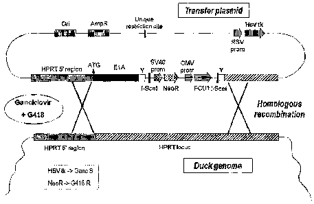
Figure 1: Vector comprising a gene coding the E1A nucleic acid sequence.
Figure 2: Schematic representation of the site specific insertion of the E1A
nucleic
acid sequence into the HPRT gene.
Figure 3: Schematic representation of the elimination of the first and the
third
selection marker from the genome of the immortalized cell obtained by the
process of the
invention.
Figure 4: Vector comprising a gene coding the Cairina moschata telomerase
reverse transcriptase gene and the E1A nucleic acid sequence.
Figure 5: Light microscopy imaging of the Cairina moschata immortalized avian
cell
line ECACC 09070701 (passage 21).
Figure 6: Light microscopy imaging of the Cairina moschata immortalized avian
cell
line ECACC 09070702 (passage 24).
Figure 7: Light microscopy imaging of the Cairina moschata immortalized avian
cell
line ECACC 09070703 (passage 28).
Figure 8: Cairina moschata immortalized avian cell line ECACC 09070701, ECACC
09070702, and ECACC 09070703 growth curve.
Figure 9: Cairina moschata immortalized avian cell line ECACC 09070703 (Figure
9A), ECACC 09070701 (Figure 9B), and ECACC 09070702 (Figure 9C) population
doubling
time evolution.
Figure 10: Production of Modified Vaccinia virus Ankara (MVA) in the Cairina
moschata immortalized avian cell line ECACC 09070701, ECACC 09070702, and
ECACC
CA 02803509 2012-12-20
WO 2012/001075 PCT/EP2011/060952
6
09070703 (MOI 0,05).
Figure 11: Production of VV-COP in the Cairina moschata immortalized avian
cell
line ECACC 09070702
Figure 12: Production of influenza A/Panama/2007/99 H3N2 virus strain in the
Cairina moschata immortalized avian cell line ECACC 09070702
Figure 13: Production of influenza B/Brisbane/60/2008 virus in the Cairina
moschata
immortalized avian cell line ECACC 09070702.
DETAILED DESCRIPTION OF THE INVENTION
The inventors have surprisingly found that avian cells, and more particularly
cairina
moschata cells, can be efficiently immortalized by E1A transfection with a non-
viral vector.
In order to solve the different problems linked to the use of CEF and/or to
the use of
previously available cell lines, the present invention provides an
immortalized avian cell
comprising an E1A nucleic acid sequence characterized in that said cell is
obtained by a
process comprising the step of transfecting the cell with a non viral vector
comprising said
E1A nucleic acid sequence and wherein said cell does not comprise an E113
nucleic acid
sequence.
The present invention also refers to a process for immortalizing an avian cell
comprising the step of transfecting said cell with a non-viral vector
comprising an E1A
nucleic acid sequence and wherein said process does not comprise a step of
transfecting
said cell with an E1 B nucleic acid sequence.
The present invention includes immortalized avian cell lines deposited at the
European Collection of Cell Cultures (ECACC) under accession numbers 09070701,
09070702, and 09070703 and derivatives thereof. The immortalized avian cells
may further
comprise one or more nucleic acid sequences that allow propagation of a
defective virus.
The immortalized avian cells may further comprise a nucleic acid coding a
substance of
interest. The immortalized avian cell lines may be used for the replication of
a virus,
including live, attenuated, or recombinant viruses. The virus to be replicated
using the
immortalized avian cell lines may be poxvirus (such as vaccinia virus, e.g., a
modified
vaccinia virus Ankara (MVA)), adenovirus, retrovirus, herpervirus, alphavirus,
foamy virus,
adenovirus-associated virus, flavivirus and influenza virus.
An immortalized cell, as used herein, refers to a cell capable of growing in
culture for
more than 35 passages.
The term passage number refers to the number of times that a cell population
has
been removed from the culture vessel and undergone a subculture (passage)
process, in
order to keep the cells at a sufficiently low density to stimulate further
growth.
CA 02803509 2012-12-20
WO 2012/001075 PCT/EP2011/060952
7
As used throughout the entire application, the terms "a" and "an" are used in
the
sense that they mean "at least one", "at least a first", "one or more" or "a
plurality" of the
referenced components or steps, unless the context clearly dictates otherwise.
For
example, the term "a cell" includes a plurality of cells, including mixtures
thereof.
The term "and/or" wherever used herein includes the meaning of "and", "or",
and "all
or any other combination of the elements connected by said term".
As used herein, the term "comprising" is intended to mean that the products,
compositions and methods include the referenced components or steps, but not
excluding
others. "Consisting essentially of" when used to define products,
compositions, and
methods, shall mean excluding other components or steps of any essential
significance.
Thus, a composition consisting essentially of the recited components would not
exclude
trace contaminants and pharmaceutically acceptable carriers. "Consisting of"
shall mean
excluding more than trace elements of other components or steps.
As used herein, the term "E1A nucleic acid sequence" refers to nucleic acid
sequence coding all gene products of the adenovirus E1A region, including the
nucleic acid
sequence coding the two major RNAs: 13S and 12S.
Preferably, the term "E1A nucleic acid sequence" refers to a nucleic acid
sequence
comprising a nucleic acid sequence which has at least 60% sequence identity to
SEQ ID
NO:1. In a more preferred embodiment of the invention, E1A refers to a nucleic
acid
sequence comprising a nucleic acid sequence which has at least 70%, preferably
at least
80%, and even more preferably at least 90%, nucleic acid sequence identity to
SEQ ID
NO:1. In a more preferred embodiment, E1A refers to the nucleic acid sequence
set forth in
SEQ ID NO:1.
As used herein, the term "E113 nucleic acid sequence" refers to all nucleic
acid
sequence of the adenovirus E113 region, including the nucleic acid sequence
coding the 3
major polypeptides, of 19 kd and 55 kd.
As employed herein, the term "substantially the same nucleic acid sequence"
refers
to nucleic acid molecule having sufficient identity to the reference
polynucleotide, such that
it will hybridize to the reference nucleotide under moderately stringent
hybridization
conditions. In one embodiment, nucleic acid molecule having substantially the
same
nucleotide sequence as the reference nucleotide sequence set forth in SEQ ID
NO:1.
Hybridization refers to the binding of complementary strands of nucleic acid
(i.e.,
sense:antisense strands or probe:target-DNA) to each other through hydrogen
bonds,
similar to the bonds that naturally occur in chromosomal DNA. Stringency
levels used to
hybridize a given probe with target-DNA can be readily varied by those of
skill in the art.
The phrase "stringent hybridization" is used herein to refer to conditions
under which
CA 02803509 2012-12-20
WO 2012/001075 PCT/EP2011/060952
8
polynucleic acid hybrids are stable. As known to those of skill in the art,
the stability of
hybrids is reflected in the melting temperature (Tm) of the hybrids. In
general, the stability
of a hybrid is a function of sodium ion concentration and temperature.
Typically, the
hybridization reaction is performed under conditions of lower stringency,
followed by
washes of varying, but higher, stringency. Reference to hybridization
stringency relates to
such washing conditions.
As used herein, the phrase "moderately stringent hybridization" refers to
conditions
that permit target-DNA to bind a complementary nucleic acid that has about 60%
identity,
preferably about 75% identity, more preferably about 85% identity to the
target DNA; with
greater than about 90% identity to target-DNA being especially preferred.
Preferably,
moderately stringent conditions are conditions equivalent to hybridization in
50%
formamide, 5*Denhart's solution, 5*SSPE, 0.2% SDS at 42 C., followed by
washing in
0.2*SSPE, 0.2% SDS, at 65 C.
As used herein, the expression "non-viral vector" notably refers to a vector
of
plasmid origin, and optionally such a vector combined with one or more
substances
improving the transfectional efficiency and/or the stability of said vector
and/or the
protection of said vector in vivo toward the immune system of the host
organism. These
substances are widely documented in the literature which is accessible to
persons skilled in
the art (see, e.g., Feigner et al., 32 PROC. WEST. PHARMACOL. SOC. 115-121
(1987);
Hodgson and Solaiman, 14 NATURE BIOTECHNOLOGY 339-342 (1996); Remy et al., 5
BIOCONJUGATE CHEMISTRY 647-654 (1994)). By way of illustration, but without
limitation,
they may be polymers, lipids, in particular cationic lipids, liposomes,
nuclear proteins, or
neutral lipids. These substances may be used alone or in combination. Examples
of such
compounds are in particular available in WO 98/08489, WO 98/17693, WO
98/34910, WO
98/37916, WO 98/53853, EP 890362, or WO 99/05183. A combination which may be
envisaged is a plasmid recombinant vector combined with cationic lipids (DOGS,
DC-CHOL, spermine-chol, spermidine-chol and the like) and neutral lipids
(DOPE).
The choice of the plasmids which can be used in the context of the present
invention
is vast. They may be cloning and/or expression vectors. In general, they are
known to a
person skilled in the art and a number of them are commercially available, but
it is also
possible to construct them or to modify them by genetic engineering
techniques. There may
be mentioned, by way of examples, the plasmids derived from pBR322 (Gibco
BRL), pUC
(Gibco BRL), pBluescript (Stratagene), pREP4, pCEP4 (Invitrogene) or p Poly
(Lathe et al.,
1987, Gene 57, 193-201). Preferably, a plasmid used in the context of the
present invention
contains a replication origin ensuring the initiation of replication in a
producing cell and/or a
host cell (for example, the ColE1 origin may be selected for a plasmid
intended to be
CA 02803509 2012-12-20
WO 2012/001075 PCT/EP2011/060952
9
produced in E. coli and the oriP/EBNA1 system may be selected if it is desired
for it to be
self-replicating in a mammalian host cell, Lupton and Levine, 5 MOS. CELL.
BIOL. 2533-2542
(1985); Yates et al., 313 NATURE 812-815 (1985). It may comprise additional
elements
improving its maintenance and/or its stability in a given cell (cer sequence
which promotes
the monomeric maintenance of a plasmid (Summers and Sherrat, 36 CELL 1097-1103
(1984), sequences for integration into the cell genome)).
The term "non-viral vector" excludes viral vectors, such as, for example
vector
deriving from a poxvirus (vaccinia virus, in particular MVA, canarypox and the
like), from an
adenovirus, from a retrovirus, from a herpesvirus, from an alphavirus, from a
foamy virus,
from an adeno-associated virusfrom a flavivirus or from an influenza virus.
The present invention also relates to cells deriving from the cell according
to the
invention. As used herein, the term "derived" refers to cells which develop or
differentiate
from or have as ancestor a cell according to the invention.
The term passage number refers to the number of times that a cell population
has
been removed from the culture vessel and undergone a subculture (passage)
process, in
order to keep the cells at a sufficiently low density to stimulate further
growth.
As used herein, the term "transfected" refers to the stable transfection or
the
transient transfection of the cell of the invention.
The term "stable transfection" or "stably transfected" refers to the
introduction and
integration of foreign nucleic acid sequence into the genome of the
transfected cell. The
term "stable transfectant" refers to a cell that has stably integrated foreign
DNA into the
genomic DNA.
According to a preferred embodiment of the invention, the avian cell of the
invention
derives from a cell of the Anatidae family or of the Phasianidae family. Among
Anatidae,
cells belonging to the Cairina or Anas genus are particularly preferred. Even
more
preferably, the cells according to the invention belong to the Cairina
moschata or to the
Anas platyrhynchos species.
Preferably, the cell according to the invention is taken from an embryonic
organism.
Methods allowing the isolation of cells from a living organism are well known
to the one
skilled in the art. For example, methods disclosed in Example 2 can be used.
According to
a preferred embodiment of the invention, the primary cell is isolated from an
embryo
belonging to the Anatidae family which is between 0 and 20 days old, more
preferably
between 5 and 15 days old, and even more preferably between 11 and 14 days
old.
According to a preferred embodiment of the invention, the E1A nucleic acid
sequence is inserted into a target DNA sequence of the cell according to the
invention.
As used herein, a "target DNA sequence" is a predetermined region within the
CA 02803509 2012-12-20
WO 2012/001075 PCT/EP2011/060952
genome of a cell which is targeted for modification by homologous
recombination with the
vector. Target DNA sequences include structural genes (i.e., DNA sequences
encoding
polypeptides including in the case of eucaryotes, introns and exons),
regulatory sequences
such as enhancers sequences, promoters and the like, and other regions within
the genome
5 of interest. A target DNA sequence may also be a sequence which, when
targeted by a
vector, has no effect on the function of the host genome.
As used herein, "inserted into a target DNA sequence" widely means that the
homologous recombination process which leads to the insertion of the
immortalizing gene
introduces a deletion or a disruption into the targeted DNA sequence.
10 To produce immortalized avian cell wherein the E1A nucleic acid sequence is
inserted into a target DNA sequence, the vector used in the process according
to the
invention can further comprise two homologous sequences capable of homologous
recombination with a region of a target DNA sequence native to the genome of
said cell
genome.
The presence of said homologous sequences allows the site specific insertion
of the
nucleic acid molecule of the invention into the target DNA sequence by
homologous
recombination.
The term "homologous recombination" refers to the exchange of DNA fragments
between two DNA molecules at the site of essentially identical nucleotide
sequences.
According to this particular embodiment of the invention, within the vector
are sequences
which are homologous with sequence portions contained within the target DNA
sequence.
In a preferred embodiment of the invention, the homologous sequences in the
transfer
vector are hundred percent homologous to the region of the target sequence.
However,
lower sequence homology can be used. Thus, sequence homology as low as about
80%
can be used.
The homologous sequences in the transfer vector comprise at least 25bp. Longer
regions are preferred, at least 500 bp, and more preferably at least 5,000 bp.
According to a more preferred embodiment of the invention, the nucleic acid
molecule is surrounded by the homologous sequences in the vector.
As used herein "surrounded" means that one of the homologous sequences is
located upstream of the nucleic acid molecule of the invention and that one of
the
homologous sequences is located downstream of the nucleic acid molecule of the
invention.
As used herein, "surrounded" does not necessarily mean that the two homologous
sequences are directly linked to the 3' or to the 5' end of the immortalizing
gene. The
immortalizing gene and the homologous sequences can be separated by an
unlimited
number of nucleotides.
CA 02803509 2012-12-20
WO 2012/001075 PCT/EP2011/060952
11
One skilled in the art is able to choose the appropriate homologous sequences
in
order to target a specific DNA sequence into the genome of the cell to be
immortalized. For
example, one homologous sequence can be homologous to a part of the targeted
sequence, wherein the other homologous sequence is homologous to a DNA
sequence
located upstream or downstream of the targeted sequence. According to another
example,
one of the homologous sequences can be homologous to a DNA sequence located
upstream of the targeted DNA sequence, wherein the other homologous sequence
is
homologous to a DNA sequence located downstream of the target DNA sequence. In
another example, both the homologous sequences are homologous to sequences
located
into the target DNA sequence.
According to a preferred embodiment of the invention, the target DNA sequence
is
the HPRT (Hypoxanthine phosphorybosyl transferase) gene.
The genomic sequence comprising the HPRT promoter and the HPRT gene of
cairina moschata is set forth in SEQ ID NO:2. The sequence coding the HPRT
starts at the
ATG codon in position 8695 of the nucleic acid sequence set forth in SEQ ID
NO:2, the
sequence upstream of this ATG codon is the HPRT promoter sequence.
One skilled in the art is able to choose the homologous sequences necessary
for the
integration of the E1A nucleic acid sequence into the HPRT gene. As between
the various
members of a family, the genomic sequences coding HPRT are highly homologous.
One
skilled in the art is also able to design the homologous sequences necessary
to target the
HPRT gene of every avian cells.
According to a more preferred embodiment of the invention, the homologous
sequences are customized in order to insert the E1A nucleic acid sequence
downstream of
the cell's HPRT promoter. In this particular embodiment, the nucleic acid
molecule of the
invention is operably linked to the cell's endogenous HPRT promoter. "Operably
linked" is
intended to mean that the E1A nucleic acid sequence is linked to the promoter
in a manner
which allows for its expression in the cell.
According to this particular embodiment, the homologous sequence, upstream of
the
nucleic acid molecule of the invention, has preferably a nucleic acid sequence
which is
homologous with at least 500 contiguous bp and more preferably at least 5,000
contiguous
bp of the nucleic acid sequence starting from the nucleotide at position 1 and
ending with
the nucleotide at position 8694 of the nucleic acid sequence set forth in SEQ
ID NO:2, with
the proviso that said homologous sequence is not homologous with the nucleic
acid
sequence starting with the nucleotide at position 8695 and ending with the
nucleotide at
position 26916 of the nucleic acid sequence set forth in SEQ ID NO:2.
Moreover, this
upstream homologous sequence is preferably directly linked to the start codon
of the E1A
CA 02803509 2012-12-20
WO 2012/001075 PCT/EP2011/060952
12
nucleic acid sequence. According to an even more preferred embodiment of the
invention,
the homologous sequence upstream the nucleic acid molecule of the invention
consists in
the nucleic acid sequence starting from the nucleotide at position 1 and
ending with the
nucleotide at position 8694 of the nucleic acid sequence set forth in SEQ ID
NO:2. The
homologous sequence, downstream of the E1A nucleic acid sequence, preferably
has a
nucleic acid sequence which is homologous with at least 500 contiguous bp and
more
preferably at least 5000 contiguous bp of the nucleic acid sequence starting
from the
nucleotide at position 10580 and ending with the nucleotide at position 18009
of the nucleic
acid sequence set forth in SEQ ID NO:2. And, more preferably, said homologous
sequence,
downstream of the E1A nucleic acid sequence, consists in the nucleic acid
sequence
starting from the nucleotide at position 10580 and ending with the nucleotide
at position
18009 of the nucleic acid sequence set forth in SEQ ID NO:2.
Accordingly, the present invention also relates to an avian cell comprising an
E1A
nucleic acid sequence characterized in that said cell is obtained by a process
comprising
the step of transfecting the cell with a non viral vector comprising said E1A
nucleic acid
sequence, wherein said cell does not comprise an El B nucleic acid sequence
and wherein
said E1A nucleic acid sequence is operably linked to the cell's endogenous
HPRT
promoter.
According to a preferred embodiment, the vector used in the process according
to
the invention comprises a first selection marker, wherein this first selection
marker is a
positive selection marker and wherein said first selection marker is
surrounded by the
homologous sequences comprised in the vector. With this respect, the
homologous
recombination process which occurs between the vector and the genome of the
cell leads to
the integration of the E1A nucleic acid sequence and of the first selection
marker. When the
transfer vector is circular, "surrounded" means that the first selection
marker and the E1A
nucleic acid sequence are positioned in the same section of the vector, said
section being
delimited by the homologous sequences.
As used herein, the term positive selection marker notably refers to a gene
encoding
a product that enables only the cells that carry the gene to survive and/or
grow under
certain conditions. Typical selection markers encode proteins that confer
resistance to
antibiotics or other toxins, e.g., ampicillin, neomycin, methotrexate, or
tetracycline,
complement auxotrophic deficiencies, or supply critical nutrients not
available from complex
media. In a preferred embodiment according to the invention, the first
selection marker
encodes a protein that confers resistance to antibiotics.
The integration of the first selection marker allows the selection of the
cells that have
incorporated the E1A nucleic acid sequence. Accordingly, the process according
to the
CA 02803509 2012-12-20
WO 2012/001075 PCT/EP2011/060952
13
invention can further comprise a step wherein said cells are cultivated in a
medium which
only allows the growth of the cells which have incorporated the first
selection marker. For
example, in a medium which comprises an antibiotic.
According to a more preferred embodiment of the invention, the first selection
marker, in the vector, is surrounded by sequences allowing its suppression.
Said sequences
allowing the suppression of the first selection marker do not surround the E1A
nucleic acid
sequence. When the vector is circular, the sequences allowing the suppression
of the first
selection marker, the first selection marker and the E1A nucleic acid sequence
are
positioned in the same section of the transfer vector, said section being
delimited by the
homologous sequences.
Sequences allowing the suppression of a nucleic acid fragment are well known
to
the one skilled in the art (Nunes-Duby, S. et al., 26 NUCLEIC ACIDS RES. 391-
406 (1998)).
These sequences can be recognized by one or more specific enzymes which induce
the
suppression of the nucleic acid comprised between said sequences, these
enzymes are
called "recombinase". For example, three well-known recombinases allowing the
suppression of a nucleic acid fragment are the FLP, ISCE1, and Cre
recombinases.
A typical site-specific recombinase is Cre recombinase. Cre is a 38-kDa
product of
the cre (cyclization recombination) gene of bacteriophage P1 and is a site-
specific DNA
recombinase of the Int family. Sternberg, N. et al., 187 J. Mop. BIOL. 197-212
(1986). Cre
recognizes a 34-bp site on the P1 genome called loxP (locus of X-over of P1)
and efficiently
catalyzes reciprocal conservative DNA recombination between pairs of loxP
sites. The loxP
site consists of two 13-bp inverted repeats flanking an 8-bp nonpalindromic
core region.
Cre-mediated recombination between two directly repeated loxP sites results in
excision of
DNA between them as a covalently closed circle. Cre-mediated recombination
between
pairs of loxP sites in inverted orientation will result in inversion of the
intervening DNA rather
than excision. Breaking and joining of DNA is confined to discrete positions
within the core
region and proceeds on strand at a time by way of transient phophotyrosine DNA-
protein
linkage with the enzyme.
Another site-specific recombinase is the I-Scel. Other intron-homing
endonuclease,
for instance I-Tlil, I-Ceul, I-Crel, I-Ppol, and Pl-Pspl, can also be
substituted for I-Scel in the
process according to the invention. Many are listed by Belfort and Roberts (25
NUCLEIC
ACIDS RESEARCH 3379-3388 (1997)). Many of these endonucleases derive from
organelle
genomes in which the codon usage differs from the standard nuclear codon
usage. To use
such genes for nuclear expression of their endonucleases it may be necessary
to alter the
coding sequence to match that of nuclear genes. I-Scel is a double-stranded
endonuclease
that cleaves DNA within its recognition site. I-Scel generates a 4 bp
staggered cut with
CA 02803509 2012-12-20
WO 2012/001075 PCT/EP2011/060952
14
3'OH overhangs.
The enzyme I-Scel has a known recognition site. The recognition site of I-Scel
is a
non-symmetrical sequence that extends over 18 bp.
5' TAGGGATAACAGGGTAAT3'
3' ATCCCTATTGTCCCATTA5'
Another site-specific recombinase is the FLP recombinase. FIp recombinase
recognizes a distinct 34-bp minimal site which tolerates only limited
degeneracy of its
recognition sequence (Jayaram,82 PROC. NATL. ACAD. SCI. USA 5875-9 (1985);
Senecoff et al., 201 J. MOL. BIOL. 405-21 (1988)). The interaction between FIp
recombinase and a FRT sequence have been examined (Panigrahi et al., 20
NUCLEIC
ACIDS RES. 5927-35 (1992)). Examples of variant FRT sequences are given by
Jayaram
(1985) and Senecoff et al. (1988), and an assay for FIp-mediated recombination
on different
substrates is described by Snaith et al. 180 GENE 225-7 (1996).
Accordingly, the process according to the invention can further comprise a
step
consisting in suppressing the first selection marker from the genome of said
primary cell. In
order to suppress said first selection marker, the cell is transfected by the
gene coding the
recombinase specific for the sequences allowing the suppression of the first
selection
marker. Methods and vectors able to transfer said gene into the cell are well
known to the
one skilled in the art, for example, the method disclosed in Example 4 of the
present
application can be used. Vectors previously described can also be used.
According to a preferred embodiment, the vector used in the process according
to
the invention comprises a second selection marker which is not surrounded by
said
homologous sequences, wherein said second selection marker is a negative
selection
marker. Said second selection marker is particularly useful when the vector,
used in the
process according to the invention, is circular. The presence of said second
selection
marker allows the destruction of the cells in which the homologous
recombination process
has led to the introduction of the section of the transfer vector that does
not comprise the
E1A nucleic acid sequence. When the vector is circular, the fact that the
second selection
marker is not surrounded by said homologous sequences means that the second
selection
marker and the E1A nucleic acid sequence are not positioned in the same
section of the
transfer vector, said section being delimited by the homologous sequences.
Accordingly, the process according to the invention can further comprise a
step
wherein the cells are cultivated in a medium which only allows the growth of
the cells which
have not incorporated the second selection marker. Said step can be made
simultaneously
with or separately from the step wherein said primary cells are cultivated in
a medium which
only allows the growth of the cells which have incorporated the first
selection marker.
CA 02803509 2012-12-20
WO 2012/001075 PCT/EP2011/060952
According to a preferred embodiment of the invention, the vector comprises a
third
selection marker wherein said third selection marker is a negative selection
marker and
wherein said third selection marker is located between the sequences allowing
the
suppression of the first selection marker. This means that the step consisting
in suppressing
5 the first selection marker will also lead to the suppression of the third
selection marker. The
presence of the third selection marker allows the destruction of the cells in
which the first
selection marker is present. When the vector is circular, the fact that the
third selection
marker is located between the sequences allowing the suppression of the first
selection
marker means that the third selection marker and the first selection marker
are positioned in
10 the same section of the transfer vector, said section being delimited by
the sequences
allowing the suppression of the first selection marker.
As used herein, the term negative selection marker notably refers to a gene
encoding a product that kills the cells that carry the gene under certain
conditions. These
genes notably comprise "suicide gene". The products encoded by these genes are
able to
15 transform a prodrug in a cytotoxic compound. Numerous suicide gene/prodrug
pairs are
currently available. There may be mentioned more particularly the pairs:
- herpes simplex virus type I thymidine kinase (HSV-1 TK) and acyclovir or
ganciclovir
(GCV) (Caruso et al., 90 PROC. NATL. ACAD. SCI. USA 7024-7028 (1993); Culver
et
al., 256 SCIENCE 1550-1552 (1992); Ram et al., 3 NAT. MED. 1354-1361 (1997));
- cytochrome p450 and cyclophosphophamide (Wei et al., 5 HUMAN GENE THERAPY
969-978 (1994));
- purine nucleoside phosphorylase from Escherichia coli (E. coli) and 6-
methylpurine
deoxyribonucleoside (Sorscher et al., 1 GENE THERAPY 233-238 (1994));
- guanine phosphoribosyl transferase from E. coli and 6-thioxanthine (Mzoz and
Moolten, 4 HUMAN GENE THERAPY 589-595 (1993));
- cytosine deaminase (CDase) and 5-fluorocytosine (5FC);
- FCU1 and 5-fluoro-cytosine (5FC) (WO 99/54481); and
- FCU1-8 and 5-fluoro-cytosine (5FC) (WO 2005/007857).
Said third selection marker allows the selection of the cells in which the
suppression
of the first selection marker has occurred. Accordingly, the process according
to the
invention can further comprise a step in which said cell is cultivated in a
medium which does
not allow the growth of the cell comprising the third selection marker. For
example, a
medium, which does not allow the growth of the cells comprising FCU1 as a
third selection
marker, comprises 5-Fluorocytosine.
The first, second, and third selection markers can be used separately. For
example,
the vector used in the process according to the invention can comprise the
first and the third
CA 02803509 2012-12-20
WO 2012/001075 PCT/EP2011/060952
16
selection markers but not the second one, or the second and the third
selection markers but
not the first one.
According to a preferred embodiment of the invention, the E1A nucleic acid
sequence, the first, the second, and/or the third selection marker are placed
under the
control of the elements necessary for their expression in the cell to be
immortalized.
The elements necessary for the expression consist of the set of elements
allowing
the transcription of the nucleotide sequence to RNA and the translation of the
mRNA to a
polypeptide, in particular the promoter sequences and/or regulatory sequences
which are
effective in said cell, and optionally the sequences required to allow the
excretion or the
expression at the surface of the target cells for said polypeptide. These
elements may be
regulatable or constitutive. Of course, the promoter is adapted to the vector
selected and to
the host cell. There may be mentioned, by way of example, the eukaryotic
promoters of the
genes PGK (Phospho Glycerate Kinase), MT (metallothionein; Mclvor et al., 7
MOL. CELL
BIOL. 838-848 (1987)), a-1 antitrypsin, CFTR, the promoters of the gene
encoding muscle
creatine kinase, actin pulmonary surfactant, immunoglobulin or (3-actin (Tabin
et al., 2 MOL.
CELL BIOL. 416-436 (1982)), SRa (Takebe et al., 8 MGL. CELL. 466-472 (1988)),
the SV40
virus (Simian Virus) early promoter, the RSV (Rous Sarcoma Virus) LTR, the
MPSV
promoter, the TK-HSV-1 promoter, the Cytomegalovirus (CMV) early promoter. The
CMV
early promoter is most particularly preferred.
The present invention more particularly relates, but is not limited to, a
process for
immortalizing a cell comprising:
transferring into an avian cell a vector comprising:
- an E1A nucleic acid sequence surrounded by homologous sequences;
- a first selection marker wherein said first selection marker is a positive
selection marker and wherein said first selection marker is surrounded by said
homologous
sequences;
- sequences allowing the suppression of the first selection marker;
- a second selection marker which is not surrounded by said homologous
sequences, wherein said selection marker is a negative selection marker; and
- a third selection marker wherein said third selection marker is a negative
selection marker and wherein said third selection marker is located between
the sequences
allowing the suppression of the first selection marker;
CA 02803509 2012-12-20
WO 2012/001075 PCT/EP2011/060952
17
cultivating said cells in a medium which only allows the growth of the cells
which
have incorporated the first selection marker;
cultivating said cells in a medium which does not allow the growth of the
cells which
have incorporated the second selection marker;
excluding the first selection marker from the genome of said cell; and
cultivating said cell in a medium which does not allow the growth of the cells
comprising the third selection marker.
According to a specific embodiment, the present invention also relates to an
immortalized avian cell line comprising a E1A nucleic acid sequence as set
forth in SEQ ID
NO:1 and a nucleic acid sequence coding a recombinant telomerase reverse
transcriptase
as set forth in SEQ ID NO:3, selected in the group consisting of:
an immortalized avian cell line deposited at the European Collection of Cell
Cultures
(ECACC) under accession number 09070701,
an immortalized avian cell line deposited at the ECACC under accession number
09070702,
an immortalized avian cell line deposited at the ECACC under accession number
09070703, and
derivatives thereof.
Derivatives of the deposited Cairina moschata immortalized avian cell lines of
the
invention refer to Cairina moschata immortalized avian cell lines derived from
the deposited
ones by, for example:
- subcloning of the said deposited Cairina moschata immortalized avian cell
line
(ECACC 09070701, ECACC 09070702 or ECACC 09070703);
- adaptation of the said deposited Cairina moschata immortalized avian cell
line
(ECACC 09070701, ECACC 09070702 or ECACC 09070703) to a specific
culture medium (e.g., medium allowing the growth of the cell line in
suspension)
and/or to specific culture conditions (e.g., temperature; % C02);
- deletion or mutation of one or more genes of Cairina moschata involved in
saccharide fucose modification (HARUE IMAI-NISHIYA et al., BMC
BIOTECHNOLOGY (2007)) in order to produced non-fucosylated proteins, and
more particularly non-fucosylated antibodies. According to a preferred
embodiment, said derivatives of the deposited Cairina moschata immortalized
avian cell line are obtained by deletion or mutation of al,6-
fucosyltransferase
CA 02803509 2012-12-20
WO 2012/001075 PCT/EP2011/060952
18
(FUT8) and/or GDP-mannose 4,6-dehydratase (GMD) genes;
- deletion or mutation of one or more genes of Cairina moschata involved in
interferon resistance in order to reduce the immune response of the cell line.
According to a preferred embodiment, said derivatives of the deposited Cairina
moschata immortalized avian cell line are obtained by deletion or mutation of
gene(s) selected in the group consisting of STAT1 gene, STAT2 gene, STAT3
gene and STAT5 gene;
- overexpression of one or more Cairina moschata's anti-apoptotic gene(s), or
transformation with one or more exogenous anti-apoptotic gene(s) in order to
render the cell line more resistant to the culture conditions, in particular
for
maintaining confluence.According to a preferred embodiment, said anti-
apoptotic
gene(s) is/are selected in the group consisting of p19E1B human adenovirus
gene, bcl-2 gene, mcl-1 gene, Bcl-xL gene, Bcl-w gene, al gene, ICP34.5
herpes simplex virus gene, and p35 baculovirus gene;
- overexpression of one or more Cairina moschata's genes involved in
controlling
the cell cycle using vectors which are suitable for increasing the rate of
proliferation. According to a preferred embodiment, said gene(s) involved in
controlling the cell cycle, is/are selected in the group consisting of p53
gene, p21
gene, p27 gene, and p57 gene;
- modifying the viral sensitivity spectrum of the cell line by transformation
with one
or more genes which encode receptors for the viruses of interest, with a view
to
multiplying these viruses. According to a preferred embodiment, said gene(s)
which encode receptors for the viruses of interest, is/are gene(s) which
encode
measles virus CD46 receptor.
The cell according to the invention can further comprise one or more nucleic
acid
sequence(s) allowing the propagation of a defective virus. "Defective virus"
refers to a virus
in which one or more viral gene necessary for its replication are deleted or
rendered
nonfunctional. The term "nucleic acid sequence allowing the propagation of a
defective
virus" refers to a nucleic acid sequence supplying in trans the function(s)
which allows the
replication of the defective virus. In other words, said nucleic acid
sequence(s) code(s) the
proteins(s) necessary for the replication and encapsidation of said defective
virus.
The cell according to the invention can also comprise a nucleic acid sequence
coding a substance of interest. As used herein, a substance of interest may
include, but is
not limited to, a pharmaceutically active protein, for example growth factors,
growth
regulators, antibodies, antigens, their derivatives useful for immunization or
vaccination and
the like, interleukins, insulin, G-CSF, GM-CSF, hPG-CSF, M-CSF or combinations
thereof,
CA 02803509 2012-12-20
WO 2012/001075 PCT/EP2011/060952
19
interferons, for example, interferon-a, interferon-13, interferon-y, blood
clotting factors, for
example, Factor VIII, Factor IX, or tPA or combinations thereof. "Substance of
interest" also
refers to industrial enzymes, for example for use within pulp and paper,
textile modification,
or ethanol production. Finally, "substance of interest" also refers to protein
supplement or a
value-added product for animal feed.
According to a preferred embodiment of the invention, the cell according to
the
invention also comprises a nucleic acid sequence coding a recombinant
telomerase reverse
transcriptase and more preferably, the recombinant telomerase reverse
transcriptase
described in EP-06360047.2. In a preferred embodiment of the invention
described in EP-
06360047.2, nucleic acid sequence coding the recombinant telomerase reverse
transcriptase has at least 70%, more preferably at least 90%, and more
preferably at least
95% nucleic acid sequence identity to the nucleic acid sequence set forth in
SEQ ID NO:2
of EP-06360047.2. Preferred nucleic acid sequence coding the recombinant
telomerase
reverse transcriptase described in EP-06360047.2 is as set forth in SEQ ID
NO:2 of EP-
06360047.2. Telomerase reverse transcriptase nucleic acid sequence SEQ ID NO:2
described in EP-06360047.2 corresponds to telomerase reverse transcriptase
nucleic acid
sequence SEQ ID NO:3 in the present invention. As a consequence, according to
a
preferred embodiment of the invention, the cell according to the invention
also comprises a
nucleic acid sequence coding a recombinant telomerase reverse transcriptase
and more
preferably a nucleic acid sequence coding a recombinant telomerase reverse
transcriptase
having at least 70%, more preferably at least 90%, and more preferably at
least 95%
nucleic acid sequence identity to SEQ ID NO:3. More preferably, nucleic acid
sequence
coding a recombinant telomerase reverse transcriptase is as set forth in SEQ
ID NO:3
(dTERT).
The cells obtained by the process according to the invention, the cell of the
invention
and the cells derived thereof are notably useful for the replication of a
virus. Said viruses
can be live, attenuated, recombinant, or not. More preferably, said cells are
particularly
useful for the replication of poxvirus (vaccinia virus, in particular MVA,
canarypoxvirus, etc.),
an adenovirus, a retrovirus, an herpesvirus, an alphavirus, a foamy virus, an
adenovirus-
associated virus, a flavivirus or an influenza virus.
Retroviruses have the property of infecting, and in most cases integrating
into,
dividing cells and in this regard are particularly appropriate for use in
relation to cancer. A
recombinant retrovirus generally contains the LTR sequences, an encapsidation
region and
the nucleotide sequence according to the invention, which is placed under the
control of the
retroviral LTR or of an internal promoter such as those described below. A
retroviral vector
may contain modifications, in particular in the LTRs (replacement of the
promoter region
CA 02803509 2012-12-20
WO 2012/001075 PCT/EP2011/060952
with a eukaryotic promoter) or the encapsidation region (replacement with a
heterologous
encapsidation region, for example the VL30 type) (see FR-9408300 and FR-
9705203).
An adenoviral vector can lack all or part of at least one region which is
essential for
replication and which is selected from the El, E2, E4, and L1 L5 regions. A
deletion of the
5 El region is preferred. However, it can be combined with (an)other
modification(s)/deletion(s) affecting, in particular, all or part of the E2,
E4, and/or L1 L5
regions. By way of illustration, deletion of the major part of the El region
and of the E4
transcription unit is very particularly advantageous. For the purpose of
increasing the
cloning capacities, the adenoviral vector can additionally lack all or part of
the non-essential
10 E3 region. According to another alternative, it is possible to make use of
a minimal
adenoviral vector which retains the sequences which are essential for
encapsidation,
namely the 5' and 3' ITRs (Inverted Terminal Repeat), and the encapsidation
region. The
various adenoviral vectors, and the techniques for preparing them, are known
(see, e.g.,
Graham and Prevect, 7 METHODS IN MOLECULAR BIOLOGY 109-128; Ed: E. J. Murey,
The
15 Human Press Inc (1991)).
Poxvirus family comprises viruses of the Chordopoxvirus and Entomopoxvirus
subfamilies. Among these, the poxvirus according to the invention is
preferably chosen
from the group comprising Orthopoxviruses, Parapoxviruses, Avipoxviruses,
Capripoxviruses, Leporipoxviruses, Suipoxviruses, Molluscipoxviruses, and
Yatapoxviruses.
20 According to a more preferred embodiment, the poxvirus of the invention is
an
orthopoxvirus. Sequences of the genome of various Poxviridae are available in
the art, for
example, the VV Western reserve, VV Copenhagen (VV-COP; GOEBEL et al. (1990);
Modified Vaccinia virus Ankara, Cowpoxvirus, Canarypoxvirus, Ectromelia virus,
Myxoma
virus genomes are available in Genbank (accession number NC_006998, M35027,
U94848,
NC003663, NC_005309, NC_004105, NC_001132 respectively).The Orthopoxvirus is
preferably a vaccinia virus and more preferably a modified vaccinia virus
Ankara (MVA), in
particular MVA 575 (ECACC V00120707) and MVA-BN (ECACC V00083008).
The term "recombinant virus" refers to a virus comprising an exogenous
sequence
inserted in its genome. As used herein, an exogenous sequence refers to a
nucleic acid
which is not naturally present in the parent virus.
In one embodiment, the exogenous sequence encodes a molecule having a directly
or indirectly cytotoxic function. By "directly or indirectly" cytotoxic is
meant that the molecule
encoded by the exogenous sequence may itself be toxic (for example, ricin,
tumor necrosis
factor, interleukin-2, interferon-gamma, ribonuclease, deoxyribonuclease,
Pseudomonas
exotoxin A) or it may be metabolised to form a toxic product, or it may act on
something
else to form a toxic product. The sequence of ricin cDNA is disclosed in Lamb
et al. (148
CA 02803509 2012-12-20
WO 2012/001075 PCT/EP2011/060952
21
EuR. J. BIOCHEM. 265-270 (1985)) incorporated herein by reference.
In a preferred embodiment of the invention, the exogenous sequence is a
suicide
gene. A suicide gene encodes a protein able to convert a relatively non-toxic
prodrug to a
toxic drug. For example, the enzyme cytosine deaminase converts 5-
fluorocytosine (5FC)
to 5-fluorouracil (5FU) (Mullen et al. 89 PNAS 33 (1992)); the herpes simplex
enzyme
thymidine kinase sensitises cells to treatment with the antiviral agent
ganciclovir (GCV) or
aciclovir (Moolten, 46 CANCER RES. 5276 (1986); Ezzedine et al., 3 NEW BIOL
608 (1991)).
The cytosine deaminase of any organism, for example E. coli or Saccharomyces
cerevisiae,
may be used.
Thus, in a more preferred embodiment of the invention, the gene encodes a
protein
having a cytosine deaminase activity and even more preferably a protein as
described in
WO 2005/007857 and WO 99/54481.
In a further embodiment the exogenous gene encodes a ribozyme capable of
cleaving targeted RNA or DNA. The targeted RNA or DNA to be cleaved may be RNA
or
DNA which is essential to the function of the cell and cleavage thereof
results in cell death
or the RNA or DNA to be cleaved may be RNA or DNA which encodes an undesirable
protein, for example an oncogene product, and cleavage of this RNA or DNA may
prevent
the cell from becoming cancerous.
In a still further embodiment the exogenous gene encodes an antisense RNA.
By "antisense RNA" we mean an RNA molecule which hybridises to, and interferes
with the expression from a mRNA molecule encoding a protein or to another RNA
molecule
within the cell such as pre-mRNA or tRNA or rRNA, or hybridises to, and
interferes with the
expression from a gene.
In another embodiment of the invention, the exogenous sequence replaces the
function of a defective gene in a target cell. There are several thousand
inherited genetic
diseases of mammals, including humans, which are caused by defective genes.
Examples
of such genetic diseases include cystic fibrosis, where there is known to be a
mutation in
the CFTR gene; Duchenne muscular dystrophy, where there is known to be a
mutation in
the dystrophin gene; sickle cell disease, where there is known to be a
mutation in the HbA
gene. Many types of cancer are caused by defective genes, especially
protooncogenes,
and tumor-suppressor genes that have undergone mutation.
Examples of protooncogenes are ras, src, bcl and so on; examples of tumor-
suppressor genes are p53 and Rb.
In a further embodiment of the invention, the exogenous sequence encodes a
Tumor
Associated Antigen (TAA). TAA refers to a molecule that is detected at a
higher frequency
or density in tumor cells than in non-tumor cells of the same tissue type.
Examples of TAA
CA 02803509 2012-12-20
WO 2012/001075 PCT/EP2011/060952
22
include, but are not limited to, CEA, MART-1, MAGE-1, MAGE-3, GP-100, MUC-1,
MUC-2,
pointed mutated ras oncogene, normal or point mutated p53, overexpressed p53,
CA-125,
PSA, C-erb/B2, BRCA I, BRCA II, PSMA, tyrosinase, TRP-1, TRP-2, NY-ESO-1,
TAG72,
KSA, HER-2/neu, bcr-abl, pax3-fkhr, ews-fli-1, survivin, and LRP. According to
a more
preferred embodiment the TAA is MUC1.
The recombinant virus can comprise more than one exogenous sequence and each
exogenous sequence can encodes more than one molecule. For example, it can be
useful
to associate in a same recombinant poxvirus, an exogenous sequenced coding a
TAA with
an exogenous sequence coding a cytokine.
In another embodiment of the invention, the exogenous gene encodes an antigen.
As used herein, "antigen" refers to a ligand that can be bound by an antibody;
an antigen
need not itself be immunogenic.
Preferably the antigen is derived from a virus such as for example HIV-1,
(such as
gp 120 or gp 160), any of Feline Immunodeficiency virus, human or animal
herpes viruses,
such as gD or derivatives thereof or Immediate Early protein such as ICP27
from HSV1 or
HSV2, cytomegalovirus (such as gB or derivatives thereof), Varicella Zoster
Virus (such as
gpl, II or III), or from a hepatitis virus such as hepatitis B virus for
example Hepatitis B
Surface antigen or a derivative thereof, hepatitis A virus, hepatitis C virus
(preferentially non
structural protein from genotype 1 b strain such as strain JA) and hepatitis E
virus, or from
other viral pathogens, such as Respiratory Syncytial Virus, Human Papilloma
Virus
(preferentially the E6 and E7 protein from the HPV16 strain) or Influenza
virus, or derived
from bacterial pathogens such as Salmonella, Neisseria, Borrelia (for example
OspA or
OspB or derivatives thereof), or Chlamydia, or Bordetella for example P.69, PT
and FHA, or
derived from parasites such as plasmodium or Toxoplasma.
The invention further relates to the use of any of said deposited Cairina
moschata
immortalized avian cell lines (ECACC 09070701, ECACC 09070702 and ECACC
09070703) and any derivative thereof, for producing biologics including
viruses and proteins
and related methods.
According to one embodiment, the present invention concerns a method for
producing a virus, comprising the steps of infecting said immortalized avian
cell line with a
virus to be produced ; cultivating the said infected cell line under
conditions which are
enabling virus amplification; and recovering the produced viruses;wherein said
immortalized
avian cell line is selected in the group consisting of immortalized cell line
deposited at the
European Collection of Cell Cultures (ECACC) under accession number 09070701,
an
immortalized cell line deposited at the ECACC under accession number 09070702
and an
immortalized cell line deposited at the ECACC under accession number 09070703
and
CA 02803509 2012-12-20
WO 2012/001075 PCT/EP2011/060952
23
derivatives thereof. The method can also comprise additional step(s) such as
the
purification of the recovered viruses.
In a preferred embodiment,the virus to be produced is a Poxiviridae or an
Influenza
virus. Said Poxviridae is preferably an Orthopoxvirus selected from the group
consisting of
Vaccinia virus strain Copenhagen (VV-COP) and Modified Vaccinia virus Ankara
(MVA).
Said Influenza virusis preferably of type A or of type B.
In a preferred method of the invention, the immortalized cell line is infected
at a
temperature comprised between 30 C and 37 C, and more preferably at 37 C.
Infection is
performed in an appropriate cell culture medium which can be the same or
different from
the culture medium used subsequently during the amplification step.Suitable
culture media
include Dulbecco's Modified Eagle's Medium (DMEM, Invitrogen) or Basal Medium
Eagle
(BME, Invitrogen) which can be optionally supplemented with e.g. serum (e.g.
Fetal Calf
Serum (FCS)) and/or amino acid(s) (e.g. L-Glutamine). The cell culture medium
can also be
a medium free from animal product. Many media free from animal product have
been
already described and some of them are commercially available such as for
instance 293
SFM II; 293-F Cells, SFM Adapted; 293-H Cells, SFM Adapted; 293fectinTM
Transfection
Reagent;CD 293 AGTTM; CD 293 Medium; FreeStyleTM 293 Expression System;
FreeStyleTM 293 Medium; FreeStyleTM 293-F Cells, SFM Adapted; VP-SFM; VP-SFM
AGTTM; Adenovirus Expression Medium (AEM) Growth Medium for PER.C6 Cells; CD
293
AGTTM; CD 293 Medium ; SFM Adapted; EPISERF Medium; OptiProTM SFM (all
available
from Invitrogen).
The infected cell line is then cultivated under conditions which are enabling
virus
amplification meaning that the viral genome is transcribed, translated into
viral proteins and
packaged into infectious viral particles. Preferably, the infected cell line
is cultured between
1 and 7 days once infected. The infected cell line can be cultivated as
adherent cells to
surfaces or in suspension, in presence or absence of carriers. Cell culture
can be done for
instance in dishes, roller bottles or in bioreactors, using batch, fed-batch,
continuous
systems, hollow fiber, and the like.
The viruses produced are then recovered from the supernatant and/or from the
cells.
When the produced viruses are recovered from the cells (i.e. from the cells
only, or from the
cells and from the supernatant), the step of recovering of the viruses
produced can
comprise a step allowing the disruption of the cell membrane. This step leads
to the
liberation of the viruses from the cells. The disruption of the cell membrane
can be induced
by various techniques well known by the one skilled in the art. These
techniques comprise
but are not limited to freeze/thaw, hypotonic lysis, detergent-mediated lysis,
sonication (by
using a sonicator) and microfluidization (by using a microfluidizer).
Sonicators are
CA 02803509 2012-12-20
WO 2012/001075 PCT/EP2011/060952
24
commercially available from e.g. Heraeus PSP, Biologics, Misonix or GlenMills.
Preferred
sonicators used according to the present invention are SONITUBE 20 kHz type SM
20-120-
3 and SONITUBE 35 kHz type SM 35-400-3 (Heraeus PSP). Microfluidizers are
commercially available from e.g. Microfluidics Corporation. The avian cell
membrane can
also be disrupted by using a using a SLM Aminco French press. The cell
membrane can
also be disrupted by using a high speed homogenizer. High speed homogenizers
are
commercially available from e.g. Silverson Machines or Ika-Labotechnik.
According to specific embodiment of the invention, the recovered viruses can
be
purified. Purification of the viruses produced can comprise for instance one
or more of the
following steps whatever the order of such steps such as clarification step
allowing under
suitable conditions the withdrawal of the cellular debris (Said clarification
can be performed
by e.g. depth filtration),a concentration step which can be performed by e.g.
microfiltration
or ultrafiltration; and/ora chromatography step, e.g. using a ion exchange
adsorbent, gel
filtration, hydroxyapatite, etc. with a specific preference for anion
exchange, The functional
groups of the anion exchange adsorbent can be primary, secondary, tertiary or
quaternary
amino group such as for instance dimethylaminoethyl (DMAE), diethylaminoethyl
(DEAE),
trimethylaminoethyl (TMAE), triethylaminoethyl (TEAE), the group -R-CH(OH)-CH2-
N+-
(CH3)3 (also named Q group; see Streamline resins, Pharmacia).
The present invention also relates to a method for producing an orthopoxvirus
comprising the steps of (a) infecting an immortalized avian cell line of the
invention with an
Orthopoxvirus; (b) cultivating the infected avian cell line under conditions
which are enabling
virus amplification; (c) recovering said Orthopoxvirus from the culture
supernatant and/or
the packaging cells; and (d) optionally purifying said recovered
Orthopoxvirus.
As described in Examples 4 and 5, preferred cell culture medium used for the
production of Orthopoxvirus using the avian cell lines of the invention is BME
(Invitrogen)
supplemented with FCS and L-Glutamine. Infection is preferably performed at a
low MOI
which is preferably comprised between about 0.0001 and 0.1. More particularly,
when the
produced Poxviridae is VV-COP, the MOI is more preferably about 0.0001 as
described in
Example 5. When the produced Poxviridae is a MVA, the MOI is more preferably
about 0.05
as described in Example 4. Preferably, before recovering the Orthopoxviruses,
the infected
cells are incubated in presence of one or more nucleases. Monovalent salts are
added to
the recovered Orthopoxviruses under suitable conditions to inhibit the
nuclease(s) activity.
The produced Orthopoxviruses can then be purified according to the any method
known in
the art, for example by chromatographic method, and especially anion exchange
chromatography in association with clarification, diafiltration, etc.
The present invention also relates to a method for producing an influenza
virus
CA 02803509 2012-12-20
WO 2012/001075 PCT/EP2011/060952
comprising the steps of (a) infecting an immortalized avian cell line with an
influenza virus;
(b) cultivating the infected avian cell line under conditions which are
enabling virus
amplification; (c) recovering said influenza virus from the culture
supernatant and/or the
packaging cells; (d) optionally purifying said recovered influenza virus.
5 Said Influenza virus (also called Flu viruses) can be of any type (e.g. A, B
or C) and
any subtype. "Subtypes of influenza type A virus" include the different
combinations of HA
and NA proteins possible. 19 classes of NA proteins (classified H1-H15) and 9
classes of
NA proteins (classified N1-N9) have been identified in influenza type A
viruses. As
examples, subtypes of influenza type A virus can be H1N1 virus, H1N2 virus,
H3N2 virus,
10 H3N8 virus, H5N1 virus, H7N2 virus, H7N3 virus, H7N7 virus and H9N2 virus.
Further
information on the nucleotide sequences of the genomes of flu viruses can be
obtained from
publicly accessible gene databases such as www.flugenome.org/. Further
information on
the nucleotide sequences of the genomes of influenza type A viruses can also
be obtained
from publicly accessible gene databases such as GenBank, EMBL or LANL: e.g.
J02144;
15 J02146; J02148; J02151; V00603; V01099; V01104; V01106. Further information
on the
nucleotide sequences of the genomes of influenza type B viruses can also be
obtained from
publicly accessible gene databases such as GenBank, EMBL or LANL: e.g. J02094;
J02095; J02096; K00423; K01395; M20168; M20170; M20172. Further information on
the
nucleotide sequences of the genomes of influenza type C viruses can also be
obtained from
20 publicly accessible gene databases such as GenBank, EMBL or LANL: e.g.
K01689;
M10087; M17700. Further information regarding attenuated flu virus vaccines
can be found
in HICKLING J. (A review of production for influenza virus vaccines, and their
suitability for
deployment in developing countries for influenza pandemic preparedness,
Initiative for
vaccine research, World Health Organisation (WHO) (2006); www.who.int/vaccine)
and
25 RUDENKO L. G. et al. (Vaccine 19(2-3), 308-318 (2000)). Further information
regarding
attenuated cold-adapted and temperature-sensitive flu virus vaccine such as
for instance
FluMist (Medlmmune Vaccines) can be found in GLEZEN W. (Expert Rev. Vaccines
3(2):131-9 (2004)).
Example 6 describes a preferred method for producing influenza viruses from
the
cell line deposited at the European Collection of Cell Cultures (ECACC) under
accession
number 070970702.
In order to further illustrate the present invention and the advantages
thereof, the
following specific examples are given, it being understood that same are
intended only as
illustrative and in nowise limitative. In said examples to follow, all parts
and percentages
are given by weight, unless otherwise indicated.
CA 02803509 2012-12-20
WO 2012/001075 PCT/EP2011/060952
26
Examples
Example 1: Establishment of an immortalized avian cell line comprising an
E1A nucleic acid sequence.
A. Plasmid constructs
A-1. Plasmid constructs for random insertion.
A plasmid sharing no specific sequence of homology with the Cairina moschata
genome (plasmid E1A) has been used for this purpose (Figure 1).
A-2. Plasmid constructs for targeted insertion.
A plasmid comprising two 5kb fragments homologous to the Cairina moschata
HPRT gene surrounding the E1A nucleic acid sequence (SEQ ID NO:1) and two
selection
markers has been constructed. The HPRT gene encoding for the hypoxanthine
guanine
phosphoryl transferase has been selected as an adequate site for the
constitutive
expression of the E1A nucleic acid sequence.
These two selection marker are the FCU1 gene (Erbs et al. Cancer Res. 2000.
15.
60.:3813-22) under the control of a CMV promoter (Thomsen et al.PROC. NATL.
ACAD.
SCI. USA..81. 3:659-631984) and the Neomycin (or Puromycin) resistance gene
placed
under the control of a SV40 promoter. Neomycin (or Puromycin) resistance and
FCU-1
expression cassette are surrounded by Scel cleavage sites that allow the
elimination of the
selection cassettes from the final cell line. Outside of the HPRT gene arms is
inserted a
selection marker coding the HSVTK driven by an RSV promoter (Figure 2).
B. Preparation of CEC batch from Cairina moschata eggs and
subpopulations description
B.1 Preparation of CEC batch from 12 old Cairina moschata
eggs and subpopulations description.
25 fertilized SPF eggs are incubated at 37.5 C. Eggs are opened after 12 days
incubation following available protocol.
23 embryos are minced, washed once in Phosphate Buffered Saline-Dulbecco
(PBS) and dissociated in TrypLE Select (Invitrogen) 5 hours at room
temperature.
After low speed centrifugation cells are resuspended in Basal Medium Eagle
(MBE)
supplemented with 10% fetal calf serum (FCS), gentamycine 0.04 g/L, seeded in
500cm2
triple flasks and incubated at 37 C 5% C02-
After 24h the confluent cells are removed from the flasks using TrypLE Select
(5mL/triple flask), part of the cells were reseeded in 175cm2 flasks for
second passage. The
CA 02803509 2012-12-20
WO 2012/001075 PCT/EP2011/060952
27
remaining cells were concentrated at 107cell/mL in appropriate media (60%BME,
30%FCS
and 10%DMSO) and frozen in a isopropyl alcool regulated container (NALGENE. .
"Mr.
Frosty" VC freezing. Container) at -80 C prior to transfer in liquid azote for
long term
storage, constituting the initial cell bank (50x1,5.107 cells/vial,
44x1.107cells/vial).
Cells remained in culture are passaged classically up to 18 passages, during
the 3
first passages non attached cells are collected by low centrifuging the
conditioned media,
reseeded and further passaged in the same way as the initial culture.
Subpopulations, displaying characteristic different morphological features,
have
been reproducibly isolated during the culture's lifespan.
B.2. Preparation of CEC batch from 19 old Cairina moschata eggs and
subpopulations description.
29 fertilized SPF Cairina Moschata eggs obtained from AFFSSA Ploufragan are
incubated at 37.5 C in humid atmosphere.
Eggs are opened after 19 days and embryos sterilely extracted. 20 embryos are
beheaded, limbs removed as well as the liver used for other cell preparation.
The
embryonic torsi are minced, washed once in PBS Dulbecco (Sigma, Ref. D8537,
Lot
46K2428) and dissociated in 500mL TrypLE Select (Gibco, Ref. 12563, Lots
1319986 and
1339844) 2 hours at 37 C.
After 5 minutes 2000 rpm centrifugation cells are resuspended in BME (Basal
Medium Eagle, Gibco, Ref. 41010, Lot 8270) supplemented with 10% fetal calf
serum (JRH,
Ref. 12003-1000M, Lot. 5A0102, Code TG P4001 Q), gentamycin 0.04 g/L and L-
Glutamine
4mM. A final volume of 1.5L (1.9.106 cell/mL) suspension is seeded in 10
triple flasks
(500cm2) and incubated at 37 C 5%CO2.
After 24h the confluent cells are washed with PBS and removed from the flasks
using TrypLE Select. Cells are counted and centrifuged 4-5 minutes at 2,000
rpm. The
pellet is concentrated at 5.106 or 107 cell/mL in appropriate media (60%BME,
30%FCS and
10% DMSO). The suspension is filled in cryovials (Nunc) and frozen at -80 C
with a
meanwhile 2h step at -20 C, prior to transfer in liquid nitrogen for long term
storage,
constituting the primary cell bank (110 cryovials, 107cells/vial) of CETC19p1
(Duck Torso
Embryonic Cells, 19 days old embryos, passage 1).
In a preferred embodiment of the present invention, the preparation of CEC
batch
from Cairina moschata eggs is performed according to alternative B.2. (from 19
old Cairina
moschata eggs).
C. Methods of transfection
A large number of transfection methods are known in the art to introduce a
vector
CA 02803509 2012-12-20
WO 2012/001075 PCT/EP2011/060952
28
capable of directing expression of a nucleotide sequence of interest. A non
limiting list of
these methods is listed hereafter: CaPO4 precipitation, electroporation,
lipofectin
transfection method. A given example is based on CaPO4 precipitation
procedure.
Cells should be around 80-50% confluency. The medium is change two hours
beforeCaPO4/DNA addition.The 30 pg DNA is resuspended in 31 p12M CaC12 - 161.3
mM
Tris pH 7.6. H2O is added to a final volume of 0.5 ml.
Then, 2 alternatives:
a) Per transfection, 0.5 ml of 2X HEBS is distributed in 15 ml sterile Falcon
tube and
the DNA solution is added drop wise while gently vortexing or bubbling the DNA
solution in.
The solution should become milky. The mix is let stand at room temperature for
10-30 min.
Then pipette in and out once with sterile pipette in tissue culture cabinet to
break up flakes
and apply drop wise to cells. Cells are then incubated between 6 hours to
overnight at 37 C.
A fine precipitate should cover the cell surface. In order to complete the
transfection
procedure warm up to 37 C the glycerol shock solution. The medium is aspirate
off, 5 ml
BME is added to wash the cell layer, the medium is then aspirate off and 1 ml
glycerol
shock solution is added for 2 min or less. Subsequently 10 ml BME are added
gently to
dilute the glycerol and BME-glycerol is completely removed. 10 ml of desired
medium is
then added and plates are incubated at the appropriate temperature.
or
b) Per transfection, 0.5 ml of 2X HEBS is distributed in 15 ml sterile Falcon
tube and
the DNA solution is added drop wise while gently vortexing or bubbling the DNA
solution in.
The solution should become milky. The mix is let stand at room temperature for
10-30 min.
Then pipette in and out once with sterile pipette in tissue culture cabinet to
break up flakes
and apply drop wise to cells. A fine precipitate should cover the cell
surface. Cells are then
incubated between 6 hours to overnight at 37 C.
In a preferred embodiment of the present invention, the transfection (CaPO4
precipitation) is performed according to alternative b).
D. Methods of selection
D-1. Method of selection for random insertion:
Selection pressure is applied 48 to 72 hours after transfection: cells are
dissociated
with TrypLE select, low speed centrifuged and reseeded in BME with FCS 10% and
G418 800pg/mL, preferably 500pg/mL (and optionally Ganciclovir 25pg/mL,
preferably
1Opg/mL).
Cells are serially passaged until individual growing clones can be isolated.
D-2. Method of selection for targeted insertion:
CA 02803509 2012-12-20
WO 2012/001075 PCT/EP2011/060952
29
Selection pressure is applied 48 to 72 hours after transfection: cells are
dissociated
with TrypLE select, low speed centrifuged and reseeded in BME with FCS 10%;
Ganciclovir
25pg/mL, preferably 10pg/mL; and G418 800pg/mL, preferably 500pg/mL (or
Puromycin
0.5pg/mL).
Cells are serially passaged until individual growing clones can be isolated.
Cell clones are subsequently transfected with a meganuclease I-Scel expression
plasmid following the method described below.
To select the elimination of the selection markers 5-Fluorocytosine (5-FC) is
applied
48 hours after transfection: cells are dissociated with TrypLE select, low
speed centrifuged
and reseeded in media with 5-FC concentration ranging from 10-3 to 10-7 M and
maintained
G418 (or Puromycin)/Ganciclovir selection (BME with FCS 10%; 5-FC Ganciclovir
25pg/mL,
preferably 10pg/mL; and G418 800pg/mL, preferably 500pg/mL (or Puromycin
0.5pg/mL)
(Figure 3).
Example 2: Establishment of an immortalized avian cell line comprising an
E1A nucleic acid sequence and a recombinant telomerase reverse transcriptase
nucleic acid sequence
A. Plasmid constructs
A-1. Plasmid constructs for random insertion.
A plasmid sharing no specific sequence of homology with the Cairina moschata
genome has been used for this purpose.
A-2. Plasmid constructs for targeted insertion.
A plasmid (plasmid dTERT-E1A) comprising two 5kb fragments homologous to the
Cairina moschata HPRT gene surrounding the Cairina moschata telomerase reverse
transcriptase gene (SEQ ID NO:3), the E1A nucleic acid sequence (SEQ ID NO:1)
and two
selection markers has been constructed. The HPRT gene encoding for the
hypoxanthine
guanine phosphoryl transferase has been selected as an adequate site for the
constitutive
expression of the E1A nucleic acid sequence.
These two selection marker are the FCU1 gene (Erbs et al. Cancer Res. 2000.
15.
60.:3813-22) under the control of a CMV promoter (Thomsen et al. P.N.A.S.
1984. 81.
3:659-63) and the Puromycin resistance gene placed under the control of a SV40
promoter.
Puromycin resistance and FCU-1 expression cassette are surrounded by Scel
cleavage
sites that allow the elimination of the selection cassettes from the final
cell line. Outside of
the HPRT gene arms is inserted a selection marker coding the HSVTK driven by
an RSV
promoter (Figure 4).
CA 02803509 2012-12-20
WO 2012/001075 PCT/EP2011/060952
B. Preparation of CEC batch from 19 old Cairina moschata eggs and
subpopulations description
29 fertilized SPF Cairina Moschata eggs obtained from AFFSSA Ploufragan are
5 incubated at 37.5 C in humid atmosphere.
Eggs are opened after 19 days and embryos sterilely extracted. 20 embryos are
beheaded, limbs removed as well as the liver used for other cell preparation.
The embryonic
torsi are minced, washed once in PBS Dulbecco (Sigma, Ref. D8537, Lot 46K2428)
and
dissociated in 500mL TrypLE Select (Gibco, Ref. 12563, Lots 1319986 and
1339844) 2
10 hours at 37 C.
After 5 minutes 2000 rpm centrifugation cells are resuspended in BME (Basal
Medium Eagle, Gibco, Ref. 41010, Lot 8270) supplemented with 10% fetal calf
serum (JRH,
Ref. 12003-1000M, Lot. 5A0102, Code TG P4001Q), gentamycin 0.04 g/L and L-
Glutamine
4mM. A final volume of 1.5L (1.9.106 cell/mL) suspension is seeded in 10
triple flasks
15 (500cm2) and incubated at 37 C 5%CO2.
After 24h the confluent cells are washed with PBS and removed from the flasks
using TrypLE Select. Cells are counted and centrifuged 4-5 minutes at 2000
rpm. The pellet
is concentrated at 5.106 or 107 cell/mL in appropriate media (60%BME, 30%FCS
and 10%
DMSO). The suspension is filled in cryovials (Nunc) and frozen at -80 C with a
meanwhile
20 2h step at -20 C, prior to transfer in liquid nitrogen for long term
storage, constituting the
primary cell bank (110 cryovials, 107cells/vial) of CETC19p1 (Duck Torso
Embryonic Cells,
19 days old embryos, passage 1).
C. Methods of transfection
25 A large number of transfection methods are known in the art to introduce a
vector
capable of directing expression of a nucleotide sequence of interest. A non
limiting list of
these methods is listed hereafter: CaPO4 precipitation, electroporation,
lipofectin
transfection method. A given example is based on electroporation.
Transfection is performed using Amaxa's Nucleofector device and the Basic
30 Fibroblast kit (Amaxa, Cat N VPI-1002). Cells are centrifuged 10 min at
700rpm (100g) and
resuspended in Basic Nucleofector Solution (100pL per 106cells); 100pL
suspension are
mixed with 3 to 6pg DNA and transferred to a cuvette placed in the
Nucleofector (U-12
program). After electroporation the sample is transferred to a 6cm culture
dish, filled with 5
mL culture media, preequilibrated in the 37 C/ 5%CO2 incubator. After
incubation over night
at 37 C 5%CO2 culture media is renewed and incubation pursued.
CA 02803509 2012-12-20
WO 2012/001075 PCT/EP2011/060952
31
D. Methods of selection
D-1. Method of selection for random insertion:
Selection pressure is applied 48 to 72 hours after transfection: cells are
dissociated
with TrypLE select, low speed centrifuged and reseeded in BME with FCS 10%,
Ganciclovir
25pg/mL, preferably lOpg/mL; and G418 800pg/mL, preferably 500pg/mL.
Cells are serially passaged until individual growing clones can be isolated.
D-2. Method of selection for targeted insertion:
Selection pressure is applied 48 to 72 hours after transfection: cells are
dissociated
with TrypLE select, low speed centrifuged and reseeded in BME with FCS 10%;
Ganciclovir
25pg/mL, preferably lOpg/mL; and Puromycin 0.5pg/mL.
Cells are serially passaged until individual growing clones can be isolated.
Cell clones are subsequently transfected with a meganuclease I-Scel expression
plasmid following the method described below.
To select the elimination of the selection markers 5-Fluorocytosine (5-FC) is
applied
48 hours after transfection: cells are dissociated with TrypLE select, low
speed centrifuged
and reseeded in media with 5-FC concentration ranging from 10-3 to 10-7 M and
maintained
Puromycin/Ganciclovir selection (BME with FCS 10%; 5-FC Ganciclovir 25pg/mL,
preferably lOpg/mL; and Puromycin 0.5pg/mL).
Example 3: ECACC 09070701, ECACC 09070702, and ECACC 09070703
immortalized Cairina moschata cell lines
The Cairina moschata cell lines ECACC 09070701 (T17-17703B), ECACC
09070702 (T17-17703B2), and ECACC 09070703 (T17-17703A) were obtained from
primary embryonic Cairina moschata cells as described in Example 2 by target
insertion
(electroporation) with the plasmid dTERT-E1A.
The cells lines arose between 210 and 260 days post-transfection, after a
senescence/crisis phase, as the subpopulations ECACC 09070701, ECACC 09070702,
and
ECACC 09070703 entered a new continuous exponential growth phase.
Corresponding
growth curves (Figure 8) show the populations' evolution until 456 days post-
transfection.
The numbers of population doublings (PDL) calculated for this period (ECACC
09070703:
138 PDL ; ECACC 09070701: 131 PDL ; ECACC 09070702: 166 PDL) are far beyond
the
Hayflick limit. The Cairina moschata cell lines ECACC 09070701 (T17-17703B),
ECACC
09070702 (T17-17703B2), and ECACC 09070703 (T17-17703A) are consequently
referred
as immortalized cell lines.
The three Cairina moschata cell lines are distinct cell lines which display
different
morphologies: The Cairina moschata ECACC 09070703 cells have an epithelial
like
CA 02803509 2012-12-20
WO 2012/001075 PCT/EP2011/060952
32
morphology (Figure 7), whereas the Cairina moschata ECACC 09070701 and ECACC
09070702 cells form syncitia like layers (respectively Figure 5 and Figure 6).
The cells of
the three cell lines are small cells, growing in a stable static monolayer.
The three Cairina moschata cell lines also display different generation times:
Average population doubling time (PDT) is 30 hours for ECACC 09070702, 35
hours for
ECACC 09070701 and 47 hours for ECACC 09070703 (Figure 9).
The cells of the three cell lines were tested negative for mycoplasma and
microbial
contaminations.
The population doubling level (PDL) refers to the number of cell generations
(biomass 2 fold increase). PDL calculation: PDL = Ln(final/initial cell
number) / Ln(2);
The population doubling time (PDT), also called generation time, is the time
needed
for one population doubling. PDT calculation: PDT = At * Ln(2)/
Ln(final/initial cell number).
Example 4: Production of Modified Vaccinia virus Ankara (MVA)
MVA amplification capacities of Cairina moschata immortalized avian cell lines
ECACC 09070701, ECACC 09070702, and ECACC 09070703 were evaluated and
compared to primary chicken embryo fibroblasts (CEFs) usually used as
substrate for MVA
production. An MVA (Collection Nationale de Cultures de Microorganismes (CNCM)
under
depositary N602 1-721) expressing eGFP was chosen in order to facilitate virus
propagation
follow up and titration. Cairina moschata immortalized avian cell lines ECACC
09070701,
ECACC 09070702, ECACC 09070703 and CEFs were seeded at 2.106 cells in T-flask
of 25
cm2 and cultivated for 24 hours in humid atmosphere at 37 C, 5% CO2. The
culture medium
(Basal Medium Eagle (BME) supplemented with 10% Foetal Calf Serum (FCS) and 4
mM L-
Glutamine) was then removed and cells infected at MOI 0.05 with 500pL MVA
virus diluted
in PBS 1% cations, 1% FCS. After a 30 minutes adsorption step, remaining virus
suspension was removed, cells washed once with PBS and 5mL BME 10%FCS were
then
added to each flask. Fresh culture medium was added and cells replaced in the
incubator.
Virus was recovered by a freezing-thawing step from cells and supernatant
after 0, 24, 48,
72 and 96 hours infection at 37 C, 5% CO2. Recovered virus suspensions were
sonicated
prior to titration in order to avoid aggregates. Titrations were performed in
triplicates on
CEFs seeded in 6 cm culture dishes, infected with logarithmic virus dilutions
and overlaid
with agar. Plaque forming units of MVA, expressing eGFP, were visualized with
fluorescent
binocular and counted after 72 hours.
Results: As depicted in Figure 10, the Cairina moschata immortalized avian
cell
lines ECACC 09070701, ECACC 09070702, and ECACC 09070703 were permissive to
CA 02803509 2012-12-20
WO 2012/001075 PCT/EP2011/060952
33
MVA and enabled MVA amplification comparable to the one obtained with
classical CEF
substrate. For all the tested avian cell lines, the maximal viral yield was
reached 72h post-
infection and was ranging from 2,3.1 07 to 5,7.1 07 total pfu. Concordantly,
clear cytopathic
effect was observed in all cells (data not shown).
Example 5: Production of Vaccinia virus strain Copenhagen (W-COP)
VV-COP amplification capacity of Cairina moschata immortalized avian cell
IineECACC 09070702 was evaluated and compared to the reference primary chicken
embryo fibroblasts (CEFs). A VV-COP expressing eGFP was chosen in order to
facilitate
virus propagation follow up and titration. Avian cell line ECACC 09070702 (at
passage 50)
and CEFs were seeded at 1.4x10⁷ cells in T-flask 175 and cultivated for 24
hours at 37 C,
5% CO2 atmosphere. The culture medium (Basal Medium Eagle (BME) supplemented
with
10% fetal calf serum (FCS), 4 mM L-Glutamine and 0.04g/L gentamycin) was then
removed
and cells infected with the virus diluted in PBS supplemented with 1% FCS and
cations
(Merck mixture: magnesium acetate 100pg/mL, calcium chloride 100pg/mL) at low
MOI
0.0001. After 30 min adsorption at room temperature, fresh culture medium was
added and
cells replaced in the incubator (at 37 C and 5% CO2). Cells were subsequently
frozen after
24, 48, 72 and 96h infection.
Virus was recovered following freezing-thawing step from cells and supernatant
after
0, 24, 48, 72 and 96 hours infection. Recovered virus suspensions were
sonicated prior to
titration in order to dissociate virus aggregates before titration. Titration
was performed in
triplicates onto BHK21 cells in 6 well culture plates. Cells were seeded at
5x10⁵ cells per
well in 2mL DMEM (Gibco), 10% FCS and allowed to incubate during 24h. Serial
dilutions of
VV solutions ranging from 10-2 to 10-6 were made in PBS, 1% FCS and cations
(Merck
mixture. 250pL of each dilution was added per well and contact between viral
dilutions and
cells was allowed for 30 min at room temperature. 2mL of DMEM 10% FCS were
subsequently added per well and cells incubated at 37 C in 5% CO2 atmosphere.
24H later,
media was removed and the cells overlaid with 1 mL of a 1:1 vol/vol mixture of
crystal violet
(10%; Sigma) and red neutral (2g/L; Merck). After a one hour staining at room
temperature,
plaque forming units (pfu) of VV-COP expressing eGFP, were visualized and
counted with
fluorescent binocular.
As depicted in FIG. 11, the Cairina moschata immortalized avian cell line
ECACC
09070702 was permissive to VV-COP and enabled its amplification at a level
comparable to
the one obtained with CEF (2.7x10$ pfu and 6.8x10$ pfu, respectively). In both
case, the
maximal viral yield was reached 96 h post-infection.
CA 02803509 2012-12-20
WO 2012/001075 PCT/EP2011/060952
34
This study showed that the Cairina moschata immortalized avian cell lines of
the
invention are good candidates for both MVA and VV-COP production.
Example 6: Production of Flu virus
The capacity of Cairina moschata immortalized avian cell IineECACC 09070702
(T17-17703B2)was evaluatedfor the production of influenza viruses. Several
influenza strain
were tested influenza type A (A/Panama/2007/99 H3N2 strain) and influenza type
B
(B/Brisbane/60/2008). Infection and T17 cell permissivity were evaluated by
quantification of
viral intracellular neuraminidase.
Neuraminidase Assay (NA)
T17 cells were seeded into 96-well plates at 0.15x10⁵ cells per well and
cultured in
infection medium (BME supplemented with 2 mM L-glutamine, 100 U/mL penicillin,
100
pg/mL streptomycin and 0.5 pg/mL trypsin) until 100% confluence. Cells were
infected with
influenza viruses at a MOI of 1 or 0.1 by adding 25 pL per well of virus
diluted in infection
medium. Infection was allowed to proceed for 75 h (MOI 0,1) and 48h (MOI 1) at
37 C, 5%
C02 after which 25 pL of supernatant were collected for the Neuraminidase
activity test.
Standard fluorometric endpoint assays used to monitor NA activity was recently
shown to be suitable to quantify influenza virus in a high-throughput
screening test (Virol J.
2008 5: 109). Briefly, cell supernatants (25 pl) were transferred to a black
96-well plate and
75 pl of 2'-(4-methylumbelliferyl)-alpha-N-acetylneuraminic acid (MUNANA,
Sigma
Chemical Co.) to a final concentration of 50 pM were added. After incubation
of the plate at
37 C for 1 hr, 150 pl stop solution (0.05 M glycine, pH 10.4) was added to
each well and the
fluorescence read on a FluoStar Opima (BMG Labtech) with excitation and
emission filters
of 355 nm and 460 nm respectively. Relative fluorescence units (RFU) were
corrected by
subtracting specific blanks, ie medium with or without molecules.
Potential interference of test molecules on the NA enzymatic activity was
tested by
incubating the A/Moscow/10/99 (H3N2) viral stock diluted in DMEM (107.8
TCID50/mL final)
with increasing concentrations of the test molecule (or DMEM for control) for
0.5 h at room
temperature. Specific blanks were measured for each molecule. 25 pL were used
for the NA
test as described above and results were expressed as a ratio of corrected RFU
of the
sample to RFU of controls. Two independent experiments were performed in
duplicate
Result
Measure of the neuraminidase activity showed that ECACC 09070702 cell line was
permissive to influenza type A (A/Panama/2007/99 H3N2 strain; Figure 12) and
to influenza
type B (B/Brisbane/60/2008; Figure 13). Detection of the neuraminidase
activity translates
CA 02803509 2012-12-20
WO 2012/001075 PCT/EP2011/060952
both that influenza replication is effective in ECACC 09070702 cell line and
that viral
neuraminidase is correctly processed and functional.
5 Each patent, patent application, publication, text, and literature
article/report cited or
indicated herein is hereby expressly incorporated by reference in its entirety
for all
purposes.
While the invention has been described in terms of various specific and
preferred
embodiments, the skilled artisan will appreciate that various modifications,
substitutions,
10 omissions, and changes may be made without departing from the spirit
thereof.
Accordingly, it is intended that the scope of the present invention be limited
solely by the
scope of the following claims, including equivalents thereof.
CA 02803509 2012-12-20
WO 2012/001075 PCT/EP2011/060952
36
PCT
Print Out (Original in Electronic Form)
(This sheet is not part of and does not count as a sheet of the international
application)
0-1 Form PCT/RO/134 (SAFE)
Indications Relating to Deposited
Microorganism(s) or Other Biological
Material (PCT Rule 13bis)
0-1-1 Prepared Using PCT Online Filing
Version 3.5.000.225 MT/FOP
20020701/0.20.5.20
0-2 International Application No.
0-3 Applicant's or agent's file reference 358689D26692
1 The indications made below relate to
the deposited microorganism(s) or
other biological material referred to in
the description on:
1-1 page 5; 36
1-2 line 3; 4
1-3 Identification of deposit
1-3-1 Name of depositary institution ECACC European Collection of Cell
Cultures
1-3-2 Address of depositary institution Health Protection Agency - Porton
Down,
Salisbury, Wiltshire SP4 OJG, United
Kingdom
1-3-3 Date of deposit 07 July 2009 (07.07.2009)
1-3-4 Accession Number ECACC 09070701
1-5 Designated States for Which All designations
Indications are Made
2 The indications made below relate to
the deposited microorganism(s) or
other biological material referred to in
the description on:
2-1 page 5;36
2-2 line 4;6
2-3 Identification of deposit
2-3-1 Name of depositary institution ECACC European Collection of Cell
Cultures
2-3-2 Address of depositary institution Health Protection Agency - Porton
Down,
Salisbury, Wiltshire SP4 OJG, United
Kingdom
2-3-3 Date of deposit 07 July 2009 (07.07.2009)
2-3-4 Accession Number ECACC 09070702
2-5 Designated States for Which All designations
Indications are Made
CA 02803509 2012-12-20
WO 2012/001075 PCT/EP2011/060952
37
PCT
Print Out (Original in Electronic Form)
(This sheet is not part of and does not count as a sheet of the international
application)
3 The indications made below relate to
the deposited microorganism(s) or
other biological material referred to in
the description on:
3-1 page 5; 36
3-2 line 4; 8
3-3 Identification of deposit
3-3-1 Name of depositary institution ECACC European Collection of Cell
Cultures
3-3-2 Address of depositary institution Health Protection Agency - Porton
Down,
Salisbury, Wiltshire SP4 OJG, United
Kingdom
3-3-3 Date of deposit 07 July 2009 (07.07.2009)
3-3-4 Accession Number ECACC 09070703
3-5 Designated States for Which All designations
Indications are Made
FOR RECEIVING OFFICE USE ONLY
0-4 This form was received with the
international application: YES
(yes or no)
0-4-1 Authorized officer
Dobbelaere, Julie
FOR INTERNATIONAL BUREAU USE ONLY
0-5 This form was received by the
international Bureau on:
0-5-1 Authorized officer