Note: Descriptions are shown in the official language in which they were submitted.
CA 02804078 2012-12-28
WO 2012/004592 PCT/GB2011/051262
DEVICE FOR DEPLOYING A FLEXIBLE IMPLANT
This invention relates to devices and methods for deploying an implant. The
devices
and methods allow for simple, reliable and efficient deployment of an implant.
Preferably,
the device is for deploying an implant anywhere in an eye, with particular
application to
deploying implants into the sub-retinal space or vitreous chamber. The
invention also relates
to the implants themselves.
Damage to the retina in the back of the eye and, more specifically, damage to
the sub
retinal space under the retina can cause an impairment of vision and/or
blindness. A common
cause of blindness is macula degeneration. The macula is located in the back
of the eye in
the central portion of the retina and is responsible for central vision. It
may be possible to
help prevent macula degeneration and any other diseases or abnormalities in
the eye by
providing stem cells to help regenerate or cure that part of the eye. However,
surgical
correction of diseases or abnormalities in the eye and especially at the back
of the eye is
extremely difficult and awkward. Furthermore, the tissues around the eye are
very fragile,
thus any misplaced movement or contact can damage the eye. There are at
present very few
commercially available devices that are capable of delivering implants to an
eye.
WO 98/22029 discloses an instrument for implanting retinal tissue into the sub-
retinal
space of the eye. The device operates by moving a mandrel through the inside
of a nozzle to
push a piece of retinal tissue out of the nozzle exit. In this device, the
retinal tissue is carried
in a substantially flat configuration and the mandrel has the same cross-
sectional shape as the
retinal issue and the internal nozzle. Accordingly, the incision made in the
eye to accept the
instrument must be the same width as the retinal tissue being implanted.
Furthermore,
because the retinal tissue is carried and deployed in a flat configuration, it
can crumple or
buckle under the force applied from the mandrel.
The present invention provides new devices and methods for simple and easy
deployment of implants, particularly into the eye.
According to a first aspect of the invention, there is provided a device for
deploying a
flexible implant, the device comprising: a distal end; and a proximal end,
wherein the distal
end is constructed and arranged to cause the implant to be flexed into a
curved configuration
1
CA 02804078 2012-12-28
WO 2012/004592 PCT/GB2011/051262
when in a carried position; and the device is configured to urge the flexed
implant from the
carried position to a deployed position.
This aspect of the invention allows for an implant to be deployed into an eye,
while
minimising the size of the incision required to insert the implant into the
eye. As the implant
is curved in the carried position, the width of the incision can be reduced.
When the implant
is in the deployed position, the implant can preferably unfurl and flatten.
Thus it is possible
to deploy an implant with a large width while providing a small incision in
the eye. Further,
as the implant is flexed into a curved configuration in the carried position
by the distal end,
the implant can exert a force on the device, thereby allowing it to be secured
more firmly to
the distal end while being carried. This also allows the implant to be secured
to the device in
a way such that only one surface of the implant may contact the device. This
further
increases the reliability of the implant as the surface that contains the
active agent or
medicament (such as stem cells) is not disrupted when the implant is in the
carried position or
during deployment. An additional effect of the curved configuration is that
the implant
becomes resistant to buckling or creasing during deployment, allowing it to be
pushed out of
the device, even though the implant is made form flexible material.
The distal end of the device according to the first aspect may further be
arranged so
that, as the implant is inserted into the device, the distal end causes the
implant to flex into
the curved configuration. This allows easy loading of the implant, without a
need to pre-curl
the implant before presenting it to the device.
The distal end is preferably arranged so that it only contacts one surface
(preferably
the one that does not contain the medicament) of the implant during insertion.
Thus there is
minimal contact with the surface that contains the medicament. This allows for
a more
reliable implant as there is less chance of interference or contamination of
the surface
containing the medicament.
Preferably the distal end of the above devices may also be arranged so that,
as the
implant is urged from the carried position to the deployed position, the
implant flattens. This
allows the implant to be deployed and flattened in a single movement, thus
providing simple
and efficient deployment. The flattening is preferably achieved by the
inherent nature of the
implant providing a restoring force to a flat configuration.
2
CA 02804078 2012-12-28
WO 2012/004592 PCT/GB2011/051262
According to a second aspect of the invention, there is provided a device for
deploying a flexible implant, the device comprising: a distal end; and a
proximal end,
wherein: the distal end is constructed and arranged to cause the implant to be
flexed into a
curved configuration when in a carried position; and the distal end is
constructed and
arranged such that, as the implant is inserted into the distal end of the
device, the distal end
causes the implant to flex into the curved configuration.
The distal end preferably curves the implant while it is being inserted into
the device.
Thus there is no need to apply any additional force on the implant to curve
it. This allows
simple insertion of the implant into the device and, as no force has to be
applied to the
medicament surface of the implant, provides a more reliable implant.
The simplicity of the insertion and deployment of the implant allows the
device to be
compact, thus reducing the size of the incision required to be made to the
eye.
Preferably, the distal end of the devices described above may comprise an
opening
through which the implant is inserted into, and deployed from, the device.
More preferably,
the opening may be angled away from a plane perpendicular to the direction of
insertion
and/or deployment of the implant. Preferably the angle is between 100 and 80 ,
compared to
that plane, more preferably between 20 and 70 , especially between 30 and 60
. The
angled opening provides an efficient and effective way of curving the implant
during
insertion. It may also help to flatten the implant during deployment. Shaping
the device in
this way also helps to minimise any interference between the device and the
surface of the
implant carrying the medicament. The angled opening also allows the device to
be more
easily introduced into through the sclera and retina, a smaller slit opening
in the eye being
required than if the tip opening were parallel to the plane perpendicular to
the plane of
insertion.
Preferably, the proximal end of any of the devices described above may
comprise an
actuator, that, when actuated, causes the device to urge the flexed implant
from the carried
position to the deployed position. The actuator may be arranged to be
depressed, to slide or
to rotate, for example, in order to cause movement, especially linear movement
of the
implant. More preferably, the actuator is arranged to rotate, wherein
rotational movement of
the actuator causes linear movement of the implant. This allows quick, easy
and intuitive
3
CA 02804078 2012-12-28
WO 2012/004592 PCT/GB2011/051262
deployment of the implant, by a simple operation carried out at the proximal
end of the
device. The rotational movement may be obtained by turning a wheel, either
towards or
away from the implant.
According to a third aspect of the invention there is provided a device for
deploying a
flexible implant, the device comprising: a distal end; and a proximal end,
wherein the
proximal end comprises an actuator, the actuator being arranged to rotate,
wherein rotational
movement of the actuator causes linear movement of the implant.
An actuator that rotates allows the user to make simple and easy movements to
actuate the device. The easier movements allows the user to smoothly rotate
the actuator,
and such smooth rotation provides a smoothly applied force for deployment of
the implant.
This helps prevent the implant deforming due to inconsistencies in the applied
force, such as
sudden increases in the force applied.
Preferably, in the devices described above the actuator can be a wheel. A
wheel can
be easily rotated by a user. This provides greater control when deploying the
implant.
The devices described above may be constructed and arranged to urge the
implant in a
direction generally transverse to the direction in which the implant is curved
when in the
curved configuration at the carried position.
When urging the implant a force can be applied to an edge of the curved
implant in a
direction that is transverse to the direction in which the implant is curved.
A greater force
can be applied to the curved implant in this direction as the curvature helps
resist deformation
of the implant in the direction transverse to the direction of the applied
force.
The devices described above may be arranged so that the implant may be urged
from
the carried position to a deployed position by an urging member. The urging
member helps
convey a force from the proximal end of the device to the distal end of the
device where the
implant is carried and deployed from. This allows the device to be long and
thin so that one
end of the device can be easily inserted into the eye. The urging member is
preferably
elongate and may be flexible. It can be embodied by a wire, suture or other
suitable means.
Whilst in most embodiments the urging member is solid, it could in other
embodiments by a
hydraulic piston, such as a column of fluid, used to drive a solid urging
portion which
.. contacts the implant to urge it from the device.
4
CA 02804078 2012-12-28
WO 2012/004592 PCT/GB2011/051262
Preferably, the urging member is arranged to contact a part of an edge of the
implant
and this is preferably done when it is in its curved configuration. In
particular, the urging
member is preferably arranged such that when the implant and urging member are
in contact
the implant edge is positioned substantially across the midline of the end of
the urging
member. This allows the urging member to urge the implant from the device
whilst reducing
the likelihood of the urging member passing over or under the implant. Correct
positioning
of the urging member and the implant may be achieved by appropriate shaping of
the tip
region to locate the implant and urging member in relation to each other.
When the implant is in a curved configuration, the implant is more rigid.
Therefore,
when the urging member applies a force to a part of the edge of the implant,
the implant does
not crumple as the curvature of the implant helps provide greater structural
stability along the
direction that the force is applied. Thus the device allows the implant to be
more effective
and reliable.
Preferably, the actuator may be connected to the urging member so that
actuation of
the actuator causes the urging member to urge the implant from the carried
position to the
deployed position. More preferably, the urging member is elongated along a
longitudinal
direction. The urging member may be a wire, a coiled wire or a suture. This
allows the
actuating force applied at the proximal end of the device to be conveyed to
the distal end of
the device, which can be inserted into the eye. This allows the distal end to
be compact and
small, thus being easily insertable into the eye.
In order to limit the movement of the urging member, to prevent it being
retracted too
far within the device, or extended out of the device, the device may be
provided with one or
more limiting members. Said limiting members may be provided on the urging
member, as
part of the actuating member or separately from either, but arranged to
interact with one or
both of the urging member and the actuating member. For example, the device
may be
provided with a back-stop to prevent excessive retraction of the urging
member.
In the devices of the invention, the distal end may be arranged to guide the
urging
member so that the implant moves from the carried position to the deployed
position.
Preferably, the device comprises a guiding means that is arranged to guide the
urging
member. The guiding means is usefully configured to not completely surround
the urging
5
CA 02804078 2012-12-28
WO 2012/004592 PCT/GB2011/051262
member, and may be embodied as a groove. This allows the urging member to
follow the
guiding means even at positions formerly occupied by the implant, without any
need for there
to be any contact with the top surface of the implant.
Preferably, the guiding means is located underneath the implant when the
implant is
in the carried position.
The guiding means can be configured by lips that guide either side of the
urging
member.
According to a fourth aspect of the invention, there is provided a device for
deploying
a flexible implant, the device comprising: a distal end; a proximal end; an
urging member for
urging the implant from a carried position to a deployed position; and a
guiding means
arranged to guide the urging member in a direction in which the implant is
moved from the
carried position to the deployed position and to restrain movement of the
urging member in a
direction that is perpendicular to this direction.
This allows the urging member to move in a fixed direction, which includes the
.. direction of deployment. This provides a more reliable deployment of the
implant as the
urging member cannot easily deviate from the path required to deploy the
implant.
Furthermore, the guiding means may allow the urging member to move in a
direction
that is perpendicular to the edge of the implant that the urging member
contacts. Thus, the
urging member can be positioned such that the amount of area of the edge of
the implant that
the urging member contacts is increased. This helps to prevent the urging
member slipping
above the edge of the implant and therefore helps to provide more reliable
deployment of the
implant. This also helps to stop the urging member from contacting the surface
of the
implant that carries the medicament.
In the devices of the invention, the guiding means may comprise a groove. The
groove may have protruded lips either side of it, at least at the distal end
of the groove.
In the devices of the invention, the distal end may be arranged so that, when
the
implant is in the carried position, the device contacts substantially a single
surface of the
implant to secure the implant to the device. The distal end may be further
arranged so that a
surface of the implant does not contact the device. This provides a more
effective implant as
the surface that contains the medicament is not interfered with. Thus the
surface can be kept
6
CA 02804078 2012-12-28
WO 2012/004592 PCT/GB2011/051262
clean, sterile and undisturbed. This helps improve the reliability of the
implant.
In the devices described, the distal end may comprise curved interior walls.
The
interior cross-section is preferably non-circular, so as to allow a planar
implant to be carried.
The curved walls may be curved within a plane that is perpendicular to the
longitudinal axis
of the distal end. Furthermore, the curvature of the walls may increase such
that, when the
implant is in the carried position or when the implant is urged from the
carried position to the
deployed position, the walls restrict movement of the implant in a direction
generally
transverse the direction the implant is deployed. This allows the implant to
be effectively
secured inside the device while keeping a surface of the implant from
contacting the device.
The curved walls also provide protection for the implant from outside objects,
especially
during incision of the device in to the eye, thus increasing reliability of
the implant.
In the devices of the invention, the walls can be arranged to guide the urging
member.
This helps improve the reliability of deployment of the implant.
The device may be arranged such that a longitudinal axis of the proximal end
has a
direction that is different to a direction in which the implant is deployed.
The angle at which
the implant is required to be deployed may be different to the angle at which
the device is
inserted into the eye, due to the geometry of the eye. Thus helps provide
easier deployment
of an implant in the eye.
The distal end of the device may comprise a tip that may be removably
attachable to
the proximal end. The proximal end may comprise a handle. A removably
attachable tip that
is disposable may be desirable for the purposes of hygiene. This device is
also cost efficient
as the handle can be re-used. As an alternative, the whole device may be
disposable.
The implant used in the devices described above can be flexible, substantially
planar,
substantially flat and can preferably comprise stem cells. Alternatively or
additionally, the
surface can be drug-eluting or may comprise radioactive material. The
flexibility of the
implant allows it to be carried in a curved configuration, the benefits of
which are described
above. Additionally, as the implant is flexible, when the implant is in a
deployed position,
the implant can conform to the shape of the surface on which it has been
deployed (such as
the eye). Furthermore, providing a substantially flat implant allows easy
deposition or
incorporation of the medicament onto or into one or more surfaces of the
implant, or within
7
CA 02804078 2012-12-28
WO 2012/004592 PCT/GB2011/051262
the implant.
The implant may be inserted into any of the heredescribed devices in a way
that the
leading edge of the implant contacts the angled opening described above, the
contact causing
the implant to flex into the curved configuration. An edge of the planar
implant contacts the
angled opening when inserted into the device. The angled opening will
initially contact the
edge of the implant at two points situated away from each other. This can
cause the planar
implant to bend in the middle as it is being inserted. This provides an
effective method of
curving the implant simply by pushing the implant into the device. There is no
need to pre-
curl or otherwise contact the surface of the implant that carries the
medicament.
Stem cells may be provided on a surface of the implant and the surface
preferably
does not contact any part of the device when the implant is carried in the
device or while the
implant is urged from the carried position to the deployed position. This
helps improve the
reliability and effectiveness of the implant.
Any of the heredescribed implants may be used in any of the heredescribed
devices.
According to another aspect of the invention, there is provided a method of
deploying
a flexible implant in an eye, comprising the steps of: carrying the implant,
the implant being
in a curved configuration whilst carried; and deploying the carried implant.
The implant may preferably flatten after deployment.
According to another aspect of the invention, there is provided a method of
deploying
a flexible implant, the method comprising the steps of inserting the implant
into a device,
wherein as the implant is inserted into the device, the implant is caused to
assume a curved
configuration; and deploying the implant out of the device.
As the implant leaves the device, the implant can flatten.
According to another aspect of the invention, there is provided a method of
deploying
a flexible implant, comprising the step of: rotating an actuating means,
wherein rotational
movement of the actuating means causes linear movement of the implant.
According to another aspect of the invention, there is provided a method of
deploying
a flexible implant, the method comprising the step of urging, by using an
urging member, the
implant from a carried position to a deployed position; during the urging,
guiding the urging
member in a direction in which the implant is moved from the carried position
to the
8
CA 02804078 2012-12-28
WO 2012/004592 PCT/GB2011/051262
deployed position and in a direction that is perpendicular to the direction.
According to another aspect of the invention, there is provided a device as
previously
described, further comprising an implant comprising a membrane and a layer of
cells. The
membrane is preferably non-biodegradable and porous. The cells are preferably
retinal
derivative cells, especially retinal pigmented epithelial cells.
Any number of the features of the various aspects of the inventions may be
combined
in any embodiment.
The invention will be further described below, by way of non-limitative
example only, with
reference to the accompanying drawings, in which:
Figure 1 shows a first embodiment of a device in accordance with the present
invention.
Figure 2 shows the tip portion of the first embodiment of the device;
Figure 3 is a close-up of the head portion of the first embodiment of the
device;
Figure 4 is a cross-section through the head portion of the first embodiment;
Figure 5 is a close-up of a head portion of a second embodiment of the device;
Figure 6 is a cross-section through a head portion of a third embodiment of
the
device;
Figure 7 is a cross-section through the head portion of a fourth embodiment of
the
device;
Figure 8 shows the connection of an actuator wheel to an urging member;
Figure 9 shows how the mechanism of Figure 8 fits inside a handle component;
Figures 10A to 10C depict the insertion of an implant into the head portion of
the
second embodiment;
Figures 11A to 11C depict the deployment of the implant from the head portion
of the
second embodiment;
Figure 12 is a diagram of an eye with an incision made for insertion of the
device;
Figure 13 is a diagram of the interior of an eye with the implant being
inserted into
the back of the eye;
9
CA 02804078 2012-12-28
WO 2012/004592 PCT/GB2011/051262
Figures 14A and 14B show a fifth embodiment of the device;
Figures 15A and 15B show a sixth embodiment of the device;
Figures 16A and 16B show a seventh embodiment of the device;
Figures 17A and 17B show a side view of one embodiment of the device.
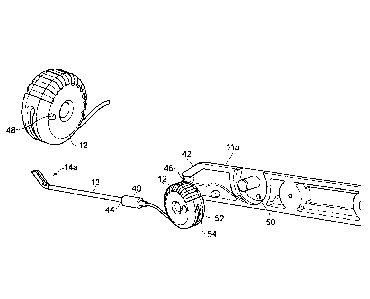
Figure 1 depicts a first embodiment of a device 10 according to the invention
that can
be used to implant or deploy an implant or other object, preferably directly
into an eye. The
device 10 may be used to deploy an implant anywhere in the eye, for example,
in the vitreous
chamber or in the sub-retinal space of an eye.
The device preferably has a means for a user to grip the device, for example a
handle
11. The handle is generally located at the proximal end of the device (as seen
from the user's
point of view). The handle may be made from stainless steel (e.g. stainless
steel 316) or
medical grade plastic. The handle can conveniently be provided in two mirror
image parts
1 la and 1 lb, with the split being along the longitudinal axis of the handle
11. Any fasteners
used to assemble the handle can by usefully made from A4 stainless steel.
Preferably, the handle 11 is shaped so as to provide a secure grip for a user
of the
device 10 and so that the device can be easily gripped. This may help provide
greater control
and prevent damage to the eye during surgery due to slippage of the device 10.
As shown in
Figure 1, the handle 11 may be shaped to have a polyhedron cross section, so
that the faces
and edges can help provide increased grip. In Figure 1, the handle has a
generally octagonal
cross-sectional shape, although any suitable shape may be used, such as
hexagonal, square or
circular.
The device may also include a user-actuatable actuator for actuating the
device 10.
The actuator may be placed on or partially within the handle 11. In the first
embodiment, the
actuator can be a wheel 12. The wheel 12 is rotated by the user to actuate the
device 10. The
actuator can be configured so that actuation can occur when the wheel is
rotated in the
clockwise direction or the actuator can be configured so that actuation can
occur when the
wheel is rotated in the anti-clockwise direction. In the Figure 1 embodiment
(see also
Figures 8 and 9), movement of the wheel generally towards the user causes the
implant to
move from the carried position to the deployed position. The wheel 12 may
comprise ridges
CA 02804078 2012-12-28
WO 2012/004592 PCT/GB2011/051262
to help increase grip during rotation.
The actuator is not limited to a wheel. For example, the actuator may be a
string (for
example as shown in the embodiment of Figures 16A and 16B), a plunger, a
switch (that, for
example, actuates a motor), a lever, a roller, a slider, a conveyer belt (for
example as shown
in the embodiment of Figures 15A and 15B) or such like.
The device 10 may include a tip portion 13 at its distal end. The tip portion
13 may
be removably attachable to the handle 11. The device may alternatively be a
single
continuous piece with an integrated handle and tip portion. The tip portion 13
may be made
from plastic or a metal such as stainless steel or gold. Stainless steel 316L
is preferred for the
tip portion.
The tip portion 13 preferably extends some distance inside the handle 11 when
the
device is assembled (see Figure 9). A proper fit of the tip portion 13 in the
handle can be
achieved by providing an annulus of flexible silicon 40 (see Figures 8 and 9)
around the
outside of the proximal end of the tip portion 13, and by clamping this
silicon within a
chamber 42 inside the handle. The flexible silicon 40 is preferably slightly
larger than the
internal dimensions of the chamber 42, so as to provide a tight clamping fit
when the handle
halves 11 a, lib are tightened together. The tip portion 13 may also be
provided with a
suitable flange portion 44, that interacts with a corresponding flange portion
46 inside the
handle 11 to prevent the tip 13 from coming out of the handle 11 during use.
The distal end of the tip portion 13 may comprise the head portion 14. The
proximal
end of the device may comprise the handle 11 only or the handle 11 and the
elongated and
narrow part of the tip portion (i.e. the tip portion not including the head
portion).
Figure 2 is a diagram of the tip portion 13 when detached from the handle 11.
The tip
portion 13 may itself be made from two separately moulded or machined
components 13a
and 13b. These components 13a and 13b may be attached together by a press-fit
or suchlike.
The tip portion 13 may be elongated and narrow so that the distal part 13a of
the tip portion
(comprising the head portion 14) can be easily inserted into the eye. The tip
portion 13 may
also be hollow so that an urging member, for example a wire, suture, string or
conveyer belt,
can be inserted into the proximal end of the tip portion 13 and move within
the inside of the
tip portion 13 along its longitudinal direction, so as to appear at the distal
end and engage
11
CA 02804078 2012-12-28
WO 2012/004592 PCT/GB2011/051262
with a carried implant there. The tip portion 13 is preferably mounted in the
handle 11 so as
to be rotatable around the longitudinal axis when in an unlocked
configuration. This allows
the user of the device 10 to adjust the location of a head portion 14 of the
tip portion 13
relative to the handle 11. The tip portion 13 can be rotated about the
longitudinal axis in
accordance with the required location for implanting the implant in the eye.
Preferably, the
tip portion can be rotated 90 , more preferably 180 , even more preferably 270
C and even
more preferably 360 or greater. The tip portion 13 may be locked into the
desired
configuration when being used to deliver the implant. The tip portion 13 can
be locked and
unlocked by respectively tightening and loosening of screws in the handle 11
which can
contact the end of the tip portion 13 inserted in the handle 11.
Figure 3 shows a close-up of the head portion 14 of Figures 1 and 2. In this
first
embodiment, the top of the head portion 14 is formed to be substantially flat.
The top of the
head provides two ledges that face inwardly and help to retain the implant in
place inside the
head portion 14. The head portion 14 comprises side walls 15 that are curved
and extend
generally upwardly, before folding over to face one another at the top of the
head portion 14.
Figure 4 shows a cross-section through the head portion 14 of the first
embodiment,
with an implant 19 shown located in position in the head portion. Numeral 18
depicts a
guiding means, which in this embodiment comprises an elongated groove that
runs
longitudinally along the bottom surface of the head portion. Accordingly, the
guiding means
18 extends in a direction parallel to the direction of deployment of the
implant 19.
Figure 5 is similar to Figure 3 but shows an alternative form for the head
portion of
the tip portion 13 according to a second embodiment of the invention. The head
portion 14a
of the second embodiment also comprises curved walls 15. As with the first
embodiment, the
end of the head portion may have an opening 16 which can be formed by the
sides of the
curved walls 15. The tip portion 13 and the end opposite to the opening 16 of
the head
portion 14a may each comprise an opening for an urging member, for example, a
wire or
suture 17, to enter the head portion 14a. By providing an urging member that
is routed
through the centre of the tip portion, the tip portion is allowed to rotate
around the urging
member with respect to the handle. The head portion 14a may also comprise a
guiding
means 18a for guiding the urging member 17 centrally along the head portion
14a. The
12
CA 02804078 2012-12-28
WO 2012/004592 PCT/GB2011/051262
guiding means 18a preferably extends along a part of the tip that is
underneath the implant
when it is carried, thereby allowing the urging member 17 to be effectively
guided for the
whole time that the urging member 17 is in contact with the implant during
deployment. The
embodiments of Figure 3 and Figure 5 each have a groove 18, 18a as the guiding
means. The
guiding means may also be formed by any suitable means, including walls, guide
posts or
such like.
The walls 15 of the first and second embodiments extend in a generally
longitudinal
direction, parallel to the direction of deployment of the implant. They curve
over themselves
towards their top portion and come close to one another at the top, although
they do not
.. touch. The walls curve in a plane that is generally transverse to the
deployment direction.
Preferably, the walls do not curve parallel to the deployment direction. As
such, the walls 15
generally provide a tunnel-shaped space for carrying the implant, whereby the
implant may
be inserted and deployed from the opening 16 formed by the distal parts of the
walls 15. The
shape of the walls is such as to cause a carried implant to the curve in a
direction generally
transverse to the deployment direction. In addition, the walls are so shaped
as to preferably
not cause any curvature of the implant in a direction parallel to the
deployment direction.
Accordingly, the head portion 14, 14a of the tip portion 13 is constructed and
arranged to accept and carry a planar flexible implant, to cause that flexible
implant to the
curve, preferably in a direction transverse to the deployment direction, and
to allow an urging
member to come into contact with the flexible implant so as to move the
implant from the
carried position to a deployed position.
In the first embodiment, the walls may preferably comprise one or more
retaining lips
22a (see Figure 3). These retaining lips 22a also help to restrict movement of
the implant in
the same way as the curved walls 15 of Figure 5. If the implant moves, the
edge of the
implant contacts a retaining lip 22a and is prevented from further transverse
movement. The
lips 22a may be formed at an angle such that they do not contact the surface
of the implant,
but only the edge of the implant. The lips 22a may be provided by the edges of
the walls 15,
as in the second embodiment.
The head portion 14 of Figures 1, 2 and 3 has approximately the same internal
configuration as the head portion 14a of Figure 5. However, the external
configuration is
13
CA 02804078 2012-12-28
WO 2012/004592 PCT/GB2011/051262
slightly different, as can be seen by comparing the drawings. In particular,
the configuration
of the first embodiment is more shrouded and less open to the outside, and is
preferred for
this reason. The first embodiment also has a wider guiding means 18 that has a
circular
radius. The guiding means 18a of the second embodiment is square-edged. This
is not a
limiting feature of the invention and any shape of guiding means 18, 18a may
be used in any
embodiment.
Figure 6 shows a cross section of a third embodiment of a head portion. The
design is
similar to the first embodiment (see Figure 4 in particular), although a
slightly modified
guiding means is shown. In this embodiment, the guiding means comprises
support lips 21b.
In particular, the edges of the guiding means 18b are preferably raised to
form the lateral
support lips 21b. The lips 21b act to surround the urging member in use and to
further
restrict the movement of the urging member transversely of the longitudinal
direction of the
groove 18b. This provides greater accuracy in guiding the urging member. In
turn, the
deployment of the implant is more reliable.
The urging member of the invention is preferably elongated. The urging member
is
usefully connected to the actuating member 12 so that actuation of the
actuating member
causes the urging member to urge the implant, for example by advancing down
the middle of
the tip portion 13. The urging member may be a wire 17. The wire may be a
suture, or the
wire may be made from nylon or from a nickel titanium alloy. The urging member
may also
be a coiled wire. Alternatively the urging member may be a hydraulic piston.
As will be evident from the Figures, the urging member preferably bends as it
advances down the inside of the device. A urging member formed as a coiled
wire facilitates
this bending and it has been found that a coiled wire goes around the corner
more easily than
a simple wire. Further, when a coiled wire is again retracted, it returns to
its original shape,
whereas a simple wire may be plastically deformed when going around the
corner.
The urging member preferably has an overall diameter of 1 mm or less, more
preferably in the range 0.5mm-0.9mm. If a coiled wire is used, the wire itself
can preferably
have a diameter in the range 0.1mm-0.2mm, with the overall diameter of the
coil being in the
range 0.5mm-1.0 mm.
The end of the urging member that contacts the implant is preferably ground or
14
CA 02804078 2012-12-28
WO 2012/004592 PCT/GB2011/051262
otherwise flattened. This increases the chance of successful implant
deployment.
All edges in the head portion 14 may preferably be blunted so that the edges
do not
cause damage to the eye upon insertion or retraction therefrom. This can be
achieved by way
of grit-blasting or similar.
The guiding means of all embodiments of the invention may be a groove that
conforms to the shape of part of the urging member. As shown in Figures 4 and
6 the groove
18, 18b may be curved. The curvature of the groove 18, 18b may be the same as
the
curvature of the urging member (such as a suture, wire or a coiled wire). This
ensures that
the urging member fits into the groove 18, 18b so that the urging member can
be accurately
guided. As will be apparent from the Figures, the guiding means 18, 18a, 18b
allows the
urging member to move substantially along the longitudinal direction, so as to
push any
implant located in the head portion 14, 14a outwards from the device, but also
helps to
restrain movement in the transverse directions (i.e. directions perpendicular
to the direction
of implant deployment).
The urging member 17 can be guided along the head portion 14, 14a by the
guiding
means, such as a groove 18, 18a, 18b. The guiding means 18, 18a, 18b helps
restrain
movement of the urging member 17 to a direction that is along the longitudinal
axis of the
head portion. The guiding means 18 may also help the urging member 17 to
remain rigid.
Furthermore, as the guiding means may be slightly depressed into the head
portion 14, the
implant 19 may rest on top of the guiding means 18, 18a, 18b (for example see
Figure 4).
The guiding means therefore does not interfere with the smooth movement of the
implant 19
into and out of the device 10.
The urging member 17, which in use runs along the guiding means 18, 18a, 18b,
can
be arranged to contact the edge of the implant 19 someway above the bottom
edge of the
urging member 17. It is particularly preferred that the implant edge is
positioned close to the
midline of the end of the urging member. This is due to the guiding means
providing a space
for movement of the urging means below the implant, as shown in Figure 4. Thus
the
guiding means 18, 18a, 18b may guide the urging member 17 so that the end of
the urging
member is directed in a direction that is perpendicular to the edge of the
implant (and/or
perpendicular to the plane of the implant). This helps ensure that the end of
the urging
CA 02804078 2012-12-28
WO 2012/004592 PCT/GB2011/051262
member 17 contacts the edge of the implant 19 and helps avoid the urging
member altogether
missing the edge of the implant 19 or being forced above the edge so that the
urging member
undesirably runs along the top surface of the implant 19. Also, by providing a
guiding means
so that the edge of the implant 19 contacts the end of the urging member
towards the central,
wider portion of the cross section of the urging member, the contact area
between the edge of
the implant and the end of the urging member can be increased. This helps to
prevent the
urging member from slipping above the edge of the implant. Thus deployment of
the implant
is more reliable.
In a fourth embodiment, longitudinal guiding ribs 26 may be provided, as shown
in
Figure 7. These guiding ribs 26 can contact a surface of the urging member 17d
that is
generally opposite to that at which the guiding means 18d contacts the urging
member 17d.
This helps to additionally guide and secure the urging member 17d inside the
device as it
moves. The support ribs 26 may be provided along the walls 15, be an extension
of the walls
15, may be formed continuously along the walls 15 or at certain points along
the walls 15 of
the head portion 14. The guiding ribs 26 may be formed at the end of the head
14b at which
the head is attached to the longitudinal part of the tip (i.e. opposite to the
opening 16). This
would help to initially guide the urging member 17d so that it properly sits
on the guiding
means 18d.
The urging member 17 need not be intimately surrounded by the head portion 14,
14a,
14b along its full length. The cross section of the urging member 17 may
clearly be different
to the cross section of the implant. As depicted herein, the urging member 17
has a
substantially circular cross-section while the implant 19 has a very elongated
rectangular
cross-section. Other shapes are however possible.
Figures 8 and 9 are diagrams of the device, similar to Figure 1, with the
handle
.. removed or partially removed to show how the tip portion can be connected
to the actuator 12
or handle 11. In Figure 8, one end of the urging member 17 is fed into the
hollow tip
member 13 and the other end of the urging member 17 is secured to the back of
the wheel 12,
for example by means of a set screw 52 (see Figure 9). The wheel 12 may have a
circumferential groove 54 which further secures the urging member 17 so that
the urging
member 17 does not axially or transversely move with respect to the wheel 12.
As the wheel
16
CA 02804078 2012-12-28
WO 2012/004592
PCT/GB2011/051262
12 rotates, the urging member 17 either unwinds or winds around the wheel 12.
In the
example of Figures 8 and 9, clockwise rotation of the wheel 12 would unwind
the urging
member 17, thus extending the urging member 17 into the tip portion. Thus,
rotational
movement of the wheel can cause linear movement of the implant as the urging
member
extends down the tip and head portions. Anticlockwise rotation of the wheel 12
would cause
the urging member to wind around the wheel and therefore partially retract
from the tip
portion 13. The tip portion 13 and the wheel 12 are, in use, a fixed distance
away from each
other so the urging member 17 moves relative to the tip portion 13. The urging
member
could be wound around the wheel in an opposite direction. This would cause the
urging
member to extend into the tip portion when rotated anti-clockwise. It is
particularly
advantageous that, whatever the configuration of the actuator, the direction
of movement of
the actuator in order to extend the urging member is opposite to the direction
in which the
device is moved in order to introduce it into the eye. For example, where the
actuator is a
wheel, the wheel is preferably rotated away from the tip region to advance the
urging
member. This helps to prevent accidental advancement of the urging member.
The wheel 12 may further comprise a means 48, 50 to limit rotation so that the
urging
member 17 does not extend too far beyond or within the head portion 14a, thus
avoiding
damage to the eye which may be caused by the urging member. The movement of
the wheel
may be limited by a pin 48 located on the wheel 12 that runs inside a guide
slot 50 located
inside the handle, and which abuts the pin 48 once a certain amount of
rotation has been
achieved.
The rotational movement of the wheel 12 causes the urging member 17 to move,
which in turn causes the implant 19 to move linearly. Rotating a wheel may
help provide
smoother urging of the implant 19. This may help prevent the implant 19 from
crumpling
due to rapid application of force by the urging member due to unsmooth
actuation. This
helps provide greater control when deploying the implant.
As mentioned above, it is preferable that the top surface of the implant 19
does not
come into contact with any part of the device or any other implant as this may
remove or
damage the medicament on the top surface. Thus by providing an urging member
17 that
contacts the edge of the implant 19 and not the top surface of the implant,
damage to the
17
CA 02804078 2012-12-28
WO 2012/004592 PCT/GB2011/051262
medicament can be avoided. Furthermore, as the implant is secured to the
device by flexing
the implant into a curved configuration so that an elastic force is provided
between the
bottom surface of the implant and the head portion 14, there is no requirement
for the top
surface of the implant to be touched when securing the implant to the device.
Furthermore,
as the walls 15 can be curved around the edges of the implant 19, but without
contacting the
top surface of the implant, the implant is further secured to the head portion
14, 14a, 14b
while avoiding contact of the device to the top surface of the implant. The
ends of the walls
may point towards the median plane of the head portion 14, 14a, 14b. The
median plane
is a plane that divides the head portion into symmetrical halves.
10 The device may be formed into a hockey-stick shape. As such, the
longitudinal axis
of the handle and/or tip may be different to the longitudinal axis of the head
portion.
Preferably, the angle between the longitudinal axis of the head portion and
the longitudinal
axis of the tip and/or handle is between 100 and 80 , preferably between 20
and 70 ,
preferably between 30 and 50 , more preferably between 40 and 50 more
preferably being
15 about 450. This angle is shown in the figures as angle A. This angle
allows easier
manipulation of the head portion when within the eye, particularly when
deploying an
implant in the curved walls of the eye. The heel or corner between the two
portions is
preferably curved rather than being a sharp corner. The corner is preferably
curved both
internally and externally to allow easy movement of the urging member around
the comer
and also easy, less traumatic entry of the device into the eye.
The tip portion 13 (with the head portion) may be removably attachable to the
handle
11. This allows the handle to be reused and the tip portion to be discarded
for the purposes of
hygiene.
Figures 10A to 10C depict the process of loading an implant 19, for example a
patch,
into the head portion 14 of the Figure 3 embodiment of the device 10. The
process is
substantially the same for the other embodiments and can be applied thereto
without further
explanation being necessary.
Figure 10A shows an embodiment of an implant 19 which is substantially flat
and
substantially planar. The implant 19 may preferably be flexible and this may
be achieved by
forming the implant 19 from a flexible plastic, such as PET (available as
Dacron TM). In
18
CA 02804078 2012-12-28
WO 2012/004592 PCT/GB2011/051262
general, any material may be used if it provides for an implant that is thin,
flexible and able
to be deformed into the shape of the inside of the head of the device.
The implant is generally rectangular and may have dimensions of about 3mm x
6mm.
The short sides of the implant here have different shapes, with one end 19a
being squarer
than the other end 19b. The more square shape of end 19a allows easier
insertion and curling
of the implant into the device 10. The more curved end 19b allows easier
deployment of the
implant into the eye or other target location. The implant 19 may have stem
cells or any
other active agent or medicament placed or formed on one or more surfaces of
the implant
19. The implant 19 may have a medicament formed within it, or may be formed by
the
medicament itself. The implant may be a drug eluting implant or a radioactive
source.
In some cases where the medicament is formed on a surface (e.g. the top
surface), it
may be preferable that the top surface of the implant does not come into
contact with any part
of the device or any other implant as this may contaminate, damage or remove
the
medicament (such as stem cells) from the implant 19. In other cases (depending
on the
medicament), a surface with a medicament may contact the device.
As shown by the arrow in Figure 10A the implant 19 may be inserted into the
head
portion 14 in a direction that is substantially parallel to the longitudinal
axis of the head
portion 14. The opening 16 of the head portion 14, 14a, 14b may be angled away
from a
plane that is perpendicular to the direction of insertion of the implant 19,
as shown in Figure
10A. The plane perpendicular to the plane of insertion is shown in the figures
as the plane X-
--X. Preferably, the plane of the opening forms an angle of around 10 to 80 ,
preferably 20 to
70 more preferably 30 to 60 even more preferably 45 to 55 from the plane of
the direction
that the implant 19 is inserted into the head portion 14, 14a, 14b. The
inventors have found
that an angle of about 50 works particularly well. This is shown in the
figures as angle B.
As the implant is inserted into the head portion 14, 14a, 14b, the leading
edge of the
implant 19a contacts the walls of the opening 16 (preferably, the walls of the
opening 16
being the sides of the curved walls 15). Due to the angle between the plane of
the implant 19
and the plane of the opening 16, the implant automatically flexes into a
curved configuration
as it is being inserted.
Figure 10B shows the implant being inserted through the opening 16 and being
part-
19
CA 02804078 2012-12-28
WO 2012/004592 PCT/GB2011/051262
way into the head portion 14, 14a, 14b. As can be seen, once the implant has
been inserted
by a small amount, it assumes a curved configuration that generally
corresponds to the
internal curvature of the interior walls of the head portion 14, 14a, 14b. The
curvature is in a
plane perpendicular to the direction of deployment of the implant. Figure 10C
shows the
implant 19 fully inserted into the head portion 14a and in the carried
configuration. When
the implant is fully inserted into the head, it is in a curved position and
the curvature of the
implant 19 preferably conforms to the inside curvature of the head portion 14,
14a, 14b. The
implant is flexed when it is curved. The implant 19 may be secured in the head
portion 14,
14a, 14b by contact of only the bottom surface of the implant 19 with the
internal surfaces
head portion 14, 14a, 14b. The implant 19 may thus be secured in place by an
elastic force
applied by the implant 19 (due to the elasticity of the implant 19) to the
head portion 14, 14a,
14b. The walls of the head portion 14, 14a, 14b may be curved, as shown. The
curved walls
can help secure the implant 19 in the head portion 14, 14a, 14b. The curved
walls 15 are
preferably curved over the transverse edges of the implant 19 so as to
restrict movement of
15 the implant in transverse directions. The walls are preferably curved
over the edges of the
implant 19 so that if the implant moves, only the edges of the implant 19 come
into contact
with the curved walls 15 and not the top surface of the implant 19. As seen in
Figure 10C,
the curvature of the walls 15 increases near the edges of the implant, so as
to firmly hold the
implant in place. Instead of curved walls per se, lips 22a may be provided, as
shown in
Figure 3.
The implant 19 may be inserted into the head portion 14, 14a, 14b by pushing
the
implant 19 into the opening 16. Alternatively, the implant may be pulled, by,
for example, a
pair of micro forceps, into the head portion 14, 14a, 14b. The micro forceps
may run along
the gap between walls 15 when pulling the implant 19 into the head portion 14,
14a, 14b.
Figures 11 A to 11C depict the process of deploying the implant 19 from the
head
portion 14, 14a, 14b of device 10. The implant is designed to linearly move
down the head
portion when being deployed. The urging member preferably causes the necessary
linear
movement.
Initially, actuator 12 is actuated so as to present the urging member 17 into
contact
with an edge of the implant 19, as shown in Figure 11A. Movement of the urging
member 17
CA 02804078 2012-12-28
WO 2012/004592 PCT/GB2011/051262
is controlled by the actuator, such as the wheel 12. The urging member
generally follows the
path of the guiding means 18a as it moves, thereby assuring proper movement in
the correct
deployment direction. As shown in Figure 11B, as the urging member moves along
the head
portion 14, 14a, 14b, it pushes the implant 19 out of the opening 16, thereby
deploying it.
The urging member 17 is preferably continuously guided by the guiding means
18, 18a, 18b
for the duration of the time that it remains in contact with the implant 19.
This is preferably
achieved by routing the urging member underneath the implant. As the implant
19 is in a
curved configuration, the implant is more rigid along its longitudinal axis
and therefore,
when the pushing force is applied to the edge of the implant 19, the implant
19 maintains its
structural integrity and does not crumple along its longitudinal axis. As the
implant 19 exits
the head portion 14, 14a, 14b of the device 10, the implant may begin to
flatten. As shown
in Figure 11C, when the implant 19 has fully exited the head portion (i.e.
when in the
deployed position), the implant 19 can flex back (under its own elastic force)
into a flat
configuration. The shape of the implant may conform to the shape of the
surface to which it
has been deployed. For example, if the implant is deployed in the back of the
eye the implant
may end up being curved to conform to the curvature of the back of the eye.
The angled opening 16 may additionally help deployment of the implant 19. When
a
part of the implant 19 has left the angled opening 16 the part can begin to
flatten, under the
implant's own elastic restoring force. This itself exerts a force onto the
angled opening and
thus can help assist the implant further out of the head portion. Thus, the
urging member 17
may not be required to completely push the implant all the way out of the head
portion 14,
14a, 14b, and may have it's travel limited to a certain proportion of the way
along the head
portion.
Figures 14A and 14B show a fifth embodiment having an alternative design of
head
portion 23 in which a pair of holder jaws 24 grip the implant 19. An outer
sheath 25 can be
used to force the holder jaws 24 together so that they grip the implant.
Alternatively, the
holders 24 can be forced to grip the implant by any other mechanism. In one
embodiment, as
the outer sheath 25 is pulled back (in the direction from the distal end to
the proximal end),
the holders separate (as shown in Figure 14B), and release the implant. The
outer tube can be
activated by using the wheel mechanism, for example. The rotational movement
of the wheel
21
CA 02804078 2012-12-28
WO 2012/004592 PCT/GB2011/051262
can cause the end of the sheath 25 to move linearly relative to the implant.
Alternatively, in
another embodiment the holder jaws 24 and the implant 19 can be linearly moved
down the
inside of the outer sheath 25 when activated (by, for example, a wheel).
Figures 15A and 15B show a sixth embodiment of the device which utilises a
conveyor mechanism as the actuator and urging member. A thumb actuated
conveyor belt 60
is provided as the actuator. The actuator 60 is connected to a modified head
portion 14b that
contains a conveyor belt. As shown in Figure 15B, the implant 19 is carried on
the head
conveyor belt 62 within the confines of the device. In Figure 15B, the implant
19 is shown in
a partially deployed position. Movement of the actuator 60 causes the conveyor
belt 62 to
move (anti-clockwise as shown in Figure 15B) and this in turn causes the
implant 19 to be
deployed from the device.
In order to insert the implant into the device, it is merely necessary to
present the edge
of the implant 19 to the opening in the head portion 14b and move the actuator
60 in the
opposite direction, so as to feed the implant 19 from a deployed position to a
carried position
inside the device.
Figures 16A and 16B show a seventh embodiment of the invention. In this
embodiment, the actuator is a string 70 that runs down the length of the tip
portion 13 to
modified head portion 14c. The head portion 14c is shown in more detail in
Figure 16B,
where the implant 19 is shown in a partially deployed position.
As can be seen from Figure 16B, the string 70 runs to the distal end of the
head
portion 14c, where it is fed around a small pulley (not shown) and is
terminated by a hook 72.
The hook 72 engages with the implant 19 and, pulling on the string 70 causes
the implant to
move from a carried position (located inside the vice) to a deployed position
(located outside
of the device).
The hook 72 automatically disengages from the implant 19 when it reaches the
pulley.
It will be appreciated that various different embodiments of the invention
have been
described. Various features of one embodiment may be used in combination with
features of
the other embodiments. For example, all embodiments of the invention may
provide that the
implant is curved, preferably in the transverse direction, during the carried
position. Further,
all embodiments of the invention may use an urging member and a guiding means
to deploy
22
CA 02804078 2012-12-28
WO 2012/004592
PCT/GB2011/051262
the implant. Various other possible modifications will be apparent to the
skilled person.
The invention also includes a method of loading and deploying the implant, as
described above.
Figure 12 shows a diagram of an eye 30 with a slit 31 formed therein, in a
position
typical for inserting the implant of the invention using the device of the
invention. The angle
between the head portion and the handle of the device allows the slit 31
formed in the eye to
be narrow. The size of the slit may also correspond to the size of the head
portion 14. As the
implant 19 is forced into a curved configuration when carried inside the head
portion 14, an
implant of a larger width can be introduced into the eye while providing a
small sized slit 31.
Figure 13 is a cut-away diagram of an interior of an eye. According to a
procedure
for deploying the implant, a blister 32 may be formed in or near the retina of
the eye 30. An
incision can then be surgically made into the blister 32 so that the implant
19 can be
delivered under the retina using the device 10, which has been introduced into
the eye via the
slit 31.
In more detail, the procedure may follow the following steps.
1. The pupil of the eye to undergo the implantation procedure is dilated
with
cyclopentolate 1% and phenylephrine 2.5%;
2. Pen-orbital skin is cleaned and a sterile drape applied to cover pen-
orbital and adjacent
skin but to allow access to the surgical field.
3. HPMC 2%
in balanced salt solution is applied to the surface of the eye periodically
throughout the surgery to prevent the eye surface drying up.
4. Lateral canthotomy is performed after lateral canthus clamping
5. Eyelids are retracted
6. Localised peritomies and 3 sclerostomies are performed 2mm from the
limbus
7. A three
port pars vitrectomy (Alcon, Ref 8065741008) is performed using an indirect
wide-angle viewing system
8. Posterior vitreous detachment is induced up to the major retinal vessels
9. Further vitrectomy continued.
10. 360 laser and cryo surgery performed. Use of equipment is recorded on
Laser/Cryo
form
23
CA 02804078 2012-12-28
WO 2012/004592 PCT/GB2011/051262
11. Localised retinal bleb detachment (4x6mm) is achieved at the vessel free
region,
superonasal to the disc, by sub-retinal injection of balanced salt solution
(Moodieids) sodium
lactate.
12. The retinotomy is enlarged using a microvitreoretinal (MVR) blade and
vertical scissors.
.. 13. The sclerostomy for patch delivery is enlarged using MVR and feather
blade while the
patch is prepared.
14. The infusion line is left open while the patch is inserted through the
sclerostomy
15. The graft is engaged at the retinotomy and advanced into the sub-retinal
space.
16. The retina is re-attached under air using back flush
17. The retinotomy is lasered and recorded. Search at periphery and laser or
cryo to tears and
record.
18. The air-to-silicone oil exchange
19. Sclerostomies are closed using polyglactin sutures, 6/0 Vicryl to delivery
sclerostomies
and 7/0 Vicryl to others
20. Intravitreal Triaminolone Acetonide lml (40mg/m1) is delivered by
injection around the
eye (in the subtenon space)
21. Chloamphenicol 0.5% and atropine sulphaste 1% is applied topically to the
eye
22. Betamethasone 0.1% is applied to the conjunctival sac
23. 1 ml Triamcinolone Acetonide 40mg/m1 is delivered by injection around the
eye (in the
subtenon space) at the end of surgery.
24