Note: Descriptions are shown in the official language in which they were submitted.
CA 02804525 2013-01-04
WO 2011/159825 PCT/US2011/040567
INTRAVASCULAR ARTERIAL TO VENOUS ANASTOMOSIS AND
TISSUE WELDING CATHETER
Background of the Invention
In the body, various fluids are transported through conduits throughout the
organism to perform various essential functions. Blood vessels, arteries,
veins, and
capillaries carry blood throughout the body, carrying nutrients and waste
products
to different organs and tissues for processing. Bile ducts carry bile from the
liver to
the duodenum. Ureters carry urine from the kidneys to the bladder. The
intestines
carry nutrients and waste products from the mouth to the anus.
In medical practice, there is often a need to connect conduits to one another
or to a replacement conduit to treat disease or dysfunction of the existing
conduits.
The connection created between conduits is called an anastomosis.
In blood vessels, anastomoses are made between veins and arteries, arteries
and arteries, or veins and veins. The purpose of these connections is to
create either
a high flow connection, or fistula, between an artery and a vein, or to carry
blood
around an obstruction in a replacement conduit, or bypass. The conduit for a
bypass
is a vein, artery, or prosthetic graft.
An anastomosis is created during surgery by bringing two vessels or a conduit
into direct contact. The vessels are joined together with suture or clips. The
anastomosis can be end-to-end, end-to-side, or side-to-side. In blood vessels,
the
anastomosis is elliptical in shape and is most commonly sewn by hand with a
continuous suture. Other methods for anastomosis creation have been used
including
carbon dioxide laser, and a number of methods using various connecting
prosthesis,
clips, and stents.
An arterio-venous fistula (AVF) is created by connecting an artery to a vein.
1
CA 02804525 2013-01-04
WO 2011/159825 PCT/US2011/040567
This type of connection is used for hemodialysis, to increase exercise
tolerance, to
keep an artery or vein open, or to provide reliable access for chemotherapy.
An alternative is to connect a prosthetic graft from an artery to a vein for
the
same purpose of creating a high flow connection between artery and vein. This
is
called an arterio-venous graft, and requires two anastomoses. One is between
artery
and graft, and the second is between graft and vein.
A bypass is similar to an arteriovenous graft. To bypass an obstruction, two
anastomoses and a conduit are required. A proximal anastomosis is created from
a
blood vessel to a conduit. The conduit extends around the obstruction, and a
second
distal anastomosis is created between the conduit and vessel beyond the
obstruction.
As noted above, in current medical practice, it is desirable to connect
arteries
to veins to create a fistula for the purpose of hemodialysis. The process of
hemodialysis requires the removal of blood from the body at a rapid rate,
passing the
blood through a dialysis machine, and returning the blood to the body. The
access to
the blood circulation is achieved with catheters placed in large veins,
prosthetic grafts
attached to an artery and a vein, or a fistula where an artery is attached
directly to the
vein.
Fistulas for hemodialysis are required by patients with kidney failure. The
fistula provides a high flow of blood that can be withdrawn from the body into
a
dialysis machine to remove waste products and then returned to the body. The
blood
is withdrawn through a large access needle near the artery and returned to the
fistula
through a second large return needle. These fistulas are typically created in
the
forearm, upper arm, less frequently in the thigh, and in rare cases, elsewhere
in the
body. It is important that the fistula be able to achieve a flow rate of 500
ml per
minute or greater. Dialysis fistulas have to be close to the skin (< 6 mm),
and large
enough (>4 mm) to access with a large needle. The fistula needs to be long
enough
(>6 cm) to allow adequate separation of the access and return needle to
prevent
2
CA 02804525 2013-01-04
WO 2011/159825 PCT/US2011/040567
recirculation of dialysed and non-dialysed blood between the needles inserted
in the
fistula.
Fistulas are created in anesthetized patients by carefully dissecting an
artery
and vein from their surrounding tissue, and sewing the vessels together with
fine
suture or clips. The connection thus created is an anastomosis. It is highly
desirable
to be able to make the anastomosis quickly, reliably, with less dissection,
and with
less pain. It is important that the anastomosis is the correct size, is
smooth, and that
the artery and vein are not twisted.
Summary of the Invention
The present disclosed invention eliminates the above described open
procedures, reduces operating time, and allows for a consistent and repeatable
fistula
creation.
It is well known that heat, whether its source is Radio Frequency (RF),
resistance, or laser, will attach and weld tissue or vessels upon direct
pressure and
contact over the targeted weld area. This is often done with jaw-type,
compression
heat delivery devices. It is also well known that radially expandable devices
such as
balloons, metal cages, and baskets are often coupled with energy in the form
of RF,
or in the case of balloons, heated saline, and used intraluminally to ablate
tissue, stop
bleeding, or create a stricture.
The present invention uses catheter based devices that are advanced from one
vessel into an adjacent vessel (i.e. a vein into an artery), join the vessel
walls by
applying heat, and cut through the two walls, creating an anastomosis.
The inventive catheter-based devices track over a guidewire which has been
placed from a first vessel, such as a vein, into a second vessel, such as an
artery, or
more broadly between any other two vascular structures. The distal tip of the
catheter has a dilating tip which allows the catheter to advance easily
through the
3
CA 02804525 2013-01-04
WO 2011/159825 PCT/US2011/040567
vessel walls. Proximal to the distal tip, the catheter has a significant
reduction in
diameter, and then a blunt, oval shaped tapered surface. As the catheter is
tracked
over the guidewire, the tapered distal tip easily passes into the adjacent
vessel. As
the catheter is further advanced, the blunt proximal surface comes into
contact with
the wall of the first vessel and encounters resistance, and cannot perforate
through
the wall into the second vessel. The distal tip, which has a matching blunt
surface on
its proximal end, is then retracted, capturing the walls of the two vessels
between the
two blunt surfaces. A known, controlled pressure (approximately 100 mN/mm2 -
300
mN/mtn2) is applied between the two surfaces. The pressure can be controlled
either
internally in the catheter or by the handle attached to the proximal end of
the catheter.
Heat is then applied to the blunt surfaces to weld the walls of the two
vessels
together. It is possible to only apply the heat to one surface as well. Heat
can be
applied through several different methods, including, but not limited to,
radiofrequency, resistance, inductance, or a combination thereof. The heat is
controlled at a known temperature ranging from between about 100-150 C. The
heat
may be applied by either applying a steady heat, pulsing heat, or a
combination
thereof.
After coaptation of the vessel walls, the heat is then increased to cut
through
the vessel walls to create the desired size fistula. It should be noted that
it is also
possible to apply the same heat to both weld the vessel walls and to cut
through the
vessel.
More particularly, there is provided a device for creating an arteriovenous
(AV) fistula, which comprises an elongate member, a distal member connected to
the
elongate member and movable relative to the elongate member, and a heating
member disposed on at least one of the movable distal member and the elongate
member. The distal member comprises structure for capturing tissue to be cut
to
create the fistula, and the heating member is adapted to cut through the
tissue to
4
CA 02804525 2013-01-04
WO 2011/159825 PCT/US2011/040567
create the fistula. The elongate member comprises an elongate outer tube.
A shaft connects the distal member to the elongate member, and is extendable
and retractable to extend and retract the distal member relative to the
elongate
member. Preferably, the elongate member comprises a distal tapered face and
the
distal member comprises a proximal tapered face, wherein the distal tapered
face and
the proximal tapered face are substantially aligned to one another. In some
embodiments, the heating member is disposed on the proximal tapered face,
while in
other embodiments, the heating member is disposed on the distal tapered face.
Some
embodiments further comprise a second heating member disposed on the distal
tapered face. At least one of the heating member and the second heating member
comprises an energized heater and a heat spreader disposed beneath the
energized
heater to spread heat away from the heater and create a temperature gradient.
The
heat spreader comprises heat conductive material, and is disposed on the
tapered face
beneath the heating member.
Preferably, the distal member is tapered and flexible, so that it can push
through a small aperture between the two vessels to be joined with a fistula.
In some
embodiments, the distal member comprises a toggle member which is pivotal
relative
to the elongate member. In certain embodiments, a shaft is provided for
connecting
the toggle member to the elongate member, the shaft being extendable and
retractable
to extend and retract the toggle member relative to the elongate member,
wherein the
toggle member is pivotally connected to the shaft.
In one disclosed embodiment, the distal member comprises a flexible clamp to
which is connected a heater, wherein the clamp is movable relative to the
elongate
member and is adapted to capture tissue to be cut to create the fistula. In
this
embodiment, the distal member further comprises a distal portion connected to
a
distal end of the elongate member, the distal portion having a side port
therein
through which the flexible clamp and connected heater extend.
CA 02804525 2013-01-04
WO 2011/159825
PCT/US2011/040567
A tissue receiving cavity may be associated with the heating member, to
capture cut tissue. As noted above, in some embodiments, the heating member
comprises an energized heater and a heat spreader disposed beneath the
energized
heater to spread heat away from the heater and create a temperature gradient.
The
heat spreader comprises heat conductive material.
In another aspect of the invention, there is disclosed a method of creating an
AV fistula between adjacent first and second vessels, which comprises a step
of
inserting a guidewire from the first vessel into the second vessel, inserting
a catheter
comprising a proximal elongate member and a distal member over the guidewire,
so
that a tapered distal tip of the distal member comes into contact with a
selected
anastomosis site, and advancing the distal member into the second vessel,
while the
elongate member remains in the first vessel, thereby enlarging an aperture
between
the two vessels. A further step involves retracting the distal member toward
the
elongate member to clamp tissue surrounding the aperture between opposed
surfaces
on each of the distal member and the elongate member, and applying energy to a
heating member on one of the distal member and the elongate member to cut and
form the aperture, and to weld the edges thereof in order to create a desired
fistula
between the two vessels.
Preferably, the opposed surfaces on each of the distal member and the
elongate member comprise aligned tapered faces, between which the tissue is
clamped, wherein a heating member is disposed on at least one of the two
aligned
tapered faces. The method may advantageously further comprise a step of
capturing
cut tissue within a cavity disposed adjacent to the heating member. Heat may
be
dispersed away from the heating member using a heat spreader comprising a
conductive material disposed on the tapered face beneath the heating member.
The invention, together with additional features and advantages thereof, may
best be understood by reference to the following description taken in
conjunction
6
CA 02804525 2013-01-04
WO 2011/159825 PCT/US2011/040567
with the accompanying illustrative drawings.
Brief Description of the Drawings
Fig. 1 is an isometric view of an embodiment of a catheter device constructed
in accordance with the principles of the present invention;
Figs. 2-8 are schematic sequential views illustrating a method for creating a
fistula performed in accordance with the principles of the present invention,
and using
an apparatus like that illustrated in Fig. 1 and disclosed herein;
Fig. 9 is a schematic view illustrating an elongate aperture formed between
two adjacent vessels to create the fistula, particularly highlighting the
welded edges
of the aperture;
Fig. 10 is a cross-sectional view of a handle portion of the embodiment shown
in Fig. 1;
Fig. 11 is an isometric view similar to Fig. 1, illustrating an alternative
embodiment of the invention;
Fig. 12 is an isometric view of yet another alternative embodiment of the
present invention;
Fig. 13 is an isometric view of still another alternative embodiment of the
present invention, wherein a distal toggle member forming part of the device
is
extended;
7
CA 02804525 2013-01-04
WO 2011/159825 PCT/US2011/040567
Fig. 14 is an isometric view similar to Fig. 13, wherein the distal toggle
member is retracted;
Fig. 15 is an isometric view of yet another alternative embodiment of the
present invention; and
Figs. 16-18 are schematic sequential views illustrating a method for creating
a
fistula using the apparatus of Fig. 15.
Description of the Preferred Embodiment
Referring now particularly to the drawings, there is shown in Fig. 1 a bi-
polar
tapered tip catheter embodiment 10, which comprises an elongate outer tube 12
having an outer diameter that can range from 3F - 12F. It may be manufactured
from
a variety of materials, either polymer or metallic. It comprises a central
lumen 14,
within which a tubular structure 16 for attaching a tip 18 may slide. There
are
separate lumina that run down the elongated core of the outer tube 12 for
wiring to
power electrodes or heating elements 20, 22 (proximal and distal,
respectively),
disposed on aligned tapered faces of the respective elongate outer tube 12 and
distal
tip 18, and to also measure the temperature during the coaptation and cutting
processes. In this configuration, the catheter is powered using bipolar energy
to the
distal RF electrode 22 and the proximal RF electrode 20. The system can also
be
used in a monopolar configuration by grounding the patient and applying energy
to
one or both of the RF electrodes to increase the length of the coaptation. The
RF
electrodes cut at matching angles to increase the surface area of the
coaptation and
fistula size relative to the catheter diameter. These angles can be adjusted
to achieve
the desired fistula sizing. The RF electrodes are only electrically conductive
on the
8
CA 2804525 2017-03-09
WO 2011/159825 PCT/US2011/040567
front faces to maximize energy density. The electrodes are oval-shaped, and
are
adapted to cut an anastomosis which is larger than the diameter of the shaft
16.
The apparatus shown and described above in connection with Fig. 1 will now
be further described in conjunction with an explanation of a particular method
by
which the system 10 may be used to create an AV fistula. This method is
illustrated
more particularly in Figs. 2-9.
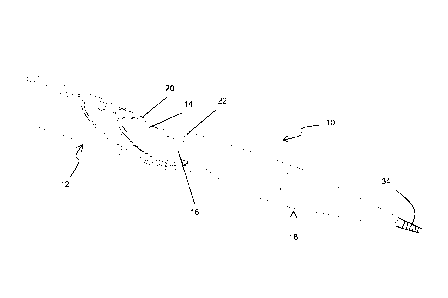
To begin the inventive method of creating an AV fistula, the practitioner
selects an appropriate procedural site having each of a first vessel 26 and a
second
vessel 28 in close proximity to one another. In currently preferred
approaches, the
first vessel 26 comprises a vein, and the second vessel 28 comprises an
artery, but the
invention is not necessarily limited to this arrangement. As illustrated in
Fig. 2, one
presently preferred location is the hand 30 of a patient. Then, generally
employing
principles of the Seldinger technique, as shown in Fig. 2, the first vessel 26
is
punctured by a needle 32, which is inserted therein, for the purpose of
introducing an
access sheath into the site. Then, using suitable techniques, such as the
technique
described in Provisional U.S. Application Serial No. 61/354,903, filed on June
15,
2010, a guidewire 34 is
inserted into the patient, from the first vessel 26 into the second vessel 28,
as shown
in Fig. 3.
The guidewire 34 creates an access path for the catheter 10. The catheter 10
is inserted into the patient by loading a proximal end of the guidewire 34
into the tip
18, which is fabricated to be flexible and tapered. The catheter 10 is
advanced further
into the patient, tracking over the guidewire 34, until the tapered dilating
distal tip 18
comes into contact with the selected anastomosis site. The device 10 can be
tracked
over the guidewire with the distal tip extended (as shown in Fig. 5) or
retracted (as
shown in Fig. 4). The distal tip 18 is extended and further advanced into the
second
vessel 28 (Fig. 5) by advancing the central tubular structure 16 distally from
the outer
9
CA 02804525 2013-01-04
WO 2011/159825 PCT/US2011/040567
tube 12, thereby dilating the fistula, so that the distal tip 18 is in the
second vessel 28,
and the tube 12 is in the first vessel 26, with its distal tapered surface
contacting the
inner wall of the fn-st vessel 26. If resistance is felt, the entire system
can be rotated
to reduce the friction. At this juncture, the opening formed in the wall of
the second
vessel 28 has recovered back to a small diameter, and fits tightly around the
shaft 16,
as shown.
After the distal tip 18 is advanced into the second vessel 28, as illustrated
in
Fig. 6, a slight tension is applied to the distal RF electrode 22 to seat it
against the
vessel wall. The blunt shape of the proximal end of the distal tip 18 prevents
the
distal tip from pulling back through the vessel wall. The proximal end of the
device
10, namely the outer tube 12, is then advanced to close the spacing between
the tube
12 and tip 18, until the walls of the first and second vessels 26, 28,
respectively, are
captured between the facing blunt surfaces of each of the outer tube 12 and
distal tip
18.
A controlled tension is maintained between the distal tip 18 and proximal
outer tube 12, and at this juncture, with the vessel walls securely clamped,
energy is
applied to the RF electrodes 20, 22 (Fig. 7). As the electrodes weld and cut
the
vessels, the electrodes will move closer to one another. When fully retracted,
the
system 10 is designed so that the two electrodes 20, 22 cannot come into
direct
contact with one another, thus preventing the electrodes from shorting. A
variety of
RF energy profiles may be applied to achieve the desired coaptation and
cutting. For
example, during the coaptation phase, a tapered sine wave may be applied to
maximize coagulation without cutting through the tissue. The energy may also
be
adjusted based upon the impedance of the tissue. Different pulse widths or
duty
cycles may be used to minimize the heat transferring into adjacent tissues.
The hot
wire is an oval shape and cuts an anastomosis larger than the diameter of the
shaft 16.
Within the oval shape of the cutting elements, there is a cavity for capturing
the
CA 2804525 2017-03-09
WO 2011/159825 PCT/US2011/040567
tissue that has been cut. The outer sliding tube is usable to push the tissue
off the
heater in case there is a sticking problem due to the heat.
Regarding the tissue welding process, more particularly, the RF energy
functions to burn and fuse or weld the vessels together, creating an elongate
aperture
36 (Fig. 8) through the opposing walls of each of the first and second
vessels, as well
as any intervening tissue. As formed, the elongate aperture 36 will typically
resemble
a slit. However, as pressurized flow 38 begins to occur through the slit or
aperture
36, which creates a communicating passage between the first vessel and the
second
vessel, the aperture widens responsive to the pressure, taking the shape of an
ellipse
as it opens to form the desired fistula. This effect is illustrated in Fig. 9.
The edges
40 of the aperture are cauterized and welded. Fig. 9 illustrates the weld from
the
venous (first vessel) side. As shown, the cut area corresponds to the shape of
the
heater wire. It can be of multiple shapes, such as round, oval, a slit, or a
combination
as shown. The area outside of the cut has been welded due to the flat face of
the
catheter in the vein (first vessel) being larger than the cutting wire. The
heat from the
wire is also preferably spread over this area by a conductive material that is
below the
heater, as will be described below. This creates a temperature gradient, which
is a
particularly advantageous feature of the present invention.
Tissue welding of the type intended to occur in the practice of these
inventive
methods is discussed in U.S. Patent No. 6,908,463, to Treat et al.
Fig. 10 is a cross-sectional view of a handle portion 42 of the embodiment
shown in Fig. 1. This is one possible approach for actuating the extension and
retraction of the distal tip 18 relative to the elongate outer tube 12, as
discussed
above, though many other suitable configurations may be used alternatively. A
trigger 44 is slidably disposed on the handle 42, slidable distally through a
slot 46 in
the direction of arrow 48, and then retractable in the reverse direction. A
spring 50
11
CA 02804525 2013-01-04
WO 2011/159825 PCT/US2011/040567
within the handle controls pressure, and a locking mechanism functions to lock
the
trigger 44 in the retracted state.
Alternative cutting approaches, such as resistive heat (hot wire), ultrasonic,
laser, or mechanical approaches, may be used instead of RF energy, if desired.
For
example, Fig. 11 illustrates an alternative embodiment, wherein a catheter 110
comprises an elongate outer tube 112 having a central lumen 114, a tubular
structure
116, and a flexible and tapered distal tip 118. In this embodiment, a single
resistive
heating wire 152 is used to provide the tissue heating, cutting, and welding
function
described above. Additionally, an RF configuration applying only monopolar
energy,
to either the venous or arterial sides, may be employed. A combination of RF
energy
and resistance heating may also be used. The tip 118. in this embodiment,
tracks
over the guidewire and dilates the anastomosis site, as in the previous
embodiment.
The tapered faces of the members 112 and 118 align. The single hot wire 152
down
the face cuts a slit in the vessel walls, and the faces are tapered to assist
in removing
the device.
Now with reference to Fig. 12, a heat spread catheter 210 is illustrated. The
catheter 210 comprises a resistive heating element 252, which is employed in a
manner similar to that described above in connection with the Fig. 11
embodiment.
However, in this embodiment, a conductive material 254 is disposed beneath the
heating element 252. In one configuration, this conductive material 254
comprises
aluminum, though other conductive bio-compatible materials may also be used.
In
operation, this conductive material 254 functions to create a heat gradient
from the
heating element 252, for the purpose of improving the welding function, as
described
above.
In this embodiment, similar to the foregoing embodiments, the tip 218 tracks
over the guidewire and dilates the anastomosis site. The tapered faces of each
of the
members 212 and 218 align, for clamping the vessel walls. The hot wire 252 is
an
12
CA 02804525 2013-01-04
WO 2011/159825 PCT/US2011/040567
oval shape and has vertical strips 256 on both sides of the artery. The hot
wire cuts
an anastomosis larger than the diameter of the shaft 216. Under the hot wire
252, the
heat conductive material 254 pulls heat away from the hot wire so that there
is a
temperature gradient across the face, with the temperature being hottest in
the center
and cooling as the distance outwardly from the center increases.
The hot wire 252 (heater) is raised above the spreader 254 to increase
pressure on the tissue, to thereby assist in the cutting process. Inside the
hot wire,
there is a cavity to capture the tissue that has been cut. The profile of the
distal tip
218 aligns with the edge of the heater when retracted. It is a lower profile
than the
heat spreader, so that it can be retracted back through the fistula. This also
increases
the pressure directly on the heater surface to assist in cutting function.
Figs. 13 and 14 illustrate still another embodiment 310, comprising a distal
toggle member 358. The cutting elements in this embodiment are substantially
identical to those shown and described in connection with Fig. 12. As in prior
embodiments, the toggle 358 tracks over the guidewire into the artery. When
retracted (Fig. 15), the toggle captures the artery and pulls against the
vein. The hot
wire is an oval shape, has vertical strips 356 on both sides of the artery,
and cuts an
anastomosis larger than the diameter of the shaft 316. Under the hot wire 352,
there
is a heat conductive material 356 that pulls heat away from the hot wire so
that there
is a temperature gradient across the face. The hot wire is raised above the
heat
spreader to increase pressure on the tissue to help it cut through. Inside the
hot wire
there is a cavity to capture the tissue that has been cut.
The profile of the toggle 358 aligns with the edge of the heater when
retracted. It is of a lower profile than the heat spreader so that it can be
retracted
back through the fistula. This also increases the pressure directly on the
heater
surface and helps it cut. Heating elements may also be disposed on the toggle
surface
to work in conjunction with the heater 352 to cut and weld tissue.
13
CA 2804525 2017-03-09
WO 2011/159825 PCT/US2011/040567
Pivotable toggles and their functionality are discussed in Provisional U.S.
Application Serial No. 61/354,903, filed on June 15, 2010.
Those teachings generally apply to this toggle
embodiment, regarding the particulars as to how the toggle is used to enter
and then
retract the second vessel toward the first vessel.
In Figs. 15-18, there is shown a different cutting approach. In this
embodiment, the cutting device 410 comprises a shaft 460 having a distal
portion
462. The distal portion comprises a side port 464, from which extends a heater
wire
466 which is supported by a flexible clamp 468, preferably fabricated from
nitinol or
similar material. The heater wire may be resistive or utilize any other energy
source
as described above.
As shown in Figs. 16-18, access to the anastomosis site is gained by methods
as described above and the function of this device, once in place, is to
manipulate the
wire 466, using the flexible clamp 468 and suitable actuation mechanisms in
order to
create a fistula of a desired configuration. Specifically, as shown in Fig.
16, the tip
462 tracks over the guidewire 34 and dilates the anastomosis site, as in
previously
described approaches. The catheter 410 is advanced so that the clip 466 is all
the
way in the artery 28, and then puled back to capture the arterial wall under
the clip,
as illustrated in Fig. 17. The wire is then activated to heat, and then drawn
back,
which cuts through the arterial and venous walls. The hot wire is then pulled
back
(Fig. 18), and pulls down the clip portion through the vessel walls.
Accordingly, although an exemplary embodiment and method according to
the invention have been shown and described, it is to be understood that all
the terms
used herein are descriptive rather than limiting, and that many changes,
modifications,
and substitutions may be made by one having ordinary skill in the art without
departing from the spirit and scope of the invention.
14