Note: Descriptions are shown in the official language in which they were submitted.
CA 02804760 2013-01-08
WO 2012/003466 PCT/US2011/042832
A. TITLE:
SINGLE-WALLED CARBON NANOTUBE/SIRNA COMPLEXES
AND METHODS RELATED THERETO
B. CROSS-REFERENCE TO RELATED APPLICATIONS:
[0001] This application claims priority to U.S. Provisional Application No.
61/360,942 entitled "Single-Walled Carbon Nanotube/SIRNA Complexes and Methods
Related Thereto" filed July 2, 2010, which is herein incorporated by reference
in its entirety.
C. GOVERNMENT INTERESTS: Not applicable
D. PARTIES TO A JOINT RESEARCH AGREEMENT: Not applicable
E. INCORPORATION BY REFERENCE OF MATERIAL SUBMITTED ON A
COMPACT DISC: Not applicable
F. BACKGROUND: Not applicable
G. SUMMARY:
[0002] Some embodiments herein are directed to a pharmaceutical composition
comprising an effective amount of one or more short-interfering ribonucleic
acid (siRNA)
complexed to single-walled carbon nanotubes; and a pharmaceutically acceptable
excipient.
In some embodiments, the single-walled carbon nanotubes may be unagglomerated
and
nonaggregated. In some embodiments, the single-walled carbon nanotubes have a
diameter
about 1 nm to about 2 nm. In some embodiments, the single-walled carbon
nanotubes have
an average length of about 600 nm or less, from about 100 nm to about 600 nm,
about 400
nm, about 200 nm, from about 25 nm to about 250 nm, or a range between any two
of these
values. In some embodiments, the effective amount comprises less than about
100 mg, less
than about 75 mg, less than about 50 mg, less than about 40 mg, less than
about 30 mg, from
about 15 mg to about 100 mg, from about 15 mg to about 75 mg, from about 15 mg
to about
50 mg, from about 15 mg to about 40 mg, or from about 15 mg to about 30 mg of
the one or
more short-interfering ribonucleic acid (siRNA) complexed to single-walled
carbon
nanotubes. In some embodiments, the effective amount of one or more short-
interfering
ribonucleic acid (siRNA) is non-covalently complexed to single-walled carbon
nanotubes.
[0003] In some embodiments, the short-interfering ribonucleic acid (siRNA) is
targeted to messenger ribonucleic acid (mRNA) transcribed from genes selected
from the
group consisting of hypoxia-inducible factor 1 alpha (HIF-1a), thioredoxin
(Trx), vascular
endothelial growth factor (VEGF) mRNA, epidermal growth factor (EGFR), human
-1-
CA 02804760 2013-01-08
WO 2012/003466
PCT/US2011/042832
epidermal growth factor receptor 2 (HER2), polo-like kinase 1 (PLK1), and
kinase family
member 11 (Kif11), epidermal growth factor receptors (EGFR, ErbB-1, HER1), and
V-Ki-
ras2 Kirsten rat sarcoma viral oncogene homolog (KRAS). In some embodiments,
the
pharmaceutically acceptable excipient is selected from water, saline,
PLURONIC,
polyethylene glycol (PEG), PEG-5000, PEG-5000 PE, PL-PEG (1,2-dimyristoyl-sn-
glycero-
3-phosphoethanolamine-N-lmethoxy(polyethylene glycol)-50001 (ammonium salt),
C18-
PMH-mPEG (poly(maleic anhydride-alt-l-octadecene)-poly(ethylene glycol)methyl
ether),
and combinations thereof.
[0004] Some embodiments described herein are directed to a method for
delivering
siRNA to tumorigenic tissue comprising administering to a subject having
tumorigenic tissue
a pharmaceutical composition comprising an effective amount of one or more
short-
interfering ribonucleic acid (siRNA) complexed to single-walled carbon
nanotubes; and a
pharmaceutically acceptable excipient. In some embodiments, administering
comprises
intravenous injection. In some embodiments, the subject is selected from a
mammal, a
mouse, and a human. In some embodiments, the subject is a human having cancer.
In some
embodiments, the effective amount comprises less than about 100 mg, less than
about 75 mg,
less than about 50 mg, less than about 40 mg, less than about 30 mg, from
about 15 mg to
about 100 mg, from about 15 mg to about 75 mg, from about 15 mg to about 50
mg, from
about 15 mg to about 40 mg, or from about 15 mg to about 30 mg of the one or
more short-
interfering ribonucleic acid (siRNA) complexed to single-walled carbon
nanotubes. In some
embodiments, a substantial portion of the one or more short-interfering
ribonucleic acid
(siRNA) complexed to single-walled carbon nanotubes accumulates in the
tumorigenic tissue
at sufficient concentrations to inhibit expression of at least one target
associated with the one
or more short-interfering ribonucleic acid (siRNA)tissue within about 1 hour
after
administration In some embodiments, substantially none of the one or more
short-interfering
ribonucleic acid (siRNA) complexed to single-walled carbon nanotubes are in
circulation
from about 5 to about 15 minutes after administration of the one or more short-
interfering
ribonucleic acid (siRNA) complexed to single-walled carbon nanotubes. In some
embodiments, the effective amount of one or more short-interfering ribonucleic
acid (siRNA)
is non-covalently complexed to single-walled carbon nanotubes.
[0005] Some embodiments herein are directed to a method for inhibiting
expression
of a gene in a subject comprising administering to the subject a
pharmaceutical composition
comprising an effective amount of one or more short-interfering ribonucleic
acid (siRNA)
complexed to single-walled carbon nanotubes; and a pharmaceutically acceptable
excipient.
-2-
CA 02804760 2013-01-08
WO 2012/003466 PCT/US2011/042832
[0006] Some embodiments may be directed to a pharmaceutical composition
comprising an effective amount of one or more short-interfering ribonucleic
acid (siRNA)
complexed to carbon nanotubes; and a pharmaceutically acceptable excipient.
[0007] Some embodiments may be directed to a method for delivering siRNA to
tumorigenic tissue comprising administering to a subject having tumorigenic
tissue a
pharmaceutical composition comprising an effective amount of one or more short-
interfering
ribonucleic acid (siRNA) complexed to carbon nanotubes; and a pharmaceutically
acceptable
excipient.
H. DESCRIPTION OF THE DRAWINGS:
[0008] For a fuller understanding of the nature and advantages of the present
invention, reference should be had to the following detailed description taken
in connection
with the accompanying drawings, in which:
[0009] FIG. 1 shows single-walled carbon nanotubes (SWCNT) in solvent (FIG.
1A), and siRNA-solubilized SWCNT in solution (FIG. 1B). FIG. 1C is a
normalized
emission spectra (using 658 nm excitation) of SWCNT solubilized with siRNA;
[0010] FIG. 2 includes bright field and near-infrared (NIR) images of
incubated
cells with internalized SWCNT.
[0011] FIG. 3 graphically depicts the cell viability of MiaPaCa-HRE pancreatic
cancer cells after delivery of biologically active siRNA via SWCNT.
[0012] FIGS. 4 graphically depicts inducement of RNAi response after delivery
of
siRNA into cells by SWCNT. FIG. 4A shows the inhibition of HIF- la activity in
cells
treated with the SWCNT-siHIF- 1 a complex as determined by luciferase assay,
and FIG. 4B
graphically depicts the inhibition of HIF-la protein expression by Western
blotting.
[0013] FIG. 5 graphically illustrates siRNA activity delivered into a variety
of
cancer cells by SWCNT induces RNAi response with similar efficiency.
[0014] FIGS. 6 shows the inhibition of HIF- la activity in a xenograft mouse
tumor
after administration of SWCNT/siRNA complexes. FIG. 6A graphically depicts the
cell
viability of MiaPaCa-HRE pancreatic cancer cells after delivery of a range of
concentrations
of toxic SWCNT/siRNA complexes. FIGS. 6B and 6C are images of HIF- 1 a
activity in
tumor bearing mice prior to addition of luciferin or 5 mm after. FIG. 6D
graphically depicts
decreased tumor HIF-1a activity in mice given intratumoral injections of
either siRNA
targeting HIF- la alone (siHIF-1a), a non-targeting siRNA complexed to SWCNT
(SWCNT/siSc), or siRNA targeting HIF- 1 a complexed to SWCNT (SWCNT-siHIF)
twice
-3-
CA 02804760 2013-01-08
WO 2012/003466 PCT/US2011/042832
per week for 3 weeks. The mice treated with SWCNT/HIF complexes were compared
to
mice treated with complexes comprising either the control SWCNT/siRNA (p<0.01
to
p<0.05) or HIF- 1 a siRNA alone, and FIG. 6E graphically depicts tumor volume
as a
function of days after cell injection of SWCNT/siRNA complexes.
[0015] FIG. 7 shows mixed SWCNT samples near-IR fluorescence that is a
superimposition of peaks from the various structural forms that are present.
[0016] FIG. 8 shows a sorted SWCNT sample with a greatly simplified absorption
spectrum because only one structural form is present.
[0017] FIG. 9 is a bar graph showing SWCNT diameter distribution deduced from
fluorometric analysis.
[0018] FIG. 10 is a bar graph showing SWCNT length distributions as measured
by
atomic force microscopy for two difference fractions obtained in an
electrophoretic length
sorting process.
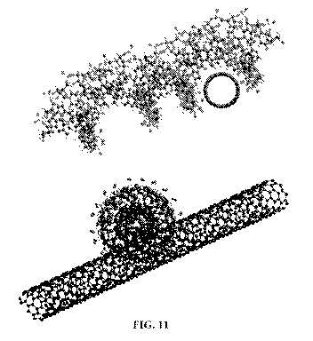
[0019] FIG. 11 shows a computer generated model of SWCNT/siRNA complexes.
[0020] FIG. 12 shows Atomic Force microscopy illustrating SWCNT coated with
siRNA. Arrows indicate areas of tube diameters. Diameter < mm: area of bare
SWCNT; > 1
nm: area of SWCNT with complexed siRNA.
[0021] FIG. 13 shows Atomic Force microscopy (AFM) of SWCNT coated with
siRNA that had been exposed to 1% BSA. Arrows indicate areas of tube
diameters. Diameter
< lnm: area of bare SWCNT; > 1 to 4 nm: area of SWCNT with complexed siRNA; >
5 nm:
area of SWCNT complexed to BSA.
[0022] FIG. 14 shows Western blots obtained from cultured MiaPaCa cells that
were exposed to SWCNT/(Trx)siRNA. FIG. 14A shows a time course of thioredoxin
expression, and FIG. 14B shows thioredoxin expression as a result of
incubation with
increasing concentrations of SWCNT/(Trx)siRNA.
[0023] FIG. 15 shows a Western blot resulting from exposure of cultured
MiaPaCa
cells to SWCNT/(Trx)siRNA, SWCNT/(EGFR)siRNA, and
SWCNT/(Trx)siRNA/(EGFR)siRNA dual payload SWCNT (FIG. 15A). FIG. 15B shows a
fluorometric analysis of a dispersion of the SWCNT/(Trx)siRNA,
SWCNT/(EGFR)siRNA,
and SWCNT/(Trx)siRNA/(EGFR)siRNA dual payload SWCNT used in FIG. 15A.
[0024] FIG. 16 shows a comparison of the size distribution of an initial
preparation
of SWCNT and a corresponding AFM (top), and the size distribution of an
optimized
preparation of SWCNT and corresponding AFM (bottom).
-4-
CA 02804760 2013-01-08
WO 2012/003466 PCT/US2011/042832
[0025] FIG. 17 shows the percent siRNA in stock solution before and after
complexing with SWCNT and the amount of siRNA remaining in solution after
complexing
in eight identical preparations.
[0026] FIG. 18 shows the blood chemistry and hematology results 24 hours and 1
week after intravenous administration of 100 itg of optimized SWCNT.
[0027] FIG. 19 shows a macroscopic image of mouse spleen 24 hrs after
administration of 100 mg SWCNT in 3% PLURONIC (top) compared to mouse spleen
24 hrs
after administration of a vehicle control (bottom).
[0028] FIG. 20 shows a bar graph comparing the average weight of liver,
spleen,
kidney, lung, heart, and brain of control mice and mice administered a
vehicle, 50 it.g of
SWCNT, and 100 jig of SWCNT.
[0029] FIG. 21 shows a fluorescence spectroscopy analysis of liver and spleen
12
hours, 24 hours, 48 hours, and 1 week after intravenous administration of 100
jig of
optimized SWCNT.
[0030] FIG. 22 shows a fluorescence spectroscopy analysis of the plasma SWCNT
content 5 mm, 1 hour, 6 hours, and 24 hours after administration of 100 jig of
optimized
SWCNT (FIG. 22A), and the average SWCNT content based on FIG. 22A plotted over
time
(FIG. 22B).
[0031] FIG. 23 shows the fluorometric analysis of SWCNT/(Trx)siRNA in the
blood of mice sacrificed at 2 mm, 15 mm, 30 mm, 1 hr, 4 hr, and 24 hr after
administration of
34 jig of SWCNT/(Trx)siRNA/PEG (FIG. 23A), and a scatter plot fit to a line of
the peak
intensity from FIG. 23A versus time (FIG. 23B).
[0032] FIG. 24 shows the fluorometric analysis of SWCNT/(Trx)siRNA in the
blood of mice sacrificed at 2 mm, 15 mm, 30 mm, 1 hr, 4 hr, and 24 hr after
administration of
67 jig of SWCNT/(Trx)siRNA/PEG (FIG. 24A), and a scatter plot fit to a line of
the peak
intensity from FIG. 24A versus time (FIG. 24B).
[0033] FIG. 25 shows a Western blot for thioredoxin and actin from the liver
and
kidney of two mice administered 67 ug of SWCNT/(Trx)siRNA.
[0034] FIG. 26 shows a Western blot for thioredoxin and actin from MiaPaCa
human pancreatic tumors excised from nude mice 24 hr, 48 hr, and 72 hr after
administration
of 39 jag of SWCNT/(Trx)siRNA (FIG. 26A). FIG. 26B shows a bar graph showing
the
percent control of thioredoxin expression in these tumors based on the Western
blot of FIG.
26A.
-5-
WO 2012/003466 CA 02804760 2013-01-08PCT/US2011/042832
[0035] FIG. 27 shows a Western blot for thioredoxin and actin from MiaPaCa
human pancreatic tumors excised from nude mice 24 hr, 48 hr, and 72 hr after
administration
of 94 [1.g of SWCNT/(Trx)siRNA (FIG. 27A). FIG. 27B shows a bar graph showing
the
percent control of thioredoxin expression in these tumors based on the Western
blot of FIG.
27A.
[0036] FIG. 28 shows the growth rate of tumors in animals treated with
SWCNT/siEGFR, SWCNT/siKRAS, and SWCNT/siEGFR/siKRAS compared to untreated
animals and vehicle controls where SWCNT/siRNA administration began 12 days
after the
initial injection of MiaPaCa-2 cells.
[0037] FIG. 29 shows the growth rate of tumors in animals treated with
SWCNT/siEGFR, SWCNT/siKRAS, and SWCNT/siEGFR/siKRAS compared to untreated
animals and vehicle controls where SWCNT/siRNA administration began 41 days
after the
initial injection of MiaPaCa-2 cells (FIG. 29A). FIG. 29B shows the change in
body weight
of the animals over the same period.
[0038] FIG. 30 shows a Western blotted for EGFR and KRAS 96 hrs following 4th
treatment of mice bearing MiaPaCa-2 tumors that had been treated weekly with
35 ug
SWCNT/siEGFR, SWCNT/siKRAS, or SWCNT/siEGFR/siKRAS
I. DETAILED DESCRIPTION
[0039] Before the present compositions and methods are described, it is to be
understood that this invention is not limited to the particular processes,
compositions, or
methodologies described, as these may vary. It is also to be understood that
the terminology
used in the description is for the purpose of describing the particular
versions or embodiments
only, and is not intended to limit the scope of the present invention which
will be limited only
by the appended claims.
[0040] It must be noted that, as used herein, and in the appended claims, the
singular
forms "a," "an," and "the" include plural reference unless the context clearly
dictates
otherwise. Unless defined otherwise, all technical and scientific terms used
herein have the
same meanings as commonly understood by one of ordinary skill in the art.
Although any
methods similar or equivalent to those described herein can be used in the
practice or testing
of embodiments of the present invention, the preferred methods are now
described. All
publications and references mentioned herein are incorporated by reference.
Nothing herein
is to be construed as an admission that the invention is not entitled to
antedate such disclosure
by virtue of prior invention.
-6-
CA 02804760 2013-01-08
WO 2012/003466 PCT/US2011/042832
[0041] As used herein, the term "about" means plus or minus 10% of the
numerical
value of the number with which it is being used. Therefore, about 50% means in
the range of
45%-55%.
[0042] The term "agglomeration," as used herein, refers to the formation of a
cohesive mass of subunits such as carbon nanotubes held together by relatively
weak forces
such as, for example, van der Waals forces or capillary action, that can be
broken during
processing resulting in a group of individual subunits. The mass of subunits
resulting from
agglomeration is an "agglomerate."
[0043] As used herein, the term "aggregation" refers to the formation of a
discrete
group of subunits such as carbon nanotubes in which the forces holding the
individual
subunits together are not easily broken. For example, carbon nanotubes bundles
can be
strongly bonded together by, for example, covalent bonds. The discrete group
of subunits is
called an "aggregate."
[0044] As used herein, the term "bioactive substance" refers to a compound
utilized
to image, impact, treat, combat, ameliorate, prevent or improve an unwanted
condition or
disease of a patient. The bioactive substance may modulate any number of
biological
functions in the cell, such as cell division, cellular infection, cellular
expression of cell
surface proteins, cellular response to a hormone, among others. The term
"bioactive
substance" may further refer to polynucleotides, small molecules, and
polypeptides that cause
a metabolic change in a cell, generally by increasing transcription,
expression or translocation
of one or more genes, or by binding to an expressed protein.
[0045] The term "carbon nanotube" refers to an allotrope of carbon having a
cylindrical or tube shape and a diameter of as small as about 1 nm. The term
"carbon
nanotube" may further include structures that can include, for example,
metals, small-gap
semiconductors, or large-gap semiconductors such as boron carbon nitride (BCN)
nanotubes.
The term carbon nanotube as used herein refers to both single-walled carbon
nanotubes
(SWCNT) and multi-walled carbon nanotubes (MWCNTs). A "single-walled carbon
nanotube" or "SWCNT" refers to a carbon nanotube that consists of a one atom
thick
graphene sheet that has been rolled into a tube, A "multi-walled carbon
nanotube" or
"MWCNT" refers to a nanotube that include 2 or more one graphene sheets roled
into
concentric tubes. The term "carbon nanotubes" may also be graphene in other
forms
including, for example, graphene spheres or "carbon nanosphere," which are
commonly
referred to as buckyballs or fullerene.
-7-
WO 2012/003466 CA 02804760 2013-01-08PCT/US2011/042832
[0046] The term "diseased tissue", as used herein, refers to tissue or cells
exhibiting
a phenotype that is inconsistent with healthy tissue. For example, "diseased
tissue" can
include tissues and cells affected by AIDS; pathogen-borne diseases, which can
be bacterial,
viral, parasitic, or fungal, examples of pathogen-borne diseases include HIV,
tuberculosis and
malaria; hormone-related diseases, such as obesity; vascular system diseases;
central nervous
system diseases, such as multiple sclerosis; and undesirable matter, such as
adverse
angiogenesis, restenosis amyloidosis, toxins, reaction-by-products associated
with organ
transplants, and other abnormal cell or tissue growth. In some embodiments,
"diseased
tissue" can refer to tissues and cells associated with solid tumors or other
cancerous growth
including, but not limited to, those associated with bone, lung, vascular,
neuronal, colon,
ovarian, breast, and prostate cancer. The term diseased tissue may also refer
to tissue or cells
of the immune system, such as tissue or cells
[0047] An "effective amount" or "therapeutically effective amount" of a
composition, as used herein, refers to an amount of a biologically active
molecule or complex
or derivative thereof sufficient to exhibit a detectable therapeutic effect
without undue
adverse side effects (such as toxicity, irritation and allergic response)
commensurate with a
reasonable benefit/risk ratio when used in the manner of the invention. The
therapeutic effect
may include, for example, inhibiting the growth of undesired tissue or
malignant cells. The
effective amount for a subject will depend upon the type of subject, the
subject's size and
health, the nature and severity of the condition to be treated, the method of
administration, the
duration of treatment, the nature of concurrent therapy (if any), the specific
formulations
employed, and the like.
[0048] "Gene silencing" as used herein can refer to the suppression of gene
expression from, for example, an endogenous gene, exogenous gene, or a
transgene, and
heterologous gene. Gene silencing may be mediated through processes that
affect
transcription, through post-transcriptional processing of RNA transcripts,
and/or translation
of the RNA transcript. In some embodiments, gene silencing can occur through
siRNA
mediated degradation of mRNA via RNA interference.
[0049] The term "knock-down" refers to gene silencing in which the expression
of a
target gene is reduced as compared with normal gene expression, but gene
expression not
completely eliminated. Knocking down gene expression can lead to the
inhibition of
production of the target gene product.
[0050] The term "non-functionalized," as used herein, refers to a chemical
composition such as, a carbon nanotube, that are substantially unmodified. As
such, each
-8-
CA 02804760 2013-01-08
WO 2012/003466 PCT/US2011/042832
carbon of the carbon nanotube is covalently bonded to a neighboring carbon
atom or an
unreactive atom such as, for example, hydrogen. Non-functionalized carbon
nanotubes do
not include reactive functional groups, i.e., a group of atoms capable of
forming a covalent
bond to a carbon atom or another functional group, covalently bonded to the
carbons of the
carbon nanotube.
[0051] The term "nucleic acid" refers to chemical compositions of monomers
having a sugar moiety, a phosphate, and a purine or pyrimidine base and
includes
deoxyribonucleic acids and ribonucleic acids as well as any single-stranded or
double-
stranded polymers thereof. Unless specifically limited, the term "nucleic
acid" further
encompasses known analogs of natural nucleotides that may have similar binding
properties
with reference to the naturally occurring nucleic acid analog and may be
metabolized in a
manner similar to naturally occurring nucleotides. Polymeric nucleic acids are
generally
referred to as "DNA" when the individual monomers making up the polymeric
nucleic acid
are deoxyribonucleic acids and "RNA" when the individual monomers making up
the
polymeric nucleic acid are ribonucleic acids. However, polymeric nucleic acids
can include
hybrid molecules that can include both deoxyribonucleic acid and ribonucleic
acid
monomers. Such polymeric nucleic acids may be arranged in any manner. For
example, a
polymeric nucleic acid may include complementary sequences that allow
intermolecular
interactions such that the polymeric nucleic acid to include secondary
structural elements, or
two single stranded polymeric nucleic acid molecules may include complementary
sequences
that allow intramolecular interactions such that the individual polymeric
nucleic acids may
bind to one another creating a double stranded polymeric nucleic acid
molecule.
[0052] The arrangement of nucleic acid monomers in a particular polymeric
nucleic
acid molecule is commonly referred to as the "sequence" of that nucleic acid
molecule. In a
phenomenon referred to as "base pairing" a purine nucleic acid monomers,
adenine (A) and
guanine (G) form hydrogen bonds selectively with pyrimidine nucleic acid
monomers
thymine (T) and cytosine (C), respectively, to create A-T and G-C "base
pairs." Ribonucleic
acids are capable of forming similar base pairs; however, thyamine (T) is
replaced with uracil
(U) to create a A-U base pair. For DNA and messenger RNA (mRNA), RNA molecules
produced as the result of transcription that have a sequence that is
complementary to the
DNA molecule from which the mRNA is produced, the nucleic acid monomers of a
sequence
may be arranged in three base pair "codons," where each codon of the mRNA
corresponds to
a specific amino acid that transported from the cytosol to a ribosome via
transfer RNA
(tRNA) during translation.
-9-
CA 02804760 2013-01-08
WO 2012/003466 PCT/US2011/042832
[0053] By "complementary sequence" is meant that the polymeric nucleic acid
molecule includes a sequence of individual monomers that allow hydrogen bonds
to form
between nucleic acid monomers. A "complementary sequence" encompasses a pair
of
nucleic acid molecules in which each base pair is exactly complementary to the
corresponding base pair to the opposing nucleic acid. "Complementary sequence"
also
encompasses a pair of nucleic acid molecules in which one of the pair include
conservatively
modified variants of naturally occurring nucleotides and degenerate codon
substitutions. For
example, degenerate codon substitutions may be achieved by generating
sequences in which
the third position of one or more selected (or all) codons is substituted with
mixed-base
and/or deoxyino sine residues.
[0054] The term "subject" or "patient," as used herein, includes human and non-
human vertebrates such as wild, domestic, and farm animals.
[0055] As used herein, a "pharmaceutically acceptable carrier" is a
pharmaceutically
acceptable solvent, suspending agent or vehicle for delivering the complexes
of the present
invention to the patient. The carrier may be liquid or solid and is selected
with the planned
manner of administration in mind. Examples of pharmaceutically acceptable
carriers that
may be utilized in accordance with the present invention include, but are not
limited to,
water, isotonic salt solution, isotonic sugar solution, polyethylene glycol
(PEG), aqueous
PEG solutions, liposomes, ethanol, organic solvent (e.g. DMSO) dissolved in
isotonic
aqueous solution, aqueous buffers, oils, and combinations thereof.
[0056] The terms "small interfering RNA," "short interfering RNA," or "siRNA"
refers to short double stranded RNA molecules in which one strand of the
double stranded
RNA is complementary to a portion of a target gene. An "RNA duplex" or "double-
stranded
RNA" refers to the structure formed by the complementary pairing between two
regions of a
RNA molecule. In some embodiments, the length of an siRNA molecule may be less
than
about 30 nucleotides. For example, the siRNA can be 29, 28, 27, 26, 25, 24,
23, 22, 21, 20,
19, 18, 17, 16, 15, 14, 13, 12, 11 or 10 nucleotides in length, and in
particular embodiments,
the length of the duplex may be 19 to 25 nucleotides in length. In some
embodiments,
siRNA may consist of two complementary RNA molecules that are held together by
the
hydrogen bonding between base pairs, and in some embodiments, the siRNA may
include a
3' or a 5' overhang of 1, 2, 3, 4 or 5 nucleotides on either end of the siRNA
molecule. In
other embodiments, the RNA duplex portion of the siRNA can be part of a
hairpin structure
prepared from a long single strand of RNA that includes at least two
complementary
sequences. siRNA including such a hairpin structure are sometime referred to
as short
-10-
CA 02804760 2013-01-08
WO 2012/003466 PCT/US2011/042832
hairpin RNA or (shRNA). In such embodiments, a loop can be formed between the
two
sequences that form the duplex. The loop can vary in length. For example, in
some
embodiments the loop may be 5, 6, 7, 8, 9, 10, 11, 12 or 13 nucleotides in
length. In other
embodiments, the hairpin structure can include 3' or 5' overhang portions.
[0057] The siRNA described herein includes double-stranded RNA molecules that
are prepared from unmodified, naturally occurring RNA bases as well as siRNA
that, for
example, include non-naturally occurring RNA base pairs or are chemically-
modified siRNA
or otherwise stabilized siRNA. siRNA can also include siRNA that are
specifically designed
to target a specific gene, "targeting siRNA," and siRNA having a randomly
generated
sequence, "non-targeting siRNA."
[0058] "RNA interference (RNAi)" is the process of sequence-specific,
posttranscriptional gene silencing initiated by siRNA. RNAi is seen in a
number of
organisms such as Drosophila, nematodes, fungi and plants and is believed to
be involved in
anti-viral defense, modulation of transposon activity, and regulation of gene
expression.
During RNAi, siRNA induces degradation of target mRNA and consequently
inhibition of
gene expression.
[0059] Various embodiments described herein are directed carbon nanotubes, and
in
some embodiments, single-walled carbon nanotubes (SWCNT), that are useful for
delivery of
a bioactive agent. In such embodiments, the bioactive agent may coat the
carbon nanotube or
SWCNT by forming covalent or non-covalent interactions with the carbon
nanotube or
SWCNT. In certain embodiments, bioactive agent coated carbon nanotube or SWCNT
may
be combined in a pharmaceutical composition that can be administered to a
subject to
facilitate delivery of the bioactive agent to the subject. Accordingly, some
embodiments
described herein include pharmaceutical compositions at least including
bioactive agent
coated carbon nanotube or SWCNT and a pharmaceutically acceptable carrier, and
other
embodiments include methods for using such pharmaceutical compositions for
treating a
subject. While such embodiments are not limited to a particular treating a
particular disease,
in certain embodiments, the disease may be cancer or another disease
characterized by
abnormal cell growth.
[0060] The carbon nanotube or SWCNT of various embodiments may be any carbon
nanotube or single-walled carbon nanotubes known in the art. In some
embodiments, the
carbon nanotube or SWCNT may have a diameter for from about 0.5 nm to about
1.5 nm, and
in other embodiments, the diameter may be about 1 nm. In still other
embodiments, the
length of the carbon nanotube or SWCNT may be about 300 nm or less. For
example, in
-11-
CA 02804760 2013-01-08
WO 2012/003466 PCT/US2011/042832
some embodiments, the carbon nanotube or SWCNT may have a length of from about
100
nm to about 400 nm, and in other embodiments, the carbon nanotube or SWCNT may
have a
length of about 150 nm to about 300 nm or about 175 nm to about 250 nm. Near-
infrared
spectral analysis provides a means for determining nanotube size distribution,
purity,
concentration in solution, and individualization, and in certain embodiments,
the carbon
nanotube or SWCNT may have a strong near-IR spectral transition in, for
example, a range of
from about 850 nm to about 1600 nm.
[0061] The carbon nanotube or SWCNT of embodiments may be derived from any
source. For example, in some embodiments, the carbon nanotube or SWCNT may be
produced by known methods including, but not limited to, arc discharge, laser
evaporation,
chemical vapor deposition, and the like, and in other embodiments, as high
quality
inexpensive carbon nanotube or SWCNT can be prepared using known catalyst
chemical
vapor deposition methods. In certain embodiments, carbon nanotube or SWCNT may
be
prepared using the high pressure carbon-monoxide method (HiPco), in which high
pressure
carbon monoxide (CO) is disproportionated on iron (Fe) nanoparticles formed in
the gas
phase from iron pentacarbonyl (Fe(C0)5) decomposition. Without wishing to be
bound by
theory, the HiPCO method may produce relatively small diameter nanotubes.
[0062] Embodiments described herein are not limited to any particular
bioactive
agent. For example, in various embodiments, the bioactive agent may be a
drugs, vaccines,
immunological agents, chemotherapeutic agent, diagnostic agent, prophylactic
agent,
nutraceutical agent, small molecule, nucleic acid, protein, peptide, lipid,
carbohydrate,
hormone, and combinations thereof. In particular embodiments, the bioactive
substance may
be siRNA. The siRNA of such embodiments may be of any sequence and may be
composed
of naturally occurring or non-naturally occurring base pairs. In some
embodiments, the
siRNA may be double-stranded RNA, and in other embodiments, the siRNA may be
hairpin
siRNA. In still other embodiments, the siRNA unmodified, and in yet other, the
siRNA may
be chemically-modified. Embodiments are not limited to a particular sequence,
and in some
embodiments, the siRNA may include a sequence that allows the siRNA to
specifically target
a specific gene thereby inhibiting expression of that particular gene. In
other embodiments,
the siRNA may be of random sequence. In certain embodiments, the siRNA may be
of a
sequence that allows the siRNA to specifically target and inhibit the
expression hypoxia-
inducible factor 1 alpha (HIF-1a), polio-like kinase 1 (PLK1), kinase-like
family 11 (K1f11),
thioredoxin (Trx), epidermal growth factor receptors (EGFR, ErbB-1, HER1), V-
Ki-ras2
Kirsten rat sarcoma viral oncogene homolog (KRAS), and the like.
-12-
CA 02804760 2013-01-08
WO 2012/003466 PCT/US2011/042832
[0063] In some embodiments, the SWCNT may be combined with a bioactive agent
to make a SWCNT/bioactive agent complex, in which the bioactive agent forms a
non-
covalent association with the SWCNT, and in other embodiments, the SWCNT may
be
combined with a bioactive agent to make a SWCNT/bioactive agent complex, in
which
covalent bonds associate the bioactive agent with the SWCNT. In still other
embodiments,
the SWCNT may be combined with siRNA to make a SWCNT/siRNA complex, in which
each delivery vehicle only includes a SWCNT and one or more siRNA molecule
associated
with the SWCNT. As used herein, the term "SWCNT complexes" shall encompass
both
SWCNT/bioactive agent complexes and, in particular, SWCNT/siRNA agent
complexes.
The amount of bioactive agent or siRNA combined with the SWCNT may vary among
embodiments and may be determined based on the amount of surface of each SWCNT
to be
covered by active agent or siRNA. For example, in some embodiments, the ratio
of
complexed to non-complexed surface area on the SWCNT may be selected to
provide
sufficient coverage to allow the SWCNT to be soluble in solution and provide a
therapeutically effective amount of bioactive agent to be delivered. In
certain embodiments,
less than about 95% of the total surface area of the SWCNT may be in complex
with the
bioactive active agent and/or solubilization agent, and in other embodiments,
less than about
50% of the surface area of the SWCNT may be in complex with the bioactive
agent and/or
solubilization agent. The amounts of SWCNT and bioactive agent or siRNA
combined to
form the SWCNT complexes may, therefore, vary accordingly. For example in some
embodiments, a composition of SWCNT complexes may include about 1 ng/u1 to
about 10
ng/u1 based on the total volume of the composition and about 10 ng/u1 to about
40 ng/u1 of
siRNA. In other embodiments, the SWCNT may be provided in a concentration of
about 2
ng/u1 to about 5 ng/u1 and about 15 ng/u1 to about 30 ng/u1 of siRNA or about
3 ng/u1 of
SWCNT and about 25 ng/u1 of siRNA. In some embodiments, the SWCNT/siRNA
complex
is administered in an effective amount. In some embodiments, an effective
amount may
comprise less than about 100 mg, less than about 75 mg, less than about 50 mg,
less than
about 40 mg, less than about 30 mg, from about 15 mg to about 100 mg, from
about 15 mg to
about 75 mg, from about 15 mg to about 50 mg, from about 15 mg to about 40 mg,
or from
about 15 mg to about 30 mg of the one or more short-interfering ribonucleic
acid (siRNA)
complexed to single-walled carbon nanotubes.
[0064] In other embodiments, the SWCNT/siRNA complex may further include one
or more solubilization agent, and in some embodiments, the solubilization
agent may be a
mild detergent that can associate with the SWCNT/siRNA complex and allow
improved the
-13-
CA 02804760 2013-01-08
WO 2012/003466 PCT/US2011/042832
solubility of the SWCNT/siRNA complex. For example, in particular embodiments,
the
detergent may be a polyalkylene oxide such as, for example, PLURONICTM, PEG
5000,
PEG5000 PE (1,2 -dimyristoyl- sn-glyc ero-3 -phosphoethanolamine-N-
lmethoxy(polyethylene
glycol)-5000] (ammonium salt), C18-PMH-mPEG (poly(maleic anhydride-alt- l-oc
tadecene)-
poly(ethylene glycol)methyl ether), or a combination thereof. The
concentration of the
detergent may vary in embodiments and may be sufficient to increase
solubilization without
effecting the stability of the SWCNT complexes or the physiological
acceptability of the
compositions. For example, in some embodiments, the solubilization may be
provided at
about 1% to about 7% of the total solution, and in other embodiments, the
detergent may be
provided at about 2% to about 5% of the total solution. In particular
embodiments, the
detergent may be provided at about 3% of the solution.
[0065] In some embodiments, the SWCNT/bioactive agent complex or
SWCNT/siRNA agent complex may be prepared in an aqueous buffer that is
physiologically
acceptable for in vivo or in vitro use, and in other embodiments, the SWCNT
complexes may
be prepared in a buffer suitable for administration to a mammal such as, for
example, a
mouse, rabbit, ape, or human. As such, in certain embodiments, the SWCNT
complexes may
be combined with one or more pharmaceutically acceptable carrier or excipient
to produce a
pharmaceutical formulation. The carrier or excipient may vary among
embodiments and may
be selected based on factors including, but not limited to, route of
administration, location of
the disease tissue, the bioactive substance being delivered, and/or time
course of delivery of
the bioactive substance. For example, in some embodiments, the
pharmaceutically
acceptable carrier may be water, and in other embodiments, the
pharmaceutically acceptable
carrier may be water combined with a physiologic salt to create an aqueous
solution that is
isotonic to blood serum. In still other embodiments, the pharmaceutical
compositions of
embodiments can include one or more preservative.
[0066] As above, in some embodiments, the pharmaceutical compositions may
include a solubilization agent such as, but not limited to, polyalkylene
oxides such as, for
example, PLURONICTM, PEG, PEG-5000, PEG-5000 PE, PL-PEG (1,2-dimyristoyl-sn-
glycero-3-phosphoethanolamine-N-lmethoxy(polyethylene glycol)-50001 (ammonium
salt),
C18-PMH-mPEG (poly(maleic anhydride-alt-l-octadecene)-poly(ethylene
glycol)methyl
ether) or a combination thereof. In particular embodiments, at least about 50%
or at least
about 75% of the total SWCNT complexes in the composition may be solubilized
in the
pharmaceutically acceptable carrier solution by association of only siRNA. The
addition of
1% to about 7% or about 2% to about 5% of the total solution of a
solubilization agent may
-14-
CA 02804760 2013-01-08
WO 2012/003466 PCT/US2011/042832
further increase the solubility of the SWCNT complexes such that up to about
85%, up to
about 95%, or up to about 100% of the SWCNT complexes.
[0067] The pharmaceutical compositions of various embodiments may be prepared
to deliver of a therapeutically effective amount of the bioactive agent to the
subject. For
example, in embodiments in which the bioactive agent is a siRNA, an effective
amount of the
SWCNT/siRNA complex may be an amount sufficient to reduce expression of the
target gene
in affected tissue. The effect of such reduced expression of the target gene
may be evident
based on physiological changes in the affected tissue such as, for example,
reduced rate of
cell proliferation in tumorigenic tissue, reduction or maintenance of tumor
size, reduction or
reversal of other symptoms associated with the disease, and combinations
thereof. Reduced
expression may be also, or alternatively, be evident by reduced expression of
the target gene
based on pre-administration expression levels in the patient or comparisons to
administration
of siRNA that is not complexed to SWCNT or vehicle controls. The
therapeutically effective
amount may vary depending on the type disease being treated, the extent of
disease, disease
progression, age of the patient, weight of the patient, and the like. In some
embodiments, a
therapeutically effective amount may be up to about 5 ug/kg or greater. In
other
embodiments, a therapeutically effective amount may be from about 0.1 ug/kg to
about 4
ug/kg or greater, and in still other embodiments, a therapeutically effective
amount may be
from about 0.5 ug/kg to about 3 ug/kg or about 2.5 ug/kg.
[0068] Without wishing to be bound by theory, the SWCNT complexes of various
embodiments may be very well tolerated when administered to a patient, such
that large
doses of a SWCNT complex may be provided to a patient with limited or no
adverse side
effects. For example, in some embodiments, a dose of greater than 10 mg or
greater than 15
mg may be administered to a human without adverse side effects. Accordingly,
various
embodiments include pharmaceutical compositions prepared for high dose
administration of
SWCNT complexes.
[0069] Further embodiments are directed to methods for delivering a bioactive
agent
to diseased tissue including the steps of administering a therapeutically
effective amount of a
SWCNT complex to a patient, methods for treating a disease by administering a
pharmaceutical composition including a therapeutically effective amount of a
SWCNT
complex to a patient in need of treatment, and methods for silencing a
targeted gene in vivo
including administering a therapeutically effective amount of a SWCNT complex
to a patient.
Any SWCNT complex including any bioactive agent described herein may be
administered
as part of a pharmaceutical composition in such methods. In certain
embodiments, the
-15-
WO 2012/003466 CA 02804760 2013-01-08PCT/US2011/042832
SWCNT complex to be delivered may be a SWCNT/siRNA complex. In some
embodiments,
delivering may include contacting diseased tissue with the bioactive agent,
and in some
embodiments, delivering may include internalization of the active agent and,
in certain
embodiments, SWCNT into cells of the diseased tissue. For example, about 0.01%
to about
30% of the total SWCNT/siRNA complex can be internalized the in vitro in media
containing
10% serum after about 1 hour, from about 20% to about 90% of the total
SWCNT/siRNA
complex can internalized after about 3 hours, and after about 24 hours about
95% of more of
the total SWCNT/siRNA complex can be internalized. The bioactive agent may
remain in
complex with the SWCNT after being internalized by the cell, or in some
embodiments, the
bioactive agent may dissociate from the SWCNT when internalized by the cell.
[0070] A broad range of diseases can be treated using the methods described
herein,
as SWCNT complexes of various embodiments can function as a serum-insensitive
transfection agent to effectuate delivery of various bioactive agents into a
cell. For example,
in some embodiments, SWCNT/siRNA complex may be used to deliver siRNA to
diseased
tissues and cells to induce an RNAi response, which can effectively treat any
disease in
which aberrant gene expression leads to a diseased state. Moreover, because
siRNA can
silence target genes with a high degree of specificity, the SWCNT/siRNA
complexes of
various embodiments may be safely administered to treat nearly any disease for
which an
aberrantly expressed gene has been identified. For example, in particular
exemplary
embodiments, the siRNA of SWCNT/siRNA complexes may targeting HIF-1a
expression.
HIF-1a has been associated with a number of disease states including, but are
not limited to,
cancers such as, for example, breast cancer, lung cancer, head and neck
cancer, brain cancer,
abdominal cancer, colon cancer, colorectal cancer, esophagus cancer,
gastrointestinal cancer,
glioma, liver cancer, tongue cancer, neuroblastoma, osteosarcoma, ovarian
cancer, pancreatic
cancer, prostate cancer, retinoblastoma, Wilms tumor, multiple myeloma, skin
cancer,
lymphoma, and blood cancer, angiogenic diseases such as, for example, diabetic
retinopathy,
age-related macular degeneration, and inflammatory diseases, inflammatory
disease such as,
for example, psoriasis and rheumatoid arthritis. Accordingly, in particular
embodiments,
SWCNT/siRNA complexes with siRNA targeting HIF- 1 a may be administered to
treat any
of these disease states.
[0071] The pharmaceutical compositions of described herein can be administered
in
any conventional manner by any route where they are active. Administration can
be systemic
or local. For example, administration can be, but is not limited to,
parenteral, subcutaneous,
-16-
WO 2012/003466 CA 02804760 2013-01-08PCT/US2011/042832
intravenous, intramuscular, intraperitoneal, transdermal, oral, buccal,
ocular, intravaginally,
or inhalation. In certain embodiments, the administration may be systemic by
intravenous
injection, and in other embodiments, the administration to subjects exhibiting
cancer may be
by intratumoral injection. In some embodiments, the pharmaceutical composition
may be
prepared in the presence or absence of stabilizing additives that favors
extended systemic
uptake, tissue half-life, and intracellular delivery. Thus, modes of
administration for the
compounds of the present invention either alone or in combination with other
pharmaceuticals can be injectable, including short-acting, depot, implant, and
pellet forms
injected subcutaneously or intramuscularly. In some embodiments, an injectable
formulation
including SWCNT complexes may be deposited to a site of the diseased tissue,
such as, for
example, in some embodiments, the pharmaceutical composition may be
administered
directly to tumorigenic tissue. In other embodiments, the pharmaceutical
composition may
be administered systemically by, for example, intravenous injection.
[0072] The frequency of administration may vary depending on the disease
indication being treated and the patients response to the treatment. For
example, in some
embodiments, a pharmaceutical composition including SWCNT complexes may be
administered at least once ever 12 hours, at least once every 24 hours, at
least once every 48
hours, or at least once every 72 hours. Without wishing to be bound by theory,
the half-life
of the SWCNT complexes may be relatively short in circulation; however,
despite this
limited half-life in circulation, the SWCNT complexes may retain activity in
affected tissue
for at least about 24 hours following administration. For example, in some
embodiments, a
SWCNT/siRNA complex may be detectable in the blood stream of a patient to whom
a
pharmaceutical composition including a SWCNT/siRNA complex for less than about
30
minutes or less than about 15 minutes, but reduction in expression of a target
gene may be
observed for up to at least 12 hours or at least 24 hours. Thus, while the
half-life of the
SWCNT/siRNA complex may be relatively short, sufficient levels of siRNA can be
delivered
to affected tissue to adequately reduce target gene expression from a single
therapeutically
effect dose once every 12 hours or 24 hours. The frequency of administration
of a
pharmaceutical composition including a SWCNT/siRNA complex may, therefore, be
reduced
based on the effect rather than concentration of the SWCNT/siRNA complex in
circulation.
[0073] The amount of SWCNT complexes in circulation following administration
may be further effected by introduction of a solubilization agent into the
pharmaceutical
composition. For example, in some embodiments, the half-life of a
pharmaceutical
composition in circulation may be increased by providing SWCNT complexes
having up to
-17-
CA 02804760 2013-01-08
WO 2012/003466 PCT/US2011/042832
about a 10:1 ratio of a solubilization agent to the bioactive agent, and in
other embodiments,
the ratio of solubilization agent to bioactive agent in SWCNT complexes having
extended
half life may be from about 8:1 to about 1:1, or about 7:1. Without wishing to
be bound by
theory, SWCNT complex having a high ratio of solubilization agent to bioactive
agent may
exhibit a half-life in circulation of about 30 minutes or more, about 15
minutes or more, about
minutes or more, or about 5 minutes or more. In still other embodiments, the
solublization
agent may be provided at less than a 1:1 ratio as compared to the bioactive
agent. For
example, in some embodiments, the ratio of solubilization agent to bioactive
agent may be
about 1:10, and in other embodiments, about 1:2 to about 1:8, or about 1:7. In
embodiments
10 with a low ratio of solubilization agent to bioactive agent, almost no
SWCNT complexes may
be detectable in circulation following administration; however, the reduced
half-life for the
SWCNT complexes in circulation may not effect of the pharmaceutical
composition on the
target tissue.
[0074] Yet further embodiments are directed to methods for preparing a SWCNT
complexes including the steps of combining a bioactive agent in an aqueous
solution with
SWCNT and sonicating the SWCNT/bioactive agent solution. In some embodiments,
the
bioactive agent may be siRNA, and the concentration of siRNA in the aqueous
solution, in
such embodiments, may be up to about 100 it.M, or in some embodiments, from
about 5 it.M
to about 50 it.M or about 10 it.M to about 30 M. In particular embodiments,
the
concentration of bioactive may be about 20 M. In some embodiments, the SWCNT
that are
combined with the bioactive agent in the aqueous solution may be provided in
an aqueous
solution at a concentration of about 1 ppm to about 10 ppm or up to
concentration of about
500 mg/mL, and when combined with the bioactive the concentration of SWCNT in
the
SWCNT/bioactive agent solution may be about 10 ug/mL to about 500 ug/mL or
about 25
ug/mL to about 300 ug/mL. In some embodiments, the SWCNT/bioactive agent
solution
may further include one or more solubilization agents. For example, in certain
embodiments,
a solubilization agent may be added to the aqueous solution to provide a final
concentration
of solubilization agent of about 1% to about 7%, about 2% to about 5%, or
about 3% of the
total solution.
[0075] The aqueous solution of various embodiments may be any buffer known in
the art that is useful during sonication and may include any number of
chemical additives.
For example, in some embodiments, the aqueous solution may be a buffer
solution of about
100 mM KC1, 30 mM HEPES-KOH, and 1 mM MgC12 or 0.9% NaCl. The pH of such
buffer
-18-
CA 02804760 2013-01-08
WO 2012/003466 PCT/US2011/042832
solutions may be approximately neutral, for example, from about 6.5 to about
8Ø In
particular embodiments, the aqueous solution may be suitable for in vivo
administration of
the SWCNT complexes. As such, in some embodiments, salt and pH concentrations
may be
within physiological ranges, and in other embodiments, the aqueous solution
may be
sterilized by known methods. The SWNCT complexes in the aqueous solution may,
therefore, be administered immediately following sonication.
[0076] Sonication may be carried out using any sonication device known in the
art,
and the parameters for sonication may vary depending, for example, on the type
of bioactive
agent being associated with the SWCNT. For example, in embodiments in which
siRNA is
associated with SWCNT, the SWCNT/siRNA solution prepared as described above,
which
may or may not include a solubilization agent, may be sonicated in 15 second
bursts with 45
second intervals between bursts. In some embodiments, the method for
sonication may
include two 15 second bursts each burst followed by a 45 second interval or
sixteen 15
second bursts each followed by a 45 second interval. In particular
embodiments, the
temperature of the samples during sonication may be maintained at about 25 C
during the
bursts, and the samples may be placed on ice during the 45 second intervals
between bursts.
The sonicator's settings during sonication may vary. For example, in some
embodiments, the
sonicator may be set to about 130 W, 20 kHz, and 40% amplitude.
[0077] The methods of some embodiments may further include the step of
removing
insoluble materials from the aqueous solution following sonication. The step
of removing
insoluble materials may be carried out by any means known in the art. For
example, in some
embodiments, insoluble materials may be removed by filtration, and in other
embodiments,
insoluble materials may be removed by centrifugation using parameters such as,
15,000 x g
for 5 minutes.
[0078] Various modifications of the invention, in addition to those described
herein,
will be apparent to those skilled in the art from the foregoing description.
Such modifications
are intended to fall within the scope of the appended claims.
EXAMPLES
[0079] Although the present invention has been described in considerable
detail
with reference to certain preferred embodiments thereof, other versions are
possible.
Therefore the spirit and scope of the appended claims should not be limited to
the description
and the preferred versions contained within this specification. Various
aspects of the present
invention will be illustrated with reference to the following non-limiting
examples. The
-19-
CA 02804760 2013-01-08
WO 2012/003466 PCT/US2011/042832
following examples are for illustrative purposes only and are not to be
construed as limiting
the invention in any manner.
EXAMPLE 1
[0080] Preparation of Noncovalent Complexes of SWCNT with siRNA.
[0081] SWCNT were produced using a high-pressure carbon monoxide (HiPco)
process. The raw HiPco SWCNT product was added to an aqueous buffer solution
(100 mM
KC1, 30 mM HEPES-KOH [pH 7.51, 1 mM MgC12) containing 20 uM solubilized pooled
siRNA [(siRNA targeting HIF- la (HIF-1 a) 5' -CCUGUGUCUAAAUCUGAAC-3' ,
5'CUAC CUUCGUGAUUCUGUUU-3', GCACAAUAGACAGCGAAAC-3', 5' -
CUACUUUCUUAA UGGCUUA) , polo-like kinase 1 (PLK1), 5' -CAACCAAAGUCG
AAUAUUGAUU-3, 5'-C AAGAAGAAUGAAUACAGUUU-3' , 5' -
GAAGAUGUCCAUGGAAAUAUU-3', 5' -CAACA CGCCUCAUCCUCUAUU-3' , Kinesin
superfamily protein (Kif11), 5' -CGUCUUUAGAU UCCUAUAU-3' , 5' -
GUUGUUCCUACUUCAGAUA-3' , 5' -GUCGUCUUUAGAUUCCU AU-3', 5' -
GAUCUACCGAAAGAGUCAU-3' [ , non-targeting siRNA 5' -UAGCGACAUU
UGUGUAGUU-3' (siTox), purchased from Dharmacon Inc, IL. This mixture was
sonicated
(Sonics, Vibra-cell) at 25 C using two 15 second pulses at settings of 130 W,
20k Hz, and
40% amplitude. The sonicated sample was centrifuged at 15,000 x g for 5
minutes. The
pellet comprising bundled SWCNT was discarded and the supernatant was
transferred into a
clean tube and centrifuged an additional 1 minute at the same settings. The
resulting
supernatant contained SWCNT non-covalently suspended by coatings of adsorbed
siRNA.
Near infrared (NIR) fluorescence spectroscopy indicated that the sample
contained
predominantly individually suspended SWCNT rather than nanotube aggregates.
[0082] The near infrared (NIR) emission spectrum of the siRNA-suspended
SWCNT was measured using 658 nm excitation in a model NS1 NanoSpectralyzer
(Applied
NanoFluorescence, Houston, TX). NIR fluorescence microscopy was performed
using a
custom-built apparatus containing diode laser excitation sources emitting at
658 and 785 nm.
Individual SWCNT internalized into cells were imaged with a custom-built NIR
fluorescence
microscope using 785 nm excitation, a 60 X oil-immersion objective, and a 946
nm long-pass
filter in the collection path. Bright field images were taken using the 60 X
objective.
[0083] The unagglomerated, SWCNT are made water-compatible by coating with
siRNA. As shown in FIG. 1A, sonication of SWCNT in aqueous buffer in the
absence of
siRNA failed to produce a stable suspension. However, as shown in FIG. 1B,
equivalent
-20-
WO 2012/003466 CA 02804760 2013-01-08PCT/US2011/042832
processing in the presence of siRNA provided stable, homogeneous suspensions.
These
suspensions displayed strong NIR fluorescence between approximately 900 and
1600 nm, as
depicted in FIG. IC, which is characteristic of dispersed or unagglomerated
SWCNT.
[0084] The SWCNT/siRNA complexes were stable and retained their biological
activity following 30 days of storage at 4 C. It is predicted that the
SWCNT/siRNA
complexes could retain biological activity following longer periods of storage
at 4 C.
EXAMPLE 2
[0085] Cell Culture and Cellular Incubation with SWCNT/siRNA Complexes.
[0086] MiaPaCa2-HRE (a pancreatic cell line with a HIF-1a/luciferase reporter)
cells were incubated in growth media consisting of high glucose DMEM
supplemented with
10% fetal calf serum (FCS) (all reagents from HyCone). To determine the
internalization
rate of non-targeting siRNA-solubilized SWCNT, 50 lut of the complex (final
SWCNT
concentration approximately 1.25 mg/L) was added to cells (approximately 2 x
105
cells/well) that had been incubated for 18 hours in 1 mL of media in a 6-well
plate.
Incubation with the SWCNT/siRNA complex continued for 1, 3 and 6 hours. After
incubation, media was removed from the wells, the cells were washed once in
phosphate
buffered saline (PBS) and then were detached from the surface by adding 0.25%
trypsin
(Invitrogen). The detached cells were washed with growth media to inactivate
the trypsin
and then washed again with PBS. The cells were resuspended in 1 mL of growth
media,
transferred onto a circular glass cover slip in a well of a new 6-well plate
and incubated at 37
C in a humid environment for approximately 20 hours. NIR fluorescence
microscopy was
utilized to identify internalized SWCNT.
[0087] The MiaPaCa2-HRE cell line was generated to stably express the promoter
sequence of a target gene of HIF-1a comprising the HIF-1a binding hypoxia
response
element (HRE) fused to the luciferase gene. At the end of the experiment, 100
[11_, of media
was removed from each well of the 96-well plate. The removed media was
replaced with 50
lut of the luciferase reagent (25 mM tricine, 0.5 mM EDTA-Na2, 0.54 mM sodium
triphosphate, 16.3 mM MgSO4=7H20, 0.3% Triton X-100, 0.1% w/v dithiothreitol,
1.2 mM
ATP, 50 mM luciferin, and 270 mM coenzyme A). The plates were incubated at
room
temperature for 5 minutes. Sample luminescence was measured relative to a
background
control using a microplate reader (Polar Star Optima; BMG Labtech).
[0088] Cell proliferation reagent (WST-1, Roche, Mannheim Germany) was added
to cells in media to a final concentration of 10% and the cells were incubated
for 30 minutes
-21-
CA 02804760 2013-01-08
WO 2012/003466 PCT/US2011/042832
at 37 C in a humidified incubator. The absorbance of the sample was then
measured relative
to a background control using a microplate reader (Polar Star Optima; BMG
Labtech) at
420 - 480 nm.
[0089] Statistical analyses were performed with commercially available
software.
Single regression analysis was used to assess the ratio of HIF-1 activity
after treatment with
100 lut sample volume, SWCNT concentration approximately 4 mg/L, siRNA
concentration
approximately 2 uM, with the percentage luciferase expression after
SWCNT/siRNA
treatment as the dependent variable. Student's t-tests were used to compare
the ratio of
luciferase intensity within the tumor between mice treated with SWCNT/siRNA.
Comparisons of mice treated with siRNA targeting HIF-1 (siHIF), SWCNT/non-
targeting
siRNA (SWCNT/SC), or SWCNT/ siRNA targeting HIF- la were computed by two-way
analysis of variance (ANOVA). Statistical significance was defined as a P
value of <0.05.
[0090] SWCNT were complexed with 20 uM of siRNA targeting polo-like kinasel
(PLK1) in a 0.9% NaC1 solution using the procedure described above. A 20 [11_,
portion of
each sample was added to cells (approximately 2 x 10⁵ cells/well) in 100 [11_,
of media
containing 10% FCS in 96-well plates. The treated cells were incubated at 37
C in a humid
chamber for 72 hours and their viability was determined by the WST-1 assay.
[0091] To investigate the biological activities of SWCNT/siRNA complexes, 20
lut
of each sample was added to cells (approximately 2 x 10⁵ cells/well) in 100
lut of media
containing 10% FCS in 96-well plates. The plates were incubated at 37 C in a
humidified
chamber for approximately 18 hours prior to and for 72 hours following
addition of the
complexes. To determine the ability of the complexes to suppress HIF-1a
activity or silence
the HIF-1a protein, treated cells incubated under normoxia for 72 hours were
incubated for a
further 18 hours under hypoxic conditions (1% oxygen).
[0092] MiaPaCa2-HRE cultures were exposed to SWCNT/siRNA complexes for 1,
3, and 6 hours to monitor internalization of the complex into tissue cells. As
shown in FIG.
2, NIR fluorescence microscopy of the treated cells revealed internalized
SWCNT. The cells
having internalized SWCNT were characterized by their emission wavelengths and
their
strong dependence of emission intensity on excitation beam polarization. In
addition, NIR
fluorescent particles were found only in cells incubated with suspended SWCNT
and not in
SWCNT-free control samples. As the sample area irradiated by the laser beam
was smaller
than the image field, some cells in each image did not show NIR emission even
though they
contain internalized SWCNT. Incubation with the SWCNT/siRNA complexes for 1
hour
-22-
CA 02804760 2013-01-08
WO 2012/003466 PCT/US2011/042832
resulted in SWCNT uptake by approximately 40% of cells. Incubation for 3 hours
or 6 hours
resulted in nanotube uptake by larger fractions of cells, and average SWCNT
content per cell
also increased with incubation time. Although the concentration of
internalized nanotubes
varied substantially from cell to cell, after 6 hours of incubation, more than
90% of the cells
showed detectable SWCNT.
[0093] A mixture of pristine SWCNT and siTox was sonicated and 20 [11_, of the
complex (containing 5 mg/L SWCNT and 5 uM siTox) was added to MiaPaCa-HRE
(human
pancreatic cancer) cells growing in a 96-well plate. Each well contained 100
[11_, of medium
with 10% FCS. Controls included untreated cells and cells treated with 20 mL
of a complex
of SWCNT and non-targeting siRNA (SWCNT/SC) (containing 5 mg/L SWCNT and 5 uM
siSC), 20 lut of SWCNT solubilized by 10% FCS, buffer alone and free
uncomplexed siTox
(final concentration 5 uM). At 72 hours after treatment, a decrease of
approximately 90%
was observed in viability of cells treated with the SWCNT/siTox complex, as
shown in FIG.
3. This effect was specific to the SWCNT/siTox complex, as none of the
controls exhibited
decreased cell viability. The preparative sonication did not damage the siRNA
and siRNA
was delivered into cells in a biologically active form. Further, the presence
of serum did not
inhibit the transfection process.
[0094] The ability of SWCNT/siRNA complexes to activate a specific RNAi
response was tested in MiaPaCa-HRE pancreatic cancer cell line. Changes in HIF-
1 a
activity were monitored in these cells by measuring the levels of luciferase
expression.
MiaPaCa-HRE cells were treated with SWCNT complexed with either an siRNA
specifically
targeting HIF-1a (siHIF), or a non-targeting siRNA (siSC), at final
concentrations of 3 mg/L
SWCNT and 5 uM siRNA. The final siRNA concentration was based on the initial
siRNA
concentration suspended in the siRNA buffer and, as such, the final siRNA
concentration
likely exceeded the actual concentration of siRNA complexed to SWCNT and the
actual
concentration taken into cells by SWCNT. Treated cells were incubated under
normoxic
conditions at 37 C for 72 hours and then were transferred into a hypoxic
chamber (1%
oxygen) for an additional 18 hours. HIF-1 activity was found to be
significantly inhibited in
cells treated with the SWCNT-siHIF-1a complex, but unchanged in cells treated
with the
SWCNT/siSC complex, as shown in FIG. 4A. Western blotting, as shown in FIG.
4B,
confirmed that the inhibition of HIF-1 activity was the result of knockdown of
the protein.
The loss of HIF-1 activity and protein knockdown correlated well in a
concentration-
dependent manner. Because knockdown of the HIF-1a protein was observed only in
cells
-23-
WO 2012/003466 CA 02804760 2013-01-08PCT/US2011/042832
treated with SWCNT/siHIF- la complexes, it is likely that siRNAs retain their
ability to
induce a specific RNAi response after delivery into cells by complexation with
SWCNT.
[0095] Transfecting cells for periods longer than 6 hours with SWCNT/siRNA
results in both a significant uptake of the complexes into the cells, as shown
in FIG. 2, and
silencing of HIf- la expression, as shown in FIG. 4B. As such, the initial
growth inhibition
observed in our ex-vivo study was most probably due to the complete inhibition
of HIF- la.
[0096] Complexes of either SWCNT/non-targeting siRNA (siSC), SWCNT/siRNA
targeting Kifll (siKif11) or SWCNT/siRNA Tox (siTox) at a final concentration
of 5mM
were added to cells growing in normal media containing 10% FCS. SWCNT/siRNA
complexes were added to cultures of pancreatic cancer cells (MiaPaCa2), breast
cancer cells
(MCF-7, MDA-MB-231), and ovarian cancer cell line (RGM1) to determine if SWCNT
could deliver siRNA into a wide range of cell types to induce the RNAi
response. Cells were
incubated at 37 C for 72 h. Cell viability was determined by the WST-1 Assay.
As shown in
FIG. 5, non-targeting siRNA (siSC) demonstrated negligible toxicity to the
cancer cells
tested while siTox and siKifll both induced cell death in transfected cells.
These results
suggest that SWCNT have the potential to function as a serum-insensitive, wide
range
transfection agent to deliver siRNA into cancer cells to induce the RNAi
response.
EXAMPLE 3
[0097] Injection of Mice with MiaPaCa-2/HRE Pancreatic Cancer Cells.
[0098] The cells were grown in humidified 95% air, 5% CO2 at 37 C in DMEM
supplemented with 10% FCS. Cells (107) in log cell growth were suspended in
0.1 mL
Matrigel (Becton Dickinson Biosciences, Pal Alto, CA) and subcutaneously
injected into the
flanks of female Swiss nu/nu mice (Charles River laboratories, Wilmington,
MA). Tumor
diameters at right angles (dshort and diong) were measured twice weekly with
electronic calipers
and converted to volume by the formula: volume = dshort2 X dlong /2. When the
tumors reached
150 mm3, the mice were stratified into groups of 8 animals having
approximately equal mean
tumor volumes. Intra-tumoral administration of the siRNA/SWCNT complexes was
then
performed twice per week for 3 weeks (100 lut sample volume, SWCNT
concentration
approximately 4 mg/L, siRNA concentration approximately 2 uM). The intra-
tumoral
injections were administered with the mice positioned dorsally and their
tumors divided into
four quadrants. Each injection was administered in a new quadrant using a
clockwise
rotation. Tumor volume was measured twice weekly until the tumor reached 1500
mm3 or
more or became necrotic, at which time the mice were euthanized.
-24-
CA 02804760 2013-01-08
WO 2012/003466 PCT/US2011/042832
[0099] After 20 days of tumor development, mice were imaged twice weekly using
the IVIS Lumina (Caliper Life Sciences). Mice were pair-matched into groups
according to
their tumor volumes. Before imaging, D-Luciferin (Caliper Life Sciences) was
given to each
mouse via intraperitoneal injection at a dose of 150 mg/kg and allowed to
distribute for 5
minutes. The mice were anesthetized in the chamber with 3% isoflurane and then
imaged
using a 12.5 cm field of view and a 15 second exposure time. Their respective
bioluminescence intensities were determined by calculating the photon flux
using Living
Image software (version 3.0). Photon flux was represented as photons/s/cm2/sr
in the region
of interest (ROI) and surrounding bioluminescence signal provided by the
tumor. The ROIs
were then used to determine the photon flux, expressed as percent photon flux
of vehicle
control values.
[00100] Cell pellets were resuspended in modified RIPA lysis buffer (10 mM
NaC1,
1% NP-40, 0.5% sodium deoxycholate, 0.1% SDS, 50 mM tris-hydrochloric acid [pH
7.51)
with inhibitors (20 ug/mL aprotinin, 1 mM sodium fluoride, 2 mM sodium
orthovanadate, 0.5
mM phenylmethanesulfonyl fluoride, and 250 mg/mL benzamidine) in ice for 30
minutes and
centrifuged at 15 000 x g for 30 minutes to collect whole cell lysates. The
lysates (50 - 60
ug) were run on 10% SDS-polyacrylamide electrophoresis (PAGE) gels and
transferred to a
polyvinylidene difluoride membrane. Western blotting was performed with
specific primary
antibodies and peroxidase-conjugated affiniPure anti-Mouse and anti-Rabbit
secondary
antibodies (Jackson ImmunoResearch Laboratories). Proteins were visualized
with ECL Plus
enhanced chemiluminescence reagents (Amersham Biosciences, Piscataway, NJ).
[00101] FIGS. 6A ¨ 6E illustrate the inhibition of HIF- 1 a activity in a
xenograft
mouse tumor after administration of SWCNT/siRNA complexes. In particular, the
xenograft
mouse tumor model was utilized to investigate the ability of SWCNT/siHIF
complexes to
inhibit HIF-1a activity in vivo. An 0.9% saline solution was utilized as an
alternative to the
siRNA buffer. In order to demonstrate that a similar biological outcome using
siRNA/SWCNT complexes in 0.9% saline can be achieved, complexes in saline were
prepared at several concentrations, as described for the siRNA buffer and
added to MiaPaCa-
HRE pancreatic cancer cells growing in normal media containing 10% FCS. siRNA
targeting
Polo-like Kinase 1 (PLK1), a protein that plays an important role in the G2-M
transition and
whose silencing results in cell death, was utilized. As shown in FIG. 6A, the
saline
environment provided no significant change in biological activity of the
SWCNT/siRNA
complexes at concentrations used for the animal study.
-25-
CA 02804760 2013-01-08
WO 2012/003466 PCT/US2011/042832
[00102] To study the effectiveness of targeting MiaPaCa-HRE cells in vivo,
cell
suspensions were subcutaneously injected into the right flanks of 6 to 8-week-
old female
athymic nude mice (nu/nu). Activation of HIF-1a in the hypoxic environment of
the growing
tumor was confirmed by imaging the bioluminescence of luciferin. Because
MiaPaCa cell
lines do not express HIF-2a, the images allowed HIF-la activity to be
monitored in vivo in
the xenograft mouse model, as depicted in FIGS. 6B and 6C. Red indicates the
highest
luciferase concentration, followed by yellow and green, while blue represents
bleed-through.
Significantly decreased tumor HIF-la activity was observed in mice treated
with
SWCNT/HIF complexes compared to those treated with complexes comprising either
the
control SWCNT/siRNA (p<0.01 to p<0.05) or HIF-la siRNA alone (FIG. 6D).
However,
no suppression of tumor volume was observed (FIG. 6E), a result possibly
attributable to
incomplete inhibition of HIF-la. To test this possibility, an ex-vivo
experiment was
conducted in which MiaPaCa-HRE parental cells, cells transfected with a
control
siRNA/SWCNT complex, and siHIF/SWCNT complex were grown in tissue culture for
24
hours prior to being injected subcutaneously into mice. Tumor growth was
monitored over a
period of 33 days. It was observed that tumors generated by the parental cells
and those
transfected with the control siRNA grew similarly and at a faster rate
compared to tumors
transfected with the siRNA targeting HIF-la. An initial period of growth
inhibition of the
tumors transfected with the siRNA targeting HIF- 1 a accounted for the slow
rate of growth
compared to the other two groups. No significant difference in the levels of
HIF- 1 a was
observed between the three groups. This may be due at least in part because
protein silencing
by siRNA is a transient effect, usually lasting up to about one week.
[00103] Even at high concentrations, toxicity was not observed following
intravenous
administration of either SWCNT or coated SWCNT of the present invention. No
mortality or
loss of weight of mice as well as no evidence of toxicity in tissues and
organs were observed
in these studies that ranged in time from 24 hours to 6 months after
treatment.
[00104] The results demonstrate that siRNA can be used to solubilize SWCNT and
that noncovalent SWCNT/siRNA complexes can transfect cancer cells and
effectively silence
a targeted gene in cell culture and also in tumors in vivo. In addition, siRNA
can be used to
silence target genes with a high degree of specificity. The results further
demonstrate that
numerous siRNA sequences can be utilized to complex the SWCNT and that
irrespective of
their nucleotide sequences, the siRNA solubilized the SWCNT equally
effectively. This
-26-
CA 02804760 2013-01-08
WO 2012/003466 PCT/US2011/042832
observation differs from observations that the ability of single stranded DNA
to solubilize
SWCNT is dependent on the guanine-cytosine (GC) content of the nucleotide
sequence.
[00105] Efficient intracellular transport and delivery of siRNA is critical to
the
potency and in vivo therapeutic activity of RNAi. Internalization of the
SWCNT/siRNA
complex was observed in about 30% of the treated cells 1 hour after addition
of the complex
to cells growing in media containing 10% serum. By 3 hours post treatment,
internalized
SWCNT were observed in more than 90% of cells and the number of internalized
SWCNT
per cell increased further by 6 hours. These findings may suggest the
capability of
introducing siRNA into nearly all of the cells in culture.
[00106] There are significant differences between SWCNT and lipid reagents as
delivery agents of siRNA. Commercial lipid reagents are cell line specific and
to obtain
optimum transfection conditions with minimum toxicity requires selecting the
best reagent
from a panel of lipid reagents. The SWCNT are much less cell line dependent
and have
negligible toxic effects on most cell lines. In addition, lipid reagent
transfections generally
have to be carried out in the absence of serum, which is toxic to cells.
Conversely, SWCNT
transfections of the present invention can be carried out in the presence of
serum.
[00107] The sonication protocol for forming SWCNT/siRNA complexes does not
functionally damage the siRNA, as cells exposed to the complexes display a
clear RNAi
response. Both HIF-1a activity and protein levels were lowered by
approximately 70% to
80% when the SWCNT delivered siRNA targeting HIF-1a mRNA into the host cancer
cells.
One possibility is that siRNA dissociates from the SWCNT inside the cell;
another is that
siRNA molecules retain their RNAi activity while complexed with the SWCNT.
EXAMPLE 4
[00108] Characterization of SWCNT/siRNA Complexes
[00109] Single walled carbon nanotubes (SWCNT)were produced using HiPco, in
which high pressure CO is disproportionated on Fe nanoparticles formed in the
gas phase
from Fe(C0)5 decomposition. This method generates the cleanest product of
single versus
multi-walled nanotubes with relatively small diameter nanotubes that have
strong near-IR
spectral transitions within the 850 to 1600 nm. Samples used for biological
work are
composed of SWCNT with a diameter of approximately 1 nm and lengths from 100-
400 nm.
Near-Infrared spectral analyses allows the determination of the composition of
the sample
with respect to nanotube size distribution, purity, concentration in solution
and
individualization. FIG. 7 shows mixed SWCNT samples having near-IR
fluorescence that is
-27-
CA 02804760 2013-01-08
WO 2012/003466 PCT/US2011/042832
a superposition of peaks from the various structural forms that are present,
and as shown in
FIG. 8, a sorted SWCNT sample shows a greatly simplified absorption spectrum
because
only one structural form is present. Arrows indicate areas of tube diameters.
Diameter <
mm: area of bare SWCNT; > 1 nm: area of SWCNT with complexed siRNA. FIG. 9
shows
the diameter distribution of mixed SWCNT determined using fluorometric
analysis.
[00110] Raw SWCNT samples (lengths of 50 nm to 1200 nm) were solubilized by
sonication for 10 mM using horn sonicator 80 W with a 50% duty cycle in sodium
dodecyl
sulfate 0.5% solution (NaDOC) and subjected to electorporetic current +15 kV
applied
current to provide samples with lengths distributions. The length distribution
of each sample
was determined using atomic force microscopy. FIG. 10 shows that each sample
exhibited
median lengths of 200 nm (25 to 250 nm) and 400 nm (100 to 600 nm). Similar
length
separation has been afforded through modification of sonication procedures to
provide
samples of different average length of SWCNT. Biological activity and toxicity
of samples
have been undertaken with these samples to determine the optimum sample
characteristics.
[00111] SWCNT sample preparation is critical for both fluorimetric analysis
and
biological utility. Nanotubes have a strong tendency to aggregate into tight
bundles bound by
van der Waals forces, and the electronic interactions within SWCNT bundles
cause
fluorescence quenching, making accurate analytical analyses impossible.
Additionally,
SWCNT solutions must be well dispersed to achieve adequate biological
activity. To
disaggregate raw samples of SWCNT to obtain dispersions of individually
suspended
nanotubes, aqueous surfactant solutions are used with ultrasonic agitation.
Physical
properties of the surfactant solutions are also important for analytical
purposes, because
emission intensities and spectral peak positions can be affected by the
immediate
environment of the nanotube. Sample concentrations for current biological
activity analyses
are in the of order several mg of SWCNT per liter up to 1 mg/mL.
[00112] Raw HiPco SWCNT product was added to an aqueous buffer solution
(100 mM KC1, 30 mM HEPES-KOH [pH 7.51, 1 mM MgC12) containing 20 uM
solubilized
pooled siRNA. This mixture was sonicated (Sonics, Vibra-cell) at 25 C using
two 15 second
pulses at 130 W, 20k Hz, and 40% amplitude with 45 second icing periods
between
sonication for a total of 2 minutes. The sonicated samples were centrifuged at
15,000 x g for
5 mM, and the pellet including bundled SWCNT was discarded. The supernatant
containing
dispersed soluble SWCNT noncovalently suspended by coatings of adsorbed siRNA
was
transferred into a clean tube and centrifuged an additional 1 mM at the same
settings. NIR
fluorescence spectroscopy is used to ensure that these samples contained
individually
-28-
CA 02804760 2013-01-08
WO 2012/003466 PCT/US2011/042832
dispersed SWCNT rather than nanotube aggregates. The sonication used for the
dispersion
and complexation of the siRNA to SWCNT has been found not to reduce the
biological
activity of the siRNA. Complexation of SWCNT with siRNA produced an adequately
dispersed, stabile solution that remained biologically active for 30 days or
more when stored
at 4 C.
[00113] The NIR emission spectrum of the siRNA-suspended SWCNT was measured
using 658 nm excitation in a model NS1 NanoSpectralyzer (Applied
NanoFluorescence,
Houston, TX). NIR fluorescence microscopy was performed using a custom-built
apparatus
containing diode laser excitation sources emitting at 658 and 785 nm.
Individual SWCNT
internalized into cells were imaged with a custom-built NIR fluorescence
microscope using
785 nm excitation, a 60 X oil-immersion objective, and a 946 nm long-pass
filter in the
collection path. Bright field images were taken using the 60 X objective.
[00114] A computer model of our siRNA complexed to the SWCNT was prepared as
shown in FIG. 11. The models are displayed A: along SWCNT axis; B: along siRNA
axis.
The model suggests that the SWCNT fits into the major groove of the siRNA
helix with the
siRNA projecting out from to the axis of the tube. This model suggests that
siRNA that is
delivered into cells may be accessed and processed while it still is complexed
to the SWCNT.
It also is possible that the siRNA is released from the SWCNT when
internalized, however
our data suggests that the siRNA/SWCNT complex is stable even in the presence
of high
BSA concentration. Therefore, it may enters the cell and is either slowly
released or remains
associated with the nanotube while processed.
[00115] FIG. 12 and 13 are Atomic Force microscopy (AFM) images of SWCNT
that were coated with siRNA (FIG. 12) before or after subsequent exposure to
1% BSA
(FIG. 13). In FIG. 12, the arrows indicate areas of tube diameters, and show
areas of bare
SWCNT (<1 nm) and area of SWCNT with complexed siRNA (>1 nm). In FIG. 13, the
arrows identify areas of bare SWCNT (<1 nm) areas or SWCNT covered with siRNA
(>1 nm
to 4 nm) and areas where BSA have associated with the SWCNT (> 5 nm). This
demonstrates that even in high concentrations of BSA the siRNA remains
complexed to the
SWCNT suggesting the siRNA/SWNCT complexes will have good stability when
administered systemically.
[00116] Displacement data suggests that siRNA solubilized SWCNT slowly become
further complexed with BSA with a saturation time of approximately 30 to 40
mm. Taken
together with the AFM data, the saturation point appears to be reached when
BSA has fully
complexed to portions of the SWCNT where no siRNA has complexed. To date, it
appears
-29-
CA 02804760 2013-01-08
WO 2012/003466 PCT/US2011/042832
that BSA does not easily displace the siRNA that is complexed to the nanotubes
prior to BSA
exposure.
EXAMPLE 5
[00117] Reduction in Expression in Cell Culture
[00118] MiaPaCa wt cells (1x10⁵) were seeded into 6-well plate and allowed to
attach for 24 hr in media containing 10% FCS 1000 L. A solution of anti-
thioredoxin
siRNA ((Trx)siRNA, 23.5 ng/it.L) was complexed with SWCNT as in Example 4 to
provide a
final SWCNT/(Trx)siRNA in 0.9% NaC1 solution containing 2.4 ng/itt SWCNT and
4.7
ng/it.L of siRNA. A time course for thioredoxin inhibition by the
SWCNT/(Trx)siRNA was
determined by providing 150 !AL of this SWCNT/(Trx)siRNA solution to each well
and
incubating the treated cells for 1 hr, 3 hr, 6 hr or 24 hr. The media
containing the
SWCNT/(Trx)siRNA complexes was removed at the appropriate time and replaced
with 2
mL of 10% FCS media. The cells were again incubated for 72 hr before
evaluating for the
cells for thioredoxin content by Western blotting (FIG. 14A). These data show
that SWCNT
can readily transfect siRNA into cells and uptake occurs within about 1 hr as
a reduction in
thioredoxin expression began within about 1 hr. This inhibition appears to
increase over the
24 hr exposure to the SWCNT/(Trx)siRNA complex.
[00119] A dose dependent reduction in thioredoxin expression as a result of
exposure
to SWCNT/siRNA complex was shown by providing increasing concentrations of
SWCNT/siRNA complex to MiaPaCa cells. In this example, 1 mL media containing
increasing concentrations of SWCNT/(Trx)siRNA complex 1.76 to 4.83 ng/uL was
added to
each well of a plate, and the cells were incubated for 24 hr (FIG. 14B). The
media was then
removed and fresh media was added and the cells harvested for Western blotting
72 hr later.
FIG. 14B shows reduction in thioredoxin expression in MiaPaCa cells exposed to
SWCNT/(Trx)siRNA even when the SWCNT/(Trx)siRNA complex is provided at a low
concentrations (50 pi). These data also show that higher concentrations of
SWCNT/(Trx)siRNA may provide improved siRNA mediated inhibition of thioredoxin
expression. Thus, these cell culture data of FIG. 14 shows both a dose
dependent
knockdown of thioredoxin protein (siRNA target) levels in as well as a time
dependent
knockdown.
[00120] The effectiveness of SWCNT complexed with multiple siRNA was explored
by complexing individual siRNAs directed toward Trx and EGFR with SWCNT either
individually (single payload) or together (dual payload). MiaPaCa cells in
culture were then
-30-
CA 02804760 2013-01-08
WO 2012/003466 PCT/US2011/042832
exposed to the increasing concentrations 0.78 to 3.2 ug/mL of the complexes
for 24 hr, the
media changed, and then the cell were incubated for 72 hrs before being
harvested, and
Western blots performed on lysates. Each sample showed well dispersed SWCNT
(FIG.
15B). Complexes including individual anti-Trx showed a concentration dependent
reduction
in Trx expression, and complexes including anti-EGFR siRNA showed a
concentration
dependent reduction in EGFR expression. Complexes including both anti-Trx and
anti-
EGFR siRNA showed a simultaneous concentration dependent reduction in both Trx
and
EGFR expression. Thus, single payload and dual payload SWCNT/siRNA complexes
produced knockdown of Trx and/or EGFR (FIG. 15A showing cells treated with
0.78
1.1 g/mL).
EXAMPLE 6
[00121] Preparation of SWCNT for Systemic Delivery
[00122] Raw HiPco SWCNT were dispersed in an aqueous solution using bio-
compatible surfactant PLURONICTM (F127). SWCNT (15.0 mg) in 15 mL of 1.0 mg/mL
pluronic solution (3% w/v) in a 20 ml glass bottle was dispersed by 1.5 hr
bath sonication
(Sharpertek, Stamina XP) at 25 C. After bath sonication 15 mL sample was
split into eight 2
mL eppendorf tubes and centrifuged at 13,000 x g for 1 hr in a bench top
centrifuge (Baxter
Scientific, Biofuge-13) to remove impurities such as catalytic metal residue
and other
insoluble impurities and aggregated SWCNT bundles. The resulting dispersed
solution of
SWCNT was collected and the nanotube concentration was measured using a
NanoSpectralyzer. A 20 !AL aliquot of the SWCNT solution was diluted 20 times
in water
and utilized for optical measurements. FIG. 16 shows a sample optimization and
size
distribution of SWCNT in solution as prepared above. When initially dispersed,
solution
contains nanotubes in lengths up to 2500 nm. Optimized preparations have
majority of
SWCNT with lengths of 200 nm. An AFM for the initial preparation and the
optimized
preparation are provided to the right. The average SWCNT concentration for the
optimized
preparation was estimated to be about 500 mg/mL and used for the mice study.
[00123] An siRNA stock solution was prepared in 0.9% NaC1 at 100 tg/mL. The
concentration of the solution was measured by nanodrop providing a 210/180
ratio. This
solution (1 mL) was combined with eight samples of 100 it.g SWCNT prepared as
described
above each in 2 mL eppendorf tubes and solubiliztion was undertaken using
standard
sonication procedure (Example 4). The solutions were centrifuged to remove any
bundled or
non-solubilized SWCNT. The resulting solution containing 38 ug/mL SWCNT was
then
-31-
CA 02804760 2013-01-08
WO 2012/003466
PCT/US2011/042832
filtered using Nanosep 100K cut off filter (Pall Life Sciences) to remove the
complexed
SWCNT from the remaining siRNA in solution. The siRNA concentration of the
filtrate was
again measured by nanodrop, and the percent of starting siRNA concentration
was
determined. The filter was washed with an additional 1 mL of 0.9% NaC1 and a
second
reading was taken to determine whether residual siRNA remained trapped by the
SWCNT
versus complexed to SWCNT. The results are reported in FIG. 17. The SWCNT
removed
20 to 25% of the siRNA from the solution equivalent to 20 to 25 ug. Hence, the
resulting
complex carried 0.53 to 0.66 ug siRNA per 1 ug SWCNT.
EXAMPLE 7
Toxicology and Pharmacokinetics of SWCNT
[00124] To evaluate toxicity at 24 hr and 1 week following SWCNT
administration,
six (6) week old C57B16 mice were administered a single 200 lut i.v. dose of
50 and 100 itg
SWCNT (7.5 mg/m2 and 15 mg/m2 i) n 3 w/v % pluronic. This dose is equivalent
to 15 and
30 mg total dose in humans. The following groups were treated as follows:
[00125] Grp 1.- Untreated control 6 mice untreated
[00126] Grp 2.- Vehicle treated control 6 mice single IV dose vehicle
(3% aqueous pluronic F127)
[00127] Grp 3.- SWCT treated 6 mice single IV dose of SWCNT
solution: 50 mg in 100 mL.
[00128] Grp 4.- SWCT treated 6 mice single IV dose of SWCNT
solution: 100mg in 100 mL.
[00129] One set of 3 animals from each of the 4 groups of mice were sacrificed
24
hours after treatment, and a second set of 3 animals were sacrificed 1 week
after the single
treatment. Animals were euthanized in CO2, and blood was drawn by cardiac
puncture and
placed into a tube for (i) hematology tests including complete CBC and white
blood cell
differential count and (ii) blood chemistry tests including tests for kidney
function using
creatinine and blood urea nitrogen (BUN) and liver function using aspartate
aminotransferase
(AST or SGOT) and alanine aminotransferase (ALT or SGPT).
[00130] There were no statistically significant changes in blood hematology or
chemistry observed at either 24 hrs or 1 week following i.v. injection of
either 50 it.g of
SWCNT or 100 ja.g of SWCNT. As indicated by FIG. 18 all measurements were
found to
have no difference from control values, and all samples were within the normal
range for
-32-
WO 2012/003466 CA 02804760 2013-01-08PCT/US2011/042832
each parameter illustrated with the red lines. Accordingly, blood chemistry
and hematology
at both time points were in normal range.
[00131] After blood was collection, a necropsy was performed and the brain,
heart,
lung, liver, spleen, and kidneys were removed. The presence of SWCNT was
visually
checked for in the IP cavity and the organs collected. The organs collected
(brain, heart,
lung, liver, spleen, and kidneys) were then embedded in paraffin. Four micron
slices from
the liver and spleen from each group were made and mounted onto slides that
were evaluated
microsopically for the presence of SWCNT. Visual inspection showed that the
organs did
not exhibit abnormal attributes, and there was no change in organ weights at
either 24 hr or 1
week. Microscopic examination of liver and spleen from animals sacrificed 24
hr after
treatment showed that these tissues contained SWCNT. FIG. 19 shows macroscopic
near-
infrared (NIR) images and processed images of the spleen of a mouse dosed with
100 itg
SWCNT at 24 hrs (top) compared with NIR and processed images of a mouse
administered
vehicle (bottom). SWCNT can be seen throughout the sample in the mouse
administered 100
it.g of SWCNT. Microscopic examination also indicated that there were a
greater number of
SWCNT in the organs of animals treated with 100 it.g versus 50 ug SWCNT.
[00132] After sacrifice of animals treated for 1 week organs (brain, heart,
lung, liver,
spleen, and kidneys) were excised and weighed. As shown in FIG. 20, the weight
of the
brain, heart, lung, liver, spleen, and kidneys were not significantly effected
by SWCNT
administration. After the organs were weighed, they were placed in 4% NaDOC
for 30 min
at room temperature and then sonicated to breakup the tissue. Tissues and
fecal samples were
further homogenized for 10 second bursts for a total of 2 minutes using a tip
sonicator
ultrasonic processor. Tissues and feces were placed on ice in between
sonication bursts and
place at 4 C. The tissue and fecal suspensions were centrifuged at 40,000 RPM
for 5
minutes, and the supernatant collected and placed in a clean eppendorf tube.
The tissue
homoganates were analyzed to quantify the SWCNT concentration using
NanoSpectralyzer.
These data were combined with animals treated for 12, 24 and 48 hr. The only
tissues with
SWCNT that could be measured were the liver and spleen, and the results are
provided in Fig
21.
[00133] A second pharmacokinetic study was undertaken to evaluate a single
i.v.
dose of 100 ng SWCNT in 6 mice at 12, 24 and 48 hrs. Six (6) 7-week old C57B16
mice
were administered 100 it.g SWCNT in a 3% (w/v) pluronic solution (250 mg/mL).
The
administration was made via 2 sequential injections into separate tail veins.
Four animals
-33-
CA 02804760 2013-01-08
WO 2012/003466 PCT/US2011/042832
were placed in metabolic cages and their urine and feces were collected at 24
hr after
treatment. Two animals in the metabolic cage were sacrificed at 24 and 48 hr
after treatment.
Two other animals were caged separately and sacrificed at 12 hr. Following
sacrifice blood
and tissues were collected as per the 1 week group above. Tissues were
macroscopically
examined and homogenized. Homogenization was carried out as follows: brain,
liver, spleen,
and feces samples from these animals were placed in 1.5 mL, 0.5 mL, and 2.5 mL
4%
NaDOC, respectively, for 30 mm at room temperature and were then sonicated to
breakup the
tissue. Tissues and feces were then homogenized for 10 second bursts for a
total of 2 minutes
using a tip sonicator ultrasonic processor. Samples were placed on ice in
between sonication
bursts and stored at 4 C. The resulting solutions were analyzed for SWCNT
content.
[00134] The concentration of SWCNT in tissue from animals sacrificed at 12 hr,
24
hr, 48 hr and 1 week were analyzed via fluorescence spectroscopy. Measureable
levels of
SWCNT were found in liver and spleen following the single i.v. dose of SWCNT
solubilized
in pluronic (see FIG. 21), with dose-dependent tissue levels in each organ.
The SWCNT
were rapidly eliminated from each organ over a 1 week period. A few loci of
SWCNT
deposits were observed on the surface of the liver (100 it.g) at 24 hr but not
at 1 wk. None
were observed on the surface of the spleen. No other tissue evaluated showed
any SWCNT
content at these time points, and no SWCNT could be identified in the urine
and feces
collected from animals placed in the metabolic chamber at any time point. No
SWCNT could
be measured in brain, urine, or feces by this methodology.
[00135] A pharmacokinetic study was performed on a group of 8 mice, each
receiving an bolus i.v. injection dose 100 ng SWCNT in 3 w/v % pluronic. Blood
samples
were taken, 2 mice per time point (2 time points per mouse) at 5 mm, 1 hr, 6
hr, and 24 hr.
Blood plasma (50 ul) was diluted in 300 ill of 2% NaDOC. The plasma SWCNT
content was
measured by fluorescence spectroscopy (FIG. 22A). The concentration of SWCNT
was
calculated for each spectra and the average for each time point was plotted
versus time (FIG.
22B). PK demonstrated first order elimination kinetics and was calculated from
the terminal
slope (t112 ehm) to be 8.2 hr. In addition, pluronic solubilized SWCNT were
readily eliminated
from the circulation over a 24 hr time period and from the liver and spleen
over a 1 week
period.
[00136] A similar pharmacokinetic study was performed with SWCNT solubilized
siRNA in 0.9% NaCl. C57B16 mice were administered 150 !AL solution containing
4.5 it.g of
SWCNT/siRNA. Blood and organs were collected at 2 mm, 5 mm, 15 mm, 30 min, 45
mm,
-34-
CA 02804760 2013-01-08
WO 2012/003466 PCT/US2011/042832
6 hr, and 24 hr. No SWCNT were detected in any blood sample probably due to
the low dose
administered.
EXAMPLE 8
In vivo Knockdown of Thioredoxin
[00137] Systemically delivered SWCNT/siRNA complexes can produce target
knockdown in organ tissue in mice. Six (6) week old C57B16 mice were
administered a
single i.v. dose (300 11.1) of SWCNT/(Trx)siRNA in aqueous PEG 5000 (57.8 M
siRNA:446
uM PEG), in the following groups:
[00138] Grp 1.- Control: untreated
Grp 2.-Control: vehicle treated single IV dose vehicle (siRNA/PEG aqueous)
Grp 3.- SWCT treated: single IV dose PEG solubilized SWCNT/(Trx)siRNA
solution: 34
ug SWCNT
Grp 4.- SWCT treated: single IV dose PEG solubilized SWCNT/(Trx)siRNA
solution: 67
ug SWCNT
[00139] Following SWCNT or vehicle administration animals were sacrificed at 2
mins, 15 min, 30 mm, 1 hr, 4 hr, and 24 hr (two animals were sacrificed per
time point), and
blood and tissue samples were collected for analyses of SWCNT concentration
and target
inhibition. Animals were euthanized in CO2, and blood was drawn by cardiac
puncture and a
portion was placed into a tube for (i) hematology tests including complete CBC
and white
blood cell differential count, (ii) blood chemistry tests including tests for
kidney function
using creatinine and blood urea nitrogen (BUN) and liver function using
aspartate
aminotransferase (AST or SGOT) and alanine aminotransferase (ALT or SGPT), and
(iii)
SWCNT concentration using NanoSpectralizer.
[00140] Blood obtained from Group 3 mice (34 itg SWCNT/(Trx)siRNA) sacrificed
at the various time points were tested for SWCNT/(Trx)siRNA using a
NanoSpectralizer
(FIG. 23A). A scatter plot of the peak intensity over time was prepared and
these data were
fit to a line. As shown in FIG. 23B the slope of the line provides the half-
life of the
SWCNT/(Trx)siRNA complexes in circulation, which was determined to be 14
minutes
based on these data. Similar SWCNT/(Trx)siRNA concentrations were determined
for Group
4 mice (67 it.g SWCNT/(Trx)siRNA) as shown in FIG. 24A, and these data were
plotted and
fit to a line (FIG. 24B). These data show a half-life for SWCNT/(Trx)siRNA in
circulation
of 8 minutes and 8.5 minutes. Based on this study, SWCNT/siRNA complexes are
expected
to have a half-life of about 8 mm to about 15 min in circulation.
-35-
WO 2012/003466 CA 02804760 2013-01-08 PCT/US2011/042832
[00141] Once the blood was collected, a necropsy was performed and the brain,
heart,
lung, liver, spleen, and kidneys removed. The presence of SWCNT was visually
checked for
in the IP cavity and the organs collected, and after the macroscopic
observation, the organs
were collected (brain, heart, lung, liver, spleen, and kidneys) and half of
each when possible
was embedded in paraffin. Slices (4 um) from the liver and spleen from each
group were
made and mounted onto slides. The slides were prepared for evaluation
microsopically as
described above for the presence of SWCNT. Organs (liver and kidney) were
homogenized
and the homogenate was evaluated for Trx levels by Western Blotting. FIG. 25
shows a
Western blot for Trx of the liver and kidney of two animals, M1 and M2, that
were sacrificed
24 hrs after administration of SWCNT/(Trx)siRNA. These data show that
SWCNT/(Trx)siRNA (x) effectively reduced thioredoxin expression in both the
kidney and
liver when compared to controls (c) 24 hr after SWCNT/siRNA complex
administration.
[00142] SWCNT/(Trx)siRNA was administered to nude mice with subcutaneous
MiaPaCa human pancreatic xenografts. The SWCNT complex was prepared in a
solution of
100 uM siRNA and 13 uM PEG 5000 (7.7:1 molar ratio) providing a solution
containing 130
mg/L of SWCNT/(Trx)siRNA. Mice were administered 39 ug total dose of SWCNT via
i.v.
tail vein injection. The mice were sacrificed at 24, 28, and 72 hrs (one
animal per time
point) after administration, organs and tumors were excised, and tumors were
evaluated for
Trx protein level by Western Blotting (FIG. 26A). FIG. 26B shows a bar graph
indicating
that Trx protein levels were reduced in a time dependent manner following a
single i.v.
administration of SWCNT/(Trx)siRNA.
[00143] In a second in vivo study in tumored animals, SWCNT/(Trx)siRNA was
administered to nude mice with subcutaneous MiaPaCa human pancreatic
xenografts with a
transfected empty vector or luciferase reporter. The SWCNT complex was
prepared in a
solution of 100 uM siRNA and 100 uM PEG 5000 uM (1:1 molar ratio) providing a
solution
containing 314 mg/L SWCNT/(Trx)siRNA. Mice were administered 94 ug total dose
of
SWCNT via i.v. tail vein injection. The mice were sacrificed at 24, 28, and 72
hrs (one
animal per time point) after administration, organs and tumors were excised,
and tumor
evaluated for Trx protein level by Western Blotting (FIG. 27A). FIG 27B shows
the Trx
levels were reduced at 72 hrs following a single i.v. administration of
SWCNT/(Trx)siRNA.
EXAMPLE 9
In vivo Knockdown of EGFR and KRAS
-36-
CA 02804760 2013-01-08
WO 2012/003466 PCT/US2011/042832
[00144] Raw HiPco SWCNTs (Lot HPR 188.4), approximately 4.0 mg, were
dispersed in 3.5 mL siRNA stock solutions (571.43ug/mL) of either siEGFR
(sequence: 5' -
GUCUCUUGGAUAUUCUCGAldTlldT1-3 ' ), siKRAS (sequence: 5' -
GAGGAAAUAUGUACUACGAldTildT1-3'), or a combination of both siEGFR and
siKRAS. These mixtures were tip sonicated for a total of 4 minutes in 15
second bursts at RT
with 45 seconds on ice between sonication (sixteen cycles of sonication). PL-
PEG (14:0
(1,2-dimyristoyl- sn-glyc ero-3 -phosphoethanolamine-N- lmethoxy(polyethylene
glycol)-50001
(ammonium salt)) stock solution (10 mg/mL in DMSO) was added to SWCNT/siRNA
mixture to provide a final PL-PEG solution of 8 M. The mixture was tip
sonicated for
another 2 minutes in 15 seconds in bursts as described above.
[00145] Following sonication the solutions were centrifuged for 10 min at
14,000
rpm and 4 C. Supernatant containing SWCNT/siRNA solution was removed and
transferred
to a sterile 15 mL conical tubes and stored refrigerated at 4 C. A 200 lut
sample of each
solution was analyzed using NS2 Nanspectralyzer to determine SWCNT
concentration.
[00146] Eight-week-old Nu/Nu female mice were inoculated subcutaneously with
MiaPaCa human carcinoma cells 1 x10⁷ in the flank. Tumor growth was measured
twice
weekly and volumes determined. When tumors reached 100 mm3 they were
randomized into
groups of 10 and administered test solutions as per study arms below through
tail vein i.v.
injections once per week for a total of 4 weeks. After the 4th injection,
animals (2 per group)
were sacrificed by CO2 euthanasia at 24, 48, 72, 96 hours post injection.
Blood samples were
collected by cardiac puncture and tissues were harvested for PD and SWCNT
analyses
including tumor, liver, spleen, heart, kidneys, lungs, brain, muscle, and
bone.
[00147] Solutions of siRNA targeting EGFR, KRAS or both complexed to SWCNT
(35 ug SWCNT/dose; ¨0.8 mg/kg siRNA in 0.9% saline/PL-PEG) were injected
weekly via
tail vein of mice bearing MiaPaCa human pancreatic tumors (groups of 8 mice).
Vehicle
control contained siRNA to EGFR and KRAS in 0.9% saline/PEG (1 mg/kg/dose).
The
initial injection of MiaPaCa cells occurred 12 days before the initial
SWCNT/siRNA
injections. Tumor volumes were measured twice weekly. Hematology and blood
chemistry
performed 24 hrs after last treatment in weekly study showed no differences in
treatment
group versus non-treated controls. Body weights did not change (see, FIG.
29B). As
illustrated in FIG. 28, growth rate of tumors in animals treated with
SWCNT/siKRAS and
SWCNT/siEGFR/siKRAS were significantly less than the control vehicle between
days 16
and 23.
-37-
CA 02804760 2013-01-08
WO 2012/003466 PCT/US2011/042832
[00148] In a similar experiment, siRNA targeting EGFR, KRAS or both complexed
to SWCNT were injected via tail vein of mice bearing MiaPaCa human pancreatic
tumors;
the cells of which were injected into the mice 41 days prior to the first
SWCNT/siRNA
injection twice weekly for 4 weeks. The total SWCNT load per dose was 35 ug
and siRNA
delivery per dose was 10 to 18 ug (0.8 mg/kg). Vehicle control contained siRNA
to EGFR
and KRAS in 0.9% saline/PEG (1 mg/kg/dose). Tumors and body weight were
measured
twice weekly. No loss in body weight was observed (FIG. 29B). As illustrated
in FIG. 29A,
growth rate of tumors in animals treated with SWCNT/siEGFR, SWCNT/siKRAS, and
SWCNT/siEGFR/siKRAS were significantly less than the control vehicle.
[00149] FIG. 30 shows a Western blotted for EGFR and KRAS 96 hrs following 4th
treatment of mice bearing MiaPaCa-2 tumors that had been treated weekly with
35 ug
SWCNT/siEGFR, SWCNT/siKRAS, or SWCNT/siEGFR/siKRAS (10 to 18 ug siRNA). As
indicated, systemic delivery produced knockdown of both protein targets, EGFR
and KRAS,
in these human pancreatic tumor xenografts. In particular, EGFR was lowered in
tumors of
animals administered SWCNT/siEGFR complexes and both EGFR and KRAS in tumors
of
animals administered SWCNT/siEGFR/siKRAS.
-38-