Note: Descriptions are shown in the official language in which they were submitted.
CA 02805614 2013-01-16
WO 2012/010327 1
PCT/EP2011/003715
Surgical implant
Description
The present invention is directed to a surgical implant for the fusion of two
adjacent vertebrae with an upper plane for contacting an upper vertebral body
and
a lower plane for contacting a lower vertebral body and a tubular structure,
wherein the tubular structure is formed by a plurality of tubes running from
the
upper plane to the lower plane and in substantially horizontal direction
throughout
one side of the surgical implant straight to the opposite side of the surgical
implant. This tubular structure has the advantage that the formation and
ingrowth
of new bone is promoted and advantaged and that the degree of formation and
ingrowth of new bone is detectable by X-ray measurements.
In the prior art solid and hollow implants are known in the area of the spine.
They
either prevent the ingrowth of bone cells due to their solid structure, or
because
bone cells display a poor adhesion to their surface, or have a cavity which is
too
large to be completely filled with endogenous bone cells within a reasonable
time
and therefore are usually filled artificially with a bone substitute material
or bone
chips. Thus the through growth of newly formed bone is achieved in moderate
time while the outer surface is overgrown at a rather sluggish rate.
Such intervertebral implants are generally denominated as cages. Metal cages
have the advantage over polymeric cages that bone cells have a better adhesion
to the metal surface. Thus the metal cages get grown through in a shorter time
in
comparison to plastic cages or cages made of polymeric material.
However
metal cages are radiopaque and thus have the disadvantage that the degree of
the formation of new bone and the degree of ingrowth and through growth of new
bone cannot be detected by X-ray spectroscopy and thus cannot be detected at
all, since other methods than radiography are not available.
The aim of a fusion of vertebrae is bone formation, for instance by cages in
the
spine area, to achieve long-term stability. The growth of bone cells into and
finally
through the implant and around the implant is desirable insofar that bone
cells can
renew themselves, like elsewhere in the body and thus guarantee long-term
stability, because body's own bones are in a continuous process of degradation
and formation. The cages thus serve as a temporary placeholder so that the
intervertebral disc space does not diminish, and thus loses height. Therefore,
the
cages primarily have to take over static functions, at least until the
formation of
CONFIRMATION COPY
CA 02805614 2013-01-16
WO 2012/010327 2
PCT/EP2011/003715
bones through the implant has taken place. A quick and stable growth of bone
cells through an artificial intervertebral implant, such as a cage, is most
desired,
because such implants come closest to the natural intervertebral disc and
represent the most advantageous embodiment for the patient.
The disadvantage of a solid implant such as a solid cage is obvious: A growth
of
bone cells through the implant is not possible, i.e. the implant must
permanently
assume the supportive function and thus is less effective in the long run. If
an
implant is used as a mere spacer there is further the risk that the implant
sinks
into the vertebrae and the desired distance is no longer guaranteed. Such
drawbacks could be avoided for example if the bones grow through the implant
naturally.
Hollow implants, such as hollow cages are used with or without bone
replacement
material. These implants, however, have the disadvantage that the bone cells
would have to fill a large cavity, if no bone replacement material is used to
fill the
implants and therefore the implant would have to assume the supportive
function
for too long with the above-described disadvantages.
If bone replacement
materials are used they serve to stimulate the growth of bone cells. Since
blood is
the catalyst for bone formation but the inner cavity of the cage is filled
with bone
replacement material and therefore is not sufficiently supplied with blood, a
natural
growth of bones through the cage partly filled with bone replacement material
is
insufficient. This in turn means that a growth of bones through a cage partly
filled
with bone replacement material doesn't take place either in the desired
manner.
Therefore it would be ideal to have a bioresorbable artificial intervertebral
disc,
which takes over the support function until the endogenous bones have replaced
it
and can take over the support functions by their own. Such embodiments have
not been realized yet due to a lack of suitable materials. One reason for this
is
the fact that no biodegradable materials are available which ensure sufficient
stability while the bone is building up. The degradation rate can't be
regulated
either with sufficient accuracy, because bone formation and the resorption of
the
implant have to occur at exactly the same speed in order to prevent that a
fragile
transition structure is formed.
Bone-joining or bone-bridging implants would be desirable which on the one
hand
provide sufficient mechanical stability and on the other hand can be grown
through
as completely as possible with endogenous bone cells.
CA 02805614 2016-07-25
3
Moreover, it is desirable to monitor bone ingrowth by spectroscopic methods
such
as X-ray spectrometry, radiography or X-ray exposures in order to determine if
and to which extent new bone is grown into and through the cage and how good
the cage structure and the cage material are accepted by the body and by the
bone cells which have to adhere and grow into the cage.
Thus it is the objective of the present invention to provide an implant for
fusion of
two adjacent vertebrae, wherein the implant should support the formation of
new
bone, should accelerate the ingrowth and growth through of new bone and should
allow detection of the degree of formation of new bone and the degree of
growth
of new bone into and through the implant.
This disadvantage is overcome by the inventive surgical implant with its
particular
tubular structure that facilitates blood flow and thus the transport of bone
cells into
the implant. It supports and accelerates the through growth of the implant and
thus the augmentation of new bone tissue inside the cavity and throughout the
implant by using capillary forces. Moreover, it is desirable that the bone
formation
inside the implant can be monitored by means of spectroscopic methods such as
X-ray spectrometry or X-ray measurements for verifying that new bone material
is
built and to which degree, thus providing a measure how well the implant has
been accepted by the patient's body. To allow such monitoring is a further
advantage of the inventive surgical implant, as will be shown in the following
in
detail, because the X-ray spectroscopy can be made through the horizontal
tubes.
The present invention discloses a surgical implant with an upper plane for
contacting an upper vertebral body and a lower plane for contacting a lower
vertebral body and a tubular structure, wherein the tubular structure is
formed by a
plurality of tubes running from the upper plane to the lower plane and in
horizontal
direction or in substantially horizontal direction throughout one side of the
surgical
implant straight to the opposite side of the surgical implant.
The present invention also discloses a surgical implant with an upper plane
for
contacting an upper vertebral body and a lower plane for contacting a lower
vertebral body and a tubular structure, wherein the tubular structure is
formed by a
CA 02805614 2013-01-16
WO 2012/010327 4
PCT/EP2011/003715
plurality of tubes running from the upper plane to the lower plane and in
horizontal
direction or in substantially horizontal direction throughout one lateral side
of the
surgical implant straight to the opposite lateral side of the surgical
implant.
Moreover the present invention discloses a surgical implant with an upper
plane
for contacting an upper vertebral body and a lower plane for contacting a
lower
vertebral body and a tubular structure, wherein the tubular structure is
formed by a
plurality of vertical tubes running from the upper plane to the lower plane
and by a
plurality of horizontal tubes running in horizontal direction or in
substantially
horizontal direction throughout one side of the surgical implant straight to
the
opposite side of the surgical implant.
Furthermore the present invention discloses a surgical implant with an upper
plane for contacting an upper vertebral body and a lower plane for contacting
a
lower vertebral body and a tubular structure, wherein the tubular structure is
formed by a plurality of vertical tubes running from the upper plane to the
lower
plane and by a plurality of horizontal tubes running in horizontal direction
or in
substantially horizontal direction throughout one lateral side of the surgical
implant
straight to the opposite lateral side of the surgical implant.
The present invention relates still to a surgical implant with an upper plane
for
contacting an upper vertebral body and a lower plane for contacting a lower
vertebral body and a tubular structure, wherein the tubular structure is
formed by a
plurality of tubes in vertical direction or in substantially vertical
direction throughout
the upper plane to the lower plane and in horizontal direction or in
substantially
horizontal direction throughout one side of the surgical implant straight to
the
opposite side of the surgical implant.
The present invention relates also to a surgical implant with an upper plane
for
contacting an upper vertebral body and a lower plane for contacting a lower
vertebral body and a tubular structure, wherein the tubular structure is
formed by a
plurality of tubes in vertical direction or in substantially vertical
direction throughout
the upper plane to the lower plane and in horizontal direction or in
substantially
horizontal direction throughout one lateral side of the surgical implant
straight to
the opposite lateral side of the surgical implant.
The above-mentioned embodiments of the present invention are directed to
implants, especially cages for fusing adjacent vertebrae, which do not
comprise an
CA 02805614 2013-01-16
WO 2012/010327 5
PCT/EP2011/003715
inner cavity or an inner volume which is fillable with bone grafts or fine
bone chips
bone replacement material or bone cement or artificial bone material or this
cavity
or volume is reduced to a single vertical tube or a group of 2 to 100 vertical
tubes.
In case the present invention is directed to embodiments having an inner
cavity or
an inner volume which could be filled with bone grafts or fine bone chips or
bone
replacement material or bone cement or artificial bone material and which is
not
reduced to or represented by a single vertical tube or a group of 2 to 100
vertical
tubes, such embodiments are defined as follows.
Disclosed is a surgical implant with an upper plane for contacting an upper
vertebral body and a lower plane for contacting a lower vertebral body, at
least
one cavity in the center of the implant and a boundary layer around the cavity
with
a tubular structure, wherein the tubular structure is formed by a plurality of
tubes
running from the upper plane to the lower plane and in horizontal direction or
in
substantially horizontal direction throughout one side of the surgical implant
straight to the opposite side of the surgical implant. The at least one cavity
is
fillable with bone grafts or fine bone chips or bone replacement material or
bone
cement or artificial bone material.
The boundary layer actually forms the implant, because the boundary layer is
the
implant with the inventive tubular structure and the inner cavity or volume
which is
just a hole in the implant which can be filled with bone taken from patient's
body or
artificial bone material. Thus the upper plane and lower plane of the implant
are
in case of implants with cavity or volume the upper plane or lower plane of
the
boundary layer.
The present invention also discloses an implant with an upper plane for
contacting
an upper vertebral body and a lower plane for contacting a lower vertebral
body, at
least one cavity in the center of the implant and a boundary layer around the
cavity between the upper plane and the lower plane with a tubular structure,
wherein the tubular structure is formed by a plurality of tubes running from
the
upper plane to the lower plane and in horizontal direction or in substantially
horizontal direction throughout one side of the surgical implant straight to
the
opposite side of the surgical implant. The at least one cavity is fillable
with bone
grafts or fine bone chips or bone replacement material or bone cement or
artificial
bone material.
CA 02805614 2013-01-16
WO 2012/010327 6
PCT/EP2011/003715
Disclosed is a surgical implant with an upper plane for contacting an upper
vertebral body and a lower plane for contacting a lower vertebral body, at
least
one cavity in the center of the implant and a boundary layer around the cavity
with
a tubular structure, wherein the tubular structure is formed by a plurality of
tubes
running from the upper plane to the lower plane and in horizontal direction or
in
substantially horizontal direction throughout one lateral side of the surgical
implant
straight to the opposite lateral side of the surgical implant. The at least
one cavity
is fillable with bone grafts or fine bone chips or bone replacement material
or bone
cement or artificial bone material.
The present invention also discloses an implant with an upper plane for
contacting
an upper vertebral body and a lower plane for contacting a lower vertebral
body, at
least one cavity in the center of the implant and a boundary layer around the
cavity between the upper plane and the lower plane with a tubular structure,
wherein the tubular structure is formed by a plurality of tubes running from
the
upper plane to the lower plane and in horizontal direction or in substantially
horizontal direction throughout one lateral side of the surgical implant
straight to
the opposite lateral side of the surgical implant. The at least one cavity is
fillable
with bone grafts or fine bone chips or bone replacement material or bone
cement
or artificial bone material.
Disclosed is a surgical implant with an upper plane for contacting an upper
vertebral body and a lower plane for contacting a lower vertebral body, at
least
one cavity in the center of the implant and a boundary layer around the cavity
with
a tubular structure, wherein the tubular structure is formed by a plurality of
vertical
tubes running from the upper plane to the lower plane and by a plurality of
horizontal tubes running in horizontal direction or in substantially
horizontal
direction throughout one side of the surgical implant straight to the opposite
side
of the surgical implant. The at least one cavity is fillable with bone grafts
or fine
bone chips or bone replacement material or bone cement or artificial bone
material.
The present invention also discloses an implant with an upper plane for
contacting
an upper vertebral body and a lower plane for contacting a lower vertebral
body, at
least one cavity in the center of the implant and a boundary layer around the
cavity between the upper plane and the lower plane with a tubular structure,
wherein the tubular structure is formed by a plurality of vertical tubes
running from
the upper plane to the lower plane and by a plurality of horizontal tubes
running in
CA 02805614 2013-01-16
WO 2012/010327 7
PCT/EP2011/003715
horizontal direction or in substantially horizontal direction throughout one
side of
the surgical implant straight to the opposite side of the surgical implant.
The at
least one cavity is fillable with bone grafts or fine bone chips or bone
replacement
material or bone cement or artificial bone material.
Disclosed is a surgical implant with an upper plane for contacting an upper
vertebral body and a lower plane for contacting a lower vertebral body, at
least
one cavity in the center of the implant and a boundary layer around the cavity
with
a tubular structure, wherein the tubular structure is formed by a plurality of
vertical
tubes running from the upper plane to the lower plane and by a plurality of
horizontal tubes running in horizontal direction or in substantially
horizontal
direction throughout one lateral side of the surgical implant straight to the
opposite
lateral side of the surgical implant. The at least one cavity is fillable with
bone
grafts or fine bone chips or bone replacement material or bone cement or
artificial
bone material.
The present invention also discloses an implant with an upper plane for
contacting
an upper vertebral body and a lower plane for contacting a lower vertebral
body, at
least one cavity in the center of the implant and a boundary layer around the
cavity between the upper plane and the lower plane with a tubular structure,
wherein the tubular structure is formed by a plurality of vertical tubes
running from
the upper plane to the lower plane and by a plurality of horizontal tubes
running in
horizontal direction or in substantially horizontal direction throughout one
lateral
side of the surgical implant straight to the opposite lateral side of the
surgical
implant. The at least one cavity is fillable with bone grafts or fine bone
chips or
bone replacement material or bone cement or artificial bone material.
Disclosed is a surgical implant with an upper plane for contacting an upper
vertebral body and a lower plane for contacting a lower vertebral body, at
least
one cavity in the center of the implant and a boundary layer around the cavity
with
a tubular structure, wherein the tubular structure is formed by a plurality of
tubes in
vertical direction or in substantially vertical direction throughout the upper
plane to
the lower plane and in horizontal direction or in substantially horizontal
direction
throughout one side of the surgical implant straight to the opposite side of
the
surgical implant. The at least one cavity is fillable with bone grafts or fine
bone
chips or bone replacement material or bone cement or artificial bone material.
CA 02805614 2013-01-16
WO 2012/010327 8
PCT/EP2011/003715
The present invention also discloses an implant with an upper plane for
contacting
an upper vertebral body and a lower plane for contacting a lower vertebral
body, at
least one cavity in the center of the implant and a boundary layer around the
cavity between the upper plane and the lower plane with a tubular structure,
wherein the tubular structure is formed by a plurality of tubes in vertical
direction
or in substantially vertical direction throughout the upper plane to the lower
plane
and in horizontal direction or in substantially horizontal direction
throughout one
side of the surgical implant straight to the opposite side of the surgical
implant.
The at least one cavity is fillable with bone grafts or fine bone chips or
bone
replacement material or bone cement or artificial bone material.
Disclosed is a surgical implant with an upper plane for contacting an upper
vertebral body and a lower plane for contacting a lower vertebral body, at
least
one cavity in the center of the implant and a boundary layer around the cavity
with
a tubular structure, wherein the tubular structure is formed by a plurality of
tubes in
vertical direction or in substantially vertical direction throughout the upper
plane to
the lower plane and in horizontal direction or in substantially horizontal
direction
throughout one lateral side of the surgical implant straight to the opposite
lateral
side of the surgical implant. The at least one cavity is fillable with bone
grafts or
fine bone chips or bone replacement material or bone cement or artificial bone
material.
The present invention also discloses an implant with an upper plane for
contacting
an upper vertebral body and a lower plane for contacting a lower vertebral
body, at
least one cavity in the center of the implant and a boundary layer around the
cavity between the upper plane and the lower plane with a tubular structure,
wherein the tubular structure is formed by a plurality of tubes in vertical
direction
or in substantially vertical direction throughout the upper plane to the lower
plane
and in horizontal direction or in substantially horizontal direction
throughout one
lateral side of the surgical implant straight to the opposite lateral side of
the
surgical implant. The at least one cavity is fillable with bone grafts or fine
bone
chips or bone replacement material or bone cement or artificial bone material.
The boundary layer has preferably a minimal thickness of 1.5 mm.
Moreover the present invention is related to a surgical implant, wherein the
implant has an upper plane for contacting an upper vertebral body and a lower
plane for contacting a lower vertebral body, at least one cavity in the center
of the
CA 02805614 2013-01-16
WO 2012/010327 9
PCT/EP2011/003715
implant and a boundary layer around the cavity between the upper plane and the
lower plane, this boundary layer having a minimal thickness of 1.5 mm and a
tubular structure, wherein the tubular structure is formed by a plurality of
tubes in
vertical direction or in substantially vertical direction throughout the upper
plane to
the lower plane and in horizontal direction or in substantially horizontal
direction
throughout one side of the boundary layer to the opposite side perpendicular
to
the tubes in vertical direction or in substantially vertical direction. The at
least one
cavity is fillable with bone grafts or fine bone chips or bone replacement
material
or bone cement or artificial bone material.
Because of their particular structure the inventive surgical implants are
grown
through and grown over by bone cells in a better, more stable and also more
rapid
fashion than those surgical implants known in the art. The cages of the state
of
the art are grown through in about 6 to 8 months while the inventive implants
are
grown through in about 3 to 4 months.
Thus they lead to an optimized fusion of the two bridged vertebral bodies. The
aim of a vertebrate fusion by means of cages for instance is an optimal growth
of
bone cells throughout the implant and around the implant because long-term
stability can be achieved best this way. When the bone grows through and
around
the implant it bears the advantage that bone cells can renew themselves as
anywhere else in the organism. This ensures the longevity of the fusion of two
adjacent vertebral bodies. Thus the cages serve as temporary placeholders and
not as permanent placeholders for preventing the vertebral bodies to sink into
the
intervertebral disc space, thereby reducing this space.
For this reason these
cages also have to be the primary static elements, at least until the implant
is
grown through and grown over with the bone cells. A rapid and stable through
growth of an artificial intervertebral disk implant such as a cage is a
principal aim
since this kind of implants resembles most a natural intervertebral disk and
therefore is the most advantageous treatment form for the patient.
Thus the inventive implants with or without inner cavity or inner volume
fillable with
bone grafts or fine bone chips or bone replacement material or bone cement or
artificial bone material support the formation and ingrowth and growth through
of
new bone into and through the implant, because blood is permanently sucked
into
the tubular structure thereby bringing bone cells into the tubular structure
which
adhere to the surfaces of the tubes and start forming new bone in and around
the
implant.
Moreover the horizontal tubes allow the recordation of X-ray
measurements through these tubes and thus through the implant so that tubes
CA 02805614 2013-01-16
WO 2012/010327 10
PCT/EP2011/003715
filled with newly formed bone can be distinguished from empty tubes and empty
tubes as well as tubes filled with bone can be distinguished from the cage
material. Moreover especially the horizontal tubes ensure that the capillary
forces
are still there even when the new bone is partly grown in and grown through
the
tubular structure of the implant and when the new bone has already filled and
occluded most of the vertical tubes especially in the vicinity of the
vertebrae.
Moreover the horizontal tubes promote and support not only the bone cell
adhesion and bone formation within the tubular structure and thus within the
implant but also the delivery of bone cells to the outer surface of the
implant and
the adhesion of bone cells to the outer surface of the implant and thus the
overgrowth of the outer surface of the implant with new bone so that finally
the
complete implant is located within newly formed bone bridging the two adjacent
vertebrae. Thus the inventive horizontal tubes have three advantages, namely
they sustain the capillary forces so that the high velocity with which the
implant is
grown through with new bone is maintained; second they are able to deliver
bone
cells to the outer surface of the implant due to the fact that the horizontal
tubes
run straight through the implant from one side, especially lateral side to the
other
side, especially lateral side, of the implant so that overgrowth of the
implant with
new bone is promoted and supported and third the horizontal tubes allow
conducting an X-ray spectrum through the horizontal tubes in order to
determine
the degree and velocity of bone formation within the horizontal tubes and
reasoned from that the degree and velocity of new bone formation throughout
the
complete implant.
The tubular structure inside the cage or the artificial surgical implant
serves for a
specific augmentation of the blood flow through the implant by using capillary
forces. It thus enables bone growth throughout the entire boundary layer or if
no
inner cavity is present throughout the entire implant. After some time the
boundary
layer o the implant is completely grown through. The outer shape of the
inventive
surgical implant may resemble that of such implants known in the art. The
inventive aspect is the tubular structure running through the boundary layer
if an
inner cavity is present or through the entire implant if no inner cavity is
present
and not the outline or shape of the implant. It has to be mentioned again that
the
inventive cages may have an inner cavity which can be filled with bone grafts
or
fine bone chips or bone replacement material or bone cement or artificial bone
material or may not have an inner cavity. However also the inventive implants
without inner cavity can be filled by filling the vertical tubes with bone
grafts or fine
bone chips or bone replacement material or bone cement or artificial bone
CA 02805614 2013-01-16
WO 2012/010327 11
PCT/EP2011/003715
material. However if the inventive cages do have an inner cavity or volume,
the
cage is formed or is represented by the boundary layer. Thus any reference to
the boundary layer is a reference to the cage itself. Cages without inner
cavity or
inner volume are referred to as cages as such, since they have no boundary
layer
around an inner cavity, because they do not have an inner cavity. Thus cages
without inner cavity are called herein "cages" and cages with an inner cavity
are
called herein "boundary layer". The term "implant" as used herein refers to
both,
cages without inner cavity and cages with inner cavity, i.e. boundary layers.
The vertical tubes or substantially vertical tubes start at the bone
contacting upper
plane of the boundary layer or implant. Therefore the openings of the tubes
are
directed towards the bone. At the same time they run through the implant to
the
lower side or also to the boundaries of the inner cavity, depending on the
embodiment. Preferably, the vertical tubes or substantially vertical tubes end
in
the openings of the lower plane facing the adjacent lower vertebral body. Thus
it
is preferred that the openings of the vertical tubes face the vertebra. The
vertical
tubes or substantially vertical tubes run preferably straight from the upper
plane of
the implant or boundary layer to the lower plane of the implant or boundary
layer.
But it is also possible that these tubes do not run straight from the upper
plane to
the lower plane. It is also possible that the vertical tubes or substantially
vertical
tubes end within the implant and/or run spiral-like, zig-zag-like, snaky,
loopy,
curved or random-like through the implant. It is only important that the
vertical
tubes are interconnected to each other so that capillary forces can occur and
that
the vertical tubes are not dead-end tubes without any opening if the top of
the
tube is sealed.
The substantially horizontal tubes run from the outer surface of the implant
with an
inner cavity, respectively from the outer surface of the boundary layer
towards the
surface facing the inner cavity. Thus these horizontal tubes which run through
the
inner cavity start at the outer surface of the boundary layer and run straight
through the boundary layer to the inner surface of the boundary layer, cross
the
inner cavity until they reach the opposite inner surface of the boundary layer
and
again continue to run straight through the opposite boundary layer until they
reach
the opposite outer surface of the opposite boundary layer. Because of this
structure it is possible that the implant is provided with blood from each
direction.
This is the reason why the through growth of the implant itself as well as of
the
cavity can be achieved in a shorter time. Moreover since these horizontal
tubes
run straight through the entire implant, X-ray measurements can be conducted
CA 02805614 2013-01-16
WO 2012/010327 12
PCT/EP2011/003715
through these tubes and thus through the entire implant in order to detect
degree
and velocity or defects of ingrowth and through-growth of new bone.
In case the implant does not have an inner cavity, the horizontal tubes run
straight
through the cage from one side, especially lateral side, to the other side,
especially lateral side and allow also the pass through of X-ray beams.
Contacting face refers to a surface of the implant that comes into contact
with the
adjacent vertebral body, either on the upper plane with the upper vertebral
body or
on the lower plane with the corresponding lower vertebral body. In the
embodiment in which the boundary layer encircles the inner cavity the
contacting
face depends directly on the thickness of the boundary layer. Preferentially,
the
upper plane of the boundary layer corresponds to the contacting face towards
the
upper vertebral body and the lower plane of the boundary layer corresponds to
the
contacting face towards the lower vertebral body.
According to the invention the vertical tubes run preferably in a
substantially
parallel manner and are also preferably straight, i.e. the vertical tubes
preferably
don't show any bends, curves, arcs or the like but run from their start to
their end
in a substantially parallel manner. In this way they run through the entire
boundary
layer. Therefore the vertical tubes preferably don't change their radius or
diameter
continuously or abruptly on their way through the implant, regardless whether
the
tubes have a round, oval and/or polygonal shape. However this is due to the
manufacturing process the preferred design of the vertical tubes but the
design of
the vertical tubes is not essential to the invention as long as the capillary
forces
arise and the vertical tubes are not dead-end tubes. Concerning the shape of
any tube the angled shapes are preferred over the round, oval or curved
shapes,
because quicker through-growth of new bone was observed by such angled tubes.
The term "in a substantially parallel manner" shall be understood this way
that
certain tolerance margins may occur which, however, don't influence
significantly
the generally parallel pattern of the tubes. The tubes don't vary in their
diameter
on their way through the implant, notwithstanding a manufacturing tolerance.
The term "straight" as used herein shall describe that the tubes don't show
any
curves, kinks, bends or the like. Ideally, one may look through each of the
tubes,
either from the upper plane to the lower plane, from one side of the implant
to the
CA 02805614 2013-01-16
WO 2012/010327 13
PCT/EP2011/003715
opposite side, or from one outer surface to the inner cavity, depending on the
embodiment. Thus a light beam may run through the implant along a straight
line.
The substantially vertical or substantially horizontal tubes may have any
shape.
They may exhibit the form of holes or cuts, round, circular, point-shaped,
punctiform, cylindrical, oval, square, wedge-shaped, triangular, quadrangular,
pentagonal, hexagonal, heptagonal, octagonal or any other configuration.
Preferred, however, are embodiments with interior angles larger than 900, i.e.
starting from a pentagon over a polygon to a circle or an oval, while angled
form
from pentagon to decagon are more preferred. Further preferred are pentagonal,
hexagonal, heptagonal and octagonal embodiments and in particular hexagonal
tubes and combinations of hexagonal and pentagonal tubes such as in a soccer
ball. Edged tubes such as quadrangular, pentagonal, hexagonal, heptagonal,
octagonal or polygonal with up to 12 sides are preferred over round or oval
tubes
without edges, as the bone cells adhere better to the angles, thereby
promoting
and accelerating bone growth and the through growth of the implant.
Dimensions of the implant and tubular structure:
The implant is to be implanted in such a way that the upper plane and the
lower
plane of the boundary layer is oriented towards the upper and the lower
vertebral
body, respectively. For those embodiments wherein the inner cavity is
open
towards the upper plane and the lower plane it can be described in an
analogous
manner, the upper plane and the lower plane of the inner cavity face the
respective adjacent vertebral body. In this case the openings of the inner
cavity
are parallel to the longitudinal axis of the spine. Only the upper plane and
the
lower plane of the boundary layer get in contact with the adjacent vertebral
bodies
in these embodiments. If the implant does not have an inner cavity, the upper
plane is the upper surface of the cage and the lower plane is the lower
surface of
the cage.
In the embodiments with inner cavity the boundary layer has a minimal
thickness
of 1.5 mm, measured at the upper and the lower side at the thinnest site of
the
boundary layer. This means that the boundary layer must have at its upper
plane
and its lower plane a minimal thickness of 1.5 mm. Preferentially, the
boundary
layer has a thickness of 1.5 mm to 15.0 mm, more preferred of 2.0 mm to 10.0
mm, further preferred of 2.5 mm to 8.0 mm, still further preferred of 3.0 to
7.0 mm,
still further preferred of 3.5 to 6.5 mm, and most preferred of 4.0 mm to 6.0
mm.
CA 02805614 2013-01-16
WO 2012/010327 14
PCT/EP2011/003715
Particularly preferred, the thickness of the material corresponds to the half
of the
height of the implant. The ratio of the height of the implant and the
thickness of
the boundary layer could be also 15:1 in an extreme case. Further, it is
preferred
that the lateral parts or sections of the boundary layer don't change their
thickness
between the upper plane and the lower plane.
In round tubes the cross-sectional area equals the circular area and can be
easily
determined with Tcr2 wherein r is the tube radius.
Preferentially, at least 55%, more preferred at least 65% and particularly
preferred
at least 75% of all vertical tubes have a cross-sectional area in the range of
7,800
pm2 to 7,500,000 pm2, more preferred of 50,000 pm2 to 3,100,000 pm2, further
preferred of 100,000 pm2 to 800.000 pm2, still further preferred of 125,000
pm2 to
650,000 pm2 and particularly preferred of 160,000 pm2 to 570,000 pm2.
The vertical tubes run preferably from the upper plane of the boundary layer
to its
lower plane wherein the vertical tubes running in the proximity of the
exterior
surface or the interior surface may have only a partial structure of the
vertical
tubes. Especially in Fig. 7 it can be seen that most vertical tubes are
hexagonal,
but in the periphery of the boundary layer there are trimmed hexagonal shapes,
i.e. tubes with four sides, three lateral sides according to the lateral sides
of the
hexagon and a side along the central diagonal of the hexagon. Also in Fig. 9
it is
shown that the vertical tubes in the periphery of the implant are cut off and
do not
show the regular hexagonal structure.
According to the invention also the horizontal tubes run preferably
substantially in
parallel and straight, i.e. the horizontal tubes don't have a bend, curve,
kink, arc or
the like but run substantially in parallel from the outer surface towards the
inner
surface of the boundary layer, or throughout the entire boundary layer.
Moreover,
the horizontal tubes don't change their radius or diameter abruptly or in a
staggered manner during their course, not regarding whether they are round,
oval
or polygonal.
Further, it is preferred that the horizontal tubes running through the inner
cavity
are straight and parallel from one exterior side of the implant to the
opposite side.
This means that these horizontal tubes that end in the inner cavity can be
thought
as continued on the opposite side of the inner cavity. In other words, a
straight
line or a light beam can be fancied through such a horizontal tube that runs
from
CA 02805614 2013-01-16
WO 2012/010327 15
PCT/EP2011/003715
one exterior side to the inner cavity and from the opposite side of the inner
cavity
in an analogous horizontal tube to the opposite exterior side of the boundary
layer.
Preferentially, at least 75%, more preferred at least 85% and particularly
preferred
at least 95% of all horizontal tubes have a cross-sectional area in the range
of
7,800 pm2 to 7,500,000 pm2, preferably 8,000 pm2 to 7,000,000 pm2, more
preferred of 50,000 pm2 to 3,100,000 pm2, further preferred of 100,000 pm2 to
800.000 pm2, still further preferred of 125,000 pm2 to 650,000 pm2 and
particularly
preferred of 160,000 pm2 to 570,000 pm2.
The expression that 85% of all tubes have a cross-sectional area inside the
aforementioned ranges means that 85 out of 100 tubes have a cross-sectional
area inside this range and the remaining 15% may have a smaller or a larger,
even an extremely smaller or an extremely larger cross-sectional area.
Normally
65% to 90% and preferably 70% to 85% of all vertical tubes have a comparable
regular size and are not cut off at the periphery of the implant. Thus at
least 60%
of all vertical tubes, preferably at least 65%, more preferably 70%, still
more
preferably 75% and most preferably 80% of all vertical tubes are not cut off
and
have a comparable size, the same diameter, the same shape and the same cross-
sectional area and have a regular shape. The term "the same" refer to
variations
of up to 10%.
It is further preferred that the upper plane of the boundary layer or of the
cage has
per cm2 surface at least 10 tubes, more preferred at least 15 tubes, further
preferred at least 20 tubes, further preferred at least 30 tubes, further
preferred at
least 40 tubes, further preferred at least 50 tubes, further preferred at
least 60
tubes, further preferred at least 70 tubes, further preferred at least 80
tubes,
further preferred at least 90 tubes, further preferred at least 100 tubes,
further
preferred at least 110 tubes, further preferred at least 120 tubes, further
preferred
at least 130 tubes, further preferred at least 140 tubes, and particularly
preferred
at least 150 tubes. It is further preferred that the lower plane of the
boundary layer
or the cage has per cm2 surface at least 10 tubes, more preferred at least 15
tubes, further preferred at least 20 tubes, further preferred at least 30
tubes,
further preferred at least 40 tubes, further preferred at least 50 tubes,
further
preferred at least 60 tubes, further preferred at least 70 tubes, further
preferred at
least 80 tubes, further preferred at least 90 tubes, further preferred at
least 100
tubes, further preferred at least 110 tubes, further preferred at least 120
tubes,
further preferred at least 130 tubes, further preferred at least 140 tubes,
and
CA 02805614 2013-01-16
WO 2012/010327 16
PCT/EP2011/003715
particularly preferred at least 150 tubes. Further it is preferred that the
exterior
surface of the boundary layer or the cage has per cm2 surface at least 2
tubes,
more preferred at least 5 tubes, more preferred at least 10 tubes, more
preferred
at least 15 tubes, more preferred at least 20 tubes, more preferred at least
25
tubes, more preferred at least 30 tubes, more preferred at least 35 tubes, and
particularly preferred at least 40 tubes.
In regard of the round or approximately round tube shapes it is preferred when
all
vertical tubes or at least 75% of them, preferred at least 85% of them, more
preferred at least 90% of them and particularly preferred at least 95% of them
have a diameter of 100 - 3000 pm, more preferred of 250 - 2000 pm, further
preferred of 350 - 1000 pm, still further preferred of 400 - 900 pm and
particularly
preferred of 450 - 850 pm.
With polygonal tube shapes the diameter is the distance of two opposite
parallel
sides in even-numbered polygons (quadratic, hexagonal, octagonal etc.), or the
distance of a corner to the center of the opposite side in odd-numbered
polygons
(triangular, pentagonal, heptagonal etc.).
In regard of the pentagonal, hexagonal, heptagonal, octagonal and especially
hexagonal tube shapes it is preferred when all vertical tubes or at least 75%
of
them, preferred at least 85%, more preferred at least 90% of them and
particularly
preferred at least 95% of them have a diameter of 100 - 3000 pm, more
preferred
of 500 - 2000 pm, further preferred of 700 - 1500 pm, still further preferred
of 800
- 1300 pm and particularly preferred of 900 - 1100 pm.
The horizontal tubes through which the radiograph or X-ray spectrum should be
measured should preferably have a diameter > 500 pm, more preferably > 750 pm
and most preferably > 900 pm. Moreover these sort of horizontal tubes should
be
parallel to each other. In addition such sort of horizontal tubes should
preferably
be equally distributed and should preferably run from one lateral side of the
implant straight to the other lateral side. Moreover it is preferred at this
sort of
horizontal tubes comprises the so-called 7" tubes which do not cross the inner
cavity and do not have a direct opening to the inner cavity.
In regard of the round or approximately round tube shapes it is preferred when
all
horizontal tubes or at least 75% of them, preferred at least 85% of them, more
preferred at least 90% of them and particularly preferred at least 95% of them
have a diameter of 200 - 4000 pm, more preferred of 300 - 3000 pm, further
preferred of 400 - 2500 pm, still further preferred of 500 - 2000 pm and
particularly
preferred of 600 - 1500 pm. With polygonal tube shapes the diameter is the
CA 02805614 2013-01-16
WO 2012/010327 17
PCT/EP2011/003715
distance of two opposite parallel sides in even-numbered polygons (quadratic,
hexagonal, octagonal etc.), or the distance of a corner to the center of the
opposite side in odd-numbered polygons (triangular, pentagonal, heptagonal
etc.).
In regard of the pentagonal, hexagonal, heptagonal, octagonal and especially
hexagonal tube shapes it is preferred when all horizontal tubes or at least
75% of
them, preferred at least 85%, more preferred at least 90% of them and
particularly
preferred at least 95% of them have a diameter of 100 - 3000 pm, more
preferred
of 500 - 2000 pm, further preferred of 700 - 1500 pm, still further preferred
of 800
- 1300 pm and particularly preferred of 900 - 1100 pm.
The wall thickness of the vertical as well as of the horizontal tubes is 50 to
800
pm, preferred 80 pm to 700 pm and further preferred 100 pm to 600 pm, still
further preferred 150 pm to 500 pm, still further preferred 200 pm to 400 pm.
Preferentially, the diameter of the vertical as well as of the horizontal
tubes
amounts to the two-fold up to the six-fold of the wall thickness.
The vertical tubes run preferably in parallel, or at least in parallel in
certain groups
of vertical tubes. It isn't absolutely necessary that all vertical tubes run
in parallel.
This means that the vertical tubes can be divided into two, three, four, five,
six,
seven, eight, nine, ten or more groups and that inside such a group all
vertical
tubes run substantially in parallel. It is further preferred that the vertical
tubes or
at least those from one group run in parallel to the longitudinal axis of the
spine.
Preferentially, there are not more than 20 groups, more preferred not more
than
10 groups and particularly preferred not more than 5 groups of vertical tubes.
The same applies for the horizontal tubes, as there can be two, three, four,
five,
six, seven, eight, nine, ten or more groups of them. Preferentially, there are
not
more than 20 groups, more preferred not more than 10 groups and particularly
preferred not more than 5 groups of vertical tubes. It is further preferred
that the
horizontal tubes or at least those from one group run in perpendicular to the
longitudinal axis of the spine. In a preferred embodiment, there are two
species
of horizontal tubes, wherein one species extends from the lateral side of the
implant to the opposite side and the second species extends in a perpendicular
or
approximately perpendicular manner or in an angle between 60 and 120 in
regard of the first species from the posterior to the anterior side. These
groups or
species of horizontal tubes can be locally separated or also alternating. Thus
each of these groups or species can be arranged in a limited section of the
boundary layer, or the horizontal tubes of one of these species can be
distributed
CA 02805614 2013-01-16
WO 2012/010327 18
PCT/EP2011/003715
all over the boundary layer. Thus all tubes that run in parallel belong to one
of
these groups, not regarding if they all are concentrated in a relative
proximity or
they are dispersed over the entire boundary layer.
Preferentially, the horizontal tubes run in perpendicular, i.e. at right
angles to the
vertical tubes. It is further preferred that the angle between the vertical
and the
horizontal tubes is between 45 and 1350, more preferred between 65 and 1150
,
further preferred between 750 and 105 and still further preferred between 85
and
950.
In the implants without inner cavity, at least one group of horizontal tubes
runs
straight through the implant from one side, especially lateral side, to the
other
side, especially lateral side so that an X-ray spectrum or a radiography
measurement can be taken through these horizontal tubes.
In the implants with inner cavity, at least one group of horizontal tubes runs
straight through the implant from one side, especially lateral side, to the
other
side, especially lateral side without crossing the inner cavity so that an X-
ray
spectrum or a radiography measurement can be taken through these horizontal
tubes. These horizontal tubes are referred herein as horizontal tubes
7".
Moreover it is preferred that at least one group of the other horizontal tubes
(7')
run straight through the boundary layer, pass the inner cavity and continue to
run
straight through the opposite boundary layer so that also X-ray beams can pass
through these horizontal tubes (7') as long as the inner cavity is not filled
with
bone grafts or fine bone chips or bone replacement material or bone cement or
artificial bone material.
The inventive implants have a porosity of the entire implant of at least 70%,
preferably of at least 75%, more preferably of at least 80% and most
preferably of
at least 85%. A porosity of 85% means that the entire volume of the implant
consists of 85% hollow space (namely the tubes and openings) and of 15% solid
material.
Moreover the tubular structure has a porosity of at least 75%, preferably of
at least
79%, more preferably of at least 83% and most preferably of at least 87%.
In addition in order to support adhesion of bone cells the inventive implants
have
preferably a roughness of all surfaces, including the surfaces of the tubes of
6.0
Ra to 8.5 Ra, preferably of 6.2 Ra to 8.0 Ra, more preferably of 6.3 Ra to 7.5
Ra,
still more preferably of 6.4 Ra to 7.0 Ra and most preferably of 6.5 Ra to 6.8
Ra.
CA 02805614 2013-01-16
WO 2012/010327 19
PCT/EP2011/003715
Moreover the inventive implant provides a total surface area for bone cell
adhesion of at least 1.500 mm2, and normally a rang of 1.900 mm2 to 4500 mm2
depending on the size of the implant. The total surface area is defined as the
sum of all surfaces of the implant to which bone cells can adhere which are
the
inner surfaces of the tubes, the surface of the inner wall of the boundary
layer
surrounding the inner cavity (if present), the surfaces of any opening within
the
tubes and any cuts through the tubes and the surface of the outer surface of
the
cage. In regard to the volume of the cage material which is only the volume of
the solid part of the cage without the volume of the tubes, the inventive
implants
have an extremely high ratio of volume of the material to total surface area.
Thus
preferably the ratio of volume of cage material to total surface area is
between
180 pm and 250 pm, preferably between 190 pm and 240 pm, more preferably
between 200 pm and 230 pm and most preferably between 205 pm and 225 pm.
Thus, if a cage has a volume of the cage material such as titanium of 708 mm3
and a total surface area of 3198 mm2, the ration of volume of cage material to
total surface area is 708 mm3 / 3198 mm2 = 0.221 mm = 221 pm.
Thus the inventive implants are characterized by the tubular structure which
consists of a plurality of horizontal tubes and a plurality of vertical tubes
which
provide an extremely high total surface area for the adhesion of bone cells
and
which make use of capillary forces in order to suck blood into the tubular
structure
which is the carrier for the blood cells. Moreover the horizontal tubes or at
least
some horizontal tubes run straight through the implant and can be used to
conduct X-ray spectra or radiographs through these tubes in order to detect
the
degree, area, completeness and velocity of through growth of new bone through
the implant or the conversion of bone replacement material or artificial bone
material or autologous bone chips or autologous bone grafts or cancellous bone
mass into new bone. In case an implant with inner cavity is filled
with bone
cement or cortical bone mass which is not distinguishable from newly formed
bone, the horizontal tubes (herein called tubes 7") which run straight through
the
implant and do not cross the inner cavity can be used for conducting X-ray
spectra
or radiographs in order to assess degree, area, completeness and velocity of
through growth of new bone through the implant or the conversion of cortical
bone
mass into new bone. Still moreover the tubular structure can perform micro-
movements, since the vertical tubes have a flexibility due to the presence of
the
horizontal tubes which allows such micro-movements although the vertical tubes
do not comprise longitudinal cuts through and along the vertical tubes. These
CA 02805614 2013-01-16
WO 2012/010327 20
PCT/EP2011/003715
micro-movements stimulate the formation of new bone so that the inventive
implants are grown through with newly formed bone much quicker than any
implant of the state of the art thereby allowing the newly formed bone to take
over
the stability function. This is important because the more a cages ensures to
be
a stable distance keeper the less the bone is forces to take over this
function and
the less stimulation for the bridged vertebrae is given to form new bone which
stably bridges these two vertebrae.
It is evident from the disclosure herein as well as the figures and examples
that
the inventive implants do not completely or exclusively consist of the tubular
structure. The tubular structure is inside the cage if no inner cavity is
present or
inside the boundary layer if an inner cavity is present.
However the tubular
structure consisting of the vertical tubes and the horizontal tubes and
optionally
any additional openings between the tubes has not sufficient stability in
order to
keep the desired distance or space between the two bridged vertebrae. In order
to avoid that the adjacent and bridged vertebrae sink into the cage, the
inventive
cages have a solid front part without tubes which also comprises a recess for
inserting an implantation tool and preferably a solid back part or solid back
plane
without tubes. Moreover the cages have lateral parts such as a lateral frame
which provides a higher stability than the tubular structure within the cage.
Of
course the horizontal tubes run through these lateral sides but no vertical
tubes
run through these lateral sides which guarantees the higher stability.
Thus the inventive cages comprise a frame which surrounds the tubular
structure
within the cage and which ensures that the cage is not deformed by the
pressure
of the spinal column. The same is true for the implants with an inner cavity
where
an outer frame is part of the boundary layer and preferably also an inner
frame
surrounds the inner cavity which is also part of the boundary layer. This
frame,
outer frame and inner frame has a thickness of preferably 0.2 mm to 7 mm and
more preferably of 1 mm to 4 mm. However such frames are not essential to
achieve the advantages of the present invention. Such frames ensure sufficient
stability of the complete implant and it is known to a skilled person how to
design
such a frame in order to provide an implant which sufficiently resists the
pressures
of the spinal column. Almost all cages with inner cavities have such frames or
other structures which provide sufficient stability like solid areas, rings or
margin
areas which do not have any tubes or which only have horizontal tubes. Fig. 10
obviously shows such frames.
Shown is one outer frame surrounding the
implant. This frame has a thickness of 3 mm. This frames becomes broader at
CA 02805614 2013-01-16
WO 2012/010327 21
PCT/EP2011/003715
the back part of the implant where the frame has a thickness of 5 mm to 6 mm.
The inner cavity is also surrounded by an inner frame having a thickness of
1.2
mm and divided into three parts by two inner walls having a thickness of 0.9
mm.
In a further preferred embodiment of the present invention the inner cavity
(2) has
one or more and preferentially one, two, three, four or five partitions as
shown for
instance in Fig. 9 and 10. They don't interfere with the filling of the inner
cavity (2)
but offer additional surfaces for the adhesion of new bone cells. In Figures 9
and
such a further preferred embodiment is shown. Herein, the inner cavity (2) is
10 divided by two partitions.
Most spine surgeons prefer to fill these surgical implants with autologous
bone
material. For this purpose, bone material is removed from the patient's hip
and
then used for the filling of the surgical implant. This method is advantageous
for
the filling of the implant but often causes complications in the hip area from
which
the bone material was removed. Such complications are well described in
literature and can be found under the tag co-morbidity. The inventive surgical
implant offers a beneficial solution also for this problem by reducing the
volume of
the inner cavity and increasing the volume of the implant itself.
The term "volume of the inner cavity (2)" refers to the volume inside the
interior
surface(s) (9) of the boundary layer (1), thus the volume to be filled with
autologous bone material (cortical bone and/or cancellous bone).
"Body volume of the surgical implant" refers to the volume resulting from the
outlines of the boundary layer (1), i.e. the mass of the boundary layer (1)
and its
height to which the volume of the tubes running through the boundary layer has
to
be added to the body volume. This means the "body volume of the surgical
implant" is the volume of the material of the boundary layer (1), respectively
the
implant, plus the volume occupied by the tubes running through the boundary
layer (1). The "body volume of the surgical implant" is thus the volume
between
the inner surface(s) (9) and the outer surface(s) (8) of the boundary layer
(1) and
the upper plane (3A) and the lower plane (3B) of the boundary layer (1).
According to the invention the ratio between the volume of the inner cavity
(2) and
the body volume of the surgical implant ranges between 1:2 (i.e. 50%) and 1:1
(i.e. 100%). In corresponding surgical implants known in the art this ratio is
over
130% and in general over 150%. If the inventive embodiment with a partition of
CA 02805614 2013-01-16
WO 2012/010327 22
PCT/EP2011/003715
the inner cavity is used (as can be seen in Fig. 9 and 10) the volume of the
partition(s) has to be subtracted from the volume of the inner cavity (2) and
has to
be added to the body volume of the surgical implant.
Furthermore, according to the invention the ratio between the volume of the
material of the surgical implant and the volume of the tubes throughout the
boundary layer (1) of the surgical implant or throughout the entire cage
ranges
from 10 vol.% : 90 vol.% or from 20 vol.% : 80 vol.% (i.e. 20% cage material
to 80
vol.')/0 air volume occupied by the tubes) up to 60 vol.% : 40 vol.% (i.e. 60%
cage
material to 40 vol.% air volume occupied by the tubes) and preferentially up
to 50
vol.% : 50 vol.% and more preferentially 40 vol.% to 60 vol.% and most
preferably
between 10 vol.% : 90 vol.% and 20 vol.% : 80 vol.%. In other words, said
ratio of
cage material to air volume generated by the tubes is thus 1:9 or 2:8 to 6:4,
preferentially 5:5 and more preferred 4:6 and most preferred between 2:8 and
1:9.
This value is also called porosity. The inventive tubular structure reaches
a
porosity of 78% to 94%, preferably of 80% to 93%, more preferably of 82% to
92%, still more preferably of 84% to 91% and most preferably of 85% to 90%.
That means within the tubular structure most preferably 10% to 15% of the
volume
are made of the solid cage material such as the metal and 90% to 85% of the
volume are hollow space.
The "volume of the material of the surgical implant" corresponds to the "body
volume of the surgical implant" minus the "tube volume". The "tube volume" can
be determined by measuring the fluid volume needed to fill all tubes with this
test
fluid. The "tube volume" refers to the volume the vertical and the horizontal
tubes
occupy together, thus the volume resulting when all tubes in the boundary
layer
(1) or in the cage are filled. The tube volume as well as the volume of the
inner
cavity are available for the new bone to be built for growing through the
implant.
The inventive surgical implant increases significantly the surface for the
adhesion
of bone cells in comparison with conventional cages. At the same time the
material requirement for the production of the inventive implant is reduced
without
incurring a loss in the stability of the implant. State of the art cages with
inner
cavity or inner volume provide between 0.1% to 10% of the surface which is
provided by the inventive implants for the adhesion of bone cells. State of
the art
cages with regular or irregular or random-like inner structure provide between
10%
and 50% of the surface which is provided by the inventive implants for the
adhesion of bone cells, but such state of the art cages have a much lower
porosity
of 20% to 60%, i.e. 20% to 60% are hollow space while 80% to 60% are cage
CA 02805614 2013-01-16
WO 2012/010327 23
PCT/EP2011/003715
material. Thus only the inventive implants provide a huge surface area for the
adhesion of bone cells in combination with a very high porosity by a structure
which is stable, wherein capillary forces occur and through which X-ray
spectra
can be made.
The inventive surgical implants can stand the same load as a conventional
massive cage, i.e. a cage with a massive boundary layer without a tubular
structure. However, they have the advantage that the surface for bone cell
adhesion from the blood is maximized and the filling volume is significantly
reduced. Therefore less autologous bone material has to be removed elsewhere
and the co-morbidity can be significantly lowered. The removal of bone
material
from the hip may even be dropped. The inventive structure of the surgical
implant
is particularly advantageous when using bioresorbable cage materials, as there
is
significantly less material that needs to be resorbed by the organism. Because
of
the tubular structure a more rapid and more stable through growth of the
surgical
implant is occurring. Thus the adjacent vertebral bodies are more rapidly
fused by
the new bone tissue lending it a more stable shape. The support and spacer
function of the surgical implant can be taken over more rapidly by the new
bone
tissue. In respect of this time course also a material can be selected for the
implant that is more rapidly resorbed.
Moreover, the vertical tubes (5) can be interconnected by holes, openings,
recesses, incisions, cuts or tapered cuts without impairing the use of the
capillary
forces. These incisions into the tube walls of the vertical channels - such as
shown
in Fig. 9 and 10 - can be disposed over the entire tube length, i.e. maximally
from
the upper plane (3A) of the boundary layer (1) or the implant to its lower
plane
(3B), or they may alternate with non incised sections. The connections between
the vertical tubes (5) can be evenly or stochastically distributed.
Longitudinal cuts,
holes, elongated holes or any other conceivable shape may only occur in such a
number and size so that the stability of the surgical implant isn't impaired.
A particularly preferred embodiment of the inventive surgical implant is now
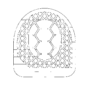
described in respect of Figure 7. This figure shows an inventive surgical
implant
with its particular tubular structure. The surgical implant is built by the
boundary
layer (1) surrounding the inner cavity (2). The boundary layer (1) has an
upper
plane (3A) that is jagged in the present example in order to generate a better
anchoring with the adjacent vertebral body, and a likewise jagged lower plane
(36). The boundary layer has a thickness of 4 mm. In ventral direction the
surgical
CA 02805614 2013-01-16
WO 2012/010327 24
PCT/EP2011/003715
implant is tapered in a pointed shape. In dorsal direction the surgical
implant has a
flattened back side (4). The vertical tubes (5) run from the upper plane (3A)
of the
boundary layer (1) in a straight and parallel manner to the lower plane (3B)
of the
boundary layer (1) through the boundary layer (1) to the lower plane (3B) of
the
boundary layer (1). These vertical tubes (5) have a hexagonal shape and a
diameter of 1.0 mm in its full size, i.e. if the vertical hexagonal tubes (5)
aren't cut
off, as may occur at the edges of the boundary layer (1). 60% to 80% of all
vertical
tubes have this full size, i.e. they aren't cut off at the edges of the
boundary layer
(1) and have aforesaid diameter. There are between 50 to 70 vertical tubes per
cm2 surface on the upper plane as well as on the lower plane. The wall
thickness
(6) of these vertical tubes amounts to 0.35 mm. The vertical tubes (5) are
interconnected by the horizontal tubes (7). The horizontal tubes (7) run in a
straight and parallel manner throughout the boundary layer (1). There are two
types of horizontal tubes (7), these tubes (7') running from the outer surface
(8) of
the boundary layer (1) to the inner surface (9) of the boundary layer (1), and
those
horizontal tubes (7") not running to and through the inner cavity (2) but only
through the boundary layer (1). As horizontal tubes (7') are denominated all
horizontal tubes (7) that run from the inner surface (9) of the boundary layer
(1) to
the outer surface (8) of the boundary layer (1). As horizontal tubes (7") are
denominated all horizontal tubes (7) that run from one side of the boundary
layer
(1) to the opposite side of the boundary layer (1) without crossing the inner
cavity
(2). The horizontal tubes (7) have a hexagonal shape and a diameter of 1.0 mm
in
their full size, i.e. when the horizontal hexagonal tubes (7) aren't cut off
at the
edges of the boundary layer (1). 96% of all horizontal tubes (7) have this
full size,
i.e. they aren't cut off at the edges of the boundary layer (1) and have this
diameter. There are between 40 and 90 horizontal tubes per cm2 outer surface
(8)
as well as per cm2 inner surface (9). The wall thickness (10) of these
horizontal
tubes is 0.35 mm.
Examples for such inventive surgical implants are in particular cages for
cervical,
thoracic or lumbar use (such as ALIF cages, PLIF cages and TLIF cages). The
inventive surgical implants are also known as interbody vertebral element,
implants for intersomatic fusion or implants for intercorporal vertebral
fusion. This
fusion can be carried out on natural vertebrae of the patient, artificial
(replaced)
vertebrae or a natural and an artificial vertebra. Mutatis mutandis this
applies also
if only parts of a natural vertebra have been replaced.
CA 02805614 2013-01-16
WO 2012/010327 25
PCT/EP2011/003715
The contact area with the bone, i.e. the upper plane as well as the lower
plane of
the boundary layer or the cage, doesn't have to be necessarily even, as in
conventional surgical implants of this kind. It may also have an asymmetrical
shape. It is also preferred that the vertical tubular structure extends to a
small
degree over the outer edge of the boundary layer in direction to the
respective
adjacent vertebral body. The portion of the vertical tubes extending beyond
the
upper plane or lower plane of the boundary layer may sink or force itself into
the
adjacent vertebral body, respectively. It thus causes an intended lesion of
the
surface of these vertebral bodies by which bone growth and blood flow are
stimulated in this area which leads to a better through growth of the implant.
Thus the inventive implant may have an even surface towards the adjacent
vertebral body on the upper plane as well as on the lower plane. However, it
is
preferred that this surface may be arched by instance, respectively that the
vertical tubes extend beyond the boundary layer and into the upper and/or
lower
vertebral body. The unevenness of the surface may amount from 0.1 mm to 3
mm, measured from the upper plane or the lower plane of the boundary layer,
respectively, to the maximal extension of the vertical tubular structure at
the
surface. Thus in these embodiments of the inventive implants a portion of the
vertical tubes doesn't end at the upper plane and/or lower plane of the
boundary
layer but extends beyond up to 3 mm maximally. .
The arrangement of the tubes and of the tubular structure preferentially has a
symmetric pattern. It should be noted that a randomly generated tubular
network,
as can be found for example in porous structures or sponges isn't suitable to
solve
the task of the present application because the capillary forces can't be used
in a
coordinated and reliable manner or are not even present. The same applies for
tubes that change their direction and/or their diameter abruptly or are
staggered or
are generated by a random sequence and/or shape of different layers of a
multilayer system for the main body of the implant. Such systems are
characterized in that the blood flow is increased only in certain parts of the
implants. Consequently, only those delimited parts will be well populated with
bone cells. It is also possible that there is only an island population
pattern in
these implants. In any case there will be no solid and homogenous through
growth
of the implant, as an entire through growth doesn't occur at all or only at a
very
slow rate. In the worst case this may even favour malpositions of the
patient's
spine caused by an uneven integration of the implant which would render
surgical
interventions indispensable.
CA 02805614 2013-01-16
WO 2012/010327 26
PCT/EP2011/003715
It should be kept in mind that the inventive implants provide a high porosity
but
also a huge surface area which is available for the adhesion and binding of
bone
cells so that new bone can grow through the implant very soon. In addition the
provided tubular structure makes use of capillary forces and also gives the
possibility to detect the degree, velocity and location of bone ingrowth and
bone
through-growth by standard X-ray spectroscopy or radiography.
It is understood that not the entire implant has to display the inventive
tubular
structure. It is preferred, however, that the vertical tubular structure
extends from
the upper plane of the boundary layer up to the lower plane of the boundary
layer
and that the horizontal tubes also extend from the outside of the boundary
layer to
the inner cavity or to the opposite side of the boundary layer, respectively.
Especially those implants that have continuous and substantially parallel
vertical
and horizontal tubes showed to be advantageous.
Further, the inventive honeycomb structure of the boundary layer of the
inventive
surgical implant combines simultaneously the features of good mechanical
stability and an optimal filling volume of the inner cavity so that a rapid
and stable
through growth of the implant with new bone tissue is effectuated while the
required bone material is reduced and thereby reducing the co-morbidity.
Bone tissue generally comprises three cell types, osteoblasts, osteocytes and
osteoclasts, whereby the developed bone also has a bone top layer of bone
lining
cells. The presence of blood is essential and needed for optimal bone
formation.
Ossification (or osteogenesis) is the process of incorporating or sedimenting
new
bone material by cells called osteoblasts.
It is synonymous to bone tissue
formation. There are two processes resulting in the formation of normal,
healthy
bone tissue: Intramembranous ossification is the direct incorporation of bone
into
the primitive connective tissue (mesenchyme), while endochondral ossification
involves cartilage as a precursor. Chondroblasts are the progenitor of
chondrocytes (which are mesenchymal stem cells) and can also differentiate
into
osteoblasts.
Endochondral ossification is an essential process during the
rudimentary formation of long bones, the growth of the length of long bones,
and
the natural healing of bone fractures.
CA 02805614 2013-01-16
WO 2012/010327 27
PCT/EP2011/003715
In the formation of bones osteoblasts, osteocytes and osteoclasts work
together.
Osteoblasts are bone-producing cells and responsible for building and
therefore
preserving the bone. Non-active osteoblasts on the bone surface are called
bone
lining cells.
Osteocytes are former osteoblasts that are incorporated into the
bone tissue by ossification. They provide for the preservation of the bone by
balancing bone resorption and bone formation. Osteoclasts are responsible for
the degradation of the bone.
Through them, the thickness of the bone is
determined and calcium and phosphate can be released from the bone. The
osteoblasts are the cells responsible for bone formation.
They develop from
undifferentiated mesenchymal cells, or chondroblasts. They attach themselves
to
bones in the form of dermal layers and indirectly form the basis for new bone
substance, the bone matrix, especially by excreting calcium phosphate and
calcium carbonate into the interstitial space. In this process they change to
a
scaffold of osteocytes no longer capable of dividing, which is slowly
mineralized
and filled with calcium.
The inventive tubular structure facilitates the influx of blood also to the
inner cavity
by using capillary forces. Therefore also osteoblasts are stimulated to
migrate in a
short period of time into the tubes and to the filled inner cavity. By this
mechanism
bone growth is promoted and thus the through growth of the implant with bone
tissue is improved and accelerated. This is a clear advantage over similar
implants
known in the art.
The inventive implant has the advantage over porous structures or sponges that
it
is hardly deformable, if at all, and is dimensionally stable, has a defined
shape
and surface and can be handled and implanted by conventional implantation
tools
without the risk to destroy or to damage the implant or its tubular structure
and
that X-ray beams can pass through the implant for radiography measurements.
In order to improve the adhesion of bone cells further the inner surfaces of
the
tubular structure(s) and to the outer surface of the implant and to the inner
surface
of the inner cavity the surface can be structured or roughened by, for
example,
any mechanical, chemical or physical roughening. To suppress the growth of
bacteria or other germs on the implant surface, it can be provided with
antibiotics
and the outer surface of the boundary layer or the cage and/or the surfaces of
the
tubular structure and the inner cavity for example can be provided with a drug
eluting coating, in which agents such as antibiotics are stored and can be
released
continuously.
CA 02805614 2013-01-16
WO 2012/010327 28
PCT/EP2011/003715
The inventive implants can be manufactured by standard techniques, for
example,
using laser technology and laser cutting procedures, rapid prototyping, laser
fusion, e.g. lasercusing or injection molding and therefore can assume in the
context of the described invention any shape.
Preferentially, the inventive implants are manufactured in one piece. They
consist
completely or at least to 90% of a metal or metal alloy, are not porous as
ceramics
for instance but have a defined interior tubular structure that stimulates the
blood
flow through the implant and by this generates optimal conditions for a
through
growth with new bone tissue. The tubular structure is not only vertical but
also
allows a blood flow through the horizontal tubes, thus accelerating the
through
growth and the conversion from an implant to natural bone tissue. This also
holds
true for inventive implants made of polymers such as carbon fibers, polyether
ketones PEEK [poly(ether ether ketone)], PEEEK [poly(ether ether ketone ether
ketone)], PEEKEK [poly(ether keton ketone)] or PEKK [poly(ether ether ether
ketone)]. The polymeric material is preferably radiolucent.
The polymeric
material is preferably radiolucent characterized by a Hounsfield unit 5 400.
The inventive surgical implants are preferentially manufactured as one piece
or
part or monolithic and don't consist of several parts nor are manufactured out
of
several pieces. The term "one-part surgical implant" or "one-part implant"
refers to
the implant only and not to any fixation means. For example, such one-part
implants can be fixated with screws to the adjacent vertebral body (-ies).
Such
fixation means are not covered by the term "one-part" and are regarded as
accessory to the implant. The same applies for implantation tools. Further,
natural
materials such as natural bone material or bone cement or bone replacement
material for the inventive surgical implant are not part of the implant.
Therefore the
inventive surgical implants are preferentially one-part, one-piece or
monolithic,
according to this definition. Embodiments with two parts are still possible,
but the
maximum is three parts, preferentially there are not more than two parts. In
these
embodiments the additional parts generally are fixation means such as
removable
plated for fixation screws or fixation hooks or fixation clamps or fixation
claws or
the like. In most cases, these additional parts are optional for the inventive
implant.
The inventive implants are not assembled according to a modular design or out
of
several parts or pieces or plates. Empirically, there are often difficulties
to connect
CA 02805614 2013-01-16
WO 2012/010327 29
PCT/EP2011/003715
or to join these different parts without a special effort, or they remain
movable one
against the other in a translational or rotational or sliding way. The
boundary layer
building the implant has a defined shape that is not modified after
implantation.
The inventive surgical implant is not smooth, or plastic or deformable.
Neither it is
spongy or porous.
In another embodiment of the invention the boundary layer builds the surgical
implant itself. These embodiments are directed to implants with an inner
cavity or
inner volume which can be filled with bone grafts or fine bone chips or bone
replacement material or bone cement or artificial bone material. Boundary
layer
refers herein to the wall around the inner cavity. It can also be defined as a
surrounding part. The inventive tubular structure of the boundary layer of
this
embodiment corresponds to the previously described embodiment. By this
structure an inner cavity is formed not in the classical sense that it is
surrounded
on all sides by the boundary layer. Instead this cavity describes a space left
open
by the boundary layer that can be described as a continuous opening between
the
walls of the boundary layer. The boundary layer is closed, so it surrounds
this
cavity by 360 and builds only the side walls delimiting this cavity. Towards
the
upper plane and the lower plane this cavity is open. Thus this cavity
essentially is
a continuous recess extending from the upper plane to the lower plane of the
implant. The body of the surgical implant is entirely built by the boundary
layer.
The boundary layer has an inner surface facing the cavity, an outer surface
facing
the exterior space and an own upper plane and lower plane shaped by the upper
plane and the lower plane of the boundary layer. The interior structure of the
boundary layer is built by substantially vertical and substantially horizontal
tubes
throughout the boundary layer. When contemplated from the outside the implant
looks as being provided with "holes", wherein each "hole" represents the
ending of
such a tube.
In this embodiment the boundary layer doesn't run circularly around the cavity
but
may have the shape of a heart (see Fig. 6), of a boat (see Fig. 5), or of a
rather
rectangular body (see Fig. 7). The space delimited by the boundary layer is
defined as cavity or inner cavity. This cavity is open to the upper side and
to the
lower side of the surgical implant.
The present invention relates also to a surgical implant consisting of a
metal, a
polymeric material or a metal alloy, wherein the implant has an upper plane
for
contacting an upper vertebral body and a lower plane for contacting a lower
CA 02805614 2013-01-16
WO 2012/010327 30
PCT/EP2011/003715
vertebral body and a boundary layer around the implant between the upper plane
and the lower plane and at least one recess opening to at least one cavity
within
the implant and a tubular structure extending from the upper plane around the
at
least one cavity through the lower plane, wherein the tubular structure is
formed
by a plurality of tubes and the at least one cavity is fillable with bone
grafts or fine
bone chips or bone replacement material or bone cement or artificial bone
material.
The two planes (upper plane and lower plane) are also called horizontal
planes.
The boundary layer is also referred to as vertical surface. The boundary layer
has an outer surface which is the surface around the implant between the upper
plane and the lower plane. In the boundary layer there is at least one recess
opening of the cavity and preferably the boundary layer has two opposite
recess
openings of the cavity. The cavity is surrounded by the inner surface of the
boundary layer. Tubular structure and the tubes of the tubular structure end
on
the inner surface of the cavity and do not cross the cavity when filled with
bone
replacement material or artificial bone material. In case the at least one
cavity is
filled with bone cement, the implant will comprise a solid core of bone cement
and
the new bone will grow from the upper vertebral body through the tubes of the
tubular structure to the bone cement core and from the lower vertebral body
through the tubes of the tubular structure to the bone cement core from the
other
side. In case the at least one cavity is filled with bone replacement material
or
especially with artificial bone material, the new bone will grow into the
implant from
the upper and the lower vertebral body through the tubes of the tubular
structure
and will convert the artificial bone material to new bone so that new bone
will also
be formed in the at least one cavity thereby bridging the two adjacent
vertebral
bodies.
In case the bone replacement material or artificial bone material is liquid or
fluid it
is preferably used together with a carrier or solid support such as particles
or
impregnated on a textile-like material.
Moreover it is preferred that the tubes of the tubular structure are parallel
to each
other or the tubes of the tubular structure are grouped into groups of
parallel
tubes.
Also preferred is that the tubes of the tubular structure extend along the
longitudinal axis of the spinal column.
CA 02805614 2013-01-16
WO 2012/010327 31
PCT/EP2011/003715
Furthermore it is preferred that the tubular structure is formed by tubes
running
along the longitudinal axis of the spinal column and tubes running
horizontally or
perpendicular to the tubes which run along the longitudinal axis of the spinal
column.
Preferably between 60% and 90% of the tubes forming the tubular structure end
at the cavity. The implant may comprise one, two, three, four, five or six
cavities.
Moreover it is preferred that at least one cavity is located in the middle of
the
implant having one opening in the upper plane or in the lower plane or in the
boundary layer or having two openings wherein one is in the upper plane and
the
other one in the lower plane or both are in the boundary layer.
The present invention relates also to a surgical implant consisting of a
polymeric
material, wherein the implant has an upper plane for contacting an upper
vertebral
body and a lower plane for contacting a lower vertebral body and a boundary
layer
around the implant between the upper plane and the lower plane and at least
one
recess opening to at least one cavity within the implant and a tubular
structure
extending from the upper plane around the at least one cavity through the
lower
plane, wherein the tubular structure is formed by a plurality of tubes and the
at
least one cavity is fillable with bone replacement material or bone cement or
artificial bone material.
Another embodiment of the present invention relates to a surgical implant for
replacing an intervertebral disc, wherein the body of the implant consists of
a
polymeric material and has two planes for contacting the two adjacent
vertebral
bodies, respectively, a boundary layer and a scaffold zone, wherein the
scaffold
zone is formed by a plurality of tubular structures and encompasses a cavity
fillable with bone replacement material or bone cement. The tubes of the
tubular
structures preferably extend parallel to one another along the longitudinal
axis of
the spinal column and the tubes of the tubular structures are preferably
interconnected by openings.
The present invention also relates to an intervertebral implant, wherein the
body of
the implant consists of a polymeric material and has two planes for contacting
two
adjacent vertebral bodies, a scaffold zone and a boundary layer which partly
surrounds the scaffold zone and wherein the scaffold zone encompasses a cavity
fillable with bone replacement material or bone cement around which a
plurality of
CA 02805614 2013-01-16
WO 2012/010327 32
PCT/EP2011/003715
vertical tubular structures preferably running along the longitudinal axis of
the
spinal column and a plurality of horizontal tubes running horizontally from
one side
to the opposite side of the implant preferably along the transversal axis of
the
body or preferably in a plane perpendicular to the longitudinal axis of the
spinal
column.
The present invention also relates to an intervertebral implant, wherein the
body of
the implant consists of a non-metallic material and has two planes each of
them
contacting an adjacent vertebral body, a scaffold zone and a boundary layer
which
partly surrounds the scaffold zone and wherein the scaffold zone is formed by
a
cavity fillable with bone replacement material or bone cement and a plurality
of
vertical tubes preferably running along the longitudinal axis of the spinal
column
and optionally a plurality of horizontal tubes running horizontally from one
side to
the opposite side of the implant preferably along the transversal axis of the
body
or preferably in a plane perpendicular to the longitudinal axis of the spinal
column.
It is preferred that the tubes will end when meeting the cavity filled with
the bone
replace material. Thus in the preferred embodiments these tubular structures
consisting of a plurality of tubes which are preferably parallel to each other
do not
cross the cavity. These dead-ends of the tubular structures which end in the
cavity
filled with bone replacement material or bone cement provide a good insertion
point or adhesion area for the bone cells because of the comparatively rough
surface of the bone replace material. This way these dead-ends become
germination centers for the continuous ossification of the tubular structures
and
thus of the implant.
As used herein the term "tubular structure" refers to the entirety of the
tubes while
also groups of tubular structures can be present which are formed of certain
numbers of tubes. The tubes form the tubular structure or groups of tubes
which
are preferably parallel to each other within one group form tubular structures
which extend around the at least one cavity in the implant.
The present invention relates also to a surgical implant, wherein the body of
the
implant consists of a polymeric material and has two planes for contacting the
adjacent vertebral body respectively, a scaffold zone and a boundary layer
which
partly surrounds the scaffold zone and wherein the scaffold zone is formed by
a
cavity fillable with bone replacement material or bone cement and a plurality
of
vertical tubes running along the longitudinal axis of the spinal column.
Preferably
CA 02805614 2013-01-16
WO 2012/010327 33
PCT/EP2011/003715
also a plurality of horizontal tubes are present running horizontally or
perpendicular to the vertical tubes through the implant.
The present invention relates further to an intervertebral implant, wherein
the body
of the implant consists of a polymeric material and has two planes for
contacting
two vertebral bodies, a scaffold zone and a boundary layer which partly
surrounds
the scaffold zone and wherein the scaffold zone is formed by a cavity fillable
with
bone replacement material and a plurality of vertical tubes running along the
longitudinal axis of the spinal column and being parallel to each other.
Preferably
also a plurality of horizontal tubes are present running horizontally from one
side
to the opposite side of the implant, with exception of the cavity area.
Furthermore the present invention relates to a surgical implant, wherein the
body
of the implant consists of a polymeric material and has two planes for
contacting
two adjacent vertebral bodies, a scaffold zone and a boundary layer which
partly
surrounds the scaffold zone and wherein the scaffold zone is formed by a
cavity
fillable with bone replacement material and a tubular structure consisting of
a
plurality of vertical tubes which extend in preferably straight lines from the
top of
the upper vertebral body contacting surface to the opposite and being
preferably
parallel to each other. Preferably a plurality of horizontal tubes are
present
running horizontally straight through the implant, with exception of the
cavity area.
The horizontal tubes preferably connect the vertical tubes with each other.
Moreover it is also possible that the horizontal tubes are connected with each
other through holes or openings or recesses between adjacent horizontal tubes.
The present invention relates to bone-joining or bone-bridging surgical
implants in
the form of artificial discs consisting of a polymeric material, wherein the
artificial
surgical implant exhibits at least one bone-contacting plane and a scaffold
zone
consisting of a cavity fillable with bone replacement material and a plurality
of
tubes with defined cross-sectional areas or radii and these tubes of the
surgical
implant are interconnected so that a three-dimensional network of tubes is
formed.
Such a three-dimensional network is also referred to as tubular structure.
The present invention further relates to bone-joining or bone-bridging
surgical
implants in the form of artificial discs consisting of a polymeric material,
wherein
the artificial disc implant exhibits at least one bone-contacting plane and a
scaffold
zone consisting of a cavity fillable with bone replacement material or bone
cement
CA 02805614 2013-01-16
WO 2012/010327 34
PCT/EP2011/003715
and a plurality of tubes with defined cross-sectional areas or radii and these
tubes
of the scaffold zone are interconnected so that a three-dimensional network of
tubes is formed, with exception of the cavity area.
It was surprisingly found that bone-joining or bone-bridging surgical implants
consisting of a preferably radiolucent polymeric material grow together
particularly
well with the contacted bone, when the surface of the implant is not smooth or
not
rough or not porous, but has a scaffold zone, consisting of at least one
cavity filled
or all filled with bone replacement material or bone cement and a plurality of
tubes
forming a tubular structure which surrounds the filled at least one cavity.
Preferably the tubes are interconnected and form a defined structure, the
tubular
structure. Concerning the tubular structures it is important that added
together at
least a total of 20% of all vertical and horizontal tubes run from one side of
the
implant through the implant to the other side of the implant. The vertical and
horizontal tubes running through the implant suck or pull blood by capillary
forces
into the vertical and horizontal tubes and thereby into the complete tubular
structure, which promotes and accelerates new bone formation within the bone-
joining or bone-bridging implant.
The present application also relates to a method for treatment of spinal
column
disorders which comprises the step of implanting a surgical implant as
described
before into the intervertebral space of a patient in need thereof.
A patient as used herein refers to any mammal including humans suffering from
a
spinal column disorder. However, it is preferred that the patient is a human.
The term "bone-joining" or "bone-bridging" implies that the implant is in
direct
contact with a bone. That means at least a part of the plane of the surgical
implant
touches a bone.
The inventive scaffold zone preferably starts at the bone-contacting plane of
the
implant, so that the openings of the vertical tubes are facing the bone, i.e.
the
upper openings are facing the upper contacted vertebral body and the lower
openings of the tubes the lower vertebral body. The vertical tubes, the
optionally
present horizontal tubes, the openings between the tubes as well as the cavity
filled with bone replacement material form the scaffold zone.
CA 02805614 2013-01-16
WO 2012/010327 35
PCT/EP2011/003715
Each vertical tube is preferably connected by a horizontal tube with at least
two
openings with the adjacent vertical tubes.
The term "radiolucent polymeric material" refers to anything that permits the
penetration and passage of X-rays or other forms of radiation. More
specifically,
the term "radiolucent polymeric material" as used herein refers to any
material that
does not impair the ability to distinguish by X-ray exposures between bones
and
specifically new grown bones and the material of the implant. Such
"radiolucent
material" can be characterized further by the Hounsfield scale, which is a
quantitative scale for describing radiodensity. The Hounsfield unit (HU) scale
is a
linear transformation of the original linear attenuation coefficient
measurement into
one in which the radiodensity of distilled water at standard pressure and
temperature (SIP) is defined as zero Hounsfield units (HU), while the
radiodensity
of air at SIP is defined as -1000 HU. For a material X with linear attenuation
coefficient px, the corresponding HU value is therefore given by
HU = lix Ilwater X 1000
Pwater ¨ Pair
where u
.--water and pair are the linear attenuation coefficients of water and air,
respectively. Thus, a change of one Hounsfield unit (HU) represents a change
of
0.1% of the attenuation coefficient of water since the attenuation coefficient
of air
is nearly zero. It is the definition for CT scanners that are calibrated
with
reference to water. Exemplary values are -1000 for air, 0 for water, and >400
for
bones.
Thus the intervertebral implant according to the invention is characterized in
that
the body of the implant consists preferably substantially of a radiolucent
polymeric
material with a Hounsfield unit 5 400, preferably 5 300, more preferably 5
200,
even more preferably 5 100, and most preferably 5 0. Such materials include
but
are not restricted to fiber-reinforced plastics (glass / carbon fibers with a
corresponding matrix), polyether ketones (PEEK -poly ether ether ketone,
PEEKEK ¨ poly ether ether ketone ether ketone, PEKK ¨ poly ether keton ketone;
PEEEK ¨ poly ether ether ether ketone) or polymer materials in general.
The body of the surgical implant according to the invention substantially
consists
of one or more radiolucent polymeric materials, which means that the implant
can
comprise one or more radio-opaque materials e.g. in the form of marking points
so
that the proportions of the implant can be seen more easier in X-ray
exposures, as
CA 02805614 2013-01-16
WO 2012/010327 36
PCT/EP2011/003715
long as the ability to distinguish between bones and specifically new grown
bones
by radiography is not impaired. Thus in a preferred embodiment the body of the
implant consists to 80% of one or more radiolucent polymeric materials, more
preferably to 90% and most preferably to .?. 95%.
"The body of the implant" as used herein refers to the structures consisting
of the
boundary layer, the scaffold zone, the tubular structures and the cavity, but
specifically does not refer to the filling of the cavity.
The tubular structures and the cavity fillable with bone replacement material
inside
of the cage or the surgical implant is used for direct stimulation of bone
growth
and less for the stabilization of the entire implant. The mechanical stability
of the
surgical implant, the cage, is conferred by the boundary layer which
completely or
partly surrounds the implant, which is designed to withstand the high
pressures of
the spine and to prevent the sinking of the implant into the vertebral bone,
so that
the distance between two vertebral bodies, defined by the height of the
boundary
layer or the height of the implant respectively, can be maintained.
As already discussed above bone cells do not adhere very well to non-metallic
bone-joining or bone-bridging implants that are made of polymeric materials.
However, if polymeric materials are preferred, the ingrowth of new bone can be
monitored by X-ray exposures.
Surprisingly, it was found that the inventive scaffold zone consisting of a
plurality
of tubular structures and a cavity fillable with bone replacement material
promotes
the ingrowth and adhesion of bone cells to implants substantially consisting
of
polymeric material. Each of the individual tubes running through the implant
can
suck or pull blood and cells by capillary forces into the whole tubular
structure and
thereby into the complete implant which promotes and accelerates formation of
new bone within the bone-joining or bone-bridging implant.
As used herein the term "scaffold zone" refers to one or more tubular
structures
like two sets of parallel tubes, while the tubular structure consists of a
plurality of
tubes.
The cavity or cavities fillable with bone replacement material is essential to
the
invention and prevents that the bone cells just flow through the implant
without
attaching themselves. After the first bone cells attach themselves to the bone
replacement material, they can start proliferating or they recruit further
cells to the
CA 02805614 2013-01-16
WO 2012/010327 37
PCT/EP2011/003715
inside of the implant, respectively the tubular structure(s). Once initial
cells are
attached it is easier for further cells to attach and the implant is grown
through
from the inside.
The cavity can be filled with any bone replacement material or bone cement or
artificial bone material suited for the use in a patient or with patient's own
bone
grafts or fine bone chips taken form patient's hip. Bone replacement material
as
used herein is a generic term comprising three major groups of materials:
The first group comprises polymeric bioresorbable materials. Suitable examples
according to the invention are (I-lactic acid) [PLLA], poly(d l-lactic acid)
[PDLLA],
poly(glycolic acid) [PGA], poly(lactic-co-glycolic acid) [PLGA],
poly(paradioxanone)
[PDS], poly(dl-glycolic acid) [PDLGA], poly(propylene fumarate) [PPF], oligo
(PEG
fumerate) [OPF], poly(ethyleneglycol) [PEG], poly(caprolactone) [PCA],
poly(hydroxybutyrate) [PHB], poly(hydroxy valerate) [PHV], poly (SA-HDA
anhydride), poly(orthoesters), poly(phosphazenes), and copolymers of di-lactic
acid and dl-glycolic acid.
The advantage of such bioresorbable materials is that the initial attachment
of
bone cells including proliferation and recruitment of further cells is
promoted by
the bioresorbable material, but afterwards when the bone replacement material
is
no longer needed it gets degraded giving way for further growth of bones
through
the implant. The rate of degradation doesn't have to be tightly controlled
because
the mechanical stability of the implant is not dependent on the bone
replacement
material. A subgroup may contain mineral blocks of animal origin.
The bone replacement material can be enriched with active substances like
antibiotics, growth factors, adhesion molecules, silver, substances that
promote
the adhesion of bone cells and others.
Thus according to the invention any
osteoinductive substance can be used. In a preferred embodiment fibroblast
growth factor (FGF) is added. Particularly preferred is the use of rhBMP-2
(Infuse), a recombinant human bone morphogenetic protein capable of initiating
bone growth in specific, targeted areas of the spine.
A second group comprises bioresorbable materials as listed for the first group
or
other biocompatible materials such as ceramic materials, enriched with human
mesenchymal stem cells or other cells suitable as a germination point for the
desired ossification. These mesenchymal stem cells can differentiate into bone
CA 02805614 2013-01-16
WO 2012/010327 38
PCT/EP2011/003715
cells either by themselves or by addition of a suitable agent. These
differentiation
procedures are known in the art.
The third group comprises bone cement. Bone cement are a form of bone
replacement material. It is often provided as two-component materials. Bone
cement consists of a powder (i.e., pre-polymerized PMMA and or PMMA or MMA
co-polymer beads and or amorphous powder, radio-opacifier, initiator) and a
liquid
(MMA monomer, stabilizer, inhibitor). The two components are mixed and a free
radical polymerization of the monomers occurs when the initiator is mixed with
the
accelerator. The bone cement viscosity changes over time from a runny liquid
into a dough like state that can be safely applied and then finally hardens
into
solid hardened material.
According to the invention also combinations of materials from the
aforementioned
groups can be used.
The cavity inside the scaffold zone or inside the implant can be filled with
bone
replacement material, bone cement or artifical bone material before the
implantation of the inventive implant. Thus the implant can be prefabricated
and
commercialized already with a filling of bone replacement material inside the
cavity of the surgical implant.
In a further embodiment the filling of the cavity of the inventive implant
with bone
replacement material takes place immediately before implantation of the
implant.
In a preferred embodiment the filling of the cavity of the inventive implant
with
bone replacement material, bone cement or artifical bone material takes place
after implantation of the implant, i.e. inside the body. It is preferred that
the filling
occurs by means of microinvasive tools. However, normally the at least one
cavity is filled by the physician just before the implantation.
Suitable for this purpose are all conventional tools for filling cavities,
such as
syringes, injection systems, catheter systems, tubing systems, pumping
systems,
jet systems, portioning systems, spoons, spatulas, pipettes, crushers,
compactors,
squeezing machines.
Thus the present application refers also to a method of loading the surgical
implant, comprising the following step:
CA 02805614 2013-01-16
WO 2012/010327 39
PCT/EP2011/003715
a) Filling the at least one cavity of the surgical implant with a bone
replacement
material or bone cement or artifical bone material.
According to this method the bone replacement material can be selected from
the
group comprising polymeric bioresorbable materials, polymeric bioresorbable
materials containing an osteoinductive agent, bioresorbable materials
containing
bone-forming cells and artifical bone material. The term "artifical bone
material"
as used herein is a subgroup of the bone replacement material and refers to
any
material which can be conferted to new bone under physiologic condition.
For this method it is preferred that the bone replacement material is a
polymeric
bioresorbable material containing an osteoinductive agent.
For this method it is even more preferred that this osteoinductive agent is
rhBMP-
2.
The filling of the inner cavity with at least one of the aforementioned
materials thus
serves for creating surfaces to which bone cells can adhere. Further, this
filling
serves for reducing the volume of the inner cavity in order to promote the
through
growth. This desired through growth and overgrowth is significantly improved
by
the special tubular structure running through the implant. This tubular
structure
enables the blood to run through the upper plane to the lower plane and
because
of the substantially horizontal tubes also from the outside of the implant to
the
inner cavity and vice versa. Thus bone cells can settle around the implant. By
the
inventive tubular structure bone cells can reach via the blood flow any site
inside
the implant so that the formation of new bone tissue doesn't occur only from
the
upper plane and/or the lower plane towards the center but also from the center
of
the implant towards the periphery.
Thus the present invention also refers to a kit which provides all materials
necessary for such an implantation. This kit comprises at least one inventive
surgical implant; and bone replacement material and/or bone cement and/or
artificial bone material suitable to fill the cavity of the surgical implant.
Such a kit
comprises the surgical implant and bone replacement material and/or bone
cement and/or artificial bone material in an amount sufficient to fill the at
least one
cavity of the surgical implant. Moreover such a kit may also comprise a
carrier or
a solid support which can be loaded with the bone replacement material or
artifical
bone material or may comprise a textile-like material which can be impregnated
with the bone replacement material or artifical bone material. Moreover this
kit
CA 02805614 2013-01-16
WO 2012/010327 40
PCT/EP2011/003715
may comprise an inplantation device for inserting the inventive implant into
the
spinal column of th patient.
In another embodiment the kit comprises additionally at least one tool for
filling the
bone replacement material into the cavity of the surgical implant.
All bone replacement materials listed above can be used with different degrees
of
viscosity. Thus the bone replacement material can set immediately after
filling
into the cavity, it can set after some time, it can set only on application of
an
external energy source such as UV hardening, on cooling, on heating, or it may
even remain in a semifluid or plastic state throughout the life time of the
implant,
respectively until being resorbed by the organism.
Thus according to the invention also substances can be added to the bone
replacement material which allow for a controlled setting upon application of
an
external energy source.
It is known to a skilled person how to modify the viscosity and thus in most
cases
the setting conditions of the bone replacement material. One way to modify the
viscosity consists in adding softening or hardening substances. One preferred
additive for increasing the viscosity of the bone replacement material is
polyvinyl
pyrrolidone. It can be added in amounts up to 1 or 2% per volume.
The term viscosity refers to the dynamic viscosity [i]:
kg Ns
[771 = ________________________________ = Pa = s =
= s m-
Typical viscosity values for the bone replacement material (while not set)
range
from aqueous solutions (ca. 1 mPas), olive oil: 102 mPas), honey (103 mPas),
syrup (105 mPas) to bitumen (109 mPas). It is understood that the viscosity
changes while setting or hardening.
The boundary layer which surrounds the implant gradually loses its supportive
function ever the more the scaffold zone is grown through with bones.
Therefore
a fast and easy evaluation of the bone structure growing in the scaffold zone
is
desirable as a long-term stability is only obtained if the scaffold zone is
grown
through as completely as possible with endogenous bone cells.
CA 02805614 2013-01-16
WO 2012/010327 41
PCT/EP2011/003715
Fig. 1 shows a top view of an inner cross section of an inventive surgical
implant.
In this embodiment the cavity is circular, but the cavity can have any desired
shape and proportion.
The cavity is preferably connected to most of the
surrounding tubes and thereby with the surrounding tubular structure(s).
Moreover, it is preferred that the tubes are arranged in a way that all tubes
are
interconnected, i.e. the entire tube-type structure could theoretically be
filled
through one opening of one tube with liquid such as blood. So preferably a
three-
dimensional interconnectivity of the entire structure is created.
The cavity can reach from one side of the implant to the other side of the
implant.
It is also possible that a part of the boundary layer is breached and the
cavity is
not completely enclosed by the cage material. The cavity can be split in two
or
more cavities of different size and proportions. Thus it is not necessary that
the
cavity is contiguous, but it is also possible that two or more cavities are
filled with
the same or different bone replacement material and are traversed by tubes or
not
independently from each other. Thus if two or more cavities exist, those
cavities
are all independent from each other regarding their properties, size, bone
replacement material filling, traversing of tubes, enrichment with active
substances
and others.
Consequently, in a further embodiment the intervertebral implant
comprises more than one cavity.
The design of the tubes and the cavity themselves is not essential to the
invention,
but their presence. It is obvious to a skilled person, that too many openings
and
especially size and proportions of the cavity can affect the stability of the
implant,
so that a skilled person knows how to determine the number, size, location and
proportions of the openings and of the cavity depending on the type of the
implant.
In order to improve the adhesion of bone cells further the inner surfaces of
the
tubular structure(s) and especially the surface of the bone replacement
material
can be structured by, for example, any mechanical, chemical or physical
roughening. To suppress the growth of bacteria or other germs on the implant
surface, it can be provided with antibiotics and the outer surface of the
boundary
layer for example can be provided with a drug eluting coating, in which agents
such as antibiotics are stored and can be released continuously.
At the posterior or anterior side of the implant a centrally round recess may
be
located which serves to hold an implantation tool during implantation. This
recess
can penetrate the boundary layer (Fig. 3B) so that directly behind the recess
the
CA 02805614 2013-01-16
WO 2012/010327 42
PCT/EP2011/003715
tubular structure starts. In a preferred embodiment the recess penetrates the
boundary layer and directly behind the recess starts the cavity. This way the
cavity can be filled conveniently with the bone replacement material.
The cavity can be filled either before insertion or after insertion of the
implant in
the human body. The cavity does not have to be filled completely and can also
be filled only partially with bone replacement material. The surface of the
bone
replacement material can be structured in any way to enlarge the available
surface area.
The inventive implants or cages are preferably made of one piece and have a
defined scaffold structure, which supports the blood flow and a cavity filled
with
bone replacement material thus creating the best possible conditions for
endogenous bone growth and have a boundary layer which is responsible for the
stability at least as long as the newly formed bone cannot yet take over this
function.
The term "one-piece surgical implants" refers only to the implant itself and
not to
any fasteners. Such implants can be screwed for example into the adjacent
vertebral bodies. The used fasteners, for example screws are not taken into
account when using the term "one-piece" and are referred to as accessories to
the
inventive surgical implant as well as the implantation tool. The inventive
implants
are thus made in accordance with this definition preferably in one-piece. Two-
piece embodiments are also possible, wherein the inventive implants are made
up
of maximal three pieces, preferably of not more than two pieces, whereby the
other parts generally relate to intended attachment means for the implant such
as
removable panels for mounting screws or hooks or fastening nails or the like,
which usually are optional for the inventive implants.
In bone-joining or bone-bridging implants of the spine area as well as with
the
inventive implants, the contact planes of the implants are generally flat to
the
respective bone.
The contact planes of the cage is understood to be the surface, which comes
into
contact with the overlying vertebral body and the opposite surface of the
cage,
which comes into contact with the underlying vertebral body.
But the contact plane with the bone has not to be designed flat, as is the
case with
the intervertebral implants of the prior art, but can also have an
asymmetrical
CA 02805614 2013-01-16
WO 2012/010327 43
PCT/EP2011/003715
form, as can be seen in Fig. 5. It is certainly more preferable, when the
inner
tubular structure extends slightly over the boundary layer in the direction of
the
overlying vertebral body as well as in the direction of the underlying
vertebral body
as will be described below in more detail. The part of the inner tubular
structure
extending over the boundary layer sinks or presses in the overlying or
underlying
vertebral body respectively and thus leads to an intended injury of the
surface of
these two vertebral bodies, whereby the growth of bones and the blood flow is
further increased.
It isn't mandatory either that all vertical tubes start on the bone-contacting
surface,
i.e. in direct contact with the bone. Up to 30%, preferably up to 20% of all
vertical
tubes, can also start in one area of the implant that is not in direct contact
with the
bone, i.e. preferably these tubes start lower than or below the bone-
contacting
plane.
Furthermore, it is essential to the invention that the tubes of the inner
tubular
structure are interconnected. The vertical tubes are connected through
the
horizontal tubes and optionally in additional through openings while the
horizontal
tubes can optionally connect with each other through openings, wherein each
horizontal tube has preferably at least one opening to an adjacent horizontal
tube.
As already described above the entire tubular structure could theoretically be
filled
through one opening of one tube with liquid. However, to achieve the best
result
it is preferred that at least 20%, preferably 30%, more preferably 40%, even
more
preferably 50% of the tubes open into the cavity. This way it is ensured that
enough blood and bone cells come into contact with the bone replacement
material.
Furthermore, implants according to the invention are preferred where the
honeycomb structure, i.e. the inner tubular structure, rises slightly over the
essentially flat bone-contacting plane. Especially, if the honeycomb structure
of
the implant protrudes over a border or solid frame or boundary layer, the
advantage of a high surface friction and therefore a very good anchorage is
given.
At the same time the low thickness of the honeycomb walls gives rise to the
possibility of mechanical movements which promotes growth stimulation of the
bone.
CA 02805614 2013-01-16
WO 2012/010327 44
PCT/EP2011/003715
Moreover, it is preferred that the openings in the inner tubular structure are
arranged in such a way that the entire structure permits micro-movements,
preferably friction-movements. Such movements are possible when the single
vertical tubes are connected by wedge-shaped longitudinal cuts in the lateral
wall
areas along the longitudinal axis of the vertical tubes. Thus, the individual
tube
walls can be shifted against each other according to the thickness of the
wedge-
shaped openings, so that micro-movements are possible.
As outlined above it is preferred for the majority of embodiments that the
inventive
surgical implant is made of a metallic material to allow a monitoring of the
ingrowth
of bone cells into the implant via x-ray spectrometry and radiography through
the
horizontal tubes.
Surprisingly, it showed that it is feasible by the inventive implants to
monitor the
through growth of the implant by means of x-rays via the horizontal tubes.
Herein, a radiography is taken from the implant, respectively the patient, in
such
an angle so that a fraction of the x-ray beams passes through the horizontal
tubes
or at least a group of horizontal tubes. Therefore it is essential that the
horizontal
tubes crossing the inner cavity take a straight way through the anterior
boundary
layer as well as through the posterior boundary layer so that an x-ray beam
can
pass through the entire implant via a single horizontal tube without being
refracted
and not only through the anterior part or the posterior part of the boundary
layer.
In the latter case the beam would eventually end on the opposite side of the
inner
cavity (i.e. on the opposite inner wall of the boundary layer) on solid
material which
is again radiopaque, thus counteracting the x-ray monitoring. In case the
implant
has an inner cavity which is filled with bone grafts or fine bone chips or
bone
replacement material or bone cement or artificial bone material, the X-ray
spectrum or the radiography can be performed in a way that the measurement is
made through the horizontal tubes (7") which do not run through the inner
cavity.
The presence of more than one group of parallel horizontal tubes allows for
taking
radiographies from different angles by performing the radiography through the
respective groups of horizontal tubes. Herein, the lumen of the tubes appears
dark
on the radiography, as long as no bone has been formed inside these tubes. As
soon as the ossification starts the interior of the tubes will appear grayish,
according to the progress.
The solid and radiopaque portions of the implant
appear in white. Therefore it is possible to monitor the through growth via
the
horizontal tubes.
Radiographies can be taken from different angles and
CA 02805614 2013-01-16
WO 2012/010327 45
PCT/EP2011/003715
combined in order to create an overview of the progress of ossification. The
differentiation between hollow tubes, ossified tubes and solid material is
straight
forward in general. Figure 8 shows the inventive surgical implant from a
lateral
view. The white sections shows the radiopaque material of the surgical
implant,
i.e. of the boundary layer. The dark or black sections are the horizontal
tubes
through which the x-ray beams could pass and expose the x-ray film at the
other
end of the implant. When new bone tissue has been built inside the tubes they
appear grayish since the x-rays can't pass anymore freely.
Bone is not as
radiopaque as the metal of the boundary layer.
The inventive surgical implants have an inner cavity that can be filled with
bone
replacement material, bone cement or autologous bone chips. According to the
indication and to the preferences of the physician different fillings of this
cavity are
favored. If the cavity is filled with bone replacement material then this bone
replacement material should preferably not contain an opacifier. Only without
an
opacifier the newly built bone tissue can be differentiated from the bone
replacement material. Herein, a radiography is taken through the horizontal
tubes
in which the bone replacement material without an opacifier appears dark, as
being widely x-ray transparent, and the newly built bone appears light gray.
So the
current degree of ossification of the implant can be monitored. If the
physician
uses cancellous bone material for filling the cavity this cancellous bone
material
appears dark to dark gray via the horizontal tubes, as the cancellous bone
mass is
widely x-ray transparent. New bone formed in the inner cavity of the implant,
however, then appears light to light gray and thus can be differentiated from
the
cancellous bone mass. If the physician uses, however, cortical bone for
filling the
cavity of the surgical implant the cortical bone appears light on
radiographies
taken via the horizontal tubes and thus can't be differentiated from newly
built
= bone tissue. In order to detect the through growth of the inventive
surgical implant
by means of x-rays when the inner cavity is filled with cortical bone
material, the
inventive surgical implants have two types (7' and 7") of horizontal tubes
(7). One
species of horizontal tubes (7') runs through the boundary layer and exits on
the
interior surface of the boundary layer. The other species (7") runs
exclusively
through the boundary layer and does not cross the inner cavity. They exit at
the
opposite side of the boundary layer.
Preferentially, this type of tubes has no
direct opening or direct cross-connection with the inner cavity. In Figure 7
such
horizontal tubes (7") are shown that are running through the tip of the
implant
towards the back side of the boundary layer. When filling the inner cavity
this tube
species remains free through their entire length so that it can be determined
by
CA 02805614 2013-01-16
WO 2012/010327 46
PCT/EP2011/003715
radiography to which degree a through growth of these tubes has occurred. This
advantage isn't offered by any conventional metal cage. It is important
for
radiographies that the x-ray device is positioned in such a way that the
radiography can be taken through the tubes. This isn't a technical challenge
anymore nowadays. On these radiographies radiopaque materials such as the
metal of the surgical implant appear light to white, newly built bone and
cortical
bone appears light gray or light gray to light, cancellous bone and bone
replacement material dark gray or dark gray to dark and free tubes allowing an
unimpeded passage of the x-ray beam appear dark to black.
Therefore according to the invention the surgical implant can be also made of
metal. This applies in particular for the scaffold zone with the tubular
structure.
According to the invention also hybrid implants made of a polymeric material
and
metal can be used. In these embodiments the percentage of weight of metal
versus polymer can range between 0.01% and 99.99%, preferably from 0.1% to
99%, more preferably from 1% to 90% and most preferably 30% to 70%. For
these embodiments all specifications on structures, materials, coatings, sizes
and
combinations with therapeutic agents made for the polymeric embodiments apply
in the same manner.
The implants of the present invention and the detection of new bone formation
within the implants of the present invention can be shown best in Figures 8,
11,
12, 13, and 14.
Figures 8 and 13 are the radiographs of two implants of the present invention
with
empty horizontal tubes and consequently also with empty vertical tubes. The
horizontal tubes are displayed dark or black since the X-ray beams can freely
pass
through the horizontal tubes. The metal of the cage is radiopaque and appears
white or very light.
Figure 11 is a radiograph of the cage of Fig. 8 which is almost completely
filled
with new bone. The new bone within the horizontal tubes appear light gray and
can be clearly distinguished from the radiopaque metal material of the cage.
It
has to be kept in mind that the physician does normally have a radiograph of
the
empty cage like shown in Fig. 8 and thus clearly knows the size, number and
location of the single horizontal tubes. In the radiograph of Fig. 11 it seems
that
only two horizontal tubes are not completely filled with new bone. This is the
tube
almost in the middle of the cage and the tube in the second column from the
right
side of the cage and in the middle of that column. These two horizontal tubes
are
still displayed dark so that they seem to be still open.
CA 02805614 2013-01-16
WO 2012/010327 47
PCT/EP2011/003715
Fig. 12 is a radiograph of another inventive cage where the bone has just
started
to grow through that cage from the top and the bottom towards the center of
the
cage. From the radiograph of Fig. 12 it is evident that only the middle of the
upper first row of horizontal tubes and the lowest row of horizontal tubes is
filled
with new bone while all other tubes are still in black which indicates that
they are
still empty. After two to three weeks after implantation such a radiograph can
be
expected.
Fig. 13 is a radiograph of another inventive cage where none of the horizontal
tubes is filled neither with new bone nor with bone grafts or fine bone chips
or
bone replacement material or bone cement or artificial bone material. All
horizontal tubes are displayed in black so that the X-ray beams could freely
pass
through these tubes.
Fig. 14 is a radiograph of the cage of Fig. 13 wherein a group of horizontal
tubes
is filled with a bone replacement material.
The bone replacement material
without an opacifier is widely x-ray transparent, but of course not completely
x-ray
transparent so that X-ray beams can almost unhindered pass through such
horizontal tubes filled with bone replacement material (without opacifier).
The
group of horizontal tubes filled with bone replacement material consists of
the
following horizontal tubes: fifth row from the bottom, tube 4 from the left
side;
fourth row from the bottom, tubes 3 and 4 from the left side; third row from
the
bottom, tubes 4 to 8 from the left side; second lowest row, all 8 tubes and
lowest row, all 8 tubes.
Thus the present inventive cages allow the detection of the degree, location
and
velocity of new bone formation and the conversion of bone replacement material
or artificial bone material or autologous bone chips or autologous bone grafts
or
cancellous bone mass into new bone, since all such materials have a
distinguishable x-ray transparency.
Only cortical bone mass used as filling material for the cage cannot be
distinguished from newly formed bone. However in such cases the degree and
velocity of the formation of new bone and the conversion of the cortical bone
mass
into new bone can be detected by X-ray spectra recorded through the horizontal
tubes (named herein as tubes 7") which do not cross the inner cavity and which
do not have a direct opening to the inner cavity and which run straight
through the
boundary layer from one side to the other. These tubes (7") remain empty
although the inner cavity of the cage is filled with cortical bone mass. Thus
only
newly formed bone can close or seal these tubes (7") so that the appearance of
these horizontal tubes (7") indicates if new bone was formed therein are not
at the
time the radiograph was taken. If all such tubes (7") are filled with new bone
it
CA 02805614 2013-01-16
WO 2012/010327 48
PCT/EP2011/003715
can be concluded that the cortical bone mass within the inner cavity of the
cage
was completely or almost completely converted to new bone.
Suitable materials for the inventive cage implant are medical steal, titanium,
titanium oxide, chromium, vanadium, tungsten, zirconium, oxidized zirconium,
molybdenum, hafnium, gold, platinum, rhodium, niobium, lead, cobalt-chromium,
tantalum, as well as alloys of these metals and biodegradable materials such
as
magnesium, zinc, calcium, iron as well as polymeric materials such as fiber-
reinforced polymers (glass / carbon fibers in a suitable matrix) chitosan,
hepara,
polyhydroxybutyrate (PHB), polyglyceride, polylactide and copolymers thereof.
Suitable metals include, but are not limited to medical stainless steel,
titanium,
chromium, vanadium, tungsten, molybdenum, gold, magnesium, iron, zinc,
calcium, lithium, sodium, potassium, aluminium, scandium, zirconium, niobium,
tantalum, silicon, manganese, iron, cobalt, nickel, copper, zinc, gallium,
yttrium,
ruthenium, rhodium, palladium, silver, indium, tin, lanthanum, cerium,
praseodymium, neodymium, promethium, samarium, europium, gadolinium,
terbium, dysprosium, holmium, erbium, thulium, ytterbium, lutetium, rhenium,
platinum, lead and/or at least one metal salt with a cation selected from the
group
comprising Li, Na, Mg2+, K+, Ca2+, Sc3+, Ti2+, Ti4+, V2+, V3+, V4+, V5+, Cr,
Cr3+
Cr4+, Cr6+, Mn2+ Mn3+, Mn4+, Mn5+, Mn6+, Mn7+, Fe2+, Fe3+, Co2+, Co3+, Ni2+,
Cu,+
Cu2+, Zn2+, Ga+ Ga3+, Al3+, Si4+, Y3+, Zr2+, Zr4+, Nb2+, Nb4+, Nb5+, Mo4+,
Mo6+, Tc2+
Tc3+, Tc4+, Tc5+ Tc6+, Tc7+, Ru3+, Ru4+, Ru5+, Ru6+, Ru7+, Ru8+, Rh3+, Rh4+,
Pd2+
Pd3+, Ag+, In+, ln3, Ta4+, Ta5+, W4+, W6+, R2+, Pt3+, Pt4+, Pt5+, Pt6+, Au,
Au3+
Au5+, Sn2+, Sn4+, Pb2+, Pb4+, La3+, Ce3+, Ce4+, Gd3+, Nd3+, Pr3+, Tb3+, Pr3+,
Pm3+
Sm3+, Eu2+, Dy3+, Ho3+, Er3+, Tm3+, Yb3+, as well as alloys of aforesaid
metals. In
addition to the aforementioned metals and metal salts small amounts of non-
metals, carbon, sulfur, nitrogen, oxygen and/or hydrogen may be present.
In preferred metal alloys metals such as aluminium, medical steel and/or gold
can
be added.
In some embodiments it is preferred that the metal is bioresorbable,
respectively
biodegradable. This group includes lithium, sodium, magnesium, aluminum,
potassium, calcium, scandium, titanium, vanadium, chromium, manganese, iron,
cobalt, nickel, copper, zinc, gallium, silicon, yttrium, zirconium, niobium,
molybdenum, ruthenium, rhodium, palladium, silver, indium, tin, lanthanum,
cerium, praseodymium, neodymium, promethium, samarium, europium,
CA 02805614 2013-01-16
WO 2012/010327 49
PCT/EP2011/003715
gadolinium, terbium, dysprosium, holmium, erbium, thulium, ytterbium,
lutetium,
tantalum, tungsten, rhenium, platinum, gold, lead.lt has shown that for a
variety of
applications it is advantageous to fill the cavity or the cavities inside the
scaffold
zone with a textile-like material, impregnated with the aforementioned
substances
suitable for filling the cavity. Such a textile-like material can be a
physiologically
acceptable felt material, medical cellulose, bandaging material, wound insert,
compress, sponge or medical textile.
In some embodiments it is preferred that this textile-like material is
bioresorbable.
Thus with some time after implantation the textile-like material is degraded,
respectively resorbed while the bone replacement material remains.
It is preferred that the textile-like material can adjust to any surfaces,
i.e. it can
follows the surface contours of a cavity inside the scaffold zone.
It is also
possible that such a textile-like material fills up to the entire volume of
the cavity or
the cavities.
It is understood that the term textile-like materials not necessarily refers
to only a
material consisting of one piece, but also to plurality of pieces. These
pieces can
be made of the same or of differing materials.
It is preferred that such a textile-like material is highly impregnable with
the bone
replacement material.
If the textile-like material is bioresorbable the remaining
bone replacement material should be able to fill a considerable part of a
cavity
inside the scaffold zone. It may also happen that after biodegradation of the
textile-like material some air pockets are generated inside the bone
replacement
material. If the textile-like material is not biodegradable the impregnated
textile-
like material will remain inside the cavity throughout the life time of the
implant.
It is understood by the term "biodegradable" or "bioresorbable" that these
materials are degraded or will have been degraded within a period of 6 month
up
to 24 months, preferably within 9 to 21 months, more preferably within 12 to
18
months and most preferably between 14 and 16 months under physiological
conditions.
Suitable materials for biodegradable textile-like materials are polyacrylic
acid,
polyacrylate, polymethyl methacrylate, polybutyl methacrylate, polyisobutyl
methacrylate, polyacrylamide, polyacrylnitrile, polyamide, polyetheramide,
polyethyleneamine, polyimide, polycarbonate, polycarbourethane,
polyvinylketone,
CA 02805614 2013-01-16
WO 2012/010327 50
PCT/EP2011/003715
polyvinylhalogenide, polyvinylidenhalogenide, polyvinylether, polyvinyl
aromatics,
polyvinyl ester, polyvinylpyrollidone,
polyoxymethylene, polyethylene,
polypropylene, polytetrafluoroethylene, polyurethane, polyolefin elastomer,
polyisobutylene, EPDM gums, fluorosilicone, carboxymethylchitosan,
polyethyleneterephtalate, polyvalerate, carboxymethylcellulose, cellulose,
rayon,
rayon triacetate, cellulose nitrate, cellulose acetate, hydroxyethyl
cellulose,
cellulose butyrate, cellulose acetate-butyrate, ethylvinylacetate copolymer,
polysulfone, polyethersulfone, epoxy resin, ABS resins, EPDM gums, silicone
pre-
polymer, silicone, polysiloxane, polyvinyl halogen, cellulose ether, cellulose
triacetate, chitosane, chitosan derivatives, polymerisable oils,
polyvalerolactones,
poly-e-decalacton, polylactide, polyglycolide, co-polymers of polylactide and
polyglycolide, poly e caprolactone, polyhydroxy butyric acid,
polyhydroxybutyrate,
polyhydroxyvalerate, polyhydroxybutyrate-co-valerate, poly(1,4-dioxan-2,3-
dione),
poly(1,3-dioxan-2-one), poly-para-dioxanone, polyanhydride, polymaleic acid
anhydride, polyhydroxy methacrylate, polycyanoacrylate, polycaprolacton
dimethylacrylate, poly-R-maleic acid, polycaprolacton butyl acrylate, multi-
block
polymers made of oligocaprolactonediol and oligodioxanondiol, polyetherester-
multi-block polymers made of PEG und poly(butyleneterephthalate),
polypivotolactone, polyglycolic acid trimethylcarbonate, polycaprolactone-
glycolide, poly(y-ethylglutamate), poly(DTH-iminocarbonate), poly(DTE-co-DT-
carbonate), poly(bisphenol A-iminocarbonate), polyorthoester, polyglycolic
acid
trimethyl-carbonate, polytrimethylcarbonate, polyiminocarbonate, polyvinylic
alcohols, polyester amides, glycolidized polyesters, polyphosphoesters,
polyphosphazenes, poly[p-carboxyphenoxy)propane], polyhydroxypentaic acid,
polyethylene oxide-propylene oxide, soft polyurethanes, polyurethanes with
amino
acid rests in the backbone, polyether esters, polyethylene oxide,
polyalkenoxalates, polyorthoesters, carrageenans, starch, collagen, protein-
based
polymers, polyamino acids, synthetic polyamino acids, zein, modified zein,
polyhydroxyalkanoates, pectic acid, actinic acid, fibrin, modified fibrin,
casein,
modified casein, carboxymethylsulphate, albumin, hyaluronic acid, heparan
sulphate, heparin, chondroitin sulphate, dextrane, cyclodextrine, co-polymers
made of PEG and polypropyleneglycol, gum arabic, guar, or other gum resins,
gelatine, collagen, collagen-N-hydroxysuccinimide, lipids, lipoids,
polymerisable
oils and their modifications, co-polymers and mixtures of the aforementioned
substances.
Suitable materials for non-biodegradable or biostable textile-like materials
are
polymethyl methacrylate, polybutyl methacrylate,
polyacrylamide,
CA 02805614 2013-01-16
WO 2012/010327 51
PCT/EP2011/003715
polyacrylonitriles, polyamides, polyetheramides, polyethyleneamine,
polyimides,
polycarbonates, polycarbourethanes, polyvinylketones, poly(vinyl halogenide)s,
poly(vinylidene halogenide)s, polyvinylethers, polyvinylic aromatics,
polyvinylic
esters, polyvinylpyrollidones, polyoxymethylenes, polyethylene, polypropylene,
polytetrafluoroethylene, polyurethanes, polyolefin elastomers,
polyisobutylene,
fluorosilicones, carboxymethyl chitosan, polyethyleneterephtalate,
polyvalerate,
carboxymethyl cellulose, cellulose, rayon, rayon triacetates, cellulose
nitrate,
cellulose acetate, hydroxyethyl cellulose, cellulose butyrate, cellulose
acetate-
butyrate, ethylvinylic acetate-co-polymeres, polysulfones, epoxy resins, ABS
resins, EPDM gums, silicones such as polysiloxanes, polyvinylic halogens and
co-
polymers, cellulose ether, cellulose triacetate, chitosan and co-polymers
and/or
mixtures thereof.
Medical cellulose
Polyhydroxybutyrate and cellulose derivatives, chitosan derivatives as well as
collagen, polyethylene glycol, polyethylene oxide and polylactides are
preferred
materials for medical celluloses as textile-like materials. Calcium alginate
products
interwoven with sodium carboxymethyl cellulose are used preferably if
alginates
are used as wound covers. SeaSorb Soft from the company Coloplast is to be
given as an example.
The products Tabotamp and Spongostan from the company Johnson and
Johnson have to be mentioned in particular. These products are produced of
regenerated cellulose by controlled oxidation.
If compresses are to be impregnated with the bone-replacement material in
particular sterile gauze compresses of 100 % cotton have to be used herein.
Examples are the product lines Stericomp und Askina .
If medical cellulose is used it is preferred that it has a cellulose content
of more
than 90%.
Trevira products are preferred if medical textiles are used.
Sponges
The medical sponges are bioresorbable implants with a spongy porous structure.
CA 02805614 2013-01-16
WO 2012/010327 52
PCT/EP2011/003715
Preferred materials for medical sponges are collagen, oxidized cellulose,
chitosan,
thrombin, fibrin, chitin, alginate, hyaluronic acid, PLGA, PGA, PLA,
polysaccharides and globin.
If medical sponges are used it is preferred that they have a collagen content
of
more than 90%.
Thus the present application also refers to a textile-like material suitable
for being
impregnated with bone replacement material for being inserted into the cavity
inside the scaffold zone of a surgical implant.
Finally the present invention is directed to a method for making an X-ray
spectrum
or a radiograph by adjusting the X-ray apparatus in a way that the X-ray beams
can pass through at least a group of horizontal tubes and conducting the X-ray
measurement through such tubes. This method is useful to detect the degree,
area, completeness and velocity of through growth of new bone through the
implant or the conversion of bone replacement material or artificial bone
material
or autologous bone chips or autologous bone grafts or cancellous bone mass
into
new bone.
Description of the figures
Figure 1 shows a top view of an inner cross section of an inventive surgical
implant with a circular cavity in the middle of the implant and openings
to the upper plane and the lower plane of the implant. Thus the cavity
is like a bore hole through the implant from the upper plane to the
lower plane along the longitudinal axis of the spinal column.
Figure 2 shows the tubular structure in the surgical implant, which is an
enlargement of the encircled area in Fig. 1.
Figure 3A shows a side view of an inventive surgical implant with a serrated
top
and serrated bottom.
The teething is located in the honeycomb
structure and in the boundary layer and serves to stabilize the position
of the implant between the vertebral bodies after implantation. The
horizontal tubes are not shown in Fig. 3A since it is a top view of the
implant.
The longitudinal cuts in the walls of the vertical tubes are
shown clearly so that zig-zag walls remain which form within their
bulges the vertical tubes.
The zig-zag walls also allow micro
CA 02805614 2013-01-16
WO 2012/010327 53
PCT/EP2011/003715
movements which stimulate the formation of new bone. This implant
does not have a defined cavity or volume fillabe with bone grafts or fine
bone chips or bone replacement material or bone cement or artificial
bone material. However the complete tubular structure or a group of
vertical tubes could be partly or completely filled with bone grafts or
fine bone chips or bone replacement material or bone cement or
artificial bone material. A technical drawing of the embodiment of Fig.
3A is shown in Fig. 3B.
Figure 3B is the technical drawing of the embodiment shown in Fig. 3A. Shown
is the top view of the implant with the hexagonal or sexangular vertical
tubes.
Also shown is at the front side of the implant the part for
inserting the implantation device. Not shown are the horizontal tubes
which are also present in this embodiment. Clearly shown are the zig-
zag walls forming the vertical tubes between these walls the thickness
of the walls and the location and thickness of the longitudinal cuts in
the walls of the vertical tubes.
Figure 3C shown an enlargement of a section of the tubular structure of the
implant shown in Fig. 3B. Shown is one complete hexagonal vertical
tube, the adjacent vertical tubes only partly. Moreover the diameter of
the vertical tube is indicated as 1 mm. The walls are
shown
surrounding the vertical tube while the walls have a thickness of 0.30
mm and two longitudinal cuts in the walls of the vertical tubes located
opposite to each other are shown which have a thickness of 0.25 mm.
A tubular structure consisting of such hexagonal vertical tubes (and
also hexagonal horizontal tubes which are not shown) guarantees
capillary forces which suck blood and bone cells into the implant while
the angled shape of the tubes advantages the adhesion of bone cells
and the formation of new bone and the longitudinal cuts connect the
vertical tubes to each other and allow the so formed zig-zag walls to
perform micro movements which promote the formation of new bone.
Figure 4 shows an implant according to the present invention with the tubular
structure consisting of a plurality of hexagonal vertical tubes and a
plurality of hexagonal horizontal tubes which run straight through the
implant so that X-ray beams can pass through the implant by passing
through the horizontal tubes.
Figure 5 shows a perspective view of another embodiment of the inventive
surgical implant, a so-called TLIF cage. The cage shape serves only as
an example and isn't mandatory. The boundary layer of the implant
CA 02805614 2013-01-16
WO 2012/010327 54
PCT/EP2011/003715
surrounds the inner cavity and is traversed by vertical and horizontal
tubes. The implant consists of a physiologically acceptable material, in
particular a metal or a metal alloy. The bone contacting surface is
rippled in this embodiment in order to stimulate bone growth and to
achieve a better anchoring at the vertebral body.
Figure 6 shows a perspective view of a further inventive surgical implant, a
so-
called ALIF cage.
Figure 7 shows a perspective view of another inventive surgical implant, a so-
called PLIF cage. The implant is built by the boundary layer (1) that
has an upper plane (3A), a lower plane (3B) and a back side (4) and it
surrounds the inner cavity (2). The boundary layer (1) has the inventive
tubular structure of vertical tubes (5) and horizontal tubes (7 or 7' or 7")
wherein the horizontal tubes (7 or 7' or 7") run from the outer surface
(8) to the inner surface (9) of the boundary layer (1) and have a
minimal wall thickness (10). Also the vertical tubes (5) have a minimal
wall thickness (6). From these 87 horizontal tubes in total 10 horizontal
tubes (7") run exclusively through the boundary layer (1) and 77
horizontal tubes (7') run through the boundary layer (1) and the inner
cavity (2). All horizontal tubes (7) have a hexagonal shape.
Figure 8 shows a radiography of an inventive surgical implant in which the
dark
sections represent the horizontal tubes and the light sections the
radiopaque cage material.
Figure 9 shows the top view of a further variant of the inventive surgical
implant
-
wherein the inner cavity (2) is separated by two partitions. The two
partitions are not interconnected and display a zigzag shape, i.e. they
have the same shape as the tube walls of the boundary layer.
Moreover, the two partitions also have the openings of the horizontal
tubes so that an x-ray beam can pass along a horizontal tube (7')
through the boundary layer (1), the corresponding opening in the first
partition, the corresponding opening in the second partition and the
horizontal tube in the opposite boundary layer section. Fine openings
can be seen between the vertical tubes (5) that interconnect the
vertical tubes.
Figure 10 shows a similar view as Figure 9 in another display mode.
Figure 11 shows a radiography of the surgical implant of Fig. 8 which is
almost
completely through grown with new bone. The dark sections visible in
Fig. 8 disappeared which indicates that all horizontal tubes are filled
with bone. Only one tube in the center of the cage and another tube
CA 02805614 2013-01-16
WO 2012/010327 55
PCT/EP2011/003715
in the middle right side of the cage seem not to be filled completely
with new bone.
The light sections are still the radiopaque cage
material which is titanium in the present case.
Figure 12 shows a radiography of another embodiment of a cage of the present
invention where the new bone has just started to grow into the cage.
The tubes in the middle part of the cage are still empty and thus
appear dark or black. The tubes at the upper section and of the lower
section of the cage appear light grey which indicates that new bone
has started to grow into these tubes. Thus this figure clearly indicates
that the new bone starts growing from the upper plane and
simultaneous from the lower plane of the implant through the vertical
tubes into the horizontal tubes in direction to the center of the implant.
Figure 13 shows a radiography of another embodiment of a cage of the present
invention wherein all tubes are empty. Similar to Fig. 8, the horizontal
tubes are black and the cage material which is titanium appear white or
light.
Figure 14 shows a radiography of the cage of Fig. 13 which is partly filled
with
bone replacement material in the lower part of the cage. The lower
two lines of horizontal tubes are filed, since they appear gray to dark
gray, i.e. the lowest line and the second lowest line of horizontal tubes
are completely filled with bone replacement material.
In the third
lowest line the 5 horizontal tubes from the right side to the middle are
also filled with bone replacement material while the three tubes at the
left side in the line are empty and thus appear black or dark.
Moreover two horizontal tubes are filled with bone replacement
material in the fourth lowest line which are the third and fourth tube
from the left side of the cage and only one horizontal tube in the fifth
lowest line is filled with bone replacement material. That tube is the
fourth tube from the left side which is also the fifth tube from the right
side of the cage. Thus, the X-ray spectra shown in Figures 8, 11, 12,
13, and 14 allow to distinguish between radiopaque cage material,
empty tubes, tubes filled with bone and tubes filled with bone
replacement material.
CA 02805614 2013-01-16
WO 2012/010327 56
PCT/EP2011/003715
Examples
The following examples are included to demonstrate preferred embodiments of
the invention. It should be appreciated by those of skill in the art that the
techniques disclosed in the examples which follow represent techniques
discovered by the inventor to function well in the practice of the invention,
and
thus can be considered to constitute preferred modes for its practice.
However,
those of skill in the art should, in light of the present disclosure,
appreciate that
many changes can be made in the specific embodiments which are disclosed and
still obtain a like or similar result without departing from the spirit and
scope of the
invention.
Further modifications and alternative embodiments of various aspects of the
invention will be apparent to those skilled in the art in view of this
description.
Accordingly, this description is to be construed as illustrative only and is
for the
purpose of teaching those skilled in the art the general manner of carrying
out the
invention. It is to be understood that the forms of the invention shown and
described herein are to be taken as examples of embodiments. Elements and
materials may be substituted for those illustrated and described herein, parts
and
processes may be reversed, and certain features of the invention may be
utilized
independently, all as would be apparent to one skilled in the art after having
the
benefit of this description of the invention. Changes may be made in the
elements
described herein without departing from the spirit and scope of the invention
as
described in the following claims.
Example 1: Cage
Example 1 relates to a PEEK cage, especially a cervical cage with a
longitudinal
diameter of 14 mm and a transverse diameter of 12 mm and a height of 8 mm.
The Cage is nearly oval and the longitudinal diameter is understood to be the
maximum diameter and the transverse diameter is understood to be the smallest
diameter.
The cage is made of PEEK with an at least 1.1 mm thick boundary layer and an
upper and lower flat plane for contact with the respective vertebral bodies.
The
boundary layer surrounds the anterior and the posterior side of the implant
while
the lateral sides do only have an upper and lower frame or ring of the
boundary
CA 02805614 2013-01-16
WO 2012/010327 57
PCT/EP2011/003715
layer. In the middle of the lateral sides the inner tubular structure starts.
At the
posterior side of the implant a centrally round recess is located, which
serves to
hold an implantation tool during implantation and through which the cavity is
filled
with artificial bone material (PMMA).
Inside the cage a honeycomb structure of tubes is formed with hexagonal walls.
The vertical tubes extend in a straight line from the top of the bone-
contacting
surface to the opposite lower vertebral contacting flat surface. In the middle
of
the implant is a circular cavity completely filled with bone cement. Per cm2
bone-
contacting surface about 34 ¨42 tubes are available.
The vertical tubes have a diameter of 870 ¨ 970 pm specified as the distance
between two opposing parallel walls.
The vertical tubes are also interconnected through openings in the tube walls.
The openings have a wedge-shaped structure so that the tube walls can be
shifted laterally only by the thickness of the notches against each other,
which
leads to an increased stability of the implant. The opening has a diameter of
60
pm.
The cage has also horizontal tubes perpendicular to the vertical tubes.
The
horizontal tubes are also formed with hexagonal walls and have the same
diameter as the vertical tubes. The horizontal tubes run straight from one
lateral
side of the implant to the opposite side. The horizontal tubes are not
connected
with openings to each other. The margin area from where no horizontal tubes
start is 1.5 cm wide and forms a square frame around the area where the
horizontal tubes start.
Example 2: Cage
Example 2 refers to a cage, especially a cervical cage with a longitudinal
diameter
of 14 mm and a transverse diameter of 12 mm and a height of 8 mm. The Cage is
nearly oval and the longitudinal diameter is understood to be the maximum
diameter and the transverse diameter is understood to be the smallest
diameter.
The cage is made of titanium and has a thickness of the boundary layer of 5 mm
for contacting the respective vertebral body.
CA 02805614 2013-01-16
WO 2012/010327 58
PCT/EP2011/003715
Inside the boundary layer (1) there is a tubular structure of round tubes. The
horizontal tubes (7) have all a diameter of 1,5 mm. The horizontal tubes (7")
that
don't run through the inner cavity are straight and in parallel so that x-ray
beams
can pass along these tubes (7"). There are two groups of vertical tubes. The
boundary layer (1) is traversed from its upper plane (3A) up to its lower
plane (3B)
with round vertical tubes (5') having a larger diameter of 1.0 mm. In the
periphery
of the boundary layer (1) close to the inner surface (9) or close to the outer
surface (8), there are smaller round vertical tubes (5") with a diameter of
0.5 mm
that are placed between the outer surface (8) and the larger tubes (5') and
also
between the inner surface (9) and the larger tubes (5').
Per cm2 upper plane (3A) of the boundary layer (1) as well as per cm2 lower
plane
(3B) of the boundary layer (1) there are between 30 and 100 vertical tubes
(5). Per
cm2 outer surface (8) of the boundary layer (1) as well as per cm2 inner
surface (9)
of the boundary layer (1) there are between 34 and 42 horizontal tubes (7). In
the
periphery of the boundary layer (1) there extend between 10 and 20 horizontal
tubes (7") that run exclusively inside the boundary layer (1) and don't cross
the
inner cavity (2) or don't end on the inner surface (9) of the boundary layer
(1).
At the thinnest site between the horizontal tubes (7) the wall thickness
amounts
still to 0.2 mm. At the thinnest site between the vertical tubes (5) the wall
thickness
amounts still to 0.15 mm.
The volume of the cage material (such as titanium) is 708 mm3 and the total
surface area is 3198 mm2 so that the ratio of volume of cage material to total
surface area is 221 pm.
Example 3: TLIF cage
An embodiment of the inventive surgical implant is now described in regard of
Figure 5. This figure shows an inventive surgical implant with a particular
tubular
structure. The boundary layer (1) builds the implant and surrounds the inner
cavity
(2). The boundary layer (1) has an upper plane (3A) that is jagged in the
present
example in order to achieve an improved anchoring at the adjacent vertebral
body,
and a lower plane (3B) that is likewise jagged. The boundary layer (1) has a
thickness of 3 mm. At the ventral side the surgical implant is tapered into a
tip out
of anatomical reasons. At the dorsal side the surgical implant has a flattened
back
side (4). The vertical tubes (5) run from the upper plane (3A) of the boundary
layer
(1) straight and in parallel throughout the boundary layer (1) up to the lower
plane
CA 02805614 2013-01-16
WO 2012/010327 59
PCT/EP2011/003715
(3B) of the boundary layer (1). The vertical tubes (5) have a hexagonal shape
and
a diameter of 0.4 mm at its full size, i.e. if the vertical hexagonal tubes
(5) are not
cut off in the periphery of the boundary layer (1). Of all vertical tubes (5)
80% to
85% have this full size, i.e. they are not cut off in the periphery of the
boundary
layer (1) and have said diameter of 0.4 mm. Per cm2 upper plane and lower
plane
the boundary layer (1) has between 150 and 200 vertical tubes. The wall
thickness
(6) of the vertical tubes amounts to 0.2 mm. The vertical tubes (5) are
interconnected via the horizontal tubes (7). The horizontal tubes (7) run
straight
and in parallel throughout the boundary layer (1). There are two species of
horizontal tubes (7), such horizontal tubes (7') that run from the exterior
surface
(8) of the boundary layer (1) to the interior surface (9) of the boundary
layer, and
those horizontal tubes (7") that don't cross the inner cavity (2) and run
exclusively
throughout the boundary layer (1). The horizontal tubes (7') are characterized
in
that they run from the inner surface (9) of the boundary layer (1) to the
exterior
surface (8) of the boundary layer (1). The horizontal tubes (7") are
characterized in
that they run from one side of the boundary layer (1) to the opposite side of
the
boundary layer (1) without crossing the inner cavity (2).
The horizontal tubes (7) have a hexagonal shape and a diameter of 2.0 mm in
their full size, i.e. if the horizontal hexagonal tubes (7) are not cut off in
the
periphery of the boundary layer (1). Of all horizontal tubes 96% have this
full size,
i.e. they aren't cut off in the periphery of the boundary layer (1) and have
said
diameter. Per cm2 outer surface (8) and inner surface (9) the boundary layer
(1)
has between 5 and 15 horizontal tubes. The wall thickness (10) of the
horizontal
tubes amounts to 0.5 mm.
The volume of the cage material (such as medical stainless steel) is 406 mm3
and
the total surface area is 1958 mm2 so that the ratio of volume of cage
material to
total surface area is 207 pm.
Example 4: ALIF cage
An embodiment of the inventive surgical implant is now described in regard of
Figure 6. This figure shows an inventive surgical implant with a particular
tubular
structure. The surgical implant is formed by the boundary layer (1) that
surrounds
the inner cavity (2). The boundary layer (1) has an upper plane (3A) that is
jagged
in the present example in order to ensure a better anchoring at the adjacent
vertebral body, and a lower plane (3B) that is likewise jagged. The boundary
layer
(1) has a thickness of 7.0 mm. The inventive surgical implant has the typical
heart
shape of an ALIF cage. The vertical tubes (5) run from the upper plane (3A) of
the
CA 02805614 2013-01-16
WO 2012/010327 60
PCT/EP2011/003715
boundary layer (1) straight and in parallel throughout the boundary layer (1)
to the
lower plane (3B) of the boundary layer (1). The vertical tubes (5) have a
hexagonal shape and a diameter of 1.8 mm in their full size, i.e. if they are
not cut
off in the periphery of the boundary layer (1). Of all vertical tubes (5) 65%
to 75%
have this full size, i.e. they aren't cut off in the periphery of the boundary
layer (1)
and have said diameter of 1.8 mm. Per cm2 upper plane and lower plane the
boundary layer (1) has between 15 and 30 vertical tubes. The wall thickness
(6) of
the vertical tubes amounts to 0.3 mm. The vertical tubes (5) are
interconnected via
the horizontal tubes (7). The horizontal tubes (7) extend straight and in
parallel
throughout the boundary layer (1). There are two species of horizontal tubes
(7),
such horizontal tubes (7') that extend from the outer surface (8) of the
boundary
layer (1) to the inner surface (9) of the boundary layer (1), and those
horizontal
tubes (7") that don't cross the inner cavity (2) but run through the boundary
layer
(1) exclusively. The horizontal tubes (7') are characterized in that they run
from the
inner surface (9) of the boundary layer (1) to the exterior surface (8) of the
boundary layer (1). The horizontal tubes (7") are characterized in that they
run
from one side of the boundary layer (1) to the opposite side of the boundary
layer
(1) without crossing the inner cavity (2).
The horizontal tubes (7) have a hexagonal shape and a diameter of 2.0 mm in
their full size, i.e. if the horizontal hexagonal tubes (7) are not cut off in
the
periphery of the boundary layer (1). Of all horizontal tubes 96% have this
full size,
i.e. they aren't cut off in the periphery of the boundary layer (1) and have
said
diameter. Per cm2 outer surface (8) and inner surface (9) the boundary layer
(1)
has between 2 and 20 horizontal tubes. The wall thickness (10) of the
horizontal
tubes amounts to 0.4 mm.
The volume of the cage material (such as titanium) is 507 mm3 and the total
surface area is 2371 mm2 so that the ratio of volume of cage material to total
surface area is 214 pm.
Example 5: Cage
An embodiment of the inventive surgical implant is now described in regard of
Figures 3A to 3C. These figures show an inventive surgical implant with a
particular tubular structure. This surgical implant does not have an inner
cavity
and consists completely of the tubular structure while only the edges, a back
of
the implant and the front part where the implantation device is inserted are
solid
and do not comprise tubes. The implant has an upper plane (3A) for contacting
CA 02805614 2013-01-16
WO 2012/010327 61
PCT/EP2011/003715
the upper vertebral body and a lower plane (3B) for contacting the lower
vertebral
body.
The vertical tubes (5) run from the upper plane (3A) of the implant straight
and in
parallel throughout the implant to the lower plane (3B) of the implant. The
vertical
tubes (5) have a hexagonal shape and a diameter of 1.0 mm in their full size,
i.e. if
they are not cut off in the periphery of the implant. The implant has in total
104
vertical tubes (5), while 25 vertical tubes do not have their full size,
because they
are cut off in the periphery of the implant and 79 vertical tubes do have
their full
size. Thus of all vertical tubes (5) 76% have the full size, i.e. they aren't
cut off in
the periphery of the implant. The wall thickness (6) of the vertical tubes
(5)
amounts to 0.3 mm. Moreover in one line from dorsal to ventral the vertical
tubes
are connected to each other by longitudinal cuts which have a breadth of 0.25
mm.
Due to the longitudinal cuts in the walls of the vertical tubes (5) zig-zag
walls
extending from the ventral side of the implant to the dorsal side are formed
which
can perform micro movements in order to stimulate bone formation.
Moreover the implant comprises 20 horizontal tubes (7) arranged in two lines
of 10
horizontal tubes (7) one line upon the other running straight through the
implant
from one lateral side to the opposite lateral side of the implant so that X-
ray
beams can pass through these horizontal tubes (7) thereby passing through the
implant. Also these horizontal tubes (7) have a hexagonal shape and all of
them
have a diameter of 1.0 mm in their full size, i.e. none of the horizontal
hexagonal
tubes (7) is cut off in the periphery of the implant. The wall thickness (10)
of the
horizontal tubes amounts to 0.3 mm.
The horizontal tubes (7) are not
interconnected to each other by longitudinal cuts or any other cuts into the
walls of
the horizontal tubes (7). However these horizontal tubes (7) run through or
cross
the vertical tubes (5).
The volume of the cage material (such as titanium) is 607 mm3 and the total
surface area is 2785 mm2 so that the ratio of volume of cage material to total
surface area is 218 pm.