Note: Descriptions are shown in the official language in which they were submitted.
CA 02808164 2013-02-12
WO 2012/019248 PCT/AU2011/001049
1
PROSTHETIC MENISCI AND METHOD OF IMPLANTING IN THE HUMAN KNEE
JOINT
This invention relates generally to surgical methods of alleviating the
discomfort
and impaired mobility resulting from deterioration or injury to the meniscus
of the human
knee. More specifically, it relates to a method of replacing either or both of
the native
menisci in the human knee with prosthetic menisci.
Arthritic deterioration of the knee joint is now extremely common in the
western
world. A progressively aging population is doubtless a contributory factor. As
a result of
the limited options available, the number of total knee joint replacements
being performed
annually in the United States is rapidly becoming an unsustainable burden on
the
healthcare system. Worldwide, the number of procedures is increasing almost
exponentially, despite the fact that it is irreversible and may ultimately
require revision.
Indeed, the number of total knee replacement revisions has become so great
that it now
constitutes a well defined sub-speciality in orthopaedic surgery. Clearly, any
alternatives
to forestall the need for total knee replacement should be considered.
The knee complex is one of the most frequently injured joints in the human
body.
The knee joint works in conjunction with the hip joint and ankle to support
the weight of
the body during static, erect posture. Dynamically, it is responsible for
moving and
supporting the body during a variety of both routine and difficult activities.
The fact that
the knee must fulfil both major stability and major mobility functions is
reflected in its
complex structure and functionality.
The two major bones of the leg are the femur, the proximal end of which pivots
at
the hip joint and the tibia, the distal end of which pivots at the ankle
joint. The femur and
tibia pivot are joined in an articulated relationship at the knee by the
tibiofemoral joint, the
largest in the body. The distal end of the femur and proximal end of the tibia
are expanded
and, although this provides some basis for stability, there is no great
adaptation of the bony
ends one to another. The distal end of the femur is developed into two
discrete condyles,
the lower surfaces of which are smoothly rounded and covered in (hyaline)
articular
cartilage which provides a smooth bearing surface. The anteroposterior
convexity of the
condyles is not consistently spherical, having a smaller radius of curvature
posteriorly.
Separated by the intercondylar notch, the condyles have considerable posterior
development to accommodate flexing of the knee joint. The medial condyle has
greater,
postetior development and a greater vertical development which compensates for
a degree
CA 02808164 2013-02-12
WO 2012/019248 PCT/AU2011/001049
2
of obliquity of the shaft of the femur. The lateral femoral condyle is shifted
anteriorly in
relation to the medial condyle and the articular surface of the lateral
condyle is shorter than
that of the medial condyle. The proximal end of the tibia comprises shallow,
concave
lateral and medial plateaus covered with articular cartilage, the medial
plateau being larger
than the lateral. The tibial plateaus are separated by the lateral and medial
intercondylar
eminences or tubercles. The femoral condyles are located by and pivotally
supported in
semi-annular fibro-cartilaginous structures, the menisci, located on the
tibial plateaus.
These accessory joint structures provide smooth, concave upper surfaces
forming
complementary bearing surfaces against which the condyles work during
articulation of
the knee. The knee is also supported by a laterally-located, long auxiliary
bone, the
fibula. The fibula is strongly bound to the tibia at its distal end, but has a
small synovial
joint at its upper end joined to the tibial epiphysis. The capsule of the
superior tibiofibular
joint is reinforced by anterior and posterior ligaments.
The patella (kneecap) is embedded in the quadriceps tendon which connects the
quadriceps musculature of the anterior upper thigh to the patella, the patella
being
connected by the patellar ligament to the tibia just beneath the knee. In
simple terms, the
posterior surface of the patella is provided with a projection which, during
knee flexion, is
slidingly displaced in the trochlea, a groove formed in the anterior surface
of the femur,
between the condyles. The contact zones of patella and femur are covered with
smooth
articular cartilage providing low friction, complementary working surfaces.
The
combination of quadriceps tendon, patella and patellar ligament acts rather
like a pulley,
transmitting forces generated by the quadriceps musculature to the tibia via
the flexed
knee to straighten the leg or decelerate the rate of flexion. The patella
obviously also
serves the further function of protecting the knee joint from impact damage.
The knee joint is stabilised by a plurality of ligaments and tendons
connecting
and/or enclosing its components. During knee joint motion, the fibre bundles
of the knee
ligaments are non-uniformly loaded in a recruitment pattern which depends on
successive
relative orientations of the insertion sites. The principal ligaments of the
knee are the
anterior and posterior cruciate ligaments, the tibial collateral ligament, the
fibular
collateral ligament, the patellar ligament, the oblique popliteal ligament,
and the arcuate
popliteal ligament. Of these, the cruciate ligaments are attached, as
designated, to the
central anterior and posterior surfaces of the tibia and pass obliquely
upwards between the
condyles to join the femur; acting to substantially locate the condyles on the
tibia in the
WO 2012/019248 CA 02808164 2013-02-12 PCT/AU2011/001049
3
sagittal plane in all knee configurations. The tibial and fibular collateral
ligaments are
attached, respectively, to the medial and lateral edges of the femur and
extend downwardly
to join the tibia and fibular capsule; acting to maintain the joint
relationship in the coronal
plane. The popliteal ligaments provide auxiliary reinforcement of the knee
joint. The
function of the patellar ligament is explained above. With the knee extended,
both the
tibial and fibular collateral ligaments, as well as the anterior part of the
anterior cruciate
ligament, are taut. During extension, the femoral condyles glide into a
position which
causes complete unfolding of the tibial collateral ligament. During the final
100 of
extension, an obligatory terminal rotation is triggered in which the knee is
rotated medially
50. The final rotation is produced by a lateral rotation of the tibia in the
non-weight-
bearing leg and by a medial rotation of the femur in the weight-bearing leg.
This terminal
rotation is made possible by the shape of the medial femoral condyle, assisted
by the
iliotibial tract and is caused by the stretching of the anterior cruciate
ligament. Both
cruciate ligaments are slightly unwound and both collateral ligaments become
taut. In the
flexed position, the collateral ligaments are relaxed while the cruciate
ligaments are taut.
Rotation is controlled by the cruciate ligaments which are brought into
twisting contact
during medial rotation of the tibia and unwound during its lateral rotation.
Because of the
oblique configuration and shaping of the crucial ligaments, at least part of
one of them is
always in tension, controlling the joint during relaxation of the collateral
ligaments. The
tibial collateral ligament also acts to limit medial rotation. While the heads
of the
gastrocnemius muscles, the muscles of the posterior calf, pass behind the
knee, most
muscles above and below the knee joint exercise their functions via
aponeuroses - thin
sheets of tendinous material which substantially surround the knee ¨ which are
thickened
locally where higher forces are transmitted. A high degree of blending and
inter-
attachment occurs between tendons and ligaments, and nerves and blood vessels
extend
throughout the various tissues of the knee area. The fact that almost all
tendons and other
tissue surrounding the knee lie parallel to the bones and move lengthwise
across the joint
creates a potential for substantial frictional forces. This explains the
existence of
numerous bursae in the knee area - essentially thin-walled capsules filled
with synovial
fluid which act to reduce friction between independently moving tissue
structures. Some
bursae communicate with or are contiguous with the synovial membrane.
The articular cavity of the knee joint is the largest joint space of the body
and is
completely enclosed by the joint capsule. The capsule is reinforced medially,
laterally and
CA 02808164 2013-02-12
WO 2012/019248 PCT/AU2011/001049
4
posteriorly by capsular ligaments and is critical in restricting excessive
joint motions to
maintain joint integrity and normal function. In general, the outer or fibrous
portion of the
capsule is firmly attached to the inferior aspect of the femur and the
superior portion of the
tibia. Posteriorly, the capsule is attached proximally to the posterior
margins of the
femoral condyles and intercondylar notch and distally to the posterior tibial
condyle. The
patella and its associated structures complete the anterior portion of the
capsule. The
capsule is strongly innervated with mechanoreceptors which may contribute to
muscular
stabilisation of the knee joint by initiating reflex-mediated muscular
responses. The
synovial membrane forms an inner lining of much of the knee joint capsule. Its
purpose is
to secrete synovial fluid into the joint space for the lubrication and
nutrition of avascular
structures, such as the menisci. The membrane is complex in its arrangement,
passing
beneath the patella and, posteriorly, breaking away from the capsule and
invaginating
anteriorly to exclude the cruciate ligaments. The tribological
interrelationship between the
synovial fluid and the cartilage surfaces which it lubricates is a complex one
and a unified
model of joint lubrication is yet to be proposed.
During displacement of the tibiofemoral joint, rotatory or angular motion
occurs
about changeable but definable axes. In addition to the angular motion,
translation in an
anteroposterior direction is common on both the medial and lateral tibial
plateaus. To a
lesser extent, medial and lateral translations can occur in response to varus
(tending to a
knock-kneed posture) and valgus (tending to a bow-legged posture) forces. The
small
amounts of anteroposterior and medial/lateral displacements that occur in the
normal knee
are the result of joint incongruence and variations in ligamentous elasticity.
Although
these translations may be seen as undesirable, they are necessary for normal
joint motion
to occur. The axis for tibiofemoral flexion and extension can be simplified as
a line
passing more or less horizontally through the approximate centres of curvature
of the
articular surfaces of the femoral condyles. However, this axis is not fixed
and shifts
throughout the range of motion, principally as a result of incongruence of the
joint
surfaces. The large articular surface of the femur and the relatively small
tibial condyle
create a potential problem as the femur commences rotation on the tibia.
During extended
flexion, in order for them to remain on the tibial plateaus, the femoral
condyles must
simultaneously glide anteriorly and, during extension, simultaneously glide
posteriorly.
As stated, the cruciate ligaments act to substantially locate the condyles on
the tibia during
flexion and extension. As illustrated in a. of Figure 1, during flexion of the
knee joint,
CA 02808164 2013-02-12
WO 2012/019248 PCT/AU2011/001049
5
tension applied by the anterior cruciate ligament restrains the condyles from
posterior
displacement. Similarly, as illustrated in b. of Figure 1, during extension of
the knee joint,
the posterior cruciate ligament restrains the condyles from anterior
displacement. These
effects are reinforced by the capsule and the layers of ligamentous and
tendinous tissue
surrounding the knee joint. For example, the iliotibial band, which transmits
forces from
thigh muscles to the tibia, provides lateral support to the knee joint and,
during flexion,
restricts excessive anterior translation of the tibia under the femur.
Medial and lateral rotation of the knee joint are angular motions that are
named for
the motion (or relative motion) of the tibia on the femur. These axial
rotations of the knee
joint occur about a longitudinal axis that runs through or close to the medial
tibial
intercondylar tubercle. Consequently, the medial condyles acts as pivot points
while the
lateral condyles move through a greater arc of motion, regardless of the
direction of
rotation. This is illustrated in Figure 2. As the tibia laterally rotates on
the femur, the
medial tibial condyle moves only slightly anteriorly on the relatively fixed
medial femoral
condyle, whereas the lateral tibial condyle moves a larger distance
posteriorly on the
relatively fixed lateral femoral condyle. During tibial medial rotation, the
medial tibial
condyle moves only slightly posteriorly, whereas the lateral condyle moves
anteriorly
through a longer arc of motion. During both medial and lateral rotation, the
knee joint
menisci will distort in the direction of movement of the corresponding femoral
condyle
and, therefore, maintain their relationship to the femoral condyles as they
did in flexion
and extension. The range of knee joint rotation possible depends upon the
flexion/extension position of the knee. When the knee is in full extension,
the ligaments
are taut, the tibial tubercles are lodged in the intercondylar notch and the
menisci are
tightly interposed between the articulating surfaces; consequently, little
axial rotation is
possible. As the knee flexes towards 90 degrees, capsular and ligamentous
laxity increase,
the tibial tubercles are no longer in the intercondylar notch, and the
condyles of the tibia
and femur are free to move in relation to each other. The maximum range of
axial rotation
is available at 90 degrees of knee flexion: the range of lateral rotation
being 0 to 20
degrees and the range of medial rotation, 0 to 15 degrees, giving a total
medial/lateral
rotation of up to 35 degrees.
The knee menisci were once thought to be just a form of vestigial tissue but
are
now understood to be vital to the proper functioning of the knee joint. In
addition to
enhancing joint congruence, the menisci play an important role in distributing
forces
WO 2012/019248 CA 02808164 2013-02-12 PCT/AU2011/001049
6
through the knee, in reducing friction between the femur and tibia and in
absorbing shock
loadings to the knee. The menisci cover between one half and two thirds of the
tibial
articular plateau and are open towards the tibial tubercles, the lateral
meniscus covering a
greater percentage of the smaller lateral tibial plateau. As a result of its
larger exposed
surface, the medial condyle has a greater susceptibility to the enormous
compressive loads
that pass through it during routine activities. Although compressive forces in
the knee
may reach one or two times body weight during gait and stair climbing and
three to four
time body weight during running, the menisci assume 50% to 70% of this imposed
load.
Meniscal motion on the tibia is limited by multiple attachments to surrounding
structures,
some common to both menisci and some unique to each. To accommodate deviations
of
the femoral condyles from sphericality, the menisci obviously possess some
freedom of
movement. The medial meniscus has greater ligamentous and capsular
constraints,
limiting its translation to a greater extent than that of the lateral
meniscus. The relative
lack of mobility of the medial meniscus may contribute to its greater
incidence of injury -
some nine times greater than that of the lateral meniscus.
The menisci are best described as crescent-shaped wedges of fibrocartilage
supported upon the peripheral aspects of the articular surfaces of the
proximal tibia. They
function to effectively deepen the medial and lateral tibial fossae for
articulation with the
condyles of the femur. They are thickest at their external margins and taper
to thin,
unattached edges as they extend radially inwards. The superior surfaces of the
menisci are
slightly concave to accommodate the condyles of the femur and providing
greater contact
surface area. The medial meniscus is larger than the lateral and more ovoid in
shape.
Anteriorly, it is thin and pointed at its attachment in the anterior
intercondylar area of the
tibia, directly outside the anterior cruciate ligament. Posteriorly, it is
broadest, attaching in
the corresponding posterior fossa, anteriorly to the origin of the posterior
cruciate
ligament. The lateral meniscus is smaller and more circular, its anterior horn
being
attached in the anterior intercondylar area, posteriorly and laterally to the
insertion of the
anterior cruciate ligament. Its posterior horn terminates in the posterior
intercondylar area,
immediately anterior to the termination of the posterior horn of the medial
meniscus. The
lateral meniscus is weakly attached around the margin of the lateral tibial
condyle, except
where crossed by the popliteal tendon and is not attached to the fibular
collateral ligament.
Near its posterior attachment, the lateral meniscus frequently sends off a
collection of
fibres which either join or lie behind the posterior cruciate ligament. The
bundle of fibres,
WO 2012/019248 CA 02808164 2013-02-12 PCT/AU2011/001049
7
termed the posterior meniscofemoral ligament, terminates in the medial condyle
of the
femur immediately behind the attachment of the posterior cruciate ligament.
Depending
upon whether it passes anteriorly or posteriorly to the posterior cruciate
ligament, the
ligament is known, respectively, as the ligament of Humphry or the ligament of
Wrisberg.
Occasionally, both meniscofemoral ligaments are present, their function
apparently being
to provide a secondary restraint to posterior tibial translation.
Occasionally, an anterior
meniscofemoral ligament is also present, with a similar but anterior
relationship to the
posterior cruciate ligament. The lateral meniscus is thus loosely attached to
the tibia and
has frequent attachment to the femur. Therefore, it tends to move forward and
backward
with the lateral femoral condyle during flexion of the knee. In contrast, the
medial
meniscus is more firmly fixed to the tibia. The convex anterior margin of the
lateral
meniscus is connected to the anterior horn of the medial meniscus (or its
convex anterior
margin) by the transverse genicular ligament. This connection allows the two
menisci to
move in unison. This ligament, which varies considerably in thickness, is
often absent.
The curved external margins of the menisci are attached to the fibrous capsule
of the knee
joint (and thus the synovial membrane) and through it, to the edges of the
articular
surfaces of the tibia. The capsular fibres attaching the meniscal margins to
the tibial
condyles are termed coronary ligaments. The medial meniscus is further
restrained by its
attachment to the deep surface of the tibial collateral ligament. The capsular
and tibial
attachments of the meniscus may be seen clearly in Figure 9. The tibial
plateaux and
meniscal horn attachment sites are illustrated in Figure 10.
The thick peripheral margins of the menisci have an extensive microvascular
network that arises from their respective superior and inferior genicular
branches of the
popliteal artery, while the thin, unattached edges of the menisci within the
joint are
avascular. The perimeniscal capillary plexus is oriented circumferentially and
it branches
extensively into smaller vessels to supply the menisci. The capillaries are
developed into
smaller vessels which extend peripherally throughout 10 to 30 per cent of the
medial
meniscus and 10 to 25 per cent of the lateral meniscus. Similarly, nerve
fibres originate in
the perimeniscal tissues and radiate into the peripheral 30 per cent of the
menisci. The
most densely innervated regions are the anterior and posterior horns, these
nerves being
thought to play a proprioceptive role for protective neuromuscular reflex
control of joint
motion and loading. The location and morphology of the menisci and associated
structures are illustrated in Figure 3.
WO 2012/019248 CA 02808164 2013-02-12 PCT/AU2011/001049
8
The anterior glide of the femoral condyles during flexion is also influenced
by the
menisci. The effective 'wedging' effect of the menisci acts to restrain the
condyles from
posterior displacement while the reaction forces applied to them act to
displace the
menisci posteriorly on the tibial plateaus. Deformation of the menisci occurs
because the
rigid attachment of their horns limits their ability to move in their
entirety. Posterior
deformation permits the menisci to remain beneath the femoral condyles as the
condyles
move on the tibial plateaus. As the knee returns to extension from full
flexion, the
menisci return to their neutral positions and, as extension continues, are
deformed
anteriorly. Appropriate posterior deformation of the menisci is assisted by
muscular
mechanisms. During knee flexion, for example, through its attachment to
posterior horn
of the medial meniscus, the semimembranous applies a force to the medial
meniscus
urging it posteriorly. An investigation has found that, in more than 40 per
cent of knees,
the semimembranous has a similar attachment to the posterior horn of the
lateral
meniscus. The popliteus applies a similar force to the lateral meniscus.
The menisci are, effectively, cartilaginous extensions of the tibia composed
principally of type I collagen. Water accounts for more than 70 per cent of
the total weight
of the meniscus. Collagen makes up the largest organic content in
cartilaginous tissue ¨
some 10 to 20 per cent of the wet weight of the extracellular matrix.
Currently, more than
types of collagen have been identified, based upon their specific amino acid
sequences.
20 The basic molecular structure of collagen begins with three intertwined
alpha helical
polypeptide chains bound together through covalent cross-links. These
tropocollagen
molecules, as they are termed, then self-aggregate into a quarter-stagger
manner to form
fibrils with a characteristic 64 to 100 run banding visible under
electromicroscopy. These
collagen fibres further aggregate into small-diameter fibrils 10 to 25 nm in
width and
larger-diameter fibres 1 to 2 p.m in width, depending upon the collagen type
and location.
Proteoglycan aggrecans constitute the second largest part of the organic
material of
cartilaginous tissues, accounting for some 1 to 2 per cent of the weight of
the meniscus.
An aggrecan consisting of a long protein core to which approximately 150
glycosaminglycan (GAG) chains are attached. Sulphated GAGs found in cartilage
are
chondroitin sulphate, keratan sulphate and hyaluronic acid (HA). Components of
proteoglycan are produced separately by the chondrocytes and extruded into the
pericellular matrix in a form soluble in the interstitial fluid. Subsequent
aggregates
become more securely immobilised in the interfibrillar space of the
surrounding collagen
CA 02808164 2013-02-12
WO 2012/019248 PCT/AU2011/001049
9
network and are held in place principally by frictional interactions. The
result is a strong,
cohesive, porous-permeable, fibre-reinforced composite material.
With reference to Figure 4, the physical structure of the meniscal collagen
networks can be roughly divided into three separate zones. In the outer,
superficial layer,
fibrils are randomly oriented and are interwoven to form a fine mesh.
Immediately beneath
this mesh is a narrow zone in which the collagen bundles show a much more
irregular
orientation. Interior to these two surface zones, the collagen fibres form
large bundles that
can be seen with the naked eye. These fibre bundles are circumferentially
arranged,
extending from the anterior attachment site to the posterior attachment site.
Between these
large, circumferentially arranged collagen fibre bundles are smaller tie
fibres or tie sheaths
orientated radially and extending from the periphery to the inner edge. Thus,
compressive
force applied to the meniscus is translated into a circumferentially directed
tensile or hoop
stress, supported by the strong circumferential fibres that dominate its
ultrastructure.
Viscoelastic behaviour of meniscus material to tensile and compressive forces
is complex,
the tensile modulus, stiffness and failure stress correlating with collagen
content and ratio
of collagen to proteoglycan (PG). When meniscus material is loaded in
compression, a
loss of volume can occur due to fluid exudation from the tissue and/or fluid
distribution
within the tissue. The concentration of PG within the tissue has been shown to
affect
permeability, suggesting a direct relationship between PG content and
compressive
stiffness. The concentration and molecular conformation of proteoglycan
aggregates in
cartilage vary with age and disease and the amount of PG present depends on
joint loading
and motion. In general, with aging and disease, the size of the PG aggregates
decreases by
shortening of the hyaluronic acid chain or by shortening of either the protein
core or
glycosaminglycan chains, or both. Another important age-related change in PG
can be
observed at the molecular level. Chondroitin sulphate (CS) has two isomeric
forms, CS4
and CS6, where the subscript indicates the location of sulphation on the
hexosamine. It
has been observed that the CS4 isomer is more common in young cartilage,
whereas the
presence of CS6 isomer increases with age. The net result is a reduction in
resilience of
the cartilage and a concomitant disposition towards mechanical damage.
The lubrication process of the knee joint is thought to be a- combination of
boundary lubrication and fluid film lubrication, but modified by the
characteristics of the
articulating cartilage surfaces. Boundary lubrication depends upon the
chemical
adsorption of a monolayer of lubricant molecules to the articulating surfaces,
the clearance
WO 2012/019248 CA 02808164 2013-02-12 PCT/AU2011/001049
10
between the articulating surfaces maintained by the lubrication mechanism
being as small
as only a few pm. During relative motion, the surfaces are protected by the
lubricant
molecules sliding over one another, preventing adhesion and abrasion of the
naturally
occurring surface asperities. In fluid film lubrication, a much thicker (10 ¨
20 Inn) layer of
lubricant is necessary, compared with the molecular size of the lubricating
glycoprotein
molecule. The lubricant layer causes relatively wide separation of the
articulating
surfaces compared with the typical surface roughness of normal articular
cartilage. The
load applied across the surfaces is supported by pressure generated in the
fluid film. The
low relative speed difference of the articulating surfaces and the high loads
applied across
them are, generally speaking, incompatible with the concept of fluid film
lubrication. This
has led to the postulation of a 'weeping lubrication' process in which
lubricant exudes
from the permeable cartilage surface as a result of applied pressure.
Experimental
investigations have been unable to validate this hypothesis and a theory of
'boosted
lubrication' is now accepted. In this process, high pressures generated in the
fluid
lubricant film causes synovial fluid without hyaluronate to flow into the
cartilage tissue,
leaving a concentrated gel in the gap to protect the articulating surfaces. It
is also now
accepted that micro-elastohydrodynamic lubrication contributes substantially
to formation
of effective lubricating films in synovial joints. Micro-elastohydrodynamic
lubrication
occurs when elastomeric layers deform under pressure, forming a fluid film in
which
asperities in the articulating surfaces are flattened as a result of local
pressure
perturbations. In light of the ability of synovial joints to maintain a high
level of
lubrication efficiency under a wide range of conditions, it is not
unreasonable to infer the
presence of a hybrid lubrication mechanism and it is notable that substantial
differences of
agreement still exist in relation to the subject. Regardless of the
lubricating mechanism,
by engineering standards, friction in the diarthrodial joint is reduced to
levels associated
with a fluid film separating the sliding surfaces (hydrodynamic lubrication),
but at sliding
velocities normally associated only with boundary (solid-to-solid) lubrication
and, hence,
with frictional levels one to two orders of magnitude higher. As an indicator
of the
efficiency of the lubrication system, coefficients of kinetic friction (p.) in
human joints are
approximately 0.002 to 0.006, compared with a value of 0.04 for Teflon, which
is one of
the best boundary lubricants used in non-biological systems. The coefficient
of friction ( )
is the ratio of the frictional force (T) resisting movement of one
articulating surface over
another and the normal force (N) urging the articulating surfaces together (pi
TN).
CA 02808164 2013-02-12
WO 2012/019248 PCT/AU2011/001049
1 1
The search for a boundary lubricant with the capability to reduce friction to
the
remarkably low levels mentioned in the preceding paragraph has attracted much
attention.
Hyaluronic acid has long been recognised for its remarkable ability to retain
water and
control the viscosity of synovial fluid, but its failure to lubricate under
any appreciable
load has ruled it out as the key. Experimental centrifugation of synovial
fluid
demonstrated that the active load-bearing constituent was located in the
`proteinacious'
layer, rather than the 'hyaluronate' layer. While further research identified
within the
load-bearing fraction a glycoprotein unique to synovial fluid, calculations of
the molecular
weight of lubricin', as it was termed, failed to account for some 9 to 13 per
cent. The
view was adopted that a component of lubricin is deposited onto the articular
surface from
the synovial fluid. This adsorption theory is reinforced by the fact that
lubrication of a
surface exposed to synovial fluid is not immediately compromised when synovial
fluid is
replaced with saline. Moreover, a surface must be in contact with synovial
fluid for
approximately three minutes before it is fully lubricated. The identification
of surface-
active phospholipid (SAPL) in association with other sliding surfaces in the
body, namely
the pleura and pericardium, led researchers to seek similar compounds in the
joints. At the
molecular level, the predominant surface-active component was identified as
the
surfactant, L-d-dipalmitoylphosphatidylcholine (DPPC). Subsequent studies have
shown
DPPC and synovial SAPL to be capable of reducing friction to the very low
levels (p. =
0.001- 0.006) characteristic of the mammalian joint and of doing so at low
sliding
velocities and under high load. While the production of surfactant by soft
tissues
surrounding the joint has been demonstrated, direct adsorption of SAPL from
the synovial
fluid should be severely restricted by its very low solubility in water and,
hence, in
synovial fluid. This difficulty would be overcome if a highly soluble
macromolecule were
to be present as a carrier. It is speculated that lubricin might provide that
carrier.
Examination of the knee joint using magnetic resonance imaging has shown the
relatively large excursions experienced by the menisci during various phases
of knee joint
flexion. Figures 6, 7 and 8 give some figures in this regard. Given the
relatively high
degree of meniscal mobility, it will be appreciated that the primary
locational mechanism
of the femoral condyles on the tibial plateaux is tension applied by the
anterior and
posterior cruciate ligaments. The menisci essentially provide a moveable,
cushioned
bearing surface for the femoral condyles and may, at extremes of knee flexion,
provide
supplementary locational assistance. This factor makes possible the provision
of
WO 2012/019248 CA 02808164 2013-02-12 PCT/AU2011/001049
12
prosthetic menisci which, while not able to accommodate the rigors of athletic
performance, will readily meet the needs of a sedentary person of middle age.
Meniscus failure commonly takes two forms: direct mechanical damage and that
resulting from degenerative breakdown. In sports persons, for example, acute
tearing of
the meniscus may result when the knee is bent and forcefully twisted.
Degenerative tears
in the meniscus are very common in older persons with some 60 per cent of
western
populations over the age of 65 years having some sort of degenerative
breakdown. While
acute tearing may result in the sudden onset of symptoms, in older subjects,
degenerative
breakdown may result from minor events and be symptomless for an extended
period. A
combination of circumstances, such as age-related degenerative changes, 'wear
and tear'
arthritis of the whole knee typically found in former athletes, inflammatory
arthritis,
decline of synovial lubrication, degradation caused by enzymes, unnatural
gait, alignment
disorders of the leg or excessive knee loadings as a result of occupational
activities may
result in progressive frictional wear of a meniscus, the cartilage having very
little power of
natural restoration. The meniscus is capable of self healing only in the
vascularised,
innervated peripheral zone while the unattached central zone is nourished only
by synovial
fluid and, generally speaking, is incapable of self healing.
Interventions to alleviate the effects of cartilage injury or failure take a
number of
forms and include:
= Flexibility exercise programmes, ice packs, unloading braces
= Analgesics, anti-inflammatory drugs, intra-articular injection, needle
lavage or
acupuncture
= Viscosupplementation
= Arthroscopy, meniscectomy
= Osteotomy
= Meniscus replacement ¨ allograft
= Meniscus replacement ¨ growth in-vivo
= Meniscus replacement ¨ growth in-vitro
= Meniscus replacement - prosthetic
= Knee joint replacement ¨ unicapsular
= Knee joint replacement - total
Those in the first group are self-explanatory. Those in the second group are
self-
explanatory, excepting needle lavage. This procedure involves washing out the
knee joint
CA 02808164 2013-02-12
WO 2012/019248 PCT/AU2011/001049
13
with a sterile saline solution, typically followed by an injection of a
corticosteroid into the
joint. The effect of the procedure is variable. In viscosupplementation, a
preparation of
hyaluronic acid is injected into the knee joint. This restores the depleted
lubrication
commonly found in subjects with osteoarthritis and has been found to relieve
pain.
Arthroscopy, performed with an arthroscope (a type of endoscope) inserted into
the
joint through a small incision, can be performed to evaluate and treat a range
of
orthopaedic conditions. Arthroscopic treatment may include repair or partial
removal of
the meniscus, anterior cruciate ligament reconstruction or articular cartilage
repair. Where
a meniscus is damaged beyond repair or partial removal, a total meniscectomy
may be
performed. This option is avoided wherever possible, owing to the increased
risk of
osteoarthritis leading, ultimately, to the need for total knee joint
replacement. Meniscal
repairs are normally limited to the young and to damage in the vascularised
zone and are
effected by suturing or the use of small fixation darts, pins or clips of a
bio-absorbable
material. Some success has been achieved with meniscal repairs in the
avascular zone
with the use of exogenous fibrin clots. Depending upon the type of treatment
received,
recovery of full use of the knee may be rapid or slow. In the case of a
meniscus repair, use
of a knee brace may be specified. Patients can normally bear weight on the
affected knee a
day or two after the surgery and return to full activity within two to four
weeks. A return
to vigorous sporting activity may be delayed for several months.
Osteotomy has been employed successfully in younger patients who have
sustained osteoarthritis on one side of the knee. The procedure involves the
removal of a
wedge-shaped section of bone from the appropriate side of the tibia
immediately beneath
the knee joint. This permits correction of a mal-alignment of the knee joint
causing the
arthritic condition, reducing the load on the deteriorated compartment and,
frequently,
stimulating the blood flow to it. The adjusted position of the tibial plateau
is stabilised
with a plate. Rehabilitation may involve the use of a continuous passive
motion machine
immediately after surgery to reduce stiffness, ease pain, prevent blood clots
from forming
and prevent extra scar tissue from forming inside the joint. Hospital stay may
be several
days, a patient usually being discharged when able to safely get in and out of
bed and walk
with crutches or a walking frame. Exercises will be prescribed to ensure the
regaining of
good contraction of the quadriceps muscle and an improved range of knee
motion. It is
common for patients to wear a knee brace for up to six weeks following surgery
to protect
the knee joint. Stitches are commonly removed in 10 to 14 days with full
recovery in two
CA 02808164 2013-02-12
WO 2012/019248 PCT/AU2011/001049
14
to three months. In the best of circumstances, a tibial osteotomy is
considered only
temporary, the benefits of the operation usually lasting for five to seven
years before a total
knee replacement becomes necessary. The procedure may not provide complete
pain
relief and there are a number of possible complications, fortunately quite
rare.
Also in the younger patient, where meniscal preservation is not possible, the
implantation of an allograft has achieved some success. An allograft is a
cadaveric
meniscus which has been selected for size and sterilised by gamma radiation.
Following
removal of the defective meniscus, the allograft is implanted by securing the
horns to a
bone bridge or plugs inserted into the tibia to provide correct location and
by suturing its
outer edge to the capsule or edge of the tibial plateau. Patients are normally
discharged on
the day of the procedure and analgesics and anti-inflammatoty drugs may be
required for
four to seven days. A cryocuff is commonly employed to reduce swelling.
Patients are
encouraged to do straight leg raises in the brace immediately after surgery. A
brace is used
to walk with the knee in extension for six weeks. Range of motion is generally
started
soon after surgery from 0-90 degrees, without any weight-bearing during
motion. The
brace is unlocked at six weeks and weaned off after eight weeks when good
quadriceps
control is demonstrated. Motion is increased as tolerated at six weeks, but
deep squats are
avoided until 12 weeks. Low impact type activities such as swimming and
exercise
machines are encouraged at 12 weeks, with advancement to cutting and pivoting
sports
generally at 16 weeks. The assistance of a physical therapist is very helpful
in achieving a
rapid full recovery. As with osteotomy, allograft transplantation has a number
of possible
complications and is not always successful.
Considerable experimental effort has been directed towards the in-vivo growth
of
meniscal material. This tissue engineering technology involves the use of
biological or
synthetic matrices. The process aims at growing on the matrix chondrocytes
recruited
from the remaining meniscus or seeded into the matrix before its implantation
into the
joint. A C-shaped disk of suitable matrix material is created, the damaged
meniscal tissue
is debrided until healthy, vascularised tissue is exposed and the implant is
trimmed to
shape and sutured into place. The matrix implant is intended to be absorbed
over time.
Although the technology is still at an experimental stage, the use of collagen
meniscal
implants has achieved some success and has been approved for use in a number
of
countries. Generally speaking, the resultant cartilage lacks the
microstructure and
biomechanical characteristics of native cartilage. It is doubtful that the
technology can be
WO 2012/019248 CA 02808164 2013-02-12 PCT/AU2011/001049
15
employed to replace a complete meniscus, due to difficulty in creating
fixation methods
for re-grown menisci. It is also doubtful that the regenerated cartilage
material would have
the requisite strength and durability. Recovery and rehabilitation following
this procedure
are unlikely to be less onerous than that following an allograft implantation.
Similarly, considerable experimental effort has been directed towards the in-
vitro
growth of meniscal material. The tissue engineering technology involved is
similar in
nature to that of the in-vivo technique, excepting that the material is
moulded and
regularly subjected to aggressive tension and compression with a view to
encouraging the
development of a microstructure similar in nature to that of the native
meniscus. The
implantation procedure is similar to that used to implant matrix material for
the in-vivo
generation of cartilage. This technology is also still at an experimental
stage, but shows
considerable promise. Assuming the eventual implantation of in-vitro-created
menisci,
recovery and rehabilitation following the procedure are also unlikely to be
less onerous
than that following an allograft implantation. It is estimated that patients
will be required
to avoid weight-bearing activities on the affected knee for up to six weeks
and may require
the use of passive continuous motion during this period.
Considerable experimental work has also been conducted into autograft meniscus
replacement. The most common method involves the harvesting of tendinous
material
(normally the free middle third of the patellar tendon), shaping it and
implanting it in a
manner similar to that used with matrix material for the in-vivo generation of
cartilage.
Examination of menisci formed in this way in animal studies have shown good
shaping
but that the cartilage does not have the microstructure and strength of the
native meniscus.
This technology is also still at an experimental stage, but must be regarded
as promising.
Should the procedure become practical for human use, recovery and
rehabilitation
following the procedure are also unlikely to be less onerous than that
following an
allograft implantation. Patients are normally required to avoid exposing the
affected knee
to any weight-bearing activity for at least one month.
Suggestions have been made for bio-compatible polymers and polypeptide
materials to be injected into an arthritic joint where they would set and
create a supporting
surface similar in character to the native meniscus. These methods are also
still at an
experimental stage, but are unlikely to be effective for other than the
restoration of small
areas of lost cartilage. Proposals have been made for the implantation of
prosthetic
menisci, but difficulties have been experienced with locating these and
creating a material
WO 2012/019248 CA 02808164 2013-02-12 PCT/AU2011/001049
16
of sufficient strength and durability. Some of the procedures for replacement
of menisci
may be performed arthroscopically.
Where one or both knee compartments have suffered irretrievable arthritic
deterioration, it is common for a unicapsular or total knee replacement to be
pertbrmed. In
a typical form of this procedure, the joint is opened and the appropriate
femoral and tibial
condyles are cut away. Elements of a mechanical joint are fixed to the distal
end of the
femur and the proximal end of the tibia. Both elements are normally made from
a suitable
metal alloy material and the femoral unit is provided at its distal end with
one or more
curved surfaces homologous with the femoral condyles. The tibial unit
replicates the tibial
plateaux and incorporates a bearing surface homologous with the menisci and
tibial
articular cartilage. The bearing unit normally takes the form of a plate of
high molecular
weight polyethylene. The surgical procedure is significant. It involves a
complete
opening of the knee joint and may extend to several hours. A blood transfusion
may be
required. Physical therapy is an important part of the recovery process and
normally
commences 48 hours after surgery. The use of a continuous passive motion is
commonly
prescribed and some degree of pain, discomfort and stiffness is to be expected
during
therapy. Patients are normally discharged from hospital on the third or fourth
day.
Therapy in various forms will continue for several months to minimise scarring
and ensure
full joint movement. The time to full recovery varies from patient to patent,
but may
require up to 12 weeks. Risks associated with the procedure are not
insignificant and
include deep venous thrombosis, pain, post-surgical infection, stiffness,
unequal limb
length and loosening of the prostheses. An important consequence of knee joint
replacement is the fact that, at any time, a prosthesis may become the focus
of an invasive
infection. As a routine precaution, persons with artificial joints are
recommended to take
antibiotics before any invasive procedure, including dental.
In light of the foregoing generally, it is postulated that, if arthritic
deterioration can
be detected at an early enough stage, intervention in the form of the
implantation of
prosthetic menisci may be sufficient to almost immediately restore normal knee
function.
Further, this has the potential to arrest the deterioration process and to
forestall the
eventual need for total or partial knee replacement. If the implantation
procedure can be
performed arthroscopically as a day procedure, the reduction in demand for
hospital bed
space and orthopaedic, anaesthetic, physiotherapy and general medical services
will
represent a significant cost saving. Concomitant benefits would be rapid
patient recovery
WO 2012/019248
CA 02808164 2013-02-12
PCT/AU2011/001049
17
with minimal discomfort and, if required, ease of revision.
Primary objects of the present invention are to provide prosthetic menisci to
replace the native human knee menisci, together with surgical procedures for
removal of
the native menisci and implantation of the prostheses; the prostheses being
readily
matchable to the dimensions of the femoral condyles, able to be securely
located on the
tibial plateaus and to replicate normal meniscal motion while providing
durable working
surfaces of low friction; having compatibility with the constituents of
synovial fluid, and
capable of accommodating the stresses imposed by the knee working under all
normal
loads; the surgical implantation procedures and effect of the prosthetic
menisci being such
as to require minimal rehabilitation for each patient. A secondary object of
the present
invention is the provision of a prosthetic meniscus which may be implanted
using
arthroscopie surgical procedures. A tertiary object of the present invention
is the provision
of a soft and protective prosthetic meniscus which may be temporarily
implanted during
repair of the femoral or tibial articular cartilage.
According to the present invention, prosthetic menisci of correct size and
shape are
made from suitable materials and treated to render their surfaces attractive
to the
lubricating constituents of synovial fluid. Access is gained to the knee
compartment via
minimal incisions and displacement of the tendinous and capsular tissue
surrounding the
joint. The native menisci are removed by surgically severing all of their
tibial and capsular
attachments with careful attention to haemostasis. The prosthetic menisci are
correctly
positioned between the femoral condyles and the tibial plateaux and secured in
place by
several means. The synovial capsule is then modified as required to fully
enclose the
joint, separated tissue is reinstated and the skin incisions are closed.
The various aspects of the present invention will be more readily understood
by
reference to the following description of preferred embodiments given in
relation to the
accompanying drawings in which:
Figures 1(a) and 1(b) are lateral, diagrammatic views of the bones of the
knee during extension and flexion;
of the right knee;Figure 2 is a superior, diagrammatic view of the proximal
end of the tibia
Figure 3 is a superior, diagrammatic, transverse cross-sectional view
through the right knee just above the menisci;
Figure 4 is a fragmentary, diagrammatic view of a meniscus partially
CA 02808164 2013-02-12
WO 2012/019248
PCT/AU2011/001049
18
sectioned to display its internal structure;
Figure 5 is a diagrammatic presentation of regional variations of Young's
Modulus in the human meniscus in tension; Figure 6 is a superior, diagrammatic
view of displacement of menisci on the tibial plateaux at 0 and 120 degrees of
knee flexion, the displaced positions depicted in solid line';
Figure 7 is a superior, diagrammatic view of displacement of menisci on
the tibial plateaux during 90 degrees of flexion from an erect stance with
weight
bearing and during 90 degrees of flexion in a sitting position, relaxed and
bearing
no weight, the displaced positions depicted in broken line2;
Figure 8 is a superior, diagrammatic view of displacement of the meniscal
inner margins on the tibial plateaux during deep knee flexion, the displaced
margins depicted in solid line3;
Figure 9 is a fragmentary cross-sectional view on a sagittal plane of the
femoral condyle, meniscus and tibial plateau, separated to allow an
appreciation of
their relative dimensions;
Figure 10 is a superior, diagrammatic view of the proximal end of the tibia
of the right knee;
Figure 11 is the view of Figure 10 showing the location of supporting
frames and positions of prosthetic menisci supported by them;
Figure 12 is an anterior view of the bones of the knee joint depicting a
native meniscus and a prosthetic meniscal installation;
Figure 13 is a transverse cross-sectional view of a representative prosthetic
meniscus, showing laminated construction;
Figure 14 is a transverse cross-sectional view of a representative prosthetic
meniscus, showing an edge stiffening band, stiffening wire and tethers;
Figure 15 is a fragmentary view on face of typical sheet reinforcing
material for prosthetic menisci;
Figure 16 is a transverse cross-sectional view of a meniscus locating band
through its attachment lug;Figures 17a, 17b, 17c and 17d depict, in
fragmentary, superior,
diagrammatic views, individual cushion elements used in elastic supporting and
locating means for prosthetic menisci and the modes of their functional
distortion;
Figure 18 is a fragmentary, superior, diagrammatic view of a working
CA 02808164 2013-02-12
WO 2012/019248 PCT/AU2011/001049
19
embodiment of the elastic supporting and locating means of Figures 17a, b, c
and
d.
The figures are drawn to a variety of scales and no meaning or significance
should
be adduced from this fact.
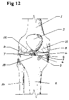
With reference to Figures 3 and 10, the positioning and attachment of the
horns of
the lateral and medial menisci are depicted. With further reference to Figures
6' and 7'
and particularly to Figure 83, it may be seen that, with progressive flexion
of the knee joint,
the menisci are progressively translated posteriorly. This is accompanied, to
varying
degrees, by posterolateral displacement in the case of the lateral meniscus
and
posteromedial displacement in the case of the medial meniscus. As the menisci
are
securely anchored by their horns, the result of such displacement is an
elastic distortion of
the menisci from their natural shapes and a stretching of the horns with a
concomitant
reduction in their heights. An inspection of Figures 1 and 2 will give a
better
understanding of factors affecting meniscal displacement or translation. The
elastic
distortion of the menisci appears to be such as to permit a very rapid
recovery of the
menisci to their relaxed positions during rapid and repeated flexion of the
knee joint. It
can be argued that this facility is a natural development almost wholly
applicable to the
young and physically active, and that a person of middle-age or older
following a
sedentary lifestyle has little need of it. For older persons, providing the
menisci are
constrained within a suitable range of translation with their positioning
regulated by
condylar movement, they will adequately perform their principal functions of
providing
shock absorption and enlarged bearing surface area for the joint. Limitation
of meniscal
translation to a range corresponding to a maximum knee joint flexion of 120
will
accommodate most sedentary activities. The principal means employed to effect
such
translational constraint of the menisci are locating bands extending
substantially around
the tibial condyles and fixed to the tibia. With reference to Figure 12, femur
1 terminates
at its distal end in medial condyle 11 and lateral condyle 12. Patella 3 is
depicted free of
its ligarnentary support and positioned above the knee joint as would be the
case with
considerable knee joint flexion. Fibula 10 is joined to tibia 2 at the
superior tibiofibular
joint and to the femur by the lateral collateral ligament 9. Femur 1 is joined
to the tibia by
medial collateral ligament 8. Lateral meniscus 7 and its anterior horn 13 are
depicted in
place. Locating band 4 is supported on one or more attachment lugs 5 fixed to
the
surfaces immediately inferior to the proximal edge of tibia 2 by suitable
fastenings 6.
CA 02808164 2013-02-12
WO 2012/019248 PCT/AU2011/001049
20
Using a suitable routing cutter, bone is removed from the tibia to permit said
attachment
lugs to be inset and positioned more or less flush with the bone surface. Said
attachment
lugs are optionally located, in the case of the lateral meniscus, in the
anterolateral lateral,
lateral and posterolateral zones. In the case of the medial meniscus, said
attachment lugs
are optionally located in the anteromedial, medial and posteromedial zones.
Access to
said zones is readily gained via an incision of approximately 30 millimetres,
by suitably
positioning the knee joint, by parting and/or displacement of the surrounding
tendinous
and ligamentary tissue and by detachment and retraction of the synovial
capsule. Said
locating band is made from a suitable metal alloy material and is sufficiently
stiff to
sustain all normal loadings. Suitable materials for said locating bands are
well known in
the art and include passivated nitinol, titanium, austenitic stainless steels
(which may have
tantalum, niobium or titanium coatings), cobalt-chromium alloys, passivated
beryllium,
beryllium-aluminium alloys, nickel-chromium alloys, nickel-chromium-beryllium
alloys,
cobalt-chromium alloys, zirconia, zirconia-toughened alumina, metal-carbon
fibre
composite and the like. Said locating bands are shaped to conform accurately
to the
peripheral shaping of the proximal edge of the tibia as determined
radiographically. Said
locating bands are optionally provided with downwardly extending locating lugs
which
abut the surfaces immediately inferior to the proximal edge of the tibia.
Suitable inwardly
directed locating pegs are optionally provided in said lugs, said pegs being
accommodated
in bores suitably located in said tibial surfaces. The free ends of said
locating bands are
optionally provided with downwardly directed pegs which are accommodated in
suitably
located bores made in the proximal (upper) surfaces of the tibia, said pegs
acting to
positively locate said ends. Other means of stabilising the ends of said
locating bands are
also optionally employed. Said prosthetic menisci are moulded from a suitable
biocompatible elastomer (the base material), in the preferred embodiment, the
elastomer
being DSM-PTG Carbosil 20 90A biocompatible silicone polycarbonate urethane,
manufactured by DSM Biomedical, of 6167 RA Geleen, The Netherlands. The
principal
mechanical properties of the material are:
Density 1.16 g/cc
Hardness, Shore A 90
Tensile Strength, Ultimate 42.6 MPa
Tensile Strength, Yield 6.4 MPa
Elongation at Break 530% - -
CA 02808164 2013-02-12
WO 2012/019248 PCT/AU2011/001049
21
Flexural Modulus 0.0407 GPa
Flexural Strength, yield 1.90 MPa
Tear Strength 87.7 kN/m
Taber Abrasion, mg/1000 Cycles 57.0
Compression Set 15.0%
The material combines the biocompatibility and biostability of conventional
silicone
elastomers with the processability and toughness of thermoplastic urethane
elastomers.
The material is non-cytotoxic and non-haemolytic, has a low-energy silicone
surface, has
outstanding oxidative stability, is hydrophobic, has high tensile strength and
is optically
clear. PurSilTM silicone-polyetherurethane and CarboSilTM silicone-
polycarbonateirethane
are true thermoplastic copolymers containing silicone in the soft segment.
These high-
strength thermoplastic elastomers are prepared through a multi-step bulk
synthesis where
polydimethylsiloxane (PSX) is incorporated into the polymer soft segment with
polytetramethyleneoxide (PTMO) (PurSil) or an aliphatic, hydroxyl-terminated
polycarbonate (CarboSil). The hard segment consists of an aromatic
diisocyanate, MDI,
with a low molecular weight glycol chain extender. The copolymer chains are
then
terminated with silicone (or other) Surface-Modifying End GroupsTM. Aliphatic
(AL)
versions of these materials, with a hard segment synthesized from an aliphatic
diisocyanate, are also available. PurSil and CarboSil can be melt fabricated
by
conventional extrusion, injection molding, or compression molding techniques.
Rod,
pellet, and tubing extruded from these materials displays an excellent surface
finish and
low gel content. In addition, these materials are heat-sealable, readily
blended with fillers,
and easily post-formed. In an alternative embodiment, said elastomer is
Tecoflex SG-
93A thermoplastic polyurethane elastomer (polyether), manufactured by Lubrizol
Advanced Materials, Inc., of Cleveland, Ohio, USA, which has a nominal Shore A
hardness of 87. This material is formulated especially for solution moulding.
In other
alternative embodiments, elastomer materials similar in characteristics to the
Carbosil and
Tecoflex products and having a hardness in the Shore A range 60 to 95 are used
with the
present invention. In the preferred embodiment, prostheses are sized and
shaped
according to radiographically-derived images of the condyles, although some
success has
been demonstrated in the selection of allograft meniscal replacements simply
in relation to
such factors as sex and height of a subject. A mould is created for the
required size and
final shaping (or selected from an available range of moulds) of a specific
prosthetic
CA 02808164 2013-02-12
WO 2012/019248 PCT/AU2011/001049
22
meniscus. With reference to Figures 13 and 15, in order to better accommodate
forces
applied to the prosthetic meniscus, in the preferred embodiment, prosthetic
meniscus 14 is
made in a plurality of more or less parallel layers of said base material
bonded or fused at
interfaces 25. Said layers of said base material are separated by thin sheets
16 of load-
carrying material of suitable tensile strength. Said layers of said base
material optionally
vary in thickness according to the location within a prosthetic meniscus and
number
between 2 and 12. In a first embodiment, said load carrying material takes the
form of a
thin, flexible sheet material, such as Kevlare. Said sheet material ranges in
thickness from
0.005 to 0.1 millimetre. The thickness and extent of said sheet material
optionally varies
according to the location within a prosthetic meniscus. With specific
reference to Figure
15, in the preferred embodiment, a plurality of apertures 22 is provided in
said sheet
material to facilitate bonding or fusing of one layer of said base material to
another, said
apertures being of any suitable shape and of an arrangement such as to leave
intact zones
capable of satisfactorily carrying the radial and circumferential loads
applied to said sheet
material. In a second embodiment (not shown), said layers of said base
material are
separated by arrays of fibres of a material of suitable tensile strength, said
fibres also being
orientated to conform to the known radial and circumferential load paths.
Photoelasticity
methods are optionally employed to determine the direction and magnitude of
stresses
applied to a meniscus at various loadings. Said fibres are captured between
said layers of
said base material when they are bonded or fused together. In the preferred
embodiment,
said fibres are made from a polymer, such as Kevlar0 or suitable carbon
fibres. In an
alternative embodiment (not shown), said fibres are distributed throughout a
said
prosthetic meniscus in a random way. With reference to Figure 5, Young's
modulus (or
tensile modulus) values for locations within the human menisci in tension are
shown
(values in MPa). It will be noted that the values are well below those of most
polymer
materials. For example, Kevlar (aramid) has a tensile modulus normally in the
range 83 to
186 GPa. With reference to Figure 4, the disposition and arrangement of the
natural
fibrous reinforcement is depicted. In the preferred embodiment, said layers of
said base
material are assembled by thermal fusion or bonding with the final shaping
occurring in a
mould created for the purpose. Said final mould is finely finished to provide
a glass-
smooth finish to the upper and lower surfaces of the final prosthetic
meniscus. In the
preferred embodiment, thermal fusing of said layers of said base material one
to another is
effected by heating two surfaces to be joined above their fusion temperatures
by contact
WO 2012/019248 CA 02808164 2013-02-12 PCT/AU2011/001049
23
with a hot plate and then firmly urging them together. Also in the preferred
embodiment,
bonding together of said layers of said base material is effected using one of
the permanent
biocompatible adhesives which are well known in the art.
In an alternative embodiment (not shown), either or both bearing surfaces of
said
menisci are provided with thin layers of a softer, more compliant base
material, the
thickness of said thin layers being preferably in the range 0.1 to 2.0
millimetres. By
providing a more compliant bearing surface, this embodiment is better able to
achieve
microelastohydrodynamic lubrication. In another alternative embodiment (not
shown),
said menisci are made completely from a softer, more compliant base material.
Menisci of
1 0 this embodiment are employed temporarily during repair of femoral or
tibial articular
cartilage and are subsequently replaced with menisci made from a harder base
material.
In alternative embodiments, said prosthetic menisci are made from one or more
of
the synthetic polypeptide materials of the type taught by Keeley et al in
Patent No. WO
2008/140703 A25. These materials comprise at least three consecutive beta-
sheet/beta-
turn structures and at least one crosslinking amino acid residue that
participates in
crosslinking, wherein the crosslinking residue is distinct from the beta-
sheet/beta-turn
structures, each polypeptide is between 150 and 500 amino acids in length and
the material
is a solid or liquid. In particular aspects, each beta-sheet structure may
comprise from 3 to
about 7 amino acid residues. In some embodiments, the amino acid sequences of
the
crosslinked polypeptides are the same; while in other embodiments the amino
acid
sequences of the crosslinked polypeptides are different. In some embodiments,
the
material further comprises a reinforcing material, such as animal material, a
synthetic
material or metal. In other embodiments, the material further comprises a non-
protein
hydrophilic polymer. In some embodiments, the material further comprises
glycosaminoglycan moieties, such as hyaluronan moieties. In some embodiments,
the
material comprises a mixture of crosslinked polypeptides and glycosaminoglycan
moieties. In other embodiments, the crosslinked polypeptides are covalently
linked to the
glycosaminoglycan moieties. In some embodiments, the material is solid and may
be in
the form of pads, sheets and ligament-like structures. In other embodiments,
the material
is a liquid, such as a solution or suspension.
With reference to Figures 14 and 16, locating band 4 is supported on one or
more
attachment lugs 5 fixed to the surfaces immediately inferior to the proximal
edge of tibia 2
by suitable fastenings passing through apertures 21. In the preferred
embodiment, said
WO 2012/019248 CA 02808164 2013-02-12PCT/AU2011/001049
24
apertures are countersunk to permit the heads of said fastenings to be flush
with the
external surfaces of said lugs. Said attachment lugs are optionally located,
in the case of
the lateral meniscus, in the anterolateral lateral, lateral and posterolateral
zones. In the
case of the medial meniscus, said attachment lugs are optionally located in
the
anteromedial, medial and posteromedial zones. Although attachment lug 5 is
depicted as
orientated parallel to the inner surface of said locating band, in
application, said lugs are
joggled or angled, as required, to conform to the tibial surface. The outer
surface 4 of said
locating band is made curved and is finely finished. With additional reference
to Figure
11, the location of the inner surface 30 of said locating band is indicated by
line 31. In a
first embodiment of the present invention, prosthetic meniscus 14 is
constrained within a
suitable range of translation by bridles 19, the inner ends of which are
embedded in said
prosthetic meniscus and the outer ends of which are fixed to said locating
band. It can be
seen from the figure that, when said prosthetic meniscus is displaced such
that its inner
margin moves from position 27 to position 27a (depicted in broken line), said
bridles are
displaced from positions 19 to positions 19a (depicted in broken line). The
lowermost
bridle (as depicted in the figure) undergoes little displacement and is merely
slightly
slackened. In order to ensure that said prosthetic meniscus is reliably
constrained within
the desired range of translation, said bridles are provided in larger numbers
around the
periphery of said prosthetic meniscus between said meniscus and said locating
band. In
the preferred embodiment, said bridles locating a specific prosthetic meniscus
number
between 5 and 40. Also in the preferred embodiment, said bridles are made in
looped
form, the inner ends passing around an anchor element 18 in the form of a
metal wire or
monofilament of a stiffly elastic polymer material embedded in said prosthetic
meniscus
and the outer ends passing in and out through a pair of closely-spaced
apertures (one
shown numbered 23) in said locating band, the join (not shown) of said bridle
ends being
recessed in circumferential groove 24 cut in the exterior surface of said
locating band. In
the preferred embodiment, said bridles are a firm fit in apertures 23 and the
inner openings
of said apertures are flared to minimise the possibility of chafing damage to
said bridles.
In the preferred embodiment, said bridles are braided from a large number of
fine Kevlar
fibres in the manner well known in the art and their outer ends are joined by
suitable knots
which are locked by impregnation with a suitable adhesive. In alternative
embodiments,
said bridles are spun or braided from a large number of fibres of any material
having a
suitable tensile strength.
WO 2012/019248 CA 02808164 2013-02-12PCT/AU2011/001049
25
With reference to Figure 9, it can be seen that the native meniscus 34 is
attached to
synovial capsule 37 via ligamentary connection 38 and thence, via said
synovial capsule,
to tibial articular cartilage 36. Femoral articular cartilage 35 has freedom
of movement in
relation to said meniscus and said tibial articular cartilage. It will be seen
that a space
exists between the meniscus proper and the synovial membrane, this space,
normally
occupied by ligamentary connection 38, becoming available when said native
meniscus
and said ligamentary connection are removed. In a first alternative embodiment
(not
shown), the annular zone between the circumferential face of a said prosthetic
meniscus
and the inner face of said locating band is filled with a cushion element in
the form of a
closed-cell foam material formed from a suitable elastic polymer. The shaping
and elastic
character of said foam material permits ready translation of said prosthetic
meniscus but
continuously urges said meniscus towards its natural position. In the
preferred
embodiment, said foam material is made locally stiffer in some zones to
provide a greater
force to urge a said prosthetic meniscus towards its natural position. In this
embodiment,
said foam material is fixed to said prosthetic meniscus and to said locating
band and has a
cross-sectional shape which is square or rectangular. In a second alternative
embodiment
(not shown), the annular zone between the circumferential face of a said
prosthetic
meniscus and the inner face of said locating band is filled with a cushion
element in the
form of a tube pressurised to a suitable pressure with a suitable gas or
partially filled with
a suitable liquid or gel which permits ready translation of said prosthetic
meniscus but
continuously urges said meniscus towards its natural position. In the
preferred
embodiment, said tube is made locally thicker in some zones to provide a
greater force to
urge a said prosthetic meniscus towards its natural position. In this
embodiment, said tube
is fixed to said prosthetic meniscus and to said locating band and has a
relaxed cross-
sectional shape which is round, oval, square or rectangular.
With reference to Figure 17a, in a third preferred embodiment, the annular
zone
between the circumferential face of a said prosthetic meniscus and the inner
face of said
locating band is filled with a plurality of cushion elements 39 made from a
suitable
elastomeric material and having a circular relaxed form. Said cushion elements
are
preferably moulded integrally with edge panels 40, 41 which are fixed to said
circumferential face of a said prosthetic meniscus and to the inner face of
said locating
band. Said cushion elements are made with an inner diameter of between 1.0 and
10
millimetres, with a wall thickness of between 0.5 and 3 millimetres and a
height to suit the
WO 2012/019248 CA 02808164 2013-02-12
PCT/AU2011/001049
26
edge thickness of said prosthetic meniscus. With reference to Figure 17b, said
cushion
elements are able to be flattened to reduce the clearance between said
prosthetic meniscus
and said locating band. With reference to Figure 17c, said cushion elements
are able to be
stretched out or extended to increase the clearance between said prosthetic
meniscus and
said locating band. With reference to Figure I 7d, said cushion elements are
able to be
rollingly distorted to permit independent longitudinal movement between
attachment
panels 40, 41 and, thereby, between said meniscus and said locating band.
With reference to Figure 18, said cushion elements are preferably moulded in
one
or more rows, multiple rows being separated by separation panels 42 and
complete arrays
being edged by attachment panels 40, 41. In said arrays, said cushion elements
are
orientated with their axes parallel to the tibial axis and are preferably
separated by
sufficient distance to accommodate the flattened mode depicted in Figure 17b
without
interference of adjacent elements. In the preferred embodiment, said arrays
are made in
more or less part-circular form to conform to the peripheral shaping of a said
prosthetic
meniscus. Also in the preferred embodiment, provision is made for a free flow
of synovial
fluid around said cushion elements, said separation panels and said attachment
panels to
prevent the development of hydraulic effects which might impede the
translation of a said
prosthetic meniscus. Said arrays permit ready translation of a said prosthetic
meniscus but
continuously urge said meniscus towards its natural position. In the preferred
embodiment, said cushion elements are made locally thicker in some zones to
provide a
greater force to urge a said prosthetic meniscus towards its natural position.
In the preferred embodiment, anchor element 18 is made in the form of a wire
of a
stiffly elastic metal material, tapering towards each end. By extending
substantially
throughout the circumferential extent of a said prosthetic meniscus, said wire
acts to
elastically restore said meniscus to a natural shape after any distortion.
Where said anchor
element takes the form of a monofilament of stiffly elastic polymer material,
a shaping
band 17 of a stiffly elastic material is optionally fixed to the
circumferential face of said
prosthetic meniscus. In the preferred embodiment, said shaping band passes
substantially
around the circumferential extent of a said prosthetic meniscus and acts to
elastically
restore said meniscus to a natural shape after any distortion. In the
preferred embodiment,
said shaping band is made from a suitable elastic metal material, from a
stiffly flexible
engineering polymer material, from carbon fibre or any suitable composite.
Where said
shaping band is used with said bridles, suitable flared apertures 20 are
provided in said
WO 2012/019248 CA 02808164 2013-02-12 PCT/AU2011/001049
27
shaping band. The ends of prosthetic menisci 14, 15 are optionally joined by
straps 26
which transmit hoop stresses generated in said menisci. Transmission of said
hoop
stresses acts to maintain a more or less natural shaping of said prosthetic
menisci during
loadings tending to distort them. In the preferred embodiment, said straps are
flat braided
from fine Kevlar fibres and their ends are securely embedded in said meniscus
ends. In
the preferred embodiment, said straps are coated with said base elastomer
material, which
is then treated in the manner described herein to make it attractive to
dipalmitoylphosphatidylcholine, the most abundant phospholipid in synovial
fluid. To
more securely locate said locating bands, in the preferred embodiment, their
ends 28, 29,
32, 33 are fixed to the proximal tibial surface. In this embodiment (not
shown), said ends
are provided with pegs which are accommodated within suitable bores made in
the tibial
surface or said ends are fixed to said tibial surface with suitable
fastenings. Where said
ends are fixed to the tibial surface, bone is removed using a suitable routing
cutter to
create short channels in which said ends are accommodated. In the preferred
embodiment,
said locating band ends are extended, turned through 900, joggled and shaped
to register
with said channels.
Said moulds for the final shaping of said prosthetic menisci are made with
polished surfaces to provide a glass-smooth finish to said menisci upper and
lower bearing
surfaces. To improve lubrication of said menisci by synovial fluid; said
bearing surfaces
are treated using a method' which renders them attractive to
dipalmitoylphosphatidylcholine (DPPC) by impregnating said surfaces with
poly[2-
methacryloyloxyethyl phosphorylcholine-co-n-butylmethacrylate] [poly(MPC-co-
BMA)].
[poly (MPC-co-BMA)] is a biocompatible, lipid-attracting polymer soluble in
solvent
systems which also dissolve many polyurethanes. DPPC is the most abundant
phospholipid in synovial fluid. In said method, the polyurethane elastomer is
immersed in
an ethanol solution containing BMA (0.3 mol I') and benzoic peroxide (1 wt A
to BMA)
as a polymerization initiator for 15 hours, resulting in a slightly swollen
surface. The
material is lightly washed with ethanol and then immersed in an ethanol
solution
containing MPC (0.3 mol 1-1) for 30 minutes. After removal from the second
solution, the
material is blotted dry and then heated at 70 C for 5 hours under an argon
atmosphere to
polymerize the monomers present in the surface of the material. Finally, the
material is
washed with ethanol and then dried en vacuo at room temperature for 24 hours.
To
improve the distribution of synovial fluid between the bearing surfaces of
said prosthetic
WO 2012/019248 CA 02808164 2013-02-12 PCT/AU2011/001049
28
menisci and the femoral and tibial articular cartilage, a network of narrow
channels is
moulded into one or both said bearing surfaces. In the preferred embodiment,
said
channels have a width of between 0.25 and 2.0 millimetres, a depth of between
0.25 and
2.0 millimetres, have a part-spherical or other suitable cross-sectional
shape, are separated
by between 1.0 and 5.0 millimetres and are orientated more or less radially
and
circumferentially. Also for the same purpose, either or both said bearing
surfaces are
provided at some or all of the points of intersection of said channels with
recesses
orientated more or less normal to the surface at each point, having a depth of
between 0.5
and 5.0 millimetres and a diameter of between 0.5 and 5.0 millimetres. For the
same
purpose, either or both said bearing surfaces are provided with recesses
orientated more or
less normal to the surface at each point, said recesses having a depth of
between 0.5 and
5.0 millimetres, a diameter of between 0.5 and 5.0 millimetres and being
separated from
each other by a distance of between 0.5 and 10 millimetres. For the same
purpose, said
prosthetic menisci are provided with a plurality of ducts passing from said
lower bearing
surface to said upper bearing surface, said ducts being orientated more or
less normal to
said lower bearing surface, having a diameter of between 0.5 and 5.0
millimetres and
being separated from each other by a distance of between 0.5 and 10
millimetres.
In the implantation of said prosthetic menisci, access is gained to the knee
compartment via minimal incisions and separation or displacement of the
tendinous and
capsular tissue surrounding the joint. The native menisci are removed as
required by
surgically severing all of their tibial, ligamentary and capsular attachments
with careful
attention to haemostasis, a process well known in the art. Where only one said
native
meniscus is removed, the transverse geniculate ligament is severed at an
appropriate
length and sutured to the base of the anterior cruciate ligament. Said
prosthetic menisci
and said locating bands are selected for size and shape from radiographic
images. Bone is
removed as necessary to accommodate said attachment lugs and ends of said
locating
bands. Said prosthetic menisci are lubricated and correctly positioned between
the
femoral condyles, varus or vagus force being applied as necessary to open the
joint.
Fastenings are inserted to fix said attachment lugs and locating band ends to
the tibia with
said locating elements positioned between said locating bands and said
prosthetic menisci.
The synovial capsule is modified as required to fully enclose the joint,
separated tissue is
reinstated and the skin incisions are closed.
In an arthroscopic procedure, a said menisetrs is made without anchor element
18
WO 2012/019248 CA 02808164 2013-02-12 PCT/AU2011/001049
29
and shaping band 17 (both as depicted in Figure 14) and with cushion elements
of the type
depicted in Figure 18. Outer attachment panel (depicted as 40 in Figure 18) is
made with
loops (not shown) to neatly accommodate an anchor element in the form of an
elongated
bar shaped, in its assembled form, to more or less conform to the shape of the
proximal
tibial edge. In the preferred embodiment, said bar is made from a suitable
strong, stiff
metal alloy material with a round or oval cross-sectional shape which tapers
towards the
ends. Said bar is made in two parts which are joined, as appropriate, at the
lateral or
medial position by joining means which are supported from the tibia by a
suitable
attachment lug. During the implantation process, the native meniscus is
removed
arthroscopically via a small incision in the manner well known in the art. A
said
prosthetic meniscus and associated cushion elements are folded into compact
form and,
using the same incision, extruded into the joint space via a suitable tubular
guide and
opened out into place. Said bar parts are then entered through said incision
and inserted
into said attachment panel loops. Said bar parts are joined and locked
together at said
joining means, bone is removed to create a suitable recess in the tibia and
the attachment
lug of said joining means is fixed to the tibia. In the preferred embodiment,
said joining
means take the form of a double-ended boss having shaped sockets which receive
complementary shaped ends of said bar parts. Also in the preferred embodiment,
bone is
removed approximately at the points of insertion of the horns of the native
meniscus to
create recesses into which suitable sockets are fixed. When said bar parts are
inserted,
their ends are entered into said sockets to stabilise their positions. In
another alternative
embodiment (not shown), said cushion element takes the form of a tube which is
empty
during said process of implantation, said tube being subsequently pressurised
to a suitable
pressure with a suitable gas or partially filled with a suitable liquid or gel
using suitable
injection means.
Any feasible combination of the apparatus and/or method described herein
should
be taken to be disclosed by the specification.
CA 02808164 2013-02-12
WO 2012/019248 PCT/AU2011/001049
30
REFERENCES:
1. Fu F. H., Thompson W. 0. Motion of the Meniscus During Knee
Flexion. In: Mow V. C., Arnoczky S. P., Jackson D. W., eds. Knee Meniscus:
Basic and Clinical Foundations. New York: Raven Press, 1992: Chapter 6, Figs
20 and 21.
2. Vedi V., Williams A., Tennant S. J., Spouse E., Hunt D. M.,
Gedroyc W. M. W. (1998). Meniscal Movement: An In-vivo Study Using
Dynamic MRI. The Journal of Bone & Joint Surgery (Br) 1999;81-B37-41.
3. Hamamoto K., Tobimatsu M. D., Zabinska-Uroda K. (2004).
Magnetic Resonance Imaging Evaluation of the Movement and Morphological
Changes of the Menisci During Deep Knee Flexion. J. Phys. Ther. Sci. Vol. 16
No. 2, 2004
4. Williams III P. F., Powell G. L., Love B., Ishihara A., Nalcabayashi
N., Johnson R., LaBerge W. (1994). Fabrication and Characterization of
Dipalmitoylphosphatidylcholine-attracting Flastomeric Material for Joint
Replacements.
5. Keeley et al. International Patent No. WO 2008/140703 A2.
Synthetic Peptide Materials for Joint Reconstruction, Repair and Cushioning.