Note: Descriptions are shown in the official language in which they were submitted.
CA 02808722 2013-02-19
WO 2012/035441 PCT/IB2011/002774
ADJUSTING DENTAL PROSTHESES BASED ON SOFT TISSUE
BACKGROUND
Field
[0001] The present application generally relates to dental planning, and
more
particularly to adjusting dental prostheses based on soft tissue.
Description of related technolou
[0002] The use of computer systems to design dental prostheses has
increased in
recent years. The computer systems allow a dentist, dental technician, or
other operator to
design dental prostheses for individual patients. These individual prosthesis
designs are often
called "situations," "dental plans," or "prosthetic plans." Operators using
the computer
systems can design plans based on a library of the teeth shapes and positions,
patient data,
and available equipment and hardware.
[0003] In prior systems, abutment shapes were manipulated by hand. In
these
systems, an operator might be given access to a 3D scan of the soft tissue in
the area where an
abutment would be placed. The operator could manipulate individual "handles"
on the 3D
model in order to move the 3D surface of the abutment relative to the soft
tissue (e.g., to
match the soft tissue's 3D surface). But manipulating the 3D surface of the
abutment can be
time consuming and difficult. The operator would have to manipulate multiple
individual
points on the 3D surface of the abutment in order to attempt to form it to the
desired shape.
The techniques, methods, systems, and computer-readable media herein provide
solutions to
some of these problems.
SUMMARY
[0004] Presented herein are techniques, methods, systems, devices, and
computer-
readable media for adjusting dental prostheses based on soft tissue. This
summary in no way
limits the invention herein, but instead is provided to summarize a few of the
embodiments.
[0005] Embodiments herein include techniques, methods, systems, devices,
and
computer-readable media for adjusting dental prostheses based on soft tissue,
including
receiving a 3D scan of soft tissue of the patient. The 3D scan of the soft
tissue of the patient
can include an emergence portion. The emergence portion of the 3D scan of the
soft tissue
1
CONFIRMATION COPY
81581208
may extend from an area associated with an implant attached to said patient,
to an area
where a dental prosthesis attached to said implant would emerge from said soft
tissue.
Emergence limit information for an emergence surface of a 3D model of the
dental
prosthesis may be received. Desired offset information for the emergence
surface of the 3D
model of the dental prosthesis may be received. The offset information
comprises a
distance between the emergence surface of the 3D model of the dental
prosthesis and the
emergence portion of the 3D scan of the soft tissue. A shape of the emergence
surface of
the 3D model of the dental prosthesis may be determined based on the emergence
portion
of the 3D scan of the soft tissue, the emergence limit information, and = the
offset
information. Further, manufacturing data related to the dental prosthesis may
be produced.
[0005a] Some
embodiments disclosed herein relate to a computer-
implemented method for adjusting the design of dental prostheses based on soft
tissue,
comprising: receiving a 3D scan of soft tissue of a patient, said 3D scan of
the soft tissue of
the patient comprising at least an emergence portion, said emergence portion
of the 3D scan
of the soft tissue extending from an area associated with an implant attached
to said patient,
to an area where a dental prosthesis attached to said implant would emerge
from said soft
tissue; receiving emergence limit information for an emergence surface of a 3D
model of the
dental prosthesis; receiving desired offset information for the emergence
surface of the 3D
model of the dental prosthesis, wherein the offset information comprises a
distance between
the emergence surface of the 3D model of the dental prosthesis and the
emergence portion of
the 3D scan of the soft tissue; determining a shape of the emergence surface
of the 3D model
of the dental prosthesis based on the emergence portion of the 3D scan of the
soft tissue, the
emergence limit information, and the offset information, wherein said shape is
automatically
determined by one or more computing devices based on the emergence portion of
the 3D
scan of the soft tissue, the emergence limit information, and the offset
information; further
modifying the emergence surface of the 3D model by manipulating manipulators
of the 3D
model of the dental prosthesis, wherein the manipulators are magnetized, such
that an
operator may change the emergence surface of the 3D model while still
maintaining said
distance between the emergence surface of the 3D model of the dental
prosthesis and the 3D
scan of the soft tissue, unless a manipulator of said manipulators is moved
beyond a
2
CA 2808722 2017-09-08
81581208
predetermined threshold magnetization distance, in which case the manipulator
moves freely
and the emergence surface is manipulated freely; and producing manufacturing
data related
to the dental prosthesis using the 3D model of the dental prosthesis.
[0005b] Some embodiments disclosed herein relate to a system for
adjusting
the design of dental prostheses based on soft tissue, comprising one or more
computing
devices, said one or more computing devices being configured to: receive a 3D
scan of soft
tissue of a patient, said 3D scan of the soft tissue of the patient comprising
at least an
emergence portion, said emergence portion of the 3D scan of the soft tissue
extendinu, from
an area associated with an implant attached to said patient, to an area where
a dental
prosthesis attached to said implant would emerge from said soft tissue;
receive emergence
limit information for an emergence surface of a 3D model of the dental
prosthesis: receive
desired offset information for the emergence surface of the 3D model of the
dental prosthesis,
wherein the offset information comprises a distance between the emergence
surface of the 3D
model of the dental prosthesis and the emergence portion of the 3D scan of the
soft tissue;
automatically determine a shape of the emergence surface of the 3D model of
the dental
prosthesis based on the emergence portion of the 3D scan of the soft tissue,
the emergence
limit information, and the offset information; wherein the emergence surface
of the 3D model
is further modified by manipulating manipulators of the 3D model of the dental
prosthesis,
wherein the manipulators are magnetized, such that an operator may change the
emergence
surface of the 3D model while still maintaining said distance between the
emergence surface
of the 3D model of the dental prosthesis and the 3D scan of the soft tissue,
unless a
manipulator of said manipulators is moved beyond a predetermined threshold
magnetization
distance, in which case the manipulator moves freely and the emergence surface
is
manipulated freely; and produce manufacturing data related to the dental
prosthesis based on
the 3D model of the dental prosthesis.
[0005c] Some embodiments disclosed herein relate to a non-transitory
computer-readable storage medium having embodied thereon in a non-transitory
manner
computer-executable instructions for adjusting the design of dental prostheses
based on soft
tissue, said computer-executable instructions, when running on one or more
computing
2a
CA 2808722 2017-09-08
81581208
devices, performing a method comprising: receiving a 3D scan of soft tissue of
a patient, said
3D scan of the soft tissue of the patient comprising at least an emergence
portion, said
emergence portion of the 3D scan of the soft tissue extending from an area
associated with an
implant attached to said patient, to an area where a dental prosthesis
attached to said implant
would emerge from said soft tissue; receiving emergence limit information for
an emergence
surface of a 3D model of the dental prosthesis; receiving desired offset
information for the
emergence surface of the 3D model of the dental prosthesis, wherein the offset
information
comprises a distance between the emergence surface of the 3D model of the
dental prosthesis
and the emergence portion of the 3D scan of the soft tissue; automatically
determining, using
one or more computing devices, a shape of the emergence surface of the 3D
model of the
dental prosthesis based on the emergence portion of the 3D scan of the soft
tissue, the
emergence limit information, and the offset information; further modifying
emergence
surface of the 3D model by tnanipulating manipulators of the 3D model of the
dental
prosthesis, wherein the manipulators are magnetized, such that an operator may
change the
emergence surface of the 3D model while still maintaining said distance
between the
emergence surface of the 3D model of the dental prosthesis and the 3D scan of
the soft tissue,
unless a manipulator of said manipulators is moved beyond a predetermined
threshold
monetization distance, in which case the manipulator moves freely and the
emergence
surface is manipulated freely; and producing manufacturing data related to the
dental
prosthesis based on the 3D model of the dental prosthesis.
100061 Numerous other embodiments are described throughout herein.
[0007] For
purposes of summarizing the invention and the advantages achieved
over the prior art, certain objects and advantages of the invention are
described herein. Of
course, it is to be understood that not necessarily all such objects or
advantages need to be
achieved in accordance with any particular embodiment. Thus, for example,
those skilled
in the art will recognize that the invention may be embodied or carried out in
a manner
that achieves or optimizes one advantage or group of advantages as taught or
suggested
herein without necessarily achieving other objects or advantages as may be
taught or
suggested herein.
2b
CA 2808722 2017-09-08
81581208
[0008] All of these embodiments are intended to be within the scope of
the
invention herein disclosed. These and other embodiments will become readily
apparent to
those skilled in the art from the following detailed description having
reference to the
attached figures, the invention not being limited to any particular disclosed
embodiment(s).
BRIEF DESCRIPTION OF THE DRAWINGS
[0009] FIG. 1 illustrates a first interface for adjusting dental
prostheses based on
soft tissue.
[0010] FIG. 2 illustrates an example system for adjusting dental
prostheses based
on soft tissue.
[0011] FIG. 3 illustrate an example method of adjusting dental
prostheses based
on soft tissue.
2c
CA 2808722 2017-09-08
CA 02808722 2013-02-19
WO 2012/035441 PCT/1B2011/002774
100121 FIG. 4 illustrates a second interface for adjusting dental
prostheses based
on soft tissue.
[0013] FIG. 5 illustrates a third interface for adjusting dental
prostheses based on
soft tissue.
[0014] FIG. 6 illustrates a fourth interface for adjusting dental
prostheses based
on soft tissue.
[0015] FIG. 7 illustrates a fifth interface for adjusting dental
prostheses based on
soft tissue.
[0016] FIG. 8 illustrates a sixth interface for adjusting dental
prostheses based on
soft tissue.
[0017] FIG. 9 illustrates a seventh interface for adjusting dental
prostheses based
on soft tissue.
[0018] FIG. 10 illustrates an eighth interface for adjusting dental
prostheses based
on soft tissue.
[0019] FIG. 11 illustrates a ninth interface for adjusting dental
prostheses based
on soft tissue.
[0020] FIG. 12 illustrates an example schematic for adjusting dental
prostheses
based on soft tissue.
[0021] FIG. 13 illustrates a tenth interface for adjusting dental
prostheses based
on soft tissue.
[0022] FIG. 14 illustrates an eleventh interface for adjusting dental
prostheses
based on soft tissue.
DETAILED DESCRIPTION OF SPECIFIC EMBODIMENTS
Overview
[0023] In various embodiments herein, an operator, such as a dentist,
dental
surgeon, or the like, can define the portion ("the emergence portion") of a
prosthesis', such as
an abutment's, surface that runs from the implant, through the soft tissue
("the emergence
surface"), to the base of the prosthesis. The emergence portion of the
prosthesis' surface may
be roughly matched to the soft tissue's surface by defining a desired offset
from the soft
tissue and effectuating the offset by, for example, pressing a button or
performing a
3
81581208
keystroke. The operator may first perform a 3D scan of the soft tissue around
the area in
which the abutment will be placed. In some cases, the 3D scan of the soft
tissue may come
from another lab or another operator. Regardless of the origin of the 30 scan
of the soft
tissue surface, the operator can use the 3D scan of the soft tissue as a guide
for creating the
abutment. The operator can define an offset to make the abutment's 3D
emergence surface
larger than, smaller than, or equal to (zero offset) the 3D scan of the soft
tissue. Once the
operator presses the button, the 3D emergence surface of the abutment is
modified
automatically to be offset from the 3D scan of the soft tissue by the desired
amount.
Embodiments herein may be used to design any type of prosthesis that may
emerge through
soft tissue.
[0024] An operator might want to design a dental prosthesis so that,
once it is
installed in the patient's mouth, it will compress the surrounding soft
tissue, thereby leaving
no gap between the dental prosthesis and the soft tissue. In other cases, the
operator may
want to match the dental prosthesis with the soft tissue, thereby reducing
both the gap
between the soft tissue in the dental prosthesis as well as the compression of
the soft tissue.
In some cases, the operator may want to leave a gap between the dental
prosthesis and the
soft tissue. Embodiments herein will allow the operator to design dental
prosthesis s in any
of these ways.
[0025] FIG. 1 depicts an interface 100 that has a global abstraction
portion 111,
an overlaid representation portion 110, and depicts the 3D scan of soft tissue
120, as well as
an abutment 130. In some embodiments, the operator can use the global
abstraction portion
111 to selectively turn on and off the viewing of certain items that will be
displayed in
overlaid representation portion 110. For example, the operator may be able to
turn on or off
the viewing of a prosthesis such as abutment 130, the soft tissue 120, and/or
any other item
that may be displayed in the overlaid representation portion 110. Examples and
embodiments of selection techniques are given in U.S. Pat. Appl. No.
12/703,601, filed
February 10, 2010, entitled Dental Prosthetics Manipulation, Selection, and
Planning
[0026] The operator can use the interface 100 to manipulate the
shape of the
surface of the abutment 130. FIG. 4 depicts an interface 400 including an
overlaid
4
CA 2808722 2017-09-08
CA 02808722 2013-02-19
WO 2012/035441 PCT/1B2011/002774
representation portion 410. An operator can define an emergence limit 440 for
a dental
prosthesis (not pictured) with respect to the 3D scan of soft tissue 420. The
3D scan of the
soft tissue has an "emergence portion." The phrase "emergence portion of the
3D scan of the
soft tissue" has its ordinary and customary meaning, which includes signifying
at least a
surface or a portion of a surface that extends from an emergence limit down
toward a scanned
implant. For example, an emergence portion of 3D scan 420 extends from an
emergence
limit 440 down to the implant, which is signaled as 445 in FIG. 4.
[0027] FIG. 5 depicts an interface 500 with an overlaid representation
portion
510 and a control menu 560. In some embodiments, the operator can define an
emergence
offset using an offset indicator 564 on control menu 560. After the emergence
offset 564 is
defined, the operator can press button 561 to adjust the dental prosthesis to
the soft tissue,
offset by the emergence offset. This dental prosthesis is depicted as 530 in
FIG. 5.
Adjustments made to the surface of the 3D model of the prosthesis based on the
emergence
offset can be in any direction. For example, in some embodiments, the
adjustments are made
in a radial direction extending from the axis of the prosthesis outward.
[0028] The emergence offset can define how the "emergence surface" of
the 3D
model of the dental prosthesis is positioned with respect to the emergence
portion of the 3D
scan of the soft tissue. In various embodiments, the emergence surface may be
the portion of
the dental prosthesis' surface that extends from the implant, through the soft
tissue, and ends
where the dental prosthesis emerges from the soft tissue ¨ at the emergence
limit (e.g.,
emergence limit 440 of FIGS. 4 and 5). The phrase "emergence surface of the 3D
model of
the dental prosthesis" has its ordinary and customary meaning, which includes
a surface on
the 3D model of the dental prosthesis that roughly corresponds to the
emergence portion of
the 3D scan of the soft tissue. For example, in some embodiments, the
emergence surface of
the 3D model of the dental prosthesis may extend from the interface of the 3D
model of the
prosthesis with the underlying implant, up to an emergence limit. For example,
see FIG. 10,
which depicts an emergence surface 1080 of 3D model of the dental prosthesis
1030 being
depicted on the overlaid representation portion 1010 of interface 1000.
[0029] In some embodiments, the operator can define positive or negative
emergence offsets. A positive offset indicates that the 3D model of the dental
prosthesis will
CA 02808722 2013-02-19
WO 2012/035441 PCT/1B2011/002774
not extend as far, in a radial direction, for example, as the 3D scan of the
gum. In particular,
the emergence surface of the 3D model of the dental prosthesis will not cross
over or extend
beyond the emergence portion of the 3D scan of the soft tissue. On the other
hand, if the
offset is negative, then the emergence surface of the 3D model of the dental
prosthesis may
extend beyond, in the radial direction, for example, and be wider or have a
greater
circumference than the emergence portion of the 3D scan of the soft tissue of
the patient. In
this way, the operator can very quickly and easily define an emergence surface
of a 3D model
of a prosthesis, such as an abutment, with the desired gap or compression of
soft tissue. In
some embodiments, the effect of the signs of the offsets may be swapped, with
negative
offsets being associated with a gap and positive offsets being associated with
the emergence
surface of the dental prosthesis being wider than the scan of the soft tissue.
Example Systems for Adjusting Prostheses Based on Soft Tissue
100301 FIG. 2 illustrates an example system 200 for adjusting prostheses
based on
soft tissue. The system 200 may include one or more computers 210 coupled to
one or more
displays 220, and one or more input devices 230. An operator 240, who may be a
dentist,
dental technician, or other person, may plan dental prostheses using system
200 by
manipulating the one or more input devices 230, such as a keyboard and / or a
mouse. In
some embodiments, while working on the dental plan, the operator 240 may view
the dental
plan and other related dental plan data on the display 220. The display 220
may include two
or more display regions or portions, each of which displays a different view
of the dental
plan. For example, in some embodiments, the display 220 may show a semi-
realistic 3D
rendering of the dental plan, a localized abstraction of the dental plan, and
/ or a cross-
sectional representation of the dental plan. Each of these displays or
portions may be linked
internally within a program and / or using data on computer 210. For example,
a program
running on a computer 210 may have a single internal representation of the
dental plan in
memory and the internal representation may be displayed in two or more
abstract or semi-
realistic manners on display 220.
100311 In some embodiments, the operator 240 may be able to perform a
command, such as select, move, manipulate, or make transparent, opaque, or
invisible, on a
particular substructure in the dental plan. The operator 240 may be able to
perform this
6
CA 02808722 2013-02-19
WO 2012/035441 PCT/1B2011/002774
command by manipulating the input device 230, such as clicking with a mouse on
a
particular region of one of the abstract or semi-realistic versions of the
dental plan displayed
on the display 220.
[0032] In various embodiments, the computer 210 may include one or more
processors, one or more memories, and one or more communication mechanisms. In
some
embodiments, more than one computer may be used to execute the modules,
methods,
blocks, and processes discussed herein. Additionally, the modules and
processes herein may
each run on one or multiple processors, on one or more computers; or the
modules herein
may run on dedicated hardware. The input devices 230 may include one or more
keyboards
(one-handed or two-handed), mice, touch screens, voice commands and associated
hardware,
gesture recognition, or any other means of providing communication between the
operator
240 and the computer 210. The display 220 may be a two-dimensional ("2D") or
3D display
and may be based on any technology, such as LCD, CRT, plasma, projection, etc.
[0033] The communication among the various components of system 200 may
be
accomplished via any appropriate coupling, including USB, VGA cables, coaxial
cables,
FireWire, serial cables, parallel cables, SCSI cables, IDE cables, SATA
cables, wireless
based on 802.11 or Bluetooth, or any other wired or wireless connection(s).
One or more of
the components in system 200 may also be combined into a single unit or
module. In some
embodiments, all of the electronic components of system 200 are included in a
single
physical unit or module.
Techniques for Adjusting Prostheses Based on Soft Tissue
[0034] FIG. 3 depicts a method 300 for adjusting a dental prosthesis
based on soft
tissue. In block 310, a 3D scan of soft tissue is received. As discussed
above, this 3D scan
may contain an emergence portion that defines the area in which a dental
prosthesis, such as
an abutment, will be proximal to the soft tissue. For example, if a dental
surgeon implanted
an implant into a patient and then placed a healing abutment into the implant,
soft tissue
would form through an emergence portion down to the implant. Thereafter, the
healing
abutment would be taken out and a scan of the soft tissue would take place.
That scan would
be received in block 310. An example of that scan is depicted as 3D model 120
in FIG. 1.
The scan may be taken using any appropriate method including an intraoral
scan, CT scan,
7
81581208
MRI's, and the like. The scan could also be, in some embodiments, a surface
scan of a
physical model, where, for example, there is an implant replica representing
the position of
the implant relative to the soft tissue. The scan may be of the physical
model, but it
nevertheless can represent the 3D surface of the soft tissue. Various
embodiments of
scanning 3D models are given in U.S. Pat. Appl. No. 12/703,596, entitled
"Dental Data
Planning," filed Feb. 10, 2010.
[0035] In block 310, placement information for a dental prosthesis
may also be
received. For example, some embodiments detect the position of the implant
(e.g., by using
an implant locator in a scan of a model). The position of the prosthesis or
other abutment can
be determined from the position of the implant. In some embodiments, a
position locator
could be attached to a model, and the model with the position locator attached
could be
scanned. The position of the implant can be defined from the position of the
position locator
in the scan. When a 3D model of a prosthesis is "attached" to the implant in
the design
software, the position of the prosthesis is thereby defined. The placement
information may
be, looking to the Example in FIG. 1, that an abutment 130 will be placed on
top of an
implant through the 3D scan of the soft tissue 120, thereby obtaining the
position of the
abutment 120 in part, and indirectly, from the position of the implant.
[0036] In block 330, emergence limit information for a dental
prosthesis is
received. The emergence limit information may be a margin line or other line
or curve
defining the upper limit of the emergence surface of the 3D model of the
dental prosthesis.
For example, looking to FIG. 4, defining the emergence limit 440 may be
defined by
manipulating manipulators 450, 451, 452. As noted elsewhere herein, the
emergence limit
440 and the base of the 3D model of the prosthesis (e.g., a fixed diameter
interface with the
implant to which the 3D model of the prosthesis is attached) together may
define the
emergence surface of the 3D model of the dental prosthesis. In some
embodiments, the
manipulators 450-452 are used to define the emergence limit 440. In some
embodiments, the
manipulators 450-452 for an emergence limit 440 may be sorted radially (e.g.,
about the
center axis of the associated implant) ¨ and the emergence limit 440 may be
determined
based on the sorted manipulators 450-452. The emergence limit 440 may be
defined as a
8
CA 2808722 2017-09-08
CA 02808722 2013-02-19
WO 2012/035441 PCT/1B2011/002774
curved line that passes through the manipulators 450-452. The emergence limit
440 may be
formed using any appropriate algorithm or interpolation among these
manipulators 450-452,
including a second-degree NURBS curve, a spline, other NURBS, etc. In various
embodiments, the operator can add manipulation points to an emergence limit
440 by
clicking on the line 440, clicking on the 3D surface 420, and the like. The
addition of
manipulators may allow the operator to refine the emergence limit 440. In
various
embodiments, the operator can also remove manipulation points 450-452 from the
emergence
limit 440 by dragging manipulators 450-452 off the screen, right-clicking on
manipulators
450-452, performing particular keystrokes, and the like.
100371 FIG. 11 illustrates an interface 1100 with an overlapped
representation
portion 1110 depicting two emergence limits 1140 and 1141 on the 3D scan of
soft tissue
1130. When there is more than one implant and more than one emergence limit
1140, 1141,
the techniques herein may sort any received or defined manipulators 1150,
1151, 1152, 1153
by their proximity to implants and thereby define which of the manipulators
1150, 1151,
1152, 1153 are associated with each emergence limit 1140, 1141 (e.g., by
associating
manipulators 1150-1153 with the implant to which it is closest). From there,
the
manipulators 1150, 1151 that are associated together are used to define the
emergence limit
1140, as described above. As described above, manipulators may be added to an
emergence
limit. For example, looking at emergence limit 1141, we see that it crosses
over an "open
space" as it interpolates between two of the manipulators 1152 and 1153. An
operator may
want to add another manipulator in order to match the emergence limit 1141 to
the surface
1130 and/or to alter the emergence limit's 1141 shape.
100381 After the emergence limit information for the dental prosthesis
is received
in block 330, then desired offset information for the emergence surface of the
3D model of
the dental prosthesis is received in block 340. As discussed above and as
depicted in FIG. 5,
the emergence limit information may be received from an operator using the
emergence
offset interface 564 on control menu 560. The operator may be able to type in
the emergence
offset or a scroll bar, dial, or other input may be used. In some embodiments,
as depicted in
FIG. 5, once the operator is satisfied with the placement of the emergence
limit, as defined
by manipulators 450, 451, 452, the operator may press a button or otherwise
cause to be
9
CA 02808722 2013-02-19
WO 2012/035441 PCT/1B2011/002774
executed the determination of the emergence surface for the 3D model of the
dental
prosthesis, such as by pressing button 561 on control menu 560. In other
embodiments, as
the emergence offset is changed and/or the emergence limit is changed, the
techniques herein
may automatically update the emergence surface for the 3D model of the dental
prosthesis.
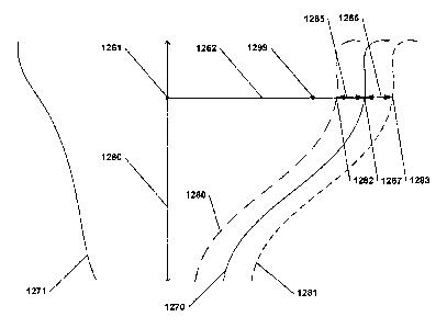
[0039]
Determining the 3D surface for the emergence surface of the 3D model of
the dental prosthesis in block 350 may include determining a surface based on
the offset and
based on the emergence portion of the 3D scan of the soft tissue. For example,
FIG. 12
depicts a cross-section of an emergence portion of a 3D scan of soft tissue
with the two sides
1270 and 1271. The dental prosthesis for which the emergence surface will be
defined has an
axis 1260. The axis may be associated with the central axis of an implant, the
central axis of
the prosthesis, an insertion axis, and the like. For example, if the axis 1260
is associated with
an implant axis, then the axis can be defined based on a scan of a physical
implant position
locator either, e.g., on the physical model or in a physical impression taken
of a patient's
mouth. In some embodiments, determining the emergence surface comprises
determining,
for each manipulator 1299 on the emergence surface of the 3D model of the
dental prosthesis
(not depicted in FIG. 12), the point 1261 along a perpendicular line 1262 from
the
manipulator 1299 to the axis 1260. From there, the point 1267 is determined as
the point
where the perpendicular line 1262 intersects the 3D soft tissue surface 1270.
The
corresponding point 1282 for the emergence surface 1280 is defined as the
point 1282 on the
perpendicular line 1262 that is offset by the desired offset 1265. As is
illustrated in the FIG.
12, the desired offset can be positive (e.g., distance 1265) and define a
point 1282, or
negative (distance 1266) and define a point 1283. This process is repeated for
each
manipulator 1299 on the emergence surface of the 3D model of the dental
prosthesis. As
discussed in more detail below with respect to FIG. 9, the manipulators may be
on the
emergence limit line (manipulators 950 and 951) as well as on other parts of
the emergence
surface (manipulators 970 and 971). Embodiments herein include offsetting each
of these
manipulators 950, 951, 970, and 971. In some embodiments, this process of
offsetting points
is repeated for only the manipulators. In other embodiments, the process of
offsetting points
is repeated for more points than just the manipulators -- for example, an
entire grid of points
may be offset.
CA 02808722 2013-02-19
WO 2012/035441 PCT/1B2011/002774
[0040] Once all of the manipulators 1299 and other points have been
offset
relative to the emergence portion of the 3D scan of the soft tissue, the
emergence surface of
the 3D model of the dental prosthesis can then be defined as a second degree
NURBS surface
through the manipulation points (and any other points that have been offset
from the 3D
surface of the soft tissue), or may be interpolated or estimated in any
appropriate way. There
are other methods of calculating the emergence surface for the 3D model of the
dental
prosthesis, and these are considered within the scope of the embodiments
herein. For
example, the emergence surface of the 3D model of the dental prosthesis may be
determined
by radially scaling the emergence portion of the 3D scan of the soft tissue in
order to offset
the emergence surface of the 3D model of the dental prosthesis by the
appropriate amount, as
defined by the offset.
[0041] After the emergence surface of the 3D model of the dental
prosthesis has
been generated in block 350, the operator may see the generated dental
prosthesis on the
overlaid representation portion of the interface. For example, turning to FIG.
7, the operator
will be able to see on interface 700 the overlaid representation portion 710
which will include
a dental prosthesis 730 and a 3D scan of soft tissue 720. As depicted in FIG.
7, if the offset
causes the dental prosthesis model to be larger than the 3D scan of the soft
tissue in its
emergence portion, then the dental prosthesis' emergence surface will overlap
with the 3D
scan, as signaled with area 721. On the other hand, if the emergence offset is
such that the
dental prosthesis is smaller than the related emergence portion of the 3D scan
of the soft
tissue, then a gap 821, shown in FIG. 8, may be seen between the 3D scan of
the soft tissue
820 and the prosthesis 830.
[0042] After the 3D surface for the emergence surface of the 3D model of
the
dental prosthesis has been determined in block 350, the operator may
optionally manipulate
the limit information (in block 330, discussed above) and offset information
(in block 340,
discussed above) again. From there, a new 3D surface for the emergence surface
of the 3D
model of the dental prosthesis may be determined in block 350. Once the
operator is satisfied
with the prosthesis or is ready to produce the prosthesis, the operator may
continue to other
steps in prosthesis design (not depicted in FIG. 3) or may produce
manufacturing data for the
prosthesis (block 360).
11
CA 02808722 2013-02-19
WO 2012/035441 PCT/1B2011/002774
Magnetization
[0043] There are numerous other embodiments of the techniques, systems,
methods, computer-readable storage media, and methods discussed herein. For
example,
different steps may be added to method 300 and/or steps in method 300 may be
performed in
a different order or not at all. For example, turning to FIG. 9, in some
embodiments, the
operator may be able to manipulate the manipulators 950, 951, 970, and/or 971
in order to
further modify the emergence surface of the 3D model of the dental prosthesis
(not depicted
in the method 300 of FIG. 3). Further, in some embodiments, the operator may
be able to
select an option 962 on control menu 960 to magnetize the emergence handles of
the dental
prosthesis. By doing this, the operator may be able to change the emergence
surface while
still maintaining the desired offset between the emergence surface of the 3D
model of the
dental prosthesis and the 3D scan of the soft tissue. In some embodiments, the
"magnetization" will be in effect for all movements of manipulators 950, 951,
970, and/or
971 that are within a predetermined distance from the 3D scan of the soft
tissue. The
distance may be predefined or may be defined by the user using a magnetization
distance
control 963 on control menu 960. If a manipulator 950, 951, 970, or 971 is
moved beyond
this threshold magnetization distance, then the manipulator will move freely
and the
emergence surface will be manipulated freely and will not be confined to the
offset with the
3D scan of the soft tissue, otherwise the manipulator will be held to the
desired offset with
the 3D scan of the soft tissue.
[0044] Magnetization may operate using any appropriate technique or
algorithm.
For example, the operator may move a manipulator 970 by clicking on the point
and holding
down a mouse button until she has placed it where she likes. If that point is
still within the
magnetization distance of the 3D scan of the soft tissue 920, then, once
released, the closest
point on the 3D surface of the soft tissue 920 will be found and the
manipulator 970 will be
placed at the desired offset from the point closest point on the 3D surface of
the soft tissue
920. If the point is not within the magnetization distance of the 3D scan of
the soft tissue
920, then it placement may not be changed after placement by the user.
12
CA 02808722 2013-02-19
WO 2012/035441 PCT/1B2011/002774
Coloring
[0045] Turning now to FIG. 10, in various embodiments the emergence
surface
of the 3D model of the dental prosthesis may be colored or shaded in order to
show the
distance between the emergence surface of the 3D model of the dental
prosthesis and the
emergence portion of the 3D scan of the soft tissue. For example, the
emergence surface of
the 3D model of the dental prosthesis may be covered with a color map, and the
color map
may have different colors or color ranges that represent different distances
between the
emergence surface of the 3D model of the prosthesis and the emergence portion
of the 3D
scan of the soft tissue. Example coloring is depicted in FIG. 10 in the
interface 1000, which
has an overlaid representation portion 1010 that shows a dental prosthesis
1030 that has a
shaded emergence surface 1080. In some embodiments and in some procedures, the
operator
may want to keep the distance between the soft tissue, such as the gingiva or
gum, within a
certain distance (e.g., 0.1 mm or 1 mm) of the emergence surface in order to
avoid a gap
larger than that size or to compress the soft tissue more than that amount.
Coloring or
shading on the emergence surface of the 3D model of the dental prosthesis can
help an
operator quickly identify the areas of the surface that are inside and outside
of a desired
range.
Other embodiments
[0046] Various of the embodiments herein show interfaces of a certain
configuration. Other configurations of interfaces are also possible. Turning
to FIG. 13, it is
possible that an interface 1300 can have an overlaid representation portion
1310, a global
selection portion 1311, and a control menu 1360, all on a single interface
1300. It is also
possible, as depicted in FIG. 14, that two separate sub-interfaces 1400 and
1401 may be
used. The control menu 1460 may be on interface portion 1401 and the overlaid
representation portion 1410 and global selection portion 1411 may be on
interface portion
1400. These various interface portions may be shown on separate screens, on
separate
displays or in separate windows. Other configurations of the various portions
on various
displays or in various windows may also be used.
13
CA 02808722 2013-02-19
WO 2012/035441 PCT/1B2011/002774
[0047] The processes and systems described herein may be performed on or
encompass various types of hardware, such as computing devices. In some
embodiments,
computer 210, display 220, and / or input device 230 may each be separate
computing
devices, applications, or processes or may run as part of the same computing
devices,
applications, or processes ¨ or one of more may be combined to run as part of
one application
or process ¨ and / or each or one or more may be part of or run on computing
devices.
Computing devices may include a bus or other communication mechanism for
communicating information, and a processor coupled with the bus for processing
information. The computing devices may have a main memory, such as a random
access
memory or other dynamic storage device, coupled to the bus. The main memory
may be used
to store instructions and temporary variables. The computing devices may also
include a
read-only memory or other static storage device coupled to the bus for storing
static
information and instructions. The computer systems may also be coupled to a
display, such
as a CRT or LCD monitor. Input devices may also be coupled to the computing
devices.
These input devices may include a mouse, a trackball, or cursor direction
keys.
[0048] Each computing device may be implemented using one or more
physical
computers, processors, embedded devices, or computer systems or a combination
or portions
thereof. The instructions executed by the computing device may also be read in
from a
computer-readable medium. The computer-readable medium may be a CD, DVD,
optical or
magnetic disk, laserdisc, carrier wave, or any other medium that is readable
by the computing
device. In some embodiments, hardwired circuitry may be used in place of or in
combination
with software instructions executed by the processor. Communication among
modules,
systems, devices, and elements may be over direct or switched connections, and
wired or
wireless networks or connections, via directly connected wires, or any other
appropriate
communication mechanism. The communication among modules, systems, devices,
and
elements may include handshaking, notifications, coordination, encapsulation,
encryption,
headers, such as routing or error detecting headers, or any other appropriate
communication
protocol or attribute. Communication may also messages related to HTTP, HTTPS,
FTP,
TCP, 1113, ebMS OASIS/ebXML, secure sockets, VPN, encrypted or unencrypted
pipes,
MIME, SMTP, MIME Multipart/Related Content-type, SQL, etc.
14
CA 02808722 2013-02-19
WO 2012/035441 PCT/1B2011/002774
[0049] Any appropriate 3D graphics processing may be used for displaying
or
rendering, including processing based on OpenGL, Direct3D, Java 3D, etc.
Whole, partial, or
modified 3D graphics packages may also be used, such packages including 3DS
Max,
SolidWorks, Maya, Form Z, Cybermotion 3D, or any others. In some embodiments,
various
parts of the needed rendering may occur on traditional or specialized graphics
hardware. The
rendering may also occur on the general CPU, on programmable hardware, on a
separate
processor, be distributed over multiple processors, over multiple dedicated
graphics cards, or
using any other appropriate combination of hardware or technique.
[0050] As will be apparent, the features and attributes of the specific
embodiments disclosed above may be combined in different ways to form
additional
embodiments, all of which fall within the scope of the present disclosure.
[0051] Conditional language used herein, such as, among others, "can,"
"could,"
"might," "may," "e.g.," and the like, unless specifically stated otherwise, or
otherwise
understood within the context as used, is generally intended to convey that
certain
embodiments include, while other embodiments do not include, certain features,
elements,
and/or states. Thus, such conditional language is not generally intended to
imply that
features, elements and/or states are in any way required for one or more
embodiments or that
one or more embodiments necessarily include logic for deciding, with or
without author input
or prompting, whether these features, elements, and/or states are included or
are to be
performed in any particular embodiment.
[0052] Any process descriptions, elements, or blocks in the flow
diagrams
described herein and/or depicted in the attached figures should be understood
as potentially
representing modules, segments, or portions of code which include one or more
executable
instructions for implementing specific logical functions or steps in the
process. Altemate
implementations are included within the scope of the embodiments described
herein in which
elements or functions may be deleted, executed out of order from that shown or
discussed,
including substantially concurrently or in reverse order, depending on the
functionality
involved, as would be understood by those skilled in the art.
[0053] A11 of the methods and processes described above may be embodied
in,
and fully automated via, software code modules executed by one or more general
purpose
CA 02808722 2013-02-19
WO 2012/035441 PCT/1B2011/002774
computers or processors, such as those computer systems described above. The
code
modules may be stored in any type of computer-readable medium or other
computer storage
device. Some or all of the methods may alternatively be embodied in
specialized computer
hardware.
100541 It should be emphasized that many variations and modifications
may be
made to the above-described embodiments, the elements of which are to be
understood as
being among other acceptable examples. All such modifications and variations
are intended
to be included herein within the scope of this disclosure and protected by the
following
claims.
16