Note: Descriptions are shown in the official language in which they were submitted.
CA 02809498 2013-02-26
WO 2012/024782
PCT/CA2011/000965
METHODS FOR ENRICHING PLURIPOTENT STEM CELL-DERIVED
CARDIOMYOCYTE PROGENITOR CELLS AND CARDIOMYOCYTE CELLS
BASED ON SIRPA EXPRESSION
RELATED APPLICATIONS
This application claims priority from U.S. Provisional Patent Application No.
61/377,665
filed on August 27, 2010.
FIELD OF THE INVENTION
The present invention relates to methods for enriching pluripotent stem cell-
derived
cardiomyocyte progenitor cells and cardiomyocyte cells based on SIRPA
expression.
BACKGROUND OF THE INVENTION
The potential of human embryonic (hESCs) and induced pluripotent stem cells
(hiPSCs)
to generate cardiovascular cells in culture provides a powerful model system
for
investigating cellular interactions and molecular regulators that govern the
specification,
commitment and maturation of these lineages, as well as a unique and unlimited
source
of human cardiomyocytes for drug testing and regenerative medicine strategies
1-4.
Translating this remarkable potential into practice is, however, dependent on
technologies that enable the reproducible generation of highly enriched
populations of
cardiomyocytes, as contaminating cell types could impact drug responses and
other
functional properties in vitro and increase the risk for abnormal growth and
teratoma
formation following transplantation in vivo 5. When induced under optimal
cardiac
conditions, human pluripotent stem cells (hPSCs) will efficiently
differentiate to generate
mixed cardiovascular populations, including cardiomyocytes, smooth muscle
cells,
fibroblasts and endothelial cells 3. While cardiomyocytes can represent up to
70% of the
population for any given hPSC line, the efficiency of generating this lineage
does vary
considerably between different stem cell lines. Further manipulation of
induction
1
CA 02809498 2013-02-26
WO 2012/024782
PCT/CA2011/000965
conditions has not yet yielded strategies for the generation of pure
populations of
cardiomyocytes from a broad range of hPSC lines.
To enrich for cardiomyocytes from the differentiation cultures, cardiomyocyte-
specific
fluorescent reporters or drug selectable elements have been introduced into
hPSCs
.. Following differentiation, cardiomyocytes can be enriched either by
fluorescent-activated
cell sorting (FACS) or the addition of appropriate selection drugs. Although
these
strategies do allow for the generation of enriched cardiomyocyte populations,
they suffer
from a major drawback as a reporter vector must be introduced into each hPSC
line
used, resulting in genetically modified cardiomyocytes, thus reducing their
utility for
clinical applications. In a more recent study, Hattori et al. demonstrated
that it was
possible to isolate cardiomyocytes by FAGS, based on their high mitochondrial
content
9. While this approach appears to be useful for isolating mature
cardiomyocytes, cells
with fewer mitochondria, such as immature hPSC-derived cardiomyocytes, may be
more
difficult to distinguish from other cell types.
SUMMARY OF THE INVENTION
In an aspect, there is provided a method of enriching a population of cells
for
cardiomyocyte cells and cardiomyocyte progenitor cells comprising providing
the
population of cells from which cardiomyocyte cells and cardiomyocyte
progenitor cells
are to be isolated; and isolating from the population, cells expressing SIRPA;
wherein
the population of cells comprises a population of human pluripotent stem cells
induced
to differentiate into cardiomyocyte cells and cardiomyocyte progenitor cells.
In a further aspect, there is provided an enriched population of cardiomyocyte
cells and
cardiomyocyte progenitor cells obtained using any one of the methods described
herein.
In a further aspect, there is provided an isolated population of cells
enriched for
cardiomyocyte cells and cardiomyocyte progenitor cells, wherein the population
of cells
comprises at least 60%, preferably at least 90%, cardiomyocyte cells and
cardiomyocyte
progenitor cells.
In a further aspect, there is provided the use of SIRPA for isolating
cardiomyocyte cells
and cardiomyocyte progenitor cells from a population of cells, wherein the
population of
2
CA 02809498 2013-02-26
WO 2012/024782
PCT/CA2011/000965
cells comprise a population of human pluripotent stem cells induced to
differentiate into
cardiomyocyte cells and cardiomyocyte progenitor cells.
In a further aspect, there is provided a method of depleting a population of
cells for
cardiomyocyte cells and cardiomyocyte progenitor cells comprising: providing
the
population of cells from which cardiomyocyte cells and cardiomyocyte
progenitor cells
are to be depleted; and depleting from the population, cells expressing SIRPA;
wherein
the population of cells comprises a population of human pluripotent stem cells
induced
to differentiate into cardiomyocyte cells, cardiomyocyte progenitor cells, and
non-
cardiomyocytes.
.. In a further aspect, there is provided a method of enriching a population
of cells for
cardiomyocyte cells and cardiomyocyte progenitor cells comprising: providing
the
population of cells from which cardiomyocyte cells and cardiomyocyte
progenitor cells
are to be isolated; and depleting from the population, cells expressing at
least one of
CD90, CD31, CD140B and CD49A; wherein the population of cells comprise a
population of human pluripotent stem cells induced to differentiate into
cardiomyocyte
cells, and cardiomyocyte progenitor cells, and non-cardiomyocytes.
DESCRIPTION OF THE DRAWINGS
Embodiments of the invention may best be understood by referring to the
following
description and accompanying drawings. In the drawings:
Figure 1 shows specification of the cardiovascular lineage from hESCs. (a)
Outline of
the protocol used to differentiate hESCs to the cardiac lineage (modified from
Yang et
al., 2008). (b) Quantitative PCR (QPCR) analysis of BRACHURY (7), MESP1,
ISLET1
(ISL1), NKX2-5, MYH6 (aMHC), MYH7 (i6MHC), MYL2 (MLC2v), MYL7 (MLC2a),
NEUROD1 and FOX1A2 in HES2-derived embryoid bodies (EBs) at different stages
during differentiation. Day 0, hES cells; LV, human fetal left ventricle; LA,
human fetal
left atria; AH, human adult heart, Ed, hESC-derived endoderm 13. Bars
represent mean
standard error of the mean, n=3.
Figure 2 shows expression of the cell surface receptor SIRPA during hESC
differentiation. (a) Flow cytometric analysis of SIRPA (SIRPA) on EBs derived
from
3
CA 02809498 2013-02-26
WO 2012/024782
PCT/CA2011/000965
NKX2-5-GFP hESCs. (b) Expression of SIRPA on HES2-derived EB populations at
the
indicated times. (c) RT-qPCR analysis of expression of SIRPA and its ligand
CD47 in
HES2-derived EBs at different times of differentiation. Day 0, ES cells; LV,
human fetal
left ventricle; LA, human fetal left atrial; AH, human adult heart. Bars
represent mean
standard error of the mean, n=4. (d) Imrinunostaining for SIRPA and cardiac
Troponin I
(cTNI) on cardiac monolayer cultures. Monolayers were generated from d20 HES2-
derived EBs.
Figure 3 shows enrichment of cardiomyocytes from hESC-derived cultures by cell
sorting based on SIRPA expression. (a) Flow cytometric analysis of SIRPA
expression
in EBs at d8, d12 and d20 of differentiation. Fluorescent-activated cell
sorting (FACS)
for SIRPA was performed at d8, d12 and d20. The presort (PS), SIRPA+ and SIRPA
fractions from each time point were analyzed for cardiac Troponin T (cTNT)
expression
by intracellular flow cytometry. The frequency of cTNT + cells at d8, d12 and
d20 was
significantly higher in the SIRPA + fraction (day8: 95.2% 1.9, 'day12: 94.4
1.7, day20:
89.6 3.6), compared to SIRPA- cells (day8: 13.0 2.1, day12: 14.3 3.9,
day20: 15.7
6.0). (b) Average enrichment of cTNT* cells from 3 different cell separation
experiments. Bars represent standard error of the mean. Asterisks indicate
statistical
significance as determined by student's t-test, *** (1350.001) (c) QPCR
analysis of PS,
SIRPA + and SIRPA- cells. Expression of SIRPA, NKX2-5, MYH6, MYH7 and MYL7 was
significantly higher in the SIRPA + fraction compared to SIRPA- fraction at
all stages
analyzed (d8, d12 and d20). Expression of markers for the non-cardiac lineages
(PECAM and DDR2) segregated to the SIRPA- fraction. Bars represent mean
standard error of the mean. Asterisks indicate statistical significance as
determined by
student's t-test, * (p$0.05), ** ()5_0.01), *** (110.001), n=3. (d)
Immunostaining of
cardiac Troponin I (cTNI) on monolayer cultures generated from PS, SIRPA+ and
SIRPA- cells sorted at day20.
Figure 4 shows enrichment of cardiomyocytes from hiPSC-derived cultures by
cell
sorting based on SIRPA expression. (a) Flow cytometric analysis of SIRPA
expression
at d20 of differentiation on 38-2 and MSC-iPS1 hiPSC-derived cells.
Fluorescent-
activated cell sorting (FACS) for SIRPA was performed at d20 and the presort
(PS),
SIRPA + and SIRPA- fractions were analyzed for cardiac Troponin T (cTNT)
expression
by intracellular flow cytometry. (b) The frequency of cTNT + cells was
significantly higher
in the SIRPA + fraction of both hiPSC-derived cultures (MSC-iPS1: 67.0 3.6,
38-2: 71.4
4
3.8), compared to SIRPA- cells (MSC-iPS1: 4.9 2.1, 38-2: 6.2 0.9). Bars
represent
mean standard error of the mean. Asterisks indicate statistical significance
as
determined by student's t-test, ¨ (p._</01), *** (pØ001), n=3. (c) QPCR
analysis of PS,
SIRPA + and SIRPA- cells derived form MSC-iPS1 and 38-2 hiPSCs after cell
sorting at
d20. Expression of markers specific for the cardiac lineage (SIRPA, NK)<'2-5,
MYH6,
MYH7, MYL2 and MYL7) was significantly higher in the SIRPA + compared to the
SIRPA-
fraction. Expression of markers for the non-cardiac lineages (DDR2, PDGFRB and
NEUROD1) segregated to the SIRPA- fraction and the PS cells. Bars represent
mean
standard error of the mean. Asterisks indicate statistical significance as
determined by
student's t-test, * (1:0.05), ** (pDD.01), *** (p_0.001), n=5.
Figure 5 shows expression of SIRPA on human fetal cardiomyocytes and in adult
human heart. (a) RT-qPCR analysis for SIRPA in human fetal heart tissue and
adult
heart. LV, left ventricle; RV, right ventricle; AP, Apex; LA, left atria; RA,
right atria, AVJ,
atrioventricular junction; HEK, human embryonic kidney cells; AH, adult heart;
day 0,
hES cells; d20, day20 of cardiac differentiation, RT, reverse transcriptase
control. Bars
represent mean standard error of the mean, n=6. (b) lmmunostaining for SIRPA
on
human fetal ventricular cells and staining with Mito Tracker Red (accumulates
in the
mitochondrial matrix) and DAPI (nuclear dye). (c) Flow cytometric analysis for
SIRPA on
human fetal heart tissue. (d) Intracellular flow cytometric analysis for cTNT
on human
fetal heart tissue.
Figure 6 shows the utilization of SIRPA to predict cardiac differentiation
efficiency. (a)
Day5 KDR/PDGFRA flow cytometry profiles of cardiac differentiation cultures
induced
with varying combinations of Activin A (ACTAO, 3, 6, 9ng/m1) and BMP4 (10,
30ng/m1).
The KDR+PDGFRB+ population has been shown to contain the cardiac mesoderm
cells
2. (b) Day 9 SIRPA flow cytometric analysis expression profiles of the
cultures described
in (a). (c) Day 20 cTNT profiles (intracellular flow cytometric analysis) of
the cultures
described in (a). (d) Quantification of a-c. Close correlation of expression
of SIRPA at
day 9 (dots) and cTNT expression at day20 (rhombuses) illustrates the
predictive
potential of SIRPA for cardiac differentiation efficiency.
Figure 7 shows enrichment of cardiomyocytes through negative selection. (a)
Flow
cytometric analysis of markers specifically expressed on non-myocyte (SIRPA-
negative)
cells in day 20 differentiation cultures (HES2). (b) Fluorescent activated
cell sorting for
the combination of markers specifically expressed on non-myocyte cells (in PE:
CD31,
5
CA 2809498 2017-12-29
= CA 02809498 2013-02-26
WO 2012/024782
PCT/CA2011/000965
CD90, CD140B, CD49A). (c) Flow cytometric analysis of the presort cells, PE-
negative
(LIN-) and PE-positive (LIN+) samples for SIRPA. (d) Quantification of non-
myocyte
markers in at day20 of differentiation (as shown in (a)), n=4. (e)
Quantification of SIRPA-
positive cells in PS, LIN- and LIN+ fractions after cell sorting. Asterisks
indicate
statistical significance as determined by student's t-test, *** (pØ001),
n=3. (f) QPCR
analysis of the presort (PS), LIN- and LIN+ samples for non-cardiac markers
(PECAM1,
PDGFRB, THY1 and DDR2) and cardiac specific genes (SIRPA, NK)C2-5, MYH6 and
MY!-! 7). Bars represent mean standard error of the mean. Asterisks indicate
statistical
significance as determined by student's t-test, * (10Ø05),** (p0.01), '
(p<1.001), n=3.
Figure 8 shows differentiation kinetics of the NKX2.5-GFP HES3 hESC line. Flow
cytometric analysis of EBs derived from the NKX2.5-GFP hESC line at various
times
during differentiation. GFP expression is first detected at day8 of
differentiation and
increases over time with maximum expression at day20.
Figure 9 shows SIRPA expression kinetics of the NKX2.5-GFP HES3 and the HES2
hESC lines. (a) Analysis and quantification of S1RPA+/NKX2.5-GFP+ cells by
flow
cytometric analysis. EBs derived from the NKX2.5-GFP hESC line were analyzed
at
various times during differentiation, n=5. (b) Analysis and quantification of
SIRPA+ cells
by flow cytometric analysis. EBs derived from the HES2 hESC line were analyzed
at
various times during differentiation, n=8. d0-undifferentiated ES cells, d5-
d20=differentiated EBs at day5-day20.
Figure 10 shows flow cytometry analysis strategy and staining controls. (a)
Flow
cytometric analysis of day20 EB-derived cells. All cells were stained with the
viability
dye DAPI and only DAP1-negative cells (=viable cells) were analyzed for each
experiment. (b) Viable single cells were further determined by FSC/SSC (cell
size and
granularity) in order to exclude debris and doublets or cell clumps. (c)
Unstained control
of EB-derived cells at day20 of differentiation. (d) Flow cytometric analysis
of day20 EB-
derived cells with the SIRPA-PE-Cy7 antibody and the corresponding IgG
control. (e)
Flow cytometric analysis of day20 EB-derived cells with the SIRPA-
biotin/Streptavidin-
ARC (SIRPA-bio/SA-APC) antibody combination, the corresponding IgG control and
secondary antibody only staining. (f) Comparison of cell size between SIRPA-
and
SIRPA+ cell populations (from (e)) by FSC and SSC.
6
CA 02809498 2013-02-26
=
WO 2012/024782
PCT/CA2011/000965
Figure 11 shows Western Blot analysis and confirmation of the specificity of
the SIRPA
antibody. (a) Western Blot analysis of 3 samples from day20 (d20)
differentiation
cultures compared to undifferentiated ES cells (d0). The SIRPA SE5A5 antibody
was
used and Ponceau staining is shown for loading control. (b) Co-
immunoprecipitation
with the SIRPA SE5A5a antibody with controls. SIRPA runs at the predicted
size, as
previously described and analyzed in Timms et al., 1999.
Figure 12 shows a comparison of SIRPA antibody staining with mito tracker dye
retention labelling. (a) Flow cytometric analysis of mito tracker dye
labelling at day 5, 8,
12 and 20 of differentiation from HES2 hESCs. (b) Flow cytometric analysis of
SIRPA at
day 5, 8, 12 and 20 of differentiation from HES2 hESCs. (c) Co-staining of
SIRPA and
mito tracker dye labelling followed by flow cytometric analysis at day 5, 8,
12 and 20 of
differentiation from HES2 hESCs.
Figure 13 shows co-expression of SIRPA and cTNT. Cells were stained for SIRPA
first,
then fixed (4 /oPFA, 20min), followed by intracellular staining for cTNT.
Since both
primary antibodies have been raised in mouse, appropriate controls are shown
as well.
Cells were stained for anti-SIRPA-biotin/Streptavidin-APC (SIRPA single
stain), anti-
SIRPA-biotin/Streptavidin-APC and anti-mouse-PE (control to demonstrate that
the
secondary antibody for cTNT does not recognize SIRPA after fixation), anti-
SIRPA-
biotin/Streptavidin-APC and anti-cTNT and anti-mouse-PE (SIRPA and cTNT co-
staining).
Figure 14 shows analysis of Sirpa expression in mouse embryonic stem cell-
derived
cardiomyocytes and adult mouse tissue samples. (a) Flow cytometric analysis of
mESC-
derived cardiac EB cultures. Cells stained for Sirpa-APC, fixed with 4 /0PFA
and stained
with cTnT/antimouse- PE. Sirpa-expressing cells did not co-stain with cTnT-
expressing
cells, suggesting that cardiomyocytes derived from mES cells do not express
Sirpa. (b)
Flow cytometric analysis of mESC-derived cardiac EB cultures. Sirpa-positive
cells co-
stain with CD45-PE-Cy7, suggesting that the Sirpa-positive cells present in
these
cultures represent hematopoietic cells, which have previously been described
to
express Sirpa (ref).(c) QPCR analysis of Sirpa in adult mouse tissue samples.
TA,
tibialis anterior muscle; GA, gastrocnemius muscle; GI, gastrointestinal
tract; RT,
reverse transcriptase control; ESCM, mouse embryonic stem cell derived
cardiomyocytes day7 of differentiation (Kattman et al., 2011). Mouse brain
tissue was
7
= CA 02809498 2013-02-26
WO 2012/024782
PCT/CA2011/000965
used as positive control. (d) Western blot analysis of adult heart, brain and
kidney tissue
from control (c) and Sirpa-deficient mice (ko)(Timms et al., 1999) and mouse
ESC-
derived cardiomyocytes (d). Sirpa expression was solely detected in the brain
tissue of
control mice, but not in any of the Sirpa-deficient samples or in the control
heart, kidney
or mESC-derived samples. Antibodies #16 and #9 (specific for cytoplasmic
domain,
common to all Sirpa isoforms, AB#16, AB#9) were used as described in Timms et
al.,
1999. ABCAM: anti-Sirpa antibodies (Abcam, 8120).
Figure 15 shows analysis of purity of SIRPA- and SIRPA+ fractions after FACS.
(a)
Flow cytometric analysis of presort, SIRPA- and SIRPA+ fraction for SIRPA
after cell
sorting. (b) quantification of SIRPA+ cells in presort, SIRPA- and SIRPA+
fraction after
cell sorting, n=3.
Figure 16 shows enrichment of cardiomyocytes from hESC-derived cultures by
cell
sorting based on SIRPA expression. (a) Flow cytometric analysis of SIRPA
expression
at day (d)8, d12 and d20 of differentiation from NKX2.5-GFP HES3 hESCs.
Fluorescent-
activated cell sorting (FACS) for SIRPA was performed at d8, d12 and d20 and
the
presort (PS), SIRPA+ and SIRPA fractions were analysed for cardiac TroponinT
(cTnT)
expression by intracellular flow cytometry. The frequency of cTnT+ cells at
d8, d12 and
d20 was significantly higher in the SIRPA+ fraction (day8: 89.8% 1.9, day12:
95.0
1.3, day20: 89.4 4.4), compared to SIRPA- cells (day8: 9.9 1.7, day12:
21.9 2.5,
day20: 5.2 0.5), n=3. (b) QPCR analysis of PS, SIRPA+ and SIRPA- cells after
cell
sorting. Expression of markers specific for the cardiac lineage (NKX2.5, MYH6,
MYH7
and MYL7) was significantly higher in the SIRPA+ compared to SIRPA- fraction
at all
stages analyzed (d8, d12 and d20). Expression of markers for the non-cardiac
lineages
(PECAM and DDR2) segregated to the SIRPA- fraction and the PS cells, n=3.
Figure 17 shows isolation of SIRPA+ cardiomyocytes via bead sorting. (a) Flow
cytometric analysis of SIRPA. HES2-derived EBs were sorted using the Miltenyi
magnetic bead sorting system and PS, SIRPA+ and SIRPA fractions after sorting
were
analyzed for SIRPA expression. (b) Intracellular cTnT flow cytometric analysis
of PS,
SIRPA+ and SIRPA- fractions.
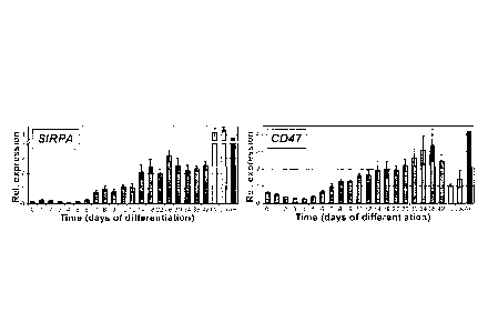
Figure 18 shows gene expression analysis of human adult tissue. (a) QPCR RT
analysis of SIRPA. (b) QPCR RT analysis of CD47.
8
CA 02809498 2013-02-26
=
WO 2012/024782
PCT/CA2011/000965
Figure 19 shows expression of non-myocyte markers in Y2-1-derived
differentiation
cultures. (a) Flow cytometric analysis of markers specifically expressed on
non-myocyte
(SIRPA-) cells in day 20 differentiation cultures. (b) Quantification of
expression of non-
myocyte markers at day 20 of differentiation from Y2-1 iPS cells.
Figure 20 is a table showing the efficiency of fluorescent-activated cell
sorting (FACS)
with the SIRPA antibody. (a) Recovery of SIRPA- cells after FACS of EB-derived
cells
from HES2 at day20 of differentiation, n=8. (b) Recovery of SIRPA+ cells after
FAGS of
EB-derived cells from HES2 at day20 of differentiation, n=9. Total cell # =
total cells
passed through the flow cytometer; SIRPA- (SIRPA-F)# = total SIRPA-(SIRPA+)
cells
recovered after the sorting procedure; SIRPA-(SIRPA+)% = percentage of SIRPA-
(SIRPA+) cells determined by staining with the SIRPA antibody; SIRPA-(SIRPA+)
exp
cell# = cells number of SIRPA-(SIRPA+) cells expected based on staining with
the
SIRPA antibody and on total cell number sorted; Eff SIRPA-(SIRPA+) =
efficiency of
SIRPA-(SIRPA+) cell recovery: SIRPA-(SIRPA+) cell# I SIRPA-(SIRPA+) exp cell#;
Eff
SIRPA-(S1RPA+) =efficiency of SIRPA-(SIRPA+) cell recovery in percentage.
Figure 21 is a table showing the efficiency of fluorescent-activated cell
sorting (FACS)
with the nonmyocyte markers. (a) Recovery of LIN- cells after FACS of EB-
derived cells
from HES2 at day20 of differentiation, n=6. (b) Recovery of LIN+ cells after
FACS of EB-
derived cells from HES2 at day20 of differentiation, n=6. Total cell #= total
cells passed
through the flow cytometer; LIN-(LIN+)# = total LIN-(LIN+) cells recovered
after the
sorting procedure; LIN-(LIN+)% = percentage of LIN-(LIN+) cells determined by
staining
with the LIN antibodies; LIN-(LIN+) exp cell# = cells number of LIN-(LIN+)
cells
expected based on staining with the LIN antibodies and on total cell number
sorted; Eff
LIN-(LIN+) = efficiency of LIN-(LIN+) cell recovery: LIN-(LIN+) cell# / LIN-
(LIN+) exp
cell#; Eff LIN-(LIN+) = efficiency of LIN-(LIN+) cell recovery in percentage.
DETAILED DESCRIPTION
There is described herein the use of a high throughput flow cytometry screen
to identify
cell surface markers specific for human cardiomyocytes. Here we report that
the cell
surface receptor SIRPA is expressed on hPSC-derived cardiomyocytes as well as
on
human fetal cardiomyocytes. Using cell sorting with an antibody against SIRPA
we
9
CA 02809498 2013-02-26
WO 2012/024782
PCT/CA2011/000965
demonstrate that it is possible to isolate populations consisting of up to 98%
cardiomyocytes from hPSC differentiation cultures.
Cell surface antigen, SIRPA (also known as CD172a, BIT, SHPS1), can be found
specifically and exclusively on cardiac progenitor cells and on troponin T-
positive
cardiomyocyte cells generated from human pluripotent stem cells (hPSCs) under
appropriate differentiation conditions.
Prior to the present application, there was no indication or evidence in the
art that
SIRPA is expressed on developing mouse or human cardiovascular cells. RNA
expression of human SIRPA has been found in different parts of the brain as
well as in
blood and at low levels in the lung. However, SIRPA RNA expression has not
been
found in the heart (http://biodps.dnf.orq). SIRPA protein expression has been
detected
in the brain, in blood and lymphoid tissues and in the colon, and at moderate
to weak
levels in placenta, pancreas, spleen, bladder and stomach
(httb://www.proteinatlas.oro/).
However, no protein expression has been reported for the adult human heart. As
such,
the discovery that SIRPA is expressed in hPSC-derived cardiac progenitor cells
and
cardiomyocyte cells is both novel and surprising.
In one example, the use of a SIRPA binding moiety, such as a SIRPA antibody,
provides a simple and novel method to identify, monitor and isolate
cardiomyocyte cells
and their progenitor cells from populations derived from human embryonic stem
cells
and induced pluripotent stem cells. Cell isolation is easy and efficient,
yielding
populations, in one embodiment, consisting of greater than 90% cardiomyocyte
cells
that remain viable and can be used for the applications disclosed herein.
SIRPA was identified as a potential cardiac marker in a screen of over 350
commercially
available antibodies supplied by the Ontario Institute for Cancer Research
Antibody
Core Facility. The antibodies were screened against hESC-derived populations
representing different stages of cardiac development generated by the directed
differentiation of the hESCs using a previously published protocol (Yang et
al., 2008).3
Antibodies that stained cell populations of similar size to the cardiomyocyte
population
in the differentiation cultures (as defined by cardiac troponin T (cTnT)
staining) were
investigated further and used for cell sorting. Of the 350 surface antibodies,
one
antibody, SIRPA, specifically and exclusively stained the hESC-derived
cardiomyocyte
population.
CA 02809498 2013-02-26
WO 2012/024782
PCT/CA2011/000965
Cells isolated based on SIRPA expression represent a novel source of highly
enriched
pluripotent stem cell-derived cardiomyocyte progenitor cells (e.g. at the
onset of Nkx2.5
expression but before cell contraction and expression of the cardiac-specific
structural
proteins) and cardiomyocyte cells for various applications, including but not
limited to
the establishment of patient-specific disease models as well as genetic,
epigenetic and
proteomic analyses of cardiac progenitor cells and cardiomyocyte cells from
normal and
patient-specific pluripotent stem cells.
The specific expression of SIRPA on cardiac cells and their precursors
suggests a
function for this receptor and its downstream signalling pathways during
cardiac
development and differentiation.
SIRPA can also be used as a negative marker for cell sorting experiments to
enrich for
non-cardiogenic PSC-derived lineages such as including those derived from the
somite
(progenitor cells of skeletal muscle, bone, and cartilage/chondrocytes).
Therefore, in one aspect, there is provided a method of enriching a population
of cells
for cardiomyocyte cells and cardiomyocyte progenitor cells comprising
providing the
population of cells from which cardiomyocyte cells and cardiomyocyte
progenitor cells
are to be isolated; and isolating from the population, cells expressing SIRPA;
wherein
the population of cells comprises a population of human pluripotent stem cells
induced
to differentiate into cardiomyocyte cells and cardiomyocyte progenitor cells.
In one embodiment, the human pluripotent stem cells are embryonic stem cells.
In
another embodiment, the human pluripotent stem cells are induced pluripotent
stem
cells.
In some embodiments, the human pluripotent stem cells are exposed to an amount
of at
least one inducing agent effective to induce cell differentiation.
In a preferable embodiment, the at least one inducing agent comprises a
cytokine. The
at least one inducing agent may comprise activin A, preferably at a
concentration of up
to 40ng/ml, further preferably at a concentration of about 6ng/m1 or about
30ng/ml. the
at least one inducing agent may also independently comprise bone morphogenetic
protein 4, preferably at a concentration of up to 40ng/ml, further preferably
at a
concentration of about lOng/ml.
11
CA 02809498 2013-02-26
=
WO 2012/024782
PCT/CA2011/000965
In some embodiments, the human pluripotent stem cells are further exposed to a
bone
morphogenetic protein inhibitor, preferably selected from the group consisting
of
Dorsomorphin, Noggin and soluble bone morphogenetic protein receptors.
In some embodiments, the human pluripotent stem cells are further exposed to
at least
.. one of VEGF, DKK and bFGF
In some embodiments, the human pluripotent stem cells are exposed to the
inducing
agent for between about 1 and about 5 days, preferably about 3 days.
In some embodiments, the time between the initiation of induction of the human
pluripotent stem cells and isolating the cells expressing SIRPA is between
about five
days and about forty-five days, preferably between about 8 and about 25 days.
In some embodiments, the cells expressing SIRPA are isolated after the onset
of SIRPA
expression by the cells, which appears around the time of onset of Nkx2.5
expression
by the cells. Preferably, the cells having the SIRPA cell surface antigen are
isolated
between the time of the onset of Nkx2.5 expression by the cells and the time
of the
onset of contraction and expression of the cardiac-specific structural
proteins by the
cells.
In some embodiments, the method further comprises depleting from the
population,
cells expressing at least one of CD90, CD31, CD140B and CD49A, preferably
using a
corresponding antibody.
Methods for isolating cells expressing a particular molecule, in this case
SIRPA, are
known to a person skilled in the art. In some embodiments, the presence of
SIRPA is
directly used to isolate cells by using a SIRPA-specific ligand, preferably
using an anti-
SIRPA antibody or antibody fragment, or antibody-like molecule, and further
preferably
an anti-SIRPA antibody. In some embodiments, the cells are then isolated using
.. magnetic beads and/or flow cytometry. Alternatively, cells expressing SIRPA
may be
indirectly selected. For example, in some embodiments, the cells in the
population
comprise a reporter gene operably linked to regulatory control elements of the
SIRPA
locus whereby the reporter gene is expressed in cells that express SIRPA and
the step
of isolating the cells expressing SIRPA comprises isolating cells expressing
the reporter
gene. In one preferable embodiment, the reporter gene confers resistance to a
cytotoxic
agent. In another preferable embodiment, the reporter gene is a cell surface
tag.
12
CA 02809498 2013-02-26
WO 2012/024782
PCT/CA2011/000965
In some embodiments, the enriched population of cells comprises at least 60%,
preferably at least 90%, further preferably 98%, cardiomyocyte cells and
cardiomyocyte
progenitor cells.
In a further aspect, there is provided an enriched population of cardiomyocyte
cells and
cardiomyocyte progenitor cells obtained using any one of the methods described
herein.
In a further aspect, there is provided an isolated population of cells
enriched for
cardiomyocyte cells and cardiomyocyte progenitor cells, wherein the population
of cells
comprises at least 60%, preferably at least 90%, further preferably 98%,
cardiomyocyte
cells and cardiomyocyte progenitor cells.
In a further aspect, there is provided the use of SIRPA for isolating
cardiomyocyte cells
and cardiomyocyte progenitor cells from a population of cells, wherein the
population of
cells comprise a population of human pluripotent stem cells induced to
differentiate into
cardiomyocyte cells and cardiomyocyte progenitor cells.
In a further aspect, there is provided a method of depleting a population of
cells for
cardiomyocyte cells and cardiomyocyte progenitor cells comprising: providing
the
population of cells from which cardiomyocyte cells and cardiomyocyte
progenitor cells
are to be depleted; and depleting from the population, cells expressing SIRPA;
wherein
the population of cells comprises a population of human pluripotent stem cells
induced
to differentiate into cardiomyocyte cells, cardiomyocyte progenitor cells, and
non-
cardiomyocytes.
In a further aspect, there is provided a method of enriching a population of
cells for
cardiomyocyte cells and cardiomyocyte progenitor cells comprising: providing
the
population of cells from which cardiomyocyte cells and cardiomyocyte
progenitor cells
are to be isolated; and depleting from the population, cells expressing at
least one of
CD90, CD31, CD140B and CD49A; wherein the population of cells comprise a
population of human pluripotent stem cells induced to differentiate into
cardiomyocyte
cells, and cardiomyocyte progenitor cells, and non-cardiomyocytes.
The term "enriching", as used in the context of the present invention,
includes any
isolation or sorting process that increases the relative abundance of a
desired cell type,
or cell types, in a population of cells.
13
CA 02809498 2013-02-26
WO 2012/024782
PCT/CA2011/000965
As used herein, the term "cardiomyocyte cells" refers to the cells that
comprise cardiac
muscle.
The term "cardiomyocyte progenitor cells" means progenitor cells derived from
human
pluripotent stem cells that have the capacity to differentiate into
cardiomyocyte cells.
.. As used herein, the process of "isolating cells" refers to any method known
to those
skilled in the art for sorting cells including, but not limited to, flow
cytometry,
fluorescence activated cell sorting, magnetic separation using antibody-coated
magnetic
beads, affinity chromatography, and the exploitation of differences in
physical properties
(e.g., density gradient centrifugation).
.. "Embryonic stem cells" ("ESC") are pluripotent stem cells that are derived
from early-
stage embryos.
"Induced pluripotent stem cells" ("iPSC"), as used in the context of the
present invention,
is a type of pluripotent stem cell that has been artificially derived from a
non-pluripotent
cell by inducing the expression of specific genes.
The term, "cell surface antigen", refers to antigens on surfaces of cells that
are capable
of being recognized by the immune system and binding specifically to an
antibody.
As used herein, the phrase "induced to differentiate" refers to any method
known in the
art used to initiate the differentiation of human pluripotent stem cells into
specialized cell
types. These methods may include exposure of the human pluripotent stem cells
to an
.. inducing agent.
As used herein, the term "inducing agent" refers to any agent capable of
initiating
differentiation of hPSCs into specialized cell types, including cardiomyocyte
cells and
cardiomyocyte progenitor cells. Inducing agent therefore includes cytokines,
including
but not limited to activin A, bone morphogenetic protein 4 (BMP4), basic
fibroblast
.. growth factor (bFGF, also known as FGF2), vascular endothelial growth
factor (VEGF,
also known as VEGFA), dickkopf homolog 1 (DKK1), and combinations therefrom.
Methods for inducing human pluripotent stem cells to differentiate into
cardiomyocyte
cells and cardiomyocyte progenitor cells are known to a person skilled in the
art (for
e.g., see Yang et al.3, and Laflamme et a1.19). In some embodiments, induction
14
= CA 02809498 2013-02-26
WO 2012/024782
PCT/CA2011/000965
conditions (e.g. concentrations of the inducing agents and timing of their
use) can be
optimized by measuring SIRPA concentration in the resulting enriched
population.
The ability to generate cells of the cardiac lineage from human pluripotent
stem cells
hPSCs (including embryonic stem cells; hESCs and induced pluripotent stem
cells;
hiPSCs) provides a novel and unlimited supply of human cardiomyocyte cells
that will be
useful for: 1) predictive drug toxicology and drug discovery, 2)
transplantation for the
treatment of cardiovascular disease and 3) modeling cardiovascular development
and
disease in vitro.
The following examples are illustrative of various aspects of the invention,
and do not
limit the broad aspects of the invention as disclosed herein.
EXAMPLES
MATERIALS AND METHODS
HPSC maintenance and differentiation
HPSCs were maintained as described 26. Embryoid bodies (EBs) were
differentiated to
the cardiovascular lineage as previously described 2'3(Fig. 1a). In brief: EBs
were
generated on day (d0) and BMP4 (1 ng/ml) was added for the first day of
differentiation
(d0-d1). At dl, EBs were harvested and resuspended in induction medium (basic
fibroblast growth factor (bFGF; 2.5 ng/ml), Activin A (6 ng/ml) and BMP-4 (10
ng/ml)).
The medium was changed on d4 and was supplemented with vascular endothelial
growth factor (VEGF; 10 ng/ml) and DKK (150 ng/ml). Media was changed again on
d8
and was supplemented with VEGF (20 ng/ml) and bFGF (10 ng/ml). EBs were
cultured
in StemPro-34 (Invitrogen) throughout the experiment. Cultures were maintained
in a
5% CO2, 5% 02, 90% N2 environment from dO-d12 and were then transferred into a
5%
CO2/air environment for the remainder of the culture period.
NKX2-5-GFP hESCs were generated by targeting sequences encoding GFP to the
NKX2-5 locus of HES3 cells using previously described protocols 27(D.E.,
A.G.E. and
E.G.S., manuscript submitted).
= = CA 02809498 2013-02-26
WO 2012/024782
PCT/CA2011/000965
Work involving human tissue collection and analysis was carried out in
accordance with
and approved through the Human Ethics Committee at the University Health
Network.
Flow cytometry and cell sorting
Dissociation procedure for day5 to day12 EBs: EBs generated from hPSC
differentiation
experiments were dissociated with 0.25% trypsin/EDTA. Dissociation procedure
for
day13 and older EBs and human fetal tissue: EBs generated from hPSC
differentiation
cultures were incubated in collagenase type 11 (1mg/m1; Worthington, LS004176)
in
Hanks solution (NaC1 136mM, NaHCO3 4.16mM, NaPO4 0.34mM, KC1 5.36 mM,
KH2PO4 0.44 mM, Dextrose 5.55mM, Hepes 5mM) over night at room temperature
with
gentle shaking28. The following day, the equivalent amount of dissociation
solution (in
Hanks solution: taurin, 10mM, EGTA 0.1mM, BSA 1nrig/ml, collagenase type II
1mg/m1)
was added to the cell suspension and the EBs were pipetted gently for complete
dissociation. Cells were centrifuged (1000rpm, 5min) and filtered. For EBs
past day 40
of differentiation, additional treatment with 0.25% trypsin/EDTA may be
required in order
to obtain complete dissociation into single cells.
Cells were stained at a concentration of 2.5x10⁶ cells/1-ni with anti-KDR -
allophycocyanin (R&D Systems; 1:10) and anti-PDGFRA - phycoerythrin (R&D
Systems; 1:20), anti-S1RPA- IgG-phycoerythrin-Cy7 (clone SE5A5; BioLegends;
1:500)1029, anti-SIRPA- IgG-biotin (clone SE5A5; BioLegends; 1:500)10, anti-
cardiac
isoform of Troponin T (cTNT)(clone 13-11; NeoMarkers; 1:400), goat anti-mouse
IgG -
allophycocyanin (BD; 1:200), Streptavidin - allophycocyanin (BD: 1:200), anti-
IgG1K -
phycoerythrin-Cy7 (clone MOPC-21; BioLegends; 1:500), anti-IgG1K-biotin (clone
MOPC-21; BioLegends; 1:500).
For cell surface markers, staining was carried out in PBS with 10% FCS. For
intracellular proteins, staining was carried out on cells fixed with 4%
paraformaldehyde
(Electron Microscopy Sciences, Hatfield, PA, USA) in PBS and stainings were
performed in PBS with 10% FCS and 0.5% saponin (Sigma). Stained cells were
analyzed using an LSRII flow cytometer (BD). For fluorescent activated cell
sorting, the
cells were sorted at a concentration of 106 cells/ml in 1MDM/6%FCS using a
FACSAriaTMII (BD) cell sorter (SickKids-UHN Flow Cytometry Facility, Toronto,
ON,
Canada). In order to prevent cell death due to pressure and sheer stress, all
sorts were
16
performed with a 100 micron nozzle. For magnetic bead sorting, the Miltenyi
MACS
bead sorting system was used and the experiments were carried out according to
the
manufacturer's guidelines and the sorting conditions for dim markers. For the
high
throughput flow cytometry analysis the BD high throughput sampler (HTS) for
the LSRII
was used according to the manufacturers guidelines. Data were analyzed using
FlowJo
software (Treestar, Ashland, OR, USA).
Immunostaininq
lmmunostaining was performed as previously described 13 using the following
primary
antibodies: rabbit anti-cardiac Troponin I (Abcam; 1:100), mouse anti-SIRPA
(BioLegends; 1:100). Secondary antibodies used were: goat anti-mouse IgGCy3
(Jackson ImmunoResearch; 1:400), donkey anti-mouse IgG-Alexa 488 (Invitrogen;
1:400). DAPI was used to counterstain nuclei. Mito Tracker Red (lnvitrogen)
was used
to stain mitochondria. The stained cells were visualized using a fluorescence
microscope (Leica CTR6000) and images captured using the Leica Application
Suite
software.
Quantitative real-time PCR
Total RNA was prepared with the RNAqueous-Micro Kit (Ambion) and treated with
RNase-free DNase (Ambion). 500 ng to 1 pg of RNA was reverse transcribed into
cDNA
using random hexamers and Oligo (dT) with SuperscriptTM III Reverse
Transcriptase
(lnvitrogen). QPCR was performed on a MasterCycler EP RealPlex (Eppendorf)
using
QuantiFast SYBRTM Green PCR Kit (Qiagen) as described previously 13.
Expression
levels were normalized to the housekeeping gene TATA box binding protein
(TBP). In
addition to TBP for normalization across samples, genomic DNA was used as a
DNA
standard. The copy number of the target gene present in the genomic DNA can be
directly calculated (Human genome size: 2.7 x 10⁹ bp (=1.78 x 10¹² daltons),
corresponds to 6.022 x 10²³ copies of a single copy gene; lug of genomic DNA
corresponds to 3.4 x 10⁵ copies of a single copy gene). The Y-axis of RT-qPCR
graphs
represents copy numbers of the gene of interest divided by copy numbers of
TBP, and
therefore represents an arbitrary but absolute unit, that can be compared
between
experiments.
Total human adult heart RNA was purchased from Ambion and a total human RNA
master panel was purchased from Clontech.
17
CA 2809498 2017-12-29
= = CA 02809498 2013-02-26
WO 2012/024782
PCT/CA2011/000965
RESULTS & DISCUSSION
Identification of novel markers expressed on hESC-derived cardiomyocytes
When induced with appropriate concentrations of Activin A and BMP4 (Fig. la),
the
HES2 hESC line efficiently and reproducibly differentiates to generate
cardiovascular
lineage cells 2'3. Kinetic analyses of the differentiation cultures revealed a
step-wise
developmental progression from a primitive streak-like population defined by
BRA CHYURY (T) expression (days 2-4) to the development of the early mesoderm
(MESP1; days 3-4) and the emergence of NKX2-5 and ISLET1 (ISL1) positive
cardiac
precursors (days 4-8). Contracting cardiomyocytes were first detected between
days 9
and 12 of differentiation, coincident with the up-regulation of MYH6 (aMHC),
MYH7
(f3MHC) and MYL7 (MLC2a) and later MYL2 (MLC2v) expression (Fig. 1b). The
levels of
expression of some of the cardiac specific genes in the hESC-derived
populations were
considerably lower than the levels found in fetal and adult heart tissue. Low
levels of
NEUROD1 and FOXA2 expression indicate that the cultures were not contaminated
with
substantial numbers of neuroectoderm or endoderm-derived cells. To be able to
monitor
cardiomyocyte development in real time, we applied the above protocol to an
NKX2-5-
GFP reporter hESC line that contains the EGFP cDNA inserted into the NKX2-5
locus of
HES3 hESCs (Elliott et at., manuscript submitted). The first NKX2-5-GFP+ cells
developed between days 7 and 8 of differentiation. The size of the NKX2-5-GFP+
population increased with time, reaching a maximum between days 12-20 (Fig.
8).
Analysis of NKX2-5-GFP ESC-derived embryoid bodies (EBs) under epifluorescence
confirmed nuclear GFP expression in the majority of the cells. The kinetics of
NKX2-5-
GFP expression closely parallels the onset of N10(2-5 expression in the HES2
cultures,
indicating that cardiac specification from both hESC lines takes place between
days 6
and 8 of differentiation (Fig. 1 b, Fig. 8). The high proportion of NKX2-5-
GFP+ cells in day
20 cultures demonstrates that the differentiation protocol used efficiently
promotes the
generation of cardiomyocytes from this hESC line.
To determine if the above developmental stages can be distinguished by cell
surface
markers, we carried out a screen of 370 known antibodies
(http://data.microarrays.ca/AntibodyWeb) using day 8, 12, and 20 populations
generated
from the GFP-NKX2-5 cell line. The initial screen focused on identifying
antibodies that
recognized antigens present on the NKX2-5-GFP+ population. From this screen,
we
identified signal-regulatory protein alpha (SIRPA, also known as SHPS-1,
SIRPA) as a
18
= CA 02809498 2013-02-26
WO 2012/024782
PCT/CA2011/000965
potential cardiac-specific marker, as the anti-SIRPA antibodyl stained the
majority of
the NKX2-5-GFP+ cells and almost none of the negative cells (Fig. 2a). From
the panel
of antibodies analyzed, SIRPA was the only one that displayed this
cardiomyocyte
specific expression pattern. SIRPA was first detected on the emerging GFP-NKX2-
5+
cells at day 8 of differentiation, a population considered to represent the
cardiac
precursor stage of development. Expression was maintained on GFP-NKX2-5+
population throughout the 20-day time course of the experiment (Fig. 2a, Fig.
9a). No
SIRPA + cells were detected in undifferentiated hESC populations or in the day
5 cardiac
mesoderm population characterized by co-expression of KDR and PDGFRA (Fig. 2a
and data not shown)2. Analyses of EBs generated from the non-genetically
modified
HES2 line revealed a similar staining pattern with the anti-SIRPA antibody.
SIRPA + cells
were first detected at days 7-8 of differentiation and the percentage of
positive cells
increased significantly over the next 2-4 days (Fig. 2b, Fig. 9b). Both the
directly
conjugated (SIRPA-PE-CY7) and the biotinylated (SIRPA-bio) antibodies stained
similar
portions of the day 20 EB population (Fig. 10a-e). Interestingly, the SIRPA +
cells
detected in day 20 EBs appear to be substantially larger than those found in
the SIRPA
-
population (Fig. 10f), suggesting that cell size of these populations can be
assessed by
flow cytometry. To confirm the specificity of the SIRPA antibody, we carried
out Western
Blot analyses and immunoprecipitation followed by Western Blot analysis (Fig.
11).
These experiments demonstrated the presence of SIRPA protein in 3 independent
day
20 EB-derived populations, but not in undifferentiated hESCs (Fig. 11a).
lmmunoprecipitation analyses revealed a band the size of that previously
described for
the SIRPA protein (Fig. 11b)11.
Co-staining of SIRPA and cTNT by flow cytometry displayed clear co-expression
of the
two markers (Fig. 12a/b), indicating that SIRPA was specifically expressed on
the
cardiomyocyte lineage in differentiated populations generated from the non
modified
HES2 cell line.
RT-qPCR analyses revealed an expression pattern for SIRPA that closely
mirrored the
flow cytometry antibody staining profile, with an up-regulation of SIRPA mRNA
between
days 6 and 8 of differentiation, followed by persistence of expression over
the 42-day
time course. Expression of CD47, the ligand for SIRPA, paralleled that
observed for
SIRPA (Fig. 2c). Flow cytometric analysis of CD47 reflected the gene
expression
pattern, showing low levels of staining on undifferentiated ES cells and on
day 5
19
= CA 02809498 2013-02-26
=
WO 2012/024782
PCT/CA2011/000965
differentiation cultures, followed by broad staining on the entire population
at days 8 and
20 (data not shown).
lmmunofluorescence analysis of monolayer cultures derived from day 20 EBs
revealed
SIRPA surface expression exclusively on cardiomyocytes, as characterized by co-
expression with cardiac Troponinl (cTNI)(Fig. 2d) The respective controls (IgG
and
secondary antibody only) did not show any signal (data not shown).
Collectively, these
kinetic studies show that expression of SIRPA uniquely marks the cardiac
lineage in
hESC differentiation cultures, beginning with the emergence of NKX2-5+
precursor cells
and persisting through the development and expansion of contracting
populations.
Hattori et al recently demonstrated it was possible to isolate cardiomyocytes
based on
mitochondria content, as measured by retention of a mito tracker dye9.
Comparison of
mito tracker dye labeling with SIRPA staining indicated that both procedures
mark the
same cardiomyocyte population in day 20 EBs (Fig. 13c). The dye retention
approach
was, however, less useful in tracking the onset of cardiovascular development,
as it
marked a less distinct population at day 12 of differentiation and almost no
cells at day 8
(Fig. 13a/b). In contrast, a substantial SIRPA + population could be clearly
resolved at
both these time points indicating that this surface marker allows one to
monitor and
isolate cells from different stages of cardiac development, whereas labeling
with the
mito tracker dye can only be used on populations containing relatively mature
cardiomyocytes.
In contrast to the human cells, Sirpa was not detected on mouse ESC-derived
cardiomyocytes by antibody staining (Fig. 14a). Sirpa + populations in the
culture were
cardiac Troponin T (cTnT) negative and CD45 positive, indicating that they
represent
hematopoietic cells (Fig. 14a/b). Gene expression analyses confirmed the flow
cytometric data, and showed only low levels of Sirpa mRNA in the mESC-derived
cardiomyocytes as well as in adult mouse atrial and ventricular tissues,
compared to
high expression in the brain (Fig. 14c). Expression of the only other known
Sirp family
member in the mouse, Sirpb, could not be detected in any of these tissues by
qPCR
(data not shown). Western blot analysis of control and Sirpa-deficient mouse
tissue
.. confirmed high Sirpa expression in the brain of control mice, but not in
any of the tissues
derived from Sirpa-deficient mice (Fig. 14d). Most importantly, no Sirpa
expression was
detected in the heart, kidney or nnESC-derived cardiomyocytes from control
mice.
= CA 02809498 2013-02-26
WO 2012/024782
PCT/CA2011/000965
Differences in SIRPA function and protein homology for mouse and human have
been
described previously for the interaction of macrophages and red blood cells12.
Purification of cardiomyocytes from hESC-derived populations
To assess whether expression of the SIRPA surface receptor can be used to
generate
enriched populations of cardiomyocytes, SIRPA-positive (SIRPA) and SIRPA-
negative
(SIRPA-) fractions were isolated by cell sorting from HES2-derived EBs at days
8, 12
and 20 of differentiation and analyzed for expression of cardiac Troponin T
(cTNT) by
intracellular flow cytometry (Fig. 3a). Analyses of the presort (unsorted, PS)
populations
demonstrated that cTNT expression closely paralleled that of SIRPA at the
corresponding stages during differentiation (PS: d8, d12, d20). Following
sorting, the
SIRPA + fractions from each stage were highly enriched for cTNT +
cardiomyocytes,
whereas the SIRPA" fractions were depleted of these cells. It is unclear if
the low
numbers of cTNT + cells present in the SIRPA" fractions are contaminants from
the
sorting procedure or represent true SIRPA-negative cardiomyocytes. FACS based
separation in multiple experiments reproducibly yielded significantly enriched
populations of cardiomyocytes (SIRPA+: day8 (95.2% 1.9), day12 (94.4%
1.7),
day20 (89.6% 3.6); SIRP-: day8 (13.0% 2.1), day12 (14.3% 3.9), day20
(15.7%
6.0))(Fig. 3b). The purity of the SIRPA + and SIRPA" sorted populations and
the
efficiency of cell recovery from the sorting procedure is summarized in Figure
15 and
Figure 20 (Table 1).
Molecular analyses revealed that the SIRPA + cells expressed significantly
higher levels
of NKX2-5, MYH6, MYH7 and MYL7 than the SIRPA- population (Fig. 3c), further
demonstrating enrichment of cardiomyocytes. As expected, SIRPA expression
segregated to the SIRPA + population. In contrast to the cardiac markers, non-
myocyte
markers such as the fibroblast markers DDR2 and THY1 (CD90, data not shown)
and
the endothelial marker PECAM (CD31) were expressed at higher levels in the
SIRPA"
population (Fig. 3c).
When plated in monolayer cultures, cells from both SIRPA" and SIRPA +
fractions formed
viable populations that could easily be maintained for several weeks.
Contracting cells
were detected in unsorted (PS) and SIRPA-derived populations, but not in the
population generated from the SIRPA- cells. Imnnunohistochemical analysis
revealed
broad cTNI expression in the SIRPA + population confirming the high proportion
of
21
= CA 02809498 2013-02-26
WO 2012/024782
PCT/CA2011/000965
cardiomyocytes in these cultures. Only few cTNI-positive cells were detected
in the
SIRPA- population (Fig. 3d)
As anticipated from the co-expression of SIRPA and NKX2-5-GFP, it was also
possible
to isolate populations enriched for cardiac lineage cells from NKX2-5-GFP HES3-
derived cultures by sorting with the anti-SIRPA antibody. Cardiac precursors
(day 8) and
cardiomyocytes (days 12 and 20) defined by gene expression and cTNT staining,
segregated to the SIRPA + fraction whereas non-myocyte cells were enriched in
the
SIRPA- population (Fig. 16).
To enable rapid processing of large numbers of cells, we also attempted to
isolate
SIRPA cells by magnetic bead sorting. Isolation of SIRPA + cells from NKX2-5-
GFP
differentiation cultures by this approach resulted in populations highly
enriched for
cardiomyocytes similar to those derived from FACS experiments (Fig. 17a-c).
However,
with current magnetic bead sorting protocols a substantial amount of cells is
lost during
the process, resulting in a lower efficiency of this approach compared to FACS
(compare Fig. 17d to Fig. 20 (Table 1)).
Taken together, the findings from these cell sorting studies clearly
demonstrate that
SIRPA expression marks the cardiac lineage in hESC-derived differentiation
cultures
and that cell sorting with the anti-SIRPA antibody allows for the isolation of
populations
highly enriched for cardiomyocytes.
Purification of cardiomyocytes from human induced pluripotent stem cells
To determine if SIRPA expression marked the cardiac lineage in other hPSC-
derived
populations, we next analyzed EBs generated from two different hiPSC lines,
MSC-iPS1
(also known as Y2-1) and 38-2 13'14. The efficiency of cardiac differentiation
from both
lines was low, as demonstrated by the proportion of cTNT+ cells (MSC-iPS1:
12.2%
5.6, 38-2: 26.7% 5.7; Fig. 4a). Similar low levels of SIRPA expression were
detected
in both EB
populations. FACS of the SIRPA + cells from both iPSC lines yielded
populations significantly enriched for cTNT+ cardiomyocytes (SIRPA: MSC-iPS1
(67.0% 3.6), 38-2(71.4% 3.8); SIRPA-: MSC-iPS1 (4.9% 2.1), 38-2(6.2%
0.9))(Fig 4a,b). These SIRPA + populations expressed significantly higher
levels of
NKX2-5, MYH6, MYH7, MYL2 and MYL7 than the corresponding SIRPA" cells. As
observed with the hESC-derived cells, non-myocyte markers including DDR2,
PDGFRB,
THY1 and NEUROD segregated to the SIRPA" fraction (Fig. 4b,c and data not
shown).
22
== CA 02809498 2013-02-26
WO 2012/024782
PCT/CA2011/000965
These data clearly document the utility of this marker for generating enriched
cardiac
populations from a range of pluripotent stem cell lines, including those that
do not
differentiate efficiently to the cardiac lineage with the current protocols.
SIRPA expression in human fetal and adult heart cells
To determine if SIRPA is expressed on primary human cardiomyocytes, we next
analyzed expression patterns in fetal (18-20 weeks of gestation) and adult
heart tissue
by RT-qPCR. As shown in Figure 5a, SIRPA transcripts were detected in all
fetal-
derived heart tissue (left (LA) and right atrial (RA) cells, left (LV) and
right ventricle (RV)
cells, apex (AP) and atrioventricular junction (AVJ)), with comparable levels
to those
found in day 20 hESC-derived cells (Fig. 3a). SIRPA was not expressed in
undifferentiated hESCs (d0) or in control HEK (human embryonic kidney) cells.
Similar
to the fetal heart, SIRPA expression was also detected in the adult heart,
suggesting
that its expression marks cardiomyocytes at different stages of human cardiac
development. High levels of SIRPA were detected in the adult human brain and
lung
(Fig. 18a) with low levels found in many other tissues. These low levels may
reflect the
presence of tissue macrophages that are known to express this receptor15=16.
CD47, the
SIRPA ligand was expressed in most tissues, confirming the pattern described
in
previous studies (Fig. 18b)15. lmmunofluorescence staining showed that SIRPA
was
localized on the surface membrane of the fetal ventricular cells but was not
present on
other membrane fractions such as the mitochondrial membrane, as indicated by
the lack
of co-staining with Mito Tracker Red (Fig. 5b). Flow cytometric analyses
revealed a high
proportion of SIRPA + cells in all fetal heart tissues at levels that
correlated with the
percentage of cINT+ cells in the respective fractions (Fig. 5c, d).
These findings demonstrate clearly that SIRPA is expressed on fetal
cardiomyocytes as
well as in adult heart, illustrating that its cardiac-specific expression is
not an artifact of
pluripotent stem cell-derived cultures.
Using SIRPA expression to monitor the efficiency of hPSC differentiation
Recently, we reported that co-expression of KDR and PDGFRA provides a reliable
method to monitor cardiac mesoderm induction following treatment with BMP4 and
Activin A 2(Fig. 6a). While this study showed that the induction of a
KDR+PDGFRA+
population was an essential first step in the generation of the cardiomyocyte
population,
not all KDR+PDGFRK populations differentiated to give rise to cardiac lineage
cells
23
= CA 02809498 2013-02-26
WO 2012/024782
PCT/CA2011/000965
(example of this type of population: induced with 30 ng/ml BMP4 and no
exogenous
Activin A (AO)). To determine if SIRPA would more accurately predict cardiac
potential
of differentiating populations at an early stage, we monitored its expression
in day 9 EBs
induced with different concentrations of Activin A and BMP4 (Fig. 6b). The
same
populations were evaluated at day 5 for expression of KDR and PDGFRA (Fig. 6a)
and
at day 20 for expression of cTNT (Fig. 6c). While there was little correlation
between the
size of the KDR4PDGFRA+ population at day 5 and the proportion of cTNT + cells
at day
20, the cultures with the largest SIRPA population at day 9 (Activin A 6
ng/ml, BMP4 10
ng/ml) contained the highest number of cTNT+ cells at the later time point.
SIRPA
expression correlated well with cTNT output for most conditions tested and the
highest
levels of SIRPA predicted the highest cardiomyocyte development at day 20
(Fig. 6d).
These data demonstrate that expression of SIRPA at day 9 is a reliable
indicator of
cardiomyocyte potential, and as such can be used to monitor and optimize
induction
protocols for directed differentiation of hPSCs to the cardiac lineage.
Enrichment of hPSC-derived cardiomyocytes through depletion of the non-mvocyte
lineage cells
In addition to antibodies that recognize cardiomyocytes, our flow cytometric
screen also
identified a panel of antibodies that marked the non-myocyte population in the
differentiation cultures. This set of antibodies, including anti-CD90 (THY1,
expressed on
fibroblast cells), anti-CD31 (PECAM1, expressed on endothelial cells), anti-
CD140B
(PDGFRB, expressed on smooth muscle cells) and anti-CD49A (INTEGRIN1A), all
recognized different proportions of the SIRPA- population of day 20 HES2-
derived EBs
(Fig. 7a/d). The combination of these antibodies marked the majority of non-
myocyte
(SIRPA-) cells in the culture (Fig. 7c, presort). To determine if it was
possible to enrich
for cardiomyocytes by depleting cells expressing the non-myocyte markers, we
combined these antibodies and sorted day 20 EBs into lineage-positive (LIN+)
and
lineage-negative (LIN-) fractions (Fig. 7b). This approach has the advantage
in
generating enriched populations free of any bound antibody or magnetic beads.
As
expected, the LIN- population was significantly enriched for SIRPA + cells,
whereas the
LIN+ population was depleted for the cardiomyocytes (Fig. 7c/e). The
efficiency of cell
recovery after FACS for LIN- and LIN+ cells is summarized in Figure 21 (Table
2). Gene
expression analyses revealed that non-myocyte specific genes including PECAM1,
PDGFRB, THY/ and DDR2 were primarily expressed in the LIN+ fraction, whereas
cardiac gene expression was restricted to the LIN- fraction (Fig. 7f). When
plated on
24
CA 02809498 2013-02-26
=
WO 2012/024782
PCT/CA2011/000965
gelatin coated dishes or re-aggregated as cell clusters, the LIN" fraction
generated
populations that contained a high proportion of contracting cardiomyocytes
(data not
shown). The same lineage cocktail of antibodies also marked the non-myocyte
(SIRPA-)
fraction of the iPSC (MSC-iPS1)-derived day 20 EB population (Figure 19),
indicating
that this depletion approach can be applied to different PSC lines with
variable
differentiation efficiencies.
Taken together, these data illustrate that cardiomyocytes can be enriched from
hPSC-
derived differentiation cultures by depletion of the non-myocte lineages. This
method
therefore represents an alternative approach to obtaining highly purified
cardiomyocyte
cultures and may as such be used for strategies that require purified
cardiomyocyte
populations free of any bound antibodies.
Advances in our understanding of the signaling pathways that regulate lineage
specification has led to strategies for the efficient and reproducible
directed
differentiation of hPSCs to specific cell types I. With respect to cardiac
lineage
development, protocols have been established that promote the generation of
mixed
cardiovascular populations representing the major cell types found in the
human heart
including cardiomyocytes, endothelial cells, vascular smooth muscle cells and
fibroblasts. Cardiomyocytes typically represent between 10% and 70% of such
mixed
populations 2'3, depending on the PSC line used. While such mixed populations
have
been used to demonstrate the potential utility of the PSC-derived cells for
predictive
toxicology 5, modeling human disease in vitro 17'15 and transplantation based
therapy for
heart disease 19, highly enriched and well defined cell populations will
ultimately be
required to translate this potential into practical applications.
Our identification of SIRPA as a cardiomyocyte-specific marker now enables,
for the first
time, easy and routine access to highly enriched populations of cardiomyocytes
from
hESCs and hiPSCs. These cardiomyocyte enriched populations can be isolated by
FACS or magnetic bead sorting, the latter approach enabling the isolation of
large
numbers of cells required for in vivo studies. Access to highly enriched
populations of
cardiomyocytes through simple sorting approaches will enable the development
of
defined high throughput drug discovery and toxicology assays, the detailed
phenotypic
evaluation of cells generated from patient specific hPSCs, and the generation
of defined
populations safe for transplantation. The fact that SIRPA is expressed on
cardiac
lineage cells from the earliest cardiac stage to contracting and more mature
CA 02809498 2013-02-26
WO 2012/024782
PCT/CA2011/000965
cardiomyocytes will allow for comparisons of the in vivo potential of the
different
populations.
In addition to SIRPA, our screen also identified a panel of markers defining
the non-
myocyte fractions of the PSC-derived cardiovascular population. The markers
used
suggest that they represent a combination of fibroblasts (CD90, THY1)20,
vascular
smooth muscle cells (CD140B, PDGFRB)21 and endothelial cells (CD31, PECAM1),
Access to enriched populations of each of these cell types together with
cardiomyocytes
will allow. Many of the proposed applications for PSC-derived cardiomyocytes
may
require three-dimensional engineered tissue to more accurately reflect drug
responses
and function in the adult heart. Recent studies suggest that appropriate
combinations of
cardiac cells, endothelial cells and fibroblasts need to be incorporated into
such tissue
constructs in order for them to function best in vitro or in vivo 22.24. Our
ability to generate
pure myocyte and non-myocyte populations will allow for the generation of
engineered
constructs consisting of varying proportions of different cell types, enabling
us to
determine the optimal proportion of each required to form heart tissue with
structural
and functional properties most similar to that of the human heart.
The specific expression pattern of SIRPA in the PSC-derived populations and in
the
fetal heart tissue suggests that this receptor plays some functional role in
the human
cardiomyocyte lineage, perhaps as early as the precursor stage of development.
The
fact that expression of the ligand, CD47, is upregulated in parallel with
SIRPA in the EBs
and that CD47 is found on a large proportion of the cells in the culture
further supports
the interpretation that this ligand/receptor pair plays a role in the human
cardiomyocyte
development and/or function. One thoroughly studied role for SIRPA is on
macrophages, where it appears to mediate a signal to eliminate cells from the
body that
do not express the ligand CD47 16. The only other suggested function in human
cells is
in the smooth muscle lineage, where SIRPA has been shown to play an important
role
in mediating IGF-1-induced mitogenic signaling 26. Given that SIRPA was not
detected
in mouse cardiomyocytes, it is possible that its function in human cells may
relate to
aspects of cardiomyocyte physiology and/or function that differ between the
two
species.
In summary, the findings reported here demonstrate that expression of SIRPA
uniquely
marks the cardiomyocyte lineage in PSC-differentiation cultures. Isolation of
SIRPA'
cells by FAGS or magnetic bead sorting provides a simple approach for
generating
26
highly enriched populations of cardiomyocytes from a broad range of PSC lines,
including those that do not differentiate efficiently to the cardiovascular
lineage using
current protocols.
Although preferred embodiments of the invention have been described herein, it
will be
understood by those skilled in the art that variations may be made thereto
without
departing from the spirit of the invention or the scope of the appended
claims.
27
CA 2809498 2017-12-29
CA 02809498 2013-02-26
WO 2012/024782
PCT/CA2011/000965
REFERENCE LIST
1 Murry, C. E. & Keller, G. Differentiation of embryonic stem cells to
clinically
relevant populations: lessons from embryonic development. Cell 132, 661-680,
(2008).
2 Kattman, S. J. et al. Stage-specific optimization of activin/nodal and
BMP
signaling promotes cardiac differentiation of mouse and human pluripotent stem
cell lines. Cell Stem Cell 8, 228-240, (2011).
3 Yang, L. et al. Human cardiovascular progenitor cells develop from a
KDR+
embryonic-stem-cell-derived population. Nature 453, 524-528, (2008).
4 Zwi, L. et a/. Cardiomyocyte differentiation of human induced pluripotent
stem
cells. Circulation 120, 1513-1523, (2009).
5 Braam, S.
R., Passier, R. & Mummery, C. L. Cardiomyocytes from human
pluripotent stem cells in regenerative medicine and drug discovery. Trends
Pharmacol Sc! 30, 536-545, (2009).
6 Anderson, D. et
al. Transgenic enrichment of cardiomyocytes from human
embryonic stem cells. Mo/ Ther 15, 2027-2036, (2007).
7 Huber, I.
et al. Identification and selection of cardiomyocytes during human
embryonic stem cell differentiation. FASEB J 21, 2551-2563, (2007).
8 Ritner, C.
et al. An engineered cardiac reporter cell line identifies human
embryonic stem cell-derived myocardial precursors. PLoS One 6, e16004,
(2011).
9 Hattori,
F. et a/. Nongenetic method for purifying stem cell-derived
cardiomyocytes. Nat Methods 7, 61-66, (2010).
10 Seiffert,
M. et a/. Signal-regulatory protein alpha (SIRPalpha) but not SIRPbeta is
involved in T-cell activation, binds to CD47 with high affinity, and is
expressed on
immature CD34(+)CD38(-) hematopoietic cells. Blood 97, 2741-2749, (2001).
11 Timms, J.
F. et al. SHPS-1 is a scaffold for assembling distinct adhesion-
regulated multi-protein complexes in macrophages. Curr Blot 9, 927-930,
(1999).
28
CA 02809498 2013-02-26
WO 2012/024782
PCT/CA2011/000965
12 Subramanian, S., Parthasarathy, R., Sen, S., Boder, E. T. & Discher,
D. E.
Species- and cell type-specific interactions between CD47 and human
SIRPalpha. Blood 107, 2548-2556, (2006).
13 Nostro, M. C. et at. Stage-specific signaling through TGFbeta family
members
and WNT regulates patterning and pancreatic specification of human pluripotent
stem cells. Development 138, 861-871, (2011).
14 Park, I. H. et al. Reprogramming of human somatic cells to
pluripotency with
defined factors. Nature 451, 141-146, (2008).
Matozaki, T., Murata, Y., Okazawa, H. & Ohnishi, H. Functions and molecular
10 mechanisms of the C047-SIRPalpha signalling pathway. Trends Cell Biol
19, 72-
80, (2009).
16 Okazawa, H. et at. Negative regulation of phagocytosis in macrophages
by the
CD47-SHPS-1 system. J Immunol 174, 2004-2011, (2005).
17 Carvajal-Vergara, X. at a/. Patient-specific induced pluripotent stem-
cell-derived
15 models of LEOPARD syndrome. Nature 465, 808-812, (2010).
18 Itzhaki, I. et al. Modelling the long QT syndrome with induced
pluripotent stem
cells. Nature 471, 225-229, (2011).
19 Laflamme, M. A. et a/. Cardiomyocytes derived from human embryonic
stem
cells in pro-survival factors enhance function of infarcted rat hearts. Nat
Biotechnol 25, 1015-1024, (2007).
20 Kisselbach, L., Merges, M., Bossie, A. & Boyd, A. CD90 Expression on
human
primary cells and elimination of contaminating fibroblasts from cell cultures.
Cytotechnology 59, 31-44, (2009).
21 Ross, R. The pathogenesis of atherosclerosis: a perspective for the
1990s.
Nature 362, 801-809, (1993).
22 Stevens, K. R. at a/. Physiological function and transplantation of
scaffold-free
and vascularized human cardiac muscle tissue. Proc Nat! Acad Sc! U S A 106,
16568-16573, (2009).
29
CA 02809498 2013-02-26
WO 2012/024782
PCT/CA2011/000965
23 Dvir, T. et al. Prevascularization of cardiac patch on the omentunn
improves its
therapeutic outcome. Proc Nat! Aced Sc! U S A 106, 14990-14995, (2009).
24 Lesman, A. et al. Transplantation of a tissue-engineered human
vascularized
cardiac muscle. Tissue Eng Part A 16, 115-125, (2010).
25 Ling, Y., Maile, L. A., Lieskovska, J., Badley-Clarke, J. & Clemmons, D.
R. Role
of SHPS-1 in the regulation of insulin-like growth factor I-stimulated Shc and
mitogen-activated protein kinase activation in vascular smooth muscle cells.
Mol
Biol Cell 16, 3353-3364, (2005).
26 Kennedy, M., D'Souza, S. L., Lynch-Kattman, M., Schwantz, S. &
Keller, G.
Development of the hemangioblast defines the onset of hematopoiesis in human
ES cell differentiation cultures. Blood 109, 2679-2687, (2007).
27 Costa, M. et al. A method for genetic modification of human embryonic
stem
cells using electroporation. Nat Protoc 2, 792-796, (2007).
28 Sharma, P., Shathasivam, T., lgnatchenko, V., Kislinger, T. &
Gramolini, A. 0.
Identification of an FHL1 protein complex containing ACTN1, ACTN4, and
PDLIM1 using affinity purifications and MS-based protein-protein interaction
analysis. Mol Biosyst 7, 1185-1196, (2011).
29 Seiffert, M. at a/, Human signal-regulatory protein is expressed on
normal, but
not on subsets of leukemic myeloid cells and mediates cellular adhesion
involving its counterreceptor CD47. Blood 94, 3633-3643, (1999).