Note: Descriptions are shown in the official language in which they were submitted.
CA 02809979 2013-02-28
WO 2012/030854 PCT/US2011/049781
-1-
COMPOSITIONS AND METHODS FOR MODULATING EMT AND USES THEREOF
Related Applications
[0001] This application claims priority to and the benefit of U.S. Application
No.
61/379,327, filed September 1,2010, the teachings of which are incorporated
herein by
reference.
Background of the Invention
100021 The epithelial-mesenchymal transition (EMT) is a transdifferentiation
program that
effects critical steps of embryo genesis by interconverting epithelial cell
types into cells with
mesenchymal attributes. EMT programs are also activated in carcinoma cells,
enabling them to
acquire cellular traits associated with high-grade malignancy, including the
ability to complete
various steps of the metastatic cascade. In addition to mesenchymal traits,
recent findings
suggest that adult epithelial cells that pass through an EMT also acquire
properties associated
with normal tissue stem cells (SCs) and tumor-initiating cells. There is
significant interest in
the art in inducing or inhibiting EMT programs.
Summary of the Invention
[0003] The present invention provides compositions and methods useful for
modulating the
epithelial-mesenchymal transition. In one aspect, the invention provides a
composition
comprising one or more compounds selected from each of at least three of the
following
groups: (a) compounds that stimulate TGF-beta pathway signaling; (b) compounds
that
stimulate canonical Wnt pathway signaling; (c) compounds that stimulate non-
canonical Writ
pathway signaling; and (d) compounds that perturb cell adhesion. In some
embodiments, the
composition comprises at least one compound from each of these four groups.
[0004] In another aspect, the invention provides a composition comprising one
or more
compounds selected from each of at least three of the following groups: (a)
compounds that
inhibit TGF-beta pathway signaling; (b) compounds that inhibit canonical Wnt
pathway
signaling; (c) compounds that inhibit non-canonical Wnt pathway signaling; and
(d)
compounds that stimulate BMP signaling. In some embodiments, the composition
comprises at
least one compound from each of these four groups.
NO322261.1 }
CA 02809979 2013-02-28
WO 2012/030854 PCT/US2011/049781
-2-
[00051 In other aspects, the invention provides methods of inducing or
inhibiting EMT,
using, e.g., an inventive composition.
[0006] Certain conventional techniques of cell biology, cell culture,
molecular biology,
microbiology, recombinant nucleic acid (e.g., DNA) technology, immunology,
etc., which are
within the skill of the art, may be of use in aspects of the invention. Non-
limiting descriptions
of certain of these techniques are found in the following publications:
Ausubel, F., et al.,
(eds.), Current Protocols in Molecular Biology, Current Protocols in
Immunology, Current
Protocols in Protein Science, and Current Protocols in Cell Biology, all John
Wiley & Sons,
N.Y., editions as of 2008; Sambrook, Russell, and Sambrook, Molecular Cloning:
A
Laboratory Manual, 3rd ed., Cold Spring Harbor Laboratory Press, Cold Spring
Harbor, 2001;
Harlow, E. and Lane, D., Antibodies ¨ A Laboratory Manual, Cold Spring Harbor
Laboratory
Press, Cold Spring Harbor, 1988; Burns, R., Immunochemical Protocols (Methods
in Molecular
Biology) Humana Press; 3rd ed., 2005, Monoclonal antibodies : a practical
approach (P.
Shepherd and C Dean, eds., Oxford University Press, 2000); Freshney, R.I.,
"Culture of Animal
Cells, A Manual of Basic Technique", 5th ed., John Wiley & Sons, Hoboken, NJ,
2005;
Cancer: Principles and Practice of Oncology (VT. De Vita et al., eds., J.B.
Lippincott
Company, 8th ed., 2008). Further information on cancer may he found in The
Biology of
Cancer, Weinberg, RA, et al., Garland Science, 2006. = All patents, patent
applications,
websites, databases, scientific articles, and other publications mentioned
herein are
incorporated herein by reference in their entirety.
Brief Description of the Drawing
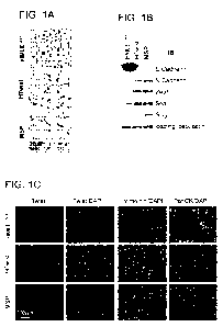
[0007] Figure 1. Characterization of a mesenchymal subpopulation isolated from
immortalized human mammary epithelial cells cells. (A) Bright phase
microscopy: images
of epithelial HMLE24', HMLE overexpressing Twist (HTwist) and MSP cells. (B)
Immunoblot:
E- and N-Cadherin, EMT transcription factors (ITs) Zebl, Snail and Slug. (C)
Immunofluorescence: expression and nuclear localization of EMT-TF Twist in
HMLE24+,
HTwist and MSP cells, levels of mesenchymal intermediary filament vimentin and
epithelial
cytokeratins (panCK = pan-cytokeratin antibody). (D) RT-PCR: expression of EMT-
TFs, E-
and N-Cadherin in HTwist and MSP relative to HMLE24' cells, L32 was loading
control, n=3
(E) Mammosphere Assay: bright phase microscopy images of mammospheres and
quantification, mammospheres/1500 cells, n-24/cell line. (F) Flow cytometry of
CD44 and
(M0322261 I)
CA 02809979 2013-02-28
WO 2012/030854 PCT/US2011/049781
-3-
CD24 cell surface markers. (G) Tumorigenicity assay: RAS-transformed HMLE24+,
HTwist
and MSP cells were implanted subcutaneously at indicated numbers in mice. (H)
Boyden
Chamber Invasion assay: Bright phase microscopy images of invaded, HMLE24+-
RAS, MSP-
RAS, MSP-II-RAS cells and quantification, ri-3. (I) Lung metastasis: HMLE24+-
RAS, MSP-
RAS and MSP-II-RAS cells were implanted in the fat pads of mice: microscope
images of the
major left lung lobe showing GFP-expressing metastatic nodules, magnified
inset and
quantification of lung surface metastases, n=5 mice/group.
[0008] Figure 2. Autocrine TGF-beta and Wnt signaling in HTwist and MSP cells.
(A)
ELISA: TGF-beta 1 secretion in HMLE24+, HTwist and MSP cells, n=-3. (B)
Luciferase reporter
assay: Smad transcriptional activity: cells were transfected with SBE4-luc
reporter plasmid,
firefly luciferase levels were normalized to pGL-SV40 renilla transfection
control, n=3. (C)
Immunoblot: smad2 phosphorylation in HMLE24+, HTwist and MSP cells. (D)
Immunoblot:
before lysis, cells were cultivated for 24 hr in either regular medium (++),
growth factors
reduced to 10% of regular concentration (+), or in growth factor-free medium (-
). (E) RT-PCR:
BMP antagonists Gremlin, Chordin-like 2, L32 was loading control, n=3. (F) RT-
PCR: SFRP1,
L32 was loading control, n=3. (G) RT-PCR: DKK1, L32 was loading control, n=3,
ELISA:
DKK1 secretion (n=3). (H) Immunoblot: activation status of pathways associated
with non-
canonical Wnt signaling. (I) Luciferase reporter assay (TOPFlash): cells were
transfected with
Super(8x)TOP (TCF-LEF reporter) or FOP construct (control, binding sites
mutated). Shown is
TOP over FOP firefly luciferase activity, normalized to pGL-SV40 renilla
transfection control,
n=3. (J) Inhibition of TOPFlash activity: DKK1 or SFRP1 recombinant protein
(1.0 12g/in])
were added to the growth medium of HTwist and MSP cells 12h before lysis, n=3.
[0009] Figure 3. Autocrine Signaling controls migration and mammosphere
formation. (A) Migration assay: cells were seeded into Boyden chambers and
recombinant
DKK1 or SFRP1 protein (0.5 p.g/m1) was added to growth medium; #HMLE control
vs. HTwist
and MSP control, p<1x10⁻⁶, *HTwist or MSP control vs. treatment with DKK1 or
SFRP1,
p<lx I 0-6, n=3. (B) Migration assay: dose-dependent inhibition of migration
by DKK1 or
SFRP1 protein at indicated concentrations (g/ml), n=3; 5H-Twist or MSP control
vs.
treatment with DKK1 or SFRP1, p p<1x10⁻⁶, ** H-Twist or MSP, high vs. low
concentrations,
p<1x10⁻⁷, n=3. (C) Migration assay: dose-dependent inhibition of migration by
recombinant
BMP4 protein at indicated concentrations (g/ml); *HTwist or MSP control,
p<0.05,
**0.6i.ig/m1 vs. 1.2 ug/ml, p<lx10-4, n=3. (D) Mammosphere formation:
recombinant DKK1,
[M0322261.1}
CA 02809979 2013-02-28
WO 2012/030854 PCT/US2011/049781
-4-
SFRP I or BMP4 protein were added daily during sphere formation (5 days) at
indicated
concentrations (ng/m1); *HTwist or MSP control vs. treatment, p<0.01, **low
vs. high
concentration or recombinant protein, p<0.05, n(mammospheres)/1000cells, n=8.
(E)
Secondary mammosphere formation: DKK1 (11tg/m1), SFRP1 (1ug/m1) and BMP4 (0.5
g/m1)
were added daily during primary sphere formation (5 days), spheres were
dispersed and seeded
for secondary sphere formation in absence of further treatment; *H-Twist or
MSP control vs
treatment, p<0.01, **primary vs. secondary sphere number, p< 0.01,
n(mammospheres)/1000cells, n=16.
100101 Figure 4. Autocrine Signaling controls tumorigenicity and metastasis of
MSP-
RAS cells. (A) Migration assay; RAS-transformed MSP (MSP-RAS) were seeded in
Boyden
Chamber migration assays and recombinant SFRP1 (1ps/m1) and BMP4 protein (0.5
g/m1)
were added; *control vs. single treatment, p<1x10⁻⁸, **single vs. double
treatment with SFRP1
and BMP4 (S+B), p<1x10⁻⁴, n=3. (B) Tumorsphere assay: SFRP1 (1ug/m1) and BMP4
protein
(0,5g/ml) were added daily during primary sphere formation (5 days), spheres
were dispersed
and seeded for secondary sphere formation in the absence of further treatment;
#control
primary vs. secondary sphere formation, p-0.001; *control vs. single
treatment, p<lx10-4,
**single vs. double treatment with SFRP I and BMP4 (S+B), p<0.01,
n(tumorspheres)/1000
cells, n-6. (C) Flow cytometry: CD44 and CD24 cell-surface markers of
dissociated primary
tumorspheres as described in (B). (D) Orthotopic tumor formation after ex vivo
treatment
during tumorsphere formation: primary tumorspheres as generated in (B) were
pooled,
dissociated and 1.0x10⁵ cells were implanted in the fat pads of mice; shown
are tumor weight
and incidence, *indicates absence of tumor, n=5mice/group. (E) Lung
metastasis: fluorescence
microscopy images of the major left lung lobe showing GFP-expressing
metastatic nodules and
quantification of the number of lung surface metastases; *control vs. SFRP1
treatment, p<0.05,
**SFRP1 vs. BMP4 or double treatment (S+B), p<0.05, n=5 mice/group. (F) Liver
metastasis:
fluorescence microscopy images of the liver surface showing GFP-expressing
micro-metastatic
nodules after implantation of cells as described in (D) and quantification: 5
fields of liver
surface were counted per mouse, *control vs. single treatment, p<0.05,
**single vs. double
treatment (S+B), p<0.05, n=5 mice/group. (G) Tumorigenicity assay: MSP-RAS
cells
suspended in PBS containing SFRP1 (11.rg/m1), BMP4 (0.5 g/m1), and a
combination of both
(S+B) were implanted subcutaneously in mice at indicated cell numbers. In
addition, 20n1 of
(M0322261. I )
CA 02809979 2013-02-28
WO 2012/030854 PCT/US2011/049781
-5-
PBS or PBS with proteins at indicated doses was injected peri-tumorally at
1,2, 3 and 7 days
after implanting the MSP-RAS cells.
100111 Figure 5. Concomitant stimulation of Wnt and TGF-beta pathways allows
HMLE24+ cells to enter into a mesenchymal and SC-like state. (A) RT-PCR: N-
Cadherin,
Zebl and Zeb2, L32 was loading control, n=3. Prior RNA isolation, HMLE244-
shGFP or -
shSFRP1 (stable knockdown of SFRP1, Figure S5A) cells were treated for 3 days
with daily
doses of indicated factors: anti-DKK I antibody (10 g/m1), anti-E-Cadherin
antibody (1p,g/m1),
TGF-beta 1 (5ng/m1), Wnt5a (250ng/m1), (B) Brightfield microscopy: images of
control, TGE-
beta- and iEMT-cocktail treated HMLE24+ cells after 14 days of treatment as
indicated (see also
Figure S5D). (C) RT-PCR: Zebl, Zeb2, E-Cadherin and N-Cadherin, L32 was
loading control,
n=3. After 14 day treatment, cells were expanded in absence of further
stimulation for 4
passages. Shown are PBS-treated control HMLE24+-shGFP and HMLE24+-shSFRP1
cells, two
HMLE24+-shGFP cultures treated with TGF-beta 1 (T0E-(3-a and -b), two HMLE24'-
shSFRP1
cultures treated with the iEMT-cocktail (iEMT-a and ¨b). (D) Immunoblot: EMT
markers and
associated autocrine pathways, cells generated as described in (C). (E)
Migration (Boyden
Chamber Assay) and Mammosphere Assay: cells generated as indicated in (C),
*control vs.
TGF-beta treatment, p<lx10-4, **control vs. iEMT-treatment, p<lx 1 0-9,
n(migration)=3,
n(mammospheres)/1000cells, n=7. (F) Flow cytometry: CD44 and CD24 cell-surface
marker
expression in control, TGF-beta-a and iEMT-a cells generated as described in
(C). (0) Lung
metastasis: cells expanded for 8 passages after cessation of treatment as
described in (C) were
transformed with RAS; 1.0x10⁵ cells were injected into the mammary fat pads of
mice; GFP-
labeled metastases on the surface of the lungs were quantified; *control vs.
all other groups, p <
0.05, n=5mice/group. (H) Migration (Boyden Chamber Assay) and Mammosphere
Assay:
HMLE-iEMT-a were expanded for 12 passages after cessation of treatment as
described in (C),
then seeded into assays in presence of recombinant SFRP1 (1t.tg/ml/d), BMP4
(0.5 g/m1/d) or
both (S+B) protein; *control vs. single treatment, p<0.01, **single vs. double
treatment (S+B),
p<0.05, n(migration)3, n(mammospheres)/1000cells, n=6.
100121 Figure 6. Basal cell populations isolated from human mammary gland
express
mesenchymal markers and EMT-TFs. (A) FACS: single-cell preparations from human
reduction mammoplasties; (1.) dead cells were excluded by 7AAD staining,
followed by (2.)
exclusion of cells positive for CD45 (white blood cells) and CD31 (endothelial
cells). (3.)
Using the resultant Lin- cells, basal (CD49fpositive/EpCAM low-negative) and
lumina!
(M0322261.1 )
CA 02809979 2013-02-28
WO 2012/030854 PCT/US2011/049781
-6-
(CD49fpositive/EpCAMpositive) cell populations were collected. (B) Bright
phase
microscopy: purified basal, lumina' and unsorted bulk MECs were cultured for 5
days after
FACS. (C) Immunofluorescence: basal and luminal MEC populations were cultured
for 5 days
after FACS; luminal lineage markers; cytokeratins (CK) 8, 18, and MUCl;
nuclear staining
with DAPI. (D) Immunofluorescence: basal lineage markers CK 14, p63;
myoepithelial lineage
marker alpha-smooth muscle actin (alpha-SMA); nuclear staining with DAPI (E)
Immunofluorescence: mesenchymal and epithelial markers; vimentin, tight-
junction protein
ZO-1 and beta catenin, nuclear staining with DAPI (F) Immunofluorescence: EMT-
associated
signaling: Smad2, TFs Twist and Zeb I , nuclear staining with DAPI. For images
of single-
fluorescent signals, see Graphic Si and S2.
[0013] Figure 7. Migratory and self-renewal properties in primary MECs:
inhibition
and induction. (A) Mammosphere Assay: microscopy images of mammospheres and
quantification of mammosphere formation by purified basal, luminal and
unsorted MEC
populations. Primary spheres were allowed to form over a period of 7 days,
dissociated and
seeded for secondary sphere formation, n(mammosphere)/1000 cells, n=48, (B)
Migration
Assay: microscopy images of migrated cells in Boyden Chambers and
quantification, n=6. (C)
Mammosphere Assay: basal cells were seeded into assay after a 5 day pre-
treatment with TGF-
beta inhibitors A83-01 and 513435142 (SB43, both inhibtors at 10aM) as well as
recombinant
SERPI (1ug/m1), n(mammospheres)/1000 cells, n=6. (D) Migration Assay: basal
cells were
seeded into migration assays treated after 5 day pre-treatment as described in
(C), n-24. (E)
Mammosphere Assay: luminal cells were seeded into the assay following a 5 day
pre-treatment
with TGF-beta 1 (5ng/m1), Wnt5a (250ng/m1), anti-E-Cadherin antibody
(11,1g/m1) plus anti-
DKK1 antibody (I 0ug/m1), and a cocktail of all factors added together (iEMT-
II),
n(mammospheres)/1000 cells, n=24. (F) Migration Assay: lumina] cells were
seeded into
Boyden Chambers following a 5 day pre-treatment as described in (E), n=6. (G)
Model of
autocrine and paracrine factors regulating mesenchymal and epithelial traits
in HMLE cell lines
(with associated cells surface markers CD44 and CD24) and, as indicated by our
data, in a
similar manner in primary MECs (with associated markers EpCAM and CD49f).
[0014] Figure Si. EMT secreted protein and gene expression profile. (A)
Experimental
design: secreted protein screening using cell culture supernatant of FIMEE24
and HTwist cells.
(B) Heat-map of 10% top differentially secreted proteins in HTwist compared to
HMLE24+
cells, based on consistent performance of each listed antibody for the three
dilutions of cell
(140322261.1 )
CA 02809979 2013-02-28
WO 2012/030854 PCT/US2011/049781
-7-
culture supernatant (2x-10x-20x diluted). (C) ELISA: randomly selected
proteins, comparison
of relative difference in signal intensity on array and fold-difference in
absolute protein levels
by ELISA. (D) Experimental design: gene expression profiling, HMLE vs, HTwist
and HSnail
cells, as well as HMLE24+ vs. MSP cells. Shown are heatmaps derived from gene
set
enrichment analysis (GSEA) of HMLE, HTwist and liSnail cell lines.
[0015] Figure S2. RT-PCR analysis. (A) BMP ligands in HMLE24-1, HTwist and MSP
cells, L32 was loading control, n=3. (B) SFRP isoforms in HMLE241 and MSP cell
lines, L32
was loading control, n=3. (C) Wnt ligands in HMLE24+, HTwist and MSP cell
lines, L32 was
loading control, n=3. (D) Summary of differential expression of secreted
proteins acting in
Wnt, TGF-beta and BMP signaling in EIMLE24', HTwist and MSP cell lines.
[0016] Figure S3. Inhibition of autocrine signaling in MSP and HTwist cell
lines (A)
Growth curves: proliferation assay (MTS) of MSP and HTwist cell lines treated
daily with
recombinant DKK1 (1 g/m1), SFRP1 (Iftg/m1) or BMP4 protein (0.5 fig/m1), n=4.
(B)
Migration Assay: HTwist cells were seeded into Boyden Chambers in the presence
of DKK1
(1ftg/m1), SFRP1 (1p,g/m1), BMP4 protein (0.5g/ml) or in combination as
indicated; *control
vs. single treatment, p<lx10-5, **single vs, double-treatment, p<0.01, 11=3.
(C) Mammosphere
assay: HTwist cells were treated daily during mammosphere formation (5 days)
with proteins
as described in (B); *control vs. single treatment, p<1x10"5, **single vs.
double-treatment,
p<0.01, n(mammospheres)/1000 cells, n=6, (D) Migration Assay: HTwist and MSP
cells were
seeded into Boyden Chambers in the presence of TGF-beta Receptor I inhibitors
A83-01 and
SB431542 (SB43, both at lOpM). (E) Mammosphere Assay: HTwist and MSP were
treated
daily during mammosphere formation (5 days) with BMP4 (0.5fIg/m1), SB431542
(10 M) or a
combination of both, n(mammospheres)/1000 cells, n=6.
[0017] Figure S4. Proliferation Assay. MSP-RAS cells were treated daily for 5
days with
SFRP1 (lpg/m1), BMP4 (0.5p,g/m1), protein or a combination of both, cumulative
proliferation
was measured by the MIS assay, n=6.
[0018] Figure S5. Concomitant stimulation of Wnt and TGF-beta pathways allows
HMLE24+ cells to enter into a mesenchymal and SC-like state. (A) RT-PCR:
SFRP1, L32
was loading control, n=3; SFRP1 mRNA levels in HMLE24+ stably expressing two
hairpin-
encoding vectors (shSFRPla and --b) compared cells expressing a control
hairpin targeting GFP
(shGFP), 1-IMLE24-1-shSFRP lb were used for iEMT experiments. (B) Flow
cytometry:
Expression of CD44 and CD24 cell-surface markers in HMLE, HMI,E24+-shGFP and -
(M0322161 I I
CA 02809979 2013-02-28
WO 2012/030854 PCT/US2011/049781
-8-
shSFRP1 cells. (C) Luciferase reporter assay: Smad transcriptional activity:
cells were
transfected with SBE4-luc reporter plasmid, firefly luciferase levels were
normalized to pGL-
SV40 renilla transfection control; cells were treated with recombinant TGF-
beta 1 (5ng/m1) for
30min, n=3, (D) Bright field microscopy: images of untreated control cells
(HMLE24+-shGFP
and -shSFRP1), one of two cultures of HMLE,24+-shGFP cells treated with TGF-
beta 1
(5ng/m1), and one of two cultures of HMLE24+-shSFRP1 cells treated with the
iEMT-cocktail:
TGF-beta 1 (5ng/m1), Wnt5a (250ng/m1), anti-DKK1- (1 Oug/m1) and anti-E-
Cadherin-
antibodies (1 Ag/m1), (E) Luciferase reporter assay: indicated cells were
transfected with smad
(SBE4) and beta-catenin/TCF-LEF (TOPFLASH) reporter plasmids 8 passages after
cessation
of 14 day treatment as described in (D), n=3. (F) Growth curves: proliferation
of indicated cell
lines was monitored by the MTS assay for 3 days, 8 passages after cessation of
14 day
treatment as described in (D), n=12.
10019] Figure S6. Immunofluoreseenee of primary MECs. (A) Expression of E-
Cadherin in basal and luminal cells cultured on glass slides for 7 days after
FACS. Arrows
point to singly migrating cells outside epithelial islands that have lost E-
Cadherin expression.
DAPI was used to stain nuclei. (B) Expression of Zebl in luminal cells
cultured on glass slides
for 7 days after FACS. Arrows point to singly migrating cells outside
epithelial islands that
have acquired high nuclear expression of Zebl.
100201 Figure S7. Modulation of migratory and self-renewal abilities in basal
and
lumina! MECs. (A) Growth curve: basal cells treated every 48 hrs with
indicated factors, TGF-
beta-type 1 receptor inhibitors A83-01 and SB431542 (10 M), SFRP1 (lug/m1);
cells were
counted at indicated time points, n=2. (B) Growth curve: luminal cells treated
every 48 hrs with
indicated factors, TGF-beta 1 (5ng/m1), Wnt5a (250ng/m1), anti-DKK1- (10ug/m1)
and anti-E-
Cadherin antibodies (1ug/m1) or a combination of all factors (iEMT-II); cells
were counted at
indicated time points, n=2, (C) Microscopy images of basal cells treated as
described in (A), at
4 days/before first passage. (D) Microscopy images of lumina' cells treated as
described in (B),
at 4 days/before first passage.
100211 Graphic S1 and S2 are images of single-fluorescent signals (see Brief
Description of
Figure 6).
(M0322261.1
CA 02809979 2013-02-28
WO 2012/030854
PCT/US2011/049781
-9-
Detailed Description of the Invention
100221 I. General
[0023] The present invention relates to the discovery that autocrine and
paracrine signaling
pathways play important roles in the induction of EMT and in the maintenance
of a
mesenchymal state and stem cell (SC)-like state associated with passage
through an EMT.
Modulation of these signaling pathways in accordance with certain embodiments
of the
invention provides means of inducing epithelial cells to undergo an EMT and/or
of maintaining
the mesenchymal and SC-like state of cells that have undergone an EMT.
Modulation of these
signaling pathways in accordance with certain other embodiments of the
invention affords
means of inhibiting epithelial cells from undergoing EMT and/or of inhibiting
cells in a
mesenchymal and SC-like state from maintaining that state.
100241 As known in the art, epithelial cells have are closely attached by
intercellular
adhesion complexes (e.g., tight junctions, adherens junctions, desrnosomes,
gap junctions) in
their lateral membranes, typically tend to grow in clusters or sheets, express
characteristic
markers such as E-cadherin, and have low or absent expression of mesenchymal
markers such
as N-cadherin, fibronectin, and vimentin. In some embodiments, of interest
epithelial cells are
CD4410 and/or CD24h1gh. In contrast to epithelial cells, mesenchymal cells
(e.g., cells that
have undergone an EMT) lack intercellular junctions and frequently exhibit an
elongated shape
and a greater tendency to be present as single cells rather than in clusters.
They express
characteristic markers such as vimentin, fibronectin, N-cadherin, and a-smooth
muscle actin,
typically have low or absent expression of epithelial markers such as E-
cadherin, a-catenin,Ii-
catenin, and 7-catenin, and frequently have an increased ability to migrate as
compared with
epithelial cells. For example, mesenchymal cells may have at least a 5-fold
greater ability to
migrate, e.g., in vitro, as assessed a migration assay, than epithelial cells.
In some
embodiments, migration is increased by at least 10, 20, 50, 100-fold or more.
Cells that exhibit
the characteristic properties of mesenchymal cells may be referred herein to
as being in a
mesenchymal state or as exhibiting a mesenchymal phenotype.
100251 Stem cells are undifferentiated cells that, among other
characteristics, can give rise
to various cell types. For the purposes of the present invention, a normal
stem cell is defined as
a normal (non-neoplastic) cell that is (a) relatively undifferentiated; (b)
capable of generating
daughter cells ("daughters") that are similarly undifferentiated; (c) capable
of generating a
lineage of such daughters that are able to reproduce themselves through a
large number of
{M0322261 I )
CA 02809979 2013-02-28
WO 2012/030854 PCT/US2011/049781
-10-
successive growth-and-division cycles; and (d) capable of generating daughters
that are able,
under appropriate conditions, to enter into a program of differentiation that
enables such cells
to acquire the specialized traits of one or another functional tissue in the
mammalian body.
Cancer stem cells (CSCs) are defined functionally as those cells within a
tumor that have the
capacity to seed and generate secondary tumors, e.g., with high efficiency.
Cancer stem cells
thus possess characteristics associated with normal stem cells, such as self-
renewal ability and
the ability to give rise to multiple cell types found in a particular cancer.
A cell that has
properties of a normal stem cell or cancer stem cell may be referred to herein
as a stem cell
(SC)-like cell or as being in a stem cell (SC)-like state or as a "progenitor
cell". In some
embodiments an SC-like cell exhibits high levels of expression or localization
of one or more
characteristic markers or lacks expression of certain markers characteristic
of differentiated cell
types.
[0026] A variety of extracellular signals, including Wnt and TGF-beta ligands,
Notch,
Sonic Hedgehog and EGF, can induce EMTs in various cell types. In response to
these
contextual signals, the expression of certain pleiotropic transcription
factors (TFs), such as
Twist, Snail, Slug, Zebl and Zeb2, is induced. These TFs then act to
orchestrate EMT
programs. EMT can also result from causing epithelial cells to express TFs
such as Twist,
Snail, Slug, Zebl, Zeb2, Goosecoid, FoxC2, and E47, e.g., through genetic
engineering
approaches. For purposes of the present invention, TFs that can induce EMT in
at least some
epithelial cell types are referred to as EMT TFs.
[0027] To characterize autocrine and paracrine mechanisms that induce EMT
programs and
subsequently maintain EMT-associated properties in normal and neoplastic
cells, the inventors
focused on molecules produced by such cells, such as growth factors and other
signaling
molecules operating extracellularly. Using an approach that included comparing
secreted
protein expression profiles and gene expression profiles between mammary
epithelial cells
(MECs) that had undergone an EMT and control cells that had not, autocrine and
paracrine
signaling pathways and molecules involved in induction of EMT and maintenance
of the
resulting mesenchymal and SC-like state were identified.
[0028] The term "autocrine signaling" is often used to refer to signaling in
which a cell
secretes a substance (an autocrine agent) that acts on that cell, leading to
changes in the cell.
Paracrine signaling typically refers to signaling in which a cell secretes a
substance that acts on
other cells in the environment of the cell, leading to changes in those cells.
For example, a
(M0322261 1)
CA 02809979 2013-02-28
WO 2012/030854 PCT/US2011/049781
-1 1 -
substance secreted by a cell in culture may act on other cells in the same
culture vessel, and a
substance secreted by a cell in vivo may act on other cells to which it can
diffuse, typically in
the same tissue or organ. Autocrine and paracrine agents may be proteins,
small molecules,
lipids, etc, and include a variety of different growth factors and hormones.
Often they are
secreted, although cell surface-bound molecules are also encompassed.
Typically, an autocrine
or paracrine agent exerts its effects by binding to receptors expressed by the
cells on which the
agent acts, leading to downstream signaling events whose details vary
depending upon the
particular agent, receptor, and/or signaling pathway involved, For purposes of
the present
invention, a paracrine signaling pathway is generally assumed to be
"homotypic", i.e.,
involving signaling between cells of the same type, unless otherwise
indicated. The terms
"autocrine" and "paracrine" are used interchangeably herein, and autocrine and
paracrine
signaling pathways will often be collectively referred to herein as "autocrine
signaling
pathways".
[00291 Autocrine signaling pathways of interest herein include the TGF-h
signaling
pathway, the BMP signaling pathway, and Wnt signaling pathways. For purposes
of facilitating
understanding of the invention, these pathways and certain of their molecular
components will
be briefly described. One of skill in the art will be aware of additional
details regarding these
pathways. One of skill in the art will also readily be able to obtain
sequences of the proteins
involved in these pathways, and the genomic and mRNA sequences encoding them,
from
publicly available databases, such as those available at the National Center
for Biotechnology
Information (NCBI; www.ncbi,n1m.nih.gov), e.g., Gene, GenBank, Proteins, etc.
For example,
the Gene database provides sequence and functional information, which can be
obtained, e.g.,
by searching on a name or Gene ID for a gene or protein of interest, Tables 1
and 2 provide
gene names and Gene IDs for certain human genes of interest herein. One of
skill in the art
will readily be able to obtain the Gene IDs of corresponding genes in other
organisms of
interest. The TGF-P superamily of proteins plays important roles in a wide
variety of
processes. The TGF-h superfamily includes the TGFOs (TGF131, TGFP2, and
TGFf33) and the
BMPs (which include BMP2, BMP3a, BMP3b (GDF-1 0), BMP4, BMP5, BMP6, BMP 7,
BMP8a, BMP8b, BMP9 (GDF-2), and BMP1 0, BMP1 1 (GDF-1 1), BMP12 (GDF-7), BMP1
3
(GDF-6), BMP14 (GDF-5), and BMP1 5, GDF-1, GDF-3, GDF-1 5, inhibin A, inhibin
B,
inhibin C and inhibin E. TGFP superfamily ligands dimerize and initiate
signaling by binding
to and bringing together type I and type II receptor serine/threonine kinases
on the cell surface.
(M0322261.1 )
CA 02809979 2013-02-28
WO 2012/030854 PCT/US2011/049781
-12-
The receptor serine/threonine kinase family in humans includes 7 type I and 5
type II receptors
that participate in TGF-f3 signaling. Type I receptors, also known as activin
receptor-like
kinases (ALKs), include ALK I, ALK2 (also known as ActR-I), ALK3 (also known
as BMPR-
IA), ALK4 (also known as ActR-IB), ALK5 (also known as TGFOR-I), ALK6 (also
known as
BMPR-IB), and ALK7. Type II receptors include TGFpR-II, BMPR-II, ACTR-IIA,
ACTR-
IIB, and AMHR-II, Different TGFp superfamily members form complexes that
contain
different subsets of these receptors. For example, TGFps form complexes with
Type I
receptors ALK I and/or ALK5 and Type II receptor TGFpR-II, while BMPs form
complexes
with Type I receptors ALK2, ALK3 and/or ALK6 and Type II receptors BMPII-R,
ACTR-IIA,
and/or ACTR-IIB.
100301 In both the TGFP and BMP signaling pathways, the type II receptors
phosphorylate
a type I receptor, which then phosphorylates and activates receptor-regulated
SMADs (R-
Smads), including SmacI2 and Smad3 for TGFb signaling and Smads 1, 5, and 8
for BMP
signaling. Activated R-Smads interact with the common partner Smad, Smad4. R-
SMAD/Smad4 complexes translocate to the nucleus, where they act as
transcription factors and
participate in the regulation of target gene expression, a process that
involves interaction with a
variety of transcriptional cofactors that help confer target gene specificity.
[0031] BMPs are subject to regulation by a variety of endogenous secreted
proteins that
function as antagonists of BMP signaling by binding to BMPs in the
extracellular space,
thereby preventing the BMPs from binding to their receptors. "Endogenous" in
this context
refers to a molecule that is native to cells or organisms that contain and/or
produce it and was
not introduced directly or indirectly by the hand of man. For example a
nucleic acid or protein
that is naturally encoded by the genome of a cell or organism that produces it
or in which it is
found (i.e., not as a result of genetic engineering or other manipulation
affecting the genome) is
considered endogenous to the cell or organism. For purposes of the present
invention, the term
"endogenous" is often used to refer to molecules, e.g., RNA or proteins, that
are encoded in the
genome of cells or organisms of interest herein (e.g., cells or organisms in
which it is desired to
induce or inhibit an EMT) without requiring introduction into such cells or
organisms (or their
ancestors) of heterologous nucleic acids encoding such molecules. As will be
evident herein,
the term "endogenous" is typically employed for purposes of referring to
certain molecules that
are naturally produced by animals and play a role in regulating the TGFb, BMP,
and/or Wnt
pathways in the organisms that produce them. It will be appreciated that an
"endogenous"
N0322261.1 )
CA 02809979 2013-02-28
WO 2012/030854
PCT/US2011/049781
-13-
molecule can be used in any of a wide variety of contexts in vitro and/or in
vivo and can be
produced or obtained using any suitable method (e.g., using recombinant DNA
technology in
any suitable cell type). The term "endogenous" is not intended to be limiting
in these regards.
Endogenous BMP inhibitor proteins include Gremlin, PRDC (protein related to
Dan and
Cerberus, also sometimes referred to as gremlin-2), Chordin, Chordin-like I,
Chordin-like 2,
Crossveinless 2, Noggin, Dan, Cerberus, Coco, Twisted Gastrulation and
Sclerostin, USAG-1,
Tsukushi, Brorin, and Brorin-like among others. BMP antagonists contain a
cysteine knot
motif and can be divided into several subfamilies based on differences in this
region. These
include the CAN (Cerberus and Dan) family (8-membered ring), Twisted
Gastrulation (9-
membered ring), and Chordin and Noggin (10-membered ring) (Avsnm-Kretchmer 0.
and
Hsueh A. J., Mol. Endocrinol. 18, 1-12, 2004). BMP3 and BMP15 can function as
antagonists
of other BMPs in certain contexts.
100321 Table 1: TGFs, BMPs, and Endogenous BMP Inhibitors
Gene name (human genes) Official S mbol Gene ID (human :eines
TGF PI TGFB1 7040
TGFP2 TGFB2 7042
TGFp3 TGFB3 7043
BMP2 BMP2 650
BMP4 BMP4 652
BMP3a BMP3 651
BMP3b (GDF-10), GDF10 2662
BMP5 BMP5 653
BMP6 BMP6 654
BMP7 BMP7 655
BMP8a BMP8A 353500
BMP8b BMP8B 656
BMP9 (GDF-2) GDF2 2658
B MP10 BMP10 27302
BMP11 (GDF-11) BMP11 10220
BMP12 (GDF-7) GDF7 151449
BMP13 (GDF-6) GDF6 392255
BMP14 (GDF-5) GDF5 8200
BMP15 BMP15 9210
Myostatin (GDF-8 MSTN 2660
GDF-1 GDF1 2657
GDF-3 GDF3 9573
GDF-15 GDF15 9518
Inhibin A INHBA 3624
)M0322261.1 )
CA 02809979 2013-02-28
WO 2012/030854 PCT/US2011/049781
-14-
Inhibin B INHBB 3625
Inhibin C INHBC 3626
Inhibin E INHBE 83729
Gremlin GREM I 26585
PRDC (Gremlin-2) GREM2 64388
Chordin CHRD 8648
Chordin-like 1 CHRDL1 91851
Chordin-like 2 CHRDL2 25884
Crossveinless 2 BMPER 608699
Noggin NOG 9241
Dan NBL1 4681
Cerberus CER 9350
Coco DAND5 199699
Twisted Gastrulation TWSGI 57045
Sclerostin SOST 50964
USAG-1 SOSTDC I 25928
Tsukushi TSKU 25987
Brorin VWC2 375567
Brorin-like VWC2L 402117
100331 Wnt signaling regulates a wide variety of cellular processes including
cell fate
determination, cell migration, and organogenesis. Wnts are secreted
glycoproteins that bind to
the extracellular domain of members of the Frizzled (Fz) receptor family.
There are 19 human
Wnt genes. Multiple transcript variants exist for some Wnts that, in some
cases, encode
different protein isoforms (e.g., Wntl6v1 and Wntl6v2). Co-receptors are
involved in
mediating Wnt signaling in many instances. For example, the low-density
lipoprotein-related
receptor protein 5/6 (LRP5/6) can act as a co-receptor for Fz. After binding
of Wnt to the
receptor complex, the Wnt-mediated signal is transduced to the cytoplasmic
protein
Dishevelled (Dsh). The Wnt signaling pathway divides into a number of distinct
pathways at
the level of Dsh. The canonical pathway involves accumulation of the adherens
junction
protein P-catenin and its translocation into the nucleus. In the absence of
Wnt signaling,
cytoplasmic p-catenin is degraded by a P-catenin destruction complex that
includes glycogen
synthase kinase 3 (GSK3) among other proteins. Phosphorylation of P-catenin by
GSK3
targets it for ubiquitination and subsequent destruction. Binding of Wnt to
its receptor complex
leads to disruption of the p-catenin destruction complex, allowing p-catenin
to accumulate. P-
catenin translocates to the nucleus, where it regulates gene transcription by
binding to a number
of different partners and functioning as a transcriptional co-activator.
{M0327261.1 )
CA 02809979 2013-02-28
WO 2012/030854
PCT/US2011/049781
-15-
[0034] Non-canonical Wnt signaling pathways (also referred to as non P-
catenin-dependent
pathways) do not affect gene transcription through p-catenin but instead exert
their effects
through various other molecules, including monomeric Rho family GTPases
(planar cell
polarity pathway), Jun N-terminal kinase (JNK), and through changes in
intracellular calcium
levels (Wnt/Ca2+ pathway). In the Wnt/Ca2+ pathway, Wnt/17z signaling leads to
release of
intracellular Ca2+ from the endoplasmic reticulum (ER) which activates a
number of Ca2+ -
sensitive proteins including protein kinase C (PKC) and calcium/calmodulin-
dependent kinase
II in a 0-protein dependent manner,
[0035] Most Wnt proteins are believed to signal mainly via the canonical
pathway, but a
number of different Wnts, including Wnt4, Wnt5a, Wntl 1, and Wntl 6 signal
primarily through
non-canonical Wnt pathways. Wnt5a, for example, can activate the non-canonical
pathway and
stimulate intracellular Ca2+ release but does not activate p-catenin
stabilization. In contrast,
Wntl 1 can also act via the canonical pathway in certain contexts.
[0036] Wnt signaling is regulated by a variety of endogenous secreted
proteins that bind to
Wnts in the extracellular environment and inhibit their interaction with Fz
and/or co-receptor(s)
and/or that bind to the receptor/co-receptor, thereby acting as antagonists of
Wnt signaling.
These inhibitors include Dikkopf (DKK) family proteins (of which there are 4
in humans),
soluble Frizzled-related proteins (SRFPs, of which there are 4 in humans:
SFRP1, SFRP2,
SFRP3 (the official name of which is frizzled-related protein (Frzb)), SFRP4,
and SFRP5), and
Wnt-inhibitor protein 1 (WIF I).
[0037] Considerable information regarding Wnts and Wnt signaling pathways is
available
at the following website: vyww.stanford.edu/¨musse/wntwindow.html.
[0038] Table 2: Wnts and Endogenous Wnt Inhibitors
Gene name (human genes) Gene Symbol Gene ID
Wntl WNT1 7471
Wnt2 WNT2 7472
Wnt2B/13 WNT2B 7482
Wnt3 WNT3 7473
Wnt3A WNT3A 89780
Wnt4 WNT4 54361
Wnt5A WNT5A 7474
Wnt5B WNT5B 81029
Wnt6 WNT6 7475
Wnt7A WNT7A 7476
Wnt7B WNT7B 7477
Wnt8A WNT8A 7478
{N10322261.1
CA 02809979 2013-02-28
WO 2012/030854
PCT/US2011/049781
-16-
Wnt8B 'WNT8B 7479
Wnt9A (previously Wnt14) WNT9A 7483
Wnt9B (previously Wnt15) WNT9B 7484
WntlOA WNT10A 80326
Wntl OB WNT1OB 7480
Wntll WNT11 7481
Wntl 6 WNT16 51384
SRFP1 SFRP1 6422
SRFP2 SFRP2 6423
FRZB FRZB 2487
SFRP4 SFRP4 6424
SFRP5 SFRP5 6425
DKK1 DKK I 22943
DKK2 DKK2 27123
DKK3 DKK3 27122
DKK4 DKK4 27121
WIF1 WIF1 11197
[00391 The invention encompasses the recognition that autocrine stimulation
of the TGFI3
pathway and of canonical and non-canonical Wnt pathways and restriction of BMP
pathway
signaling can collaborate in induction of EMT and in maintenance of the
resulting
mesenchymal and SC-like state. In accordance with certain embodiments of the
invention,
these pathways can be modulated using a variety of different approaches in
order to enhance or
inhibit EMT. For example, in some embodiments of the invention, the propensity
of epithelial
cells to undergo EMT is enhanced by mimicking the ways in which TGFP and Wnt
signaling
pathways are stimulated and/or the ways in which BMP signaling is inhibited by
endogenous
substances to induce and/or maintain an EMT. In some embodiments, maintenance
of a
mesenchymal and/or SC-like state is facilitated by mimicking or reinforcing
one or more
autocrine signaling pathways that tend to maintain such cells in a mesenchymal
and SC-like
state and/or by providing an extracellular environment that is permissive for
such signaling
pathways to be operative. In some embodiments, epithelial cells are inhibited
from undergoing
EMT by interfering with one or more autocrine signaling pathways that may
otherwise induce
such cells to undergo EMT, In some embodiments, maintenance of a mesenchymal
and/or SC-
like state is inhibited by interrupting one or more autocrine signaling
pathways that would
otherwise maintain the SC-like state. Such approaches may be of use in methods
of inducing
the formation or differentiation of stem cells, e.g., for use in cell therapy.
In some
embodiments, induction or maintenance of EMT is facilitated by perturbing cell
adhesion, e.g.,
(MO322261.1 I
CA 02809979 2013-02-28
WO 2012/030854 PCT/US2011/049781
-17-
by perturbing adherens junction formation or maintenance, in combination with
stimulating
TGFP and Wnt pathways.
[0040] As described in further detail in the Examples, mammary cells in a
mesechymal and
SC-like state were found to exhibit a variety of differences in the secretion
of certain TGFP,
BMP, and Wnt pathway ligands and endogenous antagonists as compared with
mammary
epithelial cells. These differences result in activation of TGFp signaling,
restriction of BMP
signaling, disinhibition of canonical Wnt signaling, and activation of non-
canonical Wnt
signaling. These autocrine pathways collaborate in the induction of EMT and
the maintenance
of a mesenchymal state, both in normal and transformed cells. For example, it
was discovered
that cells that had undergone an EMT exhibited increased secretion of TGFP1 as
well as
increased activity of a Smad-reporter plasmid and increased Smad2
phosphorylation (indicators
of active TGFj3 signaling), as compared with control cells that had not
undergone an EMT.
Thus, autocrine TGF-beta signaling was active in cells that had undergone an
EMT as
compared with control cells that had not, Furthermore, cells that had
undergone an EMT were
found to exhibit an extracellular signaling environment that restricted BMP
signaling via (1)
loss of BMP ligand production and (2) upregulation of secreted BMP
antagonists. Without
wishing to be bound by any theory, it is likely that restriction of BMP
signaling creates an
extracellular environment that is permissive for autocrine TGFP signaling,
thereby promoting
induction of EMT and contributing to its maintenance.
[0041] It was also discovered that secretion of endogenous Wnt antagonists was
downregulated in cells that had undergone an EMT as compared with control
cells that had not
undergone EMT. This downregulation was accompanied by increased canonical Wnt
signaling
(as evidenced, for example, by increased activity of a 13-catenin/TCF-LEF
reporter) that was
dependent on the dovvm=egulation of the endogenous inhibitors. In addition,
MECs that had
undergone an EMT exhibited upregulation of most non-canonical Wnt ligands,
accompanied by
increased activity of associated downstream noncanonical signaling pathways
(as evidenced,
for example by activated protein kinase C (PKC) proteins and elevated
phosphorylation of INK
and its downstream target, c-Jun). In particular, secretion of endogenous Wnt
inhibitors SERPI
and DKKI was decreased, while secretion of non-canonical Wnt proteins Wnt5a,
Wntl6v1,
and Wntl6v2 was increased in cells that had undergone EMT as compared with
control cells.
[0042] The invention provides a variety of compositions and methods based at
least in part
on these discoveries. In some embodiments, an inventive method is performed in
vitro (i.e.,
N03222611 )
CA 02809979 2013-02-28
WO 2012/030854 PCT/US2011/049781
-18-
outside the body of an organism, e.g., in a cell culture vessel). In other
embodiments, an
inventive method is performed in vivo, e.g., by administering one or more
compounds or
compositions to a subject. In some embodiments, an inventive method is
performed at least in
part in vitro, e.g., cells are contacted with a composition in vitro, and
cells are subsequently
introduced into a subject, e.g., for experimental or therapeutic purposes.
Thus it should be
understood that unless otherwise indicated or otherwise evident from the
context, any method
of the invention comprises in vitro and in vivo embodiments, and any
composition of the
invention can be employed in vitro or in vivo. In some embodiments of the
invention, an
inventive method is applied to and/or uses mammary cells. Cells derived from
mammary tissue
are exemplified herein, but it will be understood that the invention, in
various embodiments,
encompasses cells derived from other tissues. It will be appreciated that
certain details
regarding the particular endogenous TOF, Wnt, and/or BMP ligands and/or
antagonists
operative in the autocrine TGF, Wnt, and/or BMP signaling pathways may differ
in some other
cell types. One of skill in the art could apply the profiling approaches
described in the
Examples to identify particular TGF, Wnt, and/or BMP ligands and/or
antagonists that act to
induce or inhibit EMT and/or that act to maintain or oppose maintenance of
cells in a
mesenchymal and SC-like state, or one could examine the proteins involved in
these pathways
in a cell type of interest to identify those that are differentially secreted
and/or expressed. It will
be understood that the term "ligand" refers to a chemical entity (e.g., a
molecule or complex)
that binds to another chemical entity (e.g., a molecule or complex), such as a
cellular receptor.
For example, a BMP ligand binds to a BMP receptor. The term "agonist" refers
to a chemical
entity (e.g., a molecule or a complex) that binds to a cellular receptor or
receptor complex and
triggers a response by the cell, e.g., stimulates a signaling pathway. An
"antagonist" is a
compound that blocks or otherwise antagonizes the activity of an agonist. For
example, the
antagonist may bind to the same receptor as an agonist (or to a co-receptor)
but fail to elicit the
response typically caused by the agonist (and such binding interferes with
binding of the
agonist), or the antagonist may bind to an agonist and prevent the agonist
from binding to the
receptor. Typically a ligand as referred to herein is an agonist unless
otherwise specified or
evident from the context.
100431 It is also anticipated that inventive compositions and methods may be
employed
together with manipulations of other signaling pathways such as the Notch
pathway, Hedgehog
pathway, signaling via tyrosine kinase receptors such as Met, FGF, IGF, EGF,
HGF, VEGF,
fh40322241.1 )
CA 02809979 2013-02-28
WO 2012/030854 PCT/US2011/049781
-19-
and/or PDGF receptor family members, the NFkB pathway, hypoxia inducible
factor (HIF)
pathway, and/or microRNA regulatory pathways.
[00441 Epithelial cells (or other cells) for use in compositions and methods
of the invention
and/or to which methods of the invention may be applied, can be obtained from
any of a wide
variety of sources or, in the case of certain in vivo applications, may be
present in a variety of
tissues or organs. The cells may be primary cells, cells of a cell line,
untransfonned cells,
transformed cells, genetically modified cells, or non-genetically modified
cells, in various
embodiments. For example, cells can be obtained from a human or other
mammalian subject
who may be the intended recipient of cell-based therapy, or a relative
thereof, or an unrelated
donor, may be obtained from discarded surgical or cellular samples from a
subject, or from a
propagated cell line. Mammary tissue is a useful source of cells. For example,
primary human
mammary epithelial cells (MECS) can be derived from fresh breast reduction
tissue (reduction
mammoplasty) by mechanical dissociation and, if desired, can be further
purified by methods
such as fluorescence activated cell sorting (FACS). Cultures of such primary
MECS (or other
epithelial cell types) can be genetically modified through introduction of
various genetic
elements, such as vectors (e.g,, retroviral vectors) encoding the catalytic
subunit of the human
telomerase holoenzyme (hTERT) to generate immortalized cell lines, Such cell
lines can be
further genetically modified and transformed, e.g., through infection with
vectors (e.g.,
retroviral vectors) encoding the Simian Virus 40 (SV40), Large T antigen, and
the haRAS
oncogene. In some embodiments, gene expression is reduced by genetically
modifying cells to
express a short hairpin RNA (shRNA), microRNA (miRNA) or miRNA precursor,
miRNA
sponge, etc., It will be appreciated that a variety of different oncogenes
and/or tumor
suppressor genes can be used to genetically modify cells. One of skill in the
art would be
aware of suitable vectors (e.g., viruses, plasmids) and genetic elements
(e.g., regulatory
elements such as promoters, enhancers, etc.) for transient or stable
transfection of mammalian
cells. In some embodiments, a regulatable (e.g., inducible and/or repressible)
expression
control element (e.g., promoter) is used to achieve regulatable expression of
an RNA or protein
of interest in cells, In another embodiment, activity is regulated by using a
fusion protein
comprising a protein of interest and a ligand-binding domain of a hormone
receptor, e.g., a
steroid hormone receptor such as the estrogen receptor, or a variant thereof
that may selectively
respond to a compound that is not normally present in the body of a subject
such as a selective
estrogen receptor modulator, Further, human and murine breast cancer-derived
established
=
N0322261.1 )
CA 02809979 2013-02-28
WO 2012/030854 PCT/US2011/049781
-20-
cell lines, such as MCF7, MDA-MB-231 and 4T1 cells may be used. One of skill
in the art
would be aware of other cell lines (e.g., derived from other cancer types)
that may be used in
embodiments of the invention.
[0045] It will be appreciated that typically, inventive methods of inducing
EMT generate
cells that are in both a mesenchymal and SC-like state, and inventive methods
of inhibiting
EMT inhibit the generation of such cells. The term "and/or" in the phrase
"mesenchymal
and/or SC-like state" is used to indicate that the invention should not be
considered so limiting.
For example, phenotypes considered characteristic of mesenchymal cells or stem
cells, such as
those described above, may be of particular interest in certain embodiments of
uses of the
invention.
[0046] II. Inducing EMT or Maintaining a Mesenchymal and/or SC-like State
[0047] In some aspects, the invention provides compositions and methods useful
for
inducing epithelial cells to undergo an EMT or useful for maintaining cells in
a mesenchymal
and/or SC-like state. The invention provides a method of inducing EMT
comprising subjecting
epithelial cells to culture conditions in which at least three of the
following processes occur: (a)
stimulation of TGF-I3 pathway signaling; (b) stimulation of canonical Wnt
pathway signaling;
(c) stimulation of non-canonical Wnt pathway signaling; and (d) perturbation
of cell adhesion.
In some embodiments, epithelial cells are cultured under conditions in which
all four of these
processes occur. The invention further provides a method of maintaining cells
in a
mesenchymal and/or SC-like state, the method comprising subjecting cells in an
SC-like state
to culture conditions in which at least three of the following processes
occur: (a) stimulation of
TOF-f3 pathway signaling; (b) stimulation of canonical Wnt pathway signaling;
(c) stimulation
of non-canonical Wnt pathway signaling; and (d) perturbation of cell adhesion.
In some
embodiments, the cells are cultured under conditions in which all four of
these processes occur.
In some embodiments, the cells in a mesenchymal and/or SC-like state are
derived from
epithelial cells that have undergone an EMT. In some embodiments, the cells
were induced to
undergo an EMT according to a method of the present invention, In some
embodiments of
various aspects of the invention, stimulating a non-canonical Wnt pathway
comprises
stimulating the Wnt/Ca2+ pathway or JNIK./c-Jun pathway.
[0048] The invention encompasses the recognition that BMP signaling can
operate to
inhibit the ability of a TGFI3 agonist to induce epithelial cells to undergo
EMT and/or to
maintain cells in a mesenchymal and SC-like state. In some embodiments of the
invention,
(M037126I I )
CA 02809979 2013-02-28
WO 2012/030854 PCT/US2011/049781
-21-
stimulating TGF-f3 pathway signaling comprises providing an extracellular
environment that is
permissive for TGF-13 signaling. In some embodiments, an environment that is
permissive for
TGF-P signaling is one in which BMP pathway signaling is inhibited.
Accordingly, the
invention provides methods of enhancing the ability of a TG1713 agonist to
induce an epithelial
cell to undergo EMT, wherein the cell is in an environment in which it is
exposed to a TGFP
agonist, the methods comprising modifying the environment of the cell so that
BMP pathway
signaling is inhibited. The invention further provides methods of enhancing
the ability of a
TGFP agonist to maintain a cell in a mesenchymal and/or SC like state, the
methods
comprising exposing a cell to an environment that contains a TOFp agonist and
in which BMP
pathway signaling is inhibited. In some embodiments, inhibiting BMP pathway
signaling
comprises downregulating synthesis of one or more endogenous BMP ligands that
would
otherwise stimulate BMP signaling or providing a BMP antagonist.
[0049] In some aspects, the invention relates to the recognition that
secretion of
endogenous Wnt antagonists by cells may inhibit epithelial cells from
undergoing EMT. In
some embodiments of the invention, stimulation of Wnt pathway signaling
comprises
disinhibiting Wnt pathway signaling by inhibiting one or more endogenous Wnt
inhibitors.
Inhibiting Wnt antagonists can disinhibit Wnt signaling, thereby promoting EMT
and/or
promoting maintenance of a mesenchymal and/or SC-like state. In one aspect,
the invention
provides a method of inducing a cell to undergo an EMT, wherein the cell is a
member of a
population of cells, and wherein at least some cells in the population secrete
an endogenous
Wnt antagonist, the method comprising inhibiting the Wnt antagonist secreted
by cells in the
population. In another aspect, the invention provides a method of inducing a
cell to undergo
an EMT, wherein the cell is a member of a population of cells, and wherein at
least some cells
in the population secrete a Wnt antagonist, the method comprising contacting
the cells with an
agent that inhibits the Wnt antagonist. In some embodiments of the invention,
the method may
comprise inhibiting an SFRP family member and a DKK family member.
[0050] In some embodiments, the methods comprise contacting epithelial cells
with
compounds that stimulate or inhibit one or more of the afore-mentioned
pathways or processes.
For example, the invention provides a method of inducing EMT comprising
contacting
epithelial cells with a composition comprising one or more compounds selected
from each of at
least three of the following groups; (a) compounds that stimulate TGF-beta
pathway signaling;
(b) compounds that stimulate canonical Wnt pathway signaling; (c) compounds
that stimulate
MO322261.1 )
CA 02809979 2013-02-28
WO 2012/030854 PCT/US2011/049781
-22-
non-canonical Wnt pathway signaling; and (d) compounds that perturb cell
adhesion. In some
embodiments, the composition comprises one or more compounds from each of the
afore-
mentioned four groups. A composition useful for inducing EMT and/or for
maintaining cells in
a mesenchymal and/or SC-like state may be referred to herein as an "EMT
induction
composition" or "EMT induction cocktail". An EMT induction composition of the
invention
may further comprise a compound that inhibits BMP signaling, Exemplary
compounds are
discussed further below.
[0051] In some aspects, the invention provides methods for inducing a
mesenehymal and/or
SC-like state, the methods comprising contacting epithelial cells with an EMT
induction
composition of the invention. In some embodiments, cells are contacted for a
sufficient time
such that the cells undergo an EMT and subsequently maintain a mesenchymal and
SC-like
state for a prolonged period of time without requiring the addition of any of
the components of
the EMT-induction composition to the culture medium. In some embodiments, a
prolonged
period of time refers to at least 10 passages. For purposes of the present
invention, a state that
persists for at least 10 passages under a given set of conditions is
considered "stable". In some
embodiments, a prolonged period of time refers to at least 12 passages (about
36 population
doublings), 15, 20, 25, or more passages. In some embodiments, epithelial
cells are contacted
with an EMT induction composition for at least about 14 days. Of course cells
could be
contacted with the EMT induction composition for longer time periods, e.g.,
about 20, 25, or 30
days, or longer. One skilled in the art would appreciate that the minimum time
required for
stable induction of EMT may depend, for example, on the particular components
of the EMT
induction cocktail, the concentrations at which they are used, and the
epithelial cell type.
Furthermore, one of skill in the art could, if desired, readily vary the
particular components and
concentrations to optimize the methods for a particular epithelial cell type.
[00521 In some aspects, the invention provides a method of promoting the
ability of a
TGFP agonist to induce an epithelial cell to undergo EMT, the method
comprising: (a)
providing a composition comprising an epithelial cell and a TGFP agonist; and
(b) contacting
the cell with a compound that inhibits BMP signaling. The invention further
provides a method
of promoting the ability of a TGFp agonist to maintain a cell in a mesenchymal
and/or SC-like
state, the method comprising: (a) providing a composition comprising a
mesenchymal cell and
a TGFP agonist; and (b) contacting the cell with a compound that inhibits BMP
signaling.
{M0322261.1
CA 02809979 2013-02-28
WO 2012/030854 PCT/US2011/049781
-23-
[00531 In some embodiments, cells that have been contacted with an EMT
induction
cocktail of the invention have at least a 5-fold greater ability to migrate or
invade, e.g., in vitro,
as assessed a migration or invasion assay, than control cells that have not
been contacted with
the EMT induction cocktail. In some embodiments, migration and/or invasion is
increased by
at least 10, 20, 50, 100-fold or more. The effect of a compound or composition
on migration
or invasion can be assessed using any method known in the art. See, e.g.,
Valster A, et al.,
Methods, 37(2):208-15, 2005, and Examples. Many such assays involve a chamber
(e.g., a
Boyden chamber) consisting of two medium-filled compartments separated by a
filter, which
may be coated with various components, e.g., ECM components (e.g., Matrigel),
in order to
assess capacity to invade through such components. A cell suspension is placed
in one of the
compartments, and incubated. Cells migrate from that compartment through the
filter pores to
the other side of the filter and are then quantified. If desired, test
substances can be included in
the medium in either compartment, e.g., to assess the effect of such
substances on
migration/invasion and/or cells can be exposed to test substances prior to
introducing the cells
into the chamber.
100541 In some embodiments, cells that have been contacted with an EMT
induction
cocktail of the invention have increased self-renewal ability as compared with
control cells not
contacted with the EMT induction cocktail. For example, cells contacted with
an EMT
induction cocktail may have at least a 2-fold or at least a 5-fold greater
self-renewal ability than
control cells that have not been contacted with the EMT induction cocktail. In
some
embodiments, self-renewal ability is increased by at least 10, 20, 50, 100-
fold or more. The
effect of a compound or composition on self-renewal ability can be assessed
using any method
known in the art. For example, inhibition of primary or secondary mammosphere
formation or
tumorsphere formation can be assessed.
[0055] In some embodiments, transformed cells that have been contacted with an
EMT
induction cocktail of the invention have at least a 2-fold or at least a 5-
fold greater ability to
initiate tumors than control cells that have not been contacted with the EMT
induction cocktail.
In some embodiments, tumor-initiating ability is increased by at least 10, 20,
50, 100-fold or
more. Tumor-initiating ability may be assessed using methods known in the art,
e.g., by
introducing cells into a non-human animal host, e.g., an immunocompromised non-
human host.
Typically the host is a mammal, e.g., a rodent, e.g., a mouse or rat.
Immunocompromised
rodent strains are known in the art. For example, SCID, NOD-SCID, nude mouse
or rat could
(M0322261.I
CA 02809979 2013-02-28
WO 2012/030854 PCT/US2011/049781
-24-
be used. In another embodiment, an animal whose thymus gland has been
surgically removed
or rendered nonfunctional e.g., through a means such as radiation or chemical
agents, or whose
immune system has been suppressed by drugs or genetic manipulations (e.g.,
knockdown or
knockout of one or more genes that encode molecules important in immune system
development and/or function), is used. For example, a Ragl and/or Rag2
knockout animal
could be used.
[0056] Without wishing to be bound by any theory, methods of the present
invention for
inducing and/or maintaining EMT may offer a number of advantages. In some
embodiments,
inventive methods of inducing and/or maintaining an EMT resemble those that
occur in normal
and/or pathological states in vivo and provide a physiologically relevant
context for the study
of normal and/or pathological EMT or for the identification or
characterization of additional
agents that may alter (e.g., inhibit or promote) EMT. In some embodiments,
inventive methods
allow the induction and/or maintenance of an EMT without requiring genetic
modification of
epithelial cells. Avoiding genetic modification may be desirable, e.g., when
EMT is used to
generate progenitor cells that will subsequently be used for cell-based
therapy, as described
further below.
[0057] III. Inhibiting Induction of EMTand/or Maintenance of Mesenchymal
and/or SC-
like State
[0058] In some aspects, the invention provides compositions and methods useful
for
inhibiting induction of EMT and/or inhibiting maintenance by cells of a
mesenchymal and/or
SC-like state. In some embodiments, the inventive methods involve disrupting
(interfering
with) one or more signaling pathways that would otherwise operate to induce
epithelial cells to
undergo EMT and/or to maintain cells in a mesenchymal and/or SC-like state.
[0059] In one aspect, the invention provides a method of inhibiting epithelial
cells from
undergoing an EMT, the method comprising inhibiting the cell's canonical
and/or non-
canonical Wnt pathway signaling pathways and/or stimulating the cell's BMP
pathway
signaling pathway. In some embodiments, the method comprises inhibiting one or
more
canonical and/or noncanonical Wnt signaling pathays and stimulating the BMP
signaling
pathway of the cell. In some embodiments inhibiting Wnt pathways and
stimulating BMP
signaling has additive or synergistic effects. The invention further provides
a method of
inhibiting EMT comprising contacting epithelial cells with one or more
compounds that inhibit
canonical and/or non-canonical Wnt pathway signaling and/or one or more
compounds that
NO313261.1 I
CA 02809979 2013-02-28
WO 2012/030854 PCT/US2011/049781
-25-
stimulate BMP pathway signaling. In some embodiments of the various aspects of
the
invention, inhibiting a non-canonical Writ pathway comprises inhibiting the
Wnt/Ca2' pathway
or the JNK/c-Jun pathway. In some embodiments, the inventive methods are
useful in
situations in which the TGFP pathway is stimulated (e.g., in the presence of a
TGFb agonist),
wherein the stimulation of the TGFp pathway would otherwise promote EMT. In
some
embodiments, the method further comprises inhibiting the TGET signaling
pathway of the cell,
Without wishing to be bound by theory, the discovery that restricting BMP
signaling provides
an environment that is permissive for TGFp signaling suggests that stimulating
BMP signaling
would limit the ability of TGFp ligands to induce an EMT and/or to maintain
cells in a
mesenchymal and/or SC-like state. In some embodiments, the method comprises
contacting
the epithelial cell with a Wnt inhibitor and a BMP agonist, whereby EMT is
inhibited.
[0060] The invention further provides a method of inhibiting cells from
maintaining a
mesenchymal and/or SC-like state, the method comprising inhibiting the cells'
canonical and/or
non-canonical Wnt pathway signaling pathways and/or stimulating the cells' BMP
pathway
signaling pathway. The invention further provides a method of inhibiting cells
from
maintaining a mesenchymal and/or SC-like state, the method comprising
contacting the cells
with one or more compounds that inhibit canonical and/or non-canonical Writ
pathway
signaling and/or one or more compounds that stimulate BMP pathway signaling.
In some
embodiments, the inventive methods are useful in situations in which the TGFP
pathway is
stimulated, wherein the stimulation of the TGFP pathway would otherwise
contribute to
maintaining cells in a mesenchymal and/or SC-like state. Thus, in some
embodiments, the
inventive methods are applied to cells in an environment in which TGFb
signaling is stimulated
(e.g., in the presence of a TGFb agonist), wherein stimulation of the TGFb
signaling pathway
would otherwise operate to maintain cells in a mesenchymal and/or SC-like
state.
100611 In another aspect, the invention provides a method of inhibiting
epithelial cells from
undergoing an EMT, the method comprising contacting epithelial cells with a
composition
comprising one or more compounds selected from each of at least two of the
following groups:
(a) compounds that inhibit TGE-beta pathway signaling; (b) compounds that
inhibit canonical
and/or non-canonical Wnt pathway signaling; (c) compounds that stimulate BMP
pathway
signaling. In some embodiments, the composition comprises one or more
compounds from
each of the afore-mentioned three groups. A composition useful for inhibiting
EMT and/or for
inhibiting maintenance of a mesenchymal and/or SC-like state may be referred
to herein as an
WO322261.1
CA 02809979 2013-02-28
WO 2012/030854 PCT/US2011/049781
-26-
"EMT inhibition composition" or "EMT inhibition cocktail". In some
embodiments, the
composition comprises at least one compound that inhibits canonical and non-
canonical Wnt
pathway signaling. In some embodiments, the composition comprises at least one
compound
that inhibits canonical and non-canonical Wnt pathway signaling and at least
one compound
that stimulates BMP pathway signaling. In some embodiments, the compound that
stimulates
BMP signaling in any of the inventive compositions or methods comprises a BMP
agonist. In
some embodiments, a compound that stimulates BMP signaling in any of the
inventive
compositions or methods comprises a compound that disinhibits BMP signaling,
e.g., by
inhibiting an endogenous BMP antagonist, Exemplary compounds are discussed
further below.
[0062] In some embodiments, epithelial cells are contacted with an EMT
inhibition
composition thereof for a time period ranging from about 12 hours to about 10
days. In some
embodiments, epithelial cells are contacted with an EMT inhibition composition
for at least
about 10 days. Of course cells could be contacted with the composition for
longer time
periods, e.g., about 15, 20, 25, or 30 days, or longer, e.g., for as long as
it is desired to inhibit
EMT and/or inhibit maintenance of a mesenchymal and/or SC-like state, e.g., in
cells that have
undergone an EMT. One skilled in the art would appreciate that the particular
components of
the EMT inhibition composition, and concentrations at which they are used, can
be varied. If
desired, one of skill in the art could readily vary the particular components
and concentrations
to optimize the methods for a particular cell type.
[0063] In some aspects, the invention provides a method of reducing the
ability of a TGFI3
agonist to induce epithelial cells to undergo EMT, the method comprising: (a)
providing a
composition comprising epithelial cells and a TGFI3 agonist; and (b)
contacting the cell with a
compound that stimulates BMP signaling and/or a compound that inhibits
canonical or non-
canonical Wnt signaling. In some aspects, the invention provides a method of
reducing the
ability of a TGFP agonist to promote maintenance by cells of a mesenchymal
and/or SC-like
state, the method comprising; (a) providing a composition comprising cells in
a mesenchymal
and/or SC-like state and a TGF13 agonist; and (b) contacting the cells with a
compound that
stimulates BMP signaling and/or a compound that inhibits canonical or non-
canonical Wnt
signaling.
[0064] The invention further provides a method of enhancing the ability of a
TG-1713
inhibitor to inhibit epithelial cells from undergoing EMT, the method
comprising: (a) providing
{M0322261.1 )
CA 02809979 2013-02-28
WO 2012/030854
PCT/US2011/049781
-27-
a composition comprising epithelial cells and a TGFI3 inhibitor; and (b)
contacting the cell with
a compound that stimulates BMP signaling and/or a compound that inhibits
canonical or non-
canonical Wnt signaling. The invention further provides a method of enhancing
the ability of a
TGFP inhibitor to inhibit maintenance by cells of a mesenchymal and/or SC like
state, the
method comprising: (a) providing a composition comprising an epithelial cell
and a TGFP
inhibitor; and (b) contacting the cell with a compound that stimulates BMP
signaling and/or a
compound that inhibits canonical or non-canonical Wnt signaling, In some
embodiments, the
cell is contacted with at least one compound that stimulates BMP signaling and
at least one
compound that inhibits canonical or non-canonical Wnt signaling. In some
embodiments, the
contacting the cell with a compound that stimulates BMP signaling and/or a
compound that
inhibits canonical or non-canonical Wnt signaling allows the use of a lower
amount or
concentration of a TGFP inhibitor while still achieving the same degree of
inhibition of EMT
and/or inhibition of mesenchymal and/or SC-like properties,
[0065] In some embodiments, cells that have been contacted with one or
more components
of an EMT inhibition composition of the invention have a reduced ability to
migrate or invade,
e.g., in vitro, as assessed using a migration or invasion assay, than control
cells that have not
- been contacted with the EMT inhibition cocktail. In some embodiments,
migration and/or
invasion is reduced by at least 10, 20, 50, 100-fold or more in cells that
have been contacted
with one or more components of an inventive EMT inhibition composition.
[0066] In some embodiments, cells that have been contacted with one or
more components
an EMT inhibition composition of the invention have reduced self-renewal
capacity than
control cells that have not been so contacted. In some embodiments, self-
renewal is inhibited
by at least 10%, 20%, 30%, 40%, 50%, 60%, 70%, 80%, 90%, or more in cells
contacted with
one or more components of an inventive EMT inhibition composition.
[00671 In some embodiments, transformed cells that have been contacted
with an EMT
inhibition cocktail of the invention have a reduce ability to initiate tumors
than control cells
that have not been contacted with the EMT inhibition cocktail. For example,
tumor initiating
ability of transformed cells contacted with an EMT inhibition composition may
be reduced by
at least 20%, 30%, 40%, 50%, or more, e.g., at least 2-fold, 5-fold, 10-fold
or more, as
compared with control cells that have not been contacted with the EMT
inhibition cocktail,
D40322261.1 I
CA 02809979 2013-02-28
WO 2012/030854 PCT/US2011/049781
-28-
100681 In some embodiments, tumorigenic cells contacted with an EMT inhibition
cocktail
of the invention have a reduced ability to metastasize as compared with
control cells that have
not been contacted with an EMT inhibition composition. For example, the
ability to
metastasize may be reduced by at least 20%, 30%, 40%, 50%, or more, e.g., at
least 2-fold, 5-
fold, 10-fold, relative to control cells that have not been contacted with the
EMT inhibition
composition. Metastasis or tumor formation can be measured using in vivo
metastasis assays
as known in the art. For example, to measure metastasis, tumorigenic cells can
be implanted
into an animal host (e.g., in an orthotopic location or subcutaneously), and
the number of
metastatic foci at a second location (e.g., liver, lung) can be assessed after
a suitable time
period. The number of foci can be normalized based on primary tumor weight. To
measure
tumor forming ability, tumorigenic cells can be implanted into an animal host
(e.g., in an
orthotopic location or subcutaneously), and the number of tumors that form can
be assessed
after a suitable time period.
100691 In some embodiments, methods of inhibiting EMT are of use to promote
exit from a
mesenchymal and/or SC-like state, e.g., in the directed differentiation of
stem cells to cell types
of interest for use in cell-based therapy.
[0070] IV. Compounds for use in Modulating TGF beta, BMP, or Wnt Signaling
Pathways
or Perturbing Cell Adhesion
10071] In general, any of a variety of suitable compounds (also referred to as
"agents") may
be used to modulate the TGFP, Wnt, and/or BMP signaling pathway in accordance
with the
invention. Compounds can comprise, e.g., proteins (which term is used
interchangeably with
"polypeptide" herein), small molecules, nucleic acids, etc. A "small molecule"
as used herein,
is an organic molecule that is less than about 2 kilodaltons (KDa) in mass. In
some
embodiments, the small molecule is less than about 1.5 KDa, or less than about
1 KDa. In some
embodiments, the small molecule is less than about 800 daltons (Da), 600 Da,
500 Da, 400 Da,
300 Da, 200 Da, or 100 Da. Often, a small molecule has a mass of at least 50
Da. In some
embodiments, a small molecule is non-polymeric. In some embodiments, a small
molecule is
not an amino acid. In some embodiments, a small molecule is not a nucleotide.
In some
embodiments, a small molecule is not a saccharide. In some embodiments, a
small molecule
contains multiple carbon-carbon bonds and can comprise one or more heteroatoms
and/ or one
or more functional groups important for structural interaction with proteins
(e.g., hydrogen
bonding), e.g., an amine, carbonyl, hydroxyl, or carboxyl group, and in some
embodiments at
{M0322261.1
CA 02809979 2013-02-28
WO 2012/030854 PCT/US2011/049781
-29-
least two functional groups. Small molecules often comprise one or more cyclic
carbon or
heterocyclic structures and/or aromatic or polyaromatic structures, optionally
substituted with
one or more of the above functional groups. Polypeptides of interest herein
often contain
standard amino acids (the 20 L-amino acids that are most commonly found in
nature in
proteins). However, other amino acids and/or amino acid analogs known in the
art (e.g., found
in nature or not naturally occurring) can be used in certain embodiments of
the invention. One
or more of the amino acids in a polypeptide (e.g., at the N- or C-terminus or
in a side chain)
may be modified, for example, by addition, e.g,, covalent linkage, of a moiety
such as an alkyl
group, carbohydrate group, a phosphate group, a lipid, a linker, etc.
Polypeptides may be
modified, e.g., by addition of a moiety such as polyethylene glycol
(PEGylation), e.g., in order
to increase stability, half-life, or desirably modify other properties of the
polypeptide. In some
embodiments a nucleic acid (which term is used interchangeably with
"polynucleotide")
comprises standard nucleotides (abbreviated A, G, C, T, U). In other
embodiments a nucleic
acid comprises one or more non-standard nucleotides, In some embodiments, one
or more
nucleotides are non-naturally occurring nucleotides or nucleotide analogs. A
nucleic acid can
comprise chemically or biologically modified bases (for example, methylated
bases), modified
sugars (2'-fluororibose, arabinose, or hexose), modified phosphate groups (for
example,
phosphorothioates or 5'-N-phosphoramidite linkages), locked nucleic acids, or
morpholinos. In
some embodiments, a nucleic acid comprises nucleosides that are linked by
phosphodiester
bonds. In some embodiments, at least some nucleosides are linked by a a non-
phosphodiester
bond. A nucleic acid can be single-stranded, double-stranded, or partially
double-stranded. An
at least partially double-stranded nucleic acid can have one or more
overhangs, e.g., 5' and/or
3' overhang(s). Nucleic acid modifications (e.g., nucleoside and/or backbone
modifications),
non-standard nucleotides, delivery vehicles and approaches, etc., known in the
art as being
useful in the context of RNA interference (RNAi), aptamer, or antisense-based
molecules for
research or therapeutic purposes are contemplated for use in various
embodiments of the
instant invention. See, e.g., Crooke, ST (ed.) Antisense drug technology:
principles, strategies,
and applications, Boca Raton: CRC Press, 2008; Klimek, J. (ed.) Therapeutic
oligonucleotides,
RSC biomolecular sciences. Cambridge: Royal Society of Chemistry, 2008.
"Antibody"
encompasses immunoglobulins and derivatives thereof containing an
immunoglobulin domain
capable of binding to an antigen. An antibody can originate from a mammalian
or avian
species, e.g., human, rodent (e.g., mouse, rabbit), goat, chicken, etc., or
can be generated ex
M0322261.1 )
CA 02809979 2013-02-28
WO 2012/030854 PCT/US2011/049781
-30-
vivo using a technique such as phage display. Antibodies are of use in certain
embodiments of
the invention. Antibodies include members of the various immunoglobulin
classes, e.g., IgG,
IgM, IgA, IgD, IgE, or subclasses thereof such as IgGI, Ig02, etc., and, in
various
embodiments, encompasses antibody fragments or molecules such as an Fab',
F(ab')2, say
(single-chain variable) that retains an antigen binding site and encompasses
recombinant
molecules comprising one or more variable domains (VH or YE). An antibody can
be
monovalent, bivalent or multivalent in various embodiments, The antibody may
be a chimeric
or "humanized" antibody. An antibody may be polyclonal or monoclonal, though
monoclonal
antibodies may be preferred. RNA intereference (RNAi) may be employed to
inhibit
expression in eukaryotic cells, e.g., vertebrate cells, in a variety of ways
as known in the art.
As used herein, the term "RNAi agent" encompasses nucleic acids that can be
used to achieve
RNAi in eukaryotic cells. Exemplary RNAi agents are short interfering RNA
(siRNA) and
short hairpin RNA (shRNA). As known in the art, siRNAs typically comprise two
separate
nucleic acid strands that are hybridized to each other to form a duplex. They
can be
synthesized in vitro, e.g., using standard nucleic acid synthesis techniques
or by cleavage of a
longer dsRNA, e.g., by an RNase III or RNase III-like enzyme such as Dicer. In
certain
embodiments an siRNA or shRNA comprises a duplex portion about 15-29
nucleotides (nt)
long, e.g., between 17-25 nt long, e.g., between 19-23 nt long, wherein either
or both strands
optionally has a 3' overhang of 1-5 nucleotides long (e.g., 2 nucleotides),
which may be
composed of deoxyribonucleotides. In some embodiments, the strands are
perfectly
complementary within the duplex portion, while in other embodiments, the
duplex portion
could contain one or more mismatched nucleotide pairs or bulges. In some
embodiments, each
strand of an siRNA is between 15-29 nucleotides in length, e.g., between 19-25
nt long, e.g.,
21-23 nt long, shRNA comprise a single nucleic acid strand that contains two
complementary
portions separated by a predominantly non-self-complementary region. The
complementary
portions hybridize to form a duplex structure and the non-self-complementary
region forms a
loop connecting the 3' end of one strand of the duplex and the 5' end of the
other strand,
shRNAs can undergo intracellular processing to generate siRNAs. RNAi agents
also include
microRNA (miRNA) and miRNA precursors. As used herein, "miRNA" and "miRNA
precursor" encompasses naturally occurring and artificially designed nucleic
acids that function
in an analogous manner to naturally occurring miRNAs. In certain embodiments
an RNAi
agent is a vector that comprises a template for transcription of an siRNA
(e.g., as two separate
0140322261 1)
CA 02809979 2013-02-28
WO 2012/030854 PCT/US2011/049781
-3 1 -
strands that can hybridize to each other), shRNA, or microRNA precursor. Such
vectors can be
used to introduce the template into vertebrate cells, e.g., mammalian cells,
and result in
transient or stable expression of the siRNA, shRNA, or miRNA precursor.
100721 In some embodiments of the invention, a compound comprises a naturally
occurring
polypeptide. For purposes of the present invention, a polypeptide is said to
be "naturally
occurring" if it has the amino acid sequence of a polypeptide found in nature.
For example, a
recombinantly produced polypeptide identical in sequence to a polypeptide
found in nature is
said to be a "naturally occurring" polypeptide. Naturally occurring agonists
of TGFb, Wnt,
and/or BMP receptors can be used to stimulate signaling via the TGFb, Wnt,
and/or BMP
pathways, respectively, in certain embodiments of the invention. Naturally
occurring
antagonists of Wnt and/or BMP proteins can be used to inhibit signaling via
the Wnt, and/or
BMP pathways, respectively, in certain embodiments of the invention.
100731 In some embodiments, a compound that stimulates a signaling pathway
comprises a
protease that processes an inactive autocrine agent and thereby converts it
into an active form
or releases an autocrine agent that is bound to a cell surface or to the
extracellular matrix or
cleaves and inactivates an endogenous inhibitor of the pathway. For example,
it will be
appreciated that mature TGF-Bs may be secreted by cells in a latent dimeric
form, in which
latency is caused by the amino-terminal prodomain LAP (latency-associated
protein), and may
be bound to latent TGF-131 binding protein (LTBP), which binds to the
extracellular matrix. In
some embodiments, a protease that participates in removal of LAP and/or
release of mature
TGF-Bs from the ECM stimulates TGFb signaling.
[00741 The term "inhibitor" encompasses compounds that inhibit expression
and/or inhibit
one or more activities of a molecule or complex of interest (the "target"),
For example, a
compound is an "inhibitor" of a target if one or more activities of the target
is reduced in the
presence of the compound, or as a consequence of its use, as compared with in
the absence of
the compound, and/or if the level or amount of the target is reduced in the
presence of the
compound, or as a consequence of its use, as compared with in the absence of
the compound.
In certain embodiments, inhibitors act directly on their target in that they
physically interact
with it. In other embodiments, inhibitors act indirectly, e.g., by inhibiting
a second molecule
that is needed for synthesis or activity of the target. In some embodiments,
an inhibitor is an
antagonist. Methods of inhibiting encompass methods that result in a decreased
amount of a
target and methods that interfere with one or more functions of a target. In
some embodiments,
N0322261.1)
CA 02809979 2013-02-28
WO 2012/030854 PCT/US2011/049781
-32-
a target is inhibited by inhibiting or interfering with its expression or post-
translational
processing, so that a decreased amount of functional target is produced. As
used herein,
"expression" encompasses the cellular processes involved in producing a
polypeptide and
include transcription, mRNA processing and transport (in the case of
eukaryotic cells), and
mRNA translation. A variety of methods useful for inhibiting or interfering
with expression
can be applied in embodiments of the present invention. In general, such
methods result in
decreased synthesis of a mRNA and/or polypeptide and as a result, a reduction
in the total level
of activity present. Other means of inhibition include interfering with proper
localization,
secretion, or co- or post-translational processing, or promoting increased
degradation. Methods
of inhibiting activity can include binding to a target or to a receptor or co-
receptor for the target
and thereby blocking the target from interacting with its receptor(s) or with
other molecule(s)
needed for activity of the target. Other means of inhibiting include binding
to an active site or
catalytic residue or substrate binding site of an enzyme, blocking
dimerization or other protein-
protein interactions, etc.
[0075] In some embodiments, an inhibitor of a signaling pathway comprises a
protease that
degrades an agonist of the pathway. In some embodiments, an inhibitor of a
signaling pathway
comprises an agent that acts via RNA interference (an "RNAi agent"), e.g., an
siRNA or
shRNA, to inhibit expression of an agonist of the pathway or inhibits
production of a receptor
that functions in the pathway). In some embodiments, an inhibitor of a
signaling pathway
comprises an agent that acts via an antisense mechanism to inhibit expression
of an agonist of
the pathway or inhibits production of a receptor that functions in the
pathway). In some
embodiments, an inhibitor of a signaling pathway comprises an antibody or
aptamer that binds
to and inhibits an agonist of the pathway or binds to and inhibits a receptor
that functions in the
pathway. Additional details regarding certain compounds of use in the
invention are provided
below. One of skill in the art will be aware of other compounds that can be
used in the
practice of the invention.
[0076] TGFb signaling may be stimulated using a naturally occurring TGFb
polypeptide
(TGFbl, TGFb2, or TGFb3). In other embodiments a small molecule or peptide is
used to
stimulate TGF signaling. For example, PCT/US2008/011648 (WO/2009/051660)
discloses
small molecules reported to activate TGF beta signaling.
100771 TGFb signaling may be inhibited using any of a variety of compounds and
approaches. In some embodiments, a TGF beta inhibitor comprises a polypeptide
comprising a
[M0322261 .I )
CA 02809979 2013-02-28
WO 2012/030854 PCT/US2011/049781
-33-
soluble portion of a type II TGF-beta receptor, In some embodiments, said
polypeptide
comprises a heterologous portion, e.g., an Fe domain, e.g., soluble type II
TGF-beta receptor:Fc
fusion protein. In another embodiment, a TGFbeta inhibitor comprises an
antibody that binds
to TGFbeta, e.g., TGFbeta, and prevents the TGF beta from interacting with its
receptors. For
example, CAT-192 and GC1008 are monoclonal antibodies that bind to active
TGFbeta 1. In
another embodiment, a TGFbeta inhibitor comprises an antibody that binds to a
TGFbeta
receptor and prevents TGF beta from binding thereto. In another embodiment, a
nucleic acid
such as an RNAi agent or antisense agent is used to inhibit TGFb signaling,
e.g., to inhibit
production of TGF or of a TGFbeta receptor by cells. For example, AP12009 is a
TGF-b2
inhibitor which is an antisense agent. In other embodiments, a TGF beta
inhibitor is a small
molecule. For example, the small molecule may bind to and inhibit a TGF beta
receptor, e.g., a
Type I TGF beta receptor such as ALK1 or ALK5, and/or a Type II TGF beta
receptor. For
example, such compounds may interfere with ATP- or substrate-binding sites of
the TGF beta
type I receptor kinase. A number of such molecules are known in the art.
Examples include,
e.g., A83-01, SB431542, SM-16 and LY2109761 (a dual Type I and II receptor
inhibitor), and
structurally related compounds. One of skill in the art would be aware of
other TGFb
inhibitors.
[0078] BMP signaling may be inhibited using endogenous BMP antagonists or
functional
variants or fragments thereof. Other compounds that inhibit BMP signaling may
be used in
certain embodiments of the invention. For example, dorsomorphin and
dorsomorphin
derivatives such as LDN-193189 are small molecules that inhibit BMP signaling
(see, e.g.,
Cony, GD, et al., Bioorganic & Medicinal Chemistry Letters, 18: 4388-4392,
2008, and
references therein, and PCT/US2009/001606 (WO 2009/114180)). In other
embodiments,
expression of one or more BMP ligands and/or Type I or Type II BMPreceptors is
inhibited
using an RNAi or antisense approach. For example, shRNAs or siRNAs may be used
to inhibit
ALK2, ALK3, ALK6, BMPII-R, ACTR-IIA, and/or ACTR-IIB.
100791 BMP signaling may be stimulated using BMP proteins (e.g., BMP2, BMI34,
or
functional variants thereof. BMP signaling may also be activated using, e.g.,
small molecules
such as the amiloride derivative phenamil, which reportedly stimulates BMP
signaling
potentially by a mechanism involving upregulation of Tribble3 (Park KW, Mol,
Cell. Biol. 29:
3905-3914, 2009). It would be reasonable to expect that derivatives of
phenamil having similar
effects could be synthesized. In other embodiments, BMP signaling is
stimulated using
{M0322261.1
CA 02809979 2013-02-28
WO 2012/030854 PCT/US2011/049781
-34-
inhibitors of endogenous BMP antagonists. For example, antibodies to such BMP
antagonists
can be used. In one embodiment, an antibody to Chordin-like 2 or Gremlin is
used. In other
embodiments, synthesis of one or more naturally occurring BMP antagonists is
inhibited using
RNAi or an antisense molecule.
[0080] Wnt signaling may be inhibited using a variety of approaches. In
accordance with
certain embodiments of the invention it may be desirable to inhibit multiple
Wnt family
members. In some embodiments, a Wnt inhibition composition contains a compound
capable
of inhibiting multiple Wnt proteins, e.g., ranging between 2 Wnts, up to the
total complement
of Wnt proteins expressed by a cell or organism of interest. In some
embodiments, Wnt
signaling is inhibited using one or more endogenous Wnt antagonists such as an
SFRP or DKK
protein. For example, SFRP1 may be used. In some embodiments, at least one
SFRP family
member and at least one DKK family member are used, e.g., SFRP I and DKK1.
[0081] Other approaches to inhibting Wnt include inhibiting proteins involved
in the co- or
post-translational processing (e.g., glyeosylation, lipidation), secretion,
and/or destruction of
multiple Wnts or that otherwise interact with multiple Wnts to promote their
activity. For
example, porcupine is an endoplasmic-reticulum-resident acyltransferase
involved in lipid
modification of Wnts, which promotes and/or is necessary for their activity.
Inhibitors of
porcupine would thus inhibit multiple Wnt proteins and pathways. See, e.g.,
Chen B, et al., Nat
Chem Biol. 5(2):100-7, 2009, for description of certain small molecules that
inhibit porcupine.
Axin functions as a negative regulator of Wnts. Small molecules that stabilize
the Axin
complex or promote its activity may be used to inhibit the canonical Wnt
pathway. See, e.g.,
Lu J, et al. Bioorg Med Chem Lett. 19(14):3825-7, 2009, and Chen, supra, for
description of
certain small molecules that inhibit Axin. Evenness interrupted (Evi; also
known as Wntless
(Wls) and Sprinter) is a membrane protein that is specifically required for
secretion of multiple
Wnts (Banziger C, et al, Cell, 125(3):509-22, 2006. Inhibitors of Wntless
would thus inhibit
multiple Wnt proteins and pathways. One of skill in the art would be aware of
other proteins
that could be inhibited or stimulated to inhibit multiple Writs.
[0082] Wnt signaling may be stimulated by a variety of different methods. In
some
embodiments, Wnt signaling is activated by inhibiting one or more endogenous
Wnt
antagonists, such as those described above. Since these endogenous antagonists
function to
inhibit Writ signaling, this approach is also referred to as "disinhibition".
Endogenous Wnt
inhibitors may be inhibited using agents such as antibodies, aptamers, or
small molecules that
(M0321261.I
CA 02809979 2013-02-28
WO 2012/030854 PCT/US2011/049781
-35-
bind to the inhibitor and prevent it from effectively interacting with Wnt
receptors. In other
embodiments, synthesis and/or secretion of endogenous Wnt antagonists is
inhibited using
RNAi agents or antisense approaches. In some embodiments, SFRP1 is inhibited
using a
diarylsulfone sulfonamide such as WAY-316606, See, e.g., Moore, WJ et al., J
Med Chem.
52(1):105-16, 2009. In other embodiments, SFRP1 is inhibited using an
iminooxothiazolidine
See, e.g., Shi M, et al. Bioorg Med Chem Lett. 19(22):6337-9, 2009.
[0083] In other embodiments, Wnt signaling may be activated by contacting
cells with one
or more Wnt protein(s), optionally in combination with contacting the cells
with an inhibitor of
an endogenous Wnt antagonist. Soluble, biologically active Wnt proteins may be
prepared in
purified form using methods known in the art. See, e.g., U.S. Pat, Pub. No.
20040248803 and
Willert, K., et al., Nature, 423: 448-52, 2003. In certain embodiments the
soluble, biologically
active Wnt protein is Wnt5a or Wnt16 (Wntl6v1 or Wntl6v2).
[0084] In other embodiments, small molecules that act on proteins involved in
one or more
steps of the Wnt signaling pathway may be used. For example, GSK3 inhibitors
may be used
to activate canonical Wnt signaling. Many potent and selective small molecule
inhibitors of
GSK3 have now been identified (Wagman AS, Johnson KW, Bussiere DE, Corr Pharm
Des.,
10(10): 1105-37, 2004). Exemplary GSK3 inhibitors of use include the
following: (1) BIO:
((2'Z,3'E)-6-Bromoindirubin-3'-oxime. 6-bromoindirubin-3'-oxime), a potent,
reversible and
ATP-competitive GSK-3 inhibitor (Polychronopoulos, P. et at. J. Med. Chem. 47,
935-946,
2004). (2) AR-A0 14418: N-(4-MethoxybenzyI)- N'-(5-nitro-1,3-thiazol-2-yOurea.
AR-
A014418, inhibits GSK3 (IC50 = 104 nM), in an ATP-competitive manner (Ki = 38
nM). AR-
A014418 does not significantly inhibit cdk2 or cd1c5 ([C50> 100 itM) or 26
other kinases,
demonstrating high specificity for GSK3 (Bhat, R., et al., J. Biol. Chem. 278,
45937-45945,
2003). (3) SB 216763 : 3-(2.4-DichlorophenylV4-( 1 -methyl- I H-indo1-3-v1V 1
H-pyrrole-
2.5- dione. See, e.g., Smith, D.G., et al, Bioorg. Med. Chem, Lett. 11 , 635-
639, (2001) and
Cross, D. A., et al., J. Neurochem. 77, 94-102, (2001), (4) SB 415286 : 3-[(3-
Chloro-4-
hydroxyphenypamino]-4-(2-nitropheny1)-1H-pyrrol-2,5-dione. SB 415286 is
described in
Smith, D.G., et al, Bioorg. Med. Chem. Lett. 11, 635-639, 2001 and Coughlan,
M.P., et al,
Chem. Biol. 10, 793-803, 2000, (5) TDZD-8 : 4- Benzy1-2-methyl-1 ,2,4-
thiadiazolidine-3,5-
dione. Thi.s compound is a selective inhibitor of GSK-3, a thiadiazolidinone
derivative, a non-
ATP competitive inhibitor of GSK-311 (IC50 = 2 PM). It does not inhibit Cdk-
l/cyclin B, CK-
II, PKA or PKC at >100 tM. It has been proposed to bind to the kinase site of
GSK-30.
(I4032226 I. I )
CA 02809979 2013-02-28
WO 2012/030854 PCT/US2011/049781
-36-
(Martinez et al., J. Med. Chem. 45, 1292-1299, 2002); CHIR-91 1 and CHIR-837
(also referred
to as CT-99021 and CT-98023 respectively). Chiron Corporation (Emeryville,
Calif.) and
related compounds are of use. Lithium chloride, sodium valproate, and OSK3
inhibitor II
(Calbiochem) are other GSK3 inhibitors of use. Additional GSK3 inhibitors are
described in
U.S, Pat. Nos. 6,057,117 and 6,608,063; U.S. patent application publications
20040092535,
20040209878, 20050054663. Other GS:K3 inhibitors of use are described in
WO/2003/049739;
WO/2002/085909; WO/2003/01 1287; WO/2005/039485, and/or WO/2006/091737. See
also
WO/2009/032194 for discussion of compounds and methods of use to stimulate Wnt
signaling,
One of skill in the art would also be aware of proteins involved in the co- or
post-translational
processing, secretion, or destruction of Wnts that could be inhibited or
stimulated to stimulate
multiple Wnts.
100851 The concentration at which compounds are used in an inventive method,
e.g., the
concentration at which such compounds are present in cell culture medium
following addition
thereto, can vary. For example, suitable ranges may be between 0.1 ng/ml and
50,000 ng/ml,
e.g., between I ng/ml and 10,000 ng/ml, e.g., between 10 ng/ml and 2,500
ng/ml, etc. The
particular concentration will depend on the potency and identity of the agent,
other agents used
in combination therewith, and the desired result. Some non-limiting
concentrations for certain
compounds are provided in the Examples. Exemplary, non-limiting ranges may
vary between
0.1-fold and 10-fold from such concentrations, e.g., between 0,2-fold and 5-
fold, or between
0.5-fold and 2-fold, in various embodiments.
100861 One of skill in the art will appreciate that the particular endogenous
molecules that
are involved in regulating, e.g., stimulating or inhibiting, the TGF11, Wnt,
and BMP pathways
may differ in different cell types. For example, different epithelial cell
types may naturally
express different endogenous Wnt inhibitors. One of skill in the art could
select appropriate
inhibitors of those inhibitors in order to induce an EMT in such cells. For
example, if a cell
expresses primarily SFRP2 rather than SFRP1, an antibody to SFRP2 could be
used to
disinhibit Wnt signaling so as to promote induction of EMT and/or maintenance
of a
mesenchymal and SC-like state.
100871 One could also or alternately inhibit one or more molecules that
function
downstream of Wnts in a non-canonical pathway, e.g., the Wnt Ca2+ pathway or
in certain
embodiments of the invention. For example, the JNK./c-Jun pathway functions in
the non-
canonical Wnt pathway (e.g., downstream of Wnt5a) and downstream of TGF p. PKC
(M0:122261.1)
CA 02809979 2013-02-28
WO 2012/030854 PCT/US2011/049781
-37-
functions downstream in the non-canonical Wnt pathway (e.g., downstream of
Wnt5a).
Numerous small molecule inhibitors of these proteins are known in the art and
could be used in
an EMT inhibition composition. For example, SP60025 and BI-78D3 are small
molecule JNK
inhibitors. PKC412 and CGP41251 are small molecule PKC inhibitors.
[0088] Certain of the inventive methods comprise perturbing cell adhesion. As
noted
above, epithelial cells are attached to one another by intercellular adhesion
complexes such as
tight junctions and adherens junctions. In certain embodiments of the
invention, perturbing
formation and/or maintenance of such complexes promotes EMT and/or help
maintain cells in
a mesenchymal and/or SC-like state. Adhesion complex formation and/or
maintenance can be
perturbed by contacting cells with an antibody that binds to a protein that is
a component of
such adhesion complex, e.g., to an extracellular domain thereof. For example,
antibodies that
bind to E-Cadherin (an adherens junction protein), or to Occludin, Claudin 1,
or Claudin 2
(tight junction proteins with extracellular domains) may be used in some
embodiments. Other
agents that bind to adhesion complex proteins, such as aptamers, could be used
in certain
embodiments. RNAi or antisense molecules could be used to inhibit production
of one or more
of these proteins. In other embodiments, a protease that cleaves an adhesion
complex protein is
used. For example, a matrix metalloprotease or calpain could be used in some
embodiments.
[0089] As discussed above, in some embodiments of the invention, a naturally
occurring
polypeptide is used in a composition and/or method of the invention. In some
embodiments of
the invention, a variant of a naturally occurring polypeptide is used,
Variants of a polypeptide
include polypeptides that differ by one or more amino acid substitutions,
additions, or
deletions, relative to the polypeptode. For example, polypeptide variants of a
naturally
occurring polypeptide include polypeptides that differ by one or more amino
acid substitutions,
additions, or deletions, relative to a naturally occurring polypeptide. An
addition can be an
insertion within the polypeptide or an addition at the N- or C-terminus. In
some embodiments,
the number of amino acids substituted, deleted, or added can be for example,
about Ito 30,
e.g., about 1 to 20, e.g., about Ito 10, e.g., about 1 to 5, e.g., 1, 2, 3,4,
or 5. It will be
appreciated that naturally-occurring allelic variants of the reference
sequence for a particular
protein may exist in the population, and such variants may be used in certain
embodiments of
the invention. It will also be appreciated that splice variants may exist,
which are encompassed
herein. In some embodiments, a polypeptide variant comprises a polypeptide
whose sequence
is homologous to the sequence of polypeptide, over at least 50 amino acids, at
least 100 amino
{M0322261.1 )
CA 02809979 2013-02-28
WO 2012/030854 PCT/US2011/049781
-38-
acids, at least 150 amino acids, or over the full length of the polypeptide
(but is not identical in
sequence to the polypeptide). In some embodiments, a polypeptide variant
comprises a
polypeptide at least 70%, 75%, 80%, 85%, 90%, 95%, 96%, 97%, 98%, 99%, or more
identical
to a polypeptide over at least 50%, 60%, 70%, 80%, 85%, 90%, 95%, 96%, 97%,
98%, 99%, or
100% of the polypeptide. For example, in some embodiments, a polypeptide
variant comprises
a polypeptide whose sequence is homologous to the sequence of a naturally
occurring
polypeptide, over at least 50 amino acids, at least 100 amino acids, at least
150 amino acids, or
over the full length of the naturally occurring polypeptide (but is not
identical in sequence to
the naturally occurring polypeptide). In some embodiments, a polypeptide
variant comprises a
polypeptide at least 70%, 75%, 80%, 85%, 90%, 95%, 96%, 97%, 98%, 99%, or more
identical
to a naturally occurring polypeptide over at least 50%, 60%, 70%, 80%, 85%,
90%, 95%, 96%,
97%, 98%, 99%, or 100% of the naturally occurring polypeptide. In some
embodiments,
polypeptide comprises or consists of a fragment of a naturally occurring
polypeptide. A
fragment is shorter than the naturally occurring polypeptide and is identical
to it over the length
of the fragment. In some embodiments, a fragment is at least 50%, 60%, 70%,
80%, 85%,
90%, 95%, 96%, 97%, 98%, or 99% as long as a naturally occurring polypeptide.
In some
embodiments, one or more amino acids at the C-terminus of a naturally
occurring polypeptide
are deleted. In some embodiments, one or more amino acids at the N-terminus
are deleted. The
percent identity between a sequence of interest A and a second sequence B may
be computed
by aligning the sequences, allowing the introduction of gaps to maximize
identity, determining
the number of residues (nucleotides or amino acids) that are opposite an
identical residue,
dividing by the minimum of TGA and TUB (here TGA and TUB are the sum of the
number of
residues and internal gap positions in sequences A and B in the alignment),
and multiplying by
100. When computing the number of identical residues needed to achieve a
particular percent
identity, fractions are to be rounded to the nearest whole number. One of
skill in the art will be
aware of a variety of computer programs that may be used for sequence
alignment and/or
determining percent identity. For example, computer programs such as BLAST2,
BLASTN,
BLASTP, Gapped BLAST, etc., generate alignments. The algorithm of Karlin and
Altschul
(Karlin and Altschul, Proc. Natl. Acad. Sci. USA 87:22264-2268, 1990) modified
as in Karlin
and Altschul, Proc. Natl, Acad Sci. USA 90:5873-5877,1993 is incorporated into
the NBLAST
and )(BLAST programs of Altschul et al. (Altschul, et al., J. Mol. Biol.
215:403-410, 1990). In
some embodiments, to obtain gapped alignments for comparison purposes, Gapped
BLAST is
{M0327261.1
CA 02809979 2013-02-28
WO 2012/030854 PCT/US2011/049781
-39-
utilized as described in Altschul et al. (Altschul, et at. Nucleic Acids Res.
25: 3389-3402,
1997). When utilizing BLAST and Gapped BLAST programs, the default parameters
of the
respective programs may be used. See the Web site having URL
www.ncbi.nlm.nih,gov.
Other suitable programs include CLUSTALW (Thompson JD, Higgins DG, Gibson TJ,
Nuc Ac
Res, 22:4673-4680, 1994) and GAP (GCG Version 9.1; which implements the
Needleman &
Wunsch, 1970 algorithm (Needleman SB, Wunsch CD, J Mal Biol, 48:443-453,
1970.)
100901 In some embodiments, a polypeptide variant is a functional variant,
i.e., the variant
at least in part retains at least one biological activity of a naturally
occurring polypeptide or
non-naturally occurring polypeptide of interest. In some embodiments, a
functional variant
retains at least 10%, 20%, 30%, 40%, 50%, 60%, 70%, 80%, 90%, 95%, 96%, 97%,
98%,
99%, or more of the activity of the polypeptide, e.g., naturally occurring
polypeptide, e.g.,
about equal activity. In some embodiments, a functional variant may have
greater activity than
a naturally occurring polypeptide.
100911 One of skill in the art can readily generate functional variants or
fragments of
polypeptides of interest herein. Information is available regarding, e.g.,
identification of
various residues and domains important for activity and various residues that
may be altered
without significantly decreasing activity, as well as alignments with other
polypeptides, and
structural information in some instances. Similar analyses may readily be
performed for other
proteins in instances where such information is not already available. The
information can be
used to design functional variants and fragments. It will be appreciated that
many proteins
comprise multiple domains, wherein one or more of the domains may be
responsible for a
biological activity of interest. One of skill in the art can readily identify
such domain(s), e.g.,
using deletion analysis or generating chimeric proteins (where the particular
activity differs
between two or more different proteins). Thus, a polypeptide comprising a
functional domain
can be used rather than, e.g., a full length polypeptide. For example, a
domain responsible for
binding to a particular target may be used for purposes of binding to that
target in certain
embodiments. In some embodiments, a variant comprises one or more conservative
amino acid
substitutions relative to a naturally occurring polypeptide. Conservative
substitutions may be
made on the basis of similarity in side chain size, polarity, charge,
solubility, hydrophobicity,
hydrophilicity and/or the amphipathic nature of the residues involved. As
known in the art,
such substitutions are, in general, more likely to result in a variant that
retains activity as
{140321261 I )
CA 02809979 2013-02-28
WO 2012/030854
PCT/US2011/049781
-40-
compared with non-conservative substitutions. In one embodiment, amino acids
are classified
as follows:
Special: C
Neutral and small: A, G, P, S, T
Polar and relatively small: N, D, Q, E
Polar and relatively large: R, K
Nonpolar and relatively small: I, L, M, V
Nonpolar and relatively large: F, W, Y
Special: C
[0092] See, e.g., Zhang, J. J. Mol. Evol. 50:56-68, 2000. In some
embodiments, proline (P)
is considered to be in its own group as a second special amino acid. Within a
particular group,
certain substitutions may be of particular interest, e.g., replacements of
leucine by isoleucine
(or vice versa), serine by threonine (or vice versa), or alanine by glyeine
(or vice versa). Of
course non-conservative substitutions are often compatible with retaining
function as well. In
some embodiments, a substitution or deletion does not alter or delete an amino
acid that is
known in the art to be important for activity. In some embodiments, an
alteration is at an
amino acid that is not highly conserved between different mammalian species.
In some
embodiments, a substitution alters an amino acid to that present at a
corresponding position in a
different species. In some embodiments, a functional variant comprises a
polypeptide at least
95%, 96%, 97%, 98%, 99% or 100% identical to a naturally occurring
polypeptide, e.g., over at
least 70%, 75%, 80%, 85%, 90%, 95%, 96%, 97%, 98%, 99% or 100% of the full
length of the
naturally occurring polypeptide. If desired, polypeptide variants could be
tested in cell-free
and/or cell-based assays or in vivo to assess their activity using any
suitable method. For
example, numerous methods of assessing binding activity are known in the art.
It should also
be understood that where a particular naturally occurring protein is referred
to in the
compositions and/or methods of the invention described and/or claimed herein,
such
compositions and/or methods encompass embodiments in which such protein (e.g.,
as defined
by Sequene ID NO or accession number is used) and embodiments in which a
functional
variant of such protein is used unless otherwise indicated (e.g., by reciting
a specific sequence
4M0322261.1
CA 02809979 2013-02-28
WO 2012/030854 PCT/US2011/049781
-41-
ID NO or accession number) or otherwise evident from the context, Applicants
reserve the
right to limit the claims to any scope of variants and/or to any specific
sequences.
100931 In some embodiments, a variant of a naturally occurring polypeptide
comprises a
heterologous polypeptide portion, The heterologous portion often has a
sequence that is not
present in or homologous to the naturally occurring polypeptide. A
heterologous portion may
be, e.g., between 5 and about 5,000 amino acids long, or longer. Often it is
between 5 and
about 1,000 amino acids long. In some embodiments, a heterologous portion
comprises a
sequence that is found in a different polypeptide, e.g., a functional domain,
In some
embodiments, a heterologous portion comprises a sequence useful for purifying,
expressing,
solubilizing, and/or detecting the polypeptide. In some embodiments, a
heterologous portion
comprises a polypeptide "tag", e.g., an affinity tag or epitope tag. For
example, the tag can be
an affinity tag (e.g., HA, TAP, Myc, 6XHis, Flag, GST), fluorescent or
luminescent protein
(e.g., EGFP, ECFP, EYFP, Cerulean, DsRed, mCherry), solubility-enhancing tag
(e.g., a
SUMO tag, NUS A tag, SNUT tag, or a monomeric mutant of the Ocr protein of
bacteriophage
T7). See, e.g., Esposito D and Chatterjee DK. Corr Opin Biotechnol.; 17(4):353-
8 (2006). In
some embodiments, a tag can serve multiple functions, A tag is often
relatively small, e.g.,
ranging from a few amino acids up to about 100 amino acids long. In some
embodiments a tag
is more than 100 amino acids long, e.g., up to about 500 amino acids long, or
more. In some
embodiments, a tag is located at the N- or C- terminus, e.g., as an N- or C-
terminal fusion. The
polypeptide could comprise multiple tags. In some embodiments, a 6X His tag
and a NUS tag
are present, e.g., at the N-terminus. In some embodiments, a tag is cleavable,
so that it can be
removed from the polypeptide, e.g., by a protease. In some embodiments, this
is achieved by
including a sequence encoding a protease cleavage site between the sequence
encoding the tag
and the sequence encoding the rest of the polypeptide. Exemplary proteases
include, e.g,.
thrombin, TEV protease, Factor Xa, PreScission protease, etc. In some
embodiments, a "self-
cleaving" tag is used. See, e.g., PCT/US05/05763. Sequences encoding a tag can
be located 5'
or 3' with respect to a polynucleotide encoding the polypeptide (or both). In
some
embodiments a tag or other heterologous sequence is separated from the rest of
the polypeptide
by a polypeptide linker. For example, a linker can be a short polypeptide
(e.g., 15-25 amino
acids). Often a linker is composed of small amino acid residues such as
serine, glyeine, and/or
alanine. A heterologous domain could comprise a transmembrane domain, a
secretion signal
domain, etc. In some embodiments, a heterologous domain increases the
stability or in vivo
{A40322261.1 )
CA 02809979 2013-02-28
WO 2012/030854 PCT/US2011/049781
-42-
half-life of a polypeptide. Exemplary homologous domains include, e.g.,
albumin, albumin
binding peptides, Fc domains of immunoglobulins, etc.
10094] In other embodiments, a variant may have reduced susceptibility to
inhibition by an
endogenous antagonist. For example, one could engineer Wnt or BMP variants
that have
alterations in amino acids that play a role in the binding of endogenous Wnt
or BMP
antagonists to the naturally occurring Wnts or BMPs.
[0095] In general, compounds useful to modulate TGFI3, BMP, and/or Wnt
signaling
pathways or to perturb cell adhesion can be produced or obtained using any
suitable method.
In some embodiments, proteins of use in the instant invention are produced
using recombinant
DNA technology, A nucleic acid encoding a naturally occurring polypeptide can
readily be
obtained, e.g., from cells that express the polypeptide (e.g., by PCR or other
amplification
methods or by cloning) or by synthesis based on a known polypeptide sequence.
Typically the
nucleic acid is inserted into a suitable vector such as a plasmid or virus,
which is then
introduced into an appropriate host cell (which may be prokaryotic or
eukaryotic in various
embodiments). The host cell is maintained under conditions suitable for
production of the
polypeptide. In the case of proteins that undergo modifications such as
glycosylation, the
skilled artisan may select a host cell that performs the appropriate
modification. For example,
mammalian cells or other eukaryotic cells may be used. One of skill in the art
would know that
due to the degeneracy of the genetic code, numerous different nucleic acid
sequences would
encode the desired polypeptide. Optionally, a sequence is codon-optimized for
expression in a
host cell of choice. A nucleic that encodes a particular variant can readily
be generated, e.g., by
modifying a naturally occurring sequence using, e.g., site-directed
mutagenesis, or by other
standard methods.
[0096] In some embodiments, desired protein(s) are at least partially purified
from sources
such as conditioned cell culture medium that has been used to culture cells
that secrete (either
naturally or as a result of genetic modification) the desired protein(s).
Other methods of
producing proteins such as chemical synthesis, protein ligation, etc., may be
used in certain
embodiments. In general, the compounds or cells of interest herein may be
purified. "Purified"
refers to agents or entities (e.g., compounds) that have been separated from
most of the
components with which they are associated in nature or when originally
generated. In general,
such purification involves action of the hand of man. Purified agents or
entities may be partially
purified, substantially purified, or pure. Such agents or entities may be, for
example, at least
{M0322261.1 I
CA 02809979 2013-02-28
WO 2012/030854 PCT/US2011/049781
-43-
50%, 60%, 70%, 75%, 80%, 85%, 90%, 95%, 96%, 97%, 98%, 99%, or more than 99%
pure.
In some embodiments, a nucleic acid or polypeptide is purified such that it
constitutes at least
75%, 80%, 85%, 90%, 95%, 96%, 97%, 98%, 99%, or more, of the total nucleic
acid or
polypeptide material, respectively,present in a preparation. Purity can be
based on, e.g., dry
weight, size of peaks on a chromatography tracing, molecular abundance,
intensity of bands on
a gel, or intensity of any signal that correlates with molecular abundance, or
any art-accepted
quantification method. In some embodiments, water, buffers, ions, and/or small
molecules
(e.g., precursors such as nucleotides or amino acids), can optionally be
present in a purified
preparation. A purified molecule may be prepared by separating it from other
substances (e.g.,
other cellular materials), or by producing it in such a manner to achieve a
desired degree of
purity. In some embodiments, a purified molecule or composition refers to a
molecule or
composition that is prepared using any art-accepted method of purification. In
some
embodiments "partially purified" means that a molecule produced by a cell is
no longer present
within the cell, e.g., the cell has been lysed and, optionally, at least some
of the cellular
material (e.g., cell wall, cell membrane(s), cell organelle(s)) has been
removed. In the context
of a composition containing cells, at least 50%, 60%, 70%, 75%, 80%, 85%, 90%,
95%, 96%,
97%, 98%, 99%, or more than 99% of the cells may be of a particular type
and/or have a
particular marker expression profile, phenotypic characteristic, etc., in
various embodiments.
10097] It will be understood that the compounds and compositions herein may be
employed
in an inventive method in an amount effective to achieve a desired biological
and/or therapeutic
effect.
100981 V. Culture Media and Kits
100991 The invention further provides compositions comprising an inventive EMT
induction composition or an inventive EMT inhibition composition, wherein the
composition
further comprises cell culture medium or at least some cell culture medium
components. Any
of a variety of cell culture media could be used. Exemplary cell culture media
include, e.g.,
MEGM, DMEIV1, Ham's F-12, and mixtures thereof. One of skill in the art will
be aware of the
components of such media and methods for preparation thereof, such as
nutrients (e.g., sugars
and amino acids), vitamins, trace elements, ions, lipids, hormones, growth
factors, and other
standard cell culture medium ingredients, etc. See, e.g., Freshney, supra. One
of skill in the art
would appreciate that the precise amounts of many of the various components of
a cell culture
medium could be varied without adversely affecting the ability of the medium
to support cell
(140322261
CA 02809979 2013-02-28
WO 2012/030854
PCT/US2011/049781
.44..
growth. For example, a medium may contain between 0.1 and 100-fold the
concentration of
any one or more components, in various embodiments. In some embodiments, the
culture
medium is suitable for culturing an epithelial cell type of interest. Thus,
the invention provides
cell culture media suitable for culturing epithelial cells for induction of an
EMT and/or suitable
for culturing mesenchymal cells to maintain such cells in a mesenchymal state,
[00100] The invention provides a variety of kits. In some embodiments, a kit
comprises a
collection of agents useful for inducing and/or maintaining an EMT as
described herein. In
some embodiments, a kit comprises a collection of agents useful for inhibiting
an EMT as
described herein. Any combination of agents may be provided. For purposes of
brevity, the
various combinations will not be individually recited. The agents may be
packaged in
individual vessels, e.g., tubes. Compatible agents may be packaged together in
the same vessel
if desired. In some embodiments, cells are provided as part of or in
conjuction with the kit.
Other kit components can include, e.g., reagents (e.g., antibodies, reporter
plasmids, probes,
primers) useful for detecting expression of one or more markers characteristic
of an epithelial
cell or characteristic of a mesenchymal cell or SC-like cell. Any of the kits
can comprise
instructions for use.
[00101] VI. Applications
[00102] Compositions and/or methods described herein find use in a variety of
different
applications, which applications are aspects of the invention. Exemplary non-
limiting
applications are described in this section.
[00103] 1. Screening Methods
[00104] Cells that have been induced to undergo an EMT according to the
invention may be
used in a variety of different methods for identifying and/or characterizing
compounds. For
example, cells can be contacted with some but not all of the components of an
EMT induction
or inhibition composition and a test compound. The ability of the test
compound to provide the
activity of those compound(s) that were omitted from the composition is
assessed. In this
manner, additional compounds of use to induce or inhibit EMT may be
identified. In some
aspects, the invention provides methods of identifying a compound that
promotes EMT. In
some embodiments, the method comprises (i) contacting isolated epithelial
cells with a test
compound and with one or more compounds selected from each of at least two, or
in some
embodiments at least three, or in some embodiments at least four, of the
following groups: (a)
compounds that stimulate TGF-beta pathway signaling; (b) compounds that
inhibit BMP
N0322261.11
CA 02809979 2013-02-28
WO 2012/030854 PCT/US2011/049781
-45-
pathway signaling; (c) compounds that stimulate canonical Wnt pathway
signaling; (d)
compounds that stimulate non-canonical Wnt pathway signaling; and (e)
compounds that
perturb cell adhesion; and (ii) assessing the cells for evidence of EMT,
wherein if the cells
exhibit increased evidence of having undergone an EMT as compared with a
suitable control,
the compound is identified as a compound that promotes EMT. In this manner,
one could, for
example, identify compounds (e.g., small molecules), that contribute to
inducing EMT. In
some aspects, the inventive methods identify agents that stimulate TGFP
signaling, inhibit
BMP signaling, stimulate (e.g., disinhibit) canonical Wnt signaling, activate
non-canonical Wnt
signaling, inhibit cell adhesion (e.g., block adherens junction formation)
and/or act by other
mechanisms. Once identified, such compounds can be used in other methods in
which it is
desired to stimulate TGF13 signaling, inhibit BMP signaling, stimulate (e.g.,
disinhibit)
canonical Wnt signaling, activate non-canonical Wnt signaling, and/or block
the formation of
adherens junctions.
[00105] In other aspects, the invention provides methods of identifying a
compound that
inhibits EMT. In some embodiments, the method comprises (i) contacting
isolated epithelial
cells with a test compound and with one or more compounds selected from each
of at least two,
or in some embodiments at least three, or in some embodiments at least four,
or in some
embodiments each of the following groups: (a) compounds that stimulate TGF-
beta pathway
signaling; (b) compounds that inhibit BMP pathway signaling; (c) compounds
that stimulate
canonical Wnt pathway signaling; (d) compounds that stimulate non-canonical
Wnt pathway
signaling; and (e) compounds that perturb cell adhesion; and (ii) assessing
the cells for
evidence of EMT, wherein if the cells exhibit decreased evidence of having
undergone an EMT
as compared with a suitable control, the compound is identified as a compound
that inhibits
EMT. In some embodiments, the epithelial cells are pretreated with the test
compound prior to
contacting them with the other compounds of step (i). The test compound may or
may not be
present when the cells are contacted with the other compounds. In some
embodiments, this
method may identify compounds that inhibit TGF-beta pathway signaling,
compounds that
stimulate BMP pathway signaling, inhibit canonical and/or non-canonical Wnt
pathway
signaling, promote cell adhesion, and/or act by other mechanisms. Once
identified, such
compounds can be used in other methods in which the particular activity is
desired.
[00106] To assess the cells for evidence of EMT in the above methods (or in
other methods
of the invention), one could examine, e.g., induction of EMT TFs such as Zebl,
Zeb2, Twist,
IM0122261.1 )
CA 02809979 2013-02-28
WO 2012/030854
PCT/US2011/049781
-46-
etc., and/or mesenchymal markers such as N-Cadherin, vimentin, etc. For
example, in some
embodiments, upregulation of at least one EMT-associated TF, e.g., by at least
5-fold, 10-fo1d,
20-fold, 50-fold, or 100-fold in a population of cells contacted with a
combination of
compounds that includes the test compound, as compared with a suitable
control, indicates that
the compound promotes EMT. In some embodiments, downregulation of at least one
EMT-
associated TF, e.g., by at least 5-fold, 10-fold, 20-fold, 50-fold, or 100-
fold in a population of
cells contacted with a combination of compounds that includes the test
compound, as compared
with a suitable control, indicates that the compound inhibits EMT. One could
alternately or
additionally examine properties such as motility or capacity for self-renewal
that are increased
in cells that have undergone an EMT. One could alternately or additionally
determine the
extent to which cells exhibit alteration (reduction or increase) in epithelial
characteristics. For
example, cells that have undergone EMT exhibit reduced expression of markers
such as E-
cadherin, epithelial cytokeratins, etc. In some embodiments, expression of a
marker is reduced
by at least 5-fold, 10-fold, 20-fold, or more. In some embodiments of the
invention, epithelial
cells are CD441' and CD24hIgh while cells that have undergone an EMT are
CD44h1g)1and
CD24I0w. It will be appreciated that marker patterns of cells can be readily
determined by
techniques, such as cell fluorescence-activated cell sorting and
immunohistochemistry, etc. As
will be undertood, with respect to cell markers and their expression levels,
"neg" (-) refers to
the absence or negligible or low level of expression of the marker, and "pos"
(H-) refers to
robust expression. A transition of expression of a cellular marker from "neg"
to ''pos"
represents a change from the lack of expression or low levels of expression to
a high level or
much higher level of expression. Thus "low" refers to a low level, "high"
refers to an easily
detectable and high level of expression, and the distinction between low and
high expression
and/or the transition from low to high expression levels, or from high to low
expression levels,
would be readily apparent to the practitioner.
1001071 A suitable control for the above methods would be, e.g., extent to
which evidence of
having undergone EMT is exhibited by cells of the same type that had been
contacted with the
same combination of compounds of step (i) in the absence of the test compound
(or in the
presence of significantly lower amounts of the test compound). It will be
understood that once
the level of EMT that would occur (in the absence of a test compound) is
established, it would
not be necessary to perform the method on control cells in parallel with
screening test
compounds. Compounds that inhibit EMT have a variety of uses, e.g., in the
treatment of
(1.10322261.1
CA 02809979 2013-02-28
WO 2012/030854 PCT/US2011/049781
-47-
EMT-associated conditions, such as those discussed herein and/or known in the
art. Without
wishing to be bound by any theory, compounds that disrupt one or more
autocrine signaling
pathways that are involved in the induction and/or maintenance of EMT may be
particularly
effective for such purposes. Compounds that each disrupt at least one of these
pathways may
have synergistic effects in inhibiting EMT when used in combination.
[00108] In some embodiments, cells that have been induced to undergo an EMT
using an
inventive EMT induction method are used in a screen to identify compounds that
target cancer
stem cells (CSCs). See, e.g., PCT/US2009/002254 (W0/2009/126310). For example,
the
invention provides a method for testing the ability of a compound to inhibit
the growth and/or
survival of a cancer stem cell, the method comprising (a) contacting one or
more test cells with
the compound, wherein the one or more test cells has undergone an EMT induced
according to
the present invention, and (b) detecting the level of inhibition of the growth
and/or survival of
the one or more test cells by the compound. In some embodiments, the test
cells are epithelial
cells, e.g., transformed epithelial cells. In some embodiments, the methods
further include
contacting one or more control cells with the compound and detecting the level
of inhibition of
the growth and/or survival of the one or more control cells by the compound.
In some
embodiments, the one or more control cells comprise epithelial cell(s) that
have not undergone
an EMT, e.g., the cells have not been contacted with an EMT induction
composition of the
present invention or otherwise induced to undergo EMT. In some embodiments,
the methods
include (a) contacting one or more test cells and one or more control cells
with a compound,
wherein the one or more test cells has undergone an epithelial to tnesenchymal
transition
induced according to the present invention, and the one or more control cells
has not undergone
an EMT, (b) detecting the level of inhibition of the growth and/or survival of
the one or more
test cells and control cells by the compound; and (c) identifying the compound
as a candidate
CSC-selective chemotherapeutic agent if the compound has a greater inhibitory
effect on the
growth and/or survival of the test cells than the control cells,
[00109] A wide variety of test compounds can be used in the inventive methods.
For
example, a test compound can be a small molecule, polypeptide, peptide,
nucleic acid,
oligonucleotide, lipid, carbohydrate, or hybrid molecule. Compounds can be
obtained from
natural sources or produced synthetically. Compounds can be at least partially
pure or may be
present in extracts or other types of mixtures. Extracts or fractions thereof
can be produced
from, e.g., plants, animals, microorganisms, marine organisms, fermentation
broths (e.g., soil,
fM0322261.1
CA 02809979 2013-02-28
WO 2012/030854 PCT/US2011/049781
-48-
bacterial or fungal fermentation broths), etc. In some embodiments, a compound
collection
("library") is tested. The library may comprise, e.g., between 100 and 500,000
compounds, or
more. Compounds are often arrayed in multwell plates. They can be dissolved in
a solvent
(e.g., DMSO) or provided in dry form, e.g., as a powder or solid. Collections
of synthetic,
semi-synthetic, and/or naturally occurring compounds can be tested. Compound
libraries can
comprise structurally related, structurally diverse, or structurally unrelated
compounds.
Compounds may be artificial (having a structure invented by man and not found
in nature) or
naturally occurring. In some embodiments,a library comprises at least some
compounds that
have been identified as "hits" or "leads" in other drug discovery programs
and/or derivatives
thereof. A compound library can comprise natural products and/or compounds
generated using
non-directed or directed synthetic organic chemistry. Often a compound library
is a small
molecule library. Other libraries of interest include peptide or peptoid
libraries, cDNA
libraries, and oligonucleotide libraries. A library can be focused (e.g.,
composed primarily of
compounds having the same core structure, derived from the same precursor, or
having at least
one biochemical activity in common).
1001.101 Compound libraries are available from a number of commercial vendors
such as
Tocris BioScience, Nanosyn, BioFocus, and from government entities. For
example, the
Molecular Libraries Small Molecule Repository (MLSMR), a component of the U.S.
National
Institutes of Health (NIH) Molecular Libraries Program is designed to
identify, acquire,
maintain, and distribute a collection of >300,000 chemically diverse compounds
with known
and unknown biological activities for use, e.g., in high-throughput screening
(HTS) assays (see
https://mli.nih.gov/mli/). The NIH Clinical Collection (NCC) is a plated array
of approximately
450 small molecules that have a history of use in human clinical trials. These
compounds are
highly drug-like with known safety profiles. The NCC collection is arrayed in
six 96-well
plates. 50 ul of each compound is supplied, as an approximately 10 mM solution
in 100%
DMSO. In some embodiments, a collection of compounds comprising "approved
human
drugs" is tested. An "approved human drug" is a compound that has been
approved for use in
treating humans by a government regulatory agency such as the US Food and Drug
Administration, European Medicines Evaluation Agency, or a similar agency
responsible for
evaluating at least the safety of therapeutic agents prior to allowing them to
be marketed. The
test compound may be, e.g., an antineoplastic, antibacterial, antiviral,
antifungal, antiprotozoal,
antiparasitic, antidepressant, antipsychotic, anesthetic, antianginal,
antihypertensive,
(M0322261.1 )
CA 02809979 2013-02-28
WO 2012/030854 PCT/US2011/049781
-49-
antiarrhythmic, antiinflammatory, analgesic, antithrombotic, antiemetic,
immunomodulator,
antidiabetic, lipid- or cholesterol-lowering (e.g., statin), anticonvulsant,
anticoagulant,
antianxiety, hypnotic (sleep-inducing), hormonal, or anti-hormonal drug, etc.
In some
embodiments, a compound is one that has undergone at least some preclinical or
clinical
development or has been determined or predicted to have "drug-like"
properties. For example,
the test compound may have completed a Phase I trial or at least a preclinical
study in non-
human animals and shown evidence of safety and tolerability. In some
embodiments, a test
compound is substantially non-toxic to cells of an organism to which the
compound may be
administered or cells in which the compound may be tested, at the
concentration to be used or,
in some embodiments, at concentrations up to 10-fold, 100-fold, or 1,000-fold
higher than the
concentration to be used. For example, there may be no statistically
significant effect on cell
viability and/or proliferation, or the reduction in viability or proliferation
can be no more than
1%, 5%, or 10% in various embodiments. Cytotoxicity and/or effect on cell
proliferation can
be assessed using any of a variety of assays. For example, a cellular
metabolism assay such as
AlamarBlue, MTT, MTS, XTT, and CellTitre Glo assays, a cell membrane integrity
assay, a
cellular ATP-based viability assay, a mitochondrial reductase activity assay,
a BrdU, EdU, or
H3-Thymidine incorporation assay could be used, In other embodiments, at least
some
cytotoxicity would be acceptable or, in some embodiments, desirable. For
example, a
compound exhibiting differential cytotoxicity towards cancer stem cells as
compared with
noncancerous cells would be of significant interest. In some embodiments, a
test compound is
not a compound that is found in a cell culture medium known or used in the
art, e.g., culture
medium suitable for culturing vertebrate, e.g., mammalian cells or, if the
test compound is a
compound that is found in a cell culture medium known or used in the art, the
test compound is
used at a different, e.g., higher, concentration when used in a method of the
present invention.
[00111] In some aspects of any of the inventive compound screening and/or
characterization
methods, test compounds are contacted with test cells (and optionally control
cells) at a
predetermined dose. In one embodiment the dose may be about up to 1 nM, In
another
embodiment the dose may be between about 1 nM and about 100 nM. In another
embodiment
the dose may be between about 100 nM and about 10 M, In another embodiment
the dose may
be at or above 10 FM, e.g,. between 10 111\4 and 100 M. Following incubation
for an appropriate
time, optionally a predetermined time, the effect of compounds or composition
on a paramter of
interest in the test cells is determined by an appropriate method known to one
of ordinary skill
(M0222261.1 )
CA 02809979 2013-02-28
WO 2012/030854
PCT/US2011/049781
-50-
in the art, e.g., as described herein. Cells can be contacted with compounds
for various periods
of time. In certain embodiments cells are contacted for between 12 hours and
20 days, e.g., for
between 1 and 10 days, for between 2 and 5 days, or any intervening range or
particular value.
Cells can be contacted transiently or continuously. If desired, the compound
can be removed
prior to assessing the effect on the cells,
[00112] 2. Preparation and Use of Progenitor Cells
[00113] In some aspects, the invention provides methods of preparing
progenitor cells, the
method comprising inducing epithelial cells to undergo EMT as described above.
In one
aspect, progenitor cells are generated by contacting epithelial cells with an
EMT induction
composition of the invention for a sufficient time such that at least some of
the cells undergo
EMT, thereby generating progenitor cells. The method can further comprise
separating cells
that have undergone an EMT and exhibit progenitor cell properties from cells
that have not
undergone EMT, e.g., based on expression of markers.
[00114] Progenitor cells that are prepared as described herein are an aspect
of the invention
and have a variety of uses, Such uses include cell-based therapies in which
progenitor cells
derived from normal epithelial cells, or differentiated cells derived from
such progenitor cells,
are transplanted or implanted into a subject (e.g., as described further
below), methods for
evaluating or screening biological activity of a therapeutic or biologically-
active molecule in
progenitor cells, methods for identifying new and/or improved procedures and
compounds for
use in growing, maintaining and/or differentiating progenitor cells, and/or
for production
including manufacturing of progenitor cell-derived products such as endogenous
proteins,
recombinant proteins, peptides, fusion polypeptides, etc. Methods for
evaluating or screening
biological activities of therapeutic or biologically-active molecules such as
screening to
identify new lead compounds, as well as identifying agents and conditions that
favor the
differentiation of progenitor cells into particular cell lineages, are
examples of other uses of
progenitor cells. See, e.g., PCT/US2006/025589 (WO/2007/005611) for non-
limiting
discussion regarding progenitor cells and uses thereof.
[00115] In other embodiments, an EMT inhibition composition or EMT induction
composition may be used with stem cells obtained using any method known in the
art. For
example, an EMT inhibition composition may be useful to promote
differentiation of such cells
to a desired cell type or lineage. In another non-limiting example, an EMT
induction
composition may be useful to maintain the stem cell state of such cells during
expansion,
(M0322261.1
CA 02809979 2013-02-28
WO 2012/030854
PCT/US2011/049781
-51-
[00116] 3. Methods of Treatment and Compositions Therefor
[00117] The invention provides a variety of methods of treating a subject. The
invention
further provides compositions, e.g., pharmaceutical compositions, suitable for
performing the
methods. A subject is typically a human but other mammalian species may be
treated using
methods of the invention. For example, subjects may be non-human primates,
rodents (e.g.,
mouse, rat, rabbit), ungulates (e.g., ovine, bovine, equine, caprine species),
canines, and felines.
In some embodiments, the animal is a dog, cat, cow, horse, pig, goat, or
sheep. Certain of the
methods involve inhibiting EMT in a subject in need thereof. Other inventive
methods involve
cell therapy using progenitor cells or cells differentiated therefrom, wherein
the progenitor cells
are generated by inducing an EMT as described herein. "Treat", "treating" and
similar terms
refer to providing medical and/or surgical management of a subject. Treatment
can include, but
is not limited to, administering a compound or composition (e.g., a
pharmaceutical
composition) to a subject. Treatment is typically undertaken in an effort to
alter the course of a
disease, disorder, or undesirable condition in a manner beneficial to the
subject. The effect of
treatment can generally include reversing, alleviating, reducing severity of,
delaying the onset
of, curing, inhibiting the progression of, and/or reducing the likelihood of
occurrence or
reoccurence of the disease, disorder, or condition to which such term applies,
or one or more
symptoms or manifestations of such disease, disorder or condition. A
composition of this
invention can be administered to a subject who has developed a disease or
condition or is at
increased risk of doing so relative to a member of the general population. A
composition of
this invention can be administered prophylactically, i.e., before development
of any symptom
or manifestation of a condition. Typically in this case the subject will be at
risk of developing
the condition. For example, an inventive composition can be administered prior
to exposure of
the subject to an infectious agent or prior to the occurrence of a pathogenic
event. "Preventing"
can refer to administering a compound or composition (e.g., a pharmaceutical
composition) to a
subject who has not developed a disease or condition, so as to reduce the
likelihood that the
disease or condition will occur or so as to reduce the severity of the disease
or condition should
it occur. The subject may be identified as at risk of developing the disease
or condition (e.g., at
increased risk relative to many most other members of the population or as
having a risk factor
that increases likelihood of developing the disease).
1001181 Cell-based therapies in which progenitor cells generated according to
the inventive
methods may be employed include the treatment of a wide variety of diseases
and conditions.
{M0322261
CA 02809979 2013-02-28
WO 2012/030854 PCT/US2011/049781
-52-
Examples include neurological diseases or other conditions affecting the
nervous system such
as as Parkinson's disease, Alzheimer's disease, spinal cord injury, traumatic
brain injury, and
stroke. Traumatic injuries (e.g., tissue injuries, fractures), burns, heart
disease (e.g.,
cardiomyopathy due to any of a variety of different causes), diabetes
(e.g.,type I diabetes
involving, loss os insulin-producing beta cells), baldness, vision loss and
blindness, tooth loss,
osteoarthritis, tendon and ligament damage, osteochondrosis, and muscular
dystrophy are other
conditions that may benefit through cell-based therapies. Bone, muscle (e.g.,
cardiac, skeletal,
smooth muscle), skin, cartilage, nerve, and brain are among the cells and
tissues toward which
progenitor cell-based therapies can be directed. One of skill in the art would
be aware of
compositions and methods useful to differentiate progenitor cells towards a
desired cell lineage
or type of interest. Desired cell types may be separated from other cells
using methods such as
cell sorting, binding to resins or matrices, etc. Such separation may be
based, e.g., on
expression of markers characteristic of the cell type(s) of interest or lack
of expression of
markers not characteristic of such cell type(s). Progenitor cells or
differentiated progeny may
be implanted into failing organs (e.g., the heart) to augment function. In
some embodiments,
progenitor cells or differentiated progeny, may be used to aid in
reconstruction or sealing
tissues in the context of orthopedic, urologic, gynecologic, plastic,
colorectal, and/or oto-
laryngological surgeries, hernia repair, etc. Moreover it is envisioned that
progenitor cells
and/or differentiated progeny thereof may be used in the ex vivo and/or in
vivo construction of
tissues or organs such as skin, soft tissues, blood vessels, kidney, liver,
bladder, etc. In certain
embodiments cells may, if desired, be combined with appropriate scaffolds or
matrices
comprising naturally occurring and/or synthetic materials such as
biocompatible, optionally
biodegradable, polymers, polypeptides, etc. In some embodiments, where
progenitor cells are
introduced into the subject, substances may be administered to promote the
differentiation of
such cells in vivo. In some embodiments, the epithelial cells to be used to
derive progenitor
cells for use in cell therapy are obtained from the subject who is the
intended recipient. In
some embodiments, the epithelial cells are obtained from a different
individual, typically a
member of the same species. In other embodiments, if desired, cells can be
modified to
improve their histocompability and/or compatible donors can be selected.
[00119] In some embodiments of the invention, components of an EMT induction
composition are administered to a subject in order to induce an EMT in vivo.
In some
embodiments, the components are administered in combination. The phrase "in
combination",
(M0322261 .I }
CA 02809979 2013-02-28
WO 2012/030854 PCT/US2011/049781
-53-
as used herein, with regard to combination treatment means with respect to
administration of
first and second compounds, administration performed such that (i) a dose of
the second
compound is administered before more than 90% of the most recently
administered dose of the
first agent has been metabolized to an inactive form or excreted from the
body; or (ii) doses of
the first and second compound are administered within 48 hours of each other,
or (iii) the
agents are administered during overlapping time periods (e.g., by continuous
or intermittent
infusion); or (iv) any combination of the foregoing. Multiple compounds are
considered to be
administered in combination if the afore-mentioned criteria are met with
respect to all
compounds, or in some embodiments, if each compound can be considered a
"second
compound" with respect to at least one other compound of the combination. The
compounds
may, but need not be, administered together as components of a single
composition. In some
embodiments, they may be administered individually at substantially the same
time (by which
is meant within less than 10 minutes of one another). In some embodiments they
may be
administered individually within a short time of one another (by which is
meant less than 3
hours, sometimes less than 1 hour, apart). The compounds may, but need not, be
administered
by the same route of administration. Administration of multiple compounds in
any order is
encompassed. When administered in combination with a second compound, the
effective
amount of a first compound needed to elicit a particular biological response
may be less or
more than the effective amount of the first compound when administered in the
absence of the
second compound (or vice versa), thereby allowing an adjustment of the amount
dose of the
either or both agent(s) relative to the amount that would be needed if one
compound were
administered in the absence of the other. For example, when the compounds of
the invention
are administered in combination, a sub-therapeutic dosage of either of the
compounds, or a sub-
therapeutic dosage of both, may be used in the treatment of a subject in need
of treatment. In
some embodiments, when two compounds are used in combination, the second
compound may
in some embodiments be administered at a sub-therapeutic amount to produce a
desirable
therapeutic result. A "sub-therapeutic amount" as used herein refers to an
amount that is less
than that amount which would be expected to produce a therapeutic result in
the subject if
administered in the absence of the other compound, e.g., less than a
recommended amount.
The effects of multiple compounds may, but need not be, additive or
synergistic. One or more
of the compounds may be administered multiple times.
NO322261.1
CA 02809979 2013-02-28
WO 2012/030854
PCT/US2011/049781
-54-
[00120] In some embodiments, a BMP inhibitor is administered to provide an
environment
that is permissive for a subsequently administered TGF beta agonist. In some
embodiments,
the compounds are administered within 2,4, 8, 12, 24, or 48 hours of each
other at least once.
[00121] Inducing an EMT in vivo could be of benefit in the various diseases
and conditions
for which cell-based therapy is of use. For example, inducing an EMT in vivo
may provide an
increased number of stem cells for repair of a damaged or defective organ or
tissue. To that
end, compounds that induce EMT may be administered locally, at or near a site
of tissue or
organ damage or defect.
[00122] In another aspect, the invention provides methods of inhibiting EMT in
a subject in
need thereof, the method comprising administering the components of an EMT
inhibition
composition of the invention to the subject in combination. The subject may be
at risk of or
suffering from a condition in which EMT occurs in vivo and contributes to one
or more
pathologic features of the condition. In some embodiments of the invention,
the EMT
inhibition composition comprises a Wnt antagonist and a BMP agonist. For
example, one such
composition comprises SFRP I and BMP4. In some embodiments, administration of
a BMP
pathway activator to a subject in combination with a TGFb inhibitor enhances
the therapeutic
effect of a given dose of the TGFb inhibitor. In some embodiments of the
invention,
administration of a BMP pathway activator to a subject in combination with a
TGFb inhibitor
allows a smaller dose of TGFb inhibitor to be used with at least equivalent
therapeutic effect.
[00123] In some embodiments, the subject is in need of treatment for cancer.
As known in
the art, cancer is a disease characterized by uncontrolled or aberrantly
controlled cell
proliferation and other malignant cellular properties. The EMT process allows
cells to acquire
migratory properties, which facilitate cancer cell dissemination and
metastasis. In addition,
cancer cells that have undergone EMT exhibit increased self-renewal capacity
and tumor-
initiating capacity, properties characteristic of cancer stem cells. In some
embodiments of the
invention, interfering with autocrine signaling loops that induce and/or
maintain EMT in
accordance with the present invention reduces tumor metastasis and/or tumor
relapse or
recurrence. Interfering with autocrine signaling loops that induce and/or
maintain EMT in
accordance with the present invention may help reduce resistance to therapy
(e.g.,
chemotherapeutic agents) and render cancer cells more susceptible to
endogenous immune-
mediated defense mechanisms. In some embodiments, the cancer is also treated
using
chemotherapy, radiation, and/or surgery. It is contemplated that EMT
inhibition compositions
M0322261.1 1
CA 02809979 2013-02-28
WO 2012/030854 PCT/US2011/049781
-55-
may be administed locally, e.g., at the site of a tumor, e.g., prior to,
during, and/or following
surgery or radiation. For example, in a non-limiting embodiment, an EMT
inhibition
composition may be administered at least once within the 4 weeks preceding
surgery and/or at
least once within the 4 weeks following surgery. In another non-limiting
embodiment, an EMT
inhibition composition may be administered at least once within the 4 weeks
preceding
initiation of a course of radiation treatments and/or at least once within the
4 weeks following
completion of a course of radiation treatments, and optionally one or more
times between
radiation treatments.
1001241 In one aspect, the invention provides a method of treating a subject
in need of
treatment for cancer, the method comprising administering components of an EMT
inhibition
composition of the invention to the subject. In some embodiments, components
of the EMT
inhibition composition are administered locally, in the vicinity of the tumor,
or at a site where a
tumor has been or will be surgically removed or irradiated. As used herein,
the term cancer
includes, but is not limited to, the following types of cancer: breast cancer;
biliary tract cancer;
bladder cancer; brain cancer including glioblastomas and medulloblastomas;
cervical cancer;
choriocarcinoma; colon cancer; endometrial cancer; esophageal cancer; gastric
cancer;
hematological neoplasms including acute lymphocytic and myelogenous leukemia;
T-cell acute
lymphoblastic leukemia/lymphoma; hairy cell leukemia; chronic myelogenous
leukemia,
multiple myeloma; AIDS-associated leukemias and adult T-cell
leukemia/lymphoma;
intraepithelial neoplasms including Bowen's disease and Paget's disease; liver
cancer; lung
cancer; lymphomas including Hodgkin's disease and lymphocytic lymphomas;
neuroblastomas;
oral cancer including squamous cell carcinoma; ovarian cancer including those
arising from
epithelial cells, stromal cells, germ cells and mesenchymal cells; pancreatic
cancer; prostate
cancer; rectal cancer; sarcomas including leiomyosarcoma, rhabdomyosarcoma,
liposarcoma,
tibrosarcoma, Ewing's sarcoma, and osteosarcoma; skin cancer including
melanoma, Merkel
cell carcinoma, Kaposi's sarcoma, basal cell carcinoma, and squamous cell
cancer; testicular
cancer including germinal tumors such as seminoma, non-seminoma (teratomas,
choriocarcinomas), stromal tumors, and germ cell tumors; thyroid cancer
including thyroid
adenocarcinoma and medullar carcinoma; and renal cancer including
adenocarcinoma and
Wilms tumor. In some embodiments, cancer is a colon carcinoma, a pancreatic
cancer, a breast
cancer, an ovarian cancer, a prostate cancer, a squamous cell carcinoma, a
cervical cancer, a
lung carcinoma, a small cell lung carcinoma, a bladder carcinoma, a squamous
cell carcinoma,
fM0322261 I I
CA 02809979 2013-02-28
WO 2012/030854
PCT/US2011/049781
-56-
a basal cell carcinoma, an adenocarcinoma, a sweat gland carcinoma, a
sebaceous gland
carcinoma, a papillary carcinoma, a papillary adenocarcinoma, a
cystadenocarcinoma, a
medullary carcinoma, a bronchogenic carcinoma, a renal cell carcinoma, a
hepatocellular
carcinoma, a bile duct carcinoma, a choriocarcinoma, a seminoma, a embryonal
carcinoma, a
Wilms' tumor, or a testicular tumor. In one embodiment, cancer is a lung
carcinoma. In one
embodiment, cancer is a breast carcinoma. In some embodiments, the cancer is
believed to be
of epithelial origin. In some embodiments, the cancer is of unknown cellular
origin, but
possesses molecular or histological characteristics that are associated with
epithelial cells, such
as the production of E-cadherin, cytokeratins or intercellular bridges.
100125] For example, non-limiting examples of cancer chemotherapeutics that
can be useful
with the methods disclosed herein for treating cancer include alkylating and
alkylating-like
agents such as Nitrogen mustards (e.g., Chlorambucil, Chlormethine,
Cyclophosphamide,
Ifosfamide, and Melphalan), Nitrosoureas (e.g., Carmustine, Fotemustine,
Lomustine, and
Streptozocin), Platinum agents (i.e., alkylating-like agents) (e.g.,
Carboplatin, Cisplatin,
Oxaliplatin, BBR3464, and Satraplatin), Busulfan, Dacarbazine, Procarbazine,
Temozolomide,
ThioTEPA, Treosulfan, and Uramustine; Antimetabolites such as Folic acids
(e.g.,
Aminopterin, Methotrexate, Pemetrexed, and Raltitrexed); Purines such as
Cladribine,
Clofarabine, Fludarabine, Mercaptopurine, Pentostatin, and Thioguanine;
Pyrimidines such as
Capecitabine, Cytarabine, Fluorouracil, Floxuridine, and Gemcitabine; Spindle
poisons/mitotic
inhibitors such as Taxanes (e.g., Docetaxel, Paclitaxel) and Vincas (e.g.,
Vinblastine,
Vincristine, Vindesine, and Vinorelbine); Cytotoxic/antitumor antibiotics such
anthracyclines
(e.g., Daunorubicin, Doxorubicin, Epirubicin, Idarubicin, Mitoxantrone,
Pixantrone, and
Valrubicin), compounds naturally produced by various species of Streptomyces
(e.g.,
Actinomycin, Bleomycin, Mitomycin, Plicamycin) and Hydroxyurea; Topoisomerase
inhibitors
such as Camptotheca (e.g., Camptothecin, Topotecan and Irinotecan) and
Podophyllums (e.g.,
Etoposide, Teniposide); Monoclonal antibodies for cancer immunotherapy such as
anti-receptor
tyrosine kinases (e.g., Cetuximab, Panitumumab, Trastuzumab), anti-CD20 (e.g.,
Rituximab
and Tositumomab), and others for example Alemtuzumab, Bevacizumab, and
Gemtuzumab;
Photosensitizers such as Aminolevulinic acid, Methyl aminolevulinate, Porfimer
sodium, and
Verteportin; Tyrosine kinase inhibitors such as Cediranib, Dasatinib,
Erlotinib, Gefitinib,
Imatinib, Lapatinib, Nilotinib, Sorafenib, Sunitinib, and Vandetanib;
serine/threonine kinase
inhibitors, (e.g., inhibitors of Abl, c-Kit, insulin receptor family
member(s), EGF receptor
(M0322261 I j
CA 02809979 2013-02-28
WO 2012/030854 PCT/US2011/049781
-57-
family member(s), mTOR, Raf kinase family, phosphatidyl inositol (PI) kinases
such as PI3
kinase, PI kinase-like kinase family members, cyclin dependent kinase family
members, Aurora
kinase family), growth factor receptor antagonists, and others such as
retinoids (e.g.,
Alitretinoin and Tretinoin), Altretamine, Amsacrine, Anagrelide, Arsenic
trioxide,
Asparaginase (e.g., Pegaspargase), Bexarotene, Bortezomib, Denileukin
diftitox, Estramustine,
Ixabepilone, Masoprocol, Mitotane, and Testolactone, Hsp90 inhibitors,
proteasome inhibitors,
HDAC inhibitors, angiogenesis inhibitors, e.g., anti-vascular endothelial
growth factor agents
such as Bevacizumab, matrix metalloproteinase inhibitors, pro-apoptotic agents
(e.g., apoptosis
inducers), anti-inflammatory agents, etc.
[00126] In other embodiments, methods of inhibiting EMT in vivo are employed
in the
treatment of diseases and conditions characterized by fibrosis, which term
refers to the
formation or development of excess fibrous connective tissue in an organ or
tissue, e.g., as a
reparative or reactive process. For example, myofibroblasts may arise through
EMT and
accumulate in affected tissues, where they secrete ECM components such as
collagen.
Fibrosis can be caused, e.g., by exposure to particular agents (e.g., chemical
agents including a
variety of medication, infectious agents), as a consequence of inflammation,
or may be
idiopathic. For example, pulmonary fibrosis may occur as a consequence of
treatment or
exposure to various chemotherapy drugs (e.g., methotrexate, cyclophosphamide);
medications
used to treat heart arrhythmias and other cardiovascular problems (e.g.,
amiodarone,
propranolol); and some antibiotics (e.g., nitrofurantoin, sulfasalazine),
among others. Fibrosis
can occur after trauma or surgery. Fibrosis may affect a variety of organs and
tissues such as
the kidney, lungs, liver, hear, skin, intestine, bone marrow, eye (e.g., lens,
retina),
retroperitoneum, mediastinum, mesentery, vascular system, lymphatic system,
musculoskeletal
system (e.g., muscles, joints) etc. Fibrosis can also be systemic, affecting
multiple tissues and
organs. Fibrosis can adversely affect the function of such organs and tissues,
potentially at
least in part replacing normal tissue and compromising its function and/or
interfering with
proper repair of damaged tissue (e.g., after surgery, trauma, burns),
Myocardial, renal, and
pulmonary fibrosis, for example, can be debilitating and may be fatal, In
accordance with
embodiments of the instant invention, an EMT inhibition composition may be
used inhibit the
processes that induce EMT and/or maintain cells in a state in which they
contribute to fibrosis,
Such compositions may be used to inhibit and/or reverse accumulation of
fibrotic tissue.
[140322261.1
CA 02809979 2013-02-28
WO 2012/030854
PCT/US2011/049781
-58-
[00127] Inventive methods of treatment can include a step of identifying or
providing a
subject suffering from or at risk of a disease or condition of interest, e.g,,
a condition in which
it is desirable to inhibit or induce EMT. "At risk of" implies at increased
risk of, relative to the
risk such subject would have in the absence of one or more circumstances,
conditions, or
attributes of that subject, and/or relative to the risk that an average,
healthy member of the
population would have and/or relative to the risk that the subject had at a
previous time.. In
some embodiments the subject is at least at a 20% increased risk (1.2 fold
increased risk) of
developing a disease or condition. Examples of conditions that may place a
subject "at risk"
will vary depending on the particular disease or condition and may include,
but are not limited
to, family history of the disease or condition; exposure or possible exposure
(e.g., due to
occupation, habits, etc.) to particular physical or chemical agents known or
believed in the art
to increase risk of developing the disease or condition; a mutation, genetic
polymorphism, gene
or protein expression profile, and/or presence of particular substances in the
blood that is/are
associated with increased risk of developing or having the disease relative to
other members of
the general population not having such mutation or genetic polymorphism;
immunosuppression; presence of other diseases or conditions, age, surgery or
other trauma;
presence of symptoms; or any other condition that within the judgement and
skill of the
subject's health care provider place the subject at increased risk. In some
embodiments a
subject is suspected of having a disease or condition, e.g., as a result of
having one or more risk
factors and, typically, one or more symptoms or signs of the disease or
condition. Any suitable
methods may be employed to identify a subject in need of treatment according
to the present
invention. For example, such methods may include clinical diagnosis based at
least in part on
symptoms, medical history (if available), physical examination, laboratory
tests, imaging
studies, immunodiagnostic assays, nucleic acid based diagnostics, etc. In some
embodiments,
diagnosis can at least in part be based on serology (e.g., detection of an
antibody that
specifically reacts with a marker associated with the disease).
100128) In some embodiments the subject is at risk of cancer or cancer
recurrence. A
subject at risk of cancer may be, e.g., a subject who has not been diagnosed
with cancer but has
an increased risk of developing cancer as compared with an age-matched
control, e.g., of the
same sex. For example, the subject may have a risk at least 1.2 times that of
a matched control.
For example, a subject may be considered at risk" of developing cancer if (i)
the subject has a
mutation, genetic polymorphism, gene or protein expression profile, and/or
presence of
N0322261.1 )
CA 02809979 2013-02-28
WO 2012/030854 PCT/US2011/049781
-59-
particular substances in the blood, associated with increased risk of
developing or having
cancer relative to other members of the general population not having such
mutation or genetic
polymorphism; (ii) the subject has one or more risk factors such as having a
family history of
cancer, having been exposed to a mutagen, carcinogen or tumor-promoting agent
or condition,
e.g., asbestos, tobacco smoke, aflatoxin, radiation, chronic
infection/inflammation, etc.,
advanced age. In some embodiments the subject has one or more symptoms of
cancer but has
not been diagnosed with the disease, e.g., the subject may be suspected of
having cancer.
1001291 The compounds and compositions disclosed herein and/or identified
using a method
described herein may be administered by any suitable means such as orally,
intranasally,
subcutaneously, intramuscularly, intravenously, intra-arterially,
parenterally, intraperitoneally,
intrathecally, intratracheally, ocularly, sublingually, vaginally, rectally,
dermally, or by
inhalation, e.g., as an aerosol. Depending upon the type of disease condition
to be treated,
compounds of the invention may, for example, be inhaled, ingested,
administered locally, or
administered by systemic routes. Thus, a variety of administration modes, or
routes, are
available. The particular mode selected will depend, of course, upon the
particular compound
selected, the particular condition being treated and the dosage required for
therapeutic efficacy.
If multiple compounds are administered they may be administered using the same
or different
routes in various embodiments. The methods of this invention, generally
speaking, may be
practiced using any mode of administration that is medically or veterinarily
acceptable,
meaning any mode that produces acceptable levels of efficacy without causing
clinically
unacceptable (e.g., medically or veterinarily unacceptable) adverse effects.
In some
embodiments, a route of administration is parenteral, which includes
intravenous,
intramuscular, intraperitoneal, subcutaneous, intraosseus, and intrastemal
injection, or infusion
techniques. In some embodiments, a route of administration is oral. In some
embodiments, a
compound or composition may be delivered to or near a site of diseased or
damaged tumor. In
some embodiments, inhaled medications are of use. Such administration allows
direct delivery
to the lung, for example in subjects in need of treatment for lung cancer or
lung fibrosis,
although it could also be used to achieve systemic delivery of certain
compounds. Several
types of metered dose inhalers are regularly used for administration by
inhalation. These types
of devices include metered dose inhalers (MDI), breath-actuated MDI, dry
powder inhaler
(DPI), spacer/holding chambers in combination with MDI, and nebulizers. In
other
embodiments, intrathecal or intracranial administration may be of use, e.g.,
in a subject with a
MO32226 I I )
CA 02809979 2013-02-28
WO 2012/030854 PCT/US2011/049781
-60-
tumor of the central nervous system. Other appropriate routes and devices for
administering
therapeutic agents will be apparent to one of ordinary skill in the art.
[00130] Suitable preparations, e.g., substantially pure preparations, of one
or more
compound(s) (e.g., components of an EMT inhibition composition or an EMT
induction
composition of the invention) may be combined with one or more
pharmaceutically acceptable
carriers or excipients, etc., to produce an appropriate pharmaceutical
composition suitable for
administration to a subject. Such pharmaceutically acceptable compositions are
an aspect of the
invention. The term "pharmaceutically acceptable carrier or excipient" refers
to a carrier
(which term encompasses carriers, media, diluents, solvents, vehicles, etc.)
or excipient which
does not significantly interfere with the biological activity or effectiveness
of the active
ingredient(s) of a composition and which is not excessively toxic to the host
at the
concentrations at which it is used or administered. Other pharmaceutically
acceptable
ingredients can be present in the composition as well. Suitable substances and
their use for the
formulation of pharmaceutically active compounds is well-known in the art
(see, for example,
"Remington 's Pharmaceutical Sciences", E. W. Martin, 19th Ed., 1995, Mack
Publishing Co.:
Easton, PA, and more recent editions or versions thereof, such as Remington:
The Science and
Practice of Pharmacy. 21st Edition. Philadelphia, PA. Lippincott Williams &
Wilkins, 2005,
for additional discussion of pharmaceutically acceptable substances and
methods of preparing
pharmaceutical compositions of various types.which are incorporated herein by
reference in
their entirety). Furthermore, compounds and compositions of the invention may
be used in
combination with any compound or composition used in the art for treatment of
a particular
disease or condition of interest.
1001311 A pharmaceutical composition is typically formulated to be compatible
with its
intended route of administration. For example, preparations for parenteral
administration
include sterile aqueous or non-aqueous solutions, suspensions, and emulsions.
Aqueous
carriers include water, alcoholic/aqueous solutions, emulsions or suspensions,
including saline
and buffered media, e.g., sodium chloride solution, Ringer's dextrose,
dextrose and sodium
chloride, lactated Ringer's. Examples of non-aqueous solvents are propylene
glycol,
polyethylene glycol, vegetable oils such as olive oil, and injectable organic
esters such as ethyl
oleate. fixed oils, polyethylene glycols, glycerine, propylene glycol or other
synthetic solvents;
preservatives, e.g., antibacterial agents such as benzyl alcohol or methyl
parabens; antioxidants
such as ascorbic acid or sodium bisulfite; chelating agents such as
ethylenediaminetetraacetic
(M0322261 I )
CA 02809979 2013-02-28
WO 2012/030854
PCT/US2011/049781
-61-
acid; buffers such as acetates, citrates or phosphates, and agents for the
adjustment of tonicity
such as sodium chloride or dextrose. pH can be adjusted with acids or bases,
such as
hydrochloric acid or sodium hydroxide. Such parenteral preparations can be
enclosed in
ampoules, disposable syringes or multiple dose vials made of glass or plastic.
1001321 Pharmaceutical compositions and compounds for use in such compositions
may be
manufactured under conditions that meet standards, criteria, or guidelines
prescribed by a
regulatory agency. For example, such compositions and compounds may be
manufactured
according to Good Manufacturing Practices (GMP) and/or subjected to quality
control
procedures appropriate for pharmaceutical agents to be administered to humans.
Cells to be
administered to a subject and compositions containing them may be maintained
and handled as
appropriate for such purpose in accordance with applicable standards,
criteria, or guidelines.
[00133] For oral administration, compounds can be formulated readily by
combining the
active compounds with pharmaceutically acceptable carriers well known in the
art. Such
carriers enable the compounds of the invention to be formulated as tablets,
pills, dragees,
capsules, liquids, gels, syrups, slurries, suspensions and the like, for oral
ingestion by a subject
to be treated. Suitable excipients for oral dosage forms are, e.g., fillers
such as sugars,
including lactose, sucrose, mannitol, or sorbitol; cellulose preparations such
as, for example,
maize starch, wheat starch, rice starch, potato starch, gelatin, gum
tragacanth, methyl cellulose,
hydroxypropylmethyl cellulose, sodium carboxymethylcellulose, and/or
polyvinylpyrrolidone
(PVP). If desired, disintegrating agents may be added, such as the cross
linked polyvinyl
pyrrolidone, agar, or alginic acid or a salt thereof such as sodium alginate.
Optionally the oral
formulations may also be formulated in saline or buffers for neutralizing
internal acid
conditions or may be administered without any carriers. Dragee cores are
provided with
suitable coatings. For this purpose, concentrated sugar solutions may be used,
which may
optionally contain gum arabic, talc, polyvinyl pyrrolidone, carbopol gel,
polyethylene glycol,
and/or titanium dioxide, lacquer solutions, and suitable organic solvents or
solvent mixtures.
Dyestuffs or pigments may be added to the tablets or dragee coatings for
identification or to
characterize different combinations of active compound doses.
[00134] Pharmaceutical preparations which can be used orally include push fit
capsules
made of gelatin, as well as soft, sealed capsules made of gelatin and a
plasticizer, such as
glycerol or sorbitol. The push-fit capsules can contain the active ingredients
in admixture with
filler such as lactose, binders such as starches, and/or lubricants such as
talc or magnesium
(M0327261 I )
CA 02809979 2013-02-28
WO 2012/030854
PCT/US2011/049781
-62-
stearate and, optionally, stabilizers. In soft capsules, the active compounds
may be dissolved or
suspended in suitable liquids, such as fatty oils, liquid paraffin, or liquid
polyethylene glycols.
In addition, stabilizers may be added. Microspheres formulated for oral
administration may
also be used. Such microspheres have been well defined in the art.
1001351 Formulations for oral delivery may incorporate agents to improve
stability in the
gastrointestinal tract and/or to enhance absorption.
1001361 For administration by inhalation, inventive compositions may be
delivered in the
form of an aerosol spray from a pressured container or dispenser which
contains a suitable
propellant, e.g., a gas such as carbon dioxide, a fluorocarbon, or a
nebulizer. Liquid or dry
aerosol (e.g., dry powders, large porous particles, etc.) can be used. The
present invention also
contemplates delivery of compositions using a nasal spray or other forms of
nasal
administration.
1001371 For topical applications, pharmaceutical compositions may be
formulated in a
suitable ointment, lotion, gel, or cream containing the active components
suspended or
dissolved in one or more pharmaceutically acceptable carriers suitable for use
in such
comporisition.
[00138] For local delivery to the eye, the pharmaceutically acceptable
compositions may be
formulated as solutions or micronized suspensions in isotonic, pH adjusted
sterile saline, e.g.,
for use in eye drops, or in an ointment.
[00139] Pharmaceutical compositions may be formulated for transmucosal or
transdermal
delivery. For transmucosal or transdermal administration, penetrants
appropriate to the barrier
to be permeated may be used in the formulation. Such penetrants are generally
known in the
art. Inventive pharmaceutical compositions may be formulated as suppositories
(e.g., with
conventional suppository bases such as cocoa butter and other glycerides) or
as retention
enemas for rectal delivery.
[00140] Direct administration to a tissue, e.g., a site of disease (e.g., at
or near a tumor site)
could be accomplished, e.g., by injection or by implanting a sustained release
implant within
the tissue. In some embodiments at least one of the compounds is administered
by release
from an implanted sustained release device, by osmotic pump or other drug
delivery device. A
sustained release implant could be implanted at any suitable site. In some
embodiments, a
sustained release implant is used for prophylactic treatment of subjects at
risk of developing a
recurrent cancer or having a chronic condition (e.g., one that typically lasts
for at least 6
1M0322261 I )
CA 02809979 2013-02-28
WO 2012/030854
PCT/US2011/049781
-63-
months and often for years or indefinitely). In some embodiments, a sustained
release implant
or drug delivery device delivers therapeutic levels of the active agent(s) for
at least 30 days,
e.g., at least 60 days, e.g., up to 3 months, 6 months, or more. Compounds may
be
encapsulated or incorporated into particles, e.g., microparticles,
microcapsules, or
nanoparticles. Biodegradable, biocompatible polymers can be used, such as
ethylene vinyl
acetate, polyanhydrides, polyglycolic acid, PLGA, collagen, polyorthoesters,
polyethers, and
polylactic acid. Methods for preparation of such formulations will be apparent
to those skilled
in the art. For example, and without limitation, a number of particle-based
delivery systems are
known in the art for delivery of siRNA. The invention contemplates use of such
compositions.
Liposomes or other lipid-based particles can also be used as pharmaceutically
acceptable
carriers.
[00141] It will be appreciated that pharmaceutically acceptable salts, esters,
salts of such
esters, prodrug, active metabolite, or any derivative which upon
administration to a subject in
need thereof is capable of providing the compound, directly or indirectly may
be used in certain
embodiments. The term "pharmaceutically acceptable salt" refers to those salts
which are,
within the scope of sound medical judgment, suitable for use in contact with
the tissues of
humans and/or lower animals without undue toxicity, irritation, allergic
response and the like,
and which are commensurate with a reasonable benefit/risk ratio. A wide
variety of
appropriate pharmaceutically acceptable salts are well known in the art.
Pharmaceutically
acceptable salts include, but are not limited to, those derived from suitable
inorganic and
organic acids and bases.
[00142] A variety of approaches can be used to increase plasma half-life,
reduce clearance,
or otherwise modify properties of a compound, e.g,. a polypeptide, if desired.
See, e.g., Werle
M, et al., Strategies to improve plasma half life time of peptide and protein
drugs. Amino Acids
30(4):351-67, 2006 and Jevsevar S, et al, PEGylation of Therapeutic Proteins,
Biotechnology
Journal, 5(1): 113-128, 2010 for reviews discussing some of these approaches.
[00143] Pharmaceutical compositions of the invention, when administered to a
subject, are
preferably administered for a time and in an amount sufficient to treat the
disease or condition
for which they are administered. Therapeutic efficacy and toxicity of active
agents can be
assessed by standard pharmaceutical procedures in cell cultures or
experimental animals. The
data obtained from cell culture assays and animal studies can be used in
formulating a range of
dosages suitable for use in humans or other subjects. Different doses for
human administration
{M0322261 .I
CA 02809979 2013-02-28
WO 2012/030854 PCT/US2011/049781
-64-
can be further tested in clinical trials in humans as known in the art. The
dose used may be the
maximum tolerated dose or a lower dose. A therapeutically effective dose of an
active agent in
a pharmaceutical composition may be within a range of about 0.001 to about 100
mg/kg body
weight, about 0.01 to about 25 mg/kg body weight, about 0,1 to about 20 mg/kg
body weight,
about 1 to about 10 mg/kg. Other exemplary doses include, for example, about 1
pg/kg to
about 500 mg/kg, about 100 ug/kg to about 5 mg/kg). In some embodiments a
single dose is
administered while in other embodiments multiple doses are administered. Those
of ordinary
skill in the art will appreciate that appropriate doses in any particular
circumstance depend
upon the potency of the agent(s) utilized, and may optionally be tailored to
the particular
recipient. The specific dose level for a subject may depend upon a variety of
factors including
the activity of the specific agent(s) employed, the particular disease or
condition and its
severity, the age, body weight, general health of the subject, etc. Similarly,
the number of cells
to be administered in a cell-based therapy can be determined by those skilled
in the art based
on such considerations, the type of cell administered, etc. and will often be
in the tens to
hundreds of thousand, millions, o
[00144] It may be desirable to formulate pharmaceutical compositions,
particularly those for
oral or parenteral compositions, in unit dosage form for ease of
administration and uniformity
of dosage. Unit dosage form, as that term is used herein, refers to physically
discrete units
suited as unitary dosages for the subject to be treated; each unit containing
a predetermined
quantity of active agent(s) calculated to produce the desired therapeutic
effect in association
with an appropriate pharmaceutically acceptable carrier. It will be understood
that a therapeutic
regimen may include administration of multiple unit dosage forms over a period
of time, which
can extend over days, weeks, months, or years. In some embodiments, treatment
may be
continued indefinitely, e.g., in order to achieve prophylaxis or in the case
of a chronic disease.
A subject may receive one or more doses a day, or may receive doses every
other day or less
frequently, within a treatment period.
Examples
100145] Example I: Characterization of a mesenchymal subpopulation isolated
from
immortalized human mammary epithelial cells cells
[M0322261 I
CA 02809979 2013-02-28
WO 2012/030854 PCT/US2011/049781
-65-
[00146] We examined the properties of a minority sub-population of cells that
we isolated
from immortalized human MECs (hereafter referred to as HMLE cells,
Experimental
Procedures). Similar to HMLE cells driven through EMT by ectopic expression of
the Twist TF
(HTwist cells; Yang et al., 2004), these cells exhibited a mesenchymal
morphology and were
termed MSP for "mesenchymal sub-population" (Figure 1A), These MSP cells were
initially
isolated as a population of cells floating in the supernatant medium of a
monolayer culture of
HMLE cells. For our analyses, we also wished to eliminate from the parental
HMLE culture
the small minority of mesenchymal cells, which have been shown to display a
CD44high/CD24low-negative cell-surface profile (Mani et al., 2008). Using MACS
(Magnet-
Activated Cell Separation), HMLE cells expressing CD24, a marker tightly
associated with the
epithelial state of MECs, were positively selected to yield a purified
epithelial HMLE24+
population.
[00147] Through various analyses, we noted that, in comparison to HMLE24+
cells, epithelial
markers were downregulated in MSP cells; instead, they expressed mesenchymal
markers and
components of the EMT program similar to HTwist cells (Figure IB-D). Thus, we
found that
endogenous mRNA levels of Twist were up-regulated 10-fold in the MSP compared
to
HMLE24 cells. Further, MSP and HTwist cells expressed the Zebl, Zeb2, FoxC2
and Snail
EMT-inducing TFs at elevated levels (Figure 1B-D). Of note, increased protein
expression of
the EMT-TF Slug was found only in MSP cells (Figure 1B).
[00148] The ability of MECs to proliferate and form mammospheres when plated
at clonal
density in suspension cultures has been associated with the presence of murine
and human
mammary epithelial progenitor and SC populations (Dontu et al., 2003; Pece et
al., 2010; Yu et
al., 2007). We therefore compared the mammosphere-forming ability of HMLE244,
HTwist and
MSP cells. Relative to the HMLE24 cells, mammosphere-forming ability was
increased 30-fold
in HTwist cells and 10- to 20-fold in two independently derived MSP cell lines
(Figure 1E).
1001491 Like patient-derived human breast cancer cell populations enriched for
tumor-
initiating cells (Al-Hajj et al., 2003), HTwist cells transformed with the H-
RAS oncogene were
previously shown to exhibit a CD44high/CD24low-negative profile (termed HTwist-
RAS,
Mani et al., 2008). Similarly, 98% of MSP cells transformed with RAS (MSP-RAS)
showed a
CD44high/CD24low-negative profile (Figure 1F). In contrast, this fraction of
cells comprised
less than 2% of the RAS-transformed HMLE24+ cell populations, the bulk
residing in a
CD24high/CD44low- negative state.
1),03322261.1 )
CA 02809979 2013-02-28
WO 2012/030854
PCT/US2011/049781
-66-
[001501 HTwist-RAS cells generate tumors with enhanced efficiency compared to
HMLE-
RAS cells (Mani et al., 2008). Therefore, we tested the tumor-initiating
ability of MSP-RAS
cells relative to the HMLE24+-RAS population. In accord with previously
published results
(Mani et at., 2008), implantation of lx 105 HMLE24+-RAS cells was necessary
for subcutaneous
tumor formation, whereas HTwist-RAS and MSP-RAS populations were enriched for
tumor-
initiating cells by at least two orders of magnitude (Figure 1G). Together,
these data indicated
that MSP cells, like HTwist cells, exhibited certain properties of normal SCs,
and their
transformed derivatives of tumor-initiating cells.
[00151] Expression of Twist has been shown to be necessary for metastasis in a
mouse
model of breast cancer pathogenesis (Yang et al., 2004). These observations
caused us to
examine the invasive and metastatic abilities of two independently isolated
MSP cell lines
(MSP, MSP-II) transformed with RAS. Relative to HMLE24'-RAS cells, both MSP-
RAS cell
lines demonstrated a 20-fold increase in invasiveness in vitro (Figure 1H).
GFP-labeled MSP-
RAS and MSP-II-RAS cells implanted in the mammary fat pads of mice gave rise
to tumors
that had a 10- and 5-fold increased ability, respectively, to seed metastatic
foci in the lung
compared to the weakly metastatic HMLE241--RAS cells (Figure 11).
[00152] Taken together, these observations indicated that MSP cells reside in
a cellular state
characterized by the display of mesenchymal and motility traits as well as
properties associated
with cell populations containing normal SCs; their transformed derivatives,
the MSP-RAS
cells, continued to express traits associated with passage through an EMT and
associated
acquisition of certain SC and tumor-initiating traits. Hence, MSP are
phenotypically similar to
HTwist cells. However, the initial induction of their cellular state arose
spontaneously and was
thus not provoked by an introduced, experimentally predetermined genetic
factor.
[00153] Example 2: Comparative profiling of the autocrine signaling context in
HMLE,
HTwist and MSP cells
[00154] We speculated that maintenance of the mesenchymal and SC-like state
associated
with passage through an EMT might depend on the activation of autocrine
signaling loops. The
nature of such autocrine loops, we reasoned, might be revealed by comparing
the factors that
are released into the culture medium by MECs before and after they have passed
through an
EMT. To test this notion, we characterized the secreted protein profile of
HMLE24+ and HTwist
cells using antibody arrays (Figure 5IA). To identify differentially regulated
secreted proteins
in HTwist cells, we selected the top 10% proteins exhibiting consistent
changes across a
(mo32226I )
CA 02809979 2013-02-28
WO 2012/030854
PCT/US2011/049781
-67-
dilution series for visualization in a heatmap (Figure SIB. We also verified
differential
secretion of several proteins by ELISA (Figure Si C). Based on functional
grouping of the
antibodies on the array, this "EMT Secreted Protein Profile" was found to
contain
predominantly pro-angiogenic factors, proteins acting in Wnt, TGF-beta, and
FOP signaling
pathways, chemo- and cytokines, as well as other proteins that we were unable
to assign to
functional groups (Table 3).
[00155] Table 3: Functional grouping of top 50 differentially secreted protein
in
HTwist cells.
Antibodies Antibodies
on array Top 50
Enrichment
Functional group fraction
Angiogenesis 31 6.1 7 14.0 2.3
Wnt 22 4.3 4 8.0 1.8
FGF 22 4.3 3 6.0 1.4
TGF+BMP 43 8.5 5 10.0 1.2
Chemokines/Cytokines 143 28.2 14 28.0 1.0
Other 216 42.6 16 32.0 0.8
MMP 20 3.9 1 2.0 0.5
IGF 10 2.0 0 0.0 0.0
TOTAL 507 100.0 JO 100.0 1.0
[00156] We concluded that some of the factors in the EMT Secreted Protein
Profile were
likely to operate via heterotypic paracrine signaling channels. For example,
several
angiopoietin-like growth factors were contained among the proteins upregulated
in IITwist;
such factors might affect endothelial cells comprising the vasculature of
normal and neoplastic
tissues. For our initial studies, however, we wished to focus on those factors
that were likely to
function in an autocrine manner. For this reason, the work described below
emphasizes other
signaling pathways with components included in the Secreted Protein Profile.
Since two out of
N0322261.1 f
CA 02809979 2013-02-28
WO 2012/030854
PCT/US2011/049781
-68-
three FGF ligands contained in the EMT Secreted Protein Profile were
dovvnregulated, we
examined the two remaining functional groups, Wnt and TGF-beta signaling, in
more detail.
[00157] In order to connect changes in secreted proteins with activation of
downstream
pathways and to overcome bias created by the representation of antibodies on
the array, we
complemented the above analyses with microarray gene expression profiling of
HMLE,
HMLE24+, HTwist, IIMLE cells expressing the Snail TF (IISnail) and three
independently
derived MSP cell lines to generate an "EMT Gene Expression Profile" (Figure
SID) . To find
pathways commonly involved in the EMT programs of all of these cell
populations, we
subjected the EMT Expression Profile to Gene Set Enrichment Analysis (GSEA).
Two gene
sets showed a significant enrichment in both the HTwist, HSnail and MSP cells:
these
encompassed the Calcineurin/NFAT signaling pathway, generally indicative of
calcium
signaling and a migratory phenotype (Jauliac et al,, 2002; Yoeli-Lerner et
al., 2005), as well as
genes upregulated by TGF-beta signaling (Figure S1D). Together, the EMT
Secreted Protein
and Gene Expression Profiles provided some indication that Wnt, calcium
signaling and TGF-
beta pathways might be specifically modulated in HTwist and MSP cells. The
well-recognized
role of TGF-beta in the induction of EMTs in both the developmental and tumor
contexts
(Zavadil et al., 2001) served as one means to validate our experimental
approach.
[00158] Example 3: EMT-associated autocrine signaling: activation of TGF-beta
and
restriction of BMP signaling
[00159] In response to the above findings, we initially focused our attention
on the activation
state of TGF-beta signaling in HMLE24+, HTwist and MSP cells. We noted
increased secretion
of TGF-beta-1 protein by HTwist and MSP cells, relative to HMLE24+ cells (2-
fold and 3-4
fold, respectively by ELISA, Figure 2A). We also observed increased activity
(1.5-2-fold) of a
Smad-reporter plasmid (SBE4-luc, Figure 2B). Finally, Smad2 phosphorylation,
an indicator of
active TGF-beta signaling, was nearly absent in the epithelial HMLE24" cells,
while it was
readily detected in MSP cells (Figure 2C). When MSP cells were propagated in
culture medium
depleted of all growth factors, Smad2 phosphorylation remained robust,
providing support for
the autocrine origin of TGF-beta signaling (Figure 2D). Together, these
results indicated that
autocrine TGF-beta signaling is active in HTwist and MSP cells, while this
pathway was
relatively inactive in the parental HMLE24+ cells,
1001601 Our attention was also drawn to BMP signaling, because the EMT
Secreted Protein
and Expression Profile revealed upregulation of two secreted BMP antagonists,
Chordin-like 2
(40372261.1 )
CA 02809979 2013-02-28
WO 2012/030854 PCT/US2011/049781
-69-
and Gremlin. This was confirmed by RT-PCR analysis in MSP and in 14Twist cells
compared
to the parental HMLE24+ cell population (Figure 2E). We interrogated the
expression status of
BMP ligands. We observed a dramatic and unanticipated outcome: several BMP
ligands were
downregulated in HTwist and MSP cells (>10-fold for BMPs 2,4, 5, 6, 7, 9, 10,
Figure S2A.
Together, these observations provided indication that HTwist and MSP cells
exhibit an
extracellular signaling environment that restricts BMP signaling via (1) loss
of BMP ligand
production and (2) upregulation of the secreted BMP antagonists Chordin-like 2
and Gremlin,
We concluded that these changes, acting in concert, create an extracellular
environment that is
permissive for autocrine TGF-beta signaling.
100161] Example 4: EMT-associated autocrine signaling: canonical and non-
canonical
Wnt signaling facilitated by downregulation of Wnt antagonists
100162] Given the presence of Wnt and calcium signaling pathways in the EMT
Secreted
Protein and Gene Expression Profiles, we wished to determine whether
additional autocrine
signaling pathways might collaborate with TGF-beta signaling in HTwist and MSP
cells. The
downregulation of two classes of secreted Wnt antagonists in HTwist and MSP
cells directed
our attention to Wnt signaling pathways. To begin, mRNA levels of Secreted
Frizzled Related
Protein-1 (SFRP1) were downregulated in the EMT Expression Profile. Subsequent
RT-PCR
analysis confirmed a 5-fold downregulation of SFRPI mRNA in HTwist cells
relative to
HMLE24+ and the near absence of its expression in MSP cell lines (Figure 2F).
Importantly,
SFRP I was the only SFRP isoform that was expressed at significant levels in
HMLE24 cells
relative to MSP cells (Figure S2B). Functionally, SFRP1 acts as a secreted
decoy receptor
binding both canonical and non-canonical Wnt ligands in the extracellular
space (Galli et al.,
2006). In addition, Dickkopf-1 (DKK1) was downregulated in the EMT Secreted
Protein
Profile, which we confirmed by ELISA in both HTwist and MSP cells (8- and 5-
fold,
respectively, Figure 2G). By perturbing receptor complex formation, DKK I is
thought to
primarily inhibit the canonical Wnt pathway (Bafico et al., 2004).
1001631 We also examined whether production of Wnt ligands was changed upon
passage
through an EMT. Indeed, expression of the non-canonical Wnt5a ligand was
upregulated in the
EMT expression profile, which was confirmed by Immunoblotting (Figure 2H). RT-
PCR
analysis of the 21 remaining Wnt ligands revealed up-regulation of non-
canonical Wnt ligands
16v1 and 16v2 (both 10-fold) in HTwist and MSP cells, whereas other Wnt
ligands were
N0322261.1 )
CA 02809979 2013-02-28
WO 2012/030854 PCT/US2011/049781
-70-
expressed at similar levels (Wnt2, 2b and 6) and all others were downregulated
(5-100-fold,
Figure S2D).
[00164] Given the observed upregulation of the Wnt5a, Wntl 6v1 and 16v2 non-
canonical
ligands in both the HTwist and MSP cells, we analyzed the activity of
associated downstream
signaling pathways. Wnt5a binding to non-canonical Frizzled receptors has been
shown to
activate the calcium signaling that converges on protein kinase C (PKC)
isoforms and is
necessary for melanoma cell invasiveness (Dissanayake et al., 2007). Indeed,
using a pan-
phospho PKC antibody, we observed a significant level of activated PKCs in
HTwist and MSP
cells but not in the HMLE24+ cells (Figure 2F).
1001651 In addition, JNK signaling has been shown to be activated by Wnt5a as
an essential
component of breast cancer invasion (Pukrop et al., 2006). We found elevated
phosphorylation
of JNK and its downstream target, c-Jun, in HTwist and MSP populations
relative to the
parental HMLE24+ cells (Figure 2H). Since no exogenously added Wnt ligands
were present in
the culture medium, these results suggested that autocrine non-canonical Wnt
signaling is
active in HTwist and MSP cells, acting via at least two downstream pathways
involving the
PKC and JNK proteins.
[00166] To determine changes in the activity of canonical, beta-catenin-
dependent Wnt
signaling, we utilized the TOPFlash reporter assay (Veeman et al., 2003).
These analyses
revealed a 5-fold higher TOP (beta-catenin/TCF-LEF reporter) activity relative
to FOP activity
(a control plasmid with mutated TCF-LEF-binding sites) in HTwist cells, and a
10- to 20-fold
higher TOP/FOP activity in the two MSP lines (Figure 21). In contrast, beta-
catenin/TCF-FEF
transcriptional activity was undetectable in the HMLE24+ cells. These
observations
demonstrated that beta-catenin-dependent, canonical Wnt signaling is active in
HTwist and
MSP cells but not in the 1-IMLE24+ cells. The observed dovvnregulation of Wnt
antagonists
appears to be necessary for beta-catenin/TCF-LEF transcriptional activity:
addition of either
recombinant DKK1 or SFRP1 resulted in 1.5 to 2-fold reduction of TOPFlash
reporter activity
in the HTwist or MSP cells, respectively (Figure 2J). Indeed, expression of
DKK1 and SFRP I
has been shown to be a rate-limiting determinant of autocrine Wnt signaling in
certain breast
cancer cell lines (Bafico et al., 2004; Suzuki et al., 2004). When taken
together with our earlier
observations, these experiments indicated that autocrine signaling through the
canonical and
non-canonical Wnt pathways as well as the TGF-beta pathway, was enabled in
HTwist and
(M0322261 I)
CA 02809979 2013-02-28
WO 2012/030854
PCT/US2011/049781
-71-
MSP cells, at least in part through significant downregulation of secreted
antagonists of these
three pathways (Figure S2D).
1001671 Example 5: Autocrine Signaling controls migration and mammosphere
formation
of HTwist and MSP cells
[00168] While autocrine signaling through Wnt and TGF-beta pathways was active
in
HTwist and MSP cells, it remained unclear whether ongoing autocrine signaling
through these
pathways was required to maintain the functional properties of these cells.
Accordingly, we
determined whether continued expression of two EMT-associated cell-biological
traits
depended on such autocrine signaling: the abilities to migrate and to form
mammospheres. The
latter trait serves as an in vitro measure of anchorage-independent survival
and growth as well
as self-renewal (Dontu et al., 2003; Pece et al,, 2010; Yu et at., 2007). For
this purpose, we
added back to cultured HTwist and MSP cell cultures several of the secreted
inhibitory factors
that we had found were downregulated in these cells relative to HMLE24* cells.
1001691 Relative to the HTwist and MSP cells, HMLE24+ cells showed a 10-fold
lower
ability to.migrate in vitro, This marginal motility was not further reduced
upon addition of
either recombinant DKK I or SFRP I protein (Figure 3A). In contrast, addition
of either DKK1
or SFRP1 to the culture medium of HTwist and MSP cells inhibited their
migration in a dose-
dependent manner (Figure 3A and B). At the highest concentration, recombinant
DKK I
reduced migration 10-fold, while SFRP I reduced the motility of these cells by
a factor of 20
(Figure 3A and B). Recombinant BMP4 had a less potent effect on migration,
inducing a
maximum 2-fold reduction in HTwist or MSP cells (Figure 3C). Importantly,
proliferation of
adherent cultures of HMLE24h, HTwist and MSP cells was not affected by the
presence of any
of these recombinant proteins, excluding general cytostatic and cytotoxic
effects of these agents
(Figure S3A),
1001701 We also tested the respective abilities of DKK I, SFRP I and BMP4 to
inhibit
mammosphere formation in HTwist and MSP cells. When added daily for a period
of 5 days
beginning from the time of initial seeding of these cultures, recombinant DKK1
reduced the
number of mammospheres that formed by 40% in HTwist and by 20% in MSP cells
(Figure
3D). When cells from the initially formed mammospheres were dissociated and re-
introduced
into secondary mammosphere cultures in the absence of further DKK1 treatment,
we found that
both cell lines exhibited a reduction in secondary sphere-forming ability by
25% (Figure 3F).
Since secondary mammospheres were seeded in the absence of further treatment,
these results
N0312261.11
CA 02809979 2013-02-28
WO 2012/030854
PCT/US2011/049781
-72-
indicated a significant, albeit modest, reduction in the sub-populations of
cells with self-
renewal ability. SFRP1 had a more potent effect on primary and secondary
mammosphere
formation than DKK1: added SFRP1 protein reduced primary mammosphere formation
of
HTwist and MSP cells by 50% and 40%, respectively, and secondary sphere
formation by 90%
and 80% (Figure 3D and E). Recombinant BMP4 reduced primary mammosphere
formation by
60% and inhibited secondary sphere formation by 90% (Figure 3D and E). Taken
together,
these data indicated that SFRP1 and BMP4, and to a lesser extent DKK1, inhibit
both migration
as well as self-renewal in mammosphere cultures,
[00171] Adding either DKKI or SFRP1 in combination with BMP4 to FITwist cells
had an
additive effect on migration (8- and 7-fold respectively, compared to 3-, 5-
and 2-fold when
added individually, Figure S3B). During primary mammosphere formation, we
observed a 74%
reduction in sphere-forming efficiency when DKK1 or SFRP1 were added together
with BMP4
(Figure S3C). This compared to 40% reduction when DKK1 and SFRP1 were added
singly,
and a 64% reduction when BMP4 was added singly (Figure S3C). These responses
suggested
that the three agents work, at least in part, through distinct downstream
pathways and reinforce
the notion that maintenance of migration and self-renewal abilities is
dependent on the
combined activation and continuous signaling of autocrine Wnt signaling
pathways as well as
the inactivation of BMP signaling.
[00172] We note that inhibition of autocrine TGF-beta signaling by inhibitors
specific for
the TGF-beta Receptor Type I (A83-01 and SB431542) attenuated migration and
mammosphere formation of HTwist and MSP cells to a similar extent as added
BMP4 protein
(Figure S3D and S3E). However, the combination of these two agents did not
have an additive
effect (Figure S3D and E), suggesting that BMP4 exerts most of its inhibitory
effects by
interfering with TGF-beta signaling. Together, these data suggested that
maintenance of
migration and self-renewal ability by ongoing autocrine TGF-beta signaling in
HTwist and
MSP depends on concomitant reduction of BMP signaling.
[00173] Example 6: Autocrine Signaling controls tumorigenicity and metastasis
of MSP-
RAS cells
[00174] After establishing a role for autocrine signaling in controlling
migration and self-
renewal abilities of immortalized, non-tumorigenic MSP cells, we wished to
determine whether
their transformed derivatives ¨ the MSP-RAS cells ¨employ similar regulatory
loops to
maintain tumorigenic and metastatic behavior. Similar to their immortalized
counterparts,
[M0312261. I
CA 02809979 2013-02-28
WO 2012/030854
PCT/US2011/049781
-73-
MSP-RAS cells remained sensitive to inhibition of migration and tumorsphere
formation by
SFRP1 or BMP4, the latter trait gauging anchorage-independent proliferation of
transformed
cells at clonal densities. Exposure of MSP-RAS cells to recombinant SFRP1 led
to a 9-fold
decrease of motility in vitro, while BMP4 treatment yielded a 4-fold
inhibition of migration
(Figure 4A). The combination treatment of these cells with SFRP1 and BMP4
inhibited
migration synergistically by 30-fold (Figure 4A). Importantly, proliferation
of MSP-RAS cells
was unaffected by the presence of these recombinant proteins, once again
excluding non-
specific cytostatic and cytotoxic effects (Figure S4).
1001751 When MSP-RAS cells were treated with recombinant SFRP I and BMP4
during
primary tumorsphere formation, both primary and secondary sphere formation
were reduced by
40% by each factor added alone, and by 60% when SFRP1 and BMP4 were added in
combination (Figure 4B). Inhibition of secondary tumorsphere formation
suggested a long-term
loss of self-renewal abilities upon treatment with SFRPI and BMP4. Together,
these data
indicated that autocrine signaling continues to control migration and self-
renewal ability, as
gauged by tumorsphere formation, in MSP cells following transformation with
the RAS
oncogene. Similar to their immortalized, untransformed precursors, these
properties of MSP-
RAS could be perturbed by exposing them to naturally occurring, secreted
signaling
antagonists.
1001761 To test whether the observed attenuation of tumorsphere formation
correlated with
the loss of expression of cell-surface markers characteristic of tumor-
initiating cells (Al-Hajj et
al., 2003), we conducted flow cytometry to analyze the CD44/CD24 cell-surface
marker profile
of MSP-RAS primary tumorspheres. We found that MSP-RAS control tumorspheres
contained
only a small fraction of CD24-positive cells (6.4%, Figure 4C), with the
majority displaying the
CD44high/CD24low-negative profile that is associated with cell populations
enriched for
tumor-initiating ability. In contrast, in the smaller number of primary
tumorspheres that formed
in the presence of either SFRP1 or BMP4, we observed an increase of CD24-
positive cells from
6.4% to 40%, and to 43% in cells treated with both SFRP1 and BMP4 (Figure 4C).
However,
most of these cells continued to express CD44, with only a minority changing
to a CD44low-
negative/CD24positive profile. Therefore, while indicative of a possible
differentiation-like
process, changes:in expression of these cell-surface markers upon treatment
did not closely
correlate with the degree of tumorsphere inhibition.
NO322361.1 F
CA 02809979 2013-02-28
WO 2012/030854
PCT/US2011/049781
-74-
[00177] We also wished to determine whether in vitro exposure to SFRP I and
BMP4
influenced subsequent in vivo behavior of MSP-RAS cells. For this purpose, we
prepared
single-cell suspensions from primary control MSP-RAS tumorspheres or from the
residual
tumorspheres that had formed during a 5 day in vitro treatment with SFRP I,
BMP4 or a
combination of both. Following injection of these populations into the mammary
fat pads of
mice, we noted that the tumors that originated from mammospheres of MSP-RAS
cells that had
been exposed ex vivo to either SFRP I , BMP4 or a combination of both were
reduced in weight
by 3-fold on average (Figure 4D). However, we only observed a modest reduction
in tumor
incidence for the BMP4 and combination-treated groups (30%, Figure 4D). These
data
suggested that a sufficient number of tumor-initiating cells remained within
the treated
tumorspheres to give rise to tumors that were, however, compromised in their
ability to
vigorously proliferate and/or survive in vivo as indicated by the reduction in
tumor weight.
[00178] Further, we noted a significant reduction in lung and liver metastasis
following
orthotopic injection of ex vivo treated cells compared to control cells
(Figure 4E and F). To
account for the differences in primary tumor burden, we normalized the number
of metastatic
foci counted on the surfaces of the lungs and liver by the corresponding
primary tumor weights.
Using this metric, we observed a 2-fold reduction in lung metastases in the
SFRP1-treated
group, 7.5-fold in the BMP4, and 4.5-fold in the combination-treated group
(Figure 4E and F).
Echoing the effects on migration in vitro, combination treatment with SFRP I
and BMP4 had a
synergistic effect on decreasing the number of metastatic foci in the liver,
by >10-fold on
average, compared to a 2.5- and 5-fold reduction in the mice that had been
treated ex vivo either
with SFRP I and or BMP4 singly treated groups.
[00179] We speculated that the presence of recombinant SFRP I and BMP4 during
actual
tumor-initiation might exert a stronger effect on MSP-RAS cells compared to
the ex vivo pre-
treatment we conducted previously. For this purpose, we implanted MSP-RAS
cells
subcutaneously in mice in limiting dilutions together with SFRP1, BMP4, and a
combination of
both (Figure 4G). In addition, 201A of PBS or PBS containing recombinant
proteins at indicated
doses was injected peri-tumorally at I, 2, 3 and 7 days after implanting the
MSP-RAS cells.
Doing so reduced tumor incidence by at least one order of magnitude for SFRP
I, BMP4 or
combination treatment (Figure 40), suggesting that the presence of either
protein during tumor
initiation was sufficient to perturb the tumorigenicity of MSP-RAS cells.
(M0322261.1
CA 02809979 2013-02-28
WO 2012/030854
PCT/US2011/049781
-75-
[00180] In summary, ex vivo treatment of MSP-RAS cells with BMP4 and SFRP1
perturbed
the ability of these cells to proliferate vigorously in vivo and subsequently
give rise to
metastases from an orthotopic implantation site. Furthermore, in vivo delivery
of BMP4 and
SFRP I attenuated the ability of MSP-RAS cells to initiate tumors. Taken
together with the
earlier observations, these data indicated that ongoing autocrine signaling
plays an important
role in maintaining biological properties of immortalized, non-tumorigenic
cells that reside in a
mesenchymal and SC-like state as well as the corresponding traits of their
transformed
derivatives.
[001811 Example 7: Creation of a permissive extracellular environment allows
TGF-beta
to initiate an EMT
1001821 We wished to determine whether the signaling channels involving Wnt
and TGF-
beta pathways serve only to maintain migratory and self-renewal abilities, or
whether they
might also function to induce the initial entrance of epithelial cells into a
mesenchymal and SC-
like state. To begin, we investigated whether the same protein ligands that
functioned in an
autocrine manner in HTwist and MSP cells could be applied to HMLE24' cells to
induce an
EMT.
1001831 As an early and specific readout for activation of the EMT program, we
monitored
expression of the Zebl and Zeb2 EMT-TFs, because their mRNA expression was
nearly absent
in parental HMLE24H cells, but highly upregulated in HTwist and MSP cells
(Figure IA). In
contrast, other EMT-TFs were already expressed at significant levels in
HMLE24+ cells (Twist)
or regulated post-translationally (Snail, Slug, Figure 1B-D). In addition, we
measured
expression of the mesenchymal marker N-cadherin, whose expression we found
nearly absent
in HMLE24 cells (Figure 1B and D).
1001841 To begin, we attempted to induce an EMT in HMLE241cells through
exposure to
TGF-beta, one of the three signaling factors that our prior experiments had
implicated in
maintenance of EMT-associated properties. Though TGF-beta was historically the
first
extracellular factor reported to induce an EMT (Oft et al., 1998), treatment
of HMLE24+ cells
for a period of 3 days with recombinant TGF-beta 1 did not result in a
significant induction of
either N-Cadherin, Zebl or Zeb2 (Figure 5A). Of note, the latter have been
described as targets
of TGF-bcta signaling (Shirakihara et al., 2007). This lack of responsiveness
was not due to an
inability to process TGF-beta signals, since Smad-reporter assays demonstrated
a modest
upregulation of activity when HMLE24+ cells were treated with TGF-beta (1.5-
fold, Figure
N0322261.1 j
CA 02809979 2013-02-28
WO 2012/030854
PCT/US2011/049781
-76-
S5A). Additionally, these data suggested that the amount of added TGF-beta
sufficed to
overwhelm any inhibitory signals from BMP ligands normally produced by HMLE24+
cells.
[00185] To explore the contributions of additional, collaborating signaling
pathways to EMT
induction, we investigated the effects of the following modulators, added
singly or in
combination to HMLE24+ cells:
[00186] 1) Unblocking of autocrine Wnt signaling in HMLE24+ cells. Since SFRP1-
neutralizing antibodies were not available, we knocked down expression of the
SFRP1-
encoding mRNA in HMLE24+ cells using lentiviral vectors expressing small
hairpin (sh)RNAs
(Figure S5B and C). To complement this reduction of Wnt inhibition, we used an
anti-DKKI -
neutralizing antibody in combination with the shRNAs.
[00187] 2) Addition of Wnt5a, Since Wnt5a was among the three non-canonical
Wnt ligands
upregulated in HTwist and MSP cells, we added recombinant Wnt5a to cultures of
HMLE24+
cells.
[00188] 3) Promotion of cell scattering. Previously, we and others had noted
that cells at the
outer edges of epithelial islands lose some of their epithelial
characteristics, presumably
because they are unable to form cell-cell junctions on all sides (Godar et
al,, 2008; Savagner et
al., 2005; Zavadil et al., 2001). Hence, as an additional potential EMT-
inducing signal, we
blocked the formation of adherens junctions through the addition of an anti-E-
cadherin
monoclonal antibody to the cultured cells.
[00189] To summarize the effects of these agents, compared to single factors
or various
combinations (Figure 5A), only addition of all these EMT-inducing components
together with
TGF-beta led to a significant induction of both Zebl (6-fold) and Zeb2 (25-
fold) as well as N-
Cadherin RNA expression (9-fold, Figure 5A). Hence, disinhibition of the
canonical Wnt
pathway, activation of non-canonical Wnt signaling, and perturbation of
adherens junction
formation, collaborate with the TGF-beta pathway to induce expression of EMT-
1Ts.
Hereafter, we refer to this induction cocktail, employed with concomitant
knockdown of
SFRP I expression, as "iEMT".
[00190] Example 8: Concomitant stimulation of Wnt and TGF-beta pathways allows
HMLE24+ cells to enter into a mesenchymal and SC-like state
1001911 It remained unclear whether the EMT program triggered by the iEMT
cocktail
would yield cells that entered stably into the mesenchymal and SC-like state.
To investigate
this possibility, we treated two cultures of HMLE24+-shGFP cells independently
with TGF-beta
(M0322261 .l )
CA 02809979 2013-02-28
WO 2012/030854 PCT/US2011/049781
-77-
alone and two cultures of HMLE24'-shSFRP1 cells with the iEMT cocktail over a
period of 14
days. We compared their response to untreated HMEE24+-shGFP and ¨shSFRP I
control cells.
Three days after the first treatment, pronounced cell scattering and dispersal
of epithelial
islands was only observed in the lEMT-cultures (Figure S5D). Over a period of
14 days, these
cells acquired a mesenchymal morphology characterized by single, front-to-end
polarized cells
(Figure S5D). Following cessation of further treatment after these 14 days,
iEMT-cells did not
revert back to an epithelial phenotype, at least for 12 subsequent passages (-
36 population
doublings) in tissue culture (Figure 5B, Figure S5D). In these phenoypically
stable iEMT-cells,
we observed a switch from E- to N-Cadherin expression (Figure 5C and D, Thiery
and Chopin,
1999). In contrast, cells that had been treated with TGF-beta only (TGF-beta-
cells) displayed a
marginal upregulation of N-Cadherin, but E-Cadherin levels remained robust
(Figure 5C and
D). Repression of E-Cadherin has been shown to be a rate-limiting step for
induction of certain
EMTs (Onder et al., 2008). RT-PCR analysis of EMT-TFs Zebl, Zeb2 and Twist
provided
further evidence that iEMT-treated cells had moved stably into a mesenchymal
state: these
were all upregulated >100-fold compared to untreated control and TGFb-cells
(Figure 5C).
100192] In addition, as indications of ongoing autocrine TGF-beta signaling,
we observed a
3- to 4-fold increase in Smad-reporter activity (Figure S5E) and readily
detectable Smad2
phosphorylation in iEMT-cells, all relative to control and TGF-beta treated-
cells (Figure 5D).
We also detected increased levels of Wnt5a that were paralleled by elevated
phosphorylation of
PKCs and INK, indicative of autocrine non-canonical Wnt signaling in these
cells (Figure 5D).
We measured a 4-fold increase in beta-catenin/TCF-LEF reporter activity, which
is indicative
of canonical Wnt signaling (Figure S5E). We concluded that stable maintenance
of an EMT
was accompanied by the establishment of autocrine signaling loops involving
the same factors
that had previously triggered entrance into an EMT,
[00193] We also examined whether the stable induction of a mesenchymal
phenotype was
accompanied by the acquisition of self-renewal ability. Compared to untreated
control cells,
iEMT-cells formed 70 times more mammospheres and their ability to migrate
increased 50-fold
(Figure 5E). In contrast, TGF-beta-cells displayed a significant, but smaller
increase in
mammosphere-forming efficiency (10-fold) but not in motility. Echoing these
results, we
observed a comparably small increase in the proportion of CD44high/CD24low-
negative cells
in TGF-beta-treated cells (from 0.2% to 1.5%) compared to the 98%
representation of CD44
high/CD24 low-negative cells in iEMT-treated cells, Figure 5F). Importantly,
these effects
(M0322261.1 )
CA 02809979 2013-02-28
WO 2012/030854
PCT/US2011/049781
-78-
could not be attributed to a difference in proliferation rates between iEMT-,
TGF-beta and
control cells; indeed, proliferation rates in adherent monolayer cultures were
stably lower in
TGF-beta- or iEMT-cells (Figure S5G). Of note, the iEMT-cells remained
sensitive to
perturbation of their autocrine environment; treatment of these cells with
recombinant SFRP1,
BMP4 or a combination of both inhibited migration and mammosphere formation to
an extent
comparable to that of FITwist and MSP cells (Figure 5G).
[001941 We also obtained evidence that the mesenchymal and SC-like state of
cells treated
with the iEMT-cocktail was maintained after such cells passed through the
process of cell
transformation. Thus, when these various cell populations were transformed
with the RAS
oncogene, only the iEMT-cells, implanted into the mammary fat pads of mice,
gave rise to lung
metastatic foci with a similar elevated frequency as MSP-RAS cells. In
contrast, RAS-
transformed control HMLE cells and the corresponding TGF-beta-treated
population were only
weakly metastatic (Figure 511).
[00195] Together, these findings indicated that the same factors involved in
maintaining the
mesenchymal and SC-like state can also serve as its inducers, allowing the
derivation of cells
with migratory and SC-like properties from populations that previously
displayed these
functional attributes at very low levels. The results also demonstrate the
increased efficiency in
eliciting these responses obtained through use of more than one factor.
[001961 Example 9: Basal cell populations isolated from human mammary gland
express
mesenchymal markers and EMT-TFs
[001971 To clarify whether the migratory and self-renewal traits of HTwist,
MSP and
HMLE24+-iEMT cells reflected properties of certain cell populations residing
within the normal
mammary gland, we set out to identify populations of MECs from human mammary
glands that
were phenotypically similar to HMLE cells and their various mesenchymal and SC-
like
derivatives. To do so, we isolated and propagated in culture two distinct cell
populations
prepared from reduction mammoplasty tissue using recently reported cell-
surface markers.
Through FACS analyses we classified these populations as basal cells based on
high expression
of CD49f and low-or-absent expression of EpCAM, and as luminal lineage-
restricted cells
based on expression of CD49f and high levels of EpCAM (Figure 6A, Eirew et
al., 2008; Lim
et al., 2009; Stingl et al., 2001). The presence of stem and bi-potent
progenitor cells has been
found to be restricted to basal cells (Eirew et al., 2008; Stingl et al.,
2001).
(M0323261 I )
CA 02809979 2013-02-28
WO 2012/030854
PCT/US2011/049781
[00198] We further characterized our sorted populations by a variety of
previously described
lineage markers (reviewed in Stingl, 2009; Visvader, 2009). As anticipated,
the sorted luminal
cells expressed lineage markers cytokeratin 8, 18 and MUCI, whereas sorted
basal cells did not
(Figure 6C). Conversely, most basal cells expressed cytokeratin 14, while the
great majority of
lumina] cells, with the exception of a few clusters, did not (Figure 6D).
Significantly, only
basal cells displayed high levels of nuclear p63 (Figure 6D), a functional
marker of basal and
stem cell populations in stratified epithelia (Senoo et al., 2007), Of note,
we also observed
some cells expressing alpha-smooth muscle actin, a marker of mature
myoepithelial cells,
exclusively within basal cultures (Figure 6D). To summarize, we concluded that
our cell sorts
and subsequent propagation in monolayer cultures yielded highly purified
populations of basal
and luminal cells that continued to express appropriate markers,
[00199] We noted that basal cell populations consisted mostly of single, front-
to-end
polarized cells, whereas luminal cells formed tightly clustered epithelial
islands (Figure 6B).
We also measured the expression of epithelial and mesenchymal markers: indeed,
basal cells
expressed high levels of the mesenchymal marker vimentin, but they formed
neither ZO-1-
containing tight junctions (Figure 6E) nor adherens junctions, as gauged by
their low levels of
E-Cadherin, little of which was localized to the plasma membrane (Figure 56A).
In striking
contrast, the luminal cells expressed high levels of ZO-1 and E-Cadherin
localized to the cell
membrane, both of which are associated with differentiated epithelial cells
(Figure 6E, S6A).
1002001 We proceeded to determine whether the mesenchymal properties of basal
cells
correlated with activation of signaling pathways that we had implicated in the
induction and
maintenance of EMT-associated traits, specifically Wnt and TGF-beta pathways.
Indeed, basal
cells displayed cytoplasmic and nuclear beta-catenin expression, indicative of
active Wnt
signaling. By contrast, beta-catenin expression was almost entirely confined
to the plasma
membrane in luminal cells (Figure 6E). In addition, we found that basal cells
expressed high
levels of nuclear Smad2, indicative of TGF-beta signaling, and EMT-TFs ZEB1
and Twist
(Figure 6F). Expression levels of these three proteins were lower and
cytoplasmic in luminal
cells (Figure 6F). These data indicated that basal cells reside in a more
mesenchymal state and
express EMT-TFs similar to the previously studied HTwist, MSP and HMLE24'-iEMT
cells.
Since basal populations have been reported to contain stem cells and bipotent
progenitor cells
giving rise to both the myoepithelial and luminal lineages (Eirew et al.,
2008; Stingl et al.,
2001), our data indicated that mammary epithelial stem and progenitor
populations might
NO322261.1 I
CA 02809979 2013-02-28
WO 2012/030854 PCT/US2011/049781
-80-
undergo a mesenchymal-epithelial transition (MET)-like process upon
differentiation to
luminal-restricted cells.
[00201] Example 10: Migratory and self-renewal properties in primary MECs are
controlled by similar signaling mechanisms as in HTwist and MSP cells.
[00202] We next wished to test whether the basal cells that we had we isolated
displayed
functional properties of the mesenchymal and SC-like state, similar to those
of HTwist and
MSP cells, To begin, we employed the mammosphere assay to test for the
presence of
progenitor or SCs. In our hands, unsorted, bulk MEC populations displayed a
mammosphere-
forming frequency of 0.15%, whereas 1-2% of the basal cells formed
mammospheres,
corresponding to a 10-fold enrichment (Figure 7A). In contrast, only 0.09% of
the luminal cells
gave rise to mammospheres that were generally smaller in size (Figure 7A).
Remarkably, when
primary spheres were dissociated and reintroduced into the mammosphere assay
for secondary
sphere formation, we observed a 5-fold increase in mammosphere-forming
efficiency in basal
cells, indicating an expansion of sphere-forming cells (Figure 7A). In
unsorted cells, we
observed a 2-fold increase in mammosphere-forming efficiency. However, there
was no
expansion of the small mammospheres formed by luminal cells. These data
suggest that basal
cells self-renew in the mammosphere assay, reinforcing the notion that they
are enriched for
progenitor and SCs.
[002031 In the HMLE model system, we had observed that passage through an EMT
conferred both migratory as well self-renewal properties, prompting us to
measure the motility
of sorted primary MECs. Indeed, basal cells were highly motile, displaying a
20-fold greater
ability to migrate in vitro relative to unsorted bulk MECs. In contrast, very
few of the luminal
cells migrated, corresponding to a 2-fold lower motility relative to bulk MECs
(Figure 7B).
Together, these data demonstrate that, like HTwist and MSP cells, primary
basal cells are
enriched both for self-renewal as well as migratory abilities,
[00204] We also undertook to test whether the autocrine loops operating in
HTwist and MSP
cells enabled basal MECs to maintain their self-renewal and migratory
abilities. Because
recombinant BMP4 exerted a strong anti-proliferative effect on primary MECs
(data not
shown), we instead inhibited TGF-beta signaling directly by use of
pharmacologic inhibitors
specific for the TOE-beta Type I receptor (A83-01 and SB431542). In addition,
we blocked
autocrine Wnt signaling through application of recombinant SERPI. After a 5-
day, daily
treatment with both TGF-beta inhibitors or SFRP I in monolayer cultures, basal
cells were
(M0322261 I }
CA 02809979 2013-02-28
WO 2012/030854 PCT/US2011/049781
-81-
submitted to migration and mammosphere assays in the absence of further
treatment.
Importantly, these treatments did not reduce proliferation of basal cells in
monolayer cultures,
excluding any general cytoxic effects (Figure S7A). Of note, basal cells
treated with TGF-beta
inhibitors displayed a more epithelial morphology (Figure S713).
[00205] Remarkably, pre-treatment with the TGF-beta inhibitors or SFRP-1
reduced
mammosphere formation by 80-90% (Figure 7C). Furthermore, we observed a
complete
inhibition of motility by pre-treatment with the TGF-beta inhibitors, and a 3-
fold reduction by
pre-treatment with SFRP1 (Figure 71)). Since no Wnt and TGF-beta ligands were
added
exogenously, these data suggest that basal cells, similar to FITwist and MSP
cells, employ
autocrine TGF-beta and Wnt signaling pathways to maintain both their self-
renewal and
migratory abilities.
[00206] Analogous to our experiments using HMLE24' cells, we wished to
determine
whether migratory and self-renewal abilities could be induced in luminal cells
via a similar
EMT-induction cocktail, consisting of TGF-beta together with Wnt5a, anti-E-
Cadherin and
anti-DKK1 antibodies (iEMT-II, Figure 7E), We observed pronounced cells
scattering and
dispersal of epithelial islands during iEMT treatment, but not in the other
cultures (Figure
S7D). However, in contrast to HMLE24+ cells, in these primary cultures of
luminal cells, we
observed a significant increase in both motile and mammosphere-forming cells
following a 5-
day treatment with IGF-beta as a single agent (8-fold and 3-fold,
respectively, Figure 7E and
F), suggesting that primary luminal cells are more responsive to TGF-beta than
HMLE24 cells.
However, adding the iEMT cocktail resulted in an additive effect, enhancing
motility more than
20-fold and mammosphere-forming efficiency 5-fold (Figure 7E and F). Of note,
the baseline
levels of these properties in control lumina) cells assays were minimal with,
on average,
0.0003% of cells migrating, and 0,03% giving rise to mammospheres. We also
observed that, in
contrast to the mammosphere assay, proliferation in monolayers was
significantly reduced in
TGF-beta- and iEMT ¨treated luminal cultures, suggesting that these cells
respond to the
growth-inhibitory effects of TGF-beta (de Winter et al., 1997).
[00207] Together, these results indicated that basal MECs isolated from human
mammary
gland tissue are enriched for migratory and self-renewal properties similar to
immortalized
Hiwist, MSP and iEMT populations. The maintenance of these properties in
freshly isolated
basal cells appears to depend on similar, if not identical autocrine loops as
those operating in
both immortalized and transformed HMLE cells residing in the mesenchymal/SC-
like state.
(M0322261 I )
CA 02809979 2013-02-28
WO 2012/030854 PCT/US2011/049781
-82-
Importantly, these autocrine factors can be applied to luminal cells to induce
migratory and
self-renewal properties, similar to induction of an EMT in HMLE cells as
demonstrated in prior
experiments. Taken together, these data indicate that passage through an EMT
in HMLE cell
lines reflects the adoption of properties that are naturally present in basal
MECs.
[00208] Materials and Experimental Procedures for Examples 1 ¨ 10
1002091 Cell lines. Human mammary epithelial cell lines (HMLE) were generated
as
described and propagated in MEGM medium (Lonza) according to standard protocol
(Elenbaas
et al., 2001). Detailed descriptions of cell lines are provided below.
[00210] Primary mammary epithelial cell culture. Single-cell suspensions of
primary
human mammary epithelial cells (MECs) were generated from reduction
mammoplasties of
pre-menopausal women as previously described (Stingl et al., 2001), Data from
three donors
are included in this study. Primary MECs were cultured on collagen 1-coated
plates (Millipore)
in MEGM medium (Lonza) supplemented with 1% Fetal Bovine Serum in a humidified
4% 02,
5% CO2, atmosphere.
[00211] Migration and invasion assays. For migration assays, 2x10⁴ cells were
seeded into
24-well cell culture inserts with 8-um pores (Boyden Chambers, BD Falcon). For
invasion
assays, matrigel-coated inserts (BD Falcon) were used. After 12-48 hr, the
cells on the upper
surface of the filters were removed with a cotton swab. For visualization,
cells on lower filter
surfaces were fixed and stained with a Diff-Quick staining kit (Dade Behring).
[00212] Mammosphere and tumorsphere assay. Assays were performed as previously
described with modifications (Dontu et al., 2003): 1000 cells/well were seeded
in 96-well ultra-
low adhesion plates (Corning) in MEGM medium containing 1,3% methylcellulose
(Stem Cell
Technologies) supplemented with 2Ong/m1EGF, 1Ong/m1 bEGF (both Sigma) and B27
(Gibco). Primary spheres were dissociated by trypsinization and replated for
secondary sphere
formation after a period of 5-7 days.
[00213] Animal studies. All research involving animals complied with protocols
approved
by the MIT Committee on Animal Care. For tumorigenicity studies, indicated
numbers of cells
in 100 tiL of Matrigel (BD Biosciences) diluted 1:4 in PBS were injected
subcutaneously in the
left and right flanks of age-matched female nude (nu/nu) mice. Mice were
sacrificed and
necropsied after 10 weeks. For orthotopic injections, l x105 cells suspended
in PBS were
injected into each of the two inguinal mammary glands of age-matched female
NOD-SCID
mouse. Mice were sacrificed after 10 weeks or when tumors reached a diameter
>lcm. Lung
{1410322261.1
CA 02809979 2013-02-28
WO 2012/030854 PCT/US2011/049781
-83-
surface metastases were counted using a fluorescent microscope within 3 hr of
specimen
isolation.
[00214] Immunoblots. Protein was extracted with RIPA lysis buffer and
concentration
determined by the Lowry assay (Biorad). Protein lysates were resolved on a 4%-
12% Bis-Tris
Gel, transferred to PVDF membranes, probed with HRP-linked secondary
antibodies (GE
Healthcare) and visualized with ECL reagent (Thermo Scientific).
[00215] Immunofluorescence. Cells were grown on Labtek II-CC2 Chamber slides
(Nunc),
fixed with 4% paraformaldehyde and permeabilized with 0.2% Triton-X-100/PBS
prior
blocking with 10% goat serum (Caltag). Secondary antibodies were goat-anti-
mouse or ¨rabbit
coupled to Alexa-488 or -594 (Invitrogen). Cell nuclei were visualized with
DAPI (Sigma).
Slides were mounted with SlowFade Gold antifade reagent (Invitrogen).
[00216] Fluorescent activated cell sorting (FACS) and flow cytometry. Cells
were
prepared according to standard protocols and suspended in 0.1%FBS/PBS on ice
prior FACS.
7-AAD (BD Biosciences) was used to exclude dead cells. Samples were sorted on
a BD
FACSAria SORP and analyzed on a BD LSRII using BD FACSDiva Software (BD
Biosciences).
[00217] Antibody arrays. Culture medium of HMLE24+ and HTwist cells was
collected
following an incubation period of 48h and filtered through a 201.Lm mesh (BD
Falcon). Each
sample was biotinylated and hybridized in three dilutions to L-series 500-
antibody arrays
(Raybiotech). For each array, protein intensity values were background
subtracted, scaled by
the internal control, and floored at 1 unit. For each dilution, t-test p-
values for triplicate arrays
were used to rank proteins, and antibodies showing changes in different
directions were
excluded. The top 10% assayed proteins were selected by mean rank across three
dilutions.
[00218] ELISA. Culture medium was collected following an incubation period of
48h and
filtered through a 20pfn mesh (BD Falcon), All ELISAs were performed using
commercially
available kits according the manufacturer's instructions. For DKK1, Pentraxin
3, TGF-beta 1,
VEGF-c and uPA, kits and ancillary products were purchased from R&D systems.
All ELISA
readouts were normalized by cell number.
[00219] Microarray hybridization, data collection, and analysis. Total RNA was
extracted from three independent culture plates for HMLE, HTwist and HSnail
cells, and from
two independent plates of HMLE24+ and three independently generated MSP cell
lines with the
RNeasy Mini kit (Qiagen). Synthesis of cRNA and hybridization/scanning of
microarrays were
(M0322261 }
CA 02809979 2013-02-28
WO 2012/030854 PCT/US2011/049781
-84-
done with Affymetrix GeneChip products as described in the GeneChip manual.
Raw data
(CEL) files were normalized and summarized into probeset values with RMA using
Bioconductor (Irizarry et al., 2003). Microarray data were subjected to Gene
Set Enrichment
Analysis (GSEA) as described in (Subramanian et al., 2005). For this purpose,
990 curated
gene sets from the Molecular Signature Database (MSigDB,
http://www.broadinstitute.org/gsea/msigdlil) were queried.
[00220] Cell Lines. Human mammary epithelial cell lines (HMLE) were generated
as
described and immortalized using retroviral vectors to express the catalytic
subunit of the
human telomerase enzyme, hTERT and the SV-40 Large T antigen (Elenbaas et al.,
2001).
HMLE cells were propagated in MEGM medium (Lonza) according to standard
protocols.
CD24-positive HMLE (HMLE24+) cells were purified by MACS, using the Cellection
Kit for
mouse IgG (Invitrogen) for positive selection with a CD24 antibody (BD
Biosciences)
according to the manufacturers instructions. Mesenchymal sub-population (MSP)
cells were
detected as a minority population of free-floating cells in confluent
monolayer cultures of
HMLE cells. When removed from HMLE cultures, these MSP populations re-adhered
in new
culture dishes and were subsequently propagated in monolayer cultures.
[00221] Plastnids, Virus production and infection of target cells. HMLE cells
were cells
were infected with pBABE-Twist or ¨Snail vector to generate HTwist and IISnail
cells as
previously described (Mani et al., 2008; Yang et al., 2004). To generate
tumorigenic and green
fluorescent protein (GFP)¨expressing HMLE, HTwist and MSP cells, cells were
infected with
pBabe-V12H-RAS (Elenbaas et al., 2001) and pRRL-GFP vectors. For stable
knockdown of
SERPI, pLKO1 small hairpin-expressing vectors were purchased from Open
Biosystems
(shSRPl_a, clone ID TRCN0000062168, shSFRP1b, clone ID TRCNO0000621672).
Production and infection of target cells were previously described (Stewart et
al., 2003).
Infected cells were selected with 2 ng/mL puromycin, 200 ug/mL hygromycin, and
200 ug/mL
neomycin.
[00222] Luciferase assays. Reporter Plasmids were previously described: Super
8x
TOPFlash, corresponding FOPFlash control plasmid (Veeman et al,, 2003) and
SBE4-luc
(Zawel etal., 1998). 5.0 x 10⁴ cells were co-transfected with 50Ong of the
indicated firefly
luciferase reporter plasmid and 50 ng of pGL-SV 40-Renilla luciferase
normalization control
plasmid using Fugene 6 transfection reagent (Roche). Lysates were collected 24-
48 hr after
N0322261.1 )
CA 02809979 2013-02-28
WO 2012/030854 PCT/US2011/049781
-85-
transfection, and firefly and Renilla luciferase activities were measured with
a Dual-Luciferase
Reporter System (Promega).
[00223] Recombinant proteins and inhibitors. Recombinant human DKK1, SFRP1 and
mouse Wnt5a were purchased from R&D Systems. Recombinant BMP4 was obtained
from
Stemgent. Small molecule inhibitors were from Tocris (A83-01, Tojo et al.,
2005) and Sigma
(SB431542, Laping et al., 2002). Anti-DKK1 antibody was obtained from R&D
Systems, anti-
E-Cadherin antibody (HECD-1) from Invitrogen.
[00224] Antibodies
[00225] Immunoblotting: Beta-actin, Snail, Twist (Abeam); E-Cadherin, N-
Cadherin (BD
Transduction); ZEB1 (Bethyl Labs); phospho-SAPK/JNK (T183/Y185, clone 09),
JNK,
phospho-c-JUN (S63), JUN, phospho-PKC (pan, beta II/Ser660), Slug (C1907),
phospho-
smad2, smad2/3 (Cell Signaling); Wnt5a (R&D Systems),
[00226] Immunofluorescence: E-Cadherin, smad2/3, Twist, ZEB1, see
Invnunoblotting;
alpha-smooth muscle actin-Cy3 (Sigma), beta-catenin, total (BD Transduction),
cytokeratin 8
(TROMA-1, Developmental Studies Hybridoma Bank), cytokeratin 14 (AF64,
Covance),
cytokeratin 18 (DC10, NeoMarkers), MUC-1 (HMPV, BD Pharmingen), pan-
cytokeratin
(Biogenex), p63 (4A4, Santa Cruz), Vimentin (V9, Sigma), Z0-1 (Invitrogen).
[00227] FACS: EpCAM-FITC (StemCell Technologies), CD24-FITC (BD Pharmingen),
CD31-APC (WM59, BD Pharmingen), CD44-APC (BD-Pharmingen) CD45-V450 (BD
Horizon), CD49f-PE (BD Pharmingen).
[00228] RNA preparation and RT-PCR analysis. Total RNA was isolated using the
RNeasy Micro kit (Qiagen). Reverse transcription was performed with the
Superscript III First
Strand Synthesis kit (Invitrogen). SYBR Green Mix I (Roche Diagnostics) was
used for
amplification and samples were run on a Lightcycler-II Instrument (Roche
Diagnostics).
Analysis was described previously (Yang et al., 2004). A list of primers is
provided below.
[00229] Primers used for RT-PCR
Target Sequences: Forward, Reverse (5'->3' orientation)
Wntl TTCTCCGGGTCCTCCTAAGT, ATGGCTCCACGACAGAGACT
Wnt2 GTGGATGCAAAGGAAAGGAA, AGCCAGCA'fGTCCTGAGAGT
Wnt3a CAAGATTGGCATCCAGGAGT, ATGAGCGTGTCACTGCAAAG
(M0322261.1 j
WO 2012/030854 CA 02809979 2013-02-28 PCT/US2011/049781
-86-
Target Sequences: Forward, Reverse (5'->3' orientation)
Wnt4 ATGGAAGTCACACCCTCTGG, CCTGGAAGGACCCACAGATA
Wnt5A GGGTGGGAACCAAGAAAAAT, TGGAACCTACCCATCCCATA
Wnt5B GCGAGAAGACTGGAATCAGG, AACATCTCGGGTCTCTGCAC
Wnt6 GTCACGCAGGCCTGTTCTAT, CGTCCATAAAGAGCCTCGAC
Wnt 8A GAACTGCCCTGAAAATGCTC, ATCCTTTCCCCAAATTCCAC
Wnt 8B TGGGCTTTGAGAATTCCATC, CTGCCACACTGCTGGAGTAA
Wnt 9A GCAAGCATCTGAAGCACAAG, TGCTCTCGCAGTTCTTCTCA
Wnt 10A GGTTGCTCCACACCCTAAAA, ATGATGAAGGGAATGGTGGA
Wnt 10B TCTGACAAGGGGACAGAACC, TCATTGCTTAGAGCCCGACT
Wnt 11 CAGGCAGTGCAACAAGACAT, TGAGGGTCCTTGAGCAGAGT
Wntl 6 AAATGCGCAGGAGAGAAAAA, ACCCTCTGATGTACGGTTGC
Gremlin GCTCTGGCATTCAGAGAACC, AAATTCGCCTAGCGTGAGAA
BMP2 GTCCGCAGTCTTACGAGGAG, TGGAGGACCTGGTAGAGGAA
BMP4 CAGAGCCTTTCCAGCAACTC, TCGCTCGACTTCTTGCTGTA
BMP5 CCCCACTTTTTGACGAAGAA, CAGTTTGATCCCAGCGTTTT
BMP6 GGTTCTTCTGCGCTACTGCT, TAGGGCTGCTGGAAGGTAAA
BMP7 CCAGTTTGCATTTGGATGTG, GGTCAGAACGGCCAGTATGT
BMP9 TTCATCCAGCAGTGTTGCTC, GGTGTGGGGTAGTGGAGAGA
BMP 1 0 TTTCTGTTGGCAAGTTGCTG, CGGGTCTCTTCTTCAAGTGC
DI(K1 TCCGAGGAGAAATTGAGGAA, CCTGAGGCACAGTCTGATGA
SFRP1 CCAGTTTGCATTTGGATGTG, GGTCAGAACGGCCAGTATGT
SFRP2 GCCTCGATGACCTAGACGAG, GATGCAAAGGTCGTTGTCCT
SFRP4 GCCTGGGACAGCCTATGTAA, TCTGTACCAAAGGGCAAACC
SFRP5 TGGAGCCCAGAAAAAGAAGA, GCAGGGGTAGGAGAACATGA
hTwist GTCCGCAGTCTTACGAGGAG, TGGAGGACCTGGTAGAGGAA
Snaill GGTTCTTCTGCGCTACTGCT, TAGGGCTGCTGGAAGGTAAA
[140322261 1 )
CA 02809979 2013-02-28
WO 2012/030854
PCT/US2011/049781
-87-
Target Sequences: Forward, Reverse (5%>3' orientation)
FoxC2 GCC TAAGGACCTGGTGAAGC, TTGACGAAGCACTCGTTGAG
ZEB1 GCACAAGAAGAGCCACAAGTA, GCAAGACAAGTTCAAGGGTTC
ZEB2 TTCCTGGGCTACGACCATAC, TGTGCTCCATCAAGCAATTC
E-Cadherin TGCCCAGAAAATGAAAAAGG, G'1'GTATGTGGCAATGCGTTC
N-Cadherin ACAGTGGCCACCTACAAAGG, CCGAGATGGGGTTGATAATG
L32 CAGGGTTCGTAGAAGATTCAAGGG,
CTGGAGGAAACATTGTGAGCGATC
1002301 Proliferation Assays. To measure cell growth rates, 1000 cells were
seeded onto
96-well plates in triplicate. Cell viability was measured using CellTiter-Glo
(Promega)
according to the manufacturer's instructions.
[00231] Statistical analysis. Data are presented as mean+/-SEM. Student's t
test (two-
tailed) was used to compare two groups (p<0,05 was considered significant)
unless otherwise
indicated.
[00232] References
Abou-Khalil, R., Le Grand, F., Pallafacchina, G., Valable, S., Authier, F.J.,
Rudnicki, M.A.,
Gherardi, R.K., Germain, S., Chretien, F., Sotiropoulos, A., et al, (2009).
Autocrine and
paracrine angiopoietin 1/Tie-2 signaling promotes muscle satellite cell self-
renewal. Cell stem
cell 5, 298-309.
Acloque, H., Adams, M.S., Fishwick, K., Bronner-Fraser, M., and Nieto, MA,
(2009).
Epithelial-mesenchymal transitions: the importance of changing cell state in
development and
disease. J Clin Invest 119, 1438-1449,
Al-Hajj, M., Wicha, M.S., Benito-Hernandez, A., Morrison, S.J=, and Clarke,
M,F. (2003).
Prospective identification of tumorigenic breast cancer cells. Proc Natl Acad
Sci U S A 100,
3983-3988.
Bafico, A., Liu, G., Goldin, L., Harris, V., and Aaronson, S.A. (2004). An
autocrine mechanism
for constitutive WM pathway activation in human cancer cells. Cancer Cell 6,
497-506.
Brabletz, S., Schmalhofer, 0., and Brabletz, T. (2009). Gastrointestinal stem
cells in
development and cancer. The Journal of pathology 217, 307-317.
Brabletz, T., Jung, A., Spaderna, S., Hlubek, F., and Kirchner, T. (2005).
Opinion: migrating
cancer stem cells - an integrated concept of malignant tumour progression. Nat
Rev Cancer 5,
744-749.
Brown, K.A., Aakre, ME., Gorska, A.E., Price, JO., Eltom, S.E., Pietenpol,
J.A., and Moses,
H.L. (2004). Induction by transforming growth factor-betal of epithelial to
mesenchymal
transition is a rare event in vitro. Breast Cancer Res 6, R215-231.
{M0322261. I )
CA 02809979 2013-02-28
WO 2012/030854 PCT/US2011/049781
-88-
de Winter, J.P., Roelen, B.A., ten Dijke, P., van der Burg, B., and van den
Eijnden-van Raaij,
A.J. (1997). DPC4 (SMAD4) mediates transforming growth factor-betal (TGF-
betal)
induced growth inhibition and transcriptional response in breast tumour cells.
Oneogene 14,
1891-1899.
Dissanayake, S,K., Wade, M., Johnson, CE., O'Connell, M.P., Leotlela, P.D.,
French, A.D.,
Shah, K.V., Hewitt, KJ., Rosenthal, D.T., Indig, FE., et at (2007). The
Wnt5A/protein
kinase C pathway mediates motility in melanoma cells via the inhibition of
metastasis
suppressors and initiation of an epithelial to mesenchymal transition. J Biol
Chem 282,
17259-17271.
Dontu, G., Abdallah, W.M., Foley, J.M., Jackson, K.W., Clarke, MY., Kawamura,
MI, and
Wicha, M.S. (2003). In vitro propagation and transcriptional profiling of
human mammary
stem/progenitor cells. Genes Dev 17, 1253-1270.
Eirew, P., Stingl, J., Raouf, A., Turashvili, G,, Aparicio, S., Emerman, J.T.,
and Eaves, C.J.
(2008). A method for quantifying normal human mammary epithelial stem cells
with in vivo
regenerative ability. Nature medicine 14, 1384-1389.
Elenbaas, B., Spirio, L., Koerner, F., Fleming, M.D., Zimonjie, D.B,, Donaher,
J.L., Popeseu,
N.C., Hahn, W.C., and Weinberg, R.A. (2001). Human breast cancer cells
generated by
oncogenic transformation of primary mammary epithelial cells. Genes Dev 15, 50-
65.
Galli, L.M., Barnes, T., Cheng, T., Acosta, L., Anglade, A., Willert, K.,
Nusse, R., and Burrus,
L.W. (2006). Differential inhibition of Wnt-3a by Sfrp-1, Sfrp-2, and Sfrp-3.
Dev Dyn 235,
681-690.
Godar, S., Ince, TA., Bell, G.W., Feldser, D., Donaher, J.L., Bergh, J., Liu,
A., Miu, K.,
Watnick, R.S., Reinhardt, F., et al (2008). Growth-inhibitory and tumor-
suppressive
functions of p53 depend on its repression of CD44 expression. Cell 134, 62-73.
Guo, X., and Wang, X.F. (2009). Signaling cross-talk between TGF-beta/BMP and
other
pathways. Cell research 19, 71-88.
Jauliac, S., Lopez-Rodriguez, C., Shaw, L,M., Brown, L.F., Rao, A., and Toker,
A. (2002). The
role of NEAT transcription factors in integrin-mediated carcinoma invasion.
Nat Cell Biol 4,
540-544.
Laping, N.J., Grygielko, E., Mathur, A., Butter, S., Bomberger, J., Tweed, C.,
Martin, W.,
Fornwald, J., Lehr, R., Harling, J., et al. (2002). Inhibition of transforming
growth factor
(TGF)-betal -induced extracellular matrix with a novel inhibitor of the TGF-
beta type I
receptor kinase activity: SB-431542. Molecular pharmacology 62, 58-64.
Lim, E., Vaillant, F., Wu, D., Forrest, N.C., Pal, B., Hart, A.H., Asselin-
Labat, ML., Gyorki,
D.E., Ward, T., Partanen, A., et al (2009). Aberrant luminal progenitors as
the candidate
target population for basal tumor development in BRCA1 mutation carriers.
Nature medicine
15, 907-913.
Mani, S.A., Guo, W., Liao, M.J., Eaton, EN., Ayyanan, A., Zhou, AY., Brooks,
M., Reinhard,
F., Zhang, C.C., Shipitsin, M., et al. (2008). The epithelial-mesenchymal
transition generates
cells with properties of stem cells. Cell 133, 704-715.
Morel, A.P., Lievre, M., Thomas, C., Hinkal, G., Ansieau, S., and Puisieux, A.
(2008).
Generation of breast cancer stem cells through epithelial-mesenchymal
transition. PLoS ONE
3, e2888.
Oft, M., Heider, K.H., and Beug, H. (1998). TGF beta signaling is necessary
for carcinoma cell
invasiveness and metastasis. Curr Biol 8, 1243-1252.
Oft, M., Peli, J., Rudaz, C., Schwarz, H., Beug, H., and Reichmann, E. (1996).
TGF-beta 1 and
Ha-Ras collaborate in modulating the phenotypic plasticity and invasiveness of
epithelial
tumor cells. Gene Dev 10, 2462-2477.
(M0322261 I )
CA 02809979 2013-02-28
WO 2012/030854 PCT/US2011/049781
-89-
Ogawa, K., Saito, A., Matsui, H., Suzuki, H., Ohtsuka, S., Shimosato, D.,
Morishita, Y.,
Watabe, T., Niwa, H., and Miyazono, K. (2007). Aetivin-Nodal signaling is
involved in
propagation of mouse embryonic stem cells. J Cell Sci 120, 55-65.
Onder, T.T., Gupta, P.B., Mani, S.A., Yang, J., Lander, ES., and Weinberg,
R.A. (2008). Loss
of E-cadherin promotes metastasis via multiple downstream transcriptional
pathways. Cancer
Res 68, 3645-3654.
Pece, S., Tosoni, D., Confalonieri, S., Mazzarol, G., Vecchi, M., Ronzoni, S.,
Bernard, L.,
Viale, G., Pelicci, PG., and Di Fiore, P.P. (2010), Biological and molecular
heterogeneity of
breast cancers correlates with their cancer stem cell content. Cell 140, 62-
73.
Pukrop, T., Klemm, F., Hagemann, T., Gradl, D., Schulz, M., Siemes, S.,
Trumper, L., and
Binder, C. (2006). Wnt 5a signaling is critical for macrophage-induced
invasion of breast
cancer cell lines. Proe Natl Acad Sci U S A 103, 5454-5459.
Rider, D.A., Dombrowski, C., Sawyer, A.A,, Ng, G.H., Leong, D., Hutmacher,
D.W.,
Nurcombe, V., and Cool, S.M. (2008). Autocrine fibroblast growth factor 2
increases the
multipotentiality of human adipose-derived mesenchymal stem cells. Stem cells
(Dayton,
Ohio) 26, 1598-1608,
Savagner, P., Kusewitt, D.F., Carver, E.A., Magnino, F., Choi, C., Gridley,
T., and Hudson,
L.G. (2005). Developmental transcription factor slug is required for effective
re-
epithelialization by adult keratinocytes. J Cell Physiol 202, 858-866.
Senoo, M., Pinto, F., Crum, C.P., and McKeon, F. (2007). p63 Is essential for
the proliferative
potential of stem cells in stratified epithelia. Cell 129, 523-536.
Shimono, Y., Zabala, M., Cho, R.W., Lobo, N., Dalerba, P., Qian,D., Diehn, M.,
Liu, H.,
Panula, S.P., Chiao, E., et al. (2009). Downregulation of miRNA-200c links
breast cancer
stem cells with normal stem cells. Cell 138, 592-603.
Shirakihara, T., Saitoh, M., and Miyazono, K. (2007), Differential regulation
of epithelial and
mesenchymal markers by deltaEF1 proteins in epithelial mesenchymal transition
induced by
TGF-beta. Mol Biol Cell 18, 3533-3544.
Singh, A., and Settleman, J. EMT, cancer stem cells and drug resistance: an
emerging axis of
evil in the war on cancer. Oncogene.
Stingl, J. (2009). Detection and analysis of mammary gland stem cells. The
Journal of
pathology 217, 229-241.
Stingl, J., Eaves, C.J., Zandieh, I., and Emerman, J.T. (2001).
Characterization of bipotent
mammary epithelial progenitor cells in normal adult human breast tissue.
Breast cancer
research and treatment 67, 93-109.
Suzuki, H., Watkins, D,N., Jair, K.W., Schuebel, K.E., Markowitz, S.D., Chen,
W.D., Pretlow,
T.P., Yang, B., Akiyama, Y., Van Engeland, M., et al. (2004). Epigenetic
inactivation of
SFRP genes allows constitutive WNT signaling in colorectal cancer. Nature
genetics 36, 417-
422.
Thiery, J.P., Acloque, H., Huang, R.Y., and Nieto, M.A. (2009). Epithelial-
mesenchymal
transitions in development and disease. Cell 139, 871-890.
Thiery, J.P., and Chopin, D. (1999). Epithelial cell plasticity in development
and tumor
progression. Cancer Metastasis Rev 18, 31-42.
Toda, H., Tsuji, M., Nakano, I., Kobuke, K., Hayashi, T., Kasahara, H.,
Takahashi, J.,
Mizoguchi, A., Houtani, T., Sugimoto, T., et al. (2003). Stem cell-derived
neural
stem/progenitor cell supporting factor is an autocrine/paracrine survival
factor for adult neural
stem/progenitor cells. J Biol Chem 278, 35491-35500.
(K1322261 I)
WO 2012/030854 CA 02809979 2013-02-28 PCT/US2011/049781
-90-
Van Obberghen-Schilling, E., Roche, N.S., Flanders, K.C., Sporn, MB., and
Roberts, A.B.
(1988). Transforming growth factor beta 1 positively regulates its own
expression in normal
and transformed cells. J Biol Chem 263, 7741-7746,
Veeman, MT., Slusarski, D.C., Kaykas, A., Louie, S,H., and Moon, R.T. (2003).
Zebrafish
prickle, a modulator of noncanonical Wnt/Fz signaling, regulates gastrulation
movements.
Curr Biol 13, 680-685.
Visvader, J.E. (2009). Keeping abreast of the mammary epithelial hierarchy and
breast
tumorigenesis. Genes Dev 23, 2563-2577.
Weliner, U., Schubert, J., Burk, U.C., Schmalhofer, 0,, Zhu, F., Sonntag, A.,
Waldvogel, B.,
Vannier, C., Darling, D., zur Hansen, A., et al. (2009). The EMT-activator
ZEB1 promotes
tumorigenicity by repressing sternness-inhibiting microRNAs. Nat Cell Biol 11,
1487-1495.
Yang, J., Mani, S.A., Donaher, J.L., Ramaswamy, S., Itzykson, R.A., Come, C.,
Savagner, P.,
Gitelman, I., Richardson, A., and Weinberg, R.A. (2004). Twist, a master
regulator of
morphogenesis, plays an essential role in tumor metastasis, Cell 117, 927-939.
Yoeli-Lerner, M., Yiu, G.K., Rabinovitz, L, Erhardt, P., Jauliac, S., and
Toker, A. (2005). Akt
blocks breast cancer cell motility and invasion through the transcription
factor NEAT.
Molecular cell 20, 539-550.
Yu, F., Yao, H., Zhu, P., Zhang, X., Pan, Q,, Gong, C., Huang, Y., Hu, X., Su,
F., Lieberman,
J., et al. (2007). let-7 regulates self renewal and tumorigenicity of breast
cancer cells. Cell
1.31, 1109-1123.
Zavadil, J., Bitzer, M., Liang, D., Yang, Y.C., Massimi, A., Kneitz, S., Piek,
E., and Bottinger,
E.P. (2001). Genetic programs of epithelial cell plasticity directed by
transforming growth
factor-beta. Proc Natl Acad Sci U S A 98, 6686-6691.
Zawel, L., Dai, J.L., Buckhaults, P., Zhou, S., Kinzler, K.W., Vogelstein, B.,
and Kern, S.E.
(1998). Human Smad3 and Smad4 are sequence-specific transcription activators.
Molecular
cell 1,611-617.
Zeisberg, M., Hanai, J., Sugimoto, H., Mammoto, T., Charytan, D., Strutz, F.,
and Kalluri, R.
(2003). BMP-7 counteracts TGE-betal -induced epithelial-to-mesenchymal
transition and
reverses chronic renal injury. Nature medicine 9, 964-968.
Irizarry, R.A., Hobbs, B., Collin, F., Beazer-Barclay, Y.D., Antonellis, K,J.,
Scherf, U., and
Speed, T.P. (2003). Exploration, normalization, and summaries of high density
oligonucleotide array probe level data. Biostatistics (Oxford, England) 4, 249-
264.
Stewart, Dykxhoorn, D.M., Palliser, D., Mizuno, H., Yu, EY., An, D. S.,
Sabatini, D.M.,
Chen, IS., Hahn, W.C., Sharp, P.A., et al. (2003). Lentivirus-delivered stable
gene silencing
by RNAi in primary cells. RNA (New York, NY 9, 493-501.
Subramanian, A., Tamayo, P., Mootha, V.K., Mukherjee, S., Ebert, B.L.,
Gillette, M.A.,
Paulovich, A., Pomeroy, S.L., Golub, T.R., Lander, ES., et al. (2005). Gene
set enrichment
analysis: a knowledge-based approach for interpreting genome-wide expression
profiles. Proc
Natl Acad Sci US A 102, 15545-15550.
Tojo, M., Hamashima, Y., Hanyu, A., Kajimoto, T., Saitoh, M., Miyazono, K.,
Node, M., and
Imamura, T. (2005). The ALK-5 inhibitor A-83-01 inhibits Smad signaling and
epithelial-to-
mesenchymal transition by transforming growth factor-beta. Cancer science 96,
791-800.
(M0322261.1 )
CA 02809979 2013-02-28
WO 2012/030854 PCT/US2011/049781
-91-
[00233] Those skilled in the art will recognize, or be able to ascertain using
no more than
routine experimentation, many equivalents to the specific embodiments of the
invention
described herein. The scope of the present invention is not intended to be
limited to the
Description or the details set forth therein. Articles such as "a", "an" and
"the" may mean one
or more than one unless indicated to the contrary or otherwise evident from
the context. It will
be understood that many of the inventive methods are often practiced using
populations of
cells, e.g., in vitro or in vivo. Thus references to "a cell" should be
understood as including
embodiments in which the cell is a member of a population of cells, e.g., a
population
comprising or consisting of cells that are substantially genetically
identical. However, the
invention encompasses embodiments in which inventive methods is/are applied to
an individual
cell. Thus, references to "cells" should be understood as including
embodiments applicable to
individual cells within a population of cells and embodiments applicable to
individual isolated
cells.
[00234] Claims or descriptions that include "or" between one or more members
of a group
are considered satisfied if one, more than one, or all of the group members
are present in,
employed in, or otherwise relevant to a given product or process unless
indicated to the
contrary or otherwise evident from the context. The invention includes
embodiments in which
exactly one member of the group is present in, employed in, or otherwise
relevant to a given
product or process. The invention also includes embodiments in which more than
one, or all of
the group members are present in, employed in, or otherwise relevant to a
given product or
process. It is contemplated that all embodiments described herein are
applicable to all different
aspects of the invention. It is also contemplated that any of the embodiments
can be freely
combined with one or more other such embodiments whenever appropriate.
Furthermore, it is
to be understood that the invention encompasses all variations, combinations,
and permutations
in which one or more limitations, elements, clauses, descriptive terms, etc.,
from one or more
of the claims (whether original or subsequently added claims) is introduced
into another claim
(whether original or subsequently added). For example, any claim that is
dependent on another
claim can be modified to include one or more elements or limitations found in
any other claim
that is dependent on the same base claim, and any claim that refers to an
element present in a
different claim can be modified to include one or more elements or limitations
found in any
other claim that is dependent on the same base claim as such claim.
Furthermore, where the
claims recite a composition, the invention provides methods of making the
composition, e.g.,
fM0322261.1
CA 02809979 2013-02-28
WO 2012/030854
PCT/US2011/049781
-92-
according to methods disclosed herein, and methods of using the composition,
e.g., for
purposes disclosed herein. Where the claims recite a method, the invention
provides
compositions suitable for performing the method, and methods of making the
composition.
Also, where the claims recite a method of making a composition, the invention
provides
compositions made according to the inventive methods and methods of using the
composition,
unless otherwise indicated or unless one of ordinary skill in the art would
recognize that a
contradiction or inconsistency would arise.
1002351 Where elements are presented as lists, e.g., in Markush group format,
each subgroup
of the elements is also disclosed, and any element(s) can be removed from the
group. For
purposes of conciseness only some of these embodiments have been specifically
recited herein,
but the invention includes all such embodiments. It should also be understood
that, in general,
where the invention, or aspects of the invention, is/are referred to as
comprising particular
elements, features, etc., certain embodiments of the invention or aspects of
the invention
consist, or consist essentially of, such elements, features, etc.
100236] Where numerical ranges are mentioned herein, the invention includes
embodiments
in which the endpoints are included, embodiments in which both endpoints are
excluded, and
embodiments in which one endpoint is included and the other is excluded. It
should be
assumed that both endpoints are included unless indicated otherwise.
Furthermore, unless
otherwise indicated or otherwise evident from the context and understanding of
one of ordinary
skill in the art, values that are expressed as ranges can assume any specific
value or subrange
within the stated ranges in different embodiments of the invention, to the
tenth of the unit of the
lower limit of the range, unless the context clearly dictates otherwise. Where
phrases such as
"less than X", "greater than X", or "at least X" is used (where X is a number
or percentage), it
should be understood that any reasonable value can be selected as the lower or
upper limit of
the range. It is also understood that where a list of numerical values is
stated herein (whether
or not prefaced by "at least"), the invention includes embodiments that relate
to any intervening
value or range defined by any two values in the list, and that the lowest
value may be taken as a
minimum and the greatest value may be taken as a maximum. Furthermore, where a
list of
numbers, e.g., percentages, is prefaced by "at least", the term applies to
each number in the list.
For any embodiment of the invention in which a numerical value is prefaced by
"about" or
"approximately", the invention includes an embodiment in which the exact value
is recited.
For any embodiment of the invention in which a numerical value is not prefaced
by "about" or
(M0322261 )
CA 02809979 2013-02-28
WO 2012/030854
PCT/US2011/049781
-93-
"approximately", the invention includes an embodiment in which the value is
prefaced by
"about" or "approximately". "Approximately" or "about" generally includes
numbers that fall
within a range of 1% or in some embodiments 5% or in some embodiments 10% of a
number in
either direction (greater than or less than the number) unless otherwise
stated or otherwise
evident from the context (e.g., where such number would impermissibly exceed
100% of a
possible value).
[00237] In addition, any particular embodiment(s), aspect(s), element(s),
feature(s), etc., of
the present invention, e.g., any compound, cell type, condition, disease,
etc., may be explicitly
excluded.
[M0322261 I )