Note: Descriptions are shown in the official language in which they were submitted.
di=
CA 2810252
DETECTION OF RNA-INTERACTING REGIONS IN DNA
CROSS REFERENCE TO RELATED PATENT APPLICATIONS
[0001] This application claims benefit of priority to US Patent Application
No. 61/381,835,
.. filed September 10, 2010.
BACKGROUND
[0002] RNA interaction with genomic DNA is able to influence and regulate the
transcription
of DNA. Non-coding RNAs such as microRNAs (miRNAs) have been shown to regulate
transcription by mediating DNA modification and by changing chromatin
structure, such as by
changing chromatin from an active state to an inactive state, although in many
cases, the
mechanisms by which RNA regulate DNA transcription are unknown.
BRIEF SUMMARY
[0003] The present specification provides methods of detecting RNA-interacting
regions in
genomic DNA. In some embodiments, the method comprises: introducing an RNA-
degrading
agent and a DNA-degrading agent into a nucleus, whereby at least one DNA
region in the
genomic DNA is degraded by the DNA-degrading agent due to the presence of the
RNA-
degrading agent; and detecting the at least one DNA region in the genomic DNA
that is
degraded by the DNA-degrading agent, wherein an absence or reduction in the
quantity of
copies of a DNA region indicates that the DNA region is degraded by the DNA-
degrading
agent; thereby detecting the RNA-interacting regions.
[003A] The invention that is disclosed and claimed herein pertains to an in
vitro method of
detecting RNA-interacting regions in genomic DNA, the method comprising:
introducing an
RNA-degrading agent and a DNA-degrading agent into a nucleus, whereby at least
one DNA
region in the genomic DNA is degraded by the DNA- degrading agent due to the
presence of the
RNA-degrading agent; and detecting the at least one DNA region in the genomic
DNA that is
degraded by the DNA-degrading agent, wherein (i) reduction in the quantity of
copies of a DNA
region, or (ii) the resistance of the DNA region in the genomic DNA to
amplification, or (iii)
smaller segments of a DNA sequence detected by size fractionation indicates
that the DNA
region is degraded by the DNA-degrading agent; thereby detecting the RNA-
interacting regions.
[0004] In some embodiments, the DNA-degrading agent is a single-stranded DNA-
degrading
agent. In some embodiments, the DNA-degrading agent is a double-stranded DNA-
degrading
agent. In some embodiments, the DNA-degrading agent is an agent that degrades
an
RNA:DNA duplex.
1
CA 2810252 2018-01-19
CA 2810252
[0005] In some embodiments, the nucleus is in a cell and the RNA-degrading
agent and the
DNA-degrading agent are introduced into the cell. In some embodiments, the
method
comprises permeabilizing or disrupting a cell membrane of the cell before or
during the
introducing step, thereby enhancing introduction of the RNA-degrading agent
and/or the DNA-
degrading agent into the cell.
[0006] In some embodiments, the nucleus is an isolated nucleus and the
introducing step
comprises introducing the RNA-degrading agent and the DNA-degrading agent into
the isolated
nucleus. In some embodiments, the RNA-degrading agent is introduced into the
nucleus before
the DNA-degrading agent is introduced into the nucleus. In some embodiments,
the RNA-
degrading agent and the DNA-degrading agent are introduced into the nucleus
simultaneously.
[0007] In some embodiments, the RNA-degrading agent and/or the DNA-degrading
agent is a
protein. In some embodiments, the RNA-degrading agent and/or the DNA-degrading
agent is
encoded by a heterologous expression cassette in the cell and the introducing
step comprises
expressing the agent in the cell.
[0008] In some embodiments, the RNA-degrading agent is an RNase. In some
embodiments,
the RNase is RNase H.
[0009] In some embodiments, the DNA-degrading agent is a DNase. In some
embodiments,
the DNase is S1 nuclease.
[0010] In some embodiments, the detecting step comprises nucleotide sequencing
or
hybridizing a nucleic acid to the at least one DNA region in the genomic DNA
that is not
degraded. In some embodiments, the detecting step comprises DNA amplification
of the at
least one DNA region, wherein a region that is refractory to amplification is
likely degraded by
the DNA-degrading agent. In some embodiments, the DNA amplification comprises
a
polymerase chain reaction (PCR).
[0011] In some embodiments, the genomic DNA is fragmented by the DNA-degrading
agent
and the method further comprises enriching the DNA for either intact or
fragmented DNA
and/or size selecting the DNA, wherein intact or relatively larger DNA
fragments indicate the
relative absence of RNA-interacting regions in the DNA and wherein fragmented
or relatively
smaller DNA fragments indicate the presence of RNA-interacting regions in the
DNA.
[0012] The present specification also provides kits comprising: a RNA-
degrading agent; a
DNA-degrading agent; and a cell membrane permeabilizing or disrupting agent.
[012A] In some embodiments, the RNA-degrading agent and/or the DNA-degrading
agent is a
protein. In some embodiments, the RNA-degrading agent is a RNase. In some
embodiments,
2
CA 2810252 2018-01-19
CA 2810252
the RNase is RNase H. In some embodiments, the DNA-degrading agent is a DNase.
In some
embodiments, the DNase is S1 nuclease.
[0013] The invention that is disclosed and claimed herein also pertains to a
kit comprising: a
RNA-degrading agent, wherein the RNA-degrading agent is a RNase; a DNA-
degrading agent,
wherein the DNA-degrading agent is a DNase; and a cell membrane permeabilizing
or
disrupting agent, wherein the RNA-degrading agent and the DNA-degrading agent
are in the
same buffer.
[0014] In some embodiments, the kit comprises a lysolipid cell membrane
permeabilizing
agent. In some embodiments, the kit further comprises materials for isolating
DNA.
DEFINITIONS
[0015] An "RNA-interacting region," as used herein, refers to a sequence of
genomic DNA
with which RNA interacts directly (e.g., by hybridizing to a genomic DNA
sequence by
canonical Watson-Crick base pairing, or by associating with the major or minor
groove of
genomic DNA in a triple helix-like structure) or indirectly (e.g., through a
mediator such as a
protein). In some embodiments, an RNA-interaction region is a region in which
an RNA:DNA
duplex has formed. As used herein, "RNA" refers to both coding RNA (mRNA) as
well as non-
coding RNA. Non-limiting examples of non-coding RNA include microRNA (miRNA),
small
interfering RNA (siRNA), and long non-coding RNA.
[0016] An "RNA-degrading agent," as used herein, refers to a molecule that
digests or
degrades RNA in a detectable manner. In some embodiments, the RNA-degrading
agent
digests or degrades RNA at a site of RNA-DNA interaction. Exemplary RNA-
degrading agents
include, but are not limited to, enzymes, proteins, chemicals, and
pharmaceutical compounds.
[0017] A "DNA-degrading agent," as used herein, refers to a molecule that
digests or
degrades DNA in a detectable manner. In some embodiments, the DNA-degrading
agent
digests or degrades DNA at a site of RNA-DNA interaction due to the presence
of an RNA-
degrading agent that has digested or degraded the RNA at the site of the RNA-
DNA interaction.
Exemplary DNA-degrading agents include, but are not limited to, enzymes,
proteins, chemicals,
and pharmaceutical compounds.
[0018] A ''DNA region," as used herein, refers to a target sequence of
interest within genomic
DNA. The DNA region can be of any length that is of interest and that
interacts with RNA. In
some embodiments, the DNA region can include a single base pair, but can
3
CA 2810252 2018-01-19
CA 02810252 2013-03-01
WO 2012/034013 PCT/US2011/050992
also be a short segment of sequence within genomic DNA (e.g., 2-100, 2-500, 50-
500 bp) or
a larger segment (e.g., 100-10,000, 100-1000, or 1000-5000 bp). The amount of
DNA in a
DNA region is sometimes determined by the amount of sequence to be amplified
in a PCR
reaction. For example, standard PCR reactions generally can amplify between
about 35 to
5000 base pairs.
[0019] The number of copies of a DNA region can be measured and quantified for
a sample
of interest. The number of copies of the DNA region can be quantified as an
actual number
of copies or as relative to a control value. For determining whether the
number of copies of a
DNA region in a sample is relatively "increased," "reduced," or "absent," the
number of
copies of the DNA region in the sample is quantitated according to any method
known in the
art (e.g., quantitative PCR) and compared to the number of copies of the DNA
region that is
present in a control sample. The quantity of copies of a DNA region is
"increased" in a
sample when the number of copies of the DNA region is greater than the number
of copies of
the DNA region in the control by at least about 5%, 10%, 15%, 20%, 30%, 40%,
50%, 60%,
70%, 80%, 90%, or more. The quantity of copies of a DNA region is "reduced" in
a sample
when the number of copies of the DNA region is decreased relative to the
number of copies
of the DNA region in the control by at least about 5%, 10%, 15%, 20%, 30%,
40%, 50%,
60%, 70%, 80%, 90%, or more. A DNA region is "absent" when the number of
copies of the
DNA in the sample is below a detectable level.
[0020] "Permeabilizing" a cell membrane, as used herein, refers to reducing
the integrity of
a cell membrane to allow for entry of an RNA- or DNA-degrading agent into the
cell. A cell
with a permeabilized cell membrane will generally retain the cell membrane
such that the
cell's structure remains substantially intact. In contrast, "disrupting" a
cell membrane, as used
herein, refers to reducing the integrity of a cell membrane such that the
cell's structure does
not remain intact. For example, contacting a cell membrane with a nonionic
detergent will
remove and/or dissolve a cell membrane, thereby allowing access of an RNA- or
DNA-
degrading agent to genomic DNA that retains at least some chromosomal
structure.
[0021] The tern's "oligonucleotide" or "polynucleotide" or "nucleic acid"
interchangeably
refer to a polymer of monomers that can be corresponded to a ribose nucleic
acid (RNA) or
deoxyribose nucleic acid (DNA) polymer, or analog thereof. This includes
polymers of
nucleotides such as RNA and DNA, as well as modified forms thereof, peptide
nucleic acids
(PNAs), locked nucleic acids (LNATm), and the like. In certain applications,
the nucleic acid
can be a polymer that includes multiple monomer types, e.g., both RNA and DNA
subunits.
4
u.
CA 2810252
[0022] A nucleic acid is typically single-stranded or double-stranded and will
generally
contain phosphodiester bonds, although in some cases, as outlined herein,
nucleic acid
analogs are included that may have alternate backbones, including, for example
and without
limitation, phosphoramide (Beaucage et al. (1993) Tetrahedron 49(10):1925 and
the
references therein; Letsinger (1970) J. Org. Chem. 35:3800; Sprinzl et al.
(1977) Eur. J.
Biochem. 81:579; Letsinger et al. (1986) Nucl. Acids Res. 14: 3487; Sawai et
al. (1984)
Chem. Lett. 805; Letsinger et al. (1988) J. Am. Chem. Soc. 110:4470; and
Pauwels et al.
(1986) Chemica Scripta 26:1419), phosphorothioate (Mag et al. (1991) Nucleic
Acids Res.
19:1437 and U.S. Pat. No. 5,644,048), phosphorodithioate (Briu etal. (1989) J.
Am. Chem.
Soc. 111:2321), 0-methylphophoroamidite linkages (Eckstein, Oligonucleotides
and
Analogues: A Practical Approach, Oxford University Press (1992)), and peptide
nucleic
acid backbones and linkages (Egholm (1992) J. Am. Chem. Soc. 114:1895; Meier
et al.
(1992) Chem. Int. Ed. Engl, 31:1008; Nielsen (1993) Nature 365:566; and
Carlsson et al.
(1996) Nature 380:207). Other analog nucleic acids include those with
positively charged
backbones (Denpcy et al. (1995) Proc. Natl. Acad. Sci. USA 92:6097); non-ionic
backbones
(U.S. Pat. Nos. 5,386,023, 5,637,684, 5,602,240, 5,216,141 and 4,469,863;
Angew (1991)
Chem. Intl. Ed. English 30: 423; Letsinger et al. (1988) J. Am. Chem. Soc.
110:4470;
Letsinger etal. (1994) Nucleoside & Nucleotide 13:1597; Chapters 2 and 3, ASC
Symposium Series 580, "Carbohydrate Modifications in Antisensc Research", Ed.
Y. S.
Sanghvi and P. Dan Cook; Mesmaeker et al. (1994) Bioorganic & Medicinal Chem.
Lett. 4:
395; Jeffs et al. (1994) J. Biomolecular NMR 34:17; Tetrahedron Lett. 37:743
(1996)) and
non-ribose backbones, including those described in U.S. Pat. Nos. 5,235,033
and 5,034,506,
and Chapters 6 and 7, ASC Symposium Series 580, Carbohydrate Modifications in
Antisense Research, Ed. Y. S. Sanghvi and P. Dan Cook. Nucleic acids
containing one or
more carbocyclic sugars are also included within the definition of nucleic
acids (Jenkins et
al. (1995) Chem. Soc. Rev. pp169-176. Several nucleic acid analogs are also
described in,
e.g., Rawls, C & E News Jun. 2, 1997 page 35. These modifications of the
ribose-
phosphate backbone may be done to facilitate the addition of additional
moieties such as
labeling moieties, or to alter the stability and half-life of such molecules
in physiological
environments.
[0023] In addition to naturally occurring heterocyclic bases that are
typically found in
nucleic acids (e.g., adenine, guanine, thymine, cytosine, and uracil), nucleic
acid analogs also
include those having non-naturally occurring heterocyclic or other modified
bases, many of
5
CA 2810252 2018-01-19
CA 2810252
which are described, or otherwise referred to, herein. In particular, many non-
naturally
occurring bases are described further in, e.g., Seela et al. (1991) Hely.
Chim. Acta 74:1790,
Grein et al. (1994) Bioorg. Med. Chem. Lett. 4:971-976, and Seela et al.
(1999) Helv. Chim,
Acta 82:1640. To further illustrate, certain bases used in nucleotides that
act as melting
temperature (Tm) modifiers are optionally included. For example, some of these
include 7-
deazapurines (e.g., 7-deazaguanine, 7-deazaadenine, etc.), pyrazolo[3,4-
d]pyrimidines,
propynyl-dN (e.g., propynyl-dU, propynyl-dC, etc.), and the like. See, e.g.,
U.S. Pat. No.
5,990,303, entitled "SYNTHESIS OF 7-DEAZA-2'-DEOXYGUANOSINE
NUCLEOTIDES," which issued Nov. 23, 1999 to Seela. Other representative
heterocyclic
bases include, e.g., hypoxanthine, inosine, xanthine; 8-aza derivatives of 2-
aminopurine, 2,6-
diaminopurine, 2-amino-6-chloropurine, hypoxanthine, inosine and xanthine; 7-
deaza-8-aza
derivatives of adenine, guanine, 2-aminopurine, 2,6-diaminopurine, 2-amino-6-
chloropurine,
hypoxanthine, inosine and xanthine; 6-azacytosine; 5-fluorocytosine; 5-
chlorocytosine; 5-
iodocytosine; 5-bromocytosine; 5-methyleytosine; 5-propynylcytosine; 5-
bromovinyluracil; 5-
fluorouracil; 5-chlorouracil; 5-iodouracil; 5-bromouracil; 5-
trifluoromethyluracil; 5-
methoxymethyluracil; 5-ethynyluracil; 5-propynyluracil, and the like.
BRIEF DESCRIPTION OF THE DRAWINGS
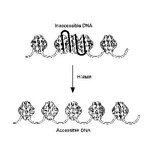
[0024] Figure 1. Principle of RNA-chromatin interaction. RNA (black)
interaction with
chromosomal DNA (white) can compact the DNA and make it inaccessible. RNA:DNA
interaction may be direct (i.e., base-pairing; interaction in the major or
minor DNA groove) or
indirect (i.e., through protein intermediates). Treatment of chromatin with
RNase can degrade
the RNA and make chromosomal regions of RNA:DNA interaction more accessible.
DETAILED DESCRIPTION OF THE INVENTION
I. Introduction
.. [0025] Methods of detecting genomic DNA regions that interact with RNA are
provided. The
methods involve introducing an RNA-degrading agent and a DNA-degrading agent
into a
nucleus and then detecting the one or more regions in the genomic DNA that are
degraded by
the DNA-degrading agent, wherein the one or more regions of the genomic DNA
are
degraded by the DNA-degrading agent due to the presence of the RNA-degrading
agent.
Regions of genomic DNA degradation, which may be detected by an absence or
reduction in
6
CA 2810252 2018-01-19
CA 02810252 2013-03-01
WO 2012/034013 PCT/US2011/050992
the number of copies of the DNA region or by its inability to be amplified by
PCR, likely
correspond to genomic DNA regions that interact with RNA.
[0026] The methods of the present invention are useful, for example, for any
diagnostic,
prognostic, or other personalized medicine application where RNA interaction
with one or
more DNA regions is or may be correlated with a particular disease or
condition.
II. General method
[0027] The methods of the invention involve introducing an RNA-degrading agent
and a
DNA-degrading agent into a nucleus, whereby at least one DNA region of genomic
DNA in
the nucleus is degraded by the DNA-degrading agent due to the presence of the
RNA-
degrading agent, and then detecting the at least one DNA region of genomic DNA
that is
degraded by the DNA-degrading agent.
[0028] In some embodiments, the nucleus into which the RNA-degrading agent
and/or
DNA-degrading agent are introduced is in a cell, and the RNA-degrading agent
and/or DNA-
degrading agent are introduced into the cell. Alternatively, the RNA-degrading
agent and/or
DNA-degrading agent are introduced directly into the nucleus of the cell.
Alternatively, the
nucleus is an isolated nucleus and the RNA-degrading agent and/or DNA-
degrading agent are
introduced into the isolated nucleus.
[0029] The methods of the invention can include permeabilizing or disrupting a
cell
membrane of the cell, thereby enhancing introduction of the RNA-degrading
agent and/or
DNA-degrading agent into the cell. The permeabilization or disruption of the
cell membrane
can occur before the RNA-degrading agent and/or DNA-degrading agent are
introduced into
the cell, or permeabilization or disruption of the cell membrane can occur
simultaneously
with the introduction of the RNA-degrading agent and/or DNA-degrading agent.
[0030] A variety of eukaryotic cells can be used in the present invention. In
some
embodiments, the cells are animal cells, including but not limited to, human,
or non-human,
mammalian cells. Non-human mammalian cells include but are not limited to,
primate cells,
mouse cells, rat cells, porcine cells, and bovine cells. In some embodiments,
the cells are
plant or fungal (including but not limited to yeast) cells. Cells can be, for
example, cultured
primary cells, immortalized culture cells or can be from a biopsy or tissue
sample, optionally
cultured and stimulated to divide before assayed. Cultured cells can be in
suspension or
adherent prior to and/or during the permeabilization and/or DNA modification
steps. Cells
can be from animal tissues, biopsies, etc. For example, the cells can be from
a tumor biopsy.
7
CA 02810252 2013-03-01
WO 2012/034013 PCT/US2011/050992
[0031] The methods of the invention provide for detecting the one or more DNA
regions of
the genomic DNA that are degraded by the DNA-degrading agent, wherein an
absence of a
DNA region or a reduction in the number of copies of a DNA region indicates
that the DNA
region is degraded by the DNA-degrading agent. A wide variety of methods are
known and
can be used to detect the absence or reduction in DNA copies of the DNA
region, and include
but are not limited to, DNA sequencing, PCR amplification to analyze a
targeted region,
genomic DNA library screening, and size selection of DNA fragments.
[0032] The present methods can include correlating degradation of one or more
DNA
regions of the genomic DNA with RNA interactions with those one or more DNA
regions. In
some embodiments, FtNA interaction with a DNA region correlates with a greater
amount of
degradation of the DNA region relative to a genomic DNA region that does not
interact with
RNA. In some embodiments, RNA interaction with a DNA region correlates with an
absence
of the DNA region following degradation by RNA- and DNA-degrading agents.
III. RNA-degrading agents and DNA-degrading agents
[0033] According to the methods of the present invention, an RNA-degrading
agent and a
DNA-degrading agent are introduced into a nucleus, or into a cell having a
nucleus, and at
least one DNA region in genomic DNA in the nucleus is degraded by the DNA-
degrading
agent due to the presence of the RNA-degrading agent. At sites of RNA-DNA
interaction,
e.g., a RNA:DNA duplex, the presence of the RNA-degrading agent will result in
degradation
of the RNA (e.g., the RNA strand in a RNA:DNA duplex), while the presence of
the DNA-
degrading agent will result in degradation of the DNA (e.g., the DNA strand in
a RNA:DNA
duplex or a single-stranded DNA following digestion of the RNA that was
interacting with
the DNA). At sites of RNA:DNA interaction that are not due to base pairing, or
at sites
where RNA associates with chromatin through protein intermediates but does not
physically
contact the DNA, degradation of RNA may alter the local chromatin structure
and change the
accessibility of the DNA-degrading agent to the DNA component of chromatin.
[0034] In some embodiments, the RNA-degrading agent and the DNA-degrading
agent are
introduced into the nucleus or the cell having the nucleus simultaneously. In
some
embodiments, the RNA-degrading agent is introduced into the nucleus or the
cell having the
nucleus before the DNA-degrading agent is introduced into the nucleus or the
cell having the
nucleus.
[0035] In some embodiments, the RNA-degrading agent and/or the DNA-degrading
agent
are introduced into a nucleus or a cell having a nucleus by passive transport,
e.g., diffusion or
8
CA 02810252 2013-03-01
WO 2012/034013 PCT/US2011/050992
facilitated diffusion. Alternatively, the RNA-degrading agent and/or the DNA-
degrading
agent can be introduced into a nucleus or a cell having a nucleus through the
use of a natural
or artificial carrier, transporter, or solvent. The carrier, transporter, or
solvent can be any
polynucleotide, polypeptide, small molecule, organic compound, or inorganic
compound that
can facilitate transport of the RNA- and/or DNA-degrading agent through a cell
membrane
into a nucleus or a cell. In some embodiments, the RNA- and/or DNA-degrading
agent is
encoded by a heterologous expression cassette (i.e., a nucleic acid construct
that is not
endogenous to the cell, which when introduced into the cell, results in
transcription and/or
translation of a RNA or polypeptide, respectively) that is introduced into the
cell.
[0036] In some embodiments, a cell membrane of a cell into which the RNA-
degrading
agent and DNA-degrading agent are to be introduced is penneabilized or
disrupted in order to
enhance introduction of the RNA-degrading agent and/or DNA-degrading agent
into the cell.
The RNA-degrading agent and/or DNA-degrading agent can be introduced into the
cell
simultaneously with permeabilization (e.g, during electroporation or during
incubation with
permeabilizing agent) or following permeabilization (e.g., following removal
of the
peinteabilizing agent, optionally with a change of the buffer). Alternatively,
in some
embodiments, the RNA-degrading agent and/or DNA-degrading agent is contacted
to the
genomic DNA without one or more intervening steps (e.g., without an exchange
of buffers,
washing of the cells, etc.). This latter approach can be convenient for
reducing the amount of
labor and time necessary and also removes a potential source of error and
contamination in
the assay.
[0037] The quantity of RNA-degrading agent and/or DNA-degrading agent used, as
well as
the length of time of the reaction with the RNA-degrading agent and/or DNA-
degrading
agent will depend on the agent used. Those of skill in the art will appreciate
how to adjust
conditions depending on the agent used. Generally, the conditions of the RNA
degrading
and/or DNA degrading step are adjusted such that detectable degradation is
achieved.
"Detectable" degradation, as used herein, refers to contacting the RNA and/or
DNA with a
degrading agent for sufficient time and under appropriate conditions to allow
for cleavage of
at least 5% and typically at least 10%, of all of the RNA-DNA interaction
sites for the target
DNA region of interest. Conditions, including the time, buffers and other
reagents necessary
for detectable degradation, are typically provided by manufacturers of the
degradation agents.
Those of skill in the art will recognize that the quality of the sample may
inhibit nucleic acid
degradation.
9
CA 02810252 2013-03-01
WO 2012/034013 PCT/US2011/050992
A. RNA-degrading agents
[0038] An RNA-degrading agent of the present invention is any reagent that is
capable of
cutting, digesting, or degrading RNA in a RNA:DNA duplex or at a site of
direct or indirect
RNA-DNA interaction, for example RNA at sites of RNA-DNA interaction in
chromatin. In
some embodiments, the RNA-degrading agent is an enzyme. In some embodiments,
the
RNA-degrading agent is a chemical or pharmaceutical compound.
[0039] In some embodiments, an enzyme that cuts or digests RNA in a sequence
non-
specific manner is used as an RNA-degrading agent. In some embodiments, the
RNA-
degrading enzyme is a sequence non-specific endoribonuclease, or "RNase." Any
RNase that
cleaves RNA may be used in the present invention. Examples of suitable RNases
include,
but are not limited to, RNase H (i.e., RNase H, RNase H1, and RNase H2) and
RNase A.
RNases used can include naturally occurring RNases, recombinant RNases, and
modified
RNases (e.g., RNases comprising mutations, insertions, or deletions). An
example of a
modified RNase is HybridaseTM Thermostable RNase H (Epicentre), which includes
mutations that allow for greater therrnostability.
[0040] In some embodiments, the RNA-degrading agent is a ribozyme. Ribozymes,
which
are enzymatic RNA molecules capable of catalyzing the specific cleavage of
RNA, are
known in the art. See, e.g., Heidenreich et al., Nucleic Acids Res., 23:2223-
2228 (1995).
Ribozymes suitable for use in the present invention include both naturally
occurring
ribozymes or synthetic ribozymes.
[0041] Alternatively, the RNA-degrading agent may be any protein, small
molecule,
chemical, or drug that digests, cleaves, or degrades RNA in a RNA:DNA duplex
or in a
RNA-DNA interaction.
B. DNA-degrading agents
[0042] A DNA-degrading agent of the present invention is any reagent that is
capable of
cutting, digesting, or degrading single-stranded DNA, double-stranded DNA, or
DNA in a
RNA:DNA duplex or at a site of direct or indirect RNA-DNA interaction. In some
embodiments, the DNA-degrading agent is an enzyme. In some embodiments, the
DNA-
degrading agent is a chemical or pharmaceutical compound.
[0043] In some embodiments, an enzyme that cuts or digests DNA, or "DNase," is
used as
a DNA-degrading agent. Any DNase that cleaves single-stranded DNA, double-
stranded
DNA, or DNA in a RNA:DNA duplex or RNA-DNA interaction may be used according
to
CA 02810252 2013-03-01
WO 2012/034013 PCT/US2011/050992
the present invention. DNases used can include naturally occurring DNases,
recombinant
DNases, and modified DNases (e.g., DNases comprising mutations, insertions, or
deletions).
[0044] In some embodiments, the DNase is an enzyme that preferentially cleaves
single-
stranded DNA or DNA in a RNA:DNA duplex (i.e., does not cleave double-stranded
DNA or
cleaves double-stranded DNA at only a very low level). Examples of suitable
single-strand
DNA-specific DNases include, but are not limited to, Si nuclease, P1 nuclease,
and mung
bean nuclease.
[0045] In some embodiments, the DNase is an enzyme that cleaves double-
stranded DNA
but which also cleaves single-stranded DNA or DNA in a RNA:DNA duplex to a
lesser
extent. For these DNases, the amount of DNase that is required to cleave
single-stranded
DNA or DNA in a RNA:DNA duplex can be experimentally determined by one of
skill in the
art by titering the DNase. Examples of suitable double-strand DNA-specific
DNases include,
but are not limited to, DNase I and Bat 31 nuclease.
[0046] Alternatively, the DNA-degrading agent may be any protein, small
molecule,
chemical, or drug that digests, cleaves, or degrades single-stranded DNA,
double-stranded
DNA, or DNA in a RNA:DNA duplex or at a site of RNA-DNA interaction.
[0047] In some embodiments, the DNA-degrading agent (e.g, a DNase) degrades
regions
of genomic DNA, such as DNA in a RNA:DNA duplex or at a site of direct or
indirect RNA-
DNA interaction, due to the presence of an RNA-degrading agent (e.g., an
RNase). In these
embodiments, degradation of the RNA that is bound, associated with, or
hybridized to
genomic DNA results in the formation of single-stranded DNA or changes the
local
chromatin structure, thus making the DNA more or less accessible to the DNA
degrading
agent for degradation of the DNA. Whether degradation of genomic DNA by a DNA-
degrading agent is due to the presence of an RNA-degrading agent can be
experimentally
determined by one of skill in the art. For example, a control experiment can
be performed in
which a genomic DNA is contacted by a DNA-degrading agent but not an RNA-
degrading
agent. The DNA regions that are degraded by the DNA-degrading agent in the "no
RNA-
degrading agent" control sample can be compared to the DNA regions that are
degraded in a
corresponding genomic DNA which has been contacted by both the DNA-degrading
agent
and an RNA-degrading agent, and those DNA regions which are degraded when RNA-
degrading agent is present, but which are not degraded when RNA-degrading
agent is absent,
are the DNA regions that are degraded by the DNA-degrading agent due to the
presence of
the RNA-degrading agent.
11
CA 2810252
Permeabilizing and disrupting cells
[0048] Cell membranes can be permeabilized or disrupted in any way known in
the art.
According to the methods of the present invention, the membrane of a cell may
be
permeabilized or disrupted before or during the step of introducing the RNA-
degrading
agent and/or DNA-degrading agent to the cell.
[0049] In some embodiments, the cell membrane is contacted with an agent that
permeabilizes or disrupts the cell membrane. Lysolipids are an exemplary class
of agents
that permeabilize cell membranes. Exemplary lysolipids include, but are not
limited to,
lysophosphatidylcholine (also known in the art as lysolecithin) or
monopalmitoylphosphatidylcholine. A variety of lysolipids are also described
in, e.g.,
W0/2003/052095.
[0050] Non-ionic detergents are an exemplary class of agents that disrupt cell
membranes.
Exemplary non-ionic detergents include, but are not limited to, NP40,
Tween20Tm, and
Triton X100TM.
[0051] Alternatively, electroporation or biolistic methods can be used to
permeabilize a
cell membrane such that a DNA modifying agent is introduced into the cell and
can thus
contact the genomic DNA. A wide variety of electroporation methods are well
known and
can be adapted for delivery of DNA modifying agents as described herein.
Exemplary
electroporation methods include, but are not limited to, those described in
WO/2000/062855. Biolistic methods include but are not limited to those
described in US
Patent No. 5,179,022.
III. Detecting DNA after degradation
[0052] In some embodiments, following RNA degradation and DNA degradation
genomic DNA is isolated from the cells according to any method known in the
art.
Essentially any DNA purification procedure can be used so long as it results
in DNA of
acceptable purity for the subsequent detecting step(s). For example, standard
cell lysis
reagents can be used to lyse cells. Optionally a protease (including but not
limited to
proteinase K) can be used. DNA can be isolated from the mixture as is known in
the art. In
some embodiments, phenol/chloroform extractions are used and the DNA can be
.. subsequently precipitated (e.g., by ethanol) and purified. Alternatively,
DNA can be
isolated on a nucleic-acid binding column.
[0053] Optionally, genomic DNA is amplified or otherwise detected directly
from the cell
lysate without an intermediate purification step.
12
CA 2810252 2018-01-19
CA 02810252 2013-03-01
WO 2012/034013 PCT/US2011/050992
A. Target DNA region
[0054] Detection of DNA involves detecting the presence or absence of at least
one DNA
region in the genomic DNA. A DNA region is a target sequence of interest
within genomic
DNA. In some embodiments, a target DNA region is a region of genomic DNA to
which
RNA binds or hybridizes. Any DNA sequence in genomic DNA of a cell can be
evaluated
for RNA interaction as described herein.
[0055] Genomic DNA can be screened to identify a DNA region of interest that
displays a
different pattern or level of interaction with RNA in different cell types,
for example,
between untreated cells and cells exposed to a drug, chemical or environmental
stimulus, or
.. between normal and diseased tissue. Thus, in some embodiments, the methods
of the
invention are used to identify a DNA region whose change in pattern or level
of RNA
interaction acts as a marker for a disease, or lack thereof. Exemplary
diseases include but are
not limited to cancers. A number of genes have been described that have
altered
transcriptional activity and/or chromatin structure in cancer cells compared
to non-cancer
cells.
B. Detecting RNA interaction with the target DNA region
[0056] A variety of methods can be used to detect and quantify the extent of
RNA
interaction with one or more target DNA regions. In some embodiments,
detecting the one or
more target DNA regions for RNA interaction involves detecting and quantifying
the amount
of target DNA region that is present. In some embodiments, detecting the one
or more target
DNA regions for RNA interaction involves detecting and quantifying a decrease
in the
number of copies of the target DNA region or detecting the absence of copies
of the target
DNA region.
[0057] As discussed below, quantitative amplification (including, but not
limited to, real-
time PCR) methods allow for determination of the amount of intact (i.e., non-
degraded)
copies of a DNA region, and can be used with various controls to determine the
relative
amount of intact copies of the DNA region in a sample of interest, thereby
indicating whether
and to what extent RNA is interacting with the DNA region. In such
embodiments, a DNA
region that is resistant or refractory to amplification would likely indicate
degradation of the
DNA region by the DNA-degrading agent, thereby indicating RNA interaction with
the DNA
region.
13
CA 02810252 2013-03-01
WO 2012/034013 PCT/US2011/050992
[0058] Quantitative amplification methods (e.g., quantitative PCR or
quantitative linear
amplification) involve amplification of nucleic acid template, directly or
indirectly (e.g.,
determining a Ct value) deteunining the amount of amplified DNA, and then
calculating the
amount of initial template based on the number of cycles of the amplification.
Amplification
of a DNA locus using reactions is well known (see U.S. Pat. Nos. 4,683,195 and
4,683,202;
PCR PROTOCOLS: A GUIDE TO METHODS AND APPLICATIONS (Innis et al., eds,
1990)). Typically, PCR is used to amplify DNA templates. However, alternative
methods of
amplification have been described and can also be employed, as long as the
alternative
methods amplify intact DNA to a greater extent than the methods amplify
cleaved or
degraded DNA. Methods of quantitative amplification are disclosed in, e.g.,
U.S. Pat. Nos.
6,180,349; 6,033,854; and 5,972,602, as well as in, e.g., Gibson et al.,
Genome Research
6:995-1001 (1996); DeGraves, et al., Biotechniques 34(1):106-10, 112-5 (2003);
Deiman B,
et al., Mol Biotechnol. 20(2):163-79 (2002). Amplifications can be monitored
in "real time."
[0059] In some embodiments, quantitative amplification is based on the
monitoring of the
signal (e.g., fluorescence of a probe) representing copies of the template in
cycles of an
amplification (e.g., PCR) reaction. In the initial cycles of the PCR, a very
low signal is
observed because the quantity of the amplicon formed does not support a
measurable signal
output from the assay. After the initial cycles, as the amount of formed
amplicon increases,
the signal intensity increases to a measurable level and reaches a plateau in
later cycles when
the PCR enters into a non-logarithmic phase. Through a plot of the signal
intensity versus the
cycle number, the specific cycle at which a measurable signal is obtained from
the PCR
reaction can be deduced and used to back-calculate the quantity of the target
before the start
of the PCR. The number of the specific cycles that is determined by this
method is typically
referred to as the cycle threshold (Ct). Exemplary methods are described in,
e.g., Heid et al.
Genome Methods 6:986-94 (1996) with reference to hydrolysis probes.
[0060] One method for detection of amplification products is the 5'-3'
exonuclease
"hydrolysis" PCR assay (also referred to as the TaqManTm assay) (U.S. Pat.
Nos. 5,210,015
and 5,487,972; Holland et al., PNAS USA 88: 7276-7280 (1991); Lee et al.,
Nucleic Acids
Res. 21: 3761-3766 (1993)). This assay detects the accumulation of a specific
PCR product
by hybridization and cleavage of a doubly labeled fluorogenic probe (the
TaqManTm probe)
during the amplification reaction. The fluorogenic probe consists of an
oligonucleotide
labeled with both a fluorescent reporter dye and a quencher dye. During PCR,
this probe is
cleaved by the 5'-exonuclease activity of DNA polymerase if, and only if, it
hybridizes to the
14
CA 02810252 2013-03-01
WO 2012/034013 PCT/US2011/050992
segment being amplified. Cleavage of the probe generates an increase in the
fluorescence
intensity of the reporter dye.
[0061] Another method of detecting amplification products that relies on the
use of energy
transfer is the "beacon probe" method described by Tyagi and Kramer, Nature
Biotech.
14:303-309 (1996), which is also the subject of U.S. Pat. Nos. 5,119,801 and
5,312,728. This
method employs oligonucleotide hybridization probes that can foim hairpin
structures. On
one end of the hybridization probe (either the 5' or 3' end), there is a donor
fluorophore, and
on the other end, an acceptor moiety. In the case of the Tyagi and Kramer
method, this
acceptor moiety is a quencher, that is, the acceptor absorbs energy released
by the donor, but
then does not itself fluoresce. Thus, when the beacon is in the open
conformation, the
fluorescence of the donor fluorophore is detectable, whereas when the beacon
is in hairpin
(closed) conformation, the fluorescence of the donor fluorophore is quenched.
When
employed in PCR, the molecular beacon probe, which hybridizes to one of the
strands of the
PCR product, is in the open conformation and fluorescence is detected, while
those that
remain unhybridized will not fluoresce (Tyagi and Kramer, Nature Biotechnol.
14: 303-306
(1996)). As a result, the amount of fluorescence will increase as the amount
of PCR product
increases, and thus may be used as a measure of the progress of the PCR. Those
of skill in
the art will recognize that other methods of quantitative amplification are
also available.
[0062] Various other techniques for performing quantitative amplification of
nucleic acids
are also known. For example, some methodologies employ one or more probe
oligonucleotides that are structured such that a change in fluorescence is
generated when the
oligonucleotide(s) is hybridized to a target nucleic acid. For example, one
such method
involves is a dual fluorophore approach that exploits fluorescence resonance
energy transfer
(FRET), e.g., LightCyclerTM hybridization probes, where two oligo probes
anneal to the
amplicon. The oligonucleotides are designed to hybridize in a head-to-tail
orientation with
the fluorophores separated at a distance that is compatible with efficient
energy transfer.
Other examples of labeled oligonucleotides that are structured to emit a
signal when bound to
a nucleic acid or incorporated into an extension product include: ScorpionsTM
probes (e.g.,
Whitcombe etal., Nature Biotechnology 17:804-807, 1999, and U.S. Pat. No.
6,326,145),
SunriseTM (or AmplifluorTM) probes (e.g., Nazarenko etal., Nuc. Acids Res.
25:2516-2521,
1997, and U.S. Pat. No. 6,117,635), and probes that form a secondary structure
that results in
reduced signal without a quencher and that emits increased signal when
hybridized to a target
(e.g., Lux probesTm).
CA 02810252 2013-03-01
WO 2012/034013 PCT/US2011/050992
[0063] In some embodiments, intercalating agents that produce a signal when
intercalated
in double stranded DNA may be used. Exemplary agents include SYBR GREENTM,
SYBR
GOLDTM, and EVAGREENTM. Since these agents are not template-specific, it is
assumed
that the signal is generated based on template-specific amplification. This
can be confirmed
.. by monitoring signal as a function of temperature because melting point of
template
sequences will generally be much higher than, for example, primer-dimers, etc.
[0064] In some embodiments, the number of copies of a DNA region is compared
to a
control value. Control values can be conveniently used, for example, where one
wants to
know whether the number of copies of intact (i.e., non-degraded and therefore
non-RNA-
interacting) DNA region exceeds or is under a particular value. For example,
in the situation
where a particular DNA region typically does not interact with an RNA in
normal cells, but
does interact with the RNA in diseased cells (or vice versa), one may simply
compare the
number of intact copies of the DNA region to a control value.
[0065] In some embodiments, a DNA region that interacts with RNA is identified
or
detected by sequencing. For example, a genomic DNA sequence for a sample of
interest can
be sequenced and compared to corresponding known genomic DNA sequences in
order to
determine sites of DNA degradation in the sample of interest. In such
embodiments, a site of
DNA degradation in the sample of interest (e.g., a DNA region that is absent
in the sample of
interest but present in the corresponding known genomic DNA sequence) is
indicative of a
region of RNA-DNA interaction. Methods of nucleic acid sequencing are well-
known in the
art. Examples of sequence analysis include, but are not limited to, Maxam-
Gilbert
sequencing, Sanger sequencing, capillary array DNA sequencing, thermal cycle
sequencing
(Sears et at., Biotechniques, 13:626-633 (1992)), solid-phase sequencing
(Zimmerman etal.,
Methods Mol. Cell Biol., 3:39-42 (1992)), sequencing with mass spectrometry
such as matrix-
assisted laser desorption/ionization time-of-flight mass spectrometry (MALDI-
TOF/MS; Fu
et at., Nature Biotech., 16:381-384 (1998)), and sequencing by hybridization
(Chee et at.,
Science, 274:610-614 (1996); Drmanac et at., Science, 260:1649-1652 (1993);
Drmanac et
at., Nature Biotech., 16:54-58 (1998)).
[0066] In some embodiments, an RNA-interacting target DNA region, or a larger
genomic
DNA sequence comprising the target DNA region, is isolated and cloned into a
library. In
some cases, one or more target DNA regions, or one or more genomic DNA
sequences
comprising one or more target DNA regions, is isolated and/or cloned.
Alternatively, a
sample having one or more target DNA regions is used to prepare a library
enriched for such
regions. In such embodiments, a target DNA region, or a larger genomic DNA
sequence
16
CA 02810252 2013-03-01
WO 2012/034013 PCT/US2011/050992
comprising the target DNA region, is purified (e.g., separated) from non-
target DNA regions
prior to cloning, thereby enriching the cloning pool for one class of DNA.
[0067] In some embodiments, subtractive libraries are generated. For example,
libraries
can be generated that are enriched for RNA-interacting target DNA regions in a
diseased cell
and subsequently subtracted with a corresponding library from a healthy cell,
thereby
generating a library of differential DNA sequences that both comprise the RNA-
interacting
target DNA region and are specific for the particular disease. Any diseased
cell can be used,
including but not limited to, cancer cells. Alternate subtractive strategies
can also be
employed, e.g., between different cell types, cell stages, drug treatments,
etc.
[0068] In some embodiments, RNA interaction with a DNA region is detected by
size
selection. This is useful, for example, to enrich for regions that interact
with RNA (i.e.,
shorter fragments) or those regions that do not interact with RNA (larger
fragments or intact
regions). This is useful, for example, in generating populations of nucleic
acids that are
enriched for DNA regions that interact (or those that do not) with RNA.
Alternatively, size
selection can be performed to assist in detection of particular DNA region(s).
Where the
DNA region of interest is known, size fractionation or size selection can be
used to detect
whether there is degradation of the sequence (e.g., by detecting whether DNA
fragments are
intact and relatively longer or fragmented and relatively shorter). For
example, in some
embodiments, DNA is isolated for a section of genomic DNA comprising the DNA
region of
interest (or from a library enriched for the section of genomic DNA comprising
the DNA
region of interest) and subjected to size separation according to any known
method.
Examples of nucleic acid size separation techniques include, but are not
limited to, agarose
gel electrophoresis (e.g., Quertermous, Curr. Protoc. Mol. Biol., Chapter
5:Unit 5.4 (May
2001)) and sucrose gradient (e.g., Weis and Querternious, Curr. Protoc. Mol.
Biol., Chapter
5:Unit 5.3 (May 2001)).
[0069] In such embodiments, the presence or absence of degradation at the DNA
region
may be determined by detecting for the fractionation of the DNA sequence into
smaller
segments, which indicates degradation of a DNA region within that larger DNA
sequence.
The presence of fragmented or relatively shorter DNA fragments indicates the
presence of
RNA-interacting regions in that DNA sequence, while the presence of intact or
relatively
longer DNA fragments indicates the relative absence of RNA-interacting regions
in that DNA
sequence. "Relative absence," as used herein, refers to a reduced extent of
RNA interaction
in a DNA region relative to a normal control or to a level of RNA interaction
in a DNA
region that is below a threshold detection level.
17
CA 02810252 2013-03-01
WO 2012/034013 PCT/US2011/050992
[0070] In some embodiments, RNA-interacting DNA region are identified using a
tiling
array. Chip-based tiling arrays which allow for screening of an entire genome
or portions
thereof are known in the art and are commercially available (e.g., Roche
NimbleGen Whole-
Genome Tiling Array or Targeted-Tiling Array, Madison, WI). For example, in
some
embodiments, following RNA degradation and DNA degradation genomic DNA is
isolated
and amplified according to known methods. Amplified products are end-labeled
(e.g., using
a fluorescent label) to indicate DNA regions that border sites of RNA-DNA
interaction, then
incubated with a tiling array according to the manufacturer's instructions in
order to hybridize
the samples to nucleic acid on the array. The identity of the DNA region that
interacts with
RNA can be determined by determining the nucleic acid sequence on the tiling
array where
hybridization occurred, while the label indicates the location in the nucleic
acid sequence
where RNA-DNA interaction occurred.
[0071] In some embodiments, a select number of DNA regions are analyzed by the
methods of the present invention. Alternatively, a genome-wide map of RNA-DNA
interactions can be created. Without intending to limit the invention to a
particular use, it is
believed that a select number of regions will be examined in situations where
RNA
interaction with a DNA region is known to have a particular association, e.g.,
with a disease
or cell phenotype, whereas a genome-wide assessment will be made where it is
desired to
identify regions of interest that differ between two treatments, cell types,
phenotypes,
diseases, etc.
[0072] The calculations for the methods described herein can involve computer-
based
calculations and tools. The tools are advantageously provided in the form of
computer
programs that are executable by a general purpose computer system (referred to
herein as a
"host computer") of conventional design. The host computer may be configured
with many
different hardware components and can be made in many dimensions and styles
(e.g., desktop
PC, laptop, tablet PC, handheld computer, server, workstation, mainframe).
Standard
components, such as monitors, keyboards, disk drives, CD and/or DVD drives,
and the like,
may be included. Where the host computer is attached to a network, the
connections may be
provided via any suitable transport media (e.g., wired, optical, and/or
wireless media) and any
suitable communication protocol (e.g., TCP/IP); the host computer may include
suitable
networking hardware (e.g., modem, Ethernet card, WiFi card). The host computer
may
implement any of a variety of operating systems, including UNIX, Linux,
Microsoft
Windows, MacOS, or any other operating system.
18
CA 02810252 2013-03-01
WO 2012/034013 PCT/US2011/050992
[0073] Computer code for implementing aspects of the present invention may be
written in
a variety of languages, including PERL, C, C++, Java, JavaScript, VB Script,
AWK, or any
other scripting or programming language that can be executed on the host
computer or that
can be compiled to execute on the host computer. Code may also be written or
distributed in
low level languages such as assembler languages or machine languages.
[0074] The host computer system advantageously provides an interface via which
the user
controls operation of the tools. In the examples described herein, software
tools are
implemented as scripts (e.g., using PERL), execution of which can be initiated
by a user from
a standard command line interface of an operating system such as Linux or
UNIX. Those
skilled in the art will appreciate that commands can be adapted to the
operating system as
appropriate. In other embodiments, a graphical user interface may be provided,
allowing the
user to control operations using a pointing device. Thus, the present
invention is not limited
to any particular user interface.
[0075] Scripts or programs incorporating various features of the present
invention may be
encoded on various computer readable media for storage and/or transmission.
Examples of
suitable media include magnetic disk or tape, optical storage media such as
compact disk
(CD) or DVD (digital versatile disk), flash memory, and carrier signals
adapted for
transmission via wired, optical, and/or wireless networks conforming to a
variety of
protocols, including the Internet.
VI. Diagnostic and prognostic methods
[0076] The present invention also provides methods for diagnosing or providing
a
prognosis for a disease or condition or determining a course of treatment for
a disease or
condition based on the detection of RNA-interaction regions in genomic DNA.
[0077] In some embodiments, RNA interaction with a DNA region of interest is
increased
(or at least is present) or decreased (or absent) in a diseased cell or tissue
as compared to a
normal (i.e., non-diseased) cell or tissue. In these embodiments, the methods
of the present
invention to detect the presence or absence of the DNA region of interest can
be used as a
diagnostic or prognostic tool. For example, in some embodiments, RNA
interaction with a
target DNA region may not occur in a normal cell or tissue, whereas RNA
interaction with
the target DNA region is increased in a diseased (e.g., cancerous) cell or
tissue. By
introducing an RNA-degrading agent and a DNA-degrading agent to the normal and
diseased
cells or tissues, and subsequently detecting the extent of degradation of the
target DNA
region in the normal and diseased cells or tissues, it is possible to compare
the differential
19
CA 02810252 2013-03-01
WO 2012/034013 PCT/US2011/050992
RNA interaction between the normal and diseased cells or tissues. In these
embodiments,
increased RNA interaction with the target DNA region in the diseased cell or
tissue is
expected to result in increased degradation of the target DNA region, and
therefore a
decreased number of copies of the DNA region will be detectable for the
diseased cell or
.. tissue, or the number of copies of the DNA region will be sufficiently low
as to be
undetectable for the diseased cell or tissue, as compared to the normal cell
or tissue.
[0078] Alternatively, in some embodiments, RNA interaction with a target DNA
region
may occur in a normal cell or tissue, whereas RNA interaction with the target
DNA region is
decreased or absent in a diseased cell or tissue. In these embodiments,
decreased or absent
RNA interaction with the target DNA region in the diseased cell or tissue is
expected to result
in decreased degradation of the target DNA region, and therefore an increased
number of
copies of the DNA region will be detectable for the diseased cell or tissue as
compared to the
normal cell or tissue.
[0079] Once a diagnosis or prognosis is established using the methods of the
invention, a
.. regimen of treatment can be established or an existing regimen of treatment
can be altered in
view of the diagnosis or prognosis. For instance, detection of a cancer cell
according to the
methods of the invention can lead to the administration of chemotherapeutic
agents and/or
radiation to an individual from whom the cancer cell was detected.
[0080] A variety of DNA regions can be detected either for research purposes
and/or as a
control DNA region to confirm that the reagents were performing as expected.
For example,
in some embodiments, a DNA region is assayed that is known to interact with
RNA, for
example, an inactivated X chromosome in female cells that is known to interact
with Xist
RNA. Such DNA regions are useful, for example, as positive controls for RNA-
DNA
interaction.
VII. Kits
[0081] The present invention also provides kits for performing the RNA
interaction assays
of the present invention. A kit can optionally include written instructions or
electronic
instructions (e.g., on a CD-ROM or DVD). Kits of the present invention can
include, e.g., an
RNA-degrading agent, a DNA-degrading agent. In some embodiments, the kits
further
comprise a cell permeabilizing and/or cell disrupting agent. RNA-degrading
agents and
DNA-degrading agents can include those described herein in detail, e.g.,
enzymes, proteins,
chemicals, pharmaceutical compounds, and small molecules that degrade RNA or
DNA in a
RNA:DNA duplex or single-stranded DNA. In some embodiments, the RNA-degrading
CA 2810252
agent is an RNase, e.g, RNase H. In some embodiments, the DNA-degrading agent
is a
DNase, e.g., Si nuclease. Kits of the invention can comprise the RNA-degrading
agent, the
DNA-degrading agent, and permeabilizing agent in the same vial/container (and
thus in the
same buffer). Alternatively, one or more of the RNA-degrading agent, the DNA-
degrading
agent, and permeabilizing agent can be in a separate vial/container.
[0082] The kits of the invention can also include one or more control cells
and/or nucleic
acids. In some embodiments, the kits include one or more sets of primers for
amplifying
such genomic sequences (whether or not the actual genomic sequences or cells
are included
in the kits). For example, in some embodiments, the kits include an RNA-
degrading agent,
a DNA-degrading agent, a cell permeabilizing and/or cell disrupting agent, and
one or more
primer sets for amplifying a control DNA region, and optionally one or more
primer sets for
amplifying a second DNA region, e.g., a target DNA region. In some
embodiments, the kits
further comprise materials for the isolation of DNA. Such materials include,
but are not
limited to, "stop" solutions capable of preventing further degradation by the
RNA-degrading
agent and/or DNA-degrading agent, spin columns for purification of genomic DNA
and/or
removal of non-DNA components such as components of a "stop" solution, and
buffers.
[00831 In some embodiments, the kits of the invention comprise one or more of
the
following:
(i) a cell membrane permeabilizing or disrupting agent;
(ii) a RNA-degrading agent;
(iii) a DNA-degrading agent;
(iv) a "stop" solution capable of preventing further degradation by the RNA-
degrading
agent and/or DNA-degrading agent;
(v) materials for the isolation of nucleic acids (e.g., spin columns)
(vi) reagents for PCR/qPCR amplification of DNA, optionally one mixture
containing all
components necessary for PCR or for qPCR aside from the template and/or
polymerase;
(vii) primer sets for PCR/qPCR amplification of specific target DNA regions.
[0084] It is understood that the examples and embodiments described herein are
for
illustrative purposes only and that various modifications or changes in light
thereof will be
suggested to persons skilled in the art and are to be included within the
spirit and purview of
this application and scope of the appended claims.
21
CA 2810252 2018-01-19