Note: Descriptions are shown in the official language in which they were submitted.
CA 02810527 2013-03-05
WO 2012/034041 PCT/US2011/051040
Attorney Docket No.: 65654- 818005 (091220PC)
Client Reference No.: BRP00544-LSG-GXD-4514
DETECTION OF CHROMATIN STRUCTURE
CROSS-REFERENCE TO RELATED PATENT APPLICATIONS
[0001] This application claims benefit of priority to US Provisional Patent
Application No.
61/381,825, filed September 10, 2010, and US Provisional Patent Application
No.
61/436,138, filed January 25, 2011, each of which is incorporated by reference
for all
purposes.
BACKGROUND OF THE INVENTION
[0002] Most DNA in a cell is packaged around a set of histone proteins in a
coiled structure
known as a nucleosome. Nucleosomes, in turn, are further coiled into a highly
condensed
structure that tightly compacts the DNA. This combination of DNA and protein
packaging is
generally referred to as chromatin. Chromatin has two forms: euchromatin, a
loosely
packaged form of chromatin in which the DNA is accessible to transcriptional
machinery and
is usually, but not always, transcriptionally active, and heterochromatin, a
tightly packaged
form in which the DNA is inaccessible to transcriptional machinery and is
usually, but not
always, transcriptionally silent.
[0003] The transition between euchromatin and heterochromatin is mainly
controlled by
three epigenetic events, DNA methylation, histone modification, and RNA
interaction. These
epigenetic events affect whether genomic DNA in a cell is in a loosely
packaged,
transcriptionally active form or a tightly packaged, transcriptionally silent
form.
BRIEF SUMMARY OF THE INVENTION
[0004] The present invention provides methods for analyzing chromosomal DNA.
In some
embodiments, the method comprises:
(a) introducing a DNA modifying agent into a nucleus having genomic DNA
under conditions such that the DNA modifying agent modifies the genomic DNA in
the
nucleus, wherein different regions of the genomic DNA are modified to a
different extent by
the DNA modifying agent, thereby forming modified DNA; and
1
WO 2012/034041 (b) nucleotide sequencing at
least one DNA region in the modified DNA, CA 02810527 2013-03-05
PCT/US2011/051040
wherein the sequencing comprises simultaneously determining (1) the nucleotide
sequence
and (2) whether sequenced nucleotides are modified.
[0005] In some embodiments, the nucleus is an isolated nucleus. In some
embodiments,
the nucleus is in a cell.
[0006] In some embodiments, before or during step (a), the method comprises
permeabilizing or disrupting a cell membrane of the cell, and step (a)
comprises contacting
the cell with the DNA modifying agent. In some embodiments, step (a) comprises
expressing
the DNA modifying agent in the cell, thereby introducing the DNA modifying
agent into the
cell.
[0007] In some embodiments, the modifying agent is a DNA methyltransferase. In
some
embodiments, the DNA methyltransferase methylates adenosines in DNA. In some
embodiments, the DNA methyltransferase methylates cytosines in DNA.
[0008] In some embodiments, the sequencing comprises monitoring DNA polymerase
kinetics. In some embodiments, the sequencing does not utilize a polymerase.
[0009] In some embodiments, the sequencing comprises nanopore sequencing.
[0010] In some embodiments, the sequencing comprises template-dependent
replication of
the DNA region that results in incorporation of labeled nucleotides, and
wherein an arrival
time and/or duration of an interval between signal generated from different
incorporated
nucleotides is determinative of the presence or absence of the modification
and/or the identity
of an incorporated nucleotide. In some embodiments, the label of the labeled
nucleotides is a
fluorescent label.
[0011] In some embodiments, the permeabilizing step comprises contacting the
cell with an
agent that permeabilizes the cell membrane. In some embodiments, the agent
that
permeabilizes the cell membrane is a lysolipid. In some embodiments, the
permeabilizing or
disrupting and the contacting of the cell with a DNA modifying agent are
performed
simultaneously.
[0012] In some embodiments, the method further comprises quantifying the
extent of
modification in at least one DNA region as compared to a control DNA region,
wherein the
control DNA region comprises a sequence that is either:
(i) accessible in essentially all cells of an animal; or
2
WO 2012/034041 (ii) inaccessible in essentially all
cells of an animal.CA 02810527 2013-03-05
PCT/US2011/051040
[0013] The present invention also provides a method of analyzing chromosomal
DNA in a
cell, comprising:
(a) introducing a DNA modifying agent into a nucleus
having genomic DNA under
conditions such that the DNA modifying agent modifies the genomic DNA in the
nucleus,
wherein different regions of the genomic DNA are modified to a different
extent by the DNA
modifying agent, thereby forming modified DNA;
(b) purifying the DNA thereby generating purified DNA;
(c) fragmenting the purified DNA;
(d) affinity purifying modified DNA from the purified
and fragmented DNA, thereby
generating a DNA sample enriched for modified DNA; and
(e) detecting a presence, absence, or quantity of one
or more DNA region in the DNA
sample enriched for modified DNA or cloning, isolating, or nucleotide
sequencing at least
one DNA fragment from the DNA sample enriched for modified DNA.
[0014] In some embodiments, the nucleus is an isolated nucleus. In some
embodiments,
the nucleus is in a cell. In some embodiments, before or during step (a), the
method
comprises permeabilizing or disrupting a cell membrane of the cell, and
wherein step (a)
comprises contacting the cell with the DNA modifying agent. In some
embodiments, step (a)
comprises expressing the DNA modifying agent in the cell, thereby introducing
the DNA
modifying agent into the cell.
[0015] In some embodiments, the modifying agent is a DNA methyltransferase. In
some
embodiments, the DNA methyltransferase methylates adenosines in DNA. In some
embodiments, the DNA methyltransferase methylates cytosines in DNA.
[0016] In some embodiments, the affinity purifying comprises contacting the
fragmented
and purified DNA with a protein affinity agent having affinity for modified
DNA under
conditions to allow for binding of the affinity agent to modified DNA, and
removing DNA
that does not bind to the affinity agent. In some embodiments, the protein
affinity agent
comprises an antibody specific for modified DNA. In some embodiments, the
modification
of the modified DNA is methylation of adenosine or methylation of cytosine.
3
CA 02810527 2013-03-05
WO 2012/034041 PCT/US2011/051040
[0017] In some embodiments, the detecting step comprises detecting the
quantity of copies
of at least one DNA region in the DNA sample enriched for modified DNA.
[0018] In some embodiments, the method comprises amplifying the at least one
DNA
region. In some embodiments, the amplifying step comprises real-time PCR.
[0019] In some embodiments, the detecting step comprises nucleotide sequencing
at least
one DNA region. In some embodiments, the nucleotide sequencing comprises
monitoring
DNA polymerase kinetics. In some embodiments, the nucleotide sequencing
comprises
simultaneously determining (1) the nucleotide sequence and (2) whether
sequenced
nucleotides are modified.
[0020] In some embodiments, the detecting step comprises hybridizing the DNA
sample
enriched for modified DNA to a plurality of nucleic acid probes and detecting
hybridization
between the DNA sample and the nucleic acid probes. In some embodiments, the
nucleic
acid probes are linked to a solid support. In some embodiments, the solid
support is selected
from the group consisting of a microarray and beads.
[0021] In some embodiments, the fragmenting comprises shearing or sonicating
the DNA
or digesting the DNA with a sequence non-specific nuclease.
DEFINITIONS
[0022] A "DNA modifying agent," as used herein, refers to a molecule that
alters DNA in a
detectable manner. In some embodiments, the DNA modifying agent is a molecule
that
methylates specific bases within a DNA strand at specific positions. Exemplary
DNA
modifying agents include, but are not limited to, enzymes, proteins, and
chemicals.
[0023] A "DNA region," as used herein, refers to a target sequence of interest
within
genomic DNA. The DNA region can be of any length that is of interest and that
is accessible
by the DNA modifying agent being used. In some embodiments, the DNA region can
include a single base pair, but can also be a short segment of sequence within
genomic DNA
(e.g., 2-100, 2-500, 50-500 bp) or a larger segment (e.g., 100-10,000, 100-
1000, or 1000-
5000 bp). The amount of DNA in a DNA region is sometimes determined by the
amount of
sequence to be amplified in a PCR reaction. For example, standard PCR
reactions generally
can amplify between about 35 to 5000 base pairs.
[0024] A different "extent" of modifications refers to a different number
(actual or relative)
of modified copies of one or more DNA regions between samples or between two
or more
4
WO 2012/034041 CA 02810527 2013-03-05PCT/US2011/051040
DNA regions in one or more samples. For example, if 100 copies of two DNA
regions
(designated for convenience as "region A" and "region B") are each present in
chromosomal
DNA in a cell, an example of modification to a different extent would be if 10
copies of
region A were modified whereas 70 copies of region B were modified.
[0025] "Permeabilizing" a membrane, as used herein, refers to reducing the
integrity of a
cell membrane to allow for entry of a modifying agent into the cell. A cell
with a
permeabilized cell membrane will generally retain the cell membrane such that
the cell's
structure remains substantially intact. In contrast, "disrupting" a cell
membrane, as used
herein, refers to reducing the integrity of a cell membrane such that the
cell's structure does
not remain intact. For example, contacting a cell membrane with a nonionic
detergent will
remove and/or dissolve a cell membrane, thereby allowing access of a modifying
agent to
genomic DNA that retains at least some chromosomal structure.
[0026] The terms "oligonucleotide," "polynucleotide," and "nucleic acid"
interchangeably
refer to a polymer of monomers that can be corresponded to a ribose nucleic
acid (RNA) or
deoxyribose nucleic acid (DNA) polymer, or analog thereof. This includes
polymers of
nucleotides such as RNA and DNA, as well as modified forms thereof, peptide
nucleic acids
(PNAs), locked nucleic acids (LNATm), and the like. In certain applications,
the nucleic acid
can be a polymer that includes multiple monomer types, e.g., both RNA and DNA
subunits.
[0027] A nucleic acid is typically single-stranded or double-stranded and will
generally
contain phosphodiester bonds, although in some cases, as outlined herein,
nucleic acid
analogs are included that may have alternate backbones, including, for example
and without
limitation, phosphoramide (Beaucage et al. (1993) Tetrahedron 49(10):1925 and
the
references therein; Letsinger (1970) J. Org. Chem. 35:3800; Sprinzl et al.
(1977) Eur. J.
Biochem. 81:579; Letsinger et al. (1986) Nucl. Acids Res. 14: 3487; Sawai et
al. (1984)
Chem. Lett. 805; Letsinger et al. (1988) J. Am. Chem. Soc. 110:4470; and
Pauwels et al.
(1986) Chemica Scripta 26:1419), phosphorothioate (Mag et al. (1991) Nucleic
Acids
19:1437 and U.S. Pat. No. 5,644,048), phosphorodithioate (Briu et al. (1989)3.
Am. Chem.
Soc. 111:2321), 0-methylphophoroamidite linkages (Eckstein, Oligonucleotides
and
Analogues: A Practical Approach, Oxford University Press (1992)), and peptide
nucleic acid
backbones and linkages (Egholm (1992) J. Am. Chem. Soc. 114:1895; Meier et al.
(1992)
Chem. Int. Ed. Engl. 31:1008; Nielsen (1993) Nature 365:566; and Carlsson et
al. (1996)
Nature 380:207), which references are each incorporated by reference. Other
analog nucleic
acids include those with positively charged backbones (Denpcy et al. (1995)
Proc. Natl.
Acad. Sci. USA 92:6097); non-ionic backbones (U.S. Pat. Nos. 5,386,023,
5,637,684,
5
WO 2012/034041 CA 02810527 2013-03-05PCT/US2011/051040
5,602,240, 5,216,141 and 4,469,863; Angew (1991) Chem. Intl. Ed. English 30:
423;
Letsinger et al. (1988) J. Am. Chem. Soc. 110:4470; Letsinger et al. (1994)
Nucleoside &
Nucleotide 13:1597; Chapters 2 and 3, ASC Symposium Series 580, "Carbohydrate
Modifications in Antisense Research", Ed. Y. S. Sanghvi and P. Dan Cook;
Mesmaeker et al.
(1994) Bioorganic & Medicinal Chem. Lett. 4: 395; Jeffs et al. (1994) J.
Biomolecular NMR
34:17; Tetrahedron Lett. 37:743 (1996)) and non-ribose backbones, including
those described
in U.S. Pat. Nos. 5,235,033 and 5,034,506, and Chapters 6 and 7, ASC Symposium
Series
580, Carbohydrate Modifications in Antisense Research, Ed. Y. S. Sanghvi and
P. Dan Cook,
which references are each incorporated by reference. Nucleic acids containing
one or more
carbocyclic sugars are also included within the definition of nucleic acids
(Jenkins et al.
(1995) Chem. Soc. Rev. pp169-176, which is incorporated by reference). Several
nucleic acid
analogs are also described in, e.g., Rawls, C & E News Jun. 2, 1997 page 35,
which is
incorporated by reference. These modifications of the ribose-phosphate
backbone may be
done to facilitate the addition of additional moieties such as labeling
moieties, or to alter the
stability and half-life of such molecules in physiological environments.
[0028] In addition to naturally occurring heterocyclic bases that are
typically found in
nucleic acids (e.g., adenine, guanine, thymine, cytosine, and uracil), nucleic
acid analogs also
include those having non-naturally occurring heterocyclic or other modified
bases, many of
which are described, or otherwise referred to, herein. In particular, many non-
naturally
occurring bases are described further in, e.g., Seela et al. (1991) Hely.
Chim. Acta 74:1790,
Grein et al. (1994) Bioorg. Med. Chem. Lett. 4:971-976, and Seela et al.
(1999) Hely. Chim.
Acta 82:1640, which are each incorporated by reference. To further illustrate,
certain bases
used in nucleotides that act as melting temperature (Tm) modifiers are
optionally included.
For example, some of these include 7-deazapurines (e.g., 7-deazaguanine, 7-
deazaadenine,
etc.), pyrazolo[3,4-d]pyrimidines, propynyl-dN (e.g., propynyl-dU, propynyl-
dC, etc.), and
the like. See, e.g., U.S. Pat. No. 5,990,303, entitled "SYNTHESIS OF 7-DEAZA-
2'-
DEOXYGUANOSINE NUCLEOTIDES," which issued Nov. 23, 1999 to Seela, which is
incorporated by reference. Other representative heterocyclic bases include,
e.g.,
hypoxanthine, inosine, xanthine; 8-aza derivatives of 2-aminopurine, 2,6-
diaminopurine, 2-
amino-6-chloropurine, hypoxanthine, inosine and xanthine; 7-deaza-8-aza
derivatives of
adenine, guanine, 2-aminopurine, 2,6-diaminopurine, 2-amino-6-chloropurine,
hypoxanthine,
inosine and xanthine; 6-azacytosine; 5-fluorocytosine; 5-chlorocytosine; 5-
iodocytosine; 5-
bromocytosine; 5-methylcytosine; 5-propynylcytosine; 5-bromovinyluracil; 5-
fluorouracil; 5-
6
WO 2012/034041 CA 02810527 2013-03-05PCT/US2011/051040
chlorouracil; 5-iodouracil; 5-bromouracil; 5-trifluoromethyluracil; 5-
methoxymethyluracil; 5-
ethynyluracil; 5-propynyluracil, and the like.
[0029] "Accessibility" of a DNA region to a DNA modifying agent, as used
herein, refers
to the ability of a particular DNA region in a chromosome of a cell to be
contacted and
modified by a particular DNA modifying agent. Without intending to limit the
scope of the
invention, it is believed that the particular chromatin structure comprising
the DNA region
will affect the ability of a DNA modifying agent to modify the particular DNA
region. For
example, the DNA region may be wrapped around histone proteins and further may
have
additional nucleosomal structure that prevents, or reduces access of, the DNA
modifying
agent to the DNA region of interest.
[0030] "Nucleotide sequencing," as used herein, refers to a process of
determining the
nucleotide composition of a polynucleotide or nucleic acid fragment. In some
embodiments,
nucleotide sequencing comprises determining both the order of nucleotides of a
particular
nucleic acid fragment and whether one or more of the sequenced nucleotides are
modified,
e.g., by methylation of a nucleotide at a specific position. Exemplary
nucleotide sequencing
methods of the present invention include, but are not limited to, single-
molecule real-time
sequencing and nanopore sequencing.
[0031] "DNA polymerase kinetics," as used herein, refers to the rate of DNA
synthesis by a
DNA polymerase. The rate of DNA synthesis is influenced by numerous factors,
including
nucleotide binding and polymerase translocation, as well as by the presence of
modified
nucleotides (e.g., methylated nucleotides), which decrease the rate of DNA
synthesis.
"Monitoring DNA polymerase kinetics," as used herein, refers to a method of
measuring the
rate of DNA synthesis by a DNA polymerase. In some embodiments, the rate of
DNA
synthesis is monitored in real time. In some embodiments, DNA polymerase
kinetics are
measured by fluorescently labeling nucleotides and measuring the fluorescence
pulse of a
nucleotide as it is incorporated into the growing DNA strand. DNA polymerase
kinetics can
be measured by any of several metrics, including but not limited to pulse
width (the duration
of a fluorescence pulse) and interpulse duration (the interval between
successive pulses).
BRIEF DESCRIPTION OF THE DRAWINGS
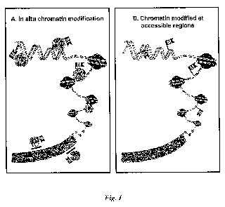
[0032] Figure 1. Principle of DNA modification. (A) Cells are treated with a
DNA
modifying agent in situ to modify accessible chromatin; inaccessible chromatin
regions are
7
CA 02810527 2013-03-05
WO 2012/034041 PCT/US2011/051040
refractory to modification. (B) The modified DNA is purified and sequenced
directly using
technology that can detect sites of DNA modification.
[0033] Figure 2. DAM modification of accessible chromatin. Permeabilized cells
were
treated in situ with the DAM methyltr'ansferase to modify accessible chromatin
(DAM
methylates the A residue at the 6-position in a GATC motif); control cells
were treated with
permeabilization buffer only. The DNA was purified and digested with DpnII, a
methylation-
sensitive restriction enzyme that only digests GATC motifs that have not been
DAM
modified; control reactions were treated with buffer only. The DNA samples
were then
amplified using primers specific for the B2M (A), RHO (B), p14 (C), and CDH13
(D)
promoters. Triangle, no DAM/no DpnII; square, no DAM/plus DpnII; diamond, plus
DAM/no DpnII; circle, plus DAM/plus DpnII.
DETAILED DESCRIPTION
I. Introduction
[0034] Methods are provided for analyzing chromatin structure of chromosomal
DNA by
modifying genomic DNA in a nucleus with a DNA modifying agent and then
nucleotide
sequencing at least one DNA region in the modified DNA. The extent of
modification in a
DNA region can be quantified and is indicative of the accessibility of that
region of DNA to
the modifying agent, and thus reflects the chromatin structure of that region.
[0035] One advantage of the present invention is that one can analyze modified
DNA and
simultaneously determine the nucleotide sequence of the DNA strand and whether
the
sequenced nucleotides are modified, e.g., methylated. This direct detection of
modified bases
during sequencing allows for the rapid generation of results.
General method
[0036] The methods of the invention can involve introducing a DNA modifying
agent into
a nucleus having genomic DNA under conditions such that the DNA modifying
agent
modifies the genomic DNA in the nucleus, wherein different regions of the
genomic DNA
are modified to a different extent by the DNA modifying agent (due to
differences in
chromatin structure) and then nucleotide sequencing at least one DNA region in
the modified
DNA, wherein the sequencing comprises simultaneously determining the
nucleotide sequence
and whether sequenced nucleotides are modified. In some embodiments, the
extent of
modification in at least one DNA region is quantified. The varying
accessibility of the DNA
8
WO 2012/034041 CA 02810527 2013-03-05 PCT/US2011/051040
can reflect the nucleosomal/chromosomal structure of the genomic DNA. For
example, in
some embodiments, DNA regions that are more accessible to DNA modifying agents
are
likely in more "loose" chromatin structures.
[0037] In some embodiments, the nucleotide sequencing step comprises
monitoring DNA
polymerase kinetics. DNA polymerization for a DNA sequence of interest can be
observed
in real-time, e.g., using a single-molecule, real-time (SMRT) sequencing
method. In some
embodiments, nucleotide-specific differences in catalyzing the incorporation
of nucleotides
can be detected and correlated with the identity of an incorporated nucleotide
and/or the
presence or absence of a modification of an incorporated nucleotide.
[0038] In some embodiments, the nucleotide sequencing step comprises nanopore
sequencing. A DNA sequence of interest can be threaded through a nanopore,
e.g., a protein
nanopore, under an applied potential while recording modulations of the ionic
current passing
through the pore. Because modulations in pore current and dwell time differ
for varying
nucleotides, these modulations in pore current and dwell times can be detected
and correlated
with the identity of an incorporated nucleotide and/or the presence or absence
of a
modification of an incorporated nucleotide.
[0039] In some embodiments, the nucleus into which the DNA modifying agent is
introduced is an isolated nucleus. In some embodiments, the nucleus is in a
cell.
[0040] When the nucleus into which the DNA modifying agent is introduced is in
a cell,
the methods of the invention can include permeabilizing or disrupting a cell
membrane of the
cell, thereby introducing the agent into the cell and/or enhancing
introduction of the agent
into the cell. The permeabilization or disruption of the cell membrane can
occur before the
DNA modifying agent is introduced into the cell, or permeabilization or
disruption of the cell
membrane can occur simultaneously with the introduction of the DNA modifying
agent into
the cell. Alternatively, the DNA modifying agent can be introduced into
isolated nuclei.
[0041] A variety of eukaryotic cells can be used in the present invention. In
some
embodiments, the cells are animal cells, including but not limited to, human,
or non-human,
mammalian cells. Non-human mammalian cells include but are not limited to,
primate cells,
mouse cells, rat cells, porcine cells, and bovine cells. In some embodiments,
the cells are
plant or fungal (including but not limited to yeast) cells. Cells can be, for
example, cultured
primary cells, immortalized culture cells or can be from a biopsy or tissue
sample, optionally
cultured and stimulated to divide before assayed. Cultured cells can be in
suspension or
9
CA 02810527 2013-03-05
WO 2012/034041 PCT/US2011/051040
adherent prior to and/or during the permeabilization and/or DNA modification
steps. Cells
can be from animal tissues, biopsies, etc. For example, the cells can be from
a tumor biopsy.
[0042] The present invention also provides for a method of analyzing
chromosomal DNA
in a cell by (1) introducing a DNA modifying agent into the nucleus of the
cell such that the
DNA modifying agent modifies some genomic regions more than others (e.g., due
to steric
hindrance due to variations in chromatin structure), (2) affinity purifying
the modified DNA
using an affinity agent specific for the DNA modification, and then
subsequently (3)
analyzing the affinity purified sample enriched for modified DNA.
[0043] As explained herein, a DNA modifying agent can be introduced into a
cell's nucleus
by a number of methods. Once the genomic DNA in the nucleus has been modified,
the
DNA can be purified, e.g., through standard molecular biology methods, and
optionally
fragmented. Fragmentation can be achieved, for example, by DNA shearing (e.g.,
extruding
the DNA through a small-gauge needle), sonication, or cleavage with a nucleic
acid nuclease
(e.g., a DNase).
[0044] Once purified, and optionally fragmented, the DNA can be submitted to
one or more
affinity purification steps using an affinity agent specific for the DNA
modification. DNA
fragments containing one or more DNA modification will thereby become enriched
in the
sample, while fragments having few or no modifications can be washed away. The
DNA in
the resulting enriched sample can subsequently be analyzed. Sequences enriched
in the
enriched sample will likely have a more "open" chromatin conformation in the
cell from
which the DNA was obtained such that the DNA modifying agent could contact and
modify
the sequence.
[0045] DNA affinity agents can be any molecule that has a selective affinity
for the DNA
modification. In some embodiments, the affinity agent is an antibody. For
example,
antibodies having affinity for methyl-cytosine and methyl-adenosine are known
and
commercially available. Antibodies specific for other types of DNA
modifications are also
contemplated. Alternatively, the affinity agent can be a non-antibody protein.
As an
example, methyl binding protein (MBP) can be used where the DNA modification
is methyl-
cytosine. In yet other embodiments, the affinity agent is a non-protein
molecule, such as a
carbohydrate, lipid, nucleic acid (including but not limited to an aptamer) or
other molecule.
[0046] In some embodiments, the affinity agent is linked to a solid support.
In some
embodiments, the fragmented and purified DNA is contacted to the affinity
agent linked to
10
WO 2012/034041 CA 02810527 2013-03-05PCT/US2011/051040
the solid support under conditions to allow the affinity agent to bind to
modified DNA, and
unbound DNA is washed or otherwise separated from the bound DNA.
/H. DNA modifying agents
[0047] According to the methods of the present invention, a DNA modifying
agent is
introduced into a nucleus having genomic DNA under such conditions that the
DNA
modifying agent modifies the genomic DNA in the nucleus. A wide variety of DNA
modifying agents can be used according to the present invention, including but
not limited to
enzymes, proteins, and chemicals.
[0048] In some embodiments, the DNA modifying agent is introduced into an
isolated
nucleus. In some embodiments, the DNA modifying agent is introduced into a
nucleus in a
cell following permeabilization, or simultaneously with permeabilization
(e.g., during
electroporation or during incubation with permeabilizing agent).
[0049] In some embodiments, the DNA modifying agents are contacted to
permeabilized
cells following removal of the permeabilizing agent, optionally with a change
of the buffer.
Alternatively, in some preferred embodiments, the DNA modifying agent is
contacted to the
genomic DNA without one or more intervening steps (e.g., without an exchange
of buffers,
washing of the cells, etc.). As noted above, this latter approach can be
convenient for
reducing the amount of labor and time necessary and also removes a potential
source of error
and contamination in the assay.
[0050] The quantity of DNA modifying agent used, as well as the time of the
reaction with
the DNA modifying agent will depend on the agent used. Those of skill in the
art will
appreciate how to adjust conditions depending on the agent used. Generally,
the conditions
of the DNA modifying step are adjusted such that a "complete" modification is
not achieved.
Thus, for example, in some embodiments, the conditions of the modifying step
is set such
that for the positive control ¨ i.e., the control where modification is
accessible and occurs ¨
the number of copies of that positive control DNA region that are modified is
at least about
10%, at least about 15%, 20%, 25%, 30%, 40%, or more.
A. Methyltransferases
[0051] In some embodiments of the invention, the DNA modifying agent generates
a
covalent modification to the DNA. For example, in some embodiments, the DNA
modifying
agents of the invention are methyltransferases. A variety of
methyltransferases are known in
the art and can be used in the invention.
11
CA 02810527 2013-03-05
WO 2012/034041 PCT/US2011/051040
[0052] In some embodiments, the methyltransferase used adds a methyl moiety to
adenosine in DNA. Examples of such methyltransferases include, but are not
limited to, E.
coli DAM methyltransferase, M.TaqI, M.EcoRV, M.FokI, and M.EcoRI. Because
adenosine
generally is not methylated in eukaryotic cells, the presence of a methylated
adenosine in a
particular DNA region indicates that a DAM methyltransferase, M.TaqI, M.EcoRV,
M.FokI,
and M.EcoRI (or other methyltransferase with similar activity) was able to
access the DNA
region.
[0053] In some embodiments, the methyltransferase methylates cytosines in GC
sequences.
Examples of such methyltransferases include, but are not limited to, M.CviPI.
See, e.g.,Xu
et at., Nuc. Acids Res. 26(17): 3961-3966 (1998). Because GC sequences
generally are not
methylated in eukaryotic cells, the presence of a methylated GC sequence in a
particular
DNA region indicates that the DNA modifying agent (i.e., a methyltransferase
that
methylates cytosines in GC sequences) was able to access the DNA region.
[0054] In some embodiments, the methyltransferase methylates cytosines in CG
(also
known as "CpG") sequences. Examples of such methyltransferases include, but
are not
limited to, M.SssI. Use of such methyltransferases will generally be limited
to use for those
DNA regions that are not typically methylated. This is because CG sequences
are
endogenously methylated in eukaryotic cells and thus it is not generally
possible to assume
that a CG sequence is methylated by the modifying agent rather than an
endogenous
methyltransferase except in such DNA regions where methylation is rare.
[0055] Other suitable methyltransferases that are known in the art include,
for example,
methyltransferases that methylate cytosine at the N4 position (e.g., M.BamHI
and M.Pvull)
and methyltransferases that methylate cytosine at the C5 position (e.g.,
M.HhaI).
Alternatively, mutated or genetically engineered methyltransferases that
exhibit altered DNA
target-site specificity or altered DNA modification specificity can be used.
B. Chemicals
[0056] In some embodiments, the DNA modifying agent comprises a DNA modifying
chemical. As most DNA modifying chemicals are relatively small compared to
chromatin,
use of DNA modifying chemicals without a fusion partner may not be effective
in some
circumstances as there will be little if any difference in the extent of
accessibility of different
DNA regions. Therefore, in some embodiments, the DNA modifying agent comprises
a
molecule having steric hindrance linked to a DNA modifying chemical. The
molecule having
steric hindrance can be any protein or other molecule that results in
differential accessibility
12
CA 02810527 2013-03-05
WO 2012/034041 PCT/US2011/051040
of the DNA modifying agent depending on chromatin structure. This can be
tested, for
example, by comparing results to those using a methyltransferase as described
herein.
[0057] In some embodiments, the molecule having steric hindrance will be at
least 5, 7, 10,
or 15 kD in size. Those of skill in the art will likely find it convenient to
use a polypeptide as
the molecule with steric hindrance. Any polypeptide can be used that does not
significantly
interfere with the DNA modifying agent's ability to modify DNA. In some
embodiments, the
polypeptide is a double-stranded sequence-non-specific nucleic acid binding
domain as
discussed in further detail below.
[0058] The DNA modifying chemicals of the present invention can be linked
directly to the
molecule having steric hindrance or via a linker. A variety of homo- and
hetero-bifunctional
linkers are known and can be used for this purpose.
[0059] Exemplary DNA modifying chemicals include but are not limited to
hydrazine (and
derivatives thereof, e.g., as described in Mathison et at., Toxicology and
Applied
Pharmacology 127(1):91-98 (1994)) and dimethyl sulfate. In some embodiments,
hydrazine
introduces a methyl group to guanine in DNA or otherwise damages DNA. In some
embodiments, dimethyl sulfate methylates guanine or results in the base-
specific cleavage of
guanine in DNA by rupturing the imidazole rings present in guanine.
C. DNA binding domains to improve DNA modifying agents
[0060] In some embodiments, the DNA modifying agents of the invention are
fused or
otherwise linked to a double-stranded sequence-non-specific nucleic acid
binding domain
(e.g., a DNA binding domain). In cases where the DNA modifying agent is a
polypeptide,
the double-stranded sequence-non-specific nucleic acid binding domain can be
synthesized,
for example, as a protein fusion with the DNA modifying agent via recombinant
DNA
technology. A double-stranded sequence-non-specific nucleic acid binding
domain is a
protein or defined region of a protein that binds to double-stranded nucleic
acid in a
sequence-independent manner, i.e., binding does not exhibit a gross preference
for a
particular sequence. In some embodiments, double-stranded nucleic acid binding
proteins
exhibit a 10-fold or higher affinity for double-stranded versus single-
stranded nucleic acids.
The double-stranded nucleic acid binding proteins in some embodiments of the
invention are
thermostable. Examples of such proteins include, but are not limited to, the
Archaeal small
basic DNA binding proteins Sac7d and Sso7d (see, e.g., Choli et at.,
Biochimica et
Biophysica Acta 950:193-203, 1988; Baumann et at., Structural Biol. 1:808-819,
1994; and
Gao et al, Nature Struc. Biol. 5:782-786, 1998), Archael HMf-like proteins
(see, e.g., Stanch
13
CA 02810527 2013-03-05
WO 2012/034041 PCT/US2011/051040
et al., J. Molec. Biol. 255:187-203, 1996; Sandman et al., Gene 150:207-208,
1994), and
PCNA homologs (see, e.g., Cann et al., J. Bacteriology 181:6591-6599, 1999;
Shamoo and
Steitz, Cell:99, 155-166, 1999; De Felice et al., J. Molec. Biol. 291, 47-57,
1999; and Zhang
et al., Biochemistry 34:10703-10712, 1995). See also European Patent 1283875B1
for
addition information regarding DNA binding domains.
Sso7d and Sac7d
[0061] Sso7d and Sac7d are small (about 7,000 kd MW), basic chromosomal
proteins from
the hyperthermophilic archaeabacteria Sulfolobus solfataricus and S.
acidocaldarius,
respectively. These proteins are lysine-rich and have high thermal, acid and
chemical
stability. They bind DNA in a sequence-independent manner and when bound,
increase the
TM of DNA by up to 40 C under some conditions (McAfee et al., Biochemistry
34:10063-
10077, 1995). These proteins and their homologs are typically believed to be
involved in
stabilizing genomic DNA at elevated temperatures.
HMf-like proteins
[0062] The HMf-like proteins are archaeal histones that'share homology both in
amino acid
sequences and in structure with eukaryotic H4 histones, which are thought to
interact directly
with DNA. The HMf family of proteins form stable dimers in solution, and
several HMf
homologs have been identified from thermostable species (e.g., Methanothermus
fervidus and
Pyrococcus strain GB-3a). The HMf family of proteins, once joined to Taq DNA
polymerase
or any DNA modifying enzyme with a low intrinsic processivity, can enhance the
ability of
the enzyme to slide along the DNA substrate and thus increase its
processivity. For example,
the dimeric HMf-like protein can be covalently linked to the N terminus of Taq
DNA
polymerase, e.g., via chemical modification, and thus improve the processivity
of the
polymerase.
[0063] Those of skill in the art will recognize that other double-stranded
sequence-non-
specific nucleic acid binding domains are known in the art and can also be
used as described
herein.
IV. Permeabilizing and disrupting cells
[0064] Cell membranes can be permeabilized or disrupted in any way known in
the art. As
explained herein, the present methods involve contacting the genomic DNA prior
to isolation
of the DNA and thus methods of permeabilizing or disrupting the cell membrane
will not
14
CA 02810527 2013-03-05
WO 2012/034041 PCT/US2011/051040
disrupt the structure of the genomic DNA of the cell such that nucleosomal or
chromatin
structure is destroyed.
[0065] In some embodiments, the cell membrane is contacted with an agent that
permeabilizes or disrupts the cell membrane. Lysolipids are an exemplary class
of agents
that permeabilize cell membranes. Exemplary lysolipids include, but are not
limited to,
lysophosphatidylcholine (also known in the art as lysolecithin) or
monopalmitoylphosphatidylcholine. A variety of lysolipids are also described
in, e.g.,
WO/2003/052095.
[0066] Non-ionic detergents are an exemplary class of agents that disrupt cell
membranes.
Exemplary non-ionic detergents, include but are not limited to, NP40, Tween 20
and Triton
X-100.
[0067] In some embodiments, the permeabilization agent and the DNA modifying
agent are
delivered simultaneously. Thus, in some embodiments, a buffer comprising both
agents is
contacted to the cell. The buffer should be adapted for maintaining activity
of both agents
while maintaining the structure of the cellular chromatin.
[0068] Alternatively, electroporation or biolistic methods can be used to
permeabilize a cell
membrane such that a DNA modifying agent is introduced into the cell and can
thus contact
the genomic DNA. A wide variety of electroporation methods are well known and
can be
adapted for delivery of DNA modifying agents as described herein. Exemplary
electroporation methods include, but are not limited to, those described in
WO/2000/062855.
Biolistic methods include but are not limited to those described in US Patent
No. 5,179,022.
V. Analyzing DNA after DNA modification step
[0069] In some embodiments, following the DNA modification step, genomic DNA
is
isolated from the nucleus according to any method available. Essentially any
DNA
purification procedure can be used so long as it results in DNA of acceptable
purity for the
subsequent sequencing step. For example, standard cell lysis reagents can be
used to lyse
cells. Optionally a protease (including but not limited to proteinase K) can
be used. DNA
can be isolated from the mixture as is known in the art. In some embodiments,
phenol/chloroform extractions are used and the DNA can be subsequently
precipitated (e.g.,
by ethanol) and purified. In some embodiments, RNA is removed or degraded
(e.g., with an
RNase or with use of a DNA purification column), if desired.
15
CA 02810527 2013-03-05
WO 2012/034041 PCT/US2011/051040
A. Target DNA region
[0070] In some embodiments, the methods of the present invention are utilized
to sequence
the whole genome. Alternatively, in some embodiments, the methods of the
present
invention are utilized to sequence a target DNA region. A DNA region is a
target sequence
of interest within genomic DNA. Any DNA sequence in genomic DNA can be
evaluated for
DNA modifying agent accessibility as described herein. DNA regions can be
screened to
identify a DNA region of interest that displays different accessibility in
different cell types,
between untreated cells and cells exposed to a drug, chemical or environmental
stimulus, or
between normal and diseased tissue, for example. Thus, in some embodiments,
the methods
of the invention are used to identify a DNA region whose change in
accessibility acts as a
marker for disease (or lack thereof). Exemplary diseases include but are not
limited to
cancers. A number of genes have been described that have altered DNA
methylation and/or
chromatin structure in cancer cells compared to non-cancer cells.
[0071] A variety of DNA regions can be detected either for research purposes
and/or as a
control DNA region to confirm that the reagents were performing as expected.
For example,
in some embodiments, a DNA region is assayed that is accessible in essentially
all cells of an
animal. Such DNA regions are useful, for example, as positive controls for
accessibility.
Such DNA regions can be found, for example, within or adjacent to genes that
are
constitutive or nearly constitutive. Such genes include those generally
referred to as
"housekeeping" genes, i.e., genes whose expression are required to maintain
basic cellular
function. Examples of such genes include, but are not limited to,
glyceraldehyde-3-
phosphate dehydrogenase (GAPDH) and beta actin (ACTB). DNA regions can include
all or
a portion of such genes, optionally including at least a portion of the
promoter.
[0072] In some embodiments, a DNA region comprises at least a portion of DNA
that is
inaccessible in most cells of an animal. Such DNA regions are useful, for
example, as
negative controls for accessibility. "Inaccessible" in this context refers to
DNA regions
whose copies are modified in no more than around 5% of the copies of the DNA
region.
Examples of such gene sequences include those generally recognized as
"heterochromatic"
and include genes that are only expressed in very specific cell types (e.g.,
expressed in a
tissue or organ-specific fashion). Exemplary genes that are generally
inaccessible (with the
exception of specific cell types) include, but are not limited to, hemoglobin-
beta chain
(HBB), immunoglobulin light chain kappa (IGK), and rhodopsin (RHO).
16
WO 2012/034041 CA 02810527 2013-03-05PCT/US2011/051040
[0073] In some embodiments, the DNA region is a gene sequence which has
different
accessibility depending on the disease state of the cell or otherwise have
variable accessibility
depending on type of cells or growth environment. For example, some genes are
generally
inaccessible in non-cancer cells but are accessible in cancer cells. Examples
of genes with
variable accessibility include, e.g., glutathione-s-transferase pi (GSTP1).
[0074] In some embodiments, the DNA regions are selected at random, for
example, to
identify regions that have differential accessibility between different cell
types, different
conditions, normal vs. diseased cells, etc.
B. Nucleotide sequencing
[0075] A variety of methods can be used to determine the nucleotide sequence
and the
extent to which sequenced nucleotides are modified, e.g., methylated. Any
sequencing
method known in the art can be used so long as it can simultaneously determine
the
nucleotide sequence and whether sequenced nucleotides are modified. As used
herein,
"simultaneously" means that as the sequencing process determines the order of
nucleotides in
a nucleic acid fragment, at the same time it can also distinguish between
modified nucleotides
(e.g., methylated nucleotides) and non-modified nucleotides (e.g., non-
methylated
nucleotides). Examples of sequencing processes that can simultaneous detect
nucleotide
sequence and distinguish whether sequenced nucleotides are modified include,
but are not
limited to, single-molecule real-time (SMRT) sequencing and nanopore
sequencing.
[0076] In some embodiments, nucleotide sequencing comprises template-dependent
replication of the DNA region that results in incorporation of labeled
nucleotides (e.g.,
fluorescently labeled nucleotides), and wherein an arrival time and/or
duration of an interval
between signal generated from different incorporated nucleotides is
determinative of the
presence or absence of the modification and/or the identity of an incorporated
nucleotide.
Single-molecule, real-time sequencing
[0077] In some embodiments, genomic DNA comprising a target DNA region is
sequenced
by single-molecule, real-time (SMRT) sequencing. SMRT sequencing is a process
by which
single DNA polymerase molecules are observed in real time while they catalyze
the
incorporation of fluorescently labeled nucleotides complementary to a template
nucleic acid
strand. Methods of SMRT sequencing are known in the art and were initially
described by
Flusberg et al., Nature Methods, 7:461-465 (2010), which is incorporated
herein by reference
for all purposes.
17
CA 02810527 2013-03-05
WO 2012/034041 PCT/US2011/051040
[0078] Briefly, in SMRT sequencing, incorporation of a nucleotide is detected
as a pulse of
fluorescence whose color identifies that nucleotide. The pulse ends when the
fluorophore,
which is linked to the nucleotide's terminal phosphate, is cleaved by the
polymerase before
the polymerase translocates to the next base in the DNA template. Fluorescence
pulses are
characterized by emission spectra as well as by the duration of the pulse
("pulse width") and
the interval between successive pulses ("interpulse duration" or "IPD"). Pulse
width is a
function of all kinetic steps after nucleotide binding and up to fluorophore
release, and IPD is
a function of the kinetics of nucleotide binding and polymerase translocation.
Thus, DNA
polymerase kinetics can be monitored by measuring the fluorescence pulses in
SMRT
sequencing.
[0079] In addition to measuring differences in fluorescence pulse
characteristics for each
fluorescently-labeled nucleotide (i.e., adenine, guanine, thymine, and
cytosine), differences
can also be measured for non-methylated versus methylated bases. For example,
the
presence of a methylated base alters the IPD of the methylated base as
compared to its non-
methylated counterpart (e.g., methylated adenosine as compared to non-
methylated
adenosine). Additionally, the presence of a methylated base alters the pulse
width of the
methylated base as compared to its non-methylated counterpart (e.g.,
methylated cytosine as
compared to non-methylated cytosine) and furthermore, different modifications
have
different pulse widths (e.g., 5-hydroxymethylcytosine has a more pronounced
excursion than
5-methylcytosine). Thus, each type of non-modified base and modified base has
a unique
signature based on its combination of IPD and pulse width in a given context.
The sensitivity
of SMRT sequencing can be further enhanced by optimizing solution conditions,
polymerase
mutations and algorithmic approaches that take advantage of the nucleotides'
kinetic
signatures, and deconvolution techiques to help resolve neighboring
methylcytosine bases.
Nanopore sequencing
[0080] In some embodiments, nucleotide sequencing does not comprise template-
dependent replication of a DNA region. In some embodiments, genomic DNA
comprising a
target DNA region is sequenced by nanopore sequencing. Nanopore sequencing is
a process
by which a polynucleotide or nucleic acid fragment is passed through a pore
(such as a
protein pore) under an applied potential while recording modulations of the
ionic current
passing through the pore. Methods of nanopore sequencing are known in the art;
see, e.g.,
Clarke et al., Nature Nanotechnology 4:265-270 (2009), which is incorporated
herein by
reference for all purposes.
18
WO 2012/034041 CA 02810527 2013-03-05PCT/US2011/051040
[0081] Briefly, in nanopore sequencing, as a single-stranded DNA molecule
passes through
a protein pore, each base is registered, in sequence, by a characteristic
decrease in current
amplitude which results from the extent to which each base blocks the pore. An
individual
nucleobase can be identified on a static strand, and by sufficiently slowing
the rate of speed
of the DNA translocation (e.g., through the use of enzymes) or improving the
rate of DNA
capture by the pore (e.g., by mutating key residues within the protein pore),
an individual
nucleobase can also be identified while moving.
[0082] In some embodiments, nanopore sequencing comprises the use of an
exonuclease to
liberate individual nucleotides from a strand of DNA, wherein the bases are
identified in
order of release, and the use of an adaptor molecule that is covalently
attached to the pore in
order to permit continuous base detection as the DNA molecule moves through
the pore. As
the nucleotide passes through the pore, it is characterized by a signature
residual current and a .
signature dwell time within the adapter, making it possible to discriminate
between non-
methylated nucleotides. Additionally, different dwell times are seen between
methylated
nucleotides and the corresponding non-methylated nucleotides (e.g., 5-methyl-
dCMP has a
longer dwell time than dCMP), thus making it possible to simultaneously
determine
nucleotide sequence and whether sequenced nucleotides are modified. The
sensitivity of
nanopore sequencing can be further enhanced by optimizing salt concentrations,
adjusting the
applied potential, pH, and temperature, or mutating the exonuclease to vary
its rate of
processivity.
C. Quantifying the extent of modification
[0083] In some embodiments, the present invention comprises quantifying the
extent of
DNA modification in at least one DNA region, wherein the extent of DNA
modification in
the DNA region is indicative of the accessibility of the DNA in chromatin in
that region. In
general, high levels of DNA modification in a DNA region, relative to a
control, are
indicative of a chromatin region that is in a loose or accessible
configuration and that is
generally transcriptionally active. Low levels of DNA modification in a DNA
region,
relative to a control, are indicative of a chromatin region that is in a
compacted or
inaccessible configuration and that is generally transcriptionally silent.
[0084] Using the nucleotide sequencing methods of the present invention, one
can quantify
the extent of DNA modification, e.g., methylation, by comparing the amount of
modification
in a DNA region to a control. In some embodiments, the amount of modification
in a DNA
region of a sample of interest can be quantified as a relative value by
comparing to the
19
CA 02810527 2013-03-05
WO 2012/034041 PCT/US2011/051040
amount of modification in a control DNA region of the sample (e.g., a DNA
region that is
known to be generally accessible or generally inaccessible in all cells of the
sample). In
some embodiments, the amount of modification in a DNA region of a sample of
interest can
be quantified as a relative value by comparing to the amount of modification
in a
corresponding DNA region of a control sample (e.g., a normal or non-diseased
sample).
[0085] Quantification of modified (or unmodified) DNA regions according to the
method
of the invention can be further improved, in some embodiments, by determining
the relative
amount (e.g., a normalized value such as a ratio or percentage) of modified or
unmodified
copies of the DNA region compared to the total number of copies of that same
region. In
some embodiments, the relative amount of modified or unmodified copies of one
DNA
region is compared to the number of modified or unmodified copies of a second
(or more)
DNA regions. In some embodiments, when comparing between two or more DNA
regions,
the relative amount of modified or unmodified copies of each DNA region can be
first
normalized to the total number of copies of the DNA region. Alternatively,
when obtained
from the same sample, in some embodiments, one can assume that the total
number of copies
of each DNA region is roughly the same and therefore, when comparing between
two or
more DNA regions, the relative amount (e.g., the ratio or percentage) of
modified or
unmodified copies between each DNA region is determined without first
normalizing each
value to the total number of copies.
[0086] In some embodiments, the actual or relative (e.g., relative to total
DNA) amount of
modified or unmodified copies is compared to a control value. Control values
can be
conveniently used, for example, where one wants to know whether the
accessibility of a
particular DNA region exceeds or is under a particular value. For example, in
the situation
where a particular DNA region is typically accessible in normal cells, but is
inaccessible in
diseased cells (or vice versa), one may simply compare the actual or relative
number of
modified or unmodified copies to a control value (e.g., greater or less than
10% modified or
unmodified, greater or less than 20% modified or unmodified, etc.).
Alternatively, a control
value can represent past or expected data regarding a control DNA region. In
these cases, the
actual or relative amount of a control DNA region are determined (optionally
for a number of
times) and the resulting data is used to generate a control value that can be
compared with
actual or relative number of modified or unmodified copies determined for a
DNA region of
interest.
[0087] The calculations for the methods described herein can involve computer-
based
calculations and tools. The tools are advantageously provided in the form of
computer
20
CA 02810527 2013-03-05
WO 2012/034041 PCT/US2011/051040
programs that are executable by a general purpose computer system (referred to
herein as a
"host computer") of conventional design. The host computer may be configured
with many
different hardware components and can be made in many dimensions and styles
(e.g., desktop
PC, laptop, tablet PC, handheld computer, server, workstation, mainframe).
Standard
components, such as monitors, keyboards, disk drives, CD and/or DVD drives,
and the like,
may be included. Where the host computer is attached to a network, the
connections may be
provided via any suitable transport media (e.g., wired, optical, and/or
wireless media) and any
suitable communication protocol (e.g., TCP/IP); the host computer may include
suitable
networking hardware (e.g., modem, Ethernet card, WiFi card). The host computer
may
implement any of a variety of operating systems, including UNIX, Linux,
Microsoft
Windows, MacOS, or any other operating system.
[0088] Computer code for implementing aspects of the present invention may be
written in
a variety of languages, including PERL, C, C++, Java, JavaScript, VBScript,
AWK, or any
other scripting or programming language that can be executed on the host
computer or that
can be compiled to execute on the host computer. Code may also be written or
distributed in
low level languages such as assembler languages or machine languages.
[0089] The host computer system advantageously provides an interface via which
the user
controls operation of the tools. In the examples described herein, software
tools are
implemented as scripts (e.g., using PERL), execution of which can be initiated
by a user from
a standard command line interface of an operating system such as Linux or
UNIX. Those
skilled in the art will appreciate that commands can be adapted to the
operating system as
appropriate. In other embodiments, a graphical user interface may be provided,
allowing the
user to control operations using a pointing device. Thus, the present
invention is not limited
to any particular user interface.
[0090] Scripts or programs incorporating various features of the present
invention may be
encoded on various computer readable media for storage and/or transmission.
Examples of
suitable media include magnetic disk or tape, optical storage media such as
compact disk
(CD) or DVD (digital versatile disk), flash memory, and carrier signals
adapted for
transmission via wired, optical, and/or wireless networks conforming to a
variety of
protocols, including the Internet.
VI. Diagnostic and prognostic methods
[0091] The present invention also provides methods for diagnosing or providing
a
prognosis for a disease or condition or determining a course of treatment for
a disease or
21
WO 2012/034041 CA 02810527 2013-03-05PCT/US2011/051040
condition based on the extent and location of DNA modification in genomic DNA.
In some
embodiments, the DNA region is known to be differentially accessible depending
on the
disease or developmental state of a particular cell. In these embodiments, the
methods of the
present invention can be used as a diagnostic or prognostic tool. For example,
in some
embodiments, DNA in a target region may be highly accessible and able to be
modified, e.g.,
by methylation, in a normal cell or tissue, whereas the DNA in that target
region may be
inaccessible and resistant to modification in a diseased cell or tissue (or
vice versa).
[0092] Once a diagnosis or prognosis is established using the methods of the
invention, a
regimen of treatment can be established or an existing regimen of treatment
can be altered in
view of the diagnosis or prognosis. For instance, detection of a cancer cell
according to the
methods of the invention can lead to the administration of chemotherapeutic
agents and/or
radiation to an individual from whom the cancer cell was detected.
VII. Reaction mixtures
[0093] The present invention also provides for reaction mixtures comprising
one or more of
the reagents as described herein, optionally with a eukaryotic cell (whose
chromatin state is
to be determined). In some embodiments, the reaction mixtures comprise, e.g.,
a DNA
modifying agent (e.g., a methyltransferase or a DNA modifying chemical) and a
cell
permeabilizing and/or cell disrupting agent and a eukaryotic cell. Other
reagents as described
herein (including but not limited to sequencing reagents) can also be included
in the reaction
mixture of the invention.
VIM Kits
[0094] The present invention also provides kits for performing the
accessibility assays of
the present invention. A kit can optionally include written instructions or
electronic
instructions (e.g., on a CD-ROM or DVD). Kits of the present invention can
include, e.g., a
DNA modifying agent and a cell permeabilizing and/or cell disrupting agent.
DNA
modifying agents can include those described herein in detail, including,
e.g., a
methyltransferase or a DNA modifying chemical. Kits of the invention can
comprise the
permeabilizing agent and the DNA modifying agent in the same vial/container
(and thus in
the same buffer). Alternatively, the permeabilizing agent and the DNA
modifying agent can
be in separate vials/containers.
[0095] The kits of the invention can also include one or more control cells
and/or nucleic
acids. Exemplary control nucleic acids include, e.g., those comprising a gene
sequence that
22
WO 2012/034041 CA 02810527 2013-03-05 PCT/US2011/051040
is either accessible in essentially all cells of an animal (e.g., a
housekeeping gene sequence or
promoter thereof) or inaccessible in most cells of an animal. In some
embodiments, the kits
include one or more sets of primers for amplifying such gene sequences
(whether or not the
actually gene sequences or cells are included in the kits). For example, in
some
embodiments, the kits include a DNA modifying agent, and a cell permeabilizing
and/or cell
disrupting agent, and one or more primer sets for amplifying a control DNA
region, and
optionally one or more primer sets for amplifying a second DNA region, e.g., a
target DNA
region.
[0096] In some embodiments, the kits of the invention comprise one or more of
the
following:
(i) a methyltransferase or other DNA modifying agent;
(ii) a cell membrane permeabilizing or disrupting agent;
(iii) a "stop" solution capable of preventing further modification by the
modifying agent;
(iv) materials for the extraction and/or purification of nucleic acids (e.g.,
a spin column for
purification of genomic DNA and/or removal of non-DNA components such as
components
of a "stop" solution); and
(v) reagents for the sequencing of the DNA (e.g., single-molecule real-time
sequencing
reagents or nanopore sequencing reagents).
EXAMPLES
[0097] The following examples are offered to illustrate, but not to limit the
claimed
invention.
[0098] The accessibility of chromatin regions to modification by a DNA
modifying agent
was tested for four genes of varying levels of accessibility in four cell
lines (Figure 2). DAM
methyltransferase is a bacterial enzyme that methylates adenine at the 6'
position in a GATC
motif.
[0099] DNA modification in four genes ¨ rhodopsin (RHO), beta-2 microglobulin
(B2M),
P14, and H-cadherin (CDH13) ¨ was analyzed as described herein using four cell
lines:
HeLa, PC3, LNCaP, and HCT15. For each gene and each cell line, permeabilized
cells were
treated with DAM methyltransferase (no DAM treatment was used as a control).
Genomic
DNA was isolated, then digested with DpnII (no DpnII treatment was used as a
control).
DpnII digestion of selected genomic regions was assessed using quantitative
PCR (qPCR)
23
CA 02810527 2013-03-05
WO 2012/034041 PCT/US2011/051040
methods known in the art. For each of the amplified regions, there was one
potential DAM
modification site (GATC).
[0100] Following DNA modification of the genomic DNA with DAM
methyltransferase,
the extent of DNA modification (and therefore the level of accessibility of
the genomic DNA
region) was quantitated using the methylation-sensitive restriction enzyme
DpnII; however,
other methods such as SMRT sequencing or nanopore sequencing would also be
suitable for
analyzing the extent of DNA modification. DpnII is an enzyme that recognizes
and digests
GATC regions in unmethylated DNA, but DpnII enzymatic activity is blocked by
DAM
methylation; therefore, adenosine-methylated GATC motifs in DNA regions are
protected
from digestion.
Analysis of the B2M promoter
[0101] B2M is a housekeeping gene that is expressed constitutively in all cell
lines. In all
cell lines, the plus DAM/plus DpnII line (circle) is left-shifted from the no
DAM/plus DpnII
line (square) (Figure 2A). This indicates that DAM has modified the B2M
promoter and has
protected it from DpnII digestion and suggests that the B2M promoter is
accessible in all cell
lines, a finding that is consistent with previous data.
Analysis of the RHO promoter
[0102] RHO is not expressed in all cell lines analyzed and its promoter is in
an inaccessible
chromatin configuration. In all cell lines, the plus DAM/plus DpnII line
(circle) co-traces
with the no DAM/plus DpnII line (square) (Figure 2B). This indicates that the
RHO promoter
is protected from DpnII digestion, consistent with its location in
inaccessible chromatin.
Analysis of the p14 promoter
[0103] p14 is not expressed in HCT15 cells and its promoter is inaccessible.
p14 is
expressed in Hela, PC3 and LNCaP cells and its promoter is accessible. Our
analysis of DAM
modification reveals that only in HCTI5 cells is the p14 promoter in a
predominately closed
chromatin conformation (Figure 2C).
Analysis of the CDH13 promoter
[0104] CDH13 is highly expressed in Hela cells and its promoter is accessible.
CH13 is
poorly expressed in PC3, LNCaP and HCT15 cells and its promoter is
inaccessible. Our
analysis of DAM modification reveals that only in Hela cells is the CDH13
promoter in a
24
WO 2012/034041 CA 02810527 2013-03-05PCT/US2011/051040
highly accessible chromatin conformation (Figure 2D). The CDH 13 promoter is
moderately
to tightly closed in the other cell lines.
[0105] This data demonstrates that DAM modification of chromatin in situ
occurs in
accessible chromatin regions but does not occur in inaccessible regions. These
results also
imply that by detecting modified DNA bases during DNA sequencing one can
identify
accessible chromatin regions.
[0106] It is understood that the examples and embodiments described herein are
for
illustrative purposes only and that various modifications or changes in light
thereof will be
suggested to persons skilled in the art and are to be included within the
spirit and purview of
this application and scope of the appended claims. All publications, patents,
and patent
applications cited herein are hereby incorporated by reference in their
entirety for all
purposes.
25