Note: Descriptions are shown in the official language in which they were submitted.
CA 02811034 2016-05-11
- 1 -
FAIL SAFE RADIATION CONCEALMENT MECHANISM
RELATED APPLICATIONS
The present application claims priority from US Provisional application number
61/344,693 filed on September 15, 2010.
FIELD OF THE INVENTION
The present invention relates generally to limiting exposure of a patient to
radiation and
more specifically to a fail safe radiation concealment mechanism in an imaging
capsule
io that is swallowed by a patient to examine the patient's gastrointestinal
tract.
BACKGROUND OF THE INVENTION
One method for examining the gastrointestinal tract for the existence of
polyps and other
clinically relevant features that may indicate regarding the potential of
cancer is
performed by swallowing an imaging capsule that will travel through the tract
and view
the patient's situation. In a typical case the trip can take between 24-48
hours after, which
the imaging capsule exits in the patient's feces. Typically the patient
swallows a contrast
agent to enhance the imaging ability of the imaging capsule. Then the patient
swallows
the imaging capsule to examine the gastrointestinal tract while flowing
through the
zo contrast agent. The imaging capsule typically includes a radiation
source, for example
including a radioisotope that emits Xrays or Gamma rays. The radiation is
typically
collimated to allow it to be controllably directed toward a specific area
during the
imaging process. In an exemplary case the imaging capsule is designed to
measure
Compton back-scattering and transmits the measurements (e.g. count rate) to an
external
analysis device, for example a computer or other dedicated instruments.
In a typical implementation a radio-opaque contrast agent is used so that a
position with
a polyp will have less contrast agent and will measure a larger back-
scattering count.
Alternatively, other methods may be used to image the gastrointestinal tract.
WO 2008/096358 A2 titled "Intra-Lumen Polyp Detection" filed on February 6,
2008, describes details related to using a swallowable capsule for detecting
anatomical anomalies in a user's colon.
CA 02811034 2016-05-11
- 2 -
Claim 1 is characterised against WO 2008/096358 A2. US Patent No. 7,787,926 to
Kimchy, describes details related to the manufacture and use of such an
imaging capsule.
Use of an imaging capsule exposes the user to radiation, which may be
potentially
harmful. Accordingly, it is of interest to limit the user's exposure to
radiation when not
necessary, for example while the imaging capsule is located in positions that
do not need
to be measured.
Typically, the imaging capsule may be designed with shutters that can be
instructed to
block the exit of radiation when not needed. However, there still exists the
hazard that in
io case of malfunction of the imaging capsule, for example in case of a
power failure
radiation may be emitted without constraint.
It is thus desirable to design a fail safe radiation blocking mechanism that
automatically
blocks the emission of radiation and only allows radiation to be emitted if
power is
available and the device provides an instruction to allow radiation to be
emitted.
SUMMARY OF THE INVENTION
An aspect of an embodiment of the invention, relates to an imaging capsule
according to
claim 1 for scanning inside a living body, with a fail-safe radiation
mechanism that
prevents the emission of radiation from the imaging capsule until the imaging
capsule is
instructed to emit radiation and power is available to activate a motor to
unblock the
emission of radiation. Optionally, when power is not available the imaging
capsule
automatically, blocks the emission of radiation.
In an exemplary embodiment of the invention, a rotatable disk with a
collimated
radiation source is attached to a motor by its rotation axis. The disk is
configured to
rotate 360 and emit radiation from the collimated radiation source on the
disk. An outer
ring which also rotates around the same rotation axis as the rotatable disk
surrounds the
circumference of the rotatable disk. The outer ring includes areas which block
radiation
and areas which don't block radiation.
CA 02811034 2016-05-11
- 3 -
In an initial rest position the outer ring is situated relative to the
rotatable disk such that
the radiation emitted through the collimators is blocked. In an exemplary
embodiment of
the invention, responsive to commands from the imaging capsule the motor
rotates the
rotatable disk to a position that allows radiation to be emitted. Optionally,
the rotatable
disk continues to rotate in the same direction and drags the outer ring along
while the
outlets of the collimators are unblocked, so that the entire circumference of
the imaging
capsule is scanned for as many rotations as desired.
In an exemplary embodiment of the invention, the rotatable disk and outer ring
are
connected together with a spring so that the emission of radiation from the
collimators
will be blocked automatically when the motor stops turning the rotatable disk.
There is thus provided according to an exemplary embodiment of the invention,
an
imaging capsule for scanning inside a living body with a fail-safe radiation
mechanism,
including: a radiation source; a rotatable disk with the radiation source
mounted on the
disk and wherein the rotatable disk has a collimator structure allowing the
emission of
radiation from the radiation source substantially only from a few locations on
the
circumference of the disk; an outer ring surrounding the circumference of the
disk and
configured to rotate relative to the disk; wherein the outer ring includes
areas that block
radiation and areas that are transparent to the emission of radiation; and
wherein in a rest
position the outer ring is situated relative to the rotatable disk such that
the areas that
block radiation are blocking the emission of radiation from the few locations
on the
circumference of the disk that allow the emission of radiation; a motor for
rotating the
rotatable disk relative to the outer ring; and wherein the rotatable disk and
outer ring are
initially in the rest position blocking the emission of radiation until the
motor is activated
to rotate the rotatable disk and allow the emission of radiation.
In an exemplary embodiment of the invention, the imaging capsule further
includes a
spring coupling the rotatable disk to the outer ring, and wherein the spring
is configured
to automatically return the rotatable disk and outer ring to the rest position
when the
motor is deactivated.
CA 02811034 2016-05-11
- 4 -
Optionally, the imaging capsule further includes flaps extending from the
outer ring and
an encasement with an inner lining enclosing the imaging capsule, wherein the
flaps are
in contact with the inner lining of the encasement and are held by a force
that prevents
the outer ring from rotating responsive to the torque of the spring and the
rotation of the
rotatable disk. In an exemplary embodiment of the invention, the force between
the flaps
and the inner lining is a friction force.
Alternatively, the force between the flaps and the inner lining is an
electromagnetic
force. In an exemplary embodiment of the invention, the force between the
flaps and the
inner lining is controllable. Optionally, if the motor is deactivated and the
force between
the flaps and the inner lining is turned off, the outer ring will rotate to
return the rotatable
disk and outer ring to the rest position. In an exemplary embodiment of the
invention, if
the motor is deactivated and the force between the flaps and the inner lining
is turned on,
the rotatable disk will rotate to return the rotatable disk and outer ring to
the rest position.
In an exemplary embodiment of the invention, the motor is connected to the
rotatable
disk with a clutch that allows the motor to rotate the rotatable disk in a
specific direction
and the rotatable disk can rotate back freely when the motor is deactivated.
Optionally,
the imaging capsule further includes an encasement with an inner lining
enclosing the
imaging capsule, wherein the inner lining applies an electromagnetic force on
the outer
ring, and wherein the electromagnetic force controllably prevents the outer
ring from
rotating responsive to the torque of the spring and the rotation of the
rotatable disk.
In an exemplary embodiment of the invention, the imaging capsule, further
includes a
first limiter attached to the rotatable disk and a second limiter attached to
the outer ring,
wherein the limiters prevent the rotatable disk and outer ring from leaving
the rest
position under the influence of the spring and the limiters force the outer
ring to rotate
with the rotatable disk under the force of the motor. Optionally, the
rotatable disk and the
outer ring are configured to controllably emit radiation 360 degrees around
the rotatable
disk. In an exemplary embodiment of the invention, the rotatable disk and the
outer ring
are configured to controllably emit radiation for a pre-selected amount of
time or a pre-
selected number of rotations.
CA 02811034 2016-05-11
- 5 -
Optionally, the imaging capsule further includes a transceiver to receive
instructions to
activate or deactivate the motor. In an exemplary embodiment of the invention,
theimaging capsule is pre-programmed to activate or deactivate the motor at
specific
times.
There is further disclosed a method of providing fail-safe radiation while
scanning inside
a living body, including: mounting a radiation source on a rotatable disk;
positioning
collimators on the disk so that the radiation is substantially allowed to be
emitted only
from a few locations on the circumference of the disk; placing an outer ring
to surround
the circumference of the disk and configured to rotate relative to the disk;
wherein the
outer ring includes areas that block radiation and areas that are transparent
to the
emission of radiation; situating the outer ring and rotatable disk initially
in a rest position
wherein the outer ring is situated relative to the rotatable disk such that
the areas that
block radiation are blocking the emission of radiation from the few locations
on the
circumference of thedisk that allow the emission of radiation; receiving
instructions to
begin emitting radiation; activating the motor to rotate the rotatable disk
relative to the
outer ring to a position that allows the emission of radiation.
The method further includes connecting between the rotatable disk and outer
ring with a
zo spring so that they will return to the rest position automatically when
the motor is
deactivated.
BRIEF DESCRIPTION OF THE DRAWINGS
The present invention will be understood and better appreciated from the
following
detailed description taken in conjunction with the drawings. Identical
structures,
elements or parts, which appear in more than one figure, are generally labeled
with the
same or similar number in all the figures in which they appear, wherein:
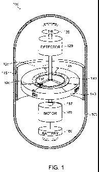
Fig. 1 is a schematic illustration of a perspective view of a failsafe imaging
capsule,
according to an exemplary embodiment of the invention;
CA 02811034 2016-05-11
- 6 -
Fig. 2 is a schematic illustration of a perspective view of a radiation
control mechanism,
according to an exemplary embodiment of the invention;
Fig. 3 is a schematic illustration of a top view of a radiation control
mechanism,
according to an exemplary embodiment of the invention;
Fig. 4 is a schematic illustration of a top view of a radiation control
mechanism in a
rotated position, according to an exemplary embodiment of the invention; and
1() Fig. 5 is a schematic illustration of a top view of a radiation control
mechanism in a
rotated position without a spring, according to an exemplary embodiment of the
invention.
DETAILED DESCRIPTION
is Fig. 1 is a schematic illustration of a perspective view of a failsafe
imaging capsule 100,
according to an exemplary embodiment of the invention. In an exemplary
embodiment of
the invention, a patient first swallows a contrast agent which mixes with the
content of
their gastrointestinal tract to increase the accuracy of the measurements.
20 Then the patient swallows imaging capsule 100 to examine the
gastrointestinal tract as
imaging capsule 100 proceeds through the gastrointestinal tract. In an
exemplary
embodiment of the invention, imaging capsule 100 is designed to automatically
block
radiation from being emitted from it until receiving instructions to release
radiation and
image its surroundings. In an exemplary embodiment of the invention, power is
required
25 to prevent blocking emission of radiation. Optionally, if imaging
capsule 100 lacks
power the radiation will be blocked.
In an exemplary embodiment of the invention, imaging capsule 100 includes an
encasement 105 for holding and protecting the elements of the device from
acids and
30 other liquids or gases along its path of motion.
CA 02811034 2016-05-11
- 7 -
Optionally, the encasement should be able to withstand external pressures for
at least 50-
100 hours to allow for imaging capsule 100 to traverse the gastrointestinal
tract and exit
while still intact. Inside encasement 105 imaging device 100 includes a power
source
180 (e.g. one or more batteries), a motor 185, a radiation source 110, a
detector 129 and a
transceiver 135. In an exemplary embodiment of the invention, radiation source
110 is
located on a rotatable disk 145 and provides radiation that is blocked by a
filling material
130 that forms the disk (e.g. made of lead or tungsten or other dense
materials).
Optionally, the radiation is only free to travel in a few specific directions
through
collimators 120.
In an exemplary embodiment of the invention, power source 180 provides power
to
motor 185, motor 185 is configured to rotate disk 145 around a rotation axis
125 with
radiation source 110 and collimators 120 mounted on disk 145. Optionally, one
or more
directed radiation beams are emitted from collimators 120 controllably
scanning the
surroundings through imaging capsule 100.
Optionally, detector 129 detects backscattered particles resulting from the
directed
radiation beam. In an exemplary embodiment of the invention, detector 129
counts the
detected particles and provides the information to transceiver 135 for
transmission to an
zo external device (e. g. a computer) for processing and optionally
constructing a visual
representation of the information. In some embodiments of the invention,
transceiver 135
uses radio frequency (RF) transmissions to receive instructions from an
external device
and to provide information to the external device. Optionally, the external
device may
instruct imaging capsule 100 to start scanning, to stop scanning, to scan in a
specific
motion pattern or at specific times.
Fig. 2 is a schematic illustration of a perspective view of a radiation
control mechanism
200, and Fig. 3 is a schematic illustration of a top view of radiation control
mechanism
200, according to an exemplary embodiment of the invention. In an exemplary
embodiment of the invention, radiation control mechanism 200 includes disk 145
and an
outer ring 140 that shares the same rotation axis 125 as disk 145 and is free
to rotate
surrounding the circumference of disk 145, for example by being connected to
axis 125
CA 02811034 2016-05-11
- 8 -
from below disk 145. Optionally, outer ring 140 includes shutters 150, which
are made
up from a material that blocks radiation and the rest of outer ring 140
(transparent area
155) does not block radiation. In an initial rest position outer ring 140 is
positioned so
that shutters 150 coincide with the outlets of collimators 120, so that the
emission of
radiation from the collimators 120 is blocked.
In an exemplary embodiment of the invention, disk 145 and outer ring 140 are
connected
together with a spring 190, for example in the shape of a spiral. Optionally,
if disk 145 is
rotated (e.g. clockwise) the spring will tighten and exert a force on outer
ring 140, so that
it will aspire to follow suit. In an exemplary embodiment of the invention,
outer ring 140
includes flaps 160 that extend from the sides of outer ring 140. Optionally,
outer ring 140
includes a hinge 175, for example with an internal spring causing flaps 160 to
extend
outward from the side of outer ring 140 and causing them to be placed in
contact with
encasement 105 or a friction lining 115. In an exemplary embodiment of the
invention,
the friction between the flaps 160 and the friction lining 115 prevent outer
ring 140 from
initially rotating while disk 145 is rotating and the spring 190 is getting
tighter.
Fig. 4 is a schematic illustration of a top view of radiation control
mechanism 200 in a
rotated position, according to an exemplary embodiment of the invention.
zo As disk 145 rotates relative to outer ring 140, in some positions,
shutters 150 stop
blocking the outlets of collimators 120 and the radiation is freely emitted to
scan the
patient.
In some embodiments of the invention, a motion limiter 170 is attached to disk
145 and
another motion limiter 170 is attached to outer ring 140. Optionally, in the
rest position
of radiation control mechanism 200, spring 190 is unwound, collimators 120 are
blocked
and the limiters prevent disk 145 from slipping and accidentally uncovering
the outlets of
collimators 120. Optionally, after rotating 360 as shown in figure 4 the
collimators are
open, and spring 190 is in a tightened position. Then motion limiters 170 meet
on their
opposite sides and the rotation of disk 145 by motor 185 forces outer ring 140
to rotate
together with disk 145 and scan the patient even though flaps 160 are rubbing
against
friction lining 115.
CA 02811034 2016-05-11
- 9 -
Optionally, scanning may be performed over 3600 (the entire circumference of
imaging
capsule 100) for a preselected amount of time or a pre-selected number of
rotations.
In an exemplary embodiment of the invention, when motor 185 is turned off,
spring 190
exerts torque on disk 145 causing it to rotate in the opposite direction (e.g.
counter
clockwise) and to return to the rest position relative to outer ring 140
blocking the
emission of radiation.
In some embodiments of the invention, limiters 170 may be placed in various
positions
io to initiate or prevent motion from various positions as explained above
and not
necessarily in the positions shown in the attached figures.
In an exemplary embodiment of the invention, motor 185 is coupled to a clutch
187 for
delivering rotational motion to disk 145. Optionally, clutch 187 allows disk
145 to move
is freely in the opposite direction when motor 185 is turned off so that
the entire motor
assembly does not need to rotate in the reverse direction under the torque of
spring 190.
Optionally, the clutch may be controlled electrically or mechanically to allow
free
motion in one state and motor controlled motion in the other state.
20 In some embodiments of the invention, other mechanisms instead of flaps
160 may be
used for causing friction between outer ring 140 and encasement 105.
Additionally, the roles of disk 145 and outer ring 140 may be reversed so that
the motor
will drive outer ring 140 and disk 145 will be held by friction with a non
moving part of
25 imaging capsule 100.
Fig. 5 is a schematic illustration of a top view of a radiation control
mechanism 200 in a
rotated position without a spring, according to an exemplary embodiment of the
invention. In some embodiments of the invention, disk 145 and outer ring 140
are not
30 connected with a spring 190 as described above. Accordingly, power is
required to turn
the motor and unblock the outlets of collimators 120 as described above.
However the
outlets are not automatically closed when the motor stops turning because of
spring 190.
CA 02811034 2016-05-11
- 10 -
Instead motor 185 is required to change the direction of rotation to restore
disk 145 to the
rest position relative to outer ring 140 so that the outlets of collimators
120 are blocked
by shutters 150.
In an exemplary embodiment of the invention, the friction between flaps 160
and lining
115 is controllable.
Optionally, when motor 185 stops turning instead of releasing motor 185 and
allowing
disk 145 to rotate back to its rest position under the influence of the torque
of spring 190,
io the friction between flaps 160 and lining 115 is canceled and outer ring
140 moves under
the influence of the torque of spring 190, so that spring 190 unwinds and disk
145 returns
to the rest position relative to outer ring 140 while disk 145 remains
stationary.
In an exemplary embodiment of the invention, the friction between flaps 160
and lining
115 is released by instructing hinge 175 to relax its hold on flaps 160
allowing them to
move closer to outer ring 140 and thus releasing the friction between them and
lining
115. Alternatively, lining 115 may include an electromagnet that is turned on
when
motor 185 starts turning. The electromagnet exerts a force on flaps 160
inhibiting motion
of outer ring 140. Optionally, when motor 185 stops the flaps are released and
the torque
of spring 190 causes outer ring 140 to rotate such that disk 145 will return
to the rest
position relative to outer ring 140 thus blocking the emission of radiation.
In some embodiments of the invention, the electromagnetic force acts directly
on outer
ring 140 and does not require the use of flaps 160. When the electromagnetic
force is
activated the outer ring will be subject to a friction force that inhibits
motion of outer
ring 140.
In some embodiments of the invention, lining 115 may be made from a material
that
expands or contracts causing the flaps to rub against the lining or be
released, for
example the lining may be a Nitonol spring or wire that changes shape when
current
passes through it causing it to heat up and expand or contract. Optionally, a
Nitinol alloy
CA 02811034 2016-05-11
- 11 -
may have 2 positions: one when current passes through it and friction is
required and the
other when no current passes through it.
In some embodiments of the invention, lining 115 may include a piezoelectric
device
that changes size responsive to an electric voltage being applied to it.
Optionally, the
piezoelectric device can form contact with flaps 160 or outer ring 140 to
inhibit motion
or the piezoelectric device can release them.
It should be appreciated that the above described methods and apparatus may be
varied
in many ways, including omitting or adding steps, changing the order of steps
and the
type of devices used. It should be appreciated that different features may be
combined in
different ways. In particular, not all the features shown above in a
particular embodiment
are necessary in every embodiment of the invention. Further combinations of
the above
features are also considered to be within the scope of some embodiments of the
invention.
It will be appreciated by persons skilled in the art that the present
invention is not limited
to what has been particularly shown and described hereinabove. Rather the
scope of the
present invention is defined only by the claims, which follow.