Language selection

Search

Patent 2811548 Summary

Third-party information liability

Some of the information on this Web page has been provided by external sources. The Government of Canada is not responsible for the accuracy, reliability or currency of the information supplied by external sources. Users wishing to rely upon this information should consult directly with the source of the information. Content provided by external sources is not subject to official languages, privacy and accessibility requirements.

Claims and Abstract availability

Any discrepancies in the text and image of the Claims and Abstract are due to differing posting times. Text of the Claims and Abstract are posted:

  • At the time the application is open to public inspection;
  • At the time of issue of the patent (grant).
(12) Patent: (11) CA 2811548
(54) English Title: TOOL FOR DRILLING BONE TISSUE, PARTICULARLY SUITABLE FOR PERFORMING A SINUS LIFT ACCORDING TO THE SUMMERS TECHNIQUE OR FOR THE FITTING OF EXTRA-SHORT IMPLANTS
(54) French Title: OUTIL DE FRAISAGE DE TISSU OSSEUX PARTICULIEREMENT INDIQUE POUR L'ELEVATION DE SINUS SELON LA TECHNIQUE DE SUMMERS OU POUR LA POSE D'IMPLANTS ULTRACOURTS
Status: Granted and Issued
Bibliographic Data
(51) International Patent Classification (IPC):
  • A61C 08/00 (2006.01)
  • A61B 17/16 (2006.01)
(72) Inventors :
  • ANITUA ALDECOA, EDUARDO (Spain)
(73) Owners :
  • BIOTECHNOLOGY INSTITUTE, I MAS D, S.L.
(71) Applicants :
  • BIOTECHNOLOGY INSTITUTE, I MAS D, S.L. (Spain)
(74) Agent: GOWLING WLG (CANADA) LLP
(74) Associate agent:
(45) Issued: 2018-04-24
(86) PCT Filing Date: 2011-09-16
(87) Open to Public Inspection: 2012-03-29
Examination requested: 2016-08-16
Availability of licence: N/A
Dedicated to the Public: N/A
(25) Language of filing: English

Patent Cooperation Treaty (PCT): Yes
(86) PCT Filing Number: PCT/ES2011/000275
(87) International Publication Number: ES2011000275
(85) National Entry: 2013-03-18

(30) Application Priority Data:
Application No. Country/Territory Date
P 201001225 (Spain) 2010-09-24

Abstracts

English Abstract


Tool for drilling bone tissue, which has the advantage of having a cutting
tip with a flat effective shape that prevents perforation of the Schneider
membrane
or injury to the dental nerve when drilling close to them. The tool is
disposed
along an longitudinal axis (7) and comprises a non-cutting main body (1), a
narrowed area (2) for retaining bone and a cutting tip (3), which comprises
cutting
blades (4), each one of which is provided with a front cutting edge (5)
substantially perpendicular to the longitudinal axis (7) and a substantially
lateral
cutting edge (6) that forms an angle of between 0 and 10° with the
longitudinal
axis (7). Spaces (9) for receiving bone are disposed between the cutting
blades (4)
and are connected to the narrowed area (2).


French Abstract

L'invention concerne un outil de fraisage de tissu osseux qui présente l'avantage de comporter une pointe de coupe présentant une forme effective plane évitant de perforer la membrane de Schneider ou de léser le nerf dentaire lorsque le fraisage est effectué à proximité de ceux-ci. Cet outil est disposé le long d'un axe longitudinal (7) et comprend un corps principal (1) non coupant, une zone en retrait (2) pour retenir le tissu osseux et une pointe de coupe (3) comprenant des lames de coupe (4) qui sont chacune dotées d'une arête de coupe frontale (5) sensiblement perpendiculaire à l'axe longitudinal (7), et d'une d'arête de coupe latérale (6) formant sensiblement un angle compris entre 0 et 10° avec l'axe longitudinal (7). Des espaces de réception de tissu osseux communiquant avec la zone en retrait (2) sont disposés entre les lames de coupe (4).

Claims

Note: Claims are shown in the official language in which they were submitted.

What is claimed is:
1. A tool for drilling bone tissue, substantially arranged along a
longitudinal axis in relation to
which said tool is designed to rotate, comprising:
a shaft, a cylindrical non-cutting main body, a narrowed area and a cutting
tip;
wherein,
the cutting tip comprises a series of cutting blades, each one of which
comprises a straight
front cutting edge substantially perpendicular to the longitudinal axis and a
straight lateral cutting
edge that forms an angle of from 1 to 10 along its entire length with the
longitudinal axis,
where said narrowed area is located right between the main body and the
straight lateral
cutting edges of the series of cutting blades, is radially narrower than the
main body and the
cutting tip, and is devoid of cylindrical outer surfaces, providing a bone-
particle-retaining area that
retains bone particles during use of the tool; and
wherein bone-particle-retaining spaces are disposed between adjacent ones of
the series
of cutting blades and are communicated with the narrowed area.
2. The tool according to claim 1, wherein the narrowed area is
substantially conical.
3. The tool according to claim 1, wherein the straight front cutting edges
of the series of
cutting blades are disposed around a central recess.
4. The tool according to claim 1, wherein the shaft and the cutting tip are
connected to each
other and may rotate in relation to the main body.
7

Description

Note: Descriptions are shown in the official language in which they were submitted.


CA 02811548 2013-03-18
1
TOOL FOR DRILLING BONE TISSUE, PARTICULARLY SUITABLE
FOR PERFORMING A SINUS LIFT ACCORDING TO THE SUMMERS
TECHNIQUE OR FOR THE FITTING OF EXTRA-SHORT IMPLANTS
DESCRIPTION
Technical field
The invention relates to a tool for drilling bone tissue, particularly (but
not
exclusively) suitable for performing a sinus lift according to the Summers
technique. Another application for which the tool may be useful is for the
fitting
of extra-short implants (of a length shorter than or equal to 7.5 mm).
Prior art
Jawbones are provided with internal bone cavities called maxillary sinuses.
Along with the nasal cavities, these cavities are situated in the central area
of the
craniofacial complex and apparently perform different functions such as
lightening the weight of the jawbone, moistening and warming air, and acting
as
an intracranial mechanical insulator in the event of a traumatism. The walls
of the
maxillary sinuses are covered by a fine layer of mucus known as the Schneider
membrane, the functions of which are to heat inhaled air and generate a
secretion
that moistens the inhaled air and that retains the solid particles it may
contain.
Said membrane can easily be removed from the walls without causing a
hemorrhage.
The bone situated beneath the maxillary sinuses, commonly known as the
subantral maxillary segment, is a potential dental-implant receptor site. The
fitting
of an implant in said subantral maxillary segment may be hampered to a greater
or
lesser extent, as is the case with any bone where an implant is to be fitted,
due to
the general condition of the bone and its dimensions. For example, a
circumstance
that frequently arises is that the subantral maxillary segment is very thin
and is not
able to host a dental implant suitably, as the implant would not have
sufficient
bone tissue around it to osseointegrate correctly and would perhaps invade the
maxillary sinus and even deteriorate the Schneider membrane. To address this
problem a surgical procedure known as sinus lift is carried out, which allows

CA 02811548 2013-03-18
2
increasing the thickness of the subantral maxillary segment (thus reducing the
height of the maxillary sinus).
The sinus lift procedure may be performed according to various
techniques. One of the most widely used is the alveolar approach or Summers
technique. In the Summers technique it is first of all necessary to ascertain
the
height of the subantral maxillary segment. Then, firstly and with the help of
osteotomes or drills, a bone cavity or alveolus is prepared in the subantral
maxillary segment until almost the very end of it is reached (in other words,
until
the Schneider membrane and the maxillary sinus is almost reached), but without
passing through it completely, so that there is no risk that the osteotomes
and,
especially, the drills perforate the membrane. Then, when there is only a thin
layer
of bone left to drill, generally 1 or 2 mm thick, osteotomes or drills of a
greater
diameter are successively used, on the tip of which bone tissue particles
produced
as a result of the drilling are accumulated. Said accumulation gradually
causes the
lifting of the Schneider membrane, beneath which the space is filled with
particles
of bone tissue. Optionally, prior to the use of the osteotomes or drills of a
greater
diameter, bone graft may be inserted in the alveolus, which is then pushed by
the
osteotomes or drills to also contribute to filling the space beneath the
raised
Schneider membrane.
A slight variation of the Summers technique involves, once the alveolus
has been drilled to within 1 or 2 mm before the end of the subantral maxillary
segment, gently hammering with a tool to break the last portion of bone. Graft
material is then inserted in the floor of the alveolus and beneath the
Schneider
membrane and pushed just enough to raise the membrane. Studies have been
published describing the adverse secondary effects of this particular
technique,
some of which may be serious (dizziness, vertigo, etc.), especially related to
the
considerable trauma involved in the technique.
In the two techniques explained above, once the subantral maxillary
segment has been drilled to within a depth of 1 or 2 mm of its total thickness
and
an additional tool (an osteotome or a piece that is able to be gently
hammered) has
been applied to break the last part of the bone, there is the risk of
perforating the
Schneider membrane, which may cause serious complications (sinusitis, etc.).

CA 02811548 2013-03-18
3
It is an objective of this invention to provide a new tool that is
particularly
suitable for performing a sinus lift according to the Summers technique, which
enables the last part of the subantral maxillary segment to be broken without
any
risk to the Schneider membrane and without causing the adverse secondary
effects
known in some techniques.
Brief description of the invention
It is an object of this invention to provide a tool for drilling bone tissue,
which has the advantage of having a cutting tip with a substantially-flat
effective
shape, to avoid perforating the Schneider membrane when drilling close to it
during a sinus lift procedure according to the alveolar process or Summers
technique. The tool is substantially disposed along a longitudinal axis in
relation
to which it may rotate. As main elements, the tool comprises a main body, a
narrowed area to retain bone, and a cutting tip. The cutting tip comprises a
series
of cutting blades, each one of which is provided with a front cutting edge
substantially perpendicular to the longitudinal axis and a lateral cutting
edge
substantially parallel to the longitudinal axis (specifically, forming an
angle of
between 0 and 100 with the longitudinal axis). Between the cutting blades
there
are spaces for receiving bone, which are in turn corrected to the narrowed
area.
As a result, when the tool rotates, the cutting tip of the tool acquires a
substantially flat shape (perpendicular to the longitudinal axis, i.e., not
pointed).
This allows that the last portion of subantral maxillary segment, in the
proximity
of the Schneider membrane, may be drilled without the risk of the drill
perforating
the membrane (as a result of which it is preferable that the approach is
gentle). In
addition, the particles of bone tissue produced during the drilling are
removed
from the cutting tip towards the narrowed area of the tool, preventing them
from
exerting additional pressure on the Schneider membrane.
As explained above, the inventive tool for drilling bone tissue is
particularly suitable for use when performing a sinus lift according to the
Summers technique. However, it is also very useful for other uses, such as
assisting with the fitting of extra-short implants (of a length shorter than
or equal
to 7.5 mm); in this case, the inventive tool allows implants with a small apex
to be
fitted and thereby enables the whole length of the implant to be used, thanks
to the

CA 02811548 2013-03-18
4
fact that the tool has a flat end rather than a pointed end and that it
therefore drills
a substantially cylindrical alveolus that may be wholly occupied by the
implant.
Brief description of the drawings
Details of the invention can be seen in the accompanying non-limiting
drawings:
- Figure 1 shows a perspective of the preferred embodiment of the
tool
according to the invention.
- Figure 2 shows a front view of the tool of the previous figure.
- Figure 3 shows a side view of the tool of Figure 2.
- Figure 4 shows a cross-sectional front view of another
embodiment of
the tool according to the invention.
Detailed description of the invention
The drawings accompanying this description show three views of the
preferred embodiment of the inventive tool.
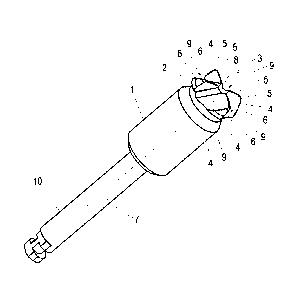
As can be seen in Figure 1, which shows a perspective of the preferred
embodiment of the tool, the tool comprises a non-cutting main body (1), a
narrowed area (2) and a cutting tip (3). The cutting tip (3) also comprises a
series
of cutting blades (4), each one of which is provided with a front cutting edge
(5)
substantially perpendicular to the longitudinal axis (7) in relation to which
the tool
is disposed and in relation to which said tool is designed to rotate. The
cutting
tip (3) also comprises a lateral cutting edge (6) substantially parallel to
the
longitudinal axis (7), more specifically forming an angle of between 0 and 100
with said longitudinal axis (7). The narrowed area (2) acts as an area for
collecting
bone. In addition, spaces (9) for receiving bone are disposed between the
cutting
blades (4) and connected to the narrowed area (2), preventing the blunting of
the
tool due to the bone debris, as explained below.
When the drilling tool is rotating, the effective cutting tip (3) is
approximately rectangular, in other words it is substantially flat due to the
fact
that the front cutting edges (5) are substantially perpendicular to the
longitudinal

CA 02811548 2013-03-18
,
axis (7). This allows the tool to cut the bone in an extremely controlled
manner,
without the tool presenting a pointed apex that may perforate the Schneider
membrane undesirably (or, in the case of its use for drilling the jawbone for
the
purpose of fitting an extra-short implant, prevents the tool from injuring the
dental
5 nerve). This
advantage is enhanced by the fact that the bone particles produced as
a result of the drilling are accumulated in the spaces (9) for receiving bone
situated between the cutting blades (4), and removed towards the narrowed
area (2) for retaining bone, preventing the accumulation of bone particles in
the
tip of the tool, which could cause the shaft tip to stop having a rectangular
effective shape.
In practice it has been shown that, given that the Schneider membrane is
elastic, the rectangular effective cutting tip (3) not only does not cut the
membrane but is also capable of pushing and moving the membrane without
breaking it, provided that the approach with the tool is performed in a
controlled
manner. In other words, the tool according to the invention contributes to
correctly perform a sinus lift according to the Summers technique, not only by
not
drilling the Schneider membrane but by moving it was well. In order to help
perform the technique, the tool may be provided with some depth marks.
In addition, as can be seen, the main body (1) is substantially cylindrical,
so that said main body (1) can act as a guide during the advance of the tool
inside
the alveolus, while not enlarging the alveolus.
As can be seen in Figure 2, which shows a front view of the tool, the
narrowed area (2) is substantially conical in shape. The conical shape allows
the
procedure for manufacturing the piece to be simplified and makes the piece
easier
to clean.
Figure 2 also shows that the lateral cutting edge (6), substantially parallel
to the longitudinal axis (7), forms in one embodiment a slight angle in
relation to
said longitudinal axis (7). Said angle is comprised between 1 and 10 , helping
center the tool during its insertion in the alveolus. Said angle also enables
the
advance to be controlled, as the bone that is perforated is approximately
conical in
shape and of a reduced size, and the slightly conical shape of the tool
therefore
allows a reduction in the risk of the bone being perforated incorrectly.

CA 02811548 2013-03-18
6
Additionally, as can be seen in the three figures and in particular in
Figure 3, which shows a side view of the tool, the front cutting edges (5) of
the
cutting blades (4) are disposed around a central recess (8). Said central
recess (8)
is capable of storing a certain amount of bone particles produced as a result
of the
drilling, which ensures, in the event of unwanted bone particles accumulating
in
the area of the cutting tip during drilling, that these particles do not
accumulate to
form a point that becomes a cutting edge, which might break the Schneider
membrane.
Figure 4 shows a second embodiment of the tool according to the
invention, the tool being shown in cross-section. As shown, in this embodiment
the shaft (10) and the cutting tip (3) are connected to each other and may
rotate in
relation to the main body (1). This allows the tool to be used in two phases.
In a
first phase the tool is inserted in the alveolus, with the main body (1) being
made
to slide all the way along the walls of the alveolus, without the need to
rotate the
tool, thus making the guiding and control of the advance easier and more
precise.
In a second phase, when the tool has already been inserted to the necessary
depth,
the tool is operated to cause the rotation of the shaft (10) and the cutting
tip (3),
initiating the drilling, without this causing the rotation of the main body
(1),
thereby preventing additional and unnecessary drilling and wear of the
alveolus.

Representative Drawing
A single figure which represents the drawing illustrating the invention.
Administrative Status

2024-08-01:As part of the Next Generation Patents (NGP) transition, the Canadian Patents Database (CPD) now contains a more detailed Event History, which replicates the Event Log of our new back-office solution.

Please note that "Inactive:" events refers to events no longer in use in our new back-office solution.

For a clearer understanding of the status of the application/patent presented on this page, the site Disclaimer , as well as the definitions for Patent , Event History , Maintenance Fee  and Payment History  should be consulted.

Event History

Description Date
Maintenance Fee Payment Determined Compliant 2024-09-06
Maintenance Request Received 2024-09-06
Common Representative Appointed 2019-10-30
Inactive: Acknowledgment of s.8 Act correction 2018-11-14
Inactive: Cover page published 2018-11-14
Correction Request for a Granted Patent 2018-11-02
Grant by Issuance 2018-04-24
Inactive: Cover page published 2018-04-23
Inactive: Final fee received 2018-03-02
Pre-grant 2018-03-02
Letter Sent 2018-02-15
Notice of Allowance is Issued 2018-02-15
Inactive: Approved for allowance (AFA) 2018-02-09
Inactive: Q2 passed 2018-02-09
Change of Address or Method of Correspondence Request Received 2018-01-10
Amendment Received - Voluntary Amendment 2017-11-14
Inactive: S.30(2) Rules - Examiner requisition 2017-05-16
Inactive: Report - No QC 2017-05-15
Letter Sent 2016-08-22
Request for Examination Received 2016-08-16
Request for Examination Requirements Determined Compliant 2016-08-16
All Requirements for Examination Determined Compliant 2016-08-16
Inactive: Cover page published 2013-05-29
Inactive: Notice - National entry - No RFE 2013-04-30
Application Received - PCT 2013-04-17
Inactive: IPC assigned 2013-04-17
Inactive: Inventor deleted 2013-04-17
Inactive: Notice - National entry - No RFE 2013-04-17
Inactive: First IPC assigned 2013-04-17
National Entry Requirements Determined Compliant 2013-03-18
Small Entity Declaration Determined Compliant 2013-03-18
Application Published (Open to Public Inspection) 2012-03-29

Abandonment History

There is no abandonment history.

Maintenance Fee

The last payment was received on 2017-09-01

Note : If the full payment has not been received on or before the date indicated, a further fee may be required which may be one of the following

  • the reinstatement fee;
  • the late payment fee; or
  • additional fee to reverse deemed expiry.

Patent fees are adjusted on the 1st of January every year. The amounts above are the current amounts if received by December 31 of the current year.
Please refer to the CIPO Patent Fees web page to see all current fee amounts.

Fee History

Fee Type Anniversary Year Due Date Paid Date
Basic national fee - small 2013-03-18
MF (application, 2nd anniv.) - small 02 2013-09-16 2013-09-05
MF (application, 3rd anniv.) - small 03 2014-09-16 2014-09-08
MF (application, 4th anniv.) - small 04 2015-09-16 2015-09-01
Request for examination - small 2016-08-16
MF (application, 5th anniv.) - small 05 2016-09-16 2016-08-31
MF (application, 6th anniv.) - small 06 2017-09-18 2017-09-01
Final fee - small 2018-03-02
MF (patent, 7th anniv.) - small 2018-09-17 2018-09-10
MF (patent, 8th anniv.) - small 2019-09-16 2019-09-06
MF (patent, 9th anniv.) - small 2020-09-16 2020-09-11
MF (patent, 10th anniv.) - small 2021-09-16 2021-09-10
MF (patent, 11th anniv.) - small 2022-09-16 2022-09-09
MF (patent, 12th anniv.) - small 2023-09-18 2023-09-08
MF (patent, 13th anniv.) - small 2024-09-16 2024-09-06
Owners on Record

Note: Records showing the ownership history in alphabetical order.

Current Owners on Record
BIOTECHNOLOGY INSTITUTE, I MAS D, S.L.
Past Owners on Record
EDUARDO ANITUA ALDECOA
Past Owners that do not appear in the "Owners on Record" listing will appear in other documentation within the application.
Documents

To view selected files, please enter reCAPTCHA code :


To view images, click a link in the Document Description column. To download the documents, select one or more checkboxes in the first column and then click the "Download Selected in PDF format (Zip Archive)" or the "Download Selected as Single PDF" button.

List of published and non-published patent-specific documents on the CPD .

If you have any difficulty accessing content, you can call the Client Service Centre at 1-866-997-1936 or send them an e-mail at CIPO Client Service Centre.


Document
Description 
Date
(yyyy-mm-dd) 
Number of pages   Size of Image (KB) 
Description 2013-03-17 6 273
Drawings 2013-03-17 3 26
Claims 2013-03-17 2 35
Abstract 2013-03-17 1 18
Representative drawing 2013-04-18 1 6
Claims 2017-11-13 1 28
Abstract 2018-02-14 1 18
Representative drawing 2018-03-26 1 5
Confirmation of electronic submission 2024-09-05 1 61
Notice of National Entry 2013-04-16 1 196
Notice of National Entry 2013-04-29 1 196
Reminder of maintenance fee due 2013-05-20 1 114
Reminder - Request for Examination 2016-05-16 1 117
Acknowledgement of Request for Examination 2016-08-21 1 177
Commissioner's Notice - Application Found Allowable 2018-02-14 1 163
Acknowledgement of Section 8 Correction 2018-11-13 2 267
Section 8 correction 2018-11-01 3 85
PCT 2013-03-17 10 344
Request for examination 2016-08-15 2 47
Examiner Requisition 2017-05-15 3 191
Amendment / response to report 2017-11-13 3 105
Final fee 2018-03-01 2 50