Note: Descriptions are shown in the official language in which they were submitted.
CA 02811648 2013-03-18
WO 2012/040355
PCT/US2011/052596
METHOD AND APPARATUS
FOR COCHLEAR IMPLANT SURGERY
CROSS-REFERENCE OF RELATED APPLICATION
[0001] This application claims priority to U.S. Provisional Application
No.
61/384,940 filed September 21, 2010, the entire contents of which are hereby
incorporated by
reference.
BACKGROUND
1. Field of Invention
[0002] The field of the currently claimed embodiments of this invention
relates to
systems, devices and methods for cochlear implant surgery.
2. Discussion of Related Art
[0003] Many different types of Cochlear implant surgery (J. Niparko (ed),
Cochlear
Implants: Principles & Practices, Philadelphia, Lippincott, Williams &
Wilkins, 2009; D. Tucci
and T, Pilkington, "Medical and surgical aspects of cochlear implantation". in
Cochlear
Implants: Principles & Practicesõ J. K. Niparko, Ed, Philadelphia: Lippincott,
Williams &
Wilkins, 2009) can be of immense auditory, linguistic and developmental
benefit to patients
with severe hearing loss due to the loss of hair cell transduction within the
cochlea.
Estimates from the National Institute of Deafness and Other Communication
Disorders
(NIDCD) are that approximately 188,000 people worldwide have received implants
("Statistics about Hearing, Balance, Ear Infections, and Deafness,
"http://www.nided.nih.gov/health/statistics/hearing.asp#1,2010)
and that rates of applying electrified implants to the ear are accelerating.
[0004] The surgical procedure is potentially complicated by difficulties
with implant
electrode array insertion (e.g., C. J, Coulson, A. P. Reid, D. W. Proops, and
P. N. Brett,
"ENT challenges at the small scale", Int J Med Robot, vol. 3- 2, pp. 91-6,
Jun2007
.http://www.ncbi .nlm,n1h,gov/entrez/query fcgi?cmd=Retrieve&db¨PubMed&
1
CA 02811648 2013-03-18
WO 2012/040355
PCT/US2011/052596
dopt=Citation&list uids=17619240 10.1002/rcs.132) and serious complications
may occur.
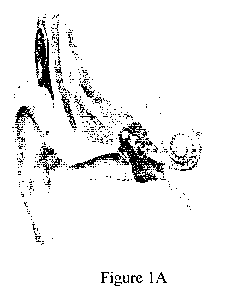
One particularly challenging step is the actual insertion of the implant into
the cochlea
(see, e.g., Figures 1A-1C). After accessing the scala tympani (via direct
round window
insertion, or drilling open a cochleostomy to gain access to the cochlea) an
electrode array
is inserted into scala tympani of the cochlea. Several designs of cochlear
implant arrays
have relied on stylet-based insertion techniques. The Advanced Bionics arrays
used in ca.
2003-2006 used a pre-curved array that was loaded onto a hand-held insertion
tool. Once
inserted into the scala tympani, the insertion tool was used to guide the
array into the
proximal 3 mm of the scala and then advance the array off of the rigid stylet
into the first
turn of the cochlea, allowing the curvature of the silastic carrier to find
the proper
trajectory through the turn. Here, if the stylet based on the hand-held tool
were to be
advanced too far into the cochlea, contact forces generated can damage the
cochlea.
[00051 Over the past 6 years, the Cochlear Corporation Freedom and C512
arrays
have used a stylet-based strategy: A stylet is used to hold the implant
straight while it is
inserted to a desired depth into the cochlea. The array is advanced over the
stylet,
which is held in a fixed position. The implant array then naturally curves to
follow the
cochlea given it's memory as a curved array once off of the stylet. The stylet
is then
withdrawn. If the stylet and implant are advanced too far into the cochlea,
the resulting
contact forces can damage the cochlea either due to direct impact or buckling
of more
proximal aspects of the carrier. Research also has been reported in which a
sheath-style
insertion device is used to perform the same function as a stylet in holding
the implant straight
while it is inserted to a desired depth into the cochlea. The implant array
naturally curves to
follow the cochlea as it is deployed further through the sheath. One example
of such a sheath
is the Modiolar Research Array (R. Briggs et al., "Development and evaluation
of the
modiolar research array ¨ multi-centre collaborative study in human temporal
bones",
Cochlear Implants Int. 2011 August 12(3) pp 129-139, PMCID: PMC3159433).
Again, if
the stylet and implant are advanced too far into the cochlea, the resulting
contact forces can
damage the cochlea either due to direct impact or buckling of more proximal
aspects of the
carrier.
2
CA 02811648 2013-03-18
WO 2012/040355
PCT/US2011/052596
[0006] Many other array designs used both historically and presently
present a
potential problem with substantial growth in resistance as the array is
inserted beyond
12mm (Tucci, et al.), with consequent risks to the integrity of intracochlear
membranous structures (Figure 2).
[0007] Several approaches to providing guidance or assistance in avoiding
damage to the cochlea during implant insertion have been reported recently.
Labadie
et al. report a microstereotactic device for aligning an implant array with
the cochlea
for percutaneous insertion based on preoperative images (R. F. Labadie, R.
Balachandran, J. Mitchell, J. H. Noble, 0. Majdani, D. Haynes, M. Bennett, B.
M.
Dawant, and M. Fitzpatrick, "Clinical Validation Study of Percutaneous
Cochlear
Access Using Patient Customized Micro-Stereotactic Frames", Otol. Neurotol,
vol. 31-
1, pp. 94-99, 2010, PMC2845321). Schurzig, Labadie, and Webster report a
system
that combines an "active canula" robot with delicate force sensing
capabilities to sense
contact between the implant and the cochlea (D. Schurzig, R. F. Labadie, and
R. J.
Webster, "A force sensing robot for cochlear electrode implantation", in IEEE
International Conference on Robotics and Automation, 2010, pp. 3674-3679),
using a
force sensor incorporated into the robotic mechanism that advances the implant
into
the cochlea, Rau et al. (T. S. Rau, A. Hussong, M. Leinung, T. Lenarz, and 0.
Majdani,
"Automated insertion of preformed cochlear implant electrodes: evaluation of
curling
behaviour and insertion forces on an artificial cochlear model", Int Comput
Assist
Radio! Surg, vol. 5-2, pp. 173-81, Mar
2010.http://www,ncbi,n1m.nih`govientrez/
query.fcgi?cmd=Retrieve&db=PubMed&dopt=Citation&list uids=20033522
10,1007/s11548-009-0299-9) have also proposed a robotic cochlear insertion
device
and have reported phantom studies of insertion forces using a load cell
attached to the
insertion mechanism. Zhang, Simaan, et al. have developed an actively
deforming,
steerable, cochlear implant that curves to follow the cochlea during insertion
(J.
Zhang, W. Wei, S, Manolidis, J. T. Roland, Jr., and N. Simaan, "Path planning
and
workspace determination for robot-assisted insertion of steerable electrode
arrays for
cochlear implant surgery", Med Image Comput Comput Assist Interv, vol. 11- Pt
2, pp.
692-700, 2008.http://www.ncbi,n1m.nih.gov/entrez/
3
CA 02811648 2013-03-18
WO 2012/040355
PCT/US2011/052596
query.fcgi?cmd=Retrieve&db=PubMed & dopt=Citation&list uids=18982665; J.
Zhang, K, Xu, N. Simaan, and S. Manolidis, "A pilot study of robot-assisted
cochlear
implant surgery using steerable electrode arrays", Med Image Comput Comput
Assist
Interv, vol. 9-Pt 1, pp. 33-40, 2006. http://www.ncbi.nlm,nih.zov/entrez/
querylegi?cmd = Retrieve&db= PubMed& dopt=Citation&list uids=17354871; J.
Zhang, W. Wei, J. Ding, J. T. Roland, S. Manolidis, and N. Simaan, "Inroads
Toward
Robot-Assisted Cochlear Implant Surgery Using Steerable Electrode Arrays",
Otology
and Neurotology, p. in Press; Published ahead of print, 2010 10.1097/
MA0.0b013e3181e7117e). They report experiments using a load cell mounted on
their
robotic manipulation device. Some limitations of these systems include
reliance on a
fairly large and cumbersome robotic insertion tool and the necessity to
implement an
extremely delicate force sensing mechanism. In the case of the reported
systems, the
difficulty is exacerbated by the moving mass of the mechanism distal to the
force
sensor and possible friction forces.
[0008] Other authors (e.g., C. J. Coulson, R. P. Taylor, A. P. Reid, M, V.
Griffiths, D. W. Proops, and P. N. Brett, "An autonomous surgical robot for
drilling a
cochleostomy: preliminary porcine trial", Clin Otolaryngol, vol. 33-4,pp. 343-
7, Aug
2008. http://www.ncbi,n1m.nih.gov/entreziquery.fcgi?cmd=Retrieve&db=PubMed&
dopt=Citation&list uids=18983344C0A1703 [pi] 10.1111/j.1749-4486.2008.01703.x;
0. Majdani, D, Schurzig, A. Hussong, T. Rau, I. Wittkopf, T. Lenarz, and R. F.
Labadie, "Force measurement of insertion of cochlear implant electrode arrays
in vitro:
comparison of surgeon to automated insertion tool", Acta Oto-Laryngologica,
vol. 130-
1, pp. 31-36, Jan 2010.<Go to ISI>://000274416300005Doi
10.3109/00016480902998281) have proposed robotic devices to assist in drilling
the
skull to gain access to the cochlea for implant insertion. These systems do
not address
the problem of inserting an implant without damage to the cochlea.
[0009] Skilled otologic surgeons have the manual dexterity and steadiness
to
insert implants without damage to the cochlea. What they lack is feedback to
know
when the implant or stylet has been introduced too far into the cochlea. In
his review
article (C. J, Coulson, et al, id), C. J. Coulson states:
4
CA 02811648 2013-03-18
WO 2012/040355
PCT/US2011/052596
If the surgeon were able to visualize or 'feel forces imparted on the
electrode array
and then guide the array around the path of least resistance, he/she would be
able to place the electrode whilst minimizing the trauma to the cochlea.
[0010] Coulson further suggests an endoscopic "flexible digit with
visualization (the
scala tympani being about 1 mm2 in cross-section) would allow the tip to be
manoeuvred
through the hollow portion of the scala tympani", but discloses no feasible
way to
implement such a device, which he describes as being "technically very
difficult" since it
would require both a light source and a visualization device in a tiny space.
As an
alternative, he suggests:
Another potential solution would be to fit the electrode array with sensing
elements at
the tip, which could feed back onto a monitor, informing the surgeon whether
the
tip was against the solid outer cochlear wall or in the middle of the hollow
scalatympani.
[0011] However, he does not disclose any feasible means for performing
such
sensing and implies that he is interested only in contact/noncontact sensing.
Some
implant manufacturers (e.g., Cochlear Corp) place fiducial marks along the
implant to
assist the surgeon in determining how deep the implant has been inserted into
the cochlea
and, hence, how much further it can be inserted before it comes into contact
with the
cochlear wall at the start of the "turn" into the high curvature portion of
the cochlea.
One limitation of this approach is that the surgeon has no clearly defined
reference
for relating the fiducial marks to the highly variable position of the opening
in the cochlea.
Similarly, the surgeon lacks a patient-specific measurement giving the exact
depth of
insertion required. There thus remains a need for improved systems, devices
and methods
for cochlear implant surgery.
SUMMARY
[0012] A system for cochlear implant surgery according to some embodiments
of the
current invention includes a reference device having at least a portion
adapted to be arranged
at a fixed position relative to a cochlea of a patient to provide a reference
position, an image
CA 02811648 2013-03-18
WO 2012/040355
PCT/US2011/052596
acquisition and processing system adapted to acquire an image of at least a
portion of the
cochlea relative to the reference position and to provide an implant plan
based at least
partially on the acquired image, and an implant system adapted for implanting
a cochlear
lead array using the reference position and the implant plan.
[0013] A reference device according to some embodiments of the current
invention
includes a spring structure such that the reference device has a size and
shape to be insertable
into and removable from an opening made during at least one of a cochleostomy
or
mastoidectomy surgical procedure while the spring structure is in a compressed
configuration
and the reference device is fixed in position while the spring structure is in
a restored
configuration.
[0014] A method of surgically implanting a prosthetic device in a
patient's cochlea
according to some embodiments of the current invention includes providing a
fiducial
reference at a spatially fixed position relative to the patient's cochlea,
acquiring an image of
at least a portion of the patient's cochlea relative to the fiducial
reference, processing the
image to determine a surgical implant plan, and providing results of the
surgical implant plan
for a surgeon to implant at least a portion of the prosthetic device using the
implant plan and
the fiducial reference.
BRIEF DESCRIPTION OF THE DRAWINGS
[0015] Further objectives and advantages will become apparent from a
consideration
of the description, drawings, and examples.
[0016] Figures 1A-1C illustrate a cochlear implant and cochlear implant
surgery.
[0017] Figure 2 shows a cross sectional view of structures that have to be
navigated
during a cochlear implant procedure.
[0018] Figure 3 is a schematic illustration of a system, method and
associated
devices for cochlear implant surgery according to an embodiment of the current
invention.
6
CA 02811648 2013-03-18
WO 2012/040355
PCT/US2011/052596
[0019] Figure 4 is an illustration of a reference device according to an
embodiment
of the current invention.
[0020] Figure 5 is an illustration of further reference devices according
to
embodiments of the current invention.
[0021] Figure 6 is a schematic illustration of a system, method and
associated
devices for cochlear implant surgery according to another embodiment of the
current
invention.
DETAILED DESCRIPTION
[0022] Some embodiments of the current invention are discussed in detail
below. In
describing embodiments, specific terminology is employed for the sake of
clarity. However,
the invention is not intended to be limited to the specific terminology so
selected. A person
skilled in the relevant art will recognize that other equivalent components
can be employed
and other methods developed without departing from the broad concepts of the
current
invention. All references cited anywhere in this specification, including the
Background and
Detailed Description sections, are incorporated by reference as if each had
been individually
incorporated.
[0023] Some embodiments of the current invention provide systems, methods
and
associated devices for avoiding damage to the cochlea during implant
insertion. In R. H.
Taylor, J. U. Kang, and J. Niparko, "Optical Sensing System for Cochlear
Implant Surgery",
The Johns Hopkins University, U.S. Provisional Patent Application No.
61/384,934, filed on
September 21, 2010, the entire contents of which are incorporated herein by
reference, some
of the current inventors disclosed multiple embodiments in which fiber-optical
OCT sensors
are embedded into a cochlear implant or insertion device such as a stylet to
provide a direct
measurement of the implant-to-cochlea geometric relationship during the
insertion
process. It will be readily understood that these embodiments may be adapted
by one
of ordinary skill in the art to be used with another insertion device such as
a sheath.
The methods and apparatus disclosed in that case provided an approach for
addressing
7
CA 02811648 2013-03-18
WO 2012/040355
PCT/US2011/052596
the problem of determining whether the stylet tip or, alternatively, the end
of the
implant is in contact with the cochlea or in the middle of the hollow scala
tympani.
Further, it provided feedback to the surgeon informing him or her of the
distance by
which either the stylet or implant may be advanced before contact occurs. It
will be readily
understood that these methods may be adapted by one of ordinary skill in the
art to be
used with another insertion device such as a sheath.
[0024] Some embodiments of the current invention provide an alternative
approach
using an indirect measurement of insertion depth into the cochlea. This is
important
information that the surgeon requires to avoid damaging the cochlea. Methods,
systems and
associated devices according to some embodiments of the current invention are
readily
adaptable to many implant designs and alternative embodiments. An aspect of
some
embodiments of the current invention is that they do not require a robot or
other elaborate
mechanical apparatus that could prove difficult to introduce into surgery.
However,
embodiments of the current invention are compatible with robotic or other
mechanical
insertion aids and some embodiments can include a robot or similar device. The
information
provided by some embodiments of the current invention can also significantly
improve the
effectiveness of a variety of technical aids for the insertion process
(including robotic devices
or mechanical aids for adjusting the curvature or shape of the implant).
However, many
embodiments of the current invention do not require a robot or other elaborate
supporting
apparatus or adjuncts that may prove difficult to introduce into routine
clinical practice,
although the basic invention is compatible with robotic aids and other
embodiments may
include a robot.
[0025] Some embodiments of the current invention provide an approach for
addressing the problem of determining whether the insertion device (e.g.
stylet or sheath)
tip or, alternatively, the end of the implant is in contact with the cochlea
or in the middle
of the hollow scala tympani. Further, some embodiments of the current
invention can
provide feedback to the surgeon informing him or her of the distance by which
either the
insertion device or implant may be advanced before contact occurs. This can be
crucial
information that the surgeon requires to avoid damaging the cochlea.
8
CA 02811648 2013-03-18
WO 2012/040355
PCT/US2011/052596
[0026] Further, since the information can provide real-time information
about the
position of the implant in the cochlea, it may be adapted readily to work with
multiple
implant designs or to improve the effectiveness of a variety of technical aids
for the
insertion process (including robotic devices or mechanical aids for adjusting
the curvature
or shape of the implant).
[0027] Figure 3 provides a schematic illustration of a system for cochlear
implant
surgery 100 according to an embodiment of the current invention. The system
for cochlear
implant surgery 100 includes a reference device 102 having at least a portion
adapted to be
arranged at a fixed position relative to a cochlea 104 of a patient to provide
a reference
position. The system for cochlear implant surgery 100 also includes an image
acquisition
and processing system 106 adapted to acquire an image of at least a portion of
the cochlea
104 relative to the reference position and to provide an implant plan based at
least partially
on the acquired image. The system for cochlear implant surgery 100 further
includes an
implant system 108 adapted for implanting a cochlear lead array 110 using the
reference
position and the implant plan.
[0028] According to some embodiments of the current invention, the
reference device
102 is structured to be fixable to and removable from the patient during
surgery. For
example, the reference device 102 can be, but is not limited to, a spring-clip
reference device
that is insertable into an opening made during at least one of a cochleostomy
or
mastoidectomy surgical procedure and reconfigurable to remain fixed during a
cochlear
implant procedure. Figure 4 shows a more detailed view of an embodiment of the
reference
device 102 in which it is a spring-clip reference device. In this embodiment,
the reference
device 102 has indents 112, 114 defined by opposing spring members 116 and
118,
respectively, that are suitable to receive a tool for compressing the spring
members 116 and
118 towards each other for inserting into and removing from the surgical
opening. The
reference device 102 further defines a slot 120 that is suitable to provide a
guide for an
imaging probe and/or a surgical device such as a stylet or sheath for
implanting an electrode
array. The slot 120 is suitable to be aligned with the opening 121 in the
cochlea.
9
CA 02811648 2013-03-18
WO 2012/040355
PCT/US2011/052596
[0029] According to some embodiments of the current invention, the image
acquisition and processing system 106 can include an imaging probe 122. The
imaging
probe 122 can be, but is not limited to, an optical coherence tomography (OCT)
imaging
probe. In another embodiment, the imaging probe 122 can be an ultrasound
imaging probe,
for example.
[0030] The image acquisition and processing system 106 can include one or
more
data processors, memory devices and data storage devices. For example, the
image
acquisition and processing system 106 can include a computer or a network of
computers,
such as, but not limited to, hand-held, tablet, laptop, personal, or
workstation computers. The
image acquisition and processing system 106 can also include one or more
display devices
and/or feeds into other peripheral devices. According to an embodiment of the
current
invention, the image acquisition and processing system 106 can be configured
to provide a
three-dimensional image of at least part of an insertion area of the cochlea
104. According to
an embodiment of the current invention, the image acquisition and processing
system 106
can be configured to provide at least one distance measurement from the
imaging probe 122
to a portion of the cochlea 104. According to an embodiment of the current
invention, the
image acquisition and processing system 106 can be configured to provide a
plurality of
distance measurements from the imaging probe 122 to portions of the cochlea
104.
[0031] According to some embodiments of the current invention, the image
acquisition and processing system 106 can include an external imaging system.
The external
imaging system can be, but is not limited to, a cone-beam x-ray system, a
computed
tomography x-ray system, and/or a magnetic resonance imaging (MRI) system.
[0032] In operation, the surgeon arranges the reference device 102 such
that it
remains fixed relative to the patient's cochlea 104. For example, in one
embodiment, the
reference device 102 is a spring-clip reference device. The surgeon can use a
tool to
compress the spring members 166, 188 such that it is insertable into an
opening made during
at least one of a cochleostomy or mastoidectomy. The reference device 102 can
have
structural features, such as teeth in the example of Figure 4, to help it
remain stable in place
during the implant procedure. An imaging device, such as, but not limited to
an OCT probe,
CA 02811648 2013-03-18
WO 2012/040355
PCT/US2011/052596
can be inserted into slot 120 defined by the reference device 102 such that it
has a defined
position relative to the reference device 102. The imaging probe can be used
to obtain at
least one distance measurement to a portion of the cochlea 104. If desired,
the imaging probe
can be used to obtain a plurality of distance measurements to portions of the
cochlea 104, or
even to obtain a three-dimensional map of a portion or substantially the
entire cochlea 104.
[0033] The image acquisition and processing system 106 can also be
configured, for
example by being programmed, to provide a plan to the surgeon for the cochlear
implant
surgery. The plan can be as simple as providing a distance value to the
surgeon
corresponding to the first sharp bend in the cochlea, for example, but it is
not limited to only
this example. In other cases, the plan can be more complex, such as, but not
limited to, a
detailed scale image and planned path. In one example, the surgeon can use an
insertion
device such as a stylet or sheath to implant a lead array using the plan to
know how deeply to
insert the stylet or sheath. After the lead array is implanted, the surgeon
can use the tool to
compress the reference device 102 to remove it.
[0034] The following describes one possible embodiment in more detail. The
broad
concepts of the current invention should not be construed as being limited to
this particular
example. Figure 3 is also useful for describing an embodiment of a method
according to the
current invention. The insertion according to this embodiment method comprises
the
following:
1. Placement of a reference fiducial object at or near the opening of the
cochlea after
the cochleostomy is performed. The detailed design of the fiducial will depend
on the
specific choice of imaging and feedback to be provided in Steps 2 and 4,
below.
Two important general characteristics are a) that it be able to be placed
firmly onto
the patient's skull so that it provides a stable reference during Steps 2 and
4 and
that it be able to be removed after insertion without disturbing the implant.
2. Imaging of the cochlea relative to the reference fiducial object.
3. Using the image data to plan an insertion path or insertion depth relative
to the
reference fiducial object.
11
CA 02811648 2013-03-18
WO 2012/040355
PCT/US2011/052596
4. Insertion of the implant into the cochlea while providing the surgeon
with information about the position, orientation, or depth of insertion of the
implant
relative to the reference fiducial object.
[0035] The reference object may then be removed and surgery may proceed
normally. There are many possible specific embodiments for each step. We will
discuss
these in subsequent sections.
Step 1: Placement of reference object at or near the opening of the cochlea
[0036] The key requirements of the reference object are 1) that it can be
firmly placed
onto the patient's skull during imaging and insertion so that its position
relative to the cochlea
is the same during imaging (Step 2) and insertion (Step 3); 2) that it provide
a suitable
reference for imaging so that its position relative to the images is known;
and 3) that it also
provide a suitable reference during insertion so that the position of the
distal end of the
cochlear implant relative to the implant may be determined.
[0037] Figure 5 shows two possible embodiments of the reference object, in
which
a spring-loaded mechanical clamp holds the fiducial object firmly against the
sides of the
hole made into the skull for the cochleostomy or the mastoidectomy made to
obtain
access for the cochleostomy. However, it will be readily appreciated that
other means
may be used to secure the fiducial object to the skull. Similarly, although
Figure 5 shows a
fiducial object of approximately the same size as the cochleostomy or
mastoidectomy
opening, this is not required in all embodiments of the invention. The
fiducial may
extend beyond the opening if necessary to provide more accurate referencing,
although it is important that the design not interfere with the surgeon's
ability to
introduce the implant into the cochlea. The fiducial object may comprise one
or more
reference surfaces to assist in positioning of imaging probes in a known
position relative
to the cochlea and in measuring the insertion of the insertion depth of the
implant
relative to the cochlea. Similarly, it may comprise additional tracking
devices or
fiducials, such as electromagnetic (EM) tracker coils or optical tracker
markers that
may be used to track the pose of the reference object relative to imaging
probes or
implant insertion tools. It may also comprise specialized fiducial marks to
assist in
12
CA 02811648 2013-03-18
WO 2012/040355
PCT/US2011/052596
locating the fiducial reference relative to x-ray tomographic images of the
skull and
cochlea or in video tracking of probes and insertion tools relative to the
reference.
[0038] Figure 4 shows one possible embodiment for a spring-clip fiducial
for
inserting into the opening made during the cochleostomy or mastoidectomy. In
this case,
two triangular "teeth" grasp the sides of the opening when the reference body
is in place,
thus holding it in place. Two holes are provided for a tool to grasp the
implant and
compress the spring during insertion. A slotted tab provides a spatial
reference for
imaging and insertion into the cochlea. The slot permits removal of the
reference
without threading it out over the implant after insertion.
[0039] There are a number of commercial EM tracking systems for medical
applications, including systems manufactured by Ascension Technologies and
Northern Digital, Incorporated. These systems use an electromagnetic field
generation
unit, together with small detector coils that may be built into surgical
instruments and
devices. The tracking system measure either 5 Degrees-of-Freedom (5 DoF),
i.e., 3
translational DoF + 2 orientation DoF, or 6 DoF, i.e., 3 translational DoF and
3 rotational
DoF, of the coils relative to the field generator. Multiple coils may be
affixed to or
embedded in a single rigid object to improve the overall accuracy of
measurement or to
provide a 6 DoF measurement if only 5 DoF coils are available. The mathematics
associated with use of such tracking systems are well known in the art.
Briefly, if Fbr
represents the measured 6 DoF pose of the reference fiducial relative to the
field
generation unit (i.e., the coordinate transformation between fiducial
reference coordinates
and field generator coordinates) and Fbt represents the measured 6 DoF pose of
a tool or
probe relative to the field generation unit, then the 6 DoF pose of the tool
relative to the
reference object is given by Frt=FbrFbt. It is usually important to put the
reference object
as close as possible to the tool or probe being tracked without interfering
with the surgical
task, in order to reduce the effects of errors in measuring orientation of the
tracked coils
and of various non-linearities and distortions in the measurement systems.
Most surgical
tracking systems are designed to be used in a fairly large work volume. If
necessary, it may
be desirable to construct a specialized EM tracking system with a much smaller
work
volume, appropriate for this application, but with higher accuracy within the
measurement
13
CA 02811648 2013-03-18
WO 2012/040355
PCT/US2011/052596
volume. In this case, one option might be to build field generation coils into
the fiducial
object itself. Alternatively, the field generator may possibly be mounted onto
the head
holder used to clamp the skull in surgery or may be placed on or near the
patient's head.
[0040] Similarly, there are many commercial optical tracking systems for
surgery,
such as the Optotrak or Polaris systems manufactured by Northern Digital
Incorporated
or Claron Technology's MicronTracker system. Similarly, there are many
research
systems, such as the optical tracking systems developed at CMU by Riviere et
al. for study
of microsurgical instrument motion. See, for example:
= M. A. Gomez-Blanco, C. N. Riviere, and P. K. Khosla, "Intraoperative
tremor
monitoring for vitreoretinal microsurgery", in Proc. Medicine Meets Virtual
Reality 8,
2000, pp. 99-101
= C. N. Riviere and P. S. Jensen, "A study of instrument motion in retinal
microsurgery", in Proc. 2 I st Annu. Conf. IEEE Eng. Med. Biol. Soc., Chicago,
2000
= R. MacLachlan and C. Riviere, "Optical tracking for performance testing
of
microsurgical instruments", Robotics Institute, Carnegie Mellon University CMU-
RI-
TR-07-01, 2007.
= R. MacLachlan and C. Riviere, "High-speed microscale optical tracking
using digital
frequency-domain multiplexing", EEE Trans Instrum Meas, p. submitted, 2007.
Typically, these systems track the position of markers in multiple cameras or
imaging
detectors whose relative poses are known and rely on triangulation to
determine 3D
positions of the markers and 6 DoF poses of constellations of markers relative
to the
cameras. Once poses are known, the mathematical methods of using them to track
relative poses of multiple objects are similar to the methods used with EM
trackers. The
markers may be "active" light emitting devices such as LEDs or passive
reflectors.
Similarly, more general computer vision methods known in the art may be used
to track
the relative 6 DoF pose of multiple objects. The accuracy of any of these
optical methods
depends on the resolution of the image detectors, the field of view,
mechanical
14
CA 02811648 2013-03-18
WO 2012/040355
PCT/US2011/052596
construction of the system, and accuracy of calibration. For the imaging
volumes
required for this application, it is relatively straightforward to produce
custom systems
with a few 10's of microns precision, more than sufficient for this
application,
[0041] One advantage of EM systems is that they are not subject to line-of-
sight
restrictions. Optical systems are typically more accurate, but it is necessary
to ensure that
there is a clear line of sight between the cameras and the tracked markers.
Step 2: Imaging of the cochlea relative to the reference device
[0042] Several systems may be used to obtain 3D images of the cochlea.
Broadly,
these fall into two classes:
[0043] 1. Imaging Probe Methods: An imaging probe may be placed at the
cochlear
opening or inserted a short distance into the opening of the cochlea, but not
so far as to
risk the probe coming into contact with the cochlear wall. Several imaging
technologies may
be used to produce 3D images of the cochlea beyond the end of the probe. One
such
imaging technology is 3D optical coherence technology (3DOCT). In one
embodiment,
such a probe would be a 3D Fourier Domain Common Path OCT probe similar to
that
demonstrated by Kang et al (J.-H. Han, X. Liu, C. G. Song, and J.U. Kang,
"vol. 45, no,
22, pp. , Oct, 2009 "Common path optical coherence tomography with fibre
bundle probe",
Electronics Lettersõ vol. 45- 22, pp. 1110-1112, Oct 2009 NIHMSID 188391). In
another
embodiment, the probe may be an ultrasound-imaging probe. Typically, the probe
may
only be able to produce an image of the cochlea only as far as the point where
it turns
into its tight spiral. In this case, the system and method may still be used
to identify
the point at which the implant must begin to coil into the cochlea. However,
it may also be
possible, especially with high frequency ultrasound, to image several turns
into the
cochlea. In this case, additional guidance assistance may be possible.
[0044] Several methods may be provided to determine the necessary
coordinate
transformation Fre relating the cochlear image coordinates to the coordinate
system
associated with the reference device. One straightforward method would be to
bring a
reference surface or fiducial mark on the probe into contact or close
proximity with a
CA 02811648 2013-03-18
WO 2012/040355
PCT/US2011/052596
reference surface or fiducial mark on the reference device prior to imaging.
In this case, it
will be necessary for the probe to be capable of imaging into the cochlea when
this
relationship is achieved. An alternative would be to use a tracking technology
such as the
optical or EM trackers discussed in Step 1 to measure the pose Frp of the
probe relative to
the reference fiducial. A suitable calibration method, well known to those of
ordinary skill
in the art, may be used to determine the transformation Fõ between probe and
image
coordinates, and Fõ may be computed from Frc = FrpFpc.
[0045] 2. Tomographic x-ray imaging: Intraoperative x-ray tomography may
be
used to provide high resolution 3D volumetric images of the cochlea. Although
conventional
CT scanners may possibly be used, high resolution "flat panel" C-arm systems
providing
"cone-beam" 3D reconstructions are preferred for this intraoperative
application. Modern
cone beam systems can readily produce image resolutions on the order of 100
microns for
the head and neck, which is sufficient for current purposes. In this case,
fiducial markers on
the reference device and visible in x-rays, together with other portions of
the tool itself, may
be located in the reconstructed 3D image, and this information may be used to
determine the
transformation Frc relating the cochlear image coordinates to the coordinate
system
associated with the reference fiducial.
Step 3: Using the image data to determine a desired depth or path of the
implant
relative to the reference device
[0046] Once the 3D image of the cochlea in obtained, suitable computer
software
may be used to display the 3D image or selected 2D slices of the 3D image for
the surgeon to
examine. These images may be used by the surgeon for planning an insertion
path and depth
of the implant into the cochlea, relative to the reference object. Computer
image
processing and/or computer graphics may be used as part of this assistance
process. For
example, the computer may use image processing to determine boundary surfaces
in the cochlear
image. Similarly, it may display computer graphic overlays of the implant at
various
insertion points into the cochlea. Such graphic displays of the implant, image
and reference
object may be shown in "image" or "reference object" coordinates. Likewise it
may
compute desired insertion depth of the implant or of some known point of the
implant relative
16
CA 02811648 2013-03-18
WO 2012/040355
PCT/US2011/052596
to the reference object or relative to some fiducial surface or mark on the
reference object. It
is important to note that this planning may be done intraoperatively. Further,
it may be
extremely simple. For example, it may consist simply of computing a desired
insertion
depth from the images or just of displaying the images on a computer display.
Further, the
computer display may be a conventional display or may be an "image injection"
display
such as found in some operating microscopes, in which the surgeon can see the
displayed
information while observing the surgical field through the microscope.
Step 4: Insertion of the implant
[0047] Several possible means may be used to assist the surgeon in
monitoring or
controlling the insertion depth of the implant into the cochlea:
1. The most straightforward methods would rely on the surgeon's natural hand-
eye
coordination. The planning done in Step 3 would include determining a desired
relative position of a reference surface or feature on the reference device to
a
reference feature (such as a mark) on the implant. The surgeon would then
insert
the implant until this point is reached, at which point the end of the implant
would be
just at the place where it is supposed to begin turning into the spiral. In
one
embodiment, index marks might be placed at regular intervals (e.g., every
millimeter) along the implant and the surgeon would be told to insert the
implant until
a particular index mark or fractional position (e.g., "5.2 mm") is reached. An
advantage of these methods is that they require minimal hardware, although
they
may be less accurate or convenient than some of the other methods described
above.
2. A tracking system or device may be used to track the position and
orientation of
the implant relative to the reference device during insertion and suitable
feedback may be provided to the surgeon to provide assistance during
insertion:
For tracking the implant relative to the reference device, a computer vision
system
may be used, possibly with additional visible marks placed on the reference
object
and/or the implant. Alternatively, a marker-based tracking system such as the
17
CA 02811648 2013-03-18
WO 2012/040355
PCT/US2011/052596
optical or EM tracking systems discussed in Step 1 may be used to track a tool
used to
hold the implant, relative to the reference device. If a tool is used, it
would be used to
grasp the implant in a known position relative to the tool. Alternatively,
this
position may be measured after the implant is grasped by touching the end of
the
implant to known reference surfaces on the reference device while the tool is
tracked.
Alternatively, an additional tracking fiducial may be incorporated into a
device
that holds the implant in a known position and orientation at the time the
tracked
tool is used to grasp the implant.
For providing feedback to the surgeon during insertion, a number of methods
known in the surgical assistance art may be used, either alone or in
combination. For example, a computer display may show a graphic
indication of the implant tip position or implant structure superimposed on
the
images obtained in Step 2. It may display numerical information showing how
deeply the implant has been inserted into the cochlea or how far it still has
to travel
before it reaches the cochlear turn. Similarly, this information may be
provided
by synthesized speech. Other "auditory sensory substitution" methods (e.g., P.
K.
Gupta, A Method to Enhance Microsurgical Tactile Perception and Performance
Through the Use of Auditory Sensory Perception, thesis in M.S. in Engineering,
The
Johns Hopkins University, Baltimore, 2001; M. Balicki, A. Uneri, I.
Iordachita, J.
Handa, P. Gehlbach, and R. H. Taylor,"Micro-force Sensing in Robot Assisted
Membrane Peeling for Vitreoretinal Surgery", in Medical Image Computing and
Computer-Assisted Intervention (MICCAI),Beijing,September 2010, p. to appear)
may be used to provide warning when the implant is approaching the cortical
turn
and should not be advanced further.
Robotic assistance methods may also be used to reduce the surgeon's hand
tremor
and provide additional manipulation constraints. For example, the
cooperatively-
controlled Johns Hopkins "Steady Hand" robots (e.g., R. H. Taylor, P, Jensen,
L.
L. Whitcomb, A. Barnes, R. Kumar, D. Stoianovici, P, Gupta, Z. X. Wang, E.
deJuan, and L. R. Kavoussi, "A Steady-Hand Robotic System for Microsurgical
Augmentation", International Journal of Robotics Research, vol. 18- 12, 1999;
P.
18
CA 02811648 2013-03-18
WO 2012/040355
PCT/US2011/052596
J. Berkelman, D. L. Rothbaum, J. Roy, Sam Lang, L, L. Whitcomb, G. Hager, P.
S. Jensen, R. H. Taylor, and J. Niparko, "Performance Evaluation of a
Cooperative
Manipulation Microsurgical Assistant Robot Applied to Stapedotomy", in Medical
Image Computing and Computer-Assisted Interventions (MICCAI 2001), Utrecht,
2001, pp. 14264429; D. L. Rothbaum, J. Roy, P. Berkelman, G. Hager, D.
Stoianovici, R. H. Taylor, L. L. Whitcomb, M. Howard Francis, and J. K.
Niparko, "Robot-assisted stapedotomy: micropick fenestration of the stapes
footplate", Otolaryngology -- Head and Neck Surgery, vol, 127- 5, pp. 417-426,
November 2002; D. Rothbaum, J. Roy, G. Hager, R. Taylor, and L. Whitcomb,
"Task Performance in stapedotomy: Comparison between surgeons of different
experience levels", Otolaryngology -- Head and Neck Surgery, vol. 128- 1, pp.
71-
77, January 2003; I. lordachita, A. Kapoor, B. Mitchell, P. Kazanzides, G.
Hager,
J. Handa, and R. Taylor, "Steady-Hand Manipulator for Retinal Surgery", in
MICCAI Workshop on Medical Robotics, Copenhagen, 2006, pp. 66-73
http://wwvv,isis.georgetown,edu/CAIMR/Workshops/miccai2006.htm ; A. Uneri,
M. Balicki, James Handa, Peter Gehlbach, R. Taylor, and I. lordachita, "New
Steady-Hand Eye Robot with Microforce Sensing for Vitreoretinal Surgery
Research", in BIOROB Conference, Tokyo, 2010, p. To appear) have been
developed for use in microsurgical applications. In these systems, the surgeon
and
the robot both hold the surgical instrument. A force sensor detects forces
exerted
by the surgeon on the tool handle, and the robot moves to comply. Since the
robot
is actually doing the motion, motion is tremor-free and extremely precise. In
the
simplest use of such a robot, the surgeon would manipulate the tool just as in
freehand surgery, using the tracking and information feedback methods
described
above. In other embodiments, the robot control may be modified to provide
"virtual fixtures" limiting motion to prevent the implant tip from reaching
the
cochlear wall or to assist the surgeon in advancing the implant down the
cochlea.
[0048] We also note that if the robot's base coordinate system remains
fixed
relative to the patient's skull, which is relatively easy to achieve if the
patient's head is
secured to a head-holding device, as is common in cochlear implant procedures,
the
19
CA 02811648 2013-03-18
WO 2012/040355
PCT/US2011/052596
robot may itself serve as a reference device. For example, the surgeon may
position an
imaging probe held by the robot into the cochlea using steady-hand guiding. An
image
would then be obtained and planning would be performed. The probe would then
be
replaced by an implant grasping tool, the implant would be grasped in a known
location, and
steady-hand guiding of the robot would then be used to insert the implant to
the desired
position.
Further Embodiments Combining Actively Manipulated Reference Guide Device for
Imaging and Insertion
[00491 Figure
6 is a schematic illustration of a system for cochlear implant surgery
200 according another embodiment of the current invention. The system for
cochlear
implant surgery 200 includes a reference guide device 202 having at least a
portion adapted
to be arranged at a fixed position relative to a cochlea 204 of a patient to
provide a reference
position. The system for cochlear implant surgery 200 also includes an image
acquisition
and processing system 206 adapted to acquire an image of at least a portion of
the cochlea
204 relative to the reference device position and to provide an implant plan
based at least
partially on the acquired image. The system for cochlear implant surgery 200
further
includes an implant system 208 adapted for implanting a cochlear lead array
210 using the
reference device position and the implant plan. In this embodiment, a guide
assembly 212 is
attachable to the patient's skull. The guide assembly 212 can be a passive
device that guides
the surgeon and includes stops and/or locking mechanisms to lock it into
particular
configurations. In other embodiments it can be a partially automated assist
device or a
substantially fully automated robotic device. In this embodiment, the fiducial
marker device
202 is a portion of the guide assembly 212. The guide assembly 212 can be
configured to
lock or otherwise hold the fiducial marker device 202 in a fixed or otherwise
substantially
known position such that the imaging and implanting can be correlated similar
to the
embodiments described above. The image acquisition and processing system 206
and implant
system 208 can be similar to or the same as image acquisition and processing
system 106 and
implant system 108 in some embodiments.
CA 02811648 2013-03-18
WO 2012/040355
PCT/US2011/052596
[0050] In the embodiment illustrated in the Figure 6, the imaging
reference is
mounted on a small adjustable and lockable manipulation aid, which is firmly
mountable onto the patient's skull during the procedure and which is lockable
into a
suitable position relative to the cochlea so that the reference may serve as
an insertion guide
and position reference for the implant during insertion into the cochlea.
Actively manipulated reference guide
[0051] Although, as will be seen from the description below, any
manipulation
device with sufficient degrees of freedom, mountability, and lockability may
be used, the
specific embodiment shown in the example of Figure 6 is a miniature 5 degree-
of-freedom
remote-center-of-motion (RCM) manipulator comprising a 3 degree-of-freedom
Cartesian stage for the translational (XYZ) motion, a revolute stage providing
rotational
motion by angle 9 about an axis F6, , and a 5-bar kinematic linkage providing
a second
rotational motion by an angle 9 about an axis /-; . The manipulator is so
constructed that
the lines of the rotation axes I-9 , and are perpendicular and intersect at
the "RCM
point" PRovi=
[0052] The reference guide in this embodiment is a simple mechanical guide
affixed
to the distal link of the RCM mechanism. A groove is cut into the surface of
the reference
guide so that when a cochlear implant is laid along the groove, the centerline
axis of the
implant ¨passes through the RCM point fiRcm The same groove and reference
guide may be
used to position an imaging probe so that images taken by the imaging probe
have a known
and fixed spatial relationship relative to the reference guide.
[0053] Any convenient means may be used to actuate (move) the degrees-of-
freedom of the manipulator. One very simple means relies entirely on motive
force provided
by the surgeon. In this mode, each degree of freedom of the manipulator is a
passive
joint equipped with a locking mechanism. When the lock is disengaged, the
surgeon can
move the corresponding joint freely simply by grasping and moving the
reference guide in
the corresponding degree of freedom. When the lock is engaged, then the motion
about
the corresponding degree of freedom is prevented. In yet other embodiments,
the
21
CA 02811648 2013-03-18
WO 2012/040355
PCT/US2011/052596
mechanism may be designed so that one locking action will cause a plurality of
the
degrees of freedom to be locked simultaneously.
[0054] An alternative means of manual actuation of the manipulation device
would use micrometer screws or their equivalents to move each degree of
freedom of the
mechanism. Such screws may be chosen so that the mechanism degrees of freedom
are not
"back drivable", i.e., so that they will not move unless the corresponding
screws are
turned. In this case, a separate locking mechanism is not required.
Alternatively, the
mechanism may be constructed so that the screws push against springs or their
equivalent built into the mechanism. So long as the springs are sufficiently
strong to
overcome any forces exerted on the reference guide during imaging or during
insertion of
the implant, then a separate locking mechanism is again not required. One
advantage of
the use of micrometer screws to adjust the mechanism is that the adjustment
can be
more precise. Similarly, the chance of inadvertently moving the reference
guide
during locking can be eliminated. One advantage of completely passive
manipulation of
the probe is that it may possibly be faster for the surgeon. However, either
method can be
sufficiently efficient and accurate for particular applications.
[0055] The manipulation mechanism may be constructed using conventional
pivot
and sliding joints. Alternatively, it may be constructed using flexures for
one or more of the
degrees of freedom. Flexures an have many advantages for small, precise
mechanisms.
They have no run-out or backlash. It is often possible to manufacture them
integrally with other structural elements of the mechanism, thus lowering
manufacturing
costs and simplifying assembly. It is possible to construct flexure-based
mechanisms
that inherently provide preload spring forces against which micrometer screw
actuators
can push. In designing flexure-based mechanisms it is important to design them
so that
the flexures will not experience fatigue fractures. This may be done by
appropriate
design methods that are well known to mechanical engineers. These include
appropriate choices of materials, limiting joint excursions and radii of
flexure curvature
to ensure that flexure strains do not exceed elastic limits, and paying
careful attention to
the number of bending cycles to which a flexure will be subjected in use. We
note that
the size of the manipulation mechanism can potentially be made quite small and
that
22
CA 02811648 2013-03-18
WO 2012/040355
PCT/US2011/052596
it is possible to design the mechanism so that it can be affixed into a
position and
orientation relative to the patient's cochlea such that the total range of
motion required to
place the reference guide into the desired relationship to the cochlea can be
small (e.g.,
5-10 mm of lateral motion and 10-15 degrees of angular rotation).
[0056] The manipulation mechanism must typically be sterilized before
clinical
use. With suitable choice of materials, this may be accomplished by any of the
usual
methods for sterilizing surgical instruments. Alternatively, a low-cost
mechanism may be
produced as a single-use device and may be sterilized using gamma rays or
other
methods commonly employed for such devices.
[0057] One advantage of RCM mechanisms is that they permit translational
and
rotational alignments to be done without interfering with each other. For
example, the translational degrees of freedom may be adjusted so that the RCM
point
p*Rcm is positioned at the center of the opening into the cochlea and then
locked. Then
the rotational degrees of freedom may be adjusted so that the axis of an
imaging probe
placed into the reference guide (and, consequently, of the implant when placed
into the
reference groove in the reference guide) aligns with the desired insertion
axis of the
implant into the cochlea. The rotational degrees of freedom may then be locked
and further
small adjustments to the translational position may be made. The process may
be
repeated until the desired insertion path is obtained.
[0058] However, we note that RCM manipulation mechanisms are not necessary
to
practice the basic method in these embodiments of this invention. Any
mechanism providing
sufficient degrees of freedom to align the reference guide to the cochlea and
then capable
of holding the guide stably in this relative position and orientation during
insertion may be
used. One example would be a simple lockable bead chain arrangement or
variations
well known in the mechanical engineering art. Another would be a serial-link
lockable
device similar to the many lockable instrument-holding devices known in the
surgical art (e.g., the "IronlnternS" manufactured and marketed by Automated
Medical
Products Corp., P.O. Box 2508, Edison, NJ 08818,
http://www.ironintem.com/amp/) though
23
CA 02811648 2013-03-18
WO 2012/040355
PCT/US2011/052596
on a smaller physical scale. Yet another example would be a parallel-link
mechanism with
lockable links, again using any one of many kinematic design principles well
known in the art.
[0059] We also note that motors, piezoelectric actuators, hydraulic
cylinders, or
other means well known in to mechanical engineers may actively actuate the
manipulation
mechanism. The surgeon can control the motion of the resulting robotic
mechanism by
means of handover-hand "steady hand" cooperative control or by teleoperator
control,
using any convenient teleoperation "master" to command motions. See, for
example:
= R. H. Taylor, P, Jensen, L. L. Whitcomb, A. Barnes, R. Kumar, D.
Stoianovici, P,
Gupta, Z. X. Wang, E. deJuan, and L. R. Kavoussi, "A Steady-Hand Robotic
System for Microsurgical Augmentation", International Journal of Robotics
Research, vol. 18- 12, 1999
= P. J. Berkelman, D. L. Rothbaum, J. Roy, Sam Lang, L, L. Whitcomb, G.
Hager, P.
S. Jensen, R. H. Taylor, and J. Niparko, "Performance Evaluation of a
Cooperative
Manipulation Microsurgical Assistant Robot Applied to Stapedotomy", in Medical
Image Computing and Computer-Assisted Interventions (MICCAI 2001), Utrecht,
2001, pp. 14264429
= D. L. Rothbaum, J. Roy, P. Berkelman, G. Hager, D. Stoianovici, R. H.
Taylor, L.
L. Whitcomb, M. Howard Francis, and J. K. Niparko, "Robot-assisted
stapedotomy: micropick fenestration of the stapes footplate", Otolaryngology --
Head and Neck Surgery, vol, 127- 5, pp. 417-426, November 2002
= D. Rothbaum, J. Roy, G. Hager, R. Taylor, and L. Whitcomb, "Task
Performance
in stapedotomy: Comparison between surgeons of different experience levels",
Otolaryngology -- Head and Neck Surgery, vol. 128- 1, pp. 71-77, January 2003
= I. lordachita, A. Kapoor, B. Mitchell, P. Kazanzides, G. Hager, J. Handa,
and R.
Taylor, "Steady-Hand Manipulator for Retinal Surgery", in MICCAI Workshop on
Medical Robotics, Copenhagen, 2006, pp. 66-73
http://wwvv,isis.georgetown,edu/CAIMR/Workshops/miccai2006.htm
24
CA 02811648 2013-03-18
WO 2012/040355
PCT/US2011/052596
= Uneri, M. Balicki, James Handa, Peter Gehlbach, R. Taylor, and I.
lordachita,
"New Steady-Hand Eye Robot with Microforce Sensing for Vitreoretinal Surgery
Research", in BIOROB Conference, Tokyo, 2010, P. To appear
[0060] Methods according to some embodiments of the current invention use
the
above-described hardware to assist the surgeon in introducing the implant into
the cochlea.
In outline, a method according to an embodiment of the current invention
includes:
1. Place the base of the combined manipulator and reference guide mechanism so
that the point is positioned approximately at the entrance to the cochlea and
the
axis of the reference groove approximately aligns with the desired insertion
axis of the implant into the cochlea, and then fix the base of the manipulator
so
that it maintains this fixed spatial relationship with the cochlea during the
procedure.
2. Place an imaging probe into a known spatial relationship with the reference
guide
and acquire one or more images of the cochlea.
3. Use information from the images to position the manipulation mechanism so
that the position and direction of the reference guide aligns with the cochlea
in a
desired spatial relationship to assist insertion into the cochlea.
4. While maintaining the reference guide in the desired spatial relationship
relative to
the cochlea, use the reference guide to assist the surgeon in inserting the
implant
into the cochlea.
[0061] In practice, Steps 2 and 3 are performed concurrently or in
multiple iterations
until the desired spatial relationship is achieved. Depending on the design of
the
manipulation mechanism, the mechanism may need to be locked at this point.
Alternatively, the mechanism may naturally stay in this posture unless
explicitly moved by
the surgeon. Similarly, Steps 2 and 3 may be combined in part with Step 1. A
more detailed
description of these steps is as follows.
Step 1: Place manipulator and reference guide
CA 02811648 2013-03-18
WO 2012/040355
PCT/US2011/052596
[0062] In this step, the surgeon places the base of the combined
manipulator and
reference guide mechanism so that the point 5 Rai is positioned approximately
at the
entrance to the cochlea and the axis of the reference groove approximately
aligns with
the desired insertion axis of the implant into the cochlea, and then fixes the
base of the
manipulator so that it maintains this fixed spatial relationship with the
cochlea during the
procedure.
[0063] The manipulator and reference guide should be placed so that the
imaging
probe used in Steps 2 and 3 does not come into contact with the cochlea during
the
repositioning in Step 3. This is easily accomplished with an RCM manipulation
mechanism
but may also be accomplished with a non-RCM mechanism, especially if suitable
care is
taken by the surgeon during manipulation.
[0064] One convenient method for doing the placement relies on the
surgeon's
natural visual assessment of the surgical field and his or her natural hand-
eye coordination,
using the reference guide as a visual indicator. For example, the degrees-of-
freedom of the manipulator can be set to the approximate center of their
motion range and
then locked (if necessary to hold them in this position). Optionally, the
imaging
probe or another indicating device can be placed into the reference guide
groove, with
the end of the probe placed at the RCM point PRovi (in the case that an RCM
manipulator
design is used) or at another convenient point. The surgeon can then manually
place the
manipulator base so that the probe is in the desired spatial relationship to
the cochlea and
secure the base to the patient's skull or otherwise secure it so that the base
remains in a fixed
spatial relationship relative to the cochlea.
[0065] If the imaging probe is in place during this step, then live images
of the
cochlea may be acquired and displayed to assist the surgeon in achieving
approximately the desired alignment. One possible optional imaging device
might
comprise a laser or LED beam generating device shining a bright visible beam
of light co-
axial with the eventual insertion axis of the implant when it is placed into
the reference
guide groove. Alternatively, in some designs, such a light beam generating
capability may
26
CA 02811648 2013-03-18
WO 2012/040355
PCT/US2011/052596
be integrated into the imaging probe itself. This option may be especially
easy to achieve if
the imaging probe is itself an optical imaging probe, such as an OCT probe.
[0066] The sketch in Figure 6 implies the use of small screws or spikes to
secure the manipulation mechanism and reference guide to the patient's skull.
However, the
specific choice of means to achieve this function is not an essential element
of this invention.
Any means of securing the manipulation device's base into a fixed spatial
relationship to
the patient's cochlea during imaging and insertion may be used. Examples may
include: alternative means for attaching the mechanism directly to the
patient's head;
mounting the mechanism onto a head-holding device used to secure the patient's
head
during surgery (e.g., the Mayfield skull clamp, http://www.integra-
ls.com/home/catalogs.aspx); or mounting to the operating table if the
patient's skull is held
fixedly to the table. Generally, a short chain of connections between the
cochlea and
manipulator base is to be preferred, since this provides less opportunity for
error
accumulation from small motions in each connection. If desired, an auxiliary
means of
support may be provided to bear some of the weight of the manipulation device,
so that a
connection to the skull is simply needed to provide a stable spatial
relationship. However,
in many embodiments of the current invention, the actual weight and size of
the
manipulation device will be small enough so that auxiliary supports are not
needed.
Step 2: Imaging of the cochlea
[0067] In this step, the surgeon will place the imaging probe into a known
spatial
relationship with the reference guide and use it to acquire one or more images
of the cochlea.
[0068] Many methods may be used to place the imaging probe into a known
spatial
relationship with the reference guide. One very simple embodiment is
illustrated
schematically in Figure 6. In this embodiment, the imaging probe comprises a
cylindrical
shaft with the same diameter as the implant, so that the central axis of the
probe aligns
with the central axis of the implant when it is placed into the groove. A
reference tab
on the proximal end of the imaging probe engages with reference surfaces at
the proximal
end of the reference guide so that the displacement of the probe along the
groove is
determined and the rotation of the probe about its axis is constrained to one
or more
27
CA 02811648 2013-03-18
WO 2012/040355
PCT/US2011/052596
preferred and known orientations. Alternatively, index marks or sensors may be
used to
measure the position and orientation of the probe relative to the reference
guide, and this
information may then be used to determine a transformation between image
coordinates and a coordinate system associated with the reference guide. In
still other
embodiments, the proximal end of the reference guide may comprise an
adjustable
mechanical guide to assist in probe alignment. For simplicity in description
of
embodiments of this invention, we will assume that images are produced with
image
coordinates in known spatial relationship to the reference guide, either from
design of the
probe or by measurement and mathematical correction in a computer.
100691 Any imaging modality capable of producing images of the cochlea may
be
used. In one embodiment, the images would be OCT images produced using a fiber-
optic
imaging bundle probe similar to that described by Han et al. (J.-H. Han, X.
Liu, C. G. Song,
and J.U. Kong, "vol. 45, no, 22, pp. , Oct, 2009 "Common path optical
coherence
tomography with fibre bundle probe", Electronics Lettersõ vol. 45- 22, pp.
1110-1112, Oct
2009 NIHMSID 188391). The fibers comprising this probe may be interrogated to
produce
"A-mode" (i.e., single OCT distance scans), "B-mode" (i.e., cross-sectional
images), or
"C-mode" (i.eõ 3D volumetric) images. In yet another embodiment, the imaging
probe
may be a high frequency imaging ultrasound probe. Ultrasound imaging probes
may be
designed to produce A-mode, B-mode", or C-mode images. We note that one way to
produce a B-mode or C-mode image is to scan an A-mode imaging device to
produce
multiple A-mode lines that are then combined to make a B-mode or C-mode image,
using
methods well known in the imaging art. Similarly, a C-mode image may be
produced by
scanning a B-mode imaging device. Such scanning may be accomplished by a
special-
purpose actuator integrated into the probe. Similarly, we note that B-mode or
A-mode
images may be obtained trivially from C-mode images by sub-sampling techniques
well
known in the art.
[0070] Two advantages of both OCT and ultrasound imaging modalities are
that they
permit real time imaging and visualization of the anatomy and that they do not
impart
potentially harmful radiation to the patient or to the surgeon. For this
reason, they are
preferred to X-ray imaging. However, X-ray tomographic images may also be
28
CA 02811648 2013-03-18
WO 2012/040355
PCT/US2011/052596
obtained, as discussed earlier. In this case, the reference guide should
comprise
sufficient fiducial geometry so that the transformation between image and
reference guide coordinates may be determined.
Step 3: Alignment of the reference guide
[0071] The surgeon will use information from the images of the cochlea to
position
the manipulation mechanism so that the position and direction of the reference
guide aligns
with the cochlea in a desired spatial relationship to assist insertion into
the cochlea.
[0072] As mentioned above, real-time imaging modalities such as OCT or
ultrasound are preferred. We will first describe the method for the use of
such modalities
and then describe modifications for the case where X-ray tomography or other
non-real time
volumetric imaging modalities (e.g., MRI) might be used.
[0073] In an embodiment, the images produced by the imaging probe are
displayed to
the surgeon on a computer monitor, combined with computer graphics indicating
the position
of the implant when it is in a known spatial relationship to the reference
guide. Typically,
this spatial relationship will correspond to the position of the implant where
fiducial
marks on the implant align with reference points, marks, or surfaces on the
reference
guide. In the sketch shown in Figure 6, two cross-sectional B-mode images of
the cochlea
are displayed, and computer graphics show corresponding cross-sectional images
of the
implant, indicating where it would be relative to the anatomic structures
shown in the
images if it were placed into the specified spatial relationship relative to
the reference
guide. Such pairs of B-mode images may be obtained from a C-mode image by sub-
sampling or from a specially constructed probe. Alternatively, they may be
obtained
sequentially. We note that other image displays and computer graphics overlays
may be
constructed, based on the characteristics of the imaging modality. For example
3D models of
the implant may be displayed with volumetric renderings of C-mode images. Or
very simple
computer graphics elements (such as lines and cross-hairs) may be substituted
for graphic
renderings of the implant. However, cross-sectional displays of implant shape
combined
with cross-sectional images have been shown to be effective in other image-
based
planning applications (e.g., R. H. Taylor, H. A. Paul, P. Kazandzides, B. D.
Mittelstadt, W. Hanson,
29
CA 02811648 2013-03-18
WO 2012/040355
PCT/US2011/052596
J, F. Zuhars, B. Williamson, B. L. Musits, E. Glassman, and W. L, Bargar, "An
Image-directed
Robotic System for Precise Orthopaedic Surgery", IEEE Transactions on Robotics
and
Automation, vol. 10- 3, pp, 261-275, 1994). Although two orthogonal B-mode
slices should
be sufficient for the current purpose, we note that it is a straightforward
matter to generate
multiple cross-sectional slices and associated graphics from a C-mode volume
if this is
desired for the surgeon.
[0074] Using the display shown in Figure 6, the surgeon would manipulate
the
manipulation mechanism (and, hence, the spatial relationship between the
reference guide
and the cochlea) while also obtaining a plurality of images, until the
computer graphics
overlay on the images indicates that the reference guide is in the proper
spatial
relationship with the cochlea. Once the desired endpoint and central axis of
the implant
relative to the cochlea are determined, additional images or cross-sections
may be obtained
or displayed to determine the desired rotational orientation of deployment for
the implant
relative to the reference guide as it curls into the cochlea.
[0075] If only B-mode images are obtainable with the imaging probe,
multiple B-
mode slices may be obtained readily by rotating the imaging probe about its
axis, and either
measuring this rotation or providing multiple referencing surfaces or geometry
on the
probe carrier and reference guide.
[0076] In one convenient embodiment, a B-mode probe may be combined
conveniently with an RCM manipulation mechanism. In this case, the B-mode
probe would
first be placed in an orientation aligned with one rotational degree of
freedom of the RCM
mechanism, and the other degree of freedom would be locked (or not moved). The
surgeon would then align the probe in this direction, based on real-time B-
mode images and
computer graphics. The probe would then be placed to align with the other
degree of
freedom, which would be manipulated while the first remains fixed. This
process would
be iterated until a desired position and axis alignment is obtained.
Additional B-mode
images may then be obtained by rotating the probe about its axis to determine
the desired
plane of deployment of the implant.
CA 02811648 2013-03-18
WO 2012/040355
PCT/US2011/052596
[0077] An A-mode probe may potentially be used as well, although in this
case
additional information (e.g., from sensing of manipulator motion or use of
calibrated
micrometer dials) may be used to supplement the information used to obtain a
desired
alignment.
[0078] In the case where a single volumetric image is obtained (e.g., from
X-ray cone
beam tomography). The surgeon would use a mouse, joystick, or other input
device to manipulate a graphic rendering of the implant (or other graphical
information)
on a display screen showing views of the image in order to determine the
desired
position of the implant upon insertion into the cochlea. From the position and
orientation of
the reference guide relative to volumetric image coordinates, the computer
would
compute a motion of the reference guide (and, hence, of the manipulation
mechanism)
that would bring the reference guide into the desired alignment with the
cochlea. The
surgeon would then move the manipulation mechanism to achieve this desired
motion.
This could be accomplished using simple graduated micrometer adjustments on
the
manipulator, feedback sensing on the manipulator degrees of freedom, or any of
the
sensing and feedback means discussed under embodiments discussed earlier
(e.g., those for
Figure 1).
Step 4: Insertion of the implant
[0079] Once the reference guide is in the desired spatial relationship
relative to the
cochlea, the surgeon will use the reference guide to assist him or her in
inserting the implant
into the cochlea, while the reference guide remains fixed relative to the
cochlea. We note
that it is not necessary for the reference guide to tightly constrain the
implant, since the
surgeon will still be doing the actual manipulation. Instead, it is intended
to provide a
guide or reference to assist the surgeon in achieving the correct insertion
path so that the
end of the implant is at the correct position in the cochlea when the surgeon
begins to
deploy it around the curl of the cochlea. However, it is likely that the
surgeon will slide
the implant along the groove until the correct insertion depth is obtained.
[0080] As noted above, the reference guide may be maintained in its
desired spatial relationship relative to the cochlea either by locking the
manipulation
31
CA 02811648 2013-03-18
WO 2012/040355
PCT/US2011/052596
mechanism or, depending on the particular means used to manipulate it (e.g.,
screw
actuators), simply by not moving it.
[0081] The simplest means for using the reference guide is for the surgeon
to
rely upon reference marks on the implant itself. For example, the implant
would be inserted
into the cochlea until a reference mark on the implant aligns with a
corresponding mark
on the reference guide. Additional marks on the reference guide may be used to
assist the surgeon in achieving the desired orientation about the implant axis
prior to
deployment.
[0082] Alternatively, a simple tool may be devised to grasp the implant or
insertion
stylet or sheath in a known spatial relationship. This tool could then be
mated to the
reference guide using simple mechanical methods well know in the mechanical
engineering art. This alignment may be achieved using the same methods used to
align
the probe. Perhaps the simplest would be a series of index marks on the
proximal end
of the guide showing different orientation rotations. The tool would comprise
a tab or
index mark that would be aligned to one of these index marks.
Further Embodiments and Combinations
[0083] The methods and apparatus disclosed in this invention may be used
in
multiple combinations in order to achieve the goals of the invention. For
example,
preoperative or intraoperative cone-beam tomographic images of the skull and
cochlea
may be obtained and used to determine a desired implant placement and
insertion
path. Intraoperative OCT or ultrasound images may be obtained subsequently
using
the imaging probe/manipulator reference combination described above. These
images may be co-registered to the tomographic images to provide a fused
computer
representation of the patient's cochlea whose coordinates are known relative
to the
reference guide device. Any of the methods described above may then be used to
guide
the implantation relative to the guide. For example, the mechanism may be
moved by a
desired amount to align the reference guide groove with the desired insertion
path,
32
CA 02811648 2013-03-18
WO 2012/040355
PCT/US2011/052596
100841 The
embodiments illustrated and discussed in this specification are intended
only to teach those skilled in the art how to make and use the invention. In
describing
embodiments of the invention, specific terminology is employed for the sake of
clarity.
However, the invention is not intended to be limited to the specific
terminology so selected.
The above-described embodiments of the invention may be modified or varied,
without
departing from the invention, as appreciated by those skilled in the art in
light of the above
teachings. It is therefore to be understood that, within the scope of the
claims and their
equivalents, the invention may be practiced otherwise than as specifically
described.
33