Note: Descriptions are shown in the official language in which they were submitted.
CA 02811699 2013-03-19
WO 2012/041899
PCT/EP2011/066854
1
FUSION PROTEIN FOR SECRETORY PROTEIN EXPRESSION
Technical field
[0001] The present invention relates generally to a novel fusion protein and a
method for
secretory protein expression.
Background art
[0002] Secretory protein expression is the expression of a protein in a host
cell, where the
protein is exported to the cell membrane and is either solubly released into
the medium or
remains attached to the cell membrane. Secretory protein expression is
mediated by a
signal peptide at the N-terminus of the protein which directs the polypeptide
to the
membrane.
[0003] Usually, recombinant proteins that are produced in prokaryotic hosts
such as E.
coli are produced intracellularly. When the protein is recovered in such a
procedure, the
cells have to be lysed which leads to contamination of the recombinant protein
with
cellular content. The protein then has to be recovered from whole cell
extracts in multi-
step purification procedures, which is time consuming and results in poor
yields.
[0004] Secretion of recombinant proteins into the medium is a better strategy
because
purification of proteins from spent medium is easier and more compatible with
continuous
culturing. However, the present systems do not have efficient yields.
[0005] Secretory protein expression where the protein remains attached to the
cell surface
has other uses. Examples of use for this type of protein expression include
live-vaccine
development, epitope mapping, biosorbent and biosensor development and the
high
throughput screening of protein and peptide libraries for drug discovery.
[0006] In both surface display and secretion, recombinant proteins face the
challenge of
translocation across the complex E. coli cell envelope that consists of two
lipid
CA 02811699 2013-03-19
WO 2012/041899 PCT/EP2011/066854
2
membranes (the inner and outer membrane) with a gel-like compartment, the
periplasm,
in between. This has been shown to be very difficult and the methods
previously used
have had low efficacy.
[0007] Autotranporters are large proteins that are secreted by Gram-negative
bacteria,
such as E. co/i. The autotransporter system is simple in the sense that the
autotransporter, as implied by its name, is suggested to carry all information
for
translocation across the periplasm and outer membrane within the protein
itself. However,
the mechanism whereby autotransporters are secreted is still not completely
understood.
[0008] Autotransporters are synthesized as large precursor proteins that
contain three
main domains: (i) an N-terminal signal peptide that targets the protein to the
Sec
translocon and initiates transfer across the inner membrane, (ii) a passenger
domain
which comprises the "cargo" protein that is to be secreted and (iii) a C-
terminal pore-
forming domain (translocator domain) comprising a beta barrel structure that
integrates
into the outer membrane and plays a crucial but unclear role in translocation
of the
passenger domain across the outer membrane into extracellular space.
[0009] After translocation, the passenger domain is cleaved from the
translocator domain
and is released into the extracellular environment. In some cases, the
passenger domain
remains non-covalently attached to the cell surface. Cleavage can be achieved
by the
action of an (external) protease on a protease motif situated between the
translocator
domain and the passenger domain. Alternatively, cleavage takes place through
an
intramolecular autocatalytic event at a specific site between the translocator
domain and
the passenger domain.
[0010] The passenger domain of an autotransporter comprises a beta stem
structure and
side domains. The beta stem is an elongated structure formed by an extended
beta helix.
The C-terminus of the passenger domain comprises an autochaperone domain which
has
been implicated in both passenger folding and translocation across the outer
membrane.
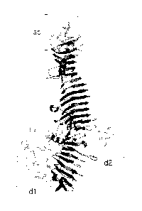
[0011] Hbp is an autotransporter protein that belongs to the subfamily of
serine protease
autotransporters of Enterobacteriaceae (SPATEs). The crystal structure of the
passenger
domain of Hbp has recently been determined (Otto et al. 2005 J Biol Chem
280(17):
17339-45), and is shown as Fig 11A. The structure shows that the polypeptide
forms a
long right-handed beta-helical structure ("beta stem"). The passenger domain
of the Hbp
comprises two larger side domains, domain dl and domain d2, of which dl
comprises the
CA 02811699 2013-03-19
WO 2012/041899 PCT/EP2011/066854
3
serine proteinase activity of the protein and d2 has an unknown function.
There are also
three smaller side domains, domain 3 (d3), domain 4 (d4) and domain 5 (d5).
[0012] Similar beta stem domains have been shown also for other
autotransporters such
as pertactin (Emsley et al 1996 Nature 381: 90-92) and IgA protease (Johnson
et al 2009
J Mol Biol 389(3): 559-74).
[0013] There have been previous attempts in using autotransporters for
secretory protein
expression in E. coli, mostly using variants of the Neisserial IgA protease
(Pyo et al 2009
Vaccine 272030-2036) and the endogenous E. coli autotranporter AIDA-1 (Van
Gerven et
al 2009 Microbiology 155:468-476) that were engineered for surface display
purposes.
[0014] Efforts using IgA protease and Al DA-I for secretion of recombinant
proteins used
constructs which resulted in poor yields of secreted and surface exposed
protein (Pyo et
al 2009 Vaccine 27 2030-2036; Van Gerven et al 2009 Microbiology 155:468-476).
In the
majority of such studies the complete, or almost complete, endogenous
passenger
domain was replaced by the recombinant protein.
[0015] So far, autotransporters have mainly been used as a display platform
rather than
for secretion of heterologous proteins in soluble form, where the protein is
secreted into
the medium.
[0016] IgA protease requires an accessory protease for processing whereas AIDA-
1
remains non-covalently attached to the outer membrane after cleavage. Thus,
these
autotransporters can only be used for surface presentation of epitopes and
proteins.
[0017] Efficient display and secretion of calmodulin fused the passenger of
Hbp has
previously been shown (Jong et al 2007 Molecular Microbiology 63:1524-1536).
In order
to minimize perturbation of the native 13-stem of the passenger, calmodulin
replaced
domain 2 of the Hbp passenger.
[0018] For certain applications the possibility to secrete or display more
than one protein
of interest (P01) from/on the cell surface is very useful. Such applications
include
vaccines, for example in which two or more epitopes are displayed on the same
cell
surface, enzyme display, in which more than one enzyme is displayed on the
cell surface
in order to carry out a range of catalytical reactions in a series of steps,
exposure of
peptide libraries and inhibitor screening.
CA 02811699 2013-03-19
WO 2012/041899 PCT/EP2011/066854
4
[0019] For multivalent vaccines it is particularly useful to have a system
wherein one
population of host cells can express and display or secrete multiple antigens,
rather than
having a mixture of cell populations, each displaying or secreting only one of
the antigens.
Having only one cell population displaying or secreting multiple multiple
antigens has the
advantage of easier production and better control of the vaccine content.
[0020] In conclusion, there is a need for improved secretory expressions
systems for the
display of heterologous proteins as well as secretion of heterologous proteins
in soluble
form into the culture medium. There is also a need for a system and a method
that enable
secretory protein expression of more than one protein of interest on the cell
surface of a
host cell or secretion of more than one protein into the culture medium.
Object of the invention
[0021] An object of the present invention is to provide efficient secretion of
a polypeptide
of interest (P01) from a host cell.
[0022] A second object of the invention is to provide efficient display of a
POI on the
surface of a host cell.
[0023] A third object of the invention is to provide efficient soluble
secretion of a P01 into
the medium in which a host cell is cultured.
[0024] Yet another object of the invention is to provide a scaffold for
efficient secretion, i.e.
display or soluble secretion, of more than one P01.
Summary of the invention
[0025] In a first aspect of the invention there is provided a host cell
capable of expressing
more than one, such as at least two, POI:s (proteins of interest). The POI:s
are comprised
in a fusion protein that also comprises a passenger domain comprising a beta
stem
domain from an autotransporter protein, a translocator domain from an
autotransporter
protein, and a signal peptide that is able to target the fusion protein to the
inner membrane
of Gram negative bacteria. The beta stem forming sequence of the passenger
domain is
essentially intact and the POI:s are fused to the passenger domain.
CA 02811699 2013-03-19
WO 2012/041899 PCT/EP2011/066854
[0026] This host cell for secretory protein expression has several advantages,
including
but not limited to more efficient secretion of more than one POI, compared to
other
systems. Also, when the goal is to display the POI, the beta stem domain will
enable a
more efficient display as the POI:s will be further away from the cell surface
and be more
stable.
[0027] In one embodiment of the host cell of the present invention, the native
form of the
passenger domain of the autotransporter comprises at least one side domain
that
protrudes from the beta stem domain. The POI:s may then be inserted into,
replace or
partly replace such side domain.
[0028] In another embodiment the native form of the passenger domain of the
autotransporter comprises at least two side domains. Each POI may then be
inserted into,
replace or partly replace a separate such side domain, or the POI:s may be
inserted into,
replace or partly replace the same side domain.
[0029] The POI:s may also be fused to an independent passenger domain,
translocator
domain and signal peptide from an autotransporter.
[0030] In a second aspect of the invention there is provided a fusion protein
comprising
more than one, such as at least two, POI:s (proteins of interest), a passenger
domain
comprising a beta stem domain from an autotransporter protein, a translocator
domain
from an autotransporter protein, and optionally, a signal peptide that targets
the fusion
protein to the inner membrane of a Gram negative bacteria. The beta stem
forming
sequence of the passenger domain is essentially intact and the POI:s are fused
to the
passenger domain.
[0031] The passenger domain of the fusion protein may in its native form
comprise at
least one side domain protruding from the beta stem domain, and the POI:s may
be
inserted onto, replace or partly replace such side domain. The passenger
domain of the
fusion protein may also in its native form comprise at least two side domains,
and each
POI may be inserted into, replace or partly replace independent domains of
such side
domains. Alternatively the POI:s may be inserted into, replace or partly
replace the same
side domain.
[0032] In another aspect of the invention there is provided a nucleic acid
arranged for
expression of a fusion protein. In one embodiment the nucleic acid comprises,
in frame,
CA 02811699 2013-03-19
WO 2012/041899 PCT/EP2011/066854
6
sequence encoding a signal peptide of the fusion protein, that is able to
target the fusion
protein to the inner membrane of Gram negative bacteria, sequence encoding a
passenger domain of the fusion protein, that comprises a beta stem domain from
an
autotransporter protein, and sequence encoding a translocator domain of the
fusion
protein, that derives from an autotransporter protein. The sequence encoding
the
passenger domain comprises at least two stretches of cloning site sequence
that allow in-
frame cloning of at least two DNA sequences that encode POI:s (proteins of
interest). The
cloning site sequences are arranged such that the encoded beta stem forming
protein
sequence of the passenger domain is essentially intact. It is also possible to
insert POI:s
into an autotransporter by merely fusing two pieces of DNA, e.g. by PCR,
without using
cloning sites thereby creating a fusion protein.
[0033] The sequence encoding the passenger domain of the autotransporter may
in its
native form comprise at least two stretches of sequence encoding side domains
protruding from the beta stem domain. The at least two stretches of cloning
site sequence
may then be inserted into, replace or partly replace separate of such
stretches encoding
side domains.
[0034] In another embodiment the nucleic acid comprises, in frame, sequence
encoding a
signal peptide of the fusion protein, that is able to target the fusion
protein to the inner
membrane of Gram negative bacteria, sequence encoding a passenger domain of
the
fusion protein, that comprises a beta stem domain from an autotransporter
protein,
sequence encoding a translocator domain of tha fusion protein, that derives
from an
autotransporter protein, and sequences encoding at least two POI:s of the
fusion protein.
The sequences encoding the POI:s are fused to the sequence encoding the
passenger
domain and are arranged such that the encoded beta stem forming protein
sequence of
the passenger domain is essentially intact.
[0035] The sequence encoding the passenger domain of the autotransporter in
its native
form may comprise at least two stretches of sequence encoding side domains
protruding
from the beta stem domain. Each of the at least two sequences encoding POI:s
may then
be inserted into, replace or partly replace each of the stretches encoding
side domains.
[0036] The host cell, fusion protein or nucleic acid may be arranged such that
the fusion
protein, when expressed, is secreted from the cell surface. For instance, the
fusion protein
may comprise a cleavage site that allows the fusion protein to be cleaved and
secreted
CA 02811699 2013-03-19
WO 2012/041899 PCT/EP2011/066854
7
from a host cell expressing the fusion protein. And the nucleic acid encoding
the fusion
protein may encode such a cleavage site.
[0037] Alternatively, the host cell, fusion protein or nucleic acid may be
arranged such that
the fusion protein, when expressed, is displayed at the cell surface. For
instance the
fusion protein may comprise no such cleavage site or may comprise a disrupted
cleavage
site. Similarly the nucleic acid encoding the fusion protein then encodes no
such a
cleavage site or encodes a disrupted cleavage site. Alternatively, the fusion
protein and
nucleic acid may comprise a cleave site and the resulting fusion protein be
cleaved, but
remains non-covalently attached to, and thus displayed at, the cell surface.
[0038] The passenger domain and the translocator domain may be derived from a
SPATE
(serine protease autotransporters of Enterobacteriaceae) protein, such as
Hemoglobin-
binding protease (Hbp), extracellular serine protease (EspC) or temperature-
sensitive
hemagglutinin (Tsh) from Escherichia coli.
[0039] In one aspect of the invention there is provided a vector comprising a
nucleic acid
of the invention.
[0040] In another aspect of the invention there is provided a host cell
comprising a nucleic
acid or a vector of the invention.
[0041] In one embodiment the host cell of the invention is a Gram negative
bacterium,
which may be selected from the family of Enterobacteriaceae, such as
Escherichia coli,
Salmonella spp., Vibrio spp., Shigella spp., Pseudomonads spp., Burkholderia
spp. or
Bordetella spp.
[0042] In one aspect there is provided an outer membrane vesicle displaying a
fusion
protein according to the invention. In another aspect there is provided a
bacterial ghost
displaying a fusion protein according to the invention.
[0043] In one aspect there is provided a method for secretory protein
expression of a
fusion protein, comprising the steps of providing a host cell according to the
invention and
inducing expression of the fusion protein.
[0044] In one embodiment the method comprising the additional step of
inhibiting a
periplasmic enzyme, such as DegP, with protease activity in the host cell.
DegP may for
example be inhibited by a mutation in its catalytic site.
CA 02811699 2013-03-19
WO 2012/041899 PCT/EP2011/066854
8
[0045] In another embodiment the method comprises the additional step of down
regulating at least one enzyme, such as DsbA or DsbB, that catalyzes the
formation of
disulphide bonds in proteins in the periplasmic space of the host cell.
[0046] The method may provide secretion of the fusion protein in a soluble
manner.
Alternatively it may provide display of the fusion protein on the cell
surface.
[0047] In one embodiment the method comprises the additional step of inducing
shedding
of vesicles from the outer membrane of the host cell, being a Gram negative
bacterium,
thus forming outer membrane vesicles displaying the fusion protein on their
surface.
[0048] In another embodiment the method comprises the additional step of
lysing the
Gram negative bacterium, thus forming bacterial ghosts displaying the fusion
protein on
their surface. The lysing may be made by use of the lethal lysis gene E from
bacteriophage PhiX174.
[0049] In one embodiment at least one of the POI:s may comprise an antigen,
for example
from an infectious organism. The antigen is for example an antigen from
Mycobacterium
tuberculosis, such as ESAT-6, Ag85B, Rv2660c, TB10.4 and TB10.3, or a protein
that is
similar to those proteins.
[0050] In one aspect there is provided a vaccine comprising a host cell, a
fusion protein,
an outer membrane vesicle or a bacterial ghost according to the invention.
Brief description of figures
[0051] The invention is now described, by way of example, with reference to
the
accompanying figures, in which:
[0052] Fig 1-10 show plasmid maps of plasmids used in the examples.
[0053] Fig 11 A-D show figures of the structure of the passenger domain of Hbp
where
certain domains are indicated.
[0054] Fig 11 E shows various constructs of fusion proteins used in examples 1-
15.
[0055] Fig 12-33 show experimental data from examples 1-19. For details, see
the
example section.
CA 02811699 2013-03-19
WO 2012/041899 PCT/EP2011/066854
9
[0056] Fig. 34 shows a map of a plasmid used in the examples.
Definitions
[0057] As used herein, the following definitions are supplied in order to
facilitate the
understanding of the present invention.
[0058] An "autotransporter" is a protein that belongs to the pfam
autotransporter family
('Autotransporter PF03797) and that also is known or predicted to form a beta
stem motif.
The BETAVVRAPPRO method for sequence analysis can be used to predict if the
passenger domain of an autotransporter will form a beta stem motif (Junker et
al 2006
Proc Natl Acad Sci U S A 103(13): 4918-23).
[0059] A "polypeptide of interest" (P01) is a polypeptide that the user of the
invention
wants a host cell to secrete in soluble form into the medium or to display on
the cell
surface, or both. Typically, the P01 is a protein that the user studies or
wants to be
expressed, whereas the other parts of the fusion protein assist in the
secretion process.
Typically, the P01 is also heterologous to the autotransporter domains to
which it is fused.
The P01 is at least 4 amino acids long, at least 10 amino acids long or at
least 20 amino
acids long.
[0060] "Beta stem forming sequence" refers to the sequence of a passenger
domain of an
autotransporter that forms a beta stem structure. The beta stem forming
sequence of a
passenger can be identified using crystal structure determination. As
described above the
beta stem forming sequence may alternatively be identified using the M4T
homology
modeling method (Rykunov et al 2009 J Struct Funct Genomics 10: 95-99) or
similar
prediction methods.
[0061] A "side domain" is a domain that is part of the passenger domain but is
not part of
the beta stem. Typically, a side domain is located in the passenger domain
between two
stretches of beta stem forming sequence. A side domain starts at the first
amino acid after
the preceding beta strand and it ends one amino acid before the starting amino
acid of the
beta strand following the side domain. The side domain can also be located at
the N-
terminus of the passenger domain. Autotransporters may have several side
domains.
CA 02811699 2013-03-19
WO 2012/041899 PCT/EP2011/066854
[0062] "Similar protein", "similar sequence" or a "like protein" refers to a
protein that has a
high degree of homology to another protein when the two amino acid sequences
are
compared. Preferably, it is at least 80%, more preferably more than 90%, more
preferably
more than 95%, even more preferably more than 97% homologous to the
comparative
sequence when the two sequences are optimally aligned. Sequence homology can
be
readily measured using public available software such as BLAST.
[0063] "Host cell" refers to a prokaryotic cell into which one or more vectors
or isolated
and purified nucleic acid sequences of the invention have been introduced. It
is
understood that the term refers not only to the particular subject cell but
also to the
progeny or potential progeny of such a cell. Because certain modifications may
occur in
succeeding generations due to either mutations or environmental influences,
such
progeny may not, in fact, be identical to the parent cell, but are still
included within the
scope of the term as used herein.
[0064] "Displayed": A secreted protein is displayed on the surface of the
secreting host
cell when it remains associated with the outer membrane of the host cell such
that it at
least partly protrudes outside the cell. The secreted protein may be attached
to the cell
membrane or a component that resides therein (such as the translocator domain
from an
autotransporter) in a covalent or non-covalent manner.
[0065] "Soluble secretion" and "secretion in a soluble manner" refers to
secretion of a
protein where the protein is secreted into the extracellular space so that is
not associated
with the host cell as opposed to when the protein remains associated to the
outer
membrane of the host cell, or a protein that is integrated into the outer
membrane of the
host cell.
[0066] "Approximately" indicates a deviation of +1-10% of the stated value,
where
applicable.
Description
[0067] The inventors have found that an autotransporter protein can be used
for improved
secretory protein expression if the beta stem forming sequence of the
passenger domain
CA 02811699 2013-03-19
WO 2012/041899 PCT/EP2011/066854
11
of the autotransporter is essentially intact. Whereas the actual beta stem-
forming
sequence is essential for optimal secretion, the side domains of the passenger
domain of
autotransporters are suitable sites for the insertion of a POI. The side
domains can be
replaced by a POI which will then be secreted. Alternatively, the POI can be
inserted so
as to replace a part of the side domain or so as to be fused to a side domain.
[0068] The inventors have also found that an autotransporter protein can be
used for
improved secretory protein expression of more than one, such as at least two,
POI:s if the
beta stem forming sequence of the passenger domain of the autotransporter is
essentially
intact. By fusing, i.e. inserting, replacing or partly replacing, the POI:s to
one or more side
domains of the passenger, while keeping the beta stem structure intact, an
efficient and
relatively easy-to-use system for simultaneous display or soluble secretion of
two or more
POI:s is achieved.
[0069] The side domains that can be replaced according to the invention are
relatively
large, such as 20, 30, 40, 60, 80 or more amino acid residues.
[0070] Thus, the passenger domain of an autotransporter can be considered as
several
sections of beta stem forming sequence linked together by non-beta stem
forming
sequences. These non-beta stem forming sequences are suitable sites for
insertion of one
or more POI:s. Thus, the POI can be placed in between two parts of beta stem
forming
sequence. The POI can also be fused to the N-terminus of the passenger domain.
[0071] Suitable methods for detecting beta stem forming sequence and side
domains of
passenger domains of autotransporters include biophysical methods such as as x-
ray
crystallography and bioinformatics software such as structure prediction
tools.
[0072] X-ray crystallography is today a standard procedure that is highly
efficient and
automatized and is known to a person skilled in the art. Examples of high
resolution
structures of passenger domains and suitable methods for determination of
structures of
the passenger domain of autotransporters are found in (Otto et al 2005 J Biol
Chem
280(17):17339-45; Emsley et al 1996 Nature 381: 90-92; Johnson et al 2009 J
Mol Biol
389(3): 559-74).
[0073] An example of a bioinformatics method that is suitable for determining
beta stem
structure is the M4T homology modeling method (Rykunov et al 2009 J Struct
Funct
Genomics 10: 95-99), which is available for free on the internet.
CA 02811699 2013-03-19
WO 2012/041899 PCT/EP2011/066854
12
[0074] Where a three-dimensional model of the protein is used for the
identification of
beta stem domains and side domains, it is suitable that the model obtained has
a
resolution of better than 4 angstrom. Side domains will then be visible as
domains that
protrude from the beta stem. By observation of the structure of the passenger
domains of
autotransporters it can be seen that parts of the sequence are not part of the
beta stem
but form domains that protrude from the beta stem.
[0075] These methods can be used for determining which domains or amino acids
of the
passenger domains that are suitable for insertion of a POI and which should be
kept
essentially intact.
[0076] The beta stem forming sequence is essentially intact according to the
invention.
Thus, as little as possible of the beta stem forming sequence should be
removed.
Predicted domain border is of help to determine where the POI(s) should be
inserted. If
too much of the beta stem forming sequence is removed, secretion will be
negatively
affected. That the beta stem forming sequence is essentially intact means that
the
efficiency of the secretory function of the protein is maintained at an
optimal level, as
compared to when the beta stem is disrupted or completely removed. It also
means that
the stability of the passenger after secretion is maintained. A person skilled
in the art can
use experimental methods to determine if a particular constructs allows
efficient secretion.
[0077] Examples of methods suitable for determining the efficiency of
secretion in vitro
include: analysis of the fraction of POI present in the medium, labeling of
surface proteins
with biotin or other labels, cell fractionation, exposure of surface proteins
to proteases
(such as proteinase K) and studies using antibodies against the POI (such as
dot blot
studies, immunofluorescene microscopy and immuno-electron microscopy).
[0078] Examples of methods suitable for determining the stability of the
passenger after
secretion include SDS-PAGE, western blotting and all of the above under
paragraph 76.
[0079] Thus, by using structural information, a person skilled in the art can
predict where
in the passenger domain the insertion of the POI can be made in order to
maintain optimal
secretion. Actual secretion can be easily determined with in vitro
experiments.
[0080] According to the invention the POI is fused to the passenger domain.
This means
that the POI is fused to the peptide that forms the passenger domain such that
they form
one continuous polypeptide. Because design of the fusion protein is carried
out at the
CA 02811699 2013-03-19
WO 2012/041899 PCT/EP2011/066854
13
DNA level, care must be taken so that the reading frame of the POI is the same
as the
reading frame of the passenger domain.
[0081] Preferably the POI has a molecular weight of less than 200 kD. More
preferably the
molecular weight is less than 200 kD such as 100 , 80, 60, 40, 30, 20, 10 or 5
kD.
[0082] The fusion protein can comprise more than one POI. Thus, the fusion
protein can
comprise two, three or more polypeptides of interest. The fusion protein can
be such that
it has at least two POls that each replaces, or partly replaces, or is fused
to, a separate or
independent side domain of the passenger domain. Alternativley, two or more
POls can
be fused to, or replace, or partly replace the same side domain.
[0083] The fusion protein is encoded by a nucleic acid and expressed in a host
cell. The
nucleic acid can be constructed with the use of standard molecular biology
techniques
involving restriction enzymes, DNA ligases, PCR, oligonucleotide synthesis,
DNA
purification and other methods well-known to a person skilled in the art.
Preferably, the
starting point is a reading frame of an autotransporter protein into which a
DNA fragment
encoding the POI is inserted so that the reading frames match. Alternatively,
the reading
frame for the fusion protein can be designed in silico and synthesized using
polynucleotide synthesis.
[0084] The reading frame encoding the fusion protein is preferably inserted in
an
expression vector for prokaryote expression carrying a promoter and other
components
well known to a person skilled in the art.
[0085] The fusion protein comprises an N-terminal signal peptide that directs
the protein
for secretion. When the host cell is a Gram negative bacteria the signal
peptide suitable is
such that it directs translocation of the protein across the inner membrane.
The signal
peptide can be derived from an autotransporter protein, suitably the same
autotransporter
from which the passenger domain is derived. The signal peptide can comprise
approximately amino acids 1 to 52 of SEQ ID NO 1, or a similar sequence.
[0086] The fusion protein suitably comprises an autochaperone domain, suitably
from the
passenger domain of the autotransporter protein used to fuse the POI. One
example of an
autochaperone domain comprises approximately amino acids 1002 to 1100 of SEQ
ID NO
1.
CA 02811699 2013-03-19
WO 2012/041899 PCT/EP2011/066854
14
[0087] The fusion protein can comprise a passenger domain from one type of
autotransporter and a translocator domain from another type of
autotransporter.
[0088] The autotransporter used in the invention can be an autotransporter
with a serine
protease domain, such as a serine protease.
[0089] The autotransporter can be a SPATE protein (Serine protease
autotransporters of
the Enterobacteriaceae). Thus, the translocator domain and the passenger
domain can be
from a SPATE protein. In one embodiment the SPATE protein is one of Hemoglobin-
binding protease (Hbp) (SwissProt 088093) and temperature-sensitive
hemagglutinin
(Tsh) (SwissProt Q47692) from E. coll. The sequence of Tsh is homologous to
that of
Hbp.
[0090] Other SPATE proteins include IgA protease of Neisseria gonorrhoeae and
Haemophilus influenzae, EspC from E. coli, Pet from E. coli, EspP from E.
coli, Pic from
E. coli, PicU from E. coli, Sat from E. coli, Vat from E. coli, Espl from E.
coli, EaaA from E.
coli, EaaC from E. coil, EatA from E. coli, EpeA from E. coli, PssA from E.
coli, AidA_B7A
from E. coli, Boa from Salmonella bongori, SepA from Shigella flexneri, SigA
from Shigella
flexneri, Pic from Shigella flexneri.
[0091] The SPATE protein can comprise the polypeptide of SEQ ID NO 1, which is
Hbp,
or SEQ ID NO 2, which is Hbp where the cleavage site between the translocator
domain
and the passenger domain has been disrupted (Hbp delta ¨cleav) or a sequence
that is
similar to those sequences. Preferably the identity is more than 80%, even
more
preferably more than 90%, even more preferably more than 95% and most
preferably
more than 97% to those sequences.
[0092] The SPATE group of proteins has several advantages for use with the
present
invention. First of all some of their structures are known, facilitating the
identification of
their beta stem and side domains. This knowledge can also be used for
prediction of side
domains and beta stem structures of related SPATEs for which the crystal
structure is not
known. Another advantage is their cleavage structure that can be used for
efficient soluble
secretion, and that is conserved within the SPATE family.
[0093] Other autotransporters, for which the structure is known, can be
predicted or will be
known, such that their beta stem and side domain structure can be determined,
may also
be used with the present invention. The autotransporter should have a beta
stem, a side
CA 02811699 2013-03-19
WO 2012/041899 PCT/EP2011/066854
domain and optionally a cleavage system that is efficient for soluble
secretion. An
example includes the autotransporter Haps from H. influenzae, which is not a
member of
the SPATE family. The structure of the passenger of Haps has recently been
published
(Meng et al 2011 Aug 12 The EMBO Journal, doi: 10.1038/emboj.2011.279. [Epub
ahead
of print]). The structure is very close to that of Hbp, having a beta-stem
with four side
domains (SD1-4).
[0094] Fig 11 A shows the crystal structure of the passenger domain of the
autotransporter Hbp (Otto et al 2005 J Biol Chem 280(17): 17339-45). Domain 1
(d1),
domain 2 (d2) and the autochaperone domain (ac) are in light grey. The
remainder of the
passenger domain, including the beta stem domain is colored black. Both
domains dl and
d2 are suitable for insertion of a POI. In addition, the domain d3 shown in
figure 11 C and
domains d4 and d5 shown in figure 11D are suitable for replacement or
insertion of a POI.
[0095] Domain dl comprises approximately the amino acids 53 to 308, d2
comprises
approximately the amino acids 533-608, d3 comprises approximately the amino
acids
657-697, d4 comprises approximately the amino acids 735 to 766 and d5
comprises
approximately amino acids 898 to 922 of SEQ ID NO 1, which is the sequence of
Hbp.
[0096] Fig 11 E shows the domain composition of wild-type Hbp. In addition,
fusion
proteins used in the examples presented herein are shown. In wild-type Hbp,
the
passenger domain comprising the beta stem (in black) and the side domains dl,
d2, d3,
d4 and d5 is shown. The translocator domain is located at the C-terminal part
of the
protein and is indicated as "p-domain". "Ac" indicates an autochaperone
domain. The
signal peptide is denoted by "ss". Numbers indicate amino acid number from the
N-
terminus.
[0097] A passenger domain that comprises approximately amino acids 53-1100 of
SEQ ID
NO 1, or a similar sequence, can be used.
[0098] A translocator domain that comprises approximately amino acids 1101-
1377 of
SEQ ID N01, or a similar sequence, can be used.
[0099] The POI can be a split protein. A split protein is a protein which in
its native form
comprises a single polypeptide or several polypeptides that are linked by
disulphide
bridges or other intermolecular bonds, and which for the present invention has
been split
in two or more parts. Each such part is fused to the passenger such that they
form a non-
CA 02811699 2013-03-19
WO 2012/041899 PCT/EP2011/066854
16
native structure, for example at a distance apart. The two or more parts may
for instance
be fused to different side domains or to the same side domain but at a
distance apart.
Each such part is considered to be one POI, such that the split protein is
considered to be
two or more POI:s. This could for example be advantageous when the native
protein has
a large or complex structure, for example comprising disulphide bridges, that
inhibits
efficient secretion. Splitting the protein may make the secretion more
efficient.
[00100] The POI can comprise at least one antigen, for example from an
infectious
organism such as Mycobacterium tuberculosis. Examples of such antigens from
Mycobacterium tubercolosis include ESAT-6-like proteins (e.g. ESAT-6, TB10.4,
TB10.3),
an Ag85B-like protein (e.g. Ag85B), and Rv 2660c,. Two or more of such
antigens may be
fused to the same passenger, for example to separate side domains.
[00101] ESAT-6 (early secretory antigenic target of 6 kDa) is a 10 kDa
protein that is
a potent 1-cell antigen and an important virulence factor.
[00102] Rv2660c is a 7.6 kDa intracellular protein of unknown function.
[00103] TB10.3 and TB10.4 are both 96 amino acid proteins.
[00104] Ag85B is a secretory mycolyltransferase of 35 kDa, comprising three
cysteins. It is also a potent T-cell antigen. This rather large and cysteine
comprising
protein is too complex, in its native form, for optimal outer membrane
translocation using
the autotransporter system.
[00105] In one embodiment the antigen is split as defined above. For
example,
Ag85B, which is a large and rather complex protein, may be split into a N'-
part
(Ag85B(N')) and a C'-part (Ag85B(C')) for more efficient secretion.
[00106] In one embodiment the POI comprises a polypeptide with a sequence
that is
at least 80%, more preferably 90%, more preferably 95% most preferably 97%
similar to
SEQ ID NO 39, which is the sequence of ESAT-6. In one embodiment the POI
comprises
the polypeptide defined in SEQ ID NO 39.
[00107] In one embodiment the POI comprises a polypeptide with a sequence
that is
at least 80%, more preferably 90%, more preferably 95% most preferably 97%
similar to
SEQ ID NO 41, which is the sequence of Rv2660c. In one embodiment the POI
comprises
the polypeptide defined in SEQ ID NO 41.
CA 02811699 2013-03-19
WO 2012/041899 PCT/EP2011/066854
17
[00108] In one embodiment the POI comprises a polypeptide with a sequence
that is
at least 80%, more preferably 90%, more preferably 95% most preferably 97%
similar to
SEQ ID NO 42, which is the sequence of TB10.4. In one embodiment the POI
comprises
the polypeptide defined in SEQ ID NO 42.
[00109] In one embodiment the POI comprises a polypeptide with a sequence
that is
at least 80%, more preferably 90%, more preferably 95% most preferably 97%
similar to
SEQ ID NO 43, which is the sequence of TB10.3. In one embodiment the POI
comprises
the polypeptide defined in SEQ ID NO 43.
[00110] In one embodiment the POI comprises a polypeptide with a sequence
that is
at least 80%, more preferably 90%, more preferably 95% most preferably 97%
similar to
at least % of SEQ ID NO 40, which is the sequence of Ag85B. In one embodiment
the POI
comprises the polypeptide defined by amino acids 1-126 or 118-285 in SEQ ID NO
40.
[00111] The POI can be flanked by one or more linker regions. A linker
region can
be a flexible peptide of 1 to 20, or more, amino acids. The linker region can
suitably be
inserted at the C- and N-termini of the POI. An advantage of a linker is that
it may allow
the various domains of the fusion protein to move more independent of each
other. A
linker can easily be designed by a person skilled in the art. Examples of
suitable linkers
include SEQ ID NO 44 and 45.
[00112] The fusion protein can comprise the polypeptide defined in any of
SEQ ID
NO:s 13¨ 19, SEQ ID NO:s 22 ¨26 or SEQ ID NO 38 or a polypeptide which is at
least
80%, more preferably 90%, more preferably 95% and most preferably 97% similar
to any
one of those sequences.
[00113] SEQ ID NO 13 is the sequence of Hbp were ESAT6 has replaced domain
dl
(Hbp(Ad1)-ESAT6, also named HbpSL-ESAT6). SEQ ID NO 14 is the same protein but
where the cleavage site between the translocator domain and the passenger
domain has
been disrupted (HbpD(Ad1)-ESAT6, also named HbpDL-ESAT6). SEQ ID NO 15 is the
sequence of Hbp where ESAT6 has replaced domain d2 (Hbp(Ad2)-ESAT6). SEQ ID NO
16 is the sequence of Hbp where ESAT6 has replaced domain d2 (HbpD(Ad2)-ESAT6,
also named HbpDD2-ESAT6) and where the cleavage site between the translocator
domain and the passenger domain has been disrupted. SEQ ID NO 17 is the
sequence of
Hbp where ESAT6 has replaced domain d3 (Hbp(Ad3)-ESAT6). SEQ ID NO 18 is the
CA 02811699 2013-03-19
WO 2012/041899 PCT/EP2011/066854
18
sequence of Hbp where ESAT6 has replaced domain d4 (Hbp(Ad4)-ESAT6). SEQ ID NO
19 is the sequence of Hbp where ESAT6 has replaced domain d5 (Hbp(Ad5)-ESAT6).
[00114] SEQ ID NO 22 is the sequence of Hbp where Rv2660c has replaced
domain
d3 (Hbp(Ad3)-Rv2660c). SEQ ID NO 23 is the sequence of Hbp where Rv2660c has
replaced domain d4 (Hbp(Ad4)-Rv2660c). SEQ ID NO 24 is the sequence of Hbp
where
Rv2660c has replaced domain d5 (Hbp(Ad5)-Rv2660c). SEQ ID NO 25 is the
sequence of
Hbp where TB10.4 has replaced domain dl (Hbp(Ad1)-TB10.4). SEQ ID NO 26 is the
sequence of Hbp where TB10.3 has replaced domain d2 (Hbp(Ad2)-TB10.3).
[00115] SEQ ID NO 38 is the sequence of EspC where ESAT6 has replaced
domain
dl (EspC(Ad1)-ESAT6).
[00116] The fusion protein can comprise a polypeptide with more than one
POI, such
as the polypeptide defined in any of SEQ ID NO:s 28¨ 35 or a polypeptide which
is at
least 80%, more preferably 90%, more preferably 95% and most preferably 97%
similar to
any one of those sequences.
[00117] SEQ ID NO 28 is the sequence of Hbp where residues 1-126 of Ag85B
has
replaced domain dl and residues 118-285 of Ag85B has replaced domain 2 (H bp-
Ag85B[N+q). SEQ ID NO 29 is the sequence of Hbp where residues 1-126 of Ag85B
has
replaced domain dl and residues 118-285 of Ag85B has replaced domain 2, and
where
the cleavage site between the translocator domain and the passenger domain has
been
disrupted (HbpD-Ag85B[N+q). SEQ ID NO 30 is the sequence of Hbp where residues
1-
126 of Ag85B has replaced domain d2 and residues 118-285 of Ag85B has replaced
domain 1 (Hbp-Ag85B[c+Ni). SEQ ID NO 31 is the sequence of Hbp where residues
1-126
of Ag85B has replaced domain d2 and residues 118-285 of Ag85B has replaced
domain
1, and where the cleavage site between the translocator domain and the
passenger
domain has been disrupted (HbpD-Ag85B[c+N]).
[00118] SEQ ID NO 32 is the sequence of Hbp where residues 1-126 of Ag85B
has
replaced domain d2, residues 118-285 of Ag85B has replaced domain 1 and ESAT6
has
replaced domain d4 (Hbp-Ag85B[c+N]-ESAT6). SEQ ID NO 33 is the same protein
but
where the cleavage site between the translocator domain and the passenger
domain has
been disrupted (HbpD-Ag85B[c+NrESAT6). SEQ ID NO 34 is the sequence of Hbp
where
residues 1-126 of Ag85B has replaced domain d2, residues 118-285 of Ag85B has
replaced domain 1, ESAT6 has replaced domain d4 and Rv2660c has replaced
domain 5
CA 02811699 2013-03-19
WO 2012/041899 PCT/EP2011/066854
19
(Hbp-Ag85B[c+NrESAT6-Rv2660c). SEQ ID NO 35 is the same protein but where the
cleavage site between the translocator domain and the passenger domain has
been
disrupted (HbpD-Ag85B[c+Ni-ESAT6-Rv2660c).
[00119] Preferably, the order of domains of the fusion protein is, from the
N-terminus
to the C-terminus: signal peptide, passenger domain, translocator domain.
[00120] In a second aspect of the invention it is provided a cell
expressing a fusion
protein as defined herein. The cell is preferably a host cell that can be
cultured and
manipulated by methods well known to a person skilled in the art and which is
able to
express heterologous proteins. Preferably the host cell is a Gram-negative
bacterium such
as E. coli, Salmonella spp., Vibrio spp., Shigella spp., Pseudomonads spp.,
Burkholderia
spp. or Bordetella spp. A wide variety of expression systems are available and
known to a
person skilled in the art. The expression may be of a stable or transient
nature. The
expression system may be inducible or non-inducible.
[00121] In one embodiment the fusion protein is at least partly solubly
secreted by
the host cell. This embodiment can be used when the invention is used for
production of a
recombinant protein, which is, for example, a commercial enzyme or a component
of a
pharmaceutical. The P01 can then be conveniently harvested from the media,
without
breaking up the host cells. Breaking up the host cells causes contamination
with cellular
debris and cellular content. Secretion of the fusion protein can be achieved
when the
fusion protein comprises a protease cleavage site between the translocator
domain and
the passenger domain. A protease activity, which may reside in the fusion
protein itself,
cleaves the fusion protein when the translocator domain has integrated into
the outer
membrane so that the passenger domain is released into the medium.
Alternatively,
cleavage may take place via an intramolecular autocatalytic cleavage mechanism
that is
unrelated to protease activity as described for the SPATE EspP from E. coli
(Dautin et al
2007 EMBO J 26(7): 1942-1952) and Al DA-I from E. coli (Charbonneau et al 2009
J Biol
Chem 284(25): 17340-17353).
[00122] For the sake of clearness, the P01 may in some cases remain
attached to
the cell membrane even though the polypeptide has been cleaved. Such
attachment will
usually be of a non-covalent nature.
[00123] In one embodiment the P01 remains covalently attached to the
translocator
domain. Where the sequence of the autotransporter harbors a cleavage site,
this can be
CA 02811699 2013-03-19
WO 2012/041899 PCT/EP2011/066854
achieved by mutating the cleavage site between the translocator domain and the
passenger domain, so that the cleavage event does not take place. Thus, the
host cell
displays at least a part of the fusion protein comprising at least one POI on
the cell
surface.
[00124] In certain aspects the invention provides outer membrane vesicles
(OMV:s)
or bacterial ghosts displaying a fusion protein according to the invention on
their surface.
[00125] Under certain conditions Gram negative bacteria may be induced to
start
shedding vesicles from their outer membrane. Such outer membrane vesicles
(OMV:s)
have for example been shown to be useful as vaccine platforms. When carrying
antigens,
as derived from their mother cells, these vesicles are capable of enhancing
the
immunogenicity of such antigen. OMV:s may easily be derived from gram negative
bacteria displaying the fusion protein of the invention on their surface.
Methods for outer
membrane vesicle production and isolation are known in the art (Chen et al
2010 PNAS
107:3099-3104; Bernadac et al 1998 J Bacteriol 180: 4872-4878; Kesty and Kuehn
2004 J
Biol Chem 279: 2069-2076); Kolling and Matthews 1999 App Env Microbial 65:
1843-
1848; Kitagawa et al 2010 J Bacterial 192: 5645-5656).
[00126] Similarly, bacterial ghosts are a nonliving vaccine platform.
Bacterial ghosts
are bacterial cell envelopes that have been emptied of their cytoplasm by
means of lysis,
for example using the lethal lysis gene E from bacteriophage PhiX174
(Langemann et al
2010 Bioeng Bugs 1:326-336; Young 1992 Microbiol rev 56: 430-481; Mayr et al
2005 Adv
Drug Deliv rev 57: 1381-1391). They retain all morphological, structural and
antigenic
features of the mother cell and comprise proteins that are expressed and
anchored to the
cell envelope before lysis. Delivery of for example antigenic proteins can be
facilitated by
the secretion system and the fusion proteins of the invention.
[00127] One aspect of the invention is a vaccine comprising a fusion
protein, a cell,
an outer membrane vesicle or a bacterial ghost according to the invention. The
vaccine
can comprise a host cell that displays a fusion protein comprising at least
one POI at the
cell surface. Preferably the POI is then an antigen as described above. The
host cell can
be an attenuated Salmonella strain, such as the strains described in Curtiss R
316 et al
2010 Crit Rev Immunol 30(3): 255-70. The vaccine can comprise living host
Salmonella
cells.
CA 02811699 2013-03-19
WO 2012/041899 PCT/EP2011/066854
21
[00128] One aspect of the invention is a nucleic acid which encodes a
fusion protein
according to the invention as has been described above. One further aspect of
the
invention is a vector carrying a nucleic acid according to the invention.
[00129] The nucleic acid or vector may be arranged for expression of more
than one
POI fused to the same passenger domain. For example, the sequence which
encodes the
passenger domain can comprise at least two stretches of cloning site sequence
that allow
in-frame cloning of at least two POI encoding sequences. This facilitates easy
cloning and
expression of any desired POI:s. Alternatively the nucleic acid may comprise
more than
one sequence encoding POI:s, fused to the passenger domain.
[00130] One aspect of the invention comprises a method for secretory
protein
expression of a POI comprising the step of expressing a fusion protein
according to the
invention in a host cell. Expression vectors are well known to a person
skilled in the art.
Suitably, the vector has a promoter suitable for the host cell which is
operatively linked to
the nucleic acid that encodes the fusion protein according to the invention.
[00131] The method can comprise the step of identifying suitable side
domains on an
autotransporter protein. This can be carried out with the biophysical methods
or the
bioinformatics methods described above.
[00132] One aspect of the method according to the invention comprises the
step of
replacing a side domain (or a part thereof) of a passenger domain of an
autotransporter
with a POI so that the beta-stem forming sequence of the passenger domain of
the
autotransporter is essentially intact. Alternatively, the method can comprise
the step of
inserting the POI into the passenger domain so that the beta stem forming
sequence is
essentially intact.
[00133] The method comprises the step of culturing the host cell under
conditions
wherein the nucleic acid encoding the fusion protein is translated to a
multitude of fusion
protein molecules and the fusion protein molecule enters the secretory
pathway.
[00134] In one embodiment, the method comprises the additional step of
inhibiting a
periplasmic enzyme with protease activity in the host cell, such as DegP. The
protease
activity of DegP can be inhibited by deleting, interrupting or inactivating
the DegP-
encoding gene on the chromosome of the host cell. Inactivation can be carried
out by the
CA 02811699 2013-03-19
WO 2012/041899 PCT/EP2011/066854
22
introduction of a mutation in the catalytic site of DegP. The inhibition of a
protease has the
advantage that yield can be improved.
[00135] In one embodiment the method comprises the additional step of down
regulation of at least one enzyme, such as DsbA or DsbB, that catalyses the
formation of
disulphide bonds in proteins in the periplasmic space of the host cell. This
has the
advantage that yield can be improved, especially for proteins that are prone
to form
disulphide bridges, such as proteins of eukaryotic origin.
[00136] In one embodiment of the method the POI is soluble secreted. In one
embodiment of the method the POI remains covalently attached to the cell
surface.
[00137] In one embodiment the method comprises the further step of inducing
shedding of vesicles from the outer membrane of the host cell, to produce
outer
membrane vesicles displaying the fusion protein of the invention on their
surface.
[00138] In another embodiment the method comprises the additional step of
lysing
the gram negative bacterium, for example using the the lethal lysis gene E
from
bacteriophage PhiX174, thus forming bacterial ghosts displaying the fusion
protein on
their surface.
[00139] One final aspect of the invention comprises a fusion protein
obtainable
according to the method of the invention.
Examples
[00140] Methods
[00141] Strains and media
[00142] E. coli strain MC1061 (araD139 A(araA-leu)7697 AlacX74 galK16
galE15(GalS) A e14" mcrA0 relA1 rpsL150(strR) spoT1 mcrB1 hsdR2) has been
described previously (Casadaban and Cohen 1980 J Mol Biol 138: 179-207).
Strain
TOP1OF' was obtained from Invitrogen.
[00143] Cells were routinely grown at 37 C in LB medium supplemented with
0.2%
glucose. Overnight cultures were grown in the presence of 0.4% glucose. Cells
were
322223322333131133132e22223 MJgsg8v/seD
223221142333323323342223324 z-r1 AJ 1j'OT91/seD
DeeDe424e3leee323424E33132e22223 TIT ml VOT91/seD
2e3334ee34;5243Eee242334e223324 OtT Ai 3099znu/seD
33e2312322232e4224233432e22223 601 ml 3099ZAH/seD
2plempeDeOee2142335333432333132e55324334e5233513233e 801 Ai (Sej/SUddqH
45eDleEle5243Dee34233e42235e325334e25e3233435e25232e529 LOT m}
(seD/sud)dqH
344344.332421e2e3ee32433ee2E333432E2232433422233243 901 u (seDisuptp)dqH
MeD9OneDuDVVeeeelSODEeDOEDDlegEeD9Dop2e022p 50T An; (seD/su!vp)dqH
3e23354.454.33e24e345 Ai TZ0E-00 dqH
342e3e24222e2334333432333432e22324334E22332432 (sej/swopv)dqH
32e22434eleeobjvig9232e322331e22e3233432e52232e229 zoT m; (seD/swory)dqH
24323e24235e3335345333435333432e52324.334e2233243233e TOT Ai
(seDitwopv)dqH
233e3223ee34434242e42232e322334e22e3233432e22232e22g 001 MJ
(sej/twopV)dqH
DleeDE22124eapleeD2 66 mi 6L8T-6S8T dqH
23e2E34.532E3543333435333432e52324334e2233243233e 86 Ai (seD/Ewopv)dqH
3e244433e44442e3233ee42232e322334E22e3233432E22232e222 L6 mj
(seD/Ewopv)dqH
212224DEDDE8ole3142 96 Ai OZ8Z-898Z dqH
Deeele24254DeEeDe2e3242235e355334e5532433435e255 56 MJ (sED/wals-dv)dqH
2e4224232E3342e4e32233ee2E333432E22324334e2233243 v6 Ai (sej/z wopv)dqH
3111.21.34E4222E32pelee42232e322334e22e3233432e02213 6 m; (seD/z
wory)dqH
2e2e35433443e2ee323333432e52e35334e253 z6 Ai (seD/T woPV)dqH
34423e3422333324E24ee42232e322334E2232433432e222 16 mj (seD/T wopv)dqH
8euelee2DeeleeElee2E3434lee01.2e3 06 Ai dqH¨luo3]
234434344emee5eDee24e3Delm4552E52e4peeeEDel4e2e43;443eel 68 ^^4
dqH¨leqX¨H3d
24242E34424244224E22 gg A-I LET Z-VST ZdqH
8Eeelpplee23Deee2e2 L8 Ai toTT-ETTdqH
35455422ee2534eDee5 98 M4Z96-17.176dqH
CE ,S) a3uanbas ON 01.03S aweN
sm Li! pesn sJewpd enei
u!ewop-dõ se 01 p8Jue48J s! upwop Joleoolsuail NT 01,1, seJn6g ul [9171.00]
.(9S1.-17Z9 :(9)C9 10!qalq!Al I0VY LOO Z le le 61-100 u! PeqP3seP ueeq seg
ID!wselq
s!t4110 uopruisuoo eui =Je;owaid gAnoei elqpnpui ue10 loJwoo Jepun s! uo!um10
uo!ssexIxe au; 'eueb dqg uOuel-lin; 8Lfl seuJeo (1, :6!j) dqH-El_od ID! wseld
[9171.001
sp!Luseld _________________________________________________ jo uo!T3ru4suo0
[12171.001
=elepdoJcIde awn `(Iwibri gz=g) eu!loAoeJlei
JO (1w/6ri
upAwoldails pue (1w/6rl oc) loopeudwecuoluo 40 equeseJd au; Li! winalb
tS8990/Iind1/I3d
6681t0/ZIOZ OAX
6I-0-TOZ 66911SZ0 VD
CA 02811699 2013-03-19
WO 2012/041899 PCT/EP2011/066854
24
Cas/Ag85B ry 114 tgccggatccgccggcgcctaacgaac
Cas/Ag85B(T118) fw 115 cggggagctccaccggcagcgctgcaatcg
Cas/Ag85B(S126) ry 116 tgccggatcccgacaagccgattgcagcg
pEH_Xba I_EspC _fw 117
taactttctagattacaaaacttaggagggtttttaccatgaataaaatatacgcattaaaata
EcoRI_EspC ry 118 Gtcagaattctcagaaagaataacggaagttag
EspC(Adom1/Cas) fw 119
gggagctccgcaggatccggcagcggtttaaaaaacaaatttactcaaaaagtc
EspC(Adoml/Cas) ry 120 cggatcctgcggagctcccagcctgagatgcgcttaaaaaag
EspC (BgIII) ry 121 Ccagagccaatgtttacgtc
p 15a fw 122 gtacgaattcgtgcgtaacggcaaaagcac
p15a ry 123 gtacgtcgacacatgagcagatcctctacg
[00147] Plasmid pEH3-Hbp[A13-cleav] (Fig. 2) is a pEH3-Hbp (Jong et al,
2007)
derivative that carries an hbp mutant that encodes a version of Hbp in which
the natural
cleavage site between the passenger domain and the translocator domain has
been
disrupted upon substitution of amino acid residues Asn" and Asn" 1 by a Gly
and Ser
residue, respectively. The construction of pEH3-Hbp[Ap-cleav] has been
described in
(Jong et al 2007 Mol Microbiol 63(5): 1524-1536).
[00148] Plasmid pHbpD(Ad1), which is the same as pHbpDL, (Fig 3) is a pEH3-
Hbp[A3-cleav] (Jong et al 2007 Mol Microbiol 63(5): 1524-1536) derivative that
carries an
hbp mutant that encodes a truncated version of Hbp[Ap-cleav] (Jong et al 2007
Mol
Microbiol 63(5): 1524-1536) in which amino acid residues 54 ¨ 307 of the full-
length Hbp
amino acid sequence have been replaced by the amino acid sequence Ser-Ser-Cys-
Gly-
Ser-Gly-Ser-Gly (SEQ ID NO 45). The DNA sequence that encodes the latter amino
acid
sequence contains Sad l and BamHI restriction sites that allow easy in-frame
cloning of
DNA sequences that encode heterologous amino acid sequences into the HbpD(Ad1)
coding sequence. To create pHbpD(Ad1), first, a variant of pEH3-Hbp[A3-cleav]
(pEH3-
Hbp[A3-cleav/ABamH1]) was created lacking BamHI restriction sites inside and
outside of
the Hbp[Af3-cleav] coding region, respectively. Subsequently, a three-step
'overlapping
extension PCR' procedure was carried out. In the first step a DNA fragment was
amplified
by PCR using pEH3-Hbp (Jong et al 2007 Mol Microbiol 63(5): 1524-1536) as a
template
and the primers pEH_Xbal_Hbp fw and Hbp(Adom1/Cas) rv. In the second step a
DNA
fragment was amplified by PCR using pEH3-Hbp (Jong et al 2007 Mol Microbiol
63(5):
1524-1536) as a template and the primers Hbp(Adom1/Cas) fw and Hbp1123-1104
rv. In
the third step a DNA fragment was amplified using a mixture of the PCR
products from
CA 02811699 2013-03-19
WO 2012/041899 PCT/EP2011/066854
step 1 and 2 as template and the primers pEH_Xbal_Hbp fw and Hbp1123-1104 rv.
The
PCR product from step three was cloned into pEH3-Hbp[Ap-cleav/ABamHI] using
the Xbal
and Ndel restriction sites, yielding plasnnid pHbpD(Ad1).
[00149] For primers used in this study see Table 1.
[00150] Plasmid pHbpD(Ad2), which is the same as pHbpDD2, (Fig 5) was
created
according to the same general procedure as pHbpD(Ad1), but with the following
modifications: Amino acid residues 534 ¨ 607 of the full-length Hbp amino acid
sequence
was replaced by the amino acid sequence Gly-Ser-Gly-Ser-Ser-Ala-Gly-Ser-Gly-
Ser-Gly
(SEQ ID NO 44). The DNA sequence that encodes the amino acid sequence also
contains Sac! and BamHI restriction sites for easy in-frame cloning of DNA
sequences
that encode heterologous amino acid sequences. For the first PCR amplification
step
primers Hbp944-962 fw and Hbp(Adom2/Cas) ry were used. For the second PCR
amplification step primers Hbp(Adom2/Cas) fw and Hbp 2154-2137 ry were used.
And for
the third step primers Hbp944-962 fw and Hbp2154-2137 ry were used. The PCR
product
from step three was cloned into pEH3-Hbp[Ap-cleav/ABamHI] using Ndel and Nsil
restriction sites.
[00151] Plasmid pHbp(Ad1), which is the same as pHbpSL, (Fig 7) is a pEH3-
Hbp
(Jong et al 2007 Mol Microbiol 63(5): 1524-1536) derivative that carries an
hbp mutant
that encodes a truncated version of Hbp [pHbp(Ad1)] in which amino acid
residues 54 ¨
307 of the full-length Hbp amino acid sequence have been replaced by the amino
acid
sequence Ser-Ser-Cys-Gly-Ser-Gly-Ser-Gly (SEQ ID NO 45). The DNA sequence that
encodes the latter amino acid sequence contains Sad l and BamHI restriction
sites that
allow easy in-frame cloning of DNA sequences that encode heterologous amino
acid
sequences into the Hbp(Ad1) coding sequence. To construct pHbp(Ad1), first a
variant of
pEH3-Hbp (pEH3-Hbp/ABamHI) was created lacking a BamHI site downstream of the
hbp
ORE. Subsequently, a three-step 'overlapping extension PCR' procedure was
carried out.
In the first step a DNA fragment was amplified by PCR using pEH3-Hbp (Jong et
al 2007
Mol Microbiol 63(5): 1524-1536) as a template and the primers pEH_Xbal_Hbp fw
and
Hbp(Adom1/Cas) rv. In the second step a DNA fragment was amplified by PCR
using
pEH3-Hbp (Jong et al 2007 Mol Microbiol 63(5): 1524-1536) as a template and
the
primers Hbp(Adom1/Cas) fw and Hbp1123-1104 rv. In the third step a DNA
fragment was
amplified using a mixture of the PCR products from step 1 and 2 as template
and the
CA 02811699 2013-03-19
WO 2012/041899 PCT/EP2011/066854
26
primers pEH_Xbal_Hbp fw and Hbp1123-1104 rv. The PCR product from step three
was
cloned into pEH3-Hbp[ABamHI], a derivative of pEH3-Hbp lacking a BamHI
restiction site
downstream of the hbp gene, using the Xbal and Ndel restriction sites,
yielding plasmid
pHbp(Ad1).
[00152] Plasmids pHbpSS (Fig 9), pHbp(Ad2), pHbp(Ad3), pHbp(Ad4),
pHbp(Ad5),
pHbp(d4ins) and pHbp(13ins) were created according to the same general
procedure as
pHbpD(Ad1), but with the following modifications:
[00153] For pHbpSS: Amino acid residues 54 ¨ 993 of the full-length Hbp
amino acid
sequence were replaced by the amino acid sequence Ser-Ser-Cys-Gly-Ser-Gly-Ser-
Gly
(SEQ ID NO 45). For the first PCR amplification step primers pEH_Xbal_Hbp fw
and
Hbp(Adom1/Cas) ry were used. For the second PCR amplification step primers
Hbp(Ap-
stem/Cas) fw and EcoRI_Hbp ry were used. And for the third step primers
pEH_Xbal_Hbp
fw and EcoRI_Hbp ry were used. The PCR product from step three was cloned into
pEH3-
Hbp[ABamH1] using the Xbal and Ndel restriction sites, yielding plasmid
pHbpSS.
[00154] For pHbp(Ad2): Amino acid residues 534 ¨ 607 of the full-length Hbp
amino
acid sequence were replaced by the amino acid sequence Gly-Ser-Gly-Ser-Ser-Ala-
Gly-
Ser-Gly-Ser-Gly (SEQ ID NO 44), the corresponding DNA sequence of which
contains
Sac! and BamHI restriction sites for easy in-frame cloning of DNA sequences.
For the first
PCR amplification step primers Hbp944-962 fw and Hbp(Adom2/Cas) ry were used.
For
the second PCR amplification step primers Hbp(Adom2/Cas) fw and Hbp 2154-2137
ry
were used. And for the third step primers Hbp944-962 fw and Hbp2154-2137 ry
were
used. The PCR product from step three was cloned into pEH3-Hbp[ABamHI] using
the
Ndel and Nsil restriction sites, yielding plasmid pHbp(Ad2).
[00155] For pHbp(Ad3): Amino acid residues 659 - 696 of the full-length Hbp
amino
acid sequence were replaced by the amino acid sequence Gly-Ser-Gly-Ser-Ser-Ala-
Gly-
Ser-Gly-Ser-Gly (SEQ ID NO 44). For the first PCR amplification step primers
Hbp944-
962 fw and Hbp(Adom3/Cas) ry were used. For the second PCR amplification step
primers Hbp(Adom3/Cas) fw and Hbp 2838-2820 ry were used. And for the third
step
primers Hbp944-962 fw and Hbp 2838-2820 ry were used. The PCR product from
step
three was cloned into pEH3-Hbp[ABamHI] using the Ndel and Kpnl restriction
sites,
yielding plasmid pHbp(Ad3).
CA 02811699 2013-03-19
WO 2012/041899 PCT/EP2011/066854
27
For pHbp(Ad4): Amino acid residues 736 - 765 of the full-length Hbp amino acid
sequence
were replaced by the amino acid sequence Gly-Ser-Gly-Ser-Ser-Ala-Gly-Ser-Gly-
Ser-Gly
(SEQ ID NO 44). For the first PCR amplification step primers Hbp1859-1879 fw
and
Hbp(Adom4/Cas) ry were used. For the second PCR amplification step primers
Hbp(Adom4/Cas) fw and Hbp 2838-2820 ry wer used. And for the third step
primers
Hbp1859-1879 fw and Hbp2838-2820 ry were used. The PCR product from step three
was cloned into pEH3-Hbp[ABamHI] using the Nsil and Kpnl restriction sites,
yielding
plasmid pHbp(Ad4).
For pHbp(Ad5): Amino acid residues 899 - 920 of the full-length Hbp amino acid
sequence were replaced by the amino acid sequence Gly-Ser-Gly-Ser-Ser-Ala-Gly-
Ser-
Gly-Ser-Gly (SEQ ID NO 44). For the first PCR amplification step primers
Hbp1859-1879
fw and Hbp(Adom5/Cas) ry were used. For the second PCR amplification step
primers
Hbp(Adom5/Cas) fw and Hbp3003-3021 ry were used. And for the third step
primers
Hbp1859-1879 fw and Hbp3003-3021 ry were used. The PCR product from step three
was cloned into pEH3-Hbp[ABamHI] using the Nsil and Kpnl restriction sites,
yielding
plasmid pHbp(Ad5).
[00156] For pHbp(d4ins): Amino acid residues 760 - 764 of the full-length
Hbp amino
acid sequence were replaced by the amino acid sequence Gly-Ser-Gly-Ser-Ser-Ala-
Gly-
Ser-Gly-Ser-Gly (SEQ ID NO 44). For the first PCR amplification step primers
Hbp1859-
1879 fw and Hbp(d4ins/Cas) ry were used. For the second PCR amplification step
primers
Hbp(d4ins/Cas) fw and Hbp 2838-2820 ry were used. And for the third step
primers
Hbp1859-1879 fw and Hbp2838-2820 ry were used. The PCR product from step three
was cloned into pEH3-Hbp[ABamHI] using the Nsil and Kpnl restriction sites,
yielding
plasmid pHbp(d4ins).
[00157] For pHbp(pins): Amino acid sequence Gly-Ser-Gly-Ser-Ser-Ala-Gly-Ser-
Gly-
Ser-Gly (SEQ ID NO 44) was inserted between residues 771 and 772 of the full-
length
Hbp amino acid sequence. For the first PCR amplification step primers Hbp1859-
1879 fw
and Hbp(pins/Cas) ry were used. For the second PCR amplification step primers
Hbp(pins/Cas) fw and Hbp 2838-2820 ry were used. And for the third step
primers
Hbp1859-1879 fw and Hbp2838-2820 ry were used. The PCR product from step three
was cloned into pEH3-Hbp[ABamHI] using the Nsil and Kpnl restriction sites,
yielding
plasmid pHbp(pins).
CA 02811699 2013-03-19
WO 2012/041899 PCT/EP2011/066854
28
[00158] ESAT6 derivatives of the plasmids above were derived by a
heterologous
insertion corresponding to the Mycobacterium tuberculosis ESAT6 protein into
the
respective plasmids. To construct the ESAT6 derivatives a synthetic ESAT6-
encoding
DNA sequence was obtained from BaseClear B.V. (Leiden, The Netherlands), the
codon-
usage of which was optimized for expression in E. coli. The synthetic DNA
fragment
possessed Sac! and BamHI sites at the 5'and 3' side of the ESAT6 coding
sequence,
respectively. This allowed cloning into the Sad l and BamHI sites of
pHbpD(Ad1),
pHbpD(Ad2), pHbp(Ad1), pHbpSS, pHbp(Ad2), pHbp(Ad3), pHbp(Ad4), pHbp(Ad5),
pHbp(d4ins) and pHbp(13ins), yielding pHbpD(Ad1)-ESAT6, which is the same as
pHbpDL-
ESAT6 (Fig 4), pHbpD(Ad2)-ESAT6, which is the same as pHbpDD2-ESAT6 (Fig 6),
pHbp(Ad1)-ESAT6, which is the same as pHbpSL-ESAT6 (Fig 8), pHbpSS-ESAT6 (Fig
10), pHbp(Ad2)-ESAT6, pHbp(Ad3)-ESAT6, pHbp(Ad4)-ESAT6, pHbp(Ad5)-ESAT6,
pHbp(d4ins)-ESAT6 and pHbp(pins)-ESAT6, respectively.
[00159] Rv2660c derivatives of plasmids above were derived by a
heterologous
insertion corresponding to the Mycobacterium tuberculosis Rv2660c protein into
the
respective plasmids. To construct the Rv2660c derivatives the gene encoding
Rv2660c
with flanking Sacl/BamHI sites was amplified by PCR using M. tuberculosis
H37Rv
genomic DNA as a template. The primers used were Cas/Rv2660c fw and
Cas/Rv2660c
rv. The PCR product was cloned into pHbp(Ad3), pHbp(Ad4) and pHbp(Ad5) using
the
Sacl/BamHI sites, creating pHbp(Ad3)-Rv2660c, pHbp(Ad4)-Rv2660c and pHbp(Ad5)-
Rv2660c, respectively.
[00160] TB10.3 and TB10.4 derivatives of plasmids above were derived by a
heterologous insertion corresponding to the Mycobacterium tuberculosis
proteins TB10.3
or TB10.4 into the respective plasmids. To construct TB10.3 and TB10.4
derivatives, the
gene encoding TB10.3 or TB10.4 with flanking Sacl/BamHI sites were amplified
by PCR
using M. tuberculosis H37Rv genomic DNA as a template. The primers used for
TB10.3
were Cas/TB10.3 fw and Cas/TB10.3 rv. The PCR product was cloned into
pHbp(Ad2)
using the Sacl/BamHI sites, creating pHbp(Ad2)-TB10.3. The primers used for
TB10.4
were Cas/TB10.4 fw and Cas/TB10.4 rv. The PCR product was cloned into
pHbp(Ad1)
using the Sacl/BamHI sites, creating pHbp(Ad1)-TB10.4.
[00161] Plasmid pHbp(Ad1)-hEGF(Oss) is a pHbp(Ad1) derivative expressing
Hbp(Ad1) containing a heterologous insertion corresponding to a cysteineless
version of
CA 02811699 2013-03-19
WO 2012/041899 PCT/EP2011/066854
29
the Homo sapiens hEGF protein. To construct pHbp(Ad1)-hEGF(Oss) a synthetic
hEGF(Oss) encoding DNA sequence was obtained possessing Sad l and BamHI sites
at
the 5'and 3' side of the hEGF(Oss) coding sequence, respectively, to allow
cloning into the
Sac! and BamHI sites of pHbp(Ad1), yielding pHbp(Ad1)-hEGF(Oss).
[00162] Plasmid pHbp-Ag85B(N+c) is a pEH3-Hbp/ABamHI derivative expressing
a
mutant of Hbp in which an amino acid sequence corresponding to residues 1-126
of the
mature region of the protein Ag85B from Mycobacterium tuberculosis (Ag85B(N))
was
inserted into a flexible linker that was located as described for pHbp(Ad1).
In addition, an
amino acid sequence corresponding to residues 118-285 of the mature region of
the
protein Ag85B (Ag85B(u)) was inserted into a flexible linker that was located
as described
for pHbp(Ad2). To construct pHbp-Ag85B(N,c), fragments of fbpA encoding
Ag85B(N) and
Ag85B(c) were generated with flanking Sacl/BamH sites using M. tuberculosis
H37Rv
genomic DNA as a template. For Ag85B(N), the primers used were Cas/Ag85B fw
and
Cas/Ag85B(S126) rv. The resulting PCR fragment was cloned into pHbp(Ad1) using
the
Sacl/BamHI restriction sites, creating pHbp(Ad1)-Ag85B(N). For Ag85B(c) the
primers used
were Cas/Ag85B(T118) fw and Cas/Ag85B rv. The resulting PCR fragment was
inserted
into pHbp(Ad2) using the Sacl/BamHI restriction sites, creating pHbp(Ad2)-
Ag85B(c).
Subsequently, the Xbal/Ndel fragment of pHbp(Ad2)-Ag85B(c) was substituted by
the
Xbal/Ndel fragment of pHbp(Ad1)-Ag85B(N), yielding pHbp-Ag85B(N,c).
[00163] Plasmids pHbp-Ag8513(c+N), pHbpD-Ag85B(N+c) and pHbpD-Ag8513(c+N)
were
created according to the same general procedure as pHbp-Ag85B(N,c), but with
the
following modifications:
[00164] For pHbp-Ag85B(c+N) the N-terminal part (residues 1-126) of the
mature
region of the protein Ag85B from Mycobacterium tuberculosis (Ag85B(N)) was
inserted into
a flexible linker that was located as described for pHbp(Ad2) and the C-
terminal part
(residues 118-285) was inserted into a flexible linker that was located as
described for
pHbp(Ad1). After PCR, using the same primers as above, the Ag85B(N) PCR
product was
cloned into pHbp(Ad2) using the Sacl/BamHI restriction sites, and the Ag85B(c)
PCR
product was cloned into pHbp(Ad1) using the Sacl/BamHI restriction sites,
creating
pHbp(Ad2)-Ag85B(N) and pHbp(Ad1)-Ag85B(c) respectively. Subsequently, the
Xbal/Ndel
fragment of pHbp(Ad2)-Ag85B(N) was substituted by the Xbal/Ndel fragment of
pHbp(Ad1)-
Ag85B(c), yielding pHbp-85B(c+N).
CA 02811699 2013-03-19
WO 2012/041899 PCT/EP2011/066854
[00165] For pHbpD-Ag85B(N,c) and pHbpD-Ag85B(c+N) the same procedures as
for
pHbpD-Ag85B(N,c) and pHbpD-Ag85B(c+N) respectively were used, except that
plasmids
pHbpD(Ad1) and pHbpD(Ad2) were used instead of plasmids pHbp(Ad1) and
pHbp(Ad2).
[00166] Plasmid pHbp-Ag85B[c+N]-ESAT6 is derivative of pH bp-Ag85B[c+N]
encoding
a version of Hbp-Ag85B[c+N] in which an amino acid sequence corresponding to
ESAT6
was inserted into a flexible linker that was located as described for
pHbp(d4ins). To
construct pHbp-Ag85B[c+NI-ESAT6, the Nsil/Kpnl fragment of pHbp-Ag85B[c+N] was
substituted by that of pHbp(d4in)-ESAT6, creating pHbp-Ag85B[c+Ni-ESAT6.
Plasmid
pHbpD-Ag85B[c+Ni-ESAT6 was created correspondingly, except that the Nsil/Kpnl
fragment of pHbpD-Ag85B[c+N] was substituted by that of pH bp(d4in)-ESAT6.
[00167] Plasmid pHbp-Ag85B[c+NrESAT6-Rv2660c is derivative of pHbp-
Ag85B[c+N]¨
ESAT6 encoding a version of Hbp-Ag85B[c+N]-ESAT6 in which an amino acid
sequence
corresponding to Rv2660c was inserted into a flexible linker that was located
as described
for pHbp(Ad5). To construct pHbp-Ag85B[c+NrESAT6-Rv2660c, the BstZ17i/Kpnl
fragment
of pHbp-Ag85B(c+N)-ESAT6 was substituted by that of pHbp(Ad5)-Rv2660c,
yielding pHbp-
Ag85B(c+N)-ESAT6-Rv2660c. Plasmid pHbpD-Ag85B(c+N)-ESAT6-Rv2660c was created
correspondingly, except that the BstZ17i/Kpnl fragment of pHbpD-Ag85B(c+N)-
ESAT6 was
substituted by that of pHbp(Ad5)-Rv2660c.
[00168] Plasmid pEH3-EspC carries the full-length espC gene, the expression
of
which is under control of an inducible LacUV5 promoter. To construct pEH3-
EspC, the
espC gene was amplified by PCR using pJLM174 (Dutta et al 2002 Infect. Immun.
70,
7105-7113) as a template and the primers pEH_Xbal_EspC fw and EcoRI_EspC rv.
The
resulting PCR product was cloned into the Xbal/EcoRI sites of pEH3-Hbp. This
step
effectively exchanged the espC ORF for that of hbp, resulting in pEH3-EspC.
[00169] Plasmid pEH3-EspC(Ad1) is a pEH3-EspC derivative that carries an
espC
mutant that encodes a truncated version of EspC in which amino acid residues
54 ¨ 300
of the full-length EspC amino acid sequence have been replaced by the amino
acid
sequence Gly-Ser-Ser-Ala-Gly-Ser-Gly-Ser-Gly (SEQ ID NO 46). The DNA sequence
that encodes the latter amino acid sequence contains Sac! and BamHI
restriction sites
that allow easy in-frame cloning of DNA sequences that encode heterologous
amino acid
sequences into the EspC(Ad1) coding sequence. To create pEH3-EspC(Ad1), a
three-
step 'overlapping extension PCR' procedure was carried out. In the first step
a DNA
CA 02811699 2013-03-19
WO 2012/041899 PCT/EP2011/066854
31
fragment was amplified by PCR using pEH3-EspC as a template and the primers
pEH_Xbal_EspC fw and EspC(Adom1/Cas) rv. In the second step a DNA fragment was
amplified by PCR using pEH3-EspC as a template and the primers EspC(Adom1/Cas)
fw
and EspC(BgIII) rv. In the third step a DNA fragment was amplified using a
mixture of the
PCR products from step 1 and 2 as template and the primers pEH_Xbal_EspC fw
and
EspC(BgIII) rv. The PCR product from step three was cloned into pEH3-EspC
using the
Xbal and BglIl restriction sites, yielding plasmid pEH3-EspC(Ad1).
[00170] Plasmid pEH3-EspC(Ad1)-ESAT6 is a pEH3-EspC(Ad1) derivative
expressing EspC(Ad1) containing a heterologous insertion corresponding to the
mycobacterium tuberculosis ESAT6 protein. To construct pEH3-EspC(Ad1)-ESAT6 a
synthetic ESAT6-encoding DNA sequence possessing Sac! and BamHI sites at the
5'and
3' side was obtained as described above. This allowed cloning of the synthetic
ESAT6-
encoding DNA sequence into the Sad l and BamHI sites of pEH3-EspC(Ad1),
yielding
pEH3-EspC(Ad1)-ESAT6.
[00171] Plasmid pEH3(,152)-HbpD-Ag8513p+Ni-ESAT6 (Fig. 34) is identical to
plasmid
pHbpD-Ag85B[c+Ni-ESAT6 except that a DNA fragment carrying the pMB1 origin of
replication has been replaced by a fragment carrying a p15a origin of
replication. To
create pEH3(,152)-HbpD-Ag8513p+Ni-ESAT6, plasmid pEH3(pi5a)-Hbp was created
first. This
plasmid was generated using pBAD33 (Guzman et al 1995 Journal of Bacteriology
177:4121-4130) as a template and the primers p15a fw and p15a ry (see Table
1). The
resulting PCR fragment, carrying the p15a origin of replication, was cloned
into pEH3-Hbp
using the Sall/EcoRI restriction sites, yielding pEH3(pi5a)-Hbp. Subsequently,
the
Xbal/EcoRI fragment of pEH3(p15a)-Hbp was substituted by that of pHbpD-
Ag85B[c+N-
ESAT6, creating pEH30,15a)-HbpD-Ag85B[c+N]-ESAT6.
Description of constructs used in the examples
[00172] For more detailed descriptions and how the constructs were made,
see
above under "Construction of plasmids".
[00173] Fig 11 A shows the crystal structure of the passenger domain of the
autotransporter Hbp (Otto et al 2005 J Biol Chem 280(17): 17339-45). Domain 1
(d1),
domain 2 (d2) and the autochaperone domain (ac) are in light grey. The
remainder of the
passenger, including the beta stem domain is colored black.
CA 02811699 2013-03-19
WO 2012/041899 PCT/EP2011/066854
32
[00174] Fig 11 B shows the crystal structure of the passenger domain of the
autotransporter Hbp (Otto et al 2005 J Biol Chem 280(17): 17339-45), rotated
around the
y-axis (50 counter clockwise) compared to the situation depicted in Fig 11 A.
The image
was created using MacPyMol.
[00175] Fig 11 C shows the crystal structure of the passenger domain of the
autotransporter Hbp as in Fig 11 B, but the residues that comprise domain 1
are hidden.
Domain 3 (d3) is depicted in light grey. The remainder of the passenger is
coloured black.
The image was created using MacPyMol.
[00176] Fig 11 D shows the crystal structure of the passenger domain of the
autotransporter Hbp as in Fig 11 C. Domain 4 (d4) and a side domain that
corresponds to
residues 898-922 (d5) of Hbp are depicted in light grey. The remainder of the
passenger
is coloured black. The image was created using MacPyMol.
[00177] Fig 11 E shows schematic representations of Hbp-derivative
constructs used
in the examples. For the examples disclosed herein the Hbp-derivative
constructs shown
in Fig 11 E were cloned into plasmids pEH3-Hbp[ABamHI] or pEH3-Hbp[A13-
cleav/ABamH1] thus forming expression vectors corresponding to the vectors
shown in
Figures 3-10. Fig 23 shows schematic representations of EspC-derivative
constructs used
in the examples. For SEQ ID NO:s of the constructs, see table 2.
CA 02811699 2013-03-19
WO 2012/041899
PCT/EP2011/066854
33
Table 2: Constructs used in this study
Protein DNA
Name SEQ ID NO SEQ ID NO
Hbp(wild-type) 1 48
Hbp(A(3-cleav) 2 49
HbpSS 3 50
Hbp(Ac11) (= HbpSL) 4 51
HbpD(AcI1) (= HbpDL) 5 52
Hbp(AcI2) 6 53
HbpD(AcI2) (= HbpDD2) 7 54
Hbp(Ac13) 8 55
Hbp(AcI4) 9 56
Hbp(Ac15) 10 57
Hbp(d4ins) 11 58
HbpSS-ESAT6 12 59
Hbp(Ac11)-ESAT6 (= HbpSL-ESAT6) 13 60
HbpD(AcI1)-ESAT6 (= HbpDL-ESAT6) 14 61
Hbp(AcI2)-ESAT6 15 62
HbpD(AcI2)-ESAT6 (= HbpDD2-ESAT6) 16 63
Hbp(Ac13)-ESAT6 17 64
Hbp(Ac14)-ESAT6 18 65
Hbp(AcI5)-ESAT6 19 66
Hbp(d4ins)-ESAT6 20 67
Hbp(13ins)-ESAT6 21 68
Hbp(Ad3)-Rv2660c 22 69
Hbp(Ac14)-Rv2660c 23 70
Hbp(Ac15)-Rv2660c 24 71
Hbp(AcI1)-TB10.4 25 72
Hbp(Ac12)-TB10.3 26 73
Hbp(Ac11)-hEGF(0ss) 27 74
Hbp-Ag85B[N,c] 28 75
HbpD-Ag85B[N,c] 29 76
Hbp-Ag85B[c+N] 30 77
HbpD-Ag85B[c+N] 31 78
Hbp-Ag85B[c+N]-ESAT6 32 79
HbpD-Ag85B[c+N]-ESAT6 33 80
Hbp-Ag85B[c+N]-ESAT6-Rv2660c 34 81
HbpD-Ag85B[c+N]-ESAT6-Rv2660c 35 82
EspC(wild-type) 36 83
EspC(AcI1) 37 84
EspC(AcI1)-ESAT6 38 85
CA 02811699 2013-03-19
WO 2012/041899 PCT/EP2011/066854
34
[00178] Hbp(wild-type) is synthesized as a 1377 amino acid (aa) precursor
that is
organized in three domains: (i) an N-terminal cleavable signal sequence (ss;
aa 1-52), (ii)
a passenger domain (aa 53-1100) and (iii) an outer membrane integrated C-
terminal
translocator domain (n-domain; aa 1101-1377). Domain 1 (d1), domain 2 (d2),
domain 3
(d3), domain 4 (d4), domain 5 (d5) and the autochaperone domain (ac) of the
passenger
domain are indicated. "FL" denotes flexible linker. The remainder of the
passenger
domain, including the beta stem domain is colored black. After passage of the
outer
membrane the passenger is cleaved from the translocator domain via an
autocatalytic
mechanism that involves hydrolysis of the peptide bond between Asnll" and
Asn1101 of
the Hbp precursor. Numbers displayed above the diagrams correspond to the
amino acid
positions of the original Hbp(wild-type) precursor, calculated from the n-
terminus.
[00179] "E-6" indicates ESAT6. "26" indicates Rv2660c. "10.3" indicates
TB10.3 and
"10.4" indicates TB10.4"."EGF" indicates hEGF(Oss). "85[N]" indicates Ag85B[N]
and
"85[C]" indicates Ag85B[c].
[00180] Hbp(Ap-cleav) represents a mutant of Hbp(wild-type) of which the
passenger
cannot be cleaved from the translocator domain due to disruption of the
cleavage site
(black cross) by substitution of Asn11" and Asnl 1 1 by a Gly and a Ser
residue,
respectively.
[00181] HbpSS represents a mutant of Hbp in which the vast majority of the
passenger, except the autochaperone domain has been substituted by a flexible
linker
(FL) hat allows insertion of heterologous protein sequences.
[00182] Hbp(Ad1), Hbp(Ad2), Hbp(Ad3), Hbp(Ad4) and Hbp(Ad5) represent
mutants
of Hbp in which domain 1, 2, 3, 4 and 5, respectively, of the passenger has
been
substituted by a flexible linker. Hbp(Ad1) is the same as HbpSL.
[00183] HbpD(Ad1) and HbpD(Ad2) are identical to Hbp(Ad1) and Hbp(Ad2),
respectively, except that the cleavage site between the passenger and the
translocator
domain was disrupted as described for Hbp(Ap-cleav). HbpD(Ad1) has also been
named
HbpDL and HbpD(Ad2) has been named HppDD2.
[00184] Hbp(d4ins) is a mutant of Hbp in which residues 760-764 - located
in domain
4 ¨ have been substituted by a flexible linker.
CA 02811699 2013-03-19
WO 2012/041899
PCT/EP2011/066854
[00185] HbpSS-ESAT6, Hbp(Ad1)-ESAT6, HbpD(Ad1)-ESAT6, Hbp(Ad2)-ESAT6,
HbpD(Ad2)-ESAT6, Hbp(Ad3)-ESAT6, Hbp(Ad4)-ESAT6, Hbp(Ad5)-ESAT6 and
Hbp(d4ins)-ESAT6 are derivatives of HbpSS, Hbp(Ad1), HbpD(Ad1), Hbp(Ad2),
HbpD(Ad2), Hbp(Ad3), Hbp(Ad4), Hbp(Ad5) and Hbp(d4ins), respectively. In these
derivatives, an amino acid sequence corresponding to the ESAT6 the protein of
Mycobacterium tuberculosis was inserted into the flexible linker, leaving
short flexible
spacers comprising Gly and Ser residues between the natural Hbp sequence and
the N'
and C' terminus of ESAT6.
[00186] Hbp(pins)-ESAT6 is a mutant of Hbp in which an amino acid sequence
corresponding to ESAT6 and short N' and C' flanking, flexible spacers has been
inserted
in a p-strand forming sequence of the Hbp passenger domain: between residues
771 and
772.
[00187] Hbp(Ad3)-Rv2660c, Hbp(Ad4)-Rv2660c and Hbp(Ad5)-Rv2660c are
derivatives of Hbp(Ad3), Hbp(Ad4) and Hbp(Ad5), respectively. In these
derivatives, an
amino acid sequence corresponding to the protein Rv2660c of Mycobacterium
tuberculosis was inserted into the flexible linker as described for Hbp(Ad3),
Hbp(Ad4) and
Hbp(Ad5) respectively.
[00188] Hbp(Ad1)-TB10.4 is a derivative of Hbp(Ad1) in which an amino acid
sequence corresponding to the protein TB10.4 of Mycobacterium tuberculosis was
inserted into the flexible linker as described for Hbp(Ad1).
[00189] Hbp(Ad2)-TB10.3 is a derivative of Hbp(Ad2) in which an amino acid
sequence corresponding to the protein TB10.3 of Mycobacterium tuberculosis was
inserted into the flexible linker as described for Hbp(Ad2).
[00190] Hbp(Ad1)-hEGF(05s) is a derivative of Hbp(Ad1) in which an amino
acid
sequence corresponding to a cysteineless mutant of the protein hEGF of Homo
sapiens
was inserted into the flexible linker as described for Hbp(Ad1).
[00191] Hbp-Ag85B[N,c] is a mutant of Hbp in which an amino acid sequence
corresponding to residues 1-126 of the mature region of the protein Ag85B from
Mycobacterium tuberculosis (Ag85B[N1) was inserted into a flexible linker that
was located
as described for Hbp(Ad1). In addition, an amino acid sequence corresponding
to
CA 02811699 2013-03-19
WO 2012/041899 PCT/EP2011/066854
36
residues 118-285 of the mature region of the protein Ag85B (Ag85B[c]) was
inserted into a
flexible linker that was located as described for Hbp(Ad2).
[00192] Hbp-Ag85B[c+N] is a mutant of Hbp in which an amino acid sequence
corresponding to residues 1-126 of the mature region of the protein Ag85B from
Mycobacterium tuberculosis (Ag85B[N]) was inserted into a flexible linker that
was located
as described for Hbp(Ad2). In addition, an amino acid sequence corresponding
to
residues 118-285 of th e mature region of the protein Ag85B (Ag85B[c]) was
inserted into
a flexible linker that was located as described for Hbp(Ad1).
[00193] Hbp-Ag85B[c+NrESAT6 is derivative of Hbp-Ag85B[c+N] in which an
amino
acid sequence corresponding to ESAT6 was inserted into a flexible linker that
was located
as described for H bp(d4ins). Thus Ag85B[c] is inserted at dl, Ag85B[N1
inserted at d2 and
ESAT6 inserted at d4.
[00194] Hbp-Ag85B[c+NrESAT6-Rv2660c is a derivative of Hbp-Ag85B[c+NrESAT6
in
which an amino acid sequence corresponding to Rv2660c was inserted into a
flexible
linker that was located as described for Hbp(Ad5). Thus Ag85B[cl is inserted
at dl,
Ag85B[N] inserted at d2, ESAT6 inserted at d4and Rv2660c inserted at d5.
[00195] HbpD-Ag8513p,Ni, HbpD-Ag85B[c+Ni-ESAT6 and HbpD-Ag85B[c+Ni-ESAT6-
Rv2660c are derivatives of Hbp-Ag85B[c+N], Hbp-Ag85B[c+NrESAT6 and Hbp-
Ag8513p+NI-
ESAT6-Rv2660c, respectively, except that the cleavage site between the
passenger and
the translocator domain was disrupted as described for Hbp(Af3-cleav).
General procedures
[00196] SDS-PAGE was performed using 4-12% NuPAGE Bis-Tris gels
(Invitrogen)
with a MES-SDS running buffer. Alternatively, SDS-PAGE was performed using
10%, 4-
15% or `any-kD' Biorad mini-Protean TGX gels, or standard 10% SDS-PAGE gels.
Before
subjection to SDS-PAGE, protein samples were dissolved in SDS-PAGE sample
buffer
(63 mM TrisHcl pH 6.8, 2% w/v SDS, 10% glycerol, 0.01% w/v bromophenol blue,
41 mM
DTT) and boiled for 5 min. Gels were stained with Coomassie Brilliant Blue G-
250 and
captured using a Molecular Imager GS-800 Calibrated Densitometer (Biorad).
CA 02811699 2013-03-19
WO 2012/041899 PCT/EP2011/066854
37
Alternatively, gels were subjected to Western blotting. Where appropriate,
Western blots
were incubated with rabbit polyclonal antibodies directed against either the
Hbp
passenger domain, the Hbp translocator domain, or the outer membrane protein
OmpA.
Alternatively, Western blots were incubated with mouse monoclonal antibodies
directed
against Mycobacterium tuberculosis ESAT6 or Ag85B, or with a rat polyclonal
antiserum
directed against Rv2660c. Subsequently, Western blots were incubated with
horse-radish
peroxidase (HRP) conjugated goat anti-rabbit antibodies (Rockland
Immunochemicals),
HRP-conjugated rabbit anti-mouse antibodies or HRP-conjugated rabbit anti-rat
antibodies, where appropriate. Western blots were developed using
chemiluminescent
LumiLight Western blotting substrate (Roche). Chemiluminescent signals were
detected
and digitalized using a ChemiDoc XRS+ Molecular Imager (BioRad).
Example 1
[00197] Expression and biogenesis of Hbp secretion and display constructs
(fig 12).
[00198] This example illustrates proper expression and biogenesis of Hbp
constructs
designed for the secretion or display of heterologous amino acid sequences.
Constructs
carrying an intact cleavage site between the passenger and the translocator
domain
(HbpSS and HbpSL) are properly processed yielding translocator domains that
are
integrated into the outer membrane and passengers that are secreted into the
medium.
Expression of constructs carrying a disrupted cleavage site between the
passenger and
the translocator domain (HbpDL and HbpDD2) yield passengers that remain
covalently
attached to the translocator domain and, hence, cell-associated.
[00199] Expression and secretion of Hbp, Hbp(Ap-cleav), HbpSS, HbpSL (also
named Hbp(Ad1)), HbpDL (also named HbpD(Ad1)) and HODD2 (also named
HbpD(Ad2)). E. coil MC1061 cells harboring the constructs cloned into the
expression
vector pEH3 from overnight cultures were subcultured in fresh medium and their
growth
was continued. When cultures reached early log phase (0D680 0.3), expression
of
Hbp(derivatives) was induced with 1 mM of IPTG. Samples were collected from
the
cultures 2 h after induction and cells (c) and spent medium (m) were separated
by low
speed centrifugation. Cells were directly solubilized in SDS-PAGE sample
buffer whereas
medium samples were subjected to TCA precipitation first. Samples
corresponding to 0.03
ODE,130 units of cells were analyzed by SDS-PAGE and Coomassie staining (A).
Samples
CA 02811699 2013-03-19
WO 2012/041899 PCT/EP2011/066854
38
corresponding to 0.003 00680 units of cells were analyzed by SDS-PAGE and
Western
blotting using either polyclonal antibodies directed against the full-length
Hbp passenger
domain (B) or polyclonal antibodies directed against an N-terminal epitope of
the Hbp
translocator domain (C). Molecular mass (kDa) markers are indicated at the
left side of the
panels. The processed passenger domains (>), processed translocator domains
(#) and
non-processed pro-forms comprising both a passenger and translocator domain
(") of the
constructs are indicated.
Example 2
[00200] Expression and biogenesis of Hbp secretion constructs carrying a
heterologous protein (Fig 13).
[00201] This example illustrates that an heterologous protein ESAT6 is
efficiently
transported to the extracellular environment (culture medium) via the Hbp
autotransporter
system when fused to the Hbp passenger at the position of domain 1 of the
passenger
(HbpSL-ESAT6). This example also shows that for the secretion of heterologous
proteins
it is neccesary to keep the beta stem of the Hbp passenger domain intact. This
follows
from the observation that fusion of ESAT6 to an Hbp construct of which the
passenger
has been N' truncated up to the autochaperone domain (HbpSS-ESAT6) does not
result
in detectable amounts of ESAT6 in the culture medium.
[00202] Expression and secretion of Hbp, HbpSS, HbpSS-ESAT6, HbpSL
(Hbp(Ad1)) and HbpSL-ESAT6 (also named Hbp(Ad1)-ESAT6). E. coli MC1061 cells
harbouring the constructs cloned into the expression vector pEH3 or an empty
vector
(lane 1) from overnight cultures were subcultured in fresh medium and their
growth was
continued. When cultures reached early log phase (00660 0.3), expression of
Hbp(derivatives) was induced with 1 mM of IPTG. Samples were collected from
the
cultures 2 h after induction and cells (c) and spent medium (m) were separated
by low
speed centrifugation. Cells were directly solubilized SOS-PAGE sample buffer
whereas
medium samples were subjected to TCA precipitation first. Samples
corresponding to 0.03
00660 units of cells were analyzed by SOS-PAGE and Coomassie staining (A).
Samples
corresponding to 0.003 Dee units of cells were analyzed by SDS-PAGE and
Western
blotting using either polyclonal antibodies directed against an N-terminal
epitope of the
Hbp translocator domain (8), polyclonal antibodies directed against the full-
length Hbp
CA 02811699 2013-03-19
WO 2012/041899 PCT/EP2011/066854
39
passenger domain (C) or monoclonal antibodies against the 10 kDa Mycobacterium
tuberculosis protein ESAT6 (E6) (0). Molecular mass (kDa) markers are
indicated at the
left side of the panels. The processed passenger domains ( ), processed
translocator
domains (#) and non-processed pro-forms comprising both a passenger and
translocator
domain (*) of the constructs are indicated.
Example 3
[00203] Expression and biogenesis of Hbp display constructs carrying a
heterologous protein (fig 14).
[00204] This example illustrates that an heterologous protein ESAT6 is
stably
expressed when fused to the Hbp passenger at the position of domain 1 (HbpDL-
ESAT6)
or domain 2 (H bpDD2-ESAT6) in an Hbp derivative carrying a disrupted cleavage
site
between the passenger and the translocator domain.
[00205] Expression and secretion of Hbp(Ap-cleav), HbpDL (HbpD(Ad1)), HbpDL-
ESAT6 (Hbp(Ad1)-ESAT6), HboDD2 (HbpD(Ad2) and HbpDD2-ESAT6 (HbpD(Ad2)-
ESAT6). E coil MC1061 cells harbouring the constructs cloned into the
expression vector
pEH3 from overnight cultures were subcultured in fresh medium and their growth
was
continued. When cultures reached early log phase (0D660 0,3), expression of
Hbp-
derivatives was induced with 1 mM of IPTG. After 2 hours of induction cells
were collected
by low speed centrifugation and solubilized in SDS-PAGE sample buffer. Samples
corresponding to 0.03 0D000 units of cells were analyzed by SOS-PAGE and
Coomassie
staining (A). Samples corresponding to 0.003 ()Dee units of cells were
analyzed by SOS-
PAGE and Western blotting using either polyclonal antibodies directed against
an N-
terminal epitope of the Hbp translocator domain (B), polyclonai antibodies
directed against
the full-length Hbp passenger domain (C) or monoclonal antibodies against the
10 kDa
Mycobacterium tuberculosis protein ESAT6 (ES) (D). Molecular mass (kDa)
markers are
indicated at the left side of the panels. The non-processed pro-forms
comprising both a
passenger and translocator domain (*) of the constructs are indicated.
Example 4
[00206] Proteinase k accessibility of Hbp-ESAT6 fusions displayed at the
cell surface
(Fig 15).
CA 02811699 2013-03-19
WO 2012/041899 PCT/EP2011/066854
[00207] This example illustrates that the passengers of HbpDL and H bpDD2
carrying
ESAT6 are accessible to and, hence, degraded by proteinase k added to intact
cells,
indicating that they are exposed to the cell surface.
[00208] Proteinase k accessibility of Hbp(Ap-cleav), HbpDL (HbpD(Ad1)),
HbpDL-
ESAT6 (HbpD(Ad1)-ESAT6), HbpDD2 (HbpD(Ad2)) and HbpDD2-ESAT6 (HbpD(Ad2)-
ESAT6). E. coli MC1061 cells harbouring the constructs cloned into the
expression vector
pEH3 from overnight cultures were subcultured in fresh medium and their growth
was
continued. When cultures reached early log phase (0D660 0.3), expression of
Hbp-
derivatives was induced with 1 mM of IPTG. Cells were collected from the
cultures 2h
after induction by low speed centrifugation and resuspended in 50 mM Tris-HCI,
PH 7.4,
containing 1 mM CaCI. In the case of Hbp(Ap-cleav) and HbpDL, half of the
cells were
lysed by sonication on ice using a tip sonicator (Branson Sonifier 250).
Subsequently, all
samples were incubated with proteinase k (pk)(100 ['gimp at 37 C for 1 hour.
The reaction
was stopped by addition of 0.1 mM phenylmethylsulfonyl fluoride (PMSF) and
incubation
on ice for 5 min. Samples were subjected to TCA precipitation before
solubilization in
SDS-PAGE sample buffer. To monitor the accessibility of Hbp constructs
displayed on
intact cells to proteinase k, samples corresponding to 0.03 0D660 units of
cells were
analyzed by SDS-PAGE and Coomassie staining (A). As a control, samples
corresponding to 0.003 00660 units of cells were analyzed by SDS-PAGE and
Western
blotting using polyclonal antibodies directed against the outer membrane
protein OmpA
which is naturally inaccessible to proteinase k unless cells are lysed by e.g.
sonication
(son)(B). An OmpA degradation product that emerges upon proteinase k treatment
is
indicated (x). Molecular mass (kDa) markers are indicated at the left side of
the panels.
The non-processed pro-forms comprising both a passenger and translocator
domain (*) of
the constructs are indicated. The position of proteinase K (pk) is indicated
at the right
hand side of the panels.
Example 5.
[00209] Display of ESAT6 at the cell surface (Fig 16).
[00210] This example illustrates that the heterologous protein ESAT6 fused
to the
passenger of HbpDL or HbpDD2 is accessible to specific antibodies added to
intact cells,
indicating efficient display of ESAT6 at the cell-surface.
CA 02811699 2013-03-19
WO 2012/041899 PCT/EP2011/066854
41
[00211] Surface display analysis of Hbp(Ap-cleav), HbpDL (HbpD(Ad1)), HbpDL-
ESAT6 (HbpD(Ad1)-ESAT6), HbpDD2 (HbpD(Ad2)) and HbpDD2-ESAT6 (HbpD(Ad2)-
ESAT6) and the secretion incompetent Hbp(ssTorA) and Hbp(OMPLA). Hbp(ssTorA)
is a
mutant of Hbp which has its native signal peptide replaced by the signal
peptide of the
protein TorA. Because the TorA signal peptide does not target Hbp to the Sec
translocon,
no translocation across the inner membrane takes place and Hbp remains in the
cytoplasm. Hbp(OMPLA) is a mutant of Hbp which has its native translocator
domain
replaced by the outer membrane protein OMPLA. OMPLA does target the Hbp
passenger
to the outer membrane but does not mediate its translocation across the outer
membrane.
Hence, the Hbp passenger remains orientated towards the periplasm and not to
the
extracellular milieu.
[00212] E. coli MC1061 cells harbouring the constructs cloned into the
expression
vector pEH3, or an empty vector (EV), from overnight cultures were subcultured
in fresh
medium and their growth was continued. When cultures reached early log phase
(00660
0.3), expression of Hbp-derivatives was induced with 1 mM of IPTG. Cells were
collected
1 hour after induction by low speed centrifugation, washed in icecold 50 mM
Tris-HCI, PH
7.4, and eventually resuspended in ice-cold 50 mM Tris-HCI, PH 7.4 and left on
ice. Half
of each sample was subjected to tip sonication on ice (Branson Sonifier 250)
to lyse the
cells, whereas the cells of the other half were left intact. Subsequently, a
five-fold dilution
range of each sample was prepared in icecold 50 mM Tris-HCI, pH 7.4. Dilutions
of each
sample were applied on presoaked nitrocellulose membranes using a vacuum
manifold
based Bio-Dot apparatus (Biorad). Membranes were blocked upon incubation in a
5%
skimmed milk solution in TBS for 20 min. To detect surface exposure of the
passenger of
Hbp-derivatives, membranes were incubated with rabbit polyclonal antibodies
directed
against the Hbp passenger in TBS for 1h, washed 3 times with TBS, incubated
with HRP
conjugated goat anti-rabbit antibodies in TBS for 45 min, washed 3 times with
TBS and
developed using di-octylsodiumsulphosuccinate (DONS) staining (A). This
confirmed
surface-exposure of the passengers of Hbp(Ap-cleav), HbpDL, HbpDL-ESAT6,
HbpDD2
and HbpDD2-ESAT6 on whole cells as opposed to the passengers of secretion-
incompetent mutants Hbp(ssTorA) and Hbp(OMPLA) the expression of which was
apparent from the corresponding sonicated samples.
[00213] To demonstrate display of ESAT6 by HbpDL-ESAT6 and HbpDD2-ESAT6
on whole cells the same procedure was followed as under A except that mouse
monoclonal antibodies directed against ESAT6 were used and HRP conjugated
rabbit
CA 02811699 2013-03-19
WO 2012/041899 PCT/EP2011/066854
42
anti-mouse antibodies (B). As a control, it was demonstrated that a
periplasmic protein
OppA could not be efficiently detected on whole cells as opposed to the
sonicated
samples. For this, the same procedure was used as described under A except
that a
rabbit polyclonal antiserum against OppA was used (C). At the left hand side
of the panels
the amount of material (in 0D660 units) applied is indicated.
Example 6
[00214] Biogenesis of Hbp upon (partial) deletion of side domains (Fig 17)
[00215] This example illustrates successful secretion of Hbp upon
replacement of
either of the side domains 1 to 5 by a flexible amino acid linker sequence (Ad
1 - Ad5).
Furthermore, successful secretion of an insertion mutant (d4ins) is shown in
which only 4
amino acids of domain 4 are replaced by a flexible linker.
[00216] Expression and secretion of Hbp, Hbp(Ad1), Hbp(Ad2), Hbp(Ad3),
Hbp(Ad4),
Hbp(Ad5) and Hbp(d4ins). E. col/ MC1061 cells harbouring the constructs cloned
into the
expression vector pEH3 or an empty vector (-) from overnight cultures were
subcultured in
fresh medium and theft growth was continued. When cultures reached early log
phase
(0D660.--- 0.3), expression of Hbp(derivatives) was induced with 1 mM of IPTG.
Samples
were collected from the cultures 2 h after induction and cells (c) and spent
medium (m)
were separated by low speed centrifugation. Cells were directly solubilized
SDS-PAGE
sample buffer whereas medium samples were subjected to TCA precipitation
first.
Samples corresponding to 0.03 ODEiao units of cells were analyzed by SDS-PAGE
and
Coomassie staining.
[00217] Proper secretion follows from the appearance of cleaved passenger
domain
(>) in the cell fraction (c) and culture medium (m), and cleaved translocator
domain (13) in
the cell fraction, similar to wild-type Hbp (wt) (Fig 17). Molecular mass
(kDa) markers are
indicated at the left side of the panel.
Example 7
[00218] Secretion of ESAT6 fused to the position of either of the domains
dl to d5
(Fig 18)
CA 02811699 2013-03-19
WO 2012/041899 PCT/EP2011/066854
43
[00219] This example illustrates efficient secretion of the Mycobacterium
tuberculosis
antigen ESAT6 upon fusion to the Hbp passenger domain at the position of
either of the
domains dl to d5, or insertion into domain 4 (d4ins).
[00220] Expression and secretion Hbp(,_Ad1)-ESAT6, Hbp(Ad2)-ESAT6, Hbp(Ad3)-
ESAT6, Hbp(Ad4)-ESAT6, Hbp(Ad5)-ESAT6 and Hbp(d4ins)-ESAT6. (A) E. coil MC1061
cells harbouring the constructs cloned into the expression vector pEH3 were
grown,
induced and analyzed as described under Example 6. (B) Samples from A
corresponding
to 0.003 0D660 units of cells were analyzed by Western blotting using
monoclonal
antibodies directed against ESAT6.
[00221] Proper secretion follows from the appearance of cleaved passenger
domain
(>) in the cell fraction (c) and culture medium (m), and cleaved translocator
domain (p) in
the cell fraction (Fig 18 A). The presence of ESAT6 in the respective
passenger domains
is confirmed by Western blotting using ESAT6 specific antibodies (Fig 18 8).
Molecular
mass (kDa) markers are indicated at the left side of the panels.
Example 8
[00222] Secretion of TB10.3 and TB10.4 upon replacement of domain dl or d2
(Fig
19)
[00223] This example illustrates efficient secretion of the Mycobacterium
tuberculosis
proteins TB10.3 and TB10.4 upon replacement of domain d2 and domain dl of the
Hbp
passenger, respectively.
[00224] Expression and secretion of Hbp(Ad1)-TB10.4 and Hbp(Ad2)-TB10.3. E.
coli
TOP1OF' cells harbouring the constructs cloned into the expression vector
pEH3, or
carrying a non-expressing plasmid (-), were grown and induced as described
under
Example 6. Samples were withdrawn from the cultures 2 h after induction.
Subsequently,
cells were isolated by centrifugation, solubilized in SDS-PAGE sample buffer
and
analyzed by Coomassie stained SDS-PAGE.
[00225] Proper secretion follows from the appearance of cleaved passenger
domain
(>) in the cell fraction (c) and culture medium (m), and cleaved translocator
domain (p) in
the cell fraction (Fig 19). Molecular mass (kDa) markers are indicated at the
left side of the
panel.
CA 02811699 2013-03-19
WO 2012/041899 PCT/EP2011/066854
44
Example 9
[00226] Secretion of Rv2660c fused to the position of either of the domains
d3, d4 or
d5 (Fig 20)
[00227] This example illustrates efficient secretion of the Mycobacterium
tuberculosis
antigen Rv2660c upon fusion to the Hbp passenger domain at the position of
domain 3,
domain 4 or domain 5.
[00228] Expression and secretion of Hbp(Ad3)/Rv2660c, Hbp(Ad4)/ Rv2660c,
Hbp(Ad5)/ Rv2660c. E. coil MC1061 cells harbouring the constructs cloned into
the
expression vector pEH3 were grown, induced and analyzed as described under
Example
6.
[00229] Proper secretion follows from the appearance of cleaved passenger
domain
(>) in the cell fraction (c) and culture medium (m), and cleaved translocator
domain (8) in
the cell fraction (Fig 20). Molecular mass (kDa) markers are indicated at the
left side of the
panel.
Example 10
[00230] Secretion of cysteinless hEGF (Fig 21).
[00231] This example illustrates efficient secretion of a cysteineless
(Oss) version of
the Homo sapiens protein hEGF (EGF) upon fusion to the Hbp passenger domain at
the
position of domain 1.
[00232] Expression and secretion of Hbp(wild-type), Hbp(Adi) and Hbp(Ad1)-
hEGF(0ss). E. coil MC1061 cells harbouring the constructs cloned into the
expression
vector pEH3 were grown overnight in M9 medium supplemented with glucose
(0.4%),
chloramphenicol (20l.tg/m1) and Streptomycin (30g/ml) at 37 C. Next morning
cells were
subcultured in fresh medium and their growth was continued. When cultures
reached
early log phase (0D560--z 0.3), expression of Hbp(derivatives) was induced
with 1 mM of
I PTG. Samples were collected from the cultures 3 h after induction and cells
(c) and spent
medium (m) were separated by low speed centrifugation. Cells were directly
solubilized
SDS-PAGE sample buffer whereas medium samples were subjected to TCA
precipitation
CA 02811699 2013-03-19
WO 2012/041899 PCT/EP2011/066854
first. Cell samples corresponding to 0.05 ()Den units and medium samples
corresponding
to 0.1 OD 660 units were analyzed by SDS-PAGE and Coomassie staining.
[00233] Proper secretion follows from the appearance of cleaved passenger
domain
(>) in the cell fraction (c) and culture medium (m), and cleaved translocator
domain (p) in
the cell fraction (Fig 21). Molecular mass (kDa) markers are indicated at the
left side of the
panel.
Example 11
[00234] Impaired secretion of ESAT6 upon insertion into p-stem forming
sequence
(Fig 22)
[00235] This example illustrates that insertion of ESAT6 in the p-stem
forming
sequence of the Hbp passenger domain (pins-ESAT6) yields inefficient secretion
as
compared to replacement of side domain d4 by ESAT6 (Ad4-ESAT6). Of note, the
ESAT6
fusion sites in the respective constructs are only 5 amino acid residues
apart.
[00236] Expression and secretion of Hbp(wild-type), Hbp(M4)-ESAT6 and
Hbp(13in5)-ESAT6. E. coil MC1061 cells harbouring the constructs cloned into
the
expression vector pEH3 were grown and induced as described under Example 6.
Samples were withdrawn from the cultures 2 h after induction. Subsequently,
cells were
isolated by centrifugation, solubilized in SDS-PAGE sample buffer and analyzed
by
Coomassie stained SOS-PAGE. Molecular mass (kDa) markers are indicated at the
left
side of the panels. Cleaved Hbp passenger species are indicated (>).
Example 12
[00237] Secretion of ESAT6 upon fusion to an alternative autotransporter
(Fig 23-24)
[00238] This example illustrates that the methodology used to secrete
heterologous
proteins via the Hbp secretion system is applicable to an alternative
autotransporter (AT);
EspC.
[00239] Fig 23 A shows a model of the EspC passenger domain structure. The
structure was predicted in silico using the M4T homology modeling method
(Rykunov et al
2009 J Struct Funct Genomics 10: 95-99). The primary amino acid sequence
CA 02811699 2013-03-19
WO 2012/041899 PCT/EP2011/066854
46
corresponding to the EspC passenger domain (residues 54-1028 of the protein
with
accession number Q9EZE7) was used as input. Side domains protruding from the p
stem
were identified. Domain 1 (dl) is in light grey. The remainder of the
passenger domain,
including the beta stem domain is colored black. Domain dl is suitable for
replacement by
a POI.
[00240] Fig 23 B shows schematic representations of EspC derivatives used
in the
examples. EspC(wild-type) is synthesized as a 1306 amino acid (aa) precursor
that is
organized in three domains: (i) an N-terminal cleavable signal sequence (ss;
aa 1-53), (ii)
a passenger domain (aa 54-1028) and (iii) an outer membrane integrated C-
terminal
translocator domain (p-domain; aa 1029-1306). The predicted domain 1 (d1) is
indicated.
The remainder of the passenger domain, including the beta stem domain, is
colored black.
"FL" denotes flexible linker. "E-6" indicates ESAT6. After passage of the
outer membrane,
the passenger is cleaved from the translocator domain (p-domain) via an
autocatalytic
mechanism that involves hydrolysis of the peptide bond between Asn1028 and
Asn1029 of
the EspC precursor. Numbers displayed above the diagrams correspond to the
amino
acid positions of the original EspC(wild-type) precursor, calculated from the
n-terminus.
[00241] Expression and secretion of EspC, EspC(Ad1), and EspC(Ad1)-ESAT6.
(Fig
24 A) E. coli MC1061 cells harbouring the constructs cloned into the
expression vector
pEH3 were grown, induced and analyzed as described under Example 6. (Fig 24 B)
Samples from A corresponding to 0.003 OD660 units of cells were analyzed by
Western
blotting using monoclonal antibodies against ESAT6.
[00242] Here, it is shown that upon replacement of the predicted dom1 of
the
passenger domain by a flexible amino acid sequence (Ad1) secretion of the EspC
passenger proceeds with the same efficiency as wild-type EspC (wt).
Furthermore, this
example illustrates that ESAT6 can be efficiently secreted upon fusion to the
EspC
passenger at the position of the predicted domain dl (Ad1iESAT6). Proper
secretion
follows from the appearance of cleaved passenger domain (>) in the medium
fraction (m)
and cleaved, and cleaved translocator domain in the cell fraction (x) (Fig 24
A). The
presence of ESAT6 in the EspC(Ad1IESAT6) passenger domain is confirmed by
Western
blotting using ESAT6 specific antibodies (Fig 24 8). Molecular mass (kDa)
markers are
indicated at the left side of the panels.
Example 13
CA 02811699 2013-03-19
WO 2012/041899 PCT/EP2011/066854
47
[00243] Secretion of split Ag85B and ESAT6 (Fig 25)
[00244] This example illustrates the simultaneous secretion of the amino
acid
stretches Ag85B[N'] and Ag85B[C'], roughly corresponding to the N' and C'
terminal half
of the mature region of the Mycobacterium tuberculosis antigen Ag85B,
respectively.
Secretion of the two moieties was achieved upon translational fusion of
Ag85B[N'] to the
Hbp passenger at the position of domain 1 and Ag85B[C'] at the position of
domain 2 of
the same Hbp passenger molecule (85[N,c]). Alternatively, Ag85B[C'] was fused
at the
position of domain 1 and Ag85B[N'] at the position of domain 2 (85[N,c1).
[00245] In addition, this example illustrates the simultaneous secretion of
Ag85B[N'],
Ag85B[C'] and the mycobacterial antigen ESAT6. This was achieved upon fusion
of
Ag85B[N'] at the position of domain 1, Ag85B[C'] at the position of domain 2
and insertion
of ESAT6 into domain 4 of the same Hbp passenger molecule (85[N+c]/E6).
Alternatively,
Ag85B[C'] was fused at the position of domain 1, Ag85B[N'] fused at the
position of
domain 2 and ESAT6 inserted into domain 4 (85[c+N]/E6).
[00246] Expression, secretion and proteinase K accessibility of Hbp,
Hbp(A13-cleav),
Hbp-Ag85B[N-fc], Hbp-Ag85B[c+N], Hbp-Ag85B[Nici/ESAT6, Hbp-Ag85B[ciNVESAT6.
(Fig 25
A) E. coil TOP1OF' cells harbouring the constructs cloned into the expression
vector pEH3
or an empty vector (-) from overnight cultures were grown, induced and
analyzed as
described under Example 6. (Fig 25 B) Cells as grown and induced under A were
resuspended in 50 mM Tris-HCI, PH 7.4, containing 1 mM CaCI. Subsequently,
samples
were incubated at 37 C for 1 hour with (+) or without (-) proteinase k
(pk)(100 g/m1). The
reaction was stopped by addition of 0.1 mM phenylmethylsulfonyl fluoride
(PMSF) and
incubation on ice for 5 min. Samples were subjected to TCA precipitation
before
solubilization in SDS-PAGE sample buffer and analysis on Coomassie stained SDS-
PAGE. Molecular mass (kDa) markers are indicated at the left side of the
panels. Cleaved
passengers (>) and translocator domains (f3),the position of proteinase K (pk)
is indicated.
[00247] Proper secretion follows from the appearance of cleaved passenger
domain
(>) and translocator domain (I3) in the cell fraction (c), similar to wild-
type Hbp (A). As a
control, no cleaved passenger and translocator domain is observed for a non-
cleavable,
but translocation competent version of Hbp (zSp). To confirm their
extracellular location,
sensitivity of the passengers towards proteinase K added to intact cells is
shown (8).
CA 02811699 2013-03-19
WO 2012/041899 PCT/EP2011/066854
48
Example 14
[00248] Simultaneous secretion of split Ag85B, ESAT6 and Rv2660 (Fig 26)
[00249] This example illustrates the simultaneous secretion of Ag85B[N'],
Ag85B[C'],
ESAT6 and the Mycobacterial antigen Rv2660c when fused to a single Hbp
passenger
domain. Secretion the moieties was achieved upon translational fusion of
Ag85B[C'] to the
Hbp passenger at the position of domain 1, fusion of Ag85B[N'] at the position
of domain
2, insertion of ESAT6 into domain 4, and fusion of Rv2660c at the position of
domain 5
(85-E6-2660"). For comparison, the secretion of constructs only carrying
Ag85B[C'] and
Ag85B[N'] ("851 or Ag85B[C'], Ag85B[N'] and ESAT6 ("85-E6') were analyzed in
parallel.
[00250] Expression and secretion of Hbp-A085B[c*N], Hbp-Ag85B[c+N]/ESAT6
and
Hbp-Ag85B[c+N]/ESAT6/Rv2660c. E. co/i MC1061 cells harbouring the constructs
cloned
into the expression vector pEH3 were grown and induced as described under
Example 6.
[00251] Proper secretion follows from the appearance of cleaved passenger
domain
(>) and translocator domain (x) in the cell fraction (c) (Fig 26). Molecular
mass (kDa)
markers are indicated at the left side of the panel.
Example 15
[00252] Simultaneous display of split Ag85B, ESAT6 and Rv2660c (Fig 27)
[00253] This example illustrates the simultaneous display of Ag85B[N'],
Ag85B[C'],
ESAT6 and Rv2660c when fused to a single passenger domain of Hbp(Af3cleav), a
non-
cleavable, yet translocation competent version of Hbp. Display was achieved
upon
translational fusion of Ag85B[C'] to the Hbp passenger at the position of
domain 1, fusion
of Ag85B[N'] at the position of domain 2, insertion of ESAT6 into domain 4,
and fusion of
Rv2660c at the position of domain 5 (85-E6-2660). For comparison, the display
of
constructs only carrying Ag85B[C'] and Ag85B[N'] (85), or Ag85B[C'], Ag85B[N']
and
ESAT6 (85-E6) were analyzed in parallel. Furthermore, display of Hbp(A[3cleav)
(ztp) was
analyzed (Fig 27 A).
[00254] Westen blotting using specific antibodies against Ag85B[C'], ESAT6
and
Rv2660c was carried out to confirm the presence of these moieties in the
respective
passenger domains where appropriate (Fig 27 B).
CA 02811699 2013-03-19
WO 2012/041899 PCT/EP2011/066854
49
[00255] To confirm translocation of the respective passenger domains across
the cell
envelope, and their display at the cell surface, sensitivity of the passengers
towards
proteinase K added to intact cells is shown (Fig 27 C).
[00256] In Fig. 27 molecular mass (kDa) markers are indicated at the left
side of the
panels. Non-cleaved pro-form Hbp species are indicated (*).
[00257] Expression and display of Hbp(Af3cleav), HbpD-Ag85Bic+Ni, HbpD-
Ag85B[c4ESAT6 and HbpD-Ag85Bp,NVESAT6/Rv2660c. (A) E. coil MC1061 cells
harbouring the constructs cloned into the expression vector pEH3 were grown
and
induced as described under Example 6. Samples were withdrawn from the cultures
2 h
after induction. Subsequently, cells were isolated by centrifugation,
solubilized in SDS-
PAGE sample buffer and analyzed by Coomassie stained SDS-PAGE. (B) Samples
from
A corresponding to 0.003 0D660 units of cells were analyzed by Western
blotting using
either monoclonal antibodies directed against an epitope of Ag85B[C],
monoclonal
antibodies directed against ESAT6 or polyclonal antibodies directed against
Rv2660c. (C)
Cells as grown and induced under A were resuspended in 50 mM Tris-HCI, PH 7.4,
containing 1 mM CaCI. Subsequently, samples were incubated at 0 C for 30 min
with (+)
or without (-) proteinase k (pk)(100 g/ml). The reaction was stopped by
addition of 0.1
mM phenylmethylsulfonyl fluoride (PMSF) and incubation on ice for 5 min.
Samples were
subjected to TCA precipitation before solubilization in SDS-PAGE sample buffer
and
analysis on Coomassie stained SDS-PAGE.
Example 16
[00258] Simultaneous secretion of Ag85B, ESAT6 and Rv2660c by an attenuated
Salmonella strain (Fig 28)
[00259] This example illustrates the simultaneous secretion of Ag85B[N'],
Ag85B[C'],
ESAT6 and the Mycobacterial antigen Rv2660c, when fused to a single Hbp
passenger
domain, by attenuated Salmonella typhimurium. Secretion the moieties was
achieved
upon translational fusion of Ag85B[C'] to the Hbp passenger at the position of
domain 1,
fusion of Ag85B[N'] at the position of domain 2, insertion of ESAT6 into
domain 4, and
fusion of Rv2660c at the position of domain 5 (85x.ENTE6-2660). Proper
secretion follows
from the appearance of cleaved passenger domain (>) in the medium fraction (m)
(Fig 28
A).
CA 02811699 2013-03-19
WO 2012/041899 PCT/EP2011/066854
[00260] Westen blotting using specific antibodies against Ag85B[C'], ESAT6
and
Rv2660c was carried out to confirm the presence of these moieties in the
secreted
passenger domain. Furthermore, Western blotting using an antiserum against the
Hbp
translocator domain (abarrel) confirmed the occurrence of cleaved translocator
domain in
the cells (Fig 28 8).
[00261] Expression and secretion of Hbp-Ag85B[c+NrESAT6-Rv2660c by
attenuated
Salmonella typhimurium. (A) Salmonella typhimurium strain SL3261 (Hoiseth and
Stocker
1981 Nature 291: 281-282) and a derivative carrying a single copy of the gene
encoding
Hbp-Ag85B[c+Ni-ESAT6-Rv2660c on the genome under control of a constitutive
lacUV5
promoter were grown overnight to saturation in LB medium at 37 C. For
construction of
Salmonella strains see Example 21. Next morning, the cells were subcultured in
fresh
medium and their growth was continued. Two hours after subculturing, samples
were
collected from the cultures and cells (c) and spent medium (m) were separated
by low
speed centrifugation. Cells were directly solubilized in SDS-PAGE sample
buffer whereas
medium samples were subjected to TCA precipitation first. Samples
corresponding to 0.03
ODB60 units of cells were analyzed by SDS-PAGE and Coomassie staining. (8)
Samples
from A corresponding to 0.003 0D660 units of cells were analyzed by Western
blotting
using either monoclonal antibodies directed against an epitope of Ag85B[C],
monoclonal
antibodies directed against ESAT6, polyclonal antibodies directed against
Rv2660c, or
polyclonal antibodies directed against the Hbp translocator domain.
[00262] A background band of unknown identity (*) is indicated. Molecular
mass
(kDa) markers are indicated at the left side of the panels.
Example 17
[00263] Simultaneous display of Ag85B, ESAT6 and Rv2660c by an attenuated
Salmonella strain (Fig 29)
[00264] This example illustrates the expression and simultaneous display of
Ag85B[N'], Ag85B[C'], ESAT6 and Rv2660c at the cell surface of attenuated
Salmonella
typhimurium, when fused to a single Hbp passenger domain of passenger domain
of
Hbp(Apcleav), a non-cleavable, yet translocation competent version of Hbp
(HbpD-
Ag858(c+NTESAT6-Rv2660c). Also shown is the surface display of a single ESAT6
unit
using the same strategy (HbpD-ESAT6).
CA 02811699 2013-03-19
WO 2012/041899 PCT/EP2011/066854
51
[00265] Proper expression of the constructs follows from analysis by SDS-
PAGE and
Coomassie staining showing the appearance of protein bands with a molecular
weight
corresponding to the calculated molecular weight of HbpD-Ag85B[c+NrESAT6-
Rv2660c
and HbpD-ESAT6, respectively (*). As a control, these bands are not present in
non-
expressing control cells (-).
[00266] To confirm translocation of the respective passenger domains across
the cell
envelope and display at the cell surface, their sensitivity towards proteinase
K (pk) added
to intact cells is shown.
[00267] Expression and display of HbpD-Ag85B[c+NrESAT6-Rv2660c and HbpD-
ESAT6 on the cell surface of attenuated Salmonella typhimurium. Salmonella
typhimurium
strain SL3261 (Hoiseth and Stocker 1981 Nature 291: 281-282) (-) and
derivative carrying
either a single copy of the gene encoding HbpD-Ag85B[c+NI-ESAT6-Rv2660c or the
gene
encoding HbpD-ESAT6 on the genome under control of a constitutive /acUV5
promoter,
were grown overnight to saturation in LB medium at 37 C. For construction of
Salmonella
strains see Example 21. Next morning, the cells were subcultured in fresh
medium and
their growth was continued. Two hours and 30 min after subculturing, samples
were
collected from the cultures and cells were resuspended in 50 mM Tris-HCI, PH
7.4,
containing 1 mM CaCI. Subsequently, samples were incubated at 37 C for 1 hour
with (+)
or without (-) proteinase k (pk)(100 [tg/m1), The reaction was stopped by
addition of 0.1
mM phenylmethylsulfonyl fluoride (PMSF) and incubation on ice for 5 min.
Samples were
subjected to TCA precipitation, solubilized in SDS-PAGE sample buffer and
analyzed on
Coomassie stained SDS-PAGE. The non-processed pro-forms of the constructs (*),
comprising both a passenger and translocator domain, are indicated. Molecular
weight
markers (kDa) are displayed at the right hand side of the panels.
Example 18
[00268] Simultaneous display of split Ag85B, ESAT6 and Rv2660c on outer
membrane vesicles (Fig 30A-D).
[00269] This sample illustrates the simultaneous display of Ag85B[C'],
Ag85B[N'] and
ESAT6 on the surface of bacterial outer membrane vesicles (OMVs) upon fusion
to a
single passenger domain of Hbp(Apcleav), a non-cleavable, yet translocation
competent
version of Hbp (858-E6-2660). In addition, the combined display of
Ag85B[ClAg85B[N]
and ESAT6 (85B-E6), as well as a single ESAT6 unit (Ad1-E6) is shown. As a
control, the
WO 2012/041899
PCT/EP2011/066854
=
52
display of HbpD(Ad1) not carying a heterologous partner (AO was analyzed. To
achieve
display on OMVs, the fusion proteins were expressed in an E. coli strain
carrying
mutations in the to/-pal genes inducing a hyper-vesiculating phenotype.
[00270] Localization of the fusion proteins in OMVs is shown by
their colocalization
with outer membrane porin proteins OmpA and OmpC in OMV isolates derived from
filtrated and, hence, cell-free culture medium fractions (Fig 30A and B).
Succesful display
of the fusion proteins at the surface of OMVs is shown by their sensitivity
towards
proteinase K added to the OMVs externally (Fig 30C and D). To confirm the
integrity of
the OMVs, it is shown that the proteinase K sensitive intracellular domain of
OmpA is not
accessible, unless the OMVs are solublized using the detergent tritonTM x-100
(tx-100).
[00271] Expression and display of HbpD(Ad1), HbpD(Ad1)-ESAT6,
HbpD-
Ag85B[c+NI-ESAT6 and Ag85B1c.Ni-ESAT6-Rv2660c on OMVs. (A) E. coli JC8031
cells
(Barnadac et al 1998 Journal of Bacteriology 180: 4872-4878) harbouring the
constructs
cloned into the expression vector pEH3 or an empty vector (-) from overnight
cultures
were subcultured in fresh medium and their growth was continued. When cultures
reached early log phase (01D660= 0.2), expression of Hbp(derivatives) was
induced with 1
mM of IPTG. Three hours after induction 50 ml culture samples were centrifuged
(5000
rpm, 4 C, 15 min) to separate the cells from the medium. Cells (Total cells)
were
solubilized in SDS-PAGE sample buffer whereas the culture medium was subjected
to
centrifugation once more (5000 rpm, 4 C, 15 min). The resulting supernatant
was filtered
through 0.2 gm-pore-size filters and subjected to high-speed centrifugation
(45,000 rpm,
4 C, 1h) using a Kontron TFT 70.38 rotor. The pellet fraction, containing the
OMVs, was
resuspended in PBS. A sample corresponding to 1 0D660 unit of cells was
solubilized in
SDS-PAGE sample buffer and analyzed by Coomassie stained SDS-PAGE in parallel
to
0.02 0D660 units of Total cells. The outer membrane proteins OmpA and OmpC,
the
identity of which was confirmed by Mass spec analysis, have been indicated at
the left
side of the panel. (B) To confirm the identity of the fusion proteins, samples
prepared
under A were analyzed by Western blotting using a polyclonal antiserum against
the Hbp
translocator domain (abarrel) and monoclonal antibodies against ESAT6. (C)
Proteinase
K treatment of OMVs. OMVs isolated under A were resuspended in 50 mM Tris-HCl,
PH
7.4, containing 1 mM CaCI. Samples were split into three equal aliquots, which
were
incubated with (+) or without (-) Proteinase K (pk) (100 pg/ml) as indicated.
Prior to
addition of proteinase k, tritonTM X-100 (tx-100) (1%) was added to one of the
aliquots. All
aliquots were incubated at 37 C for 30 min, after which the reaction was
stopped by
CA 2 8 11 69 9 2 01 8-01-0 3
CA 02811699 2013-03-19
WO 2012/041899 PCT/EP2011/066854
53
addition of 0.1 mM phenylmethylsulfonyl fluoride (PMSF) and incubation on ice
for 5 min.
Samples were subjected to TCA precipitation, solubilized in SDS-PAGE sample
buffer.
Samples corresponding to 1 00660 unit of cells were by Coomassie stained SDS-
PAGE.
(D) To confirm the identity of the fusion proteins, samples prepared under C
were
analyzed by Western blotting using a polyclonal antiserum against the Hbp
translocator
domain (abarrel) and monoclonal antibodies against ESAT6. The non-processed
pro-
forms of the constructs (*), comprising both a passenger and translocator
domain, are
indicated. Molecular weight markers (kDa) are displayed at the right hand side
of the
panels.
Example 19
[00272] Display of split Ag85B and ESAT6 on ghosts (Fig 31A-C)
[00273] In the present Example 19 plasmid pLargeRhaLysisE was used to turn
E.
coli cells into ghosts using the herein described methodology. The
construction of
pLargeRhaLysisE is described in Example 20.
[00274] This sample illustrates the simultaneous display of Ag8513p,
Ag85B[N] and
ESAT6 on the surface of bacterial ghosts upon fusion to a single passenger
domain of
Hbp(Apcleav). To achieve surface display on ghosts,HbpD-Ag85B[c+N]ESAT6 was
expressed in E. coli cells transformed with a plasmid that carries the gene
encoding the
bacteriophage phiX174 lysis protein E under control of an inducible promoter.
Following
expression of HbpD-Ag85B[c+N]ESAT6, expression of the lysis protein E was
induced,
leading to the release of the cellular cytoplasmic content into the culture
medium. This
resulted in the emergence of 'empty' bacterial cell envelopes (ghosts)
displaying HbpD-
Ag85B[c+NFESAT6 at the surface.
[00275] Succesful lyis protein E mediated ghost formation is shown by a
drop in
apparent cell density (00600) upon lyis protein E expression (Fig. 31A).
Furthermore, it is
shown that cytoplasmic marker proteins (SecB, DnaK) are released into the
medium upon
expression of lysis protein E and, hence, end up in the supernatant fraction
after
centrifugation. In contrast, a periplasmic marker protein (SurA) and an
integral membrane
protein (Lep) (mainly) localize to the centrifugation pellet containing the
bacterial cell
envelopes (ghosts) (Fig. 31B). Correct localization of HbpD-Ag85B[c+N].ESAT6
in the
ghosts is shown by its colocalization with Lep in the pellet fraction (Fig.
31B). Succesful
CA 02811699 2013-03-19
WO 2012/041899 PCT/EP2011/066854
54
display of HbpD-Ag85B[c+N_ESAT6 at the surface of ghosts and control cells is
shown by
its sensitivity towards proteinase K added to the ghosts/cells externally
(Fig. 31C).
[00276] Expression and display of HbpD-Ag85B[c+N]-ESAT6 and the subsequent
formation of ghosts. (A) E. coli MC4100 cells co-transformed with (i) a
pEH3(pi5a)- HbpD-
Ag85B[c+NI-ESAT6, and (ii) pLargeRhaLysisE, carrying the gene encoding lysis
protein E
from bacteriophage phiX174 under control of an rhamnose inducible promoter,
were
grown in LB medium at 30 C. When the culture reached an 0D600 of 0.5, 0.4 mM
of IPTG
was added to induce the expression of HbpD-Ag85B[c+Ni-ESAT6 and the culture
was split.
One hour after addition of IPTG, 0.2% rhamnose was added to one half of the
original
culture to induce the expression of lysis protein E, whereas the other half of
the culture
was used as a control. The 0D600 of the cultures was monitored over time. (8)
Three
hours after IPTG induction, the control cells (non-induced culture) and ghosts
(induced
culture) grown under A were isolated by centrifugation. The supernatant
containing the
culture medium was isolated and its protein content was TCA precipitated.
Cell/ghost
pellets (P; cell/ghost pellet) and TCA precipitated material (S; supernatant)
were then
solubilized in SDS-PAGE sample buffer and analyzed by SDS-PAGE and Western
blotting. The presence of HbpD-Ag85B[c+N]-ESAT6 was detected using polyclonal
antibodies directed against the Hbp passenger (pass.) and translocator domain
(barrel),
and monoclonal antibodies against ESAT6. The formation of ghosts was monitored
using
polyclonal antibodies against the cytoplasmic proteins DnaK and SecB, the
periplasmic
protein SurA and the integral membrane protein Lep. (C) Proteinase K treatment
of control
cells and ghosts. Part of the cells and ghosts isolated under 8 were
resuspended in 50
mM Tris-HCI, PH 7.4, containing 1 mM CaCI. Proteinase K (100 pg/ml) was added
to half
of each sample (+) whereas the other half was left untreated (-). Samples were
incubated
at 37 C for lh, after which the reaction was stopped by addition of 0.1 mM
phenylmethylsulfonyl fluoride (PMSF) and incubation on ice for 5 min. Samples
were
subjected to TCA precipitation, solubilized in SDS-PAGE sample buffer and
analyzed by
Western blotting using an antiserum against the Hbp passenger domain.
Example 20
[00277] The following example relates to construction of plasmid
pLargeRhaLysisE.
Figure 32 shows a plasmid map of pLargeRhaLysisE used in Example 20. Table 3
and
Table 4 below show, respectively, the primer sequences and the constructs used
in
Example 20.
CA 02811699 2013-03-19
WO 2012/041899 PCT/EP2011/066854
[00278] Plasmid pLargeRhaLysisE carries the gene encoding lysis protein E
from
bacteriophage phiX174 under control of a rhamnose inducible rhaBAD promoter.
To
construct pLargeRhaLysisE, plasmid pLarge was constructed first, which is
based on
pSB3398 (Wagner et al 2010 Proc Natl Acad Sci USA 107:17745-17750) and
pRha67K.
[00279] Plasmid pRha67K is a derivative of pRha67 (Giacalone et al. 2006
BioTechniques 40: 355-364 ) where the gene encoding the ampicillin marker is
replaced
from the start to the stop codon by the gene encoding the kanamycin marker
from
pET28(a+) (EMD Biosciences) using the USER cloning method (Bitinaite and
Nichols
2009 Curr Protoc Mol Biol Chapter 3:Unit 3.21; Norholm 2010 BMC Biotechnol 10:
21).
The gene encoding the kanamycin resistance marker of pET28(a+) was amplified
using
pET28(a+) as a template and the deoxyuracil (u) containing primers kanR and
kanF. DNA
encoding pRha67 without the gene encoding the ampicillin marker was amplified
using the
deoxyuracil containing primers pRhakanF and pRhakanR. The PfuX7 polymerase was
used to amplify DNA using deoxyuracil containing primers (Norholm 2010 BMC
Biotechnol 10: 21). Subsequently, USER Enzyme (New England Biolabs) was used
according to the instructions of the manufacturer for the construction of
pRha67K.
[00280] Plasmid pLarge is a derivative of pRha67K where the rhamnose
promoter
(including regulatory elements), multiple cloning site and terminator are
replaced by the
ones from pSB3398 (Wagner et al 2010 Proc Natl Acad Sci USA 107:17745-17750)
using
the USER cloning method (Bitinaite and Nichols 2009 Curr Protoc Mol Biol
Chapter 3:Unit
3.21; Norholm 2010 BMC Biotechnol 10: 21). The regulatory elements (rhaR, rhaS
PrhaBAD), and the multiple cloning site and transcriptional terminator rrnB
were amplified
using pSB3398 as a template and the deoxyuracil containing primers pSB3398
forward
and pSB3398 reverse. pRha67K was used as a template to amplify the part of
pRha67K
covering the kanamycin resistance marker (including its promoter and
terminator) and
origin of replication. The deoxyuracil containing primers used were 67kF and
pRhaR. The
PfuX7 polymerase was used to amplify the DNA and, subsequently, USER Enzyme
(New
England Biolabs) was used according to the instructions of the manufacturer to
construct
pLarge.
To construct pLargeRhaLysisE, a synthetic DNA sequence encoding lysis protein
E from
bacteriophage phiX174 was obtained from MWG. The synthetic DNA fragment
possessed
EcoRI and BamHI sites at the 5'and 3' side of the coding sequence,
respectively. This
allowed cloning into the EcoRI and BamHI sites of pLarge, yielding
pLargeRhaLysisE.
CA 02811699 2013-03-19
WO 2012/041899
PCT/EP2011/066854
56
Table 3. Primers used in Example 20
Name SEQ ID NO Sequence (5' a 3')
kanR 124 agaaaaacucatcgagcatcaaatg
kanF 125 atgagccauattcaacgggaaac
pRhakanF 126 agtttttcuaactgtcagaccaagtttactc
pRhakanR 127 atggctcauactcttcctttttcaatattattgaagc
pSB3398 fw 128 atctttcugcgaattgagatgac
pSB3398 rev 129 aagcctaguctcatgagcgg
67kF 130 actaggctugtaatcatggtcatagctgtttc
pRhaR 131 agaaagauagacgaaagggcctcgtgatac
NB: In Table 3 uracils are indicated with u.
Table 4. Constructs used in Example 20
Protein DNA
Name SEQ ID NO SEQ ID NO
lysis protein E 132 133
Example 21
[00281] The following example 21 relates to construction of Salmonella
strains. Fig.
33 shows a schematic representation of the location of hbp mutant insertions
into the
Salmonella typhimurium SL3261 chromosome. Table 5 below shows the primer
sequences used in Example 21.
[00282] Salmonella typhimurium strains carrying a single copy of either of
the genes
encoding Hbp-Ag85B(C+N)-ESAT6-Rv2660c, HbpD-ESAT6 or HbpD-Ag85B(C+N)-
ESAT6-Rv2660c on the chromosome, were constructed as follows. The concerning
hbp
mutant genes were inserted into the chromosome of S. typhimurium by allelic
exchange
through double cross-over homologues recombination (Kaniga et al 1991 Gene
109: 137-
141), replacing the malE and malK promotor regions. Briefly, the hbp mutants
including
the /acUV5 promoter region were amplified by PCR using pHbp-Ag85B(C+N)-ESAT6-
CA 02811699 2013-03-19
WO 2012/041899
PCT/EP2011/066854
57
Rv2660c, pHbpD-ESAT6 or pHbpD-Ag85B(C+N)-ESAT6-Rv2660c as a template,
respectively. The primers used were lacUV5_Scal_f and pEH3Hbpbeta_Scal_r. The
PCR
products were digested with Scal and cloned into a Smal-cut pSB890-derived
suicide
vector (Palmer et al 1998 Molecular Microbiology 27: 953-965), just in between
1000 bp
homology regions to malE and malK (Figure 33).
[00283] The
resulting hbp mutant-suicide vectors were transformed into the E. coli
donor strain SM10A pir (Miller and Mekalanos 1998 Journal of Bacteriology 170:
2575-
2583). SM10A pirwas mated over night on plate with the S. thypimurium
recipient strain
SL3261 (Hoiseth and Stocker 1981 Nature 291: 281-282). Tetracyclin resistant
S.
thypimurium transconjugants were selected on plate.
[00284] Resolution
of merodiploids and replacement of the wild-type locus with an
hbp mutant gene were achieved by selecting for resistance of the Salmonella
mutants to
sucrose (Kaniga et al 1991 Gene 109: 137-141). Positive clones were identified
by PCR of
the intergenic region between malE and malK using primers malE_insert_seq and
malK_insert_seq, and sequencing of the introduced allele.
Table 5. Primers used in Example 21
Name SEQ ID NO Sequence (5' a 3')
lacUV5_Scal_f 134 GCGC AGTACT TTG CGC CAT TCT ATG GTG TC
pEH3Hbpbeta_Scal_r 135 GCGCAGTACTCACAGCATCAGA ATGAATAACG
malLinsert_seq 136 TAT AAC CCT TGT CGC CGT TG
malK_insert_seq 137 ACG CAG CAA GGT CGA ITT AC