Note: Descriptions are shown in the official language in which they were submitted.
CA 02812848 2013-03-27
- 1 -
Device and process for machining the human eye using laser technology
The invention is concerned with the generation of incisions in the human
cornea
by means of focused and customarily pulsed laser radiation. In particular, the
invention is concerned with the implementation of a LASIK treatment and with
the preparation of a LASIK flap by means of such laser radiation.
A frequently employed technique for eliminating visual defects of the human
eye
- such as, for example, short-sightedness or long-sightedness or/and
astigmatism
- is so-called LASIK. LASIK stands for laser in-situ keratomileusis and
designates
a technique in which firstly a small disc (lamella) is cut free on the surface
of the
cornea, said disc being folded aside in order to expose the underlying tissue
regions of the cornea. These exposed tissue regions are then treated in
ablating
manner by means of focused UV laser radiation, i.e. corneal material is
resected
in accordance with an ablation profile ascertained individually for the
patient.
The surface disc of the cornea which is cut free is usually designated in
specialist
circles as a flap; it is not detached completely from the remaining cornea but
is
still connected to the remaining corneal tissue in a hinge region, commonly
des-
ignated in specialist circles as a hinge. This enables a simple folding-away
of the
flap and, above all, a simple folding-back of the flap after the ablation. On
ac-
count of the resection of material, after the flap has been folded back a
changed
shape of the anterior surface of the cornea appears. This results in a
different
refractive behaviour of the cornea and consequently of the overall system
consti-
tuted by the eye. By suitable establishment of the ablation profile, it can be
ensured that the visual defect is at least distinctly attenuated and at best
is even
almost completely eliminated.
Various procedures are known in the state of the art for the preparation of
the
flap. One procedure utilises a mechanical microkeratome, i.e. a microsurgical
plane which cuts into the cornea with a cutting blade which is ordinarily
driven in
oscillating manner. Another procedure, which will be considered in more detail
within the scope of the invention, utilises focused ultra-short-pulse laser
radiation
for the purpose of preparing the flap. Ordinarily, laser radiation with pulse
dura-
, .
. CA 02812848 2013-03-27
- 2 -
tions within the femtosecond range, but at any rate within the low picosecond
range, is employed in this case. For the placement of corneal incisions, the
laser
radiation that is used for this purpose must have a wavelength above about
300 nm, in order to enable a coupling of the radiant energy deep into the
corneal
tissue. LASIK treatments in which the flap is prepared by means of such ultra-
short-pulse laser radiation are often designated as fs LASIK.
For the generation of incisions by means of focused laser radiation in
transparent
material (transparent to the laser radiation), the so-called laser-induced
optical
breakthrough is utilised by way of physical effect. This results in a
photodisrup-
tion of the irradiated tissue in the region of the focus. By setting a
plurality of
such photodisruptions alongside one another, two-dimensional and three-
dimensional incision figures can be realised in the cornea (and also in other
tis-
sue parts of the eye, which, however, will not be considered further here).
The
radiation parameters of the laser radiation may have been set in such a way
that
each individual laser pulse results in a photodisruption. Equally, it is
conceivable
to set the radiation parameters in such a way that a photodisruption occurs
only
after beaming several (at least two) laser pulses onto substantially the same
point.
Especially in the case of the correction of a case of myopia (short-
sightedness) by
a LASIK treatment, the problem arises that after the ablation the flap can no
longer fit optimally into the wound area (corneal bed). This is because for
the
purpose of correcting a case of myopia the most intensive resection of
material
commonly takes place in the centre of the ablatively machined optical zone. As
a
result of this, the radius of curvature of the optical zone decreases in
comparison
with the state before the ablation. This is accompanied by a diminution of the
arc length of the optical zone measured along the surface. If the flap is now
folded back onto the corneal bed, it may be that it does not fit perfectly
snugly
into the bed but that creases arise in the flap. This phenomenon, also desig-
nated as striae, may give rise to unpleasant impairments of the vision of the
patient. For the purpose of eliminating the complications as a consequence of
striae of the flap, one idea may be, for example, to heat the flap after
folding it
. .
. CA 02812848 2013-03-27
- 3 -
back onto the bed and to smooth it out. However, this constitutes an
additional
burdening of the patient by virtue of a further treatment step.
The object of the invention is to make LASIK operations on the human eye, in
particular those for eliminating a case of myopia, agreeable for the patient,
with
visual impairments that are as slight as possible.
With a view to achieving this object, according to one aspect a device is
provided
for machining the human cornea with focused laser radiation, the device includ-
ing controllable components for setting the location of the radiation focus, a
control computer for controlling these components, and a control program for
the
control computer. The control program contains instructions that are designed
to
bring about, upon execution by the control computer, the generation of
incisions
in the cornea in accordance with a predetermined incision figure, the incision
figure defining a corneal bed, a flap situated on the bed and also at least
one
corneal tissue strip situated in the region of the peripheral edge of the flap
be-
tween the bed and the flap and extending along the edge of the flap.
The invention is based on the perception that by targeted shortening of the
flap
the formation of striae can be avoided better, so that subsequent elaborate
smoothing measures in respect of the cornea can be dispensed with. The short-
ening of the flap is expediently such that, after the ablative treatment, the
flap
fits exactly into the corneal (stromal) bed and does not form creases or forms
at
least only insignificant creases. For the purpose of shortening the flap, on
the
peripheral edge of the flap at least one tissue strip is cut free which is
removed
after the flap has been folded upwards. The incision figure expediently
provides
for a complete separation of this tissue strip from the flap and from the
surround-
ing corneal bed. Depending on whether and to what extent after placement of
the incisions the tissue strip is still linked with adjacent tissue via narrow
tissue
bridges between consecutive photodisruptions, it may be that in the course of
folding the flap upwards the tissue strip either follows the flap or remains
situ-
ated in the bed. For the operating surgeon it is, in any case, equally easy to
remove the tissue strip, by pulling it away from the bed or from the flap, as
the
case may be.
- .
= CA 02812848 2013-03-27
- 4 -
The tissue strip may extend substantially over the entire peripheral length of
the
edge of the flap - that is to say, substantially over the entire length of the
edge
from one end of the hinge to the other. Alternatively, the tissue strip may
extend
only over a part of the peripheral length of the edge of the flap, it even
being
conceivable that the incision figure defines a plurality of at least two
tissue strips
which extend along different peripheral regions of the edge of the flap. The
number and peripheral length of the tissue strips depend, above all, on the
abla-
tion profile, which is frequently not rotationally symmetrical but - for
example,
when an astigmatism is present - may be asymmetrical in the peripheral direc-
tion. Such asymmetries may then also be reflected in a variable cross-section
of
the tissue strip in the peripheral direction of the edge of the flap.
The tissue strip may be situated completely beneath the corneal surface, so
that
a shortening of the flap takes place only beneath the anterior surface of the
cornea. It is, of course, equally conceivable that the tissue strip reaches as
far as
the anterior surface of the cornea and possesses there a non-vanishing, finite
width. In this case a ¨ slight ¨ shortening of the flap takes place also on
the
anterior surface of the cornea. This may be necessary, depending on the inten-
sity of the resection of material in the course of the later ablation.
In view of the arc length of the optical zone which is diminished post-
ablatively in
the course of treatment of a case of myopia, it is expedient if the cross-
section of
the tissue strip has an increasing width when viewed in the direction from the
anterior surface of the cornea towards deeper regions of the cornea. The cross-
section of the tissue strip may, for example, be approximately wedge-shaped.
For the purpose of preparing the flap and the tissue strip, the incisions may
in-
clude a first incision, defining the underside of the flap, situated
completely deep
within the cornea and preferentially extending parallel to the anterior
surface of
the cornea, and also two second incisions, spaced from one another, in
particular
running into the first incision in angled manner and delimiting the tissue
strip
between themselves and the first incision, of which at least one is conducted
out
to the anterior surface of the cornea. In this case the two second incisions
may
run into one another beneath the anterior surface of the cornea. If the tissue
CA 02812848 2013-03-27
- 5 -
strip is to reach as far as the anterior surface of the cornea, however, the
two
second incisions may run into one another directly on the anterior surface of
the
cornea or, spaced from one another, may have been conducted out as far as the
anterior surface of the cornea, without intersecting one another.
According to a preferred configuration, the control computer may have access
to
ablation data that are representative of a corneal ablation profile, the
control
computer having been set up to determine, on the basis of the ablation data,
the
incision figure, in particular the cross-section of the tissue strip, in a
manner
depending on the peripheral location of the edge of the flap. By the ablation
data being made available in such a manner to the laser device making the
LASIK
incisions, the tissue strip to be removed can be optimally established in
terms of
shape and size. It will be understood, however, that, instead of being estab-
lished on the basis of patient-specific ablation data, the cross-section of
the tis-
sue strip - that is to say, its shape and its size - may be established on the
basis
of empirical data or on the basis of defined theoretical models.
A process for machining a human eye includes, according to a further aspect,
the
following steps:
- generating incisions in the cornea of the eye by means of first focused
laser
radiation in accordance with a predetermined incision figure, the incision
figure
defining a corneal bed, a flap situated on the bed and also at least one
corneal
tissue strip situated in the region of the peripheral edge of the flap between
the
bed and the flap and extending along the edge of the flap,
- folding the flap upwards,
- removing the at least one tissue strip,
- ablating the exposed bed tissue by means of second focused laser
radiation in
accordance with an ablation profile,
- folding the flap back.
The process may further include the step of determination of the incision
figure
on the basis of the ablation profile. The determination of the incision figure
may
include the ascertaining of a length-difference, existing after the ablation
in corn-
CA 02812848 2013-03-27
- 6 -
parison with before the ablation, of at least one line segment measured across
at
least one part of the bed surface and also an establishing of the cross-
section of
the tissue strip on the basis of the ascertained length-difference. The line
seg-
ment measured across the bed surface is, for example, one which passes
through the centre of the ablatively treated optical zone from one edge of the
zone to the opposite edge. To this extent, the length of this line segment
corre-
sponds to the arc length of the optical zone measured across the centre. To
the
extent that a rotationally asymmetrical resection of material is to be
effected
within the scope of the ablation, it is advisable to ascertain the difference
in arc
length (i.e. before as opposed to after the ablation) for a plurality of
different
angular positions, for example by utilising topographical data pertaining to
the
anterior surface of the cornea or to the bed surface, in order in this way to
be
able to adapt the geometry of the tissue strip individually. This enables an
opti-
mal determination of the progression of the cross-section of the tissue strip
in the
peripheral direction and hence an optimal adaptation of the cross-section of
the
strip to the circumstances of the individual patient.
The invention will be elucidated further in the following on the basis of the
ap-
pended drawings. Represented are:
Fig. 1: in schematic block representation, an embodiment of a laser device for
placing intracorneal incisions,
Fig. 2: schematically, the conditions before and after the ablation in the
case of a
LASIK treatment for correcting a case of myopia,
Fig. 3: schematically, a LASIK flap that has been shortened at the edge in the
shape of a wedge and
Figs. 4-6: various variants of the placing of an incision for the purpose of
gener-
ating a shortened LASIK flap.
The laser device shown in Fig. 1, generally denoted by 10, includes a laser-
source 12 which generates a laser beam 14 with pulse durations within the fem-
=
^ ' =
. CA 02812848 2013-03-27
- 7 -
tosecond range. In the beam path of the laser beam 14 a number of compo-
nents are arranged, inter alia a scan module 16 indicated here schematically
as a
unified functional block, an immovable deviating mirror 17 and also a focusing
objective 18. The scan module 16 serves for transverse and longitudinal
control
of the location of the focal point of the laser beam 14. 'Transverse'
designates
here a direction at right angles to the direction of propagation of the laser
beam
14; 'longitudinal' means along the direction of beam propagation. For the pur-
pose of transverse deflection of the laser beam 14, the scan module 16 may,
for
example, include a pair of galavanometrically actuated scanner mirrors which
are
capable of being tilted about mutually perpendicular axes. Alternatively, for
example, a transverse deflection by means of an electro-optical crystal is con-
ceivable.
For the longitudinal control of the focal position, the scan module 16 may,
for
example, contain a longitudinally adjustable lens or a lens of variable
refractive
power or a deformable mirror, with which the divergence of the laser beam 14
and consequently the longitudinal position of the beam focus can be
influenced.
It will be understood that the components of the scan module 16 serving for
the
transverse and the longitudinal setting of the location of the focus may be
dis-
tributed along the beam path of the laser beam 14 and, in particular, may be
accommodated in different modular units. For example, the function of the
longi-
tudinal focus control may be fulfilled by a lens arranged in a beam expander
(e.g.
Galilean telescope), whereas the components serving for the transverse focus
control may be accommodated in a separate modular unit between the beam
expander and the focusing objective 18. The representation of the scan module
16 as a unified functional block in Fig. 1 serves merely for better clarity of
layout.
The focusing objective 18 is preferably an f-theta objective and is
preferentially
separably coupled on its beam-emergence side with a patient adapter 20 which
forms an abutment interface for the cornea of an eye 22 to be treated. For
this
purpose the patient adapter 20 exhibits a contact element 24 which is transpar-
ent to the laser radiation and which on its underside facing towards the eye
ex-
hibits an abutment face (contact face) 26 for the cornea. The abutment face 26
I
CA 02812848 2013-03-27
- 8 -
is constructed, in the exemplary case that is shown, as a plane face and
serves
for levelling the cornea, by the contact element 24 being pressed against the
eye
22 with appropriate pressure or by the cornea being aspirated onto the contact
face 26 by reduced pressure. The contact element 24 (in the case of plane-
parallel construction, ordinarily designated as an applanation plate) is
attached at
the narrow end of a spacer cone 28. The connection between the contact ele-
ment 24 and the spacer cone 28 may be inseparable, for example by virtue of
adhesion bonding; alternatively it may be separable, for instance by virtue of
a
screw joint. The spacer cone 28 possesses at its wide end, in a manner not
represented in any detail, suitable coupling structures for longitudinal and
trans-
verse, positionally stable coupling to the focusing objective 18.
The laser-source 12 and the scan module 16 are controlled by a control
computer
30 which operates in accordance with a control program 34 stored in a memory
32. The control program 34 contains instructions (program code) that bring
about, upon execution by the control computer 30, such a control of the
location
of the beam focus of the laser beam 14 that a LASIK flap arises in the cornea
of
the eye 22 bearing against the contact element 24. Before considering particu-
lars of this flap, let reference briefly be made to Fig. 2, where for the eye
22 a
conventional corneal flap 36 is shown schematically which is separated from
the
remaining corneal tissue by a bed incision 38 and a marginal incision 40 and
is
situated snugly in the stromal bed delimited by the incisions 38, 40. This bed
is
denoted here by 42.
In Fig. 2 let the case be assumed that the bed 42 is treated in ablating
manner
with suitable UV laser radiation in an optical zone with diameter d. It will
be
understood that for this purpose the flap 36 previously has to be folded
aside, in
order to expose the optical zone to be machined. The ablation is to serve for
the
purpose of correcting a case of myopia, i.e. the resection of material is
greatest
in the centre of the optical zone and decreases towards the edges of the
optical
zone. Post-ablatively, therefore, a surface of the bed 42 arises such as is
indi-
cated in exemplary form in dotted manner at 44. It is readily apparent that
the
line segment measured across the centre of the optical zone on the surface of
the bed from edge to edge is shorter post-ablatively than pre-ablatively. This
CA 02812848 2013-03-27
- 9 -
holds for the edge-to-edge line segment of the ablated optical zone, just as
for
the edge-to-edge line segment of the bed as a whole. To the extent that the
resection of material is rotationally symmetrical, the shortening of the arc
length
of the bed surface in all meridional directions is at least approximately the
same.
In the case of a more complex ablation profile, which demands different
intensi-
ties of the resection in different meridional directions, the difference in
arc length
of the bed surface may vary correspondingly between pre-ablative and post-
ablative states.
Regardless of this, the shortening of the arc length of the bed surface has
the
consequence that after the ablation the flap 36 cannot fit snugly into the ¨
now
lowered ¨ bed 42: because the underside of the flap in the region of the
optical
zone has a greater arc length than the ablated bed surface, upon being folded
back the flap 36 does not bear with its full area against the bed surface.
Instead
of this, it forms relatively small creases (microstriae). Without subsequent
sup-
plementary measures these microstriae remain, and they impair the visual
acuity
considerably in some cases.
It will be understood that the observations made in connection with Fig. 2
relate
to the non-applaned state of the eye - that is to say, to a state in which the
eye
22 is no longer bearing against the contact plate 24 of the laser device shown
in
Fig. 1.
In order to obtain an improved post-ablative close fitting of the LASIK flap
against the stromal bed, the incision figure represented by the control
program
34 provides for a marginal shortening of the flap, by an approximately wedge-
shaped tissue strip being separated there from the edge of the flap. In this
re-
gard, reference will now be made to Fig. 3. Even though the flap shown therein
is a flap that has been shortened in accordance with the invention, for
reasons of
clarity of layout nevertheless in Fig. 3 and in the following Figures the same
ref-
erence symbols will be used as in Fig. 2.
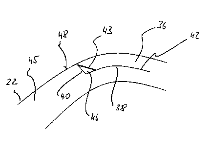
The incision figure shown in Fig. 3 includes, in addition to the bed incision
38 and
the marginal incision 40, a wedge incision 43 which proceeds radially
(relative to
CA 02812848 2013-03-27
- 10 -
an imaginary centre, not represented in any detail, of the cornea, denoted by
45,
of the eye 22) at least very largely within the marginal incision 40 in the
periph-
eral direction of the edge of the flap and, together with the marginal
incision 40
and the bed incision 38, delimits a tissue strip 46 which is approximately
wedge-
shaped in cross-section and which can be taken out after the raising and
folding-
away of the flap 36. In the exemplary case that is shown, the bed incision 38
proceeds at a substantially uniform depth of the cornea 45 parallel to the
anterior
surface of the cornea, denoted by 48. The marginal incision 40 and also the
wedge incision 43 proceed in angled manner relative to the bed incision 38 in
the
direction towards the anterior surface 48 of the cornea. The radial spacing be-
tween the marginal incision 40 and the wedge incision 43 is greatest in the re-
gion of the bed incision 38; upon advancing in the direction towards the
anterior
surface 48 of the cornea, the marginal incision 40 and the wedge incision 43
approach one another.
The size of the tissue wedge formed by the strip 46 depends on the post-
ablative
diminution of the arc length of the bed surface in the meridional direction in
question. Furthermore, the size depends on whether this reduction in arc
length
can be balanced out by a single tissue wedge or by two tissue wedges situated
in
diametrically opposed marginal regions of the flap. In those marginal regions
of
the flap which are situated opposite the hinge, the entire difference in arc
length
in the direction in question has to be compensated by a single tissue wedge.
In
the remaining meridional directions the difference in arc length can be compen-
sated by two tissue wedges at marginal points of the flap situated opposite
one
another. Accordingly, the size and shape (or generally, the cross-section) of
the
tissue strip 46 may vary upon progressing in the peripheral direction of the
edge
of the flap. In particular, in those marginal regions which are situated
opposite
the hinge the tissue strip 46 may have a larger cross-section than in the
remain-
ing peripheral regions.
Depending upon the ablation profile, the tissue strip 46 may extend over the
entire periphery of the edge of the flap. It is also conceivable that the
tissue
strip 46 extends only along a segment of the edge of the flap. It is even con-
CA 02812848 2013-03-27
- 11 -
ceivable to generate along the edge of the flap several tissue strips 46
spaced
from one another in the peripheral direction.
Even though in Fig. 3 and in the following Figures the marginal incision 40
and
the wedge incision 43 are each represented as rectilinear incisions in cross-
section, it will be understood that this is by no means imperative. In
particular,
for the wedge incision 43 an incision course that is not straight may also
readily
be chosen.
In the exemplary case shown in Fig. 3 the marginal incision 40 and the wedge
incision 43 impinge on one another substantially directly on the anterior
surface
48 of the cornea. Figures 4 to 6 show various modifications for the relative
course of the marginal incision 40 and of the wedge incision 43.
In Fig. 4 the marginal incision 40 and also the wedge incision 43 intersect be-
neath the anterior surface 48 of the cornea, the wedge incision 43 terminating
at
a spacing from the anterior surface 48 of the cornea, and the marginal
incision
40 being continuous as far as the anterior surface 48 of the cornea.
In the variant shown in Fig. 5 the marginal incision 40 and the wedge incision
43
intersect likewise beneath the anterior surface 48 of the cornea, whereby, how-
ever, in this case the marginal incision 40 terminates at a spacing before the
anterior surface 48 of the cornea and, instead, the wedge incision 43 is
continu-
ous as far as the anterior surface 48 of the cornea.
The modification according to Fig. 6 shows a case in which both the marginal
incision 40 and the wedge incision 43 are continuous as far as the anterior
sur-
face 48 of the cornea and run into the anterior surface 48 of the cornea at a
spacing from one another, without, however, intersecting one another.
By the flap being shortened at its edge in the manner elucidated above, prefer-
entially in the shape of a wedge, it is possible to modify it in such a way
that it
can be inserted exactly into the post-ablative stromal bed. As elucidated, the
shortening may be performed on the entire flap, with the exception of those
CA 02812848 2013-03-27
- 12 -
regions where the hinge is located. The calculation of the cross-section of
the
tissue strip 46, i.e. generally the calculation of the corneal incision
figure, can be
carried out by taking into account the size of the ablatively treated optical
zone,
the refractive powers of the cornea before and after the ablation, and also
the
asphericities of the anterior surface of the cornea. A possible foundation of
the
calculation is given by the mathematical formulae reproduced below.
n-1 n-1 n-1
P = _________ ¨> = _____ ;R2 = ________ ; R2 > [1]
P preop P postop
1 r 1 11 /2
S = 1 + Ql [R12 y2(1 + Q3/2 11
1 + Q2 _____________________________ [R22 ¨ y2(1 + Q2 )1
/2
1 1-R_2 oz2
1-FQ2L 2 4 __ (1+ Q2 )1/ 2 1+Q1 ____________ [R12 oz42 (1 + Q1)
[2]
= 2R,* arcsi ' oz
n
2R
( _______________________ \ 2 \
2
s(y =0)¨ Ri+ R2 + 2 OZ
4
b2 = 2R2 arcsin 1
,
R2
Ab = A ¨ 62
CA 02812848 2013-03-27
- 13 -
Ppreop: refractive power of the cornea before the operation
(e.g.
Ppreop = 43 dpt)
Ppostop: desired refractive power of the cornea after the
operation
R1: radius of curvature of the optical zone before the
ablation
R2: radius of curvature of the optical zone after the ablation
n: refractive index of the cornea (n rz 1.377)
111: arc length of the optical zone before the ablation
b2: arc length of the optical zone after the ablation
OZ diameter of the optical zone
asphericity of the anterior surface of the cornea before the ablation
(-1 < Qi < 1))
Q2: asphericity of the anterior surface of the cornea after
the ablation
(-1 <Q2 < 1))
s: depth of the ablation
y. radial indexed variable (y = 0 at the point of maximal ablation, i.e.
ablation centre)
With the aid of the above mathematical foundations, for a purely central (rota-
tionally symmetrical) resection of material, taking account of the
asphericities of
the anterior surface of the cornea, the difference in arc length Lb (post-
ablative
in comparison with pre-ablative) of the optical zone can be calculated. With
knowledge of the difference in arc length, it is readily possible to calculate
the
= cross-section of the tissue strip 46 to be removed. In this connection,
as eluci-
dated, it is to be taken into consideration that diametrically relative to the
hinge
the shortening is to be effected by a single tissue wedge, whereas on the
remain-
ing sides the shortening can be apportioned to two tissue wedges.
In purely exemplary manner the following numerical table was ascertained by
computation on the assumption of asphericity values Qi = Q2 = -0.3 and a size
(diameter) of the optical zone of 6.5 mm. This specifies, for different values
of
short-sightedness to be corrected, the resulting difference in arc length of
the
optical zone. These numerical values were calculated using the mathematical
foundations reproduced above.
=
= = ,
CA 02812848 2013-03-27
- 14 -
Values [dpt] to be corrected Difference in arc length [pm]
1 5
2 10
3 14
4 19
23
6 27
7 30
8 34
The control computer 30 of the laser device according to Fig. 1 preferentially
has
access to suitable ablation data that are representative of the resection of
mate-
rial to be realised. The ablation data may, for example, have been stored in
the
5 memory 32. In this connection it is conceivable that the memory 32 is
accessible
also for a control computer of a separate laser device, not represented in any
detail, for the later ablating treatment of the eye. The ablation data can be
writ-
ten into the memory 32 by the control computer of this ablation laser device,
in
which connection the control computer 30 of the cutting laser device shown in
Fig. 1 calculates, on the basis of the ablation data, the suitable incision
figure for
the respective patient.