Note: Descriptions are shown in the official language in which they were submitted.
CA 02814632 2013-04-12
WO 2012/050495
PCT/SE2010/051111
1
METHOD AND DEVICE FOR EXAMINING THE SURFACE TEMPERATURE OF A
BODY PART
FIELD OF INVENTION
The present invention relates to a method for examining the surface
temperature of a
body part which comprises a body surface divided into multiple measuring
zones.
The invention also relates to a device for examining the surface temperature
of a
body part and a computer program for carrying out the method and a memory
medium.
BACKGROUND
Individual performance is particularly influenced by exterior circumstances
such as
cold climate. Parameters such as low surrounding temperature or vibrations
impair
the blood circulation to the outer parts of the body, making body parts
colder.
Lack of or poor circulation can have serious consequences such as permanently
damaged tissue and sensory loss. Poor circulation also affects the mental
ability of
an individual. In very severe cases non-existent blood circulation can lead to
amputation or similar interventions.
SUMMARY OF INVENTION
One object of the invention is to provide a simple, standardized, fast, clear,
reliable,
precise and repeatable method for examination of the surface temperature of a
body
part. This aim is achieved by a method having method steps defined by claim 1.
Another object of the invention is to provide a portable, easy to use system
for
examination of the surface temperature of a body part. This object is achieved
by a
system comprising the features defined by claim 8.
Further advantageous embodiments of the invention are defined by claim 2-7 and
9-
14.
CA 02814632 2013-04-12
WO 2012/050495
PCT/SE2010/051111
2
Infrared radiation is emitted by any object that has a temperature above -273
C
and can be detected by a thermographic camera. A thermographic camera can thus
detect the infrared radiation emitted from a body part and render the surface
" temperature of the photographed or filmed part of the body. The temperature
of the
surface of the object is represented on the resulting image or film by
different colors,
and depending on the camera's sensitivity, good resolution of the temperature
variations can be obtained.
The surface temperature of a body part is considered to be an indication of
how well
the blood circulation in the body part functions.
One advantage of the invention is that a person can be helped to improve
his/her
performance at work or sports in cold climate.
The results of the examination shows how the examined body surface is
influenced
by being cooled down, and to which extent different areas of the body surface
are
affected. The results can be used for selecting and adaption of different body
protections and helping facilities such as gloves, shoes, tools and other body
proctecting means.
Another advantage is that the results of the examination can be used for
showing
how a cold surrounding climate influences a body part and the blood
circulation of the
body part in a pedagogical manner. Thereby are persons also made aware of the
risks and limitations related to work or other types of performance in cold
climate.
The method according to the invention is unique since the surface temperature
of the
entire body surface of a body part and not just individual points are
measured. For
example, the surface temperature of the entire face is measured, a whole palm
or the
entire foot sole. Both the left and right body part (hand, foot, cheek, ear)
can be
examined simultaneously, thus an automatic reference between the right and
left
body part is received and by comparing the surface temperatures on
corresponding
surfaces deviations can be identified.
The method can be used for examining if the blood circulation is affected by a
number of different parameters. Internal factors may consist of a food, such
as food
or drink, a drug or the like.
CA 02814632 2013-04-12
WO 2012/050495
PCT/SE2010/051111
3
External parameters may include cold, heat, a vibrating device, acupuncture,
light
therapy or the like. In the example of the method below the body part is
exposed to a
refrigerant. The method has proven to be particularly good for detecting frost
bites
and vibration damages on the examined body part with this type of provocation.
It is
known that frost bites and vibration damages decreases the blood circulation.
In order to simplify and increase safety in the analysis, background
information on
previous problems like injuries, smoking habits, loss of sensation and other
personal
type of information is collected with a questionnaire in advance of the
examination.
Answers from the questionnaire can be directly related to results from a
measurement zone, thus underlying causes of discrepancies can be identified.
BRIEF DESCRIPTION OF DRAWINGS
Further objects and features of the present invention will appear from the
following
detailed description of some aspects of the invention, wherein some aspects
will be
described in more detail with reference to the accompanying drawings, in
which:
Fig. 1 shows a schematic view of a system according to the invention.
Fig. 2 shows a side view and a front view of the background screen shown in
Fig.1.
Fig. 3 shows a side view and a front view of a background screen.
Fig. 4 shows a schematic view of a system according to the invention and a
side
view and a front view of a background screen.
Fig. 5 shows a detailed view of another embodiment of the background screen
shown in Fig. 4.
Fig. 6 shows an example of a body surface.
Fig. 7 shows a graph of the recovery process for a body part examined with the
method according to the invention.
The following description and the patent claims define several embodiments of
the
invention. All embodiments can be combined in a variety of ways while still
achieving
the advantages and benefits of the invention.
DETAILED DESCRIPTION
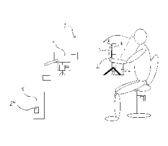
Fig. 1 shows a system 1 for examination of the surface temperature of a body
part 2
CA 02814632 2013-04-12
WO 2012/050495
PCT/SE2010/051111
4
which comprises a body surface 3 divided into a number of measuring zones
according to the invention. The system is provided with means for carrying out
the
method according to the invention.
The system 1 comprises a thermographic camera 4 for photography and filming, a
computer 5 that is connected to the therrnographic camera, a background screen
6
for the body part and support pads 7 for the body part.
The background screen 6 comprises a flat panel 8 of background material which
is
intended to prevent body heat from other parts of the body of the person to
influence
the examination and the resulting measurements.
The background screen 6 comprises a front side 6.1 and a back side 6.2, an
upper
edge 6.3, a lower edge 6.4, a first side edge 6.5 and a second side edge 6.6,
see Fig.
2.
The background screen 6 has at least one opening 9.1;10.1, or through hole,
which
has an entry channel 9.2;10.2 stretching from at least one side edge of the
background screen to a placement space 9.3;10.3. The body part 2 is positioned
next to the background screen by passing the body part through the entry
channel
9.2;10.and positioning the body part within the placement space 9.3;10.3. The
body
part 2 which is going to be examined is thereby located on the front side 6.1
of the
background screen, and other body parts of the person are located on the rear
side
6.2 of the screen.
The background screen is provided a supporting legs 12 that can be placed on a
table or a supporting stand 13 that can be placed on the floor.
The supporting legs 12 and the supporting stand 13 are provided with means for
adjusting the hight of the screen. The background screen can be tilted
angularly to
the front and rear to achieve an optimized and comfortable position for the
person.
The size of the screen is adapted such that the body part is positioned well
within the
area limited by the edges of the screen.
The openings 9.1;10.1 and placement spaces 10.1;10.2 are arranged so that body
part is exposed on the front side 6.1 of the background screen and within the
area
limited by the edges of the screen.
The screen comprises on or more adjacently positioned panels of radiant heat
insulating material or a plastic sheet or another material with low thermal
conductivity
CA 02814632 2013-04-12
WO 2012/050495
PCT/SE2010/051111
so that the background screen temperature does not influence the result of the
examination.
Fig. 4 shows another background screen 6' that has a first opening 9.1' and a
second
opening 10.1'. The first and the second opening each have a channel 9.2'1 0.2'
stretching from the lower edge of the background screen to the placement
spaces
9.3';10.3'.
Fig. 5 shows a detailed view of another embodiment of the background screen
shown in Fig. 4.
The background screen 6' is provided with means for cooling or heating the
screen.
The temperature of the screen can thereby be kept lower or higher than
temperature
of the body surface of the body part.
This is has the advantage that the temperature of the background screen is
controlled. It is important to avoid sources of errors in the examination. By
controlling
the temperature of the screen the influence of other heat sources, like other
body
parts, on the examination and temperature measurements are lowered. This
improves the results of the examination.
Preferably the temperature of the background screen is regulated so that the
background is at least 2 degrees Celsius warmer or colder (+/- 2 C) of the
body
surface throughout the whole examination process to allow and secure the
scanning
of a body parts measuring zone.
The background screen 6' comprises a front panel 14 and a rear panel 15. The
material of the panels is for example a transparent plastic panel or similar
material.
The panels 14, 15 are arranged in parallel and with a distant between them
such
that a gap 16 is formed between the panels. The front and the rear panel are
joined
to each other along the outer edges of the panels, such that the gap forms a
closed
and sealed inner space 17. The background screen 6' has at least at least one
inlet
19.1;19.2;19.3 to the inner space and at least one outlet 18 from the inner
space to
allow a cooling or heating medium, for example heated water or cold water, to
flow
through the space from the inlet to the outlet.
The front and the rear panel are also joined by distancing supports, ribs, 21
which
are located within the inner space 17 to keep the front panel and the rear
panel
securely in place.
CA 02814632 2013-04-12
WO 2012/050495
PCT/SE2010/051111
6
A pump 22 is connected by pipes to the inlet and to the outlet of the
background
screen. The pump circulates the medium through the inner space of the
background
screen. The medium can be provided by a closed container 26 connected to the
pump. The container has means for providing that the medium is kept at the
desired
temperature, such as heating means or cooling means (not shown in figures).
The
screen is provided with thermocouples 23 to measure the temperature of the
screen.
The thermocouples are connected to a controlling unit 25 and the computer 5.
Another embodiment of the background screen comprises an electrically heated
panel of a foil like material for heating the screen (not shown in the
figures).
Another embodiment of the background screen comprises an cooling panel of a
foil
like material provided with heat exchanging means for cooling the screen (not
shown
in the figures).
The system comprises support pads 7 for the body part/s which is/are located
on the
rear side of the screen.
The support pads 7 can be designed as a bowl-shaped or cup-shaped supporting
surface that a heel can rest in. When hands are examined, the palms of the
hands
are facing forwards, towards the thermographic camera. When a foot or a couple
of
feet are examined, the heels are resting in the cup-shaped support,
positioning the
soles of the feet forwards and towards the camera.
Hands and feet are placed in a high position (raised). This means that the
hands are
held at chest height. Preferably the person sits on a height adjustable chair.
When examining the feet, the person lays flat on his/her back with the legs
aligned
and straight and the feet are held such that the foot soles are facing the
camera.
The use of background screen 6 with the support pads for a body part ensures
that
the same position for the body part is found and maintained throughout the
examining period, both during the initial period and during the recovery
period when
digital photographing or filming with thermo graphic camera is carried out.
The system comprises a thermographic camera 4 for digital photography and
filming
CA 02814632 2013-04-12
WO 2012/050495
PCT/SE2010/051111
7
of traditional and thermograhic images and a computer 5 that is connected to
the
thermographic camera. The thermographic camera is used for digital photography
and digital filming of traditional images and thermographic images.
By photographing of traditional images is meant conventional monochrome or
color
photography.
The thermographic camera 4 is directly connected to the computer which has a
CPU,
a monitor, a webcam and a memory device 24 for storing the information data
collected during the examination procedure. A computer program is loaded into
an
internal memory of the computer. The computer program comprises the code parts
of
the computer program product for carrying out the method when it is executed
on the
computer. It is advantageous to provide the computer with means and programs
for
image processing. The computer and the webcam can be connected to Internet to
allow other people which are not present, to study the examination procedure.
The computer program have means for locating and programming the scanning
position for the body part to ensure that the same scanning position for the
body part
is located and scanned over the entire examination period, both at the initial
period
and during the recovery period when digital photographing and filming with
thermographic camera are carried out. For example, means for image processing
and image processing programs can be used for this.
All information data reported and/or executed by the computer and the computer
program are stored in a database after the examination procedure is completed.
A traditional image and/or a thermographic image are digitally photographed by
the
thermographic camera and the image/s are transferred to the computer and the
monitor. The images are then monitored, controlled and processed by the
computer.
The thermographic image reflects the variations of the surface temperature on
the
examined body part. The thermographic image shows the surface temperature in a
very large number of points on the examined body part.
The thermographic image has a very high resolution, for example, one camera
has a
resolution equivalent to 640x480 pixels. One pixel shows the surface
temperature on
one picture point, scanning spot, on the surface of the body part. The surface
temperature in a very large number of points on the body surface are thus
individually examined and measured at the same time.
CA 02814632 2013-04-12
WO 2012/050495
PCT/SE2010/051111
8
The computer program comprises 1.2...n predefined body surface measuring zones
hereafter referred to as M(1-n) for one or two hands, and one or two feet, one
or two
arms, one or two legs, a full face or part of a face, a neck, a back of neck,
one or two
ears, one or two cheeks.
The traditional image and/or the thermographic image of the scanned surface of
the
body part is/are displayed on the monitor, the computer screen.
The thermographic camera 4 comprises at least one predefined measuring zone
M(1-n). The traditional image and/or the thermographic image of the surface of
the
body part is/are divided into the corresponding measuring zones M (1-n). For
example, the image of each hand is divided into upto 34 zones, M (1,2 ... 34),
namely
fingertips (distal phalanges), upper intermediate fingers (intermediate
phalanges),
lower intermediate fingers (proximal phalanges), palms (metacarpals) and wrist
(carpals). The fingertips are particularly vulnerable, thus the image of each
fingertip
are further divided into four measuring zones. Fig.6 shows a right hand with
measuring zones M(1-34).
The computer program processes and saves the body surface temperature values
measured by the thermographic camera and calculates the average temperatures
and temperature anomalies from all measuring zones M (1-n) in each batch, also
called scanning, which will be further described below.
The computer 5 also has a reporting program that illustrates the recovery
process
during and after the examination and measurement procedure.
The system is also provided with a database for storing the images, all
measuring
data and information such as room temperature, medium temperature, body
temperature, heart rate and blood pressure and all the measured and calculated
values for surface temperatures and instants of time, exposure periods,
duration of
recovering period etc.
At the setup of the system for examining a body part, the distance between the
body
part/s which are to be examined and the thermographic camera and the angle of
the
thermographic camera are adjusted. The body part/s are positioned in front of
the
background screen and within the area limited by the outer edges of the
background
screen.
CA 02814632 2013-04-12
WO 2012/050495
PCT/SE2010/051111
9
After the adjusting operation, the thermographic camera scans the surface of
the
body part and transfers the information to the computer and the computer
program.
Thereafter is the body part photographed and/ or filmed by the camera.
Based on the first image, the measuring zones M (1-n) and their coordinates
can be
mutually positioned and adjusted manually by clicking and dragging with a
mouse
cursor on the computer screen.
Before the examination, the test person should be at rest at room temperature
with a
minimum of plus 20 C, at least 30 minutes. The individual must not smoke or
snuff
tobacco 4 hours before measurement since such activites lowers the body
temperature 4 degrees on the fingertips and toes. During the resting period
the test
person should be fasting and not drink hot drinks, or sit with their hands
together.
Body parts that are going to be tested must be completely at rest. It is
essential that
the person does not put the body parts that ar going to be examined on any
cold or
hot surfaces, to avoid error sources.
Measurement and documentation is carried out throughout the whole examination
period which comprises an initial period from the time the first image is
photographed,
an exposure period and a reco'very period that ends when a final image is
photographed after recovery has occurred. In the description and the example
of the
method below temperature is indicated by T and time is indicated by t.
The method for examination of the surface temperature of a body part which
comprises a body surface divided into a number of measuring zones comprises
positioning of the body part in a system for examining a body part, wherein
the
system has a thermographic camera, and digitally photographing essentially the
entire body surface that is directed towards the thermographic camera both as
a
traditional image and as a thermographic image during an initial period,
exposing the body part to a refrigerant during a predetermined exposure time
during
an exposure period, repositioning the body part in the system for examining
the body
part, and digitally photographing the body surface as a traditional image, and
filming
the body surface as a thermographic film or repetitively digitally
photographing the
body surface as thermographic images during a recovery period.
The body surface temperature in a large number of picture points are thereby
measured with the thermographic camera and indicated on the thermographic
image.
CA 02814632 2013-04-12
WO 2012/050495
PCT/SE2010/051111
The following is an embodiment of the method according to the invention:
The initial period
The body temperature is measured with an ear thermometer, heart rate and blood
pressure measured with blood pressure measuring device, the surrounding room
temperature is measured and recorded, the refrigerant temperature and the
temperature of the background screen 6' are measured and recorded with the
thermographic camera 4 and the temperature of the background screen is set and
can be regulated to be maintained within +/ - 2 degrees Celcius higher or
lower
temperature than the body parts that are examined throughout the whole
examination period. The body parts which are going to be examined are
positioned
against the background screen 2 for the examination and measuring by
photographing. The camera with the computer and the computer program, the
software, scans the body surface 3 and the measuring zones M(1..n ) to have an
intial thermographic image of the surface.
Also an initial traditional image is photographed by the camera. The initial
traditional
image, and the intial thermographic image of the initial surface temperature
are
recorded and stored in the computer memory device 24.
These images are superimposed on each other in the reporting program in the
computer, and are used for illustrating the recovery process. The initial
traditional
image is particularly important as evidence of the intial state of the body
surface and
to avoid error sources.
The computer with the software calculates an initial average surface
temperature
referred to as T: initial M(1...n) for each measuring zone M. The average
surface
temperature is based on the thermographic image of the body surface showing
the
temperature in a large number of pixels within each measuring zone.
The computer with the software also calculates an initial average temperature
referred to as T: inital for the total body surface comprising all measuring
zones M (1-
n).
The computer with the software also calculates a zone reference temperature
referred to as T:zonref (1-n) for each measuring zones M (1-n) as an average
value
of the surface temperature of the pixels in each measuring zones M (1-n)
measured
CA 02814632 2013-04-12
WO 2012/050495
PCT/SE2010/051111
11
by thermographic camera.
Thereafter is the difference between the initial average temperature T:initial
and the
zone reference temperature T zoneref M(1-n) calculated for each measuring
zone,
the difference is referred to as T: initialdiff M(1-n).
T: initialdiff M(1-n) = T: inital ¨ T zoneref M(1-n)
The traditional initial image and the thermographic initial image, the
coordinates of
the measuring zones, the initial average temperature T: initial, zone
reference
temperature T: zoneref M(1-n), and the calculated difference T: initialdiff
M(1-n) and
measuring comments are recorded and stored in the memory device in a computer
file with a unique name linked to the person and date.
Exposure period
During the exposure period is the part of the body exposed to a refrigerant
during a
predetermined exposure time. Immediately after the exposure time is finalized
the
time is started to be recorded.
The body part is exposed, cooled, with a refrigerant, in verifiable conditions
for 30
seconds, 1 minute or 5 minutes. Cooling of the hands and feet occurs in any of
the
following harmless ways:
a) Water temperature +5 to +20 C, preferably 12-15 C, the hands are
lowered
down to the wrists under water for 30 seconds, feet are lowered so that the
ankle
bump go under water for 45 seconds (more surface cooling). Before dipping the
hands into the water, plastic gloves can be placed on the hands they will
removed of
immediately after cooling. The hands can also be dipped in the water without
plastic
gloves and wiped (without rubbing) with a towel immediately after cooling. It
is
preferable to avoid the heating effect that can be cause by drying with a
towel. Feet
are lowered without protection into the water.
b) Cold rise bath (round grained rice) with a temperature of +5 to +20 C,
preferably
12-15 C, hands or feet are immersed in the same manner as above.
c) snow (dry snow) hands or feet are immersed in the same manner as above.
If the face, ears, neck, throat should be examined, these are cooled by
preferably a
CA 02814632 2013-04-12
WO 2012/050495
PCT/SE2010/051111
12
walk, in an ambiance temperature of -5 C for 10-20 min minutes, for example,
in a
cold temperature environment with a cold factor of ¨ 5 C.
A recovery period begins immediately after ending the exposure time, ie.
immediately
after the body part is removed from the refrigerant. In this example the
recovery
means that the tested body part naturally regains heat and returns to the
original
surface temperature of the body surface.
The body parts are repositioned in front and within the surface area of the
background screen 6' in the same position as at the previous initial
examination and
photographing.
The computer program have means for locating and programming the scanning
position, the measuring zone coordinates, for the body part to ensure that the
same
scanning position for the body part is located and scanned over the entire
examination period, both at the initial period and during the recovery period
when
digital photographing and filming with thermographic camera are carried out.
A traditional image and/or a traditional film of the body surface is
photographed or
filmed with the thermographic camera.
The traditional image is photgraphed to determine if a change in temperature
is due
to residual water on the skin. The resulting images or film provides a
controlling tool
for the thermographic camera measurements. A traditional still photography
shall
initally be made with a maximum one-minute intervals.
During the recovery period, ie from the recovery period begins immediately
after
exposure period has ended, the instants of time for the measurements
(photographing and/or filming) progress is recorded. Hereafter the first
instant of time
is referred to as the starting time t:cold = 0, and the finishing instant of
time is
referred to as tend, which is the instant of time when the measurements ends
and
the recovery period is finished.
The traditional and thermographic photographing with thermographic camera 4 is
repeated with a certain frequency, at least every 30 seconds the first three
minutes
and then every other minute.
CA 02814632 2013-04-12
WO 2012/050495
PCT/SE2010/051111
13
The recovery period is ended when filming with thermographic camera and the on-
going software processing of calculations and reporting program show that
some, or
all of the body surface, all measuring zones M (1-n), have reached a
predetermined
final temperature T end based on a percentage of the initial average
temperature
T:initial. The recovery period can also be ended when a predetermined maximum
recovery period has passed, where the recovery period is measured from the
first
photographing or filming with thermographic camera after the exposure period,
for
example 15 minutes.
The instant of time for each measurement, each traditional or thermographic
image,
is referred to as t:image and is measured from the starting time t:cold. Also
t:image is
recorded and saved in the memory device.
The measurement program in the computer is started when the recovery period
begins. The body surface is photographed by the thermographic camera and the
surface temperature is thereby measured. The resulting thermographic image
shows
the temperature of body surface.
The body surface is photographed, ie the surface temperature is measured, in
the
measuring zones M (1-n) at least every two minutes, preferably every minute,
preferably every 15 seconds or even every two seconds or every second. The
thermographic images are recorded and stored in the memory device.
At the first photographing, directly after cooling, the computer with the
software
calculates an cold average surface temperature of the entire body surface,
hereafter
referred to as T:cold. In addition, a cold average surface temperature
referred to as
T: zonecold M(1-n) for each measuring zone M is calculated.
The cold average surface temperatures are based on the thermographic image of
the
body surface showing the temperature in a large number of pixels within each
measuring zone.
The cold average surface temperature T:cold and the cold average surface
temperature T: zonecold M(1-n) are recorded and stored in the memory device
24.
When filming with the thermographic camera, the recovery process is recorded
and
documented and exact instants of time for each image, hereafter referred to as
tp:
M(1-n) are measured from the time t:cold.
CA 02814632 2013-04-12
WO 2012/050495
PCT/SE2010/051111
14
At each instant of photographing and measuring, a recovery temperature
hereafter
referred to as T:zonerecover M(1-n) is calculated for each measuring zones M
(1-n).
The recovery temperature is calculated as an average value of the surface
temperature of all picture points, pixels, in each measuring zones M (1-n)
measured
by the thermo graphic camera.
The instants of time of the images, tp: M(1-n) and the recovery temperature
T:zonerecover M(1-n) are recorded and stored in the memory device 24.
Thereafter are the following differences are calculated:
1) T:zonerecover M(1-n) - T:cold M(1-n) = T: recoverdiff (1-n) (temperature)
2) t: cold - t: end = t:recoverdiff (time)
3) T zonerecover M(1-n) - T:zoneref M(1-n) = T: zonediff M(1-n) (temperature)
4) tp(1-n) - t: cold = t: zonediff (time)
The calculated differences T:recoverdiff M(1-n), t: recoverdiff, T: zonediff
M(1-n) and
t: zondiff are recorded and stored in the memory device into one vector per
measuring zones M (1-n) connected to the individual-specific data file.
All thermographic film footage, traditional and thermographic photos,
measuring
zones coordinates, the average temperature T: cold, the recovery temperature
T:
zonerecover M(1-n), the time tp (1-n), t: cool, t: end, t: image, in all
measuring zones
at all measurement occasions, and the calculated differences T recoverdiff M(1-
n),
t: recoverdiff in all measuring zones at all measurement occasions, and
measurments comments are recorded and stored in the memory device into a
computer file with unique name associated with the individual and date.
Audio recording of comments during the recovery period can also be recorded
and
saved. New measuring comments are possible to insert, after which the
collected
data to can be inserted to an already previously created file. The recovery
process is
displayed in graphs on the screen and the recovery period as numerical values
per
CA 02814632 2013-04-12
WO 2012/050495
PCT/SE2010/051111
zone.
Analysis of results
For the simultaneous examination of the left and right body part, the
differences
T :initialdiff, T: recoverdiff M(1-n), T: zonediff M(1-n), t: recoverdiff and
t: zonediff for
the different body parts are calculated and stored in the memory device.
Surface
temperature deviations between left and right body and corresponding measuring
zones, are calculated, recorded, compared and stored in the memory device.
This
comparison indicates if there are measuring zones with reduced blood
circulation.
The differences are individual, and only indicates variation between the left
and the
right corresponding body surfaces of a single person.
The subsequent analysis will determine the measuring zones where strong
cooling
effect is obtained or where the recovery process is slow. The recovery process
can
be observed and compared in each measuring zone can be compared with the total
body surface of the examined body part, and also between adjacent measuring
zones.
The recovery process can take several forms, such as linear or non linear.
The collected information in the memory device such as saved temperature data
and
time data are processed and introduced into the reporting program for
visualization in
graphs and profiles like an individual profile curve on a graph.
Temperature data is converted to the profile data and plotted in a figure
which
illustrates the recovery process.
The areas indicated by the responses of the questionnaire recurs in the
analysis
automatically as possible problem areas.
The measuring zones corresponding to areas of the body part where the blood
circulation has been affected by the cold-provocation can be further examined.
Areas which appear to be sensitive and recovery time can be recorded and
documented. The presented results only represent an intermediate result, and
because there are individual differences between people, further studies need
to be
carried out to validate and confirm if there are any problems with the blood
circulation
in the indicated areas, measuring zones.
CA 02814632 2013-04-12
WO 2012/050495
PCT/SE2010/051111
16
In the embodiment described above, a number of different operations and
calculations are executed by the computer program on the computer in order to
have
results from the examination. Additional operations that the computer program
performs when it is executed on the computer and reporting to the reporting
program
during the measurement period are for example the following:
With reference to Table 1 below the computer iterates the following program
steps:
Initial Period
The difference between the surface temperature T: inital and the zone's
average
temperature T: zoneref M(1-n) is calculated during the initial period,
registered and
grouped in intervals of measuring zones with 0.01 degree difference, measuring
zones with 0.1 degree difference, etc. Each measuring zones M (1-n) can be
identified and correlated with the responses.
Directly after cooling and during the recovery period
The difference between the total surface average temperature T:cold and the
measuring zones average temperature T: zonecold M(1-n) are calculated and
recorded and grouped in intervals of zones with 0.01 degree difference, zones
with
0.1 degree difference, etc. Each measuring zones M( 1-n) can be identified and
correlated with the answers from the questionnaire. Comparison is also done
with
results from the initial period.
After recovery
The differences between the surface temperature T:end, and the measuring zones
average recovery ternperature T:zonerecover M(1-n) are calculated continuously
during the recovery period and recorded and grouped in intervals of zones with
0.01
degree difference, zone with 0.1 degree difference, etc. Each measuring zones
M (1 -n) can be identified and correlated with the responses. Comparison is
also
done with results from the initial period and immediately after cooling. Time
for
recovery per zone is also calculated.
CA 2814632 2017-04-13
17
Table 1. T = temperature, t = time
Values Measurments Measurments Measurments Time for
Initial Directly after after recovery
cooling recovery
Measuring zone T:zoneref T:zonecold T:zonerecover tp M(1-n)
temperature M(1-n) M(1-n) M(1-n)
The average T:initial T:cold T:end t:diff=t:end-
temperature of the t:cold
whole surface
Differences average T:initialdiff T:cold M(1-n)- T:diff M (1-n)=
t:diff= tend-
temperature versus M(1-n). T:zonecold T:end ¨ t:cold
measuring zone T:inital ¨ M(1-n) T:zonerecover
temperature T:zoneref M(1-n)
M(1-n)
The time for recovery of the cooled body part to regain the reference
temperature is
recorded, and if it is straight-line or according to other processes, see
Figure 7. This
figure shows a recovery process for an individual measuring zone Tzone and
recovery
process for the entire surface Tsurface. The curve for Tzone indicates that
the body part
has been more affected by the cold- provocation that the rest of the examined
body
surface. This is an indication that the particular measuring zone should be
further
examined to verify if the body part has an injury in this measuring zone, for
example a
freezing injury, which affects the blood circulation.
The present invention should not be limited to the description above and the
drawings
but can be changed and modified.
=