Note: Descriptions are shown in the official language in which they were submitted.
CA 0281482 2013-04-15
WO 2012/054466 PCT/US2011/056688
METHODS AND APPARATUS FOR INSERTING A DEVICE OR
PHARMACEUTICAL INTO A BODY CAVITY
Cross-Reference to Related Applications
[1001] This application claims benefit of priority to U.S. Provisional
Application Serial
No. 61/394,120, entitled "Methods and Apparatus for Inserting a Device or
Pharmaceutical
into a Body Cavity," filed October 18, 2010, which is incorporated herein by
reference in its
entirety.
Background
[1002] The embodiments described herein relate to apparatus and methods for
inserting a
device and/or pharmaceutical into a body cavity. More particularly, the
embodiments
described herein relate to apparatus and methods for inserting an intrauterine
device (IUD)
into the uterus.
[1003] Difficulty of insertion is a significant hurdle to the more
widespread use of known
IUDs by physicians and health care workers worldwide. Known methods of
inserting the
IUDs involve four pieces of equipment and multiple operations. In particular,
known
methods of IUD insertion include the use of a vaginal speculum, a cervical
tenaculum, an os
finder (when needed) a uterine sound, and the IUD inserter. First, a speculum
is positioned to
visualize the cervix. Second, the cervix is clamped with downward traction
using a cervical
tenaculum to substantially straighten and/or align the cervix with the uterine
cavity. In
certain circumstances, an os finder is used to locate and dilate the cervical
os. Third, a
uterine sound is used to determine the depth of the uterine cavity, which is
the depth to which
the IUD will be inserted. Fourth, the arms of the IUD are folded back and
tucked into the
tube of the inserter. Fifth, the inserter is pushed into the vagina until the
health care provider
can find the opening of the cervical canal, and then is inserted via the
cervix into the uterus to
the depth measured by the sounding process. Sixth, the tube of the inserter is
pulled back to
release the arms of the IUD from the tube at the fundus of the uterus. In some
known
procedures, the inserter tube is again pushed up against the base of the arms
of the IUD to
ensure highest achievable placement within the endometrial cavity. The
inserter is then
carefully extracted from the uterus, cervix, and vagina such that the
placement of the IUD is
1
CA 0281482 2013-04-15
WO 2012/054466 PCT/US2011/056688
not disrupted. Lastly, the practitioner must cut the IUD strings to ensure
that a sufficient
length (e.g., at least 2.5 cm) of the withdrawal string is exposed in the
vagina.
[1004] The insertion of an IUD according to such known methods can often
result in
misplacement of the IUD and/or other complications. Said another way, known
methods of
IUD insertion involve a series of precise operations to ensure proper
placement of the IUD.
Even slight procedural deviations when using known methods and tools for IUD
insertion can
lead to uterine wall perforations, increased chance of embedding of the IUD in
the
endometrium, and/or expulsion of the IUD. In addition, it is possible to push
microbes from
the vagina into the uterus during the insertion process, which can lead to
complications such
as pelvic inflammatory disease (PID).
[1005] Thus, a need exists for improved apparatus and methods for inserting
an
intrauterine device (IUD) into the uterus that will reduce these risks and
allow IUD insertions
to be performed by health care providers across all spectra of medicine.
Summary
[1006] Apparatus and methods for inserting a device and/or pharmaceutical
into a body
cavity are described herein. In some embodiments, an implant delivery device
includes a
housing, a head and an insertion member. The housing defines a housing
passageway. The
head, which defines a head passageway, is configured to rotate (e.g., the head
can flex, rotate,
pivot, etc.) relative to the housing. Collectively, the housing passageway and
the head
passageway define an insertion passageway such that at least a portion of the
insertion
passageway is nonlinear (e.g., is curved, includes nonparallel segments or the
like). The
insertion member has a distal end portion configured to be removably coupled
to an implant.
The insertion member is disposed within the housing such that, at least a
portion of a
proximal end of the insertion member is within the housing passageway. The
insertion
member is configured to bend, pivot, and/or rotate (e.g., between a proximal
and distal end)
within a portion of the insertion passageway to convey the implant to a target
tissue.
Brief Description of the Drawings
[1007] FIG. 1 is a schematic illustration of an implant delivery device
according to an
embodiment.
2
CA 0281482 2013-04-15
WO 2012/054466 PCT/US2011/056688
[1008] FIGS. 2-4 are schematic illustrations of an implant delivery device
according to an
embodiment, in a first, second, and third configuration, respectively.
[1009] FIGS. 5 and 6 are schematic illustrations of an implant delivery
device according
to an embodiment, in a first and second configuration, respectively.
[1010] FIG. 7 is a schematic illustration of an implant delivery device
according to an
embodiment, in use within a body cavity, in a first configuration.
[1011] FIG. 8 is a cross-sectional view of a contact portion included in
the implant
delivery device of FIG. 7, taken along line X1-X1 in FIG. 7.
[1012] FIG. 9 is a schematic illustration of the implant delivery device of
FIG. 7, in use
within a body cavity.
[1013] FIG. 10 is a front view of an implant delivery device, according to
an
embodiment.
[1014] FIG. 11 is a perspective view of a proximal end cap included in the
implant
delivery device of FIG. 10.
[1015] FIG. 12 is an exploded perspective view of an insertion assembly
included in the
implant delivery device of FIG. 10.
[1016] FIG. 13 is a perspective view of an articulation neck included in
the implant
delivery device of FIG. 10.
[1017] FIG. 14 is a cross-sectional view of the articulation neck of FIG.
13.
[1018] FIG. 15 is a perspective view of a head included in the implant
delivery device of
FIG. 10.
[1019] FIG. 16 is a cross-sectional view of the head of FIG. 15.
[1020] FIG. 17 is an exploded perspective view of a vacuum assembly
included in the
implant delivery device of FIG. 10.
[1021] FIG. 18 is an enlarged view of a portion of the vacuum assembly
indicated in FIG.
17 by the circle Z1.
3
CA 0281482 2013-04-15
WO 2012/054466 PCT/US2011/056688
[1022] FIG. 19 is an enlarged view of a portion of the vacuum assembly
indicated in FIG.
17 by the circle Z2.
[1023] FIGS. 20-24 are side views of an implant delivery device according
to an
embodiment, in a first, second, third, fourth, and fifth configuration,
respectively.
[1024] FIG. 25 is a perspective view of an implant delivery device
according to an
embodiment.
[1025] FIG. 26 is an enlarged view of a portion of the implant delivery
device indicated
in FIG. 25 by the region identified as Z3.
[1026] FIG. 27 is a perspective view of a vacuum actuator included in the
implant
delivery device of FIG. 25.
[1027] FIG. 28 is a perspective view of an insertion member included in the
implant
delivery device of FIG. 25.
[1028] FIG. 29 is an enlarged portion of the insertion member indicated in
FIG. 28 by the
circle Z4.
[1029] FIG. 30 is a portion of the insertion member of FIG. 28, in use with
an intrauterine
device.
[1030] FIG. 31 is a perspective view of a portion of the implant delivery
device of FIG.
25, in a first configuration.
[1031] FIG. 32 is a perspective view of the portion of the implant delivery
device of FIG.
25, in a second configuration.
[1032] FIG. 33 is a schematic illustration of an implant delivery device
according to an
embodiment, in a first configuration.
[1033] FIG. 34 is a schematic illustration of the implant delivery device
of FIG. 33, in a
second configuration.
[1034] FIG. 35 is a flowchart describing a method of using an implant
delivery device,
according to an embodiment.
4
CA 0281482 2013-04-15
WO 2012/054466 PCT/US2011/056688
[1035] FIG. 36 is a flowchart describing a method of using an implant
delivery device,
according to an embodiment.
Detailed Description
[1036] Apparatus and methods for inserting a device and/or pharmaceutical
into a body
cavity are described herein. In some embodiments, an implant delivery device
includes a
housing, a head and an insertion member. The housing defines a housing
passageway. The
head, which defines a head passageway, is configured to rotate (e.g., the head
can flex, rotate,
pivot, etc.) relative to the housing. Collectively, the housing passageway and
the head
passageway define an insertion passageway such that at least a portion of the
insertion
passageway is nonlinear (e.g., is curved, includes nonparallel segments or the
like). The
insertion member has a distal end portion configured to be removably coupled
to an implant.
The insertion member is disposed within the housing such that, at least a
portion of a
proximal end of the insertion member is within the housing passageway. The
insertion
member is configured to bend, pivot, and/or rotate (e.g., between a proximal
and distal end)
within a portion of the insertion passageway to convey the implant to a target
tissue.
[1037] In some embodiments, an implant delivery device includes a housing,
a first
insertion member, a second insertion member and a control member. The housing
defines a
housing passageway, and includes a contact portion that is configured to
contact a surface
associated with a target tissue, such as for example, an outer surface of a
uterus and/or cervix.
The first insertion member has a distal end portion configured to be removably
coupled to an
implant. The first insertion member includes a proximal end portion that is at
least partially
disposed within the housing passageway and, as such, the first insertion
member can be
configured to move, relative to the housing, between a first position and a
second position.
While in the second position, the distal end of the first insertion member is
spaced apart from
the contact portion of the housing by a predetermined distance. The second
insertion member
is coupled to the first insertion member, and is configured to move relative
to the first
insertion member to decouple (i.e., remove from contact) the implant from the
distal end
portion of the first insertion member. The implant delivery device further
includes a control
mechanism (e.g., valve, clutch, brake, ratchet, and/or the like) configured to
limit the implant
force exerted by the second insertion member on the implant when the second
insertion
member moves to decouple the implant from the first insertion member.
CA 0281482 2013-04-15
WO 2012/054466 PCT/US2011/056688
[1038] In some embodiments, an implant delivery device includes a housing
defining a
housing passageway. The housing includes a contact portion that is configured
to contact a
surface associated with a target tissue. The implant delivery device includes
at least one
insertion member, having a distal end portion configured to be removably
coupled to an
implant. The insertion member includes a proximal end portion that is at least
partially
disposed within the housing passageway. The implant delivery device further
includes an
energy storage member, such as, for example, a compressed gas container, a
biasing member
(e.g., a spring), or the like, operably coupled to the housing. The energy
storage member is
configured to produce a force, when actuated, to move the insertion member
relative to the
housing, between a first position and a second position to convey the implant
to the target
tissue.
[1039] In some embodiments, an implant delivery device includes a housing
defining a
housing passageway. The housing includes a contact portion configured to
contact a surface
associated with a target tissue. The contact portion includes a sidewall and
defines a volume.
The contact portion is configured to substantially circumscribe a bodily
cavity associated
with the target location. For example, in some embodiments, the contact
portion is
configured to substantially surround a cervical opening and/or a cervical
canal. The volume
is configured to partially circumscribe the body cavity associated with the
target tissue. In
this manner, the sidewall of the contact portion and a portion of the surface
associated with
the target tissue collectively enclose the volume defined by the contact
portion. The housing
further includes a vacuum channel in fluid communication with the volume
defined by the
contact portion and that is operably coupled to a vacuum source. The vacuum
source, when
actuated, produces a vacuum within the volume such that a vacuum force is
exerted on the
portion of the surface. The implant delivery device includes at least one
insertion member,
having a distal end portion configured to be removably coupled to an implant.
The insertion
member includes a proximal end portion that is at least partially disposed
within the housing.
The insertion member is configured to move, relative to the housing, between a
first position
and a second position to convey the implant to the target tissue via the body
cavity.
[1040] In some embodiments, a method includes inserting a contact portion
of an
implant delivery device into a body in a distal direction until the contact
portion contacts an
outer surface of a cervix of a uterus. The method further includes producing a
vacuum within
a volume defined by the contact portion of the implant delivery device such
that a suction
6
CA 0281482 2013-04-15
WO 2012/054466 PCT/US2011/056688
force is applied to at least a portion of the outer surface of the cervix.
With the suction force
applied to the outer surface, the implant delivery device is moved in a
proximal direction to
substantially align a uterine cavity and a cervical canal. More particularly,
the implant
delivery device is moved proximally until an angle between the uterine cavity
and the
cervical canal is greater than approximately 90 degrees. In some embodiments,
the implant
delivery device is moved proximally until the angle between the uterine cavity
and the
cervical canal is such that a desired level of alignment and/or "straightness"
is achieved. For
example, in some embodiments, the implant delivery device is moved proximally
until the
angle between the uterine cavity and the cervical canal is greater than
approximately 115
degrees, 135 degrees or 165 degrees. The method further includes moving an
insertion
member within a passageway defined by the implant delivery device until a
distal end portion
of the insertion member is disposed within the uterine cavity.
[1041] In some embodiments, a method includes inserting a housing of an
implant
delivery device into a body until a contact portion of the housing contacts an
outer surface of
a cervix of a uterus. The implant delivery device includes an implant
removably coupled to a
distal end of a first insertion member. The method includes moving the first
insertion
member, relative to the housing, such that the distal end portion is disposed
within a cervical
canal defined by the cervix. In some embodiments, the first insertion member
is configured
to be moved a predetermined distance (e.g., a minimum anatomical depth
associated with the
uterus). The method further includes moving a second insertion member,
relative to the first
insertion member, to decouple the implant from the first insertion member. In
some
embodiments, the force exerted by the second insertion member to decouple the
implant from
the first insertion member is maintained below a predetermined threshold.
[1042] As used in this specification and the appended claims, the words
"proximal" and
"distal" refer to direction closer to and away from, respectively, an operator
of the medical
device. Thus, for example, the end of the medicament delivery device
contacting the
patient's body would be the distal end of the medicament delivery device,
while the end
opposite the distal end would be the proximal end of the medicament delivery
device.
[1043] The term "parallel" is used herein to describe a relationship
between two
geometric constructions (e.g., two lines, two planes, a line and a plane or
the like) in which
the two geometric constructions are substantially non-intersecting as they
extend substantially
to infinity. For example, as used herein, a line is said to be parallel to
another line when the
7
CA 0281482 2013-04-15
WO 2012/054466 PCT/US2011/056688
lines do not intersect as they extend to infinity. Similarly, when a planar
surface (i.e., a two-
dimensional surface) is said to be parallel to a line, every point along the
line is spaced apart
from the nearest portion of the surface by a substantially equal distance. Two
geometric
constructions are described herein as being "parallel" or "substantially
parallel" to each other
when they are nominally parallel to each other, such as for example, when they
are parallel to
each other within a tolerance. Such tolerances can include, for example,
manufacturing
tolerances, measurement tolerances or the like.
[1044] FIG. 1 is a schematic illustration of an implant delivery device 100
according to
an embodiment. The implant delivery device 100 includes a housing 110, a head
140 and an
insertion member 161. The housing includes a proximal end portion 111 and a
distal end
portion 112, and defines a housing passageway 113 therebetween. The housing
passageway
113 defines a first centerline CLi between the proximal end 111 and the distal
end 112. The
housing 110 can be any suitable shape, size, or configuration. For example,
the housing 110
can be substantially cylindrical with a diameter suitable for insertion into a
body orifice.
[1045] The distal end portion 112 of the housing 110 is coupled to a
proximal end 141 of
the head 140 such that the head 140 can flex, rotate and/or pivot relative to
the housing 110,
as shown in FIG. 1 by arrow BB. The head 140 can be coupled to the housing via
any
suitable mechanism. For example, the distal end 112 of the housing 110 can
include a set of
apertures (not shown in FIG. 1) and the head 140 can include a set of
protrusions (not shown
in FIG. 1). The apertures can be configured to receive the protrusions to
pivotally couple the
head 140 to the housing 110. Similarly stated, the protrusions included in the
head 140 can
define an axis about which the head 140 can pivot relative to the housing 110.
In other
embodiments, the head 140 can be coupled to the housing 110 via a flexible
sleeve (not
shown in FIG. 1) configured to receive the distal end portion 112 of the
housing 110 and a
proximal end 141 of the head 140. In this manner, the sleeve can flexibly
couple the head
140 to the housing 110 such that the head 140 can rotate relative to the
housing 110 with one
or more degrees of freedom. Although the head 140 is shown as being directly
coupled to the
housing 110 (i.e., without any intervening structure), in some embodiments the
head 140 can
be coupled to the housing 110 via intervening structure. Similarly stated, in
some
embodiments the head 140 can be coupled to the housing 110 without the head
140 being in
direct physical contact with the housing 110.
8
CA 0281482 2013-04-15
WO 2012/054466 PCT/US2011/056688
[1046] The head 140 includes the proximal end 141 and a distal end 142 and
defines a
head passageway 144 therebetween. The head passageway 144 defines a second
centerline
CL2 between the proximal end 141 and the distal end 142. The housing
passageway 113 and
the head passageway 144 collectively define an insertion passageway 105 such
that, at least a
portion of the insertion passageway 105 is nonlinear. Similarly stated, the
head 140 is
configured to rotate relative to the housing 110 such that the second
centerline CL2 defined
by the head passageway 144 is nonparallel to the first centerline CLi defined
by the housing
passageway 113. Said another way, the head 140 is configured to rotate
relative to the
housing 110 such that the second centerline CL2 is angularly offset from the
first centerline
CLi. In this manner, the insertion passageway 105 includes a bend and/or curve
such that the
insertion passageway 105 does not define a straight line. As described in more
detail herein,
this configuration allows the insertion member 161 to be inserted into curved
and/or
nonlinear bodily lumen L while minimizing patient discomfort associated with
straightening
the bodily lumen.
[1047] The insertion member 161 has a proximal end 162 and a distal end
163. The
insertion member 161 can be any suitable shape, size, or configuration. For
example, in
some embodiments, the insertion member 161 can define a lumen (not shown in
FIG. 1). In
some embodiments, the insertion member 161 can be substantially solid (i.e.,
the insertion
member 161 does not define a lumen). The insertion member 161 can be formed
from any
suitable material. For example, in some embodiments, the insertion member 161
can be
formed from a flexible material such as a rubber, elastomer, and/or plastic.
[1048] The insertion member 161 is at least partially disposed within the
insertion
passageway 105. Said a different way, at least a portion of the proximal end
162 of the
insertion member 161 is disposed within the housing passageway 113. At least a
portion of
the insertion member 161 can move within the insertion passageway 105. In some
embodiments, the insertion member 161 can bend, pivot, and/or rotate as the
insertion
member 161 moves within a portion of the insertion passageway 105.
[1049] The distal end 163 of the insertion member 161 is configured to be
removably
coupled to an implant 101. In some embodiments, the implant 101 is an
intrauterine device
(IUD) configured to be implanted into a target portion of a uterus of a
patient. In other
embodiments, the implant 101 can be a pharmaceutical and or other medical
device
configured to be placed at a target location within a body of a patient.
9
CA 0281482 2013-04-15
WO 2012/054466 PCT/US2011/056688
[1050] In use, the implant delivery device 100 is inserted into a bodily
lumen L defined
by a portion of a body B of a patient, as shown in FIG. 1 by arrow AA. In some
embodiments, the bodily lumen L is substantially nonlinear (e.g., the bodily
lumen L can
have a curved portion). As described above, the head 140 can be moved relative
to the
housing 110 such that the insertion passageway 105 is substantially nonlinear.
The head 140
can be rotated relative to the housing 110 either before during or after the
insertion. In this
manner, the distal end 163 of the head 161 can be aligned with, placed into
contact with
and/or engage a surface of the target tissue T. At least a portion the
insertion member 161
can be moved within the insertion passageway 105, such that the distal end 163
of the
insertion member 161 extends beyond the distal end 142 of the head 140 to
deliver the
implant 101 to a target tissue T. As described above, a portion of the
insertion member 161
bends when the insertion member 161 is moved within the portion of the
insertion
passageway 105.
[1051] In some embodiments, for example, the implant delivery device 100
can be
inserted into a vagina of a patient in a distal direction. Similarly stated,
the implant delivery
device 101 can be moved within the vagina toward a cervix of a uterus. In some
embodiments, the head 140 can contact a portion of the cervix. With the head
140 of the
implant delivery device, in contact with and/or near an outer surface of the
cervix, the
insertion member 161 can be moved distally within the insertion passageway
105. As the
insertion member 161 is advanced in the distal direction, the insertion member
161 can bend,
rotate, and/or conform to the bend and/or curve in the insertion passageway
105. The distal
end 163 of the insertion member 161 can be configured to extend beyond the
distal end 142
of the head 140 and into (or through) a cervical os (i.e., the opening of a
uterine cavity).
With the insertion member 161 inserted into the uterine cavity, the insertion
member 161 can
deliver the implant 101 to a fundus of the uterus (i.e., a portion of the
uterus opposite the
cervical os). In some embodiments, the implant delivery device 100 can include
a control
member (not shown in FIG. 1) that limits the force exerted by the insertion
member 161 on
the implant 101 (and/or the fundus of the uterus) as it moves in a distal
direction. In some
embodiments, the implant delivery device 100 can include a mechanism (not
shown in FIG.
1) that limits the distance through which the distal end 163 of the insertion
member 161
extends beyond the distal end 142 of the head 140 during the insertion
process.
CA 0281482 2013-04-15
WO 2012/054466 PCT/US2011/056688
[1052] FIGS. 2-4 are schematic illustrations of an implant delivery device
200 according
to another embodiment, in a first, a second, and a third configuration,
respectively. The
implant delivery device 200 includes a housing 210, an insertion assembly 260
and a control
mechanism 202 (see FIG. 4). The housing 210 includes a proximal end 211 and a
distal end
212, and defines a housing passageway 213 therebetween. The distal end 212 of
the housing
210 includes a contact portion 247 that is configured to contact a surface S
associated with a
target tissue T. The surface S can be any suitable surface, such as an
external surface (e.g.,
skin) or an internal surface (e.g., an outer surface of the cervix). The
housing 210 can be any
suitable shape, size, or configuration. For example, the housing 210 can be
substantially
cylindrical with a diameter suitable for insertion into a body orifice. In
some embodiments,
the housing 210 is formed from a flexible material such as a rubber,
elastomer, and/or plastic.
In some embodiments, the housing 210 can include a set of components that are
rotatably
coupled together. For example, the housing 210 can include a head portion that
is rotatably
coupled to the housing 210, similar to the arrangement of the head 140 shown
and described
above.
[1053] The insertion assembly 260 includes a first insertion member 261 and
a second
insertion member 266. As described in more detail below, at least a portion of
the insertion
assembly 260 is configured to move relative to the housing 210 within the
housing
passageway 213. The first insertion member 261 includes a proximal end portion
262 and a
distal end portion 263. The proximal end portion 262 is at least partially
disposed within the
housing passageway 213. The distal end portion 263 is configured to be
removably coupled
to an implant 201. In some embodiments, the implant 201 is an intrauterine
device (IUD)
configured to be implanted into a target portion of a uterus of a patient. In
other
embodiments, the implant 201 can be a pharmaceutical and or other medical
device
configured to be placed at a target location within a body of a patient.
[1054] The first insertion member 261 can be any suitable shape, size, or
configuration,
and can include any suitable feature for removably coupling the implant 201
thereto. For
example, in some embodiments, distal end portion 263 of the first insertion
member 261 can
include a protrusion and/or opening (not shown in FIGS. 2-4) configured to be
matingly
coupled to a corresponding protrusion and/or opening of the implant 201. In
other
embodiments, the first insertion member 261 can define a lumen (not shown in
FIGS. 2-4)
within which at least a portion of the implant 201 can be disposed. In yet
other embodiments,
11
CA 0281482 2013-04-15
WO 2012/054466 PCT/US2011/056688
the distal end portion 263 can include a snap fit joint, threaded fitting, or
the like configured
to removably couple the implant 201 thereto. Additionally, the insertion
member 261 can be
formed from any suitable material. For example, in some embodiments, the first
insertion
member 261 can be formed from a flexible material such as a rubber, elastomer,
polymer,
and/or plastic.
[1055] The second insertion member 266 includes a proximal end portion 267
and a
distal end portion 268. The second insertion member 266 is movably coupled to
the first
insertion member 261. Specifically, the second insertion member 266 defines an
insertion
channel 270 between the proximal end portion 267 and the distal end 268 within
which at
least a portion of the first insertion member 261 is disposed. In this manner,
as described
below, the second insertion member 266 can move about the first insertion
member 261 to
decouple the implant 201 from the distal end portion 263 of the first
insertion member 261.
Although the second insertion member 266 is shown as defining a channel 270
and being
disposed about the first insertion member 261, in other embodiments, the
second insertion
member 266 can be coupled to the first insertion member 261 in any suitable
configuration.
For example, in some embodiments, the second insertion member 266 can be
disposed within
the first insertion member 261. In other embodiments, the second insertion
member 266 can
be disposed beside the first insertion member 261.
[1056] In use, the implant delivery device 200 can be moved between several
different
configurations to deliver the implant 201 to the target tissue T. In the first
configuration, as
shown in FIG. 2, the contact portion 247 is in contact with the surface S
associated with a
target tissue T. The distal end portion 263 of the first insertion member 261
and the distal
end portion 268 of the second insertion member 266 are each disposed within
the housing
passageway 213 at the distal end portion 212 of the housing 210 such that the
distal end of
the implant 201 is substantially flush with the contact portion 247. In other
embodiments, the
first insertion member 261 and the second insertion member 266 can be disposed
within the
housing passageway 213 such that the distal end of the implant 201 is spaced
apart from the
contact portion 247 in the proximal direction (i.e., recessed within the
distal end portion 212)
or in the distal direction (i.e., protruding from the distal end portion 212).
[1057] To move the device 200 from the first configuration (FIG. 2) to the
second
configuration (FIG. 3), a force is applied to advance the insertion assembly
260 in a distal
direction, as shown in FIG. 3 by the arrow CC. In this manner, the first
insertion member
12
CA 0281482 2013-04-15
WO 2012/054466 PCT/US2011/056688
261 and the second insertion member 266 move relative to the housing 210 and
advance
through an opening 0 defined by the contact surface S of the target tissue T.
More
specifically, the first insertion member 261 is configured to advance a
predetermined distance
D1 beyond the contact portion 247 of the housing 210. In some embodiments, the
housing
210 can include an engagement portion (not shown in FIGS. 2-4) or other
mechanism to limit
the distance the first insertion member 261 extends beyond the contact portion
247 when the
implant delivery device 200 is in the second configuration. For example, in
some
embodiments, the first insertion member 261 can be configured to extend
approximately 5 cm
beyond the contact portion 247. In other embodiments, the first insertion
member 261 can
extend approximately 7 cm beyond the contact portion 247. In some embodiments,
the
distance D1 can be associated with an anatomical feature related to the target
tissue T. For
example, in some embodiments, the distance D1 can be a minimum depth of the
uterus for
which insertion of an IUD is recommended.
[1058] To move the implant delivery device 200 from the second
configuration (FIG. 3)
to the third configuration (FIG. 4), a force F2 is applied to at least the
second insertion
member 266. Thus, the second insertion member 266 is moved relative to the
first insertion
member 261 in the distal direction toward the target tissue T, as shown in
FIG. 4 by arrow
DD. When the second insertion member 266 moves relative to the first insertion
member
261, it decouples the implant 201 from the first insertion member 261. In this
manner, the
implant 201 is placed in contact with and/or adjacent the target tissue T.
[1059] The control mechanism 202 configured to maintain, reduce, regulate
and/or
otherwise limit the force F2 exerted on the second insertion member 266 and/or
exerted by
the second insertion member 266 on the implant 266. In this manner, the
implant 201 can be
delivered to the target tissue T with an amount of force that minimizes the
potential damage
to the target tissue T and/or patient discomfort. The control mechanism 202
can be any
suitable mechanism, such as, for example, a valve, a clutch, a ratchet
mechanism, and/or the
like. In particular, the control mechanism 202 can be configured to receive a
first force F1
and transmit at least a portion of the force F1 to the first insertion member
261 and/or the
second insertion member 266. When the force exerted on the second insertion
member 266
increases to a threshold level, the control mechanism 202 can limit the
magnitude of the force
F2 transmitted to the first insertion member 261 and/or the second insertion
member 266.
Therefore, in use, the control mechanism 202 can regulate the force and/or
pressure exerted
13
CA 0281482 2013-04-15
WO 2012/054466 PCT/US2011/056688
on the second insertion member 266 by reducing the first force F1 transmitted
through the
control mechanism 202 to a second force F2 that is less than the first force
F1.
[1060] The force applied to the insertion assembly 260, the first insertion
member 261
and/or the second insertion member 266 (e.g., the force Fl and/or the force
F2) can be
produced in any suitable manner. For example, in some embodiments, the force
can be
produced manually (e.g., by action of the user). For example, in some
embodiments, the
force can be produced manually when the user applies a force (e.g., squeezes)
a lever (not
shown in FIGS. 2-4) coupled to the insertion assembly 260. In other
embodiments, the force
can be produced manually when the user manually pressurized a fluid in
communication with
the insertion assembly 260. In yet other embodiments, the force can be
produced by an
energy storage member (not shown in FIGS. 2-4). In some embodiments, the force
can be
produced via a biasing member (e.g., a spring system, a resilient polymer, or
the like), an
electrical energy storage member, and/or a magnetic member. In other
embodiments, the
force can be applied via a pneumatic or hydraulic system operably coupled to
the housing
passageway 213.
[1061] FIGS. 5 and 6 are schematic illustrations of an implant delivery
device 300
according to an embodiment that includes an energy storage member 303 to
produce an
insertion force. The implant delivery device 300 includes a housing 310, an
insertion
member 361 and the energy storage member 303. The housing 310 includes a
proximal end
portion 311 and a distal end portion 312, and defines a housing passageway
313. The distal
end portion 312 of the housing 310 includes a contact portion 347 configured
to contact a
surface S associated with a target tissue T. The surface S can be any suitable
surface, such as
an external surface (e.g., skin) or an internal surface (e.g., an outer
surface of the cervix).
The housing 310 can be any suitable shape, size, or configuration. For
example, the housing
310 can be substantially cylindrical with a diameter suitable for insertion
into a body orifice.
In some embodiments, the housing 310 is formed from a flexible material such
as a rubber,
elastomer, and/or plastic. In some embodiments, the housing 310 can include a
set of
components that are rotatably coupled together. For example, the housing 310
is configured
such that the contact portion 347 is rotatably coupled to the housing 310 via
a sleeve and/or
the like (not shown in FIGS. 5 and 6). In such embodiments, the housing
passageway 313
can be substantially nonlinear, such that at least a portion of the housing
passageway 313
bends and/or curves.
14
CA 0281482 2013-04-15
WO 2012/054466 PCT/US2011/056688
[1062] The insertion member 361 has a proximal end portion 362 and a distal
end portion
363. The distal end 363 of the insertion member 361 is configured to be
removably coupled
to an implant 301. In some embodiments, the implant 301 is an intrauterine
device (IUD)
configured to be implanted into a target portion of a uterus of a patient. In
other
embodiments, the implant 301 can be a pharmaceutical and or other medical
device
configured to be placed at a target location within a body of a patient.
[1063] The insertion member 361 can be any suitable shape, size, or
configuration, and
can include any suitable feature for removably coupling the implant 301
thereto. For
example, in some embodiments, distal end portion 363 of the insertion member
361 can
include a protrusion and/or opening (not shown in FIGS. 5 and 6) configured to
be matingly
coupled to a corresponding protrusion and/or opening of the implant 301. In
other
embodiments, the insertion member 361 can define a lumen (not shown in FIGS. 5
and 6)
within which at least a portion of the implant 301 can be disposed. In yet
other embodiments,
the distal end portion 363 can include a snap fit joint, threaded fitting, or
the like configured
to removably couple the implant 301 thereto. Additionally, the insertion
member 361 can be
formed from any suitable material. For example, in some embodiments, the
insertion
member 361 can be formed from a flexible material such as a rubber, elastomer,
polymer,
and/or plastic.
[1064] The insertion member 361 is configured to be disposed within the
housing
passageway 313 such that, at least a portion of the proximal end portion 362
is disposed
within the housing 310. Said a different way, at least a portion of the
proximal end portion
362 of the insertion member 361 is disposed within the housing passageway 313.
In addition,
at least a portion of the insertion member 361 is configured to move within
the housing
passageway 313 to convey the implant 301 to the target location T. In some
embodiments,
the insertion member 361 can bend, pivot, and/or rotate as the insertion
member 361 moves
within a portion of the housing passageway 313.
[1065] The energy storage member 303 configured to apply a force to the
insertion
member 361 to move the insertion member 361 between a first configuration
(FIG. 5) and a
second configuration (FIG. 6). As shown in FIG. 6, a force F3 is applied by
the energy
storage member 303 to advance the insertion member 361 in a distal direction,
as shown by
the arrow EE. In this manner, the implant 301 can be conveyed to the target
location T
without the need for the user to manually produce the force F3 during the
delivery operation.
CA 0281482 2013-04-15
WO 2012/054466 PCT/US2011/056688
[1066] In some embodiments, the energy storage member 303 can be a biasing
member
(e.g., a spring, resilient member or the like), an electrical energy storage
member, a hydraulic
system, and/or a magnetic member. In other embodiments, the energy storage
member 303
can be a pneumatic system operably coupled to the housing passageway 313. In
some such
embodiments, the pneumatic system can be controlled via a valve system
including a push
button activation, to produce a pressurized fluid flow that contacts the
insertion member 361.
In other embodiments, the pneumatic system can include an air bladder
configured to be
manually pressurized by the physician prior to the delivery operation, to
produce a
pressurized fluid flow that is stored for later use during the delivery
operation. The
pressurized fluid included in a pneumatic or hydraulic energy storage system
can flow within
a portion of the housing passageway 313, and can apply a force to a plunger
(not shown in
FIGS. 5 and 6) included in the insertion member 361.
[1067] In use, when the force F3 is applied, the insertion member 361 moves
relative to
the housing 310 and advances through an opening 0 defined by the contact
surface S of the
target tissue T. In some embodiments, the insertion member 361 can be
configured to
advance a predetermined distance beyond the contact portion 347 of the housing
310, as
described above with respect to FIGS. 2-4. In some such embodiments, the
housing 310 can
include an engagement portion that can limit the distance the insertion member
361 extends
beyond the contact portion 347. For example, the first insertion member 361
can be
configured to extend 5 cm beyond the contact portion 347.
[1068] In some embodiments, the implant delivery device 300 can include a
control
mechanism (not shown in FIGS. 5 and 6) configured to maintain, reduce,
regulate and/or
otherwise limit the force F3 exerted on the insertion member 361 and/or the
magnitude of the
force exerted by the insertion member 361 on the implant 301. The control
mechanism can
be any suitable mechanism, such as, for example, a valve, a clutch, a ratchet
mechanism,
and/or the like and can function similarly to the control mechanism described
with respect to
FIGS. 2-4.
[1069] Any of the implant delivery devices shown and described herein can
be used to
deliver an IUD into a uterus. In some embodiments, an implant delivery device
can be
configured to straighten, align and/or manipulate the uterus and/or cervix to
facilitate the
delivery of the IUD. For example, FIGS. 7-9 are schematic illustrations of an
implant
delivery device 400 according to an embodiment. The implant delivery device
400 includes a
16
CA 0281482 2013-04-15
WO 2012/054466 PCT/US2011/056688
housing 410 and an insertion member 461. The housing has a proximal end
portion 411 and
a distal end portion 412, and defines a housing passageway 413. The housing
410 also
defines a vacuum channel 481. The distal end portion 412 of the housing 410
includes a
contact portion 447 configured to contact an outer surface S of the cervix C
of a uterus U.
The contact portion 447 includes a sidewall 448 (see FIG. 8) that defines a
volume Vi.
[1070] The vacuum channel 481 defined by the housing 410 is in fluid
communication
with at least a portion of the first volume V1. The proximal end of the vacuum
channel 481
can be coupled to a vacuum source (not shown in FIGS. 7-9). In this manner, as
described
below, a vacuum can be produced within the first volume V1. In some
embodiments, the
vacuum source can be defined by a chamber defined by the housing that includes
an actuator
that is manually actuated to produce a vacuum. In such embodiments, the
actuator can
include a plunger configured to form an airtight seal with the inner surface
of the chamber.
Therefore, in use, the actuator can be retracted (e.g., moved in a proximal
direction) to
produce a vacuum force.
[1071] The housing 410 can be any suitable shape, size, or configuration.
For example,
the housing 410 can be substantially cylindrical with a diameter suitable for
insertion into a
body orifice. In some embodiments, the housing 410 is formed from a flexible
material such
as a rubber, elastomer, and/or plastic. In some embodiments, the housing 410
is configured
such that the contact portion 447 is rotatably coupled to the remainder of
housing 410 via a
sleeve, a pinned joint, a u-joint a ball joint and/or the like (not shown in
FIGS. 7-9). In some
such embodiments, the housing passageway 413 can be substantially nonlinear,
such that at
least a portion of the housing passageway 413 bends and/or curves.
[1072] The insertion member 461 has a proximal end portion 462 and a distal
end portion
463. The distal end 463 portion of the insertion member 461 is configured to
be removably
coupled to an implant 401, such as an intrauterine device (IUD) configured to
be implanted
into the uterus U of a patient. In other embodiments, the implant 401 can be a
pharmaceutical and or other medical device configured to be placed at a target
location within
a body of a patient. The insertion member 461 can be any suitable shape, size,
or
configuration, as described herein.
[1073] The insertion member 461 is disposed within the housing passageway
413 such
that, at least a portion of the proximal end 462 is disposed within the
housing 410. Said a
17
CA 0281482 2013-04-15
WO 2012/054466 PCT/US2011/056688
different way, at least a portion of the proximal end 462 of the insertion
member 461 is
disposed within the housing passageway 413. In addition, at least a portion of
the insertion
member 461 is be configured to move within the housing passageway 413 to
convey the
implant 401 to the uterus U as described below. In some embodiments, the
insertion member
461 can bend, pivot, and/or rotate before, during and/or after the insertion
member 461
moves within a portion of the housing passageway 413.
[1074] As
shown in FIG. 7, in use the implant delivery device 400 can be inserted into a
bodily lumen L defined by the body B of a patient, such as, for example, the
lumen L defined
by the inner walls of the vagina. The implant delivery device 400 can be
inserted into the
lumen L until at least a portion of the contact portion 447 contacts and/or
engages the outer
surface S of a cervix C of the uterus U. More specifically, the contact
portion 447 is
configured to contact the outer surface S of the cervix C and substantially
circumscribe a
cervical opening 0 defined by the cervix C. Similarly stated, when the contact
portion 447 is
positioned against of the cervix C, the sidewall 448 substantially surrounds
the cervical
opening 0. In this manner, the distal end portion 463 and/or the implant 401
can be
substantially aligned with the cervical opening 0 to facilitate delivery of
the implant into the
uterus U.
[1075]
When the implant delivery device 400 is positioned as shown in FIG. 7, the
first
volume V1 partially circumscribes the cervical opening 0. For example, in some
embodiments, the contact portion 447 can be configured such that the volume V1
is disposed
adjacent the anterior portion of the outer surface S of the cervix C.
Moreover, when the
contact portion 447 is positioned against the outer surface S of the cervix C
a portion of the
outer surface S and the side wall 448 substantially enclose the first volume
V1. In this
manner, when the vacuum source is actuated, a vacuum is produced within the
first volume
V1, thereby resulting in the exertion of a suction force on the portion of the
outer surface S of
the cervix C.
[1076]
When the vacuum source is actuated and the suction force is applied to the
contact
surface S of the cervix C, the implant delivery device 400 can be used to
substantially align,
straighten and/or reposition the uterine cavity Uc and the opening 0 to
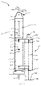
facilitate insertion of
the implant 401. Similarly stated, when the suction force is applied to the
contact surface S
of the cervix C a cervical canal, the implant delivery device 400 can be used
to place the
uterus U of the patient in more suitable position to receive the implant 401.
More
18
CA 0281482 2013-04-15
WO 2012/054466 PCT/US2011/056688
particularly, as shown in FIG. 9, with the vacuum source exerting a suction
force on the
portion of the contact surface S of the cervix C, the implant delivery device
400 can be
moved in a proximal direction, as shown in FIG. 9 by arrow FF. By moving the
implant
delivery device 400 in the proximal direction, the cervix C and/or uterus U is
straightened,
positioned and/or reoriented such that the uterine cavity Uc is more
accessible via the
cervical opening 0. In some embodiments, the implant delivery device 400 can
be moved in
a proximal direction until a desired level of alignment and/or "straightness"
between the
uterine cavity and the cervical canal is achieved. For example, in some
embodiments, the
implant delivery device 400 can be moved in a proximal direction until an
angle 0 between a
center line CL0 of the cervical canal and/or the cervical opening 0 and a
center line CLc of
the uterine cavity of the uterus is greater than approximately 90 degrees. In
other
embodiments, the implant delivery device is moved proximally until the angle
between the
uterine cavity and the cervical canal is greater than approximately 115
degrees, 135 degrees,
150 degrees or 165 degrees.
[1077] After the cervix C and/or uterus U is straightened, positioned
and/or reoriented,
the insertion member 461 can be advanced in a distal direction beyond the
contact portion
447 and into the uterine cavity Uc to place the implant 401 in any manner of
the types
described herein. In this manner, the delivery device can both straighten
and/or align the
target tissue and deliver the implant, thereby obviating the need for a
tenaculum.
[1078] Although the contact portion 447 is shown and described above as
being
configured to exert a vacuum force that spatially varies along the external
surface S of the
cervix C, in other embodiments, the contact portion 447 can be configured to
exert a
substantially uniform vacuum force along the external surface S of the cervix
C. In other
embodiments, however, the contact portion 447 can define a additional volumes
in fluid
communication with the vacuum channel 481 to produce localized and/or
noncontiguous
areas of vacuum force.
[1079] FIGS. 10-19 show an implant delivery device 500 according to an
embodiment.
The implant delivery device 500 includes a housing 510, an insertion assembly
560, a head
540, and a vacuum assembly 580. The housing 510 includes a proximal end 511
and a distal
end 512 and defines a housing passageway 513 therebetween. The housing 510
includes a
pair of holders 516 configured to couple the vacuum assembly 580 to the
housing 510. The
holders 516 can be any suitable holder and can create a friction fit with at
least a portion of
19
CA 0281482 2013-04-15
WO 2012/054466 PCT/US2011/056688
the vacuum assembly 580. The housing 510 can be any suitable size, shape, or
configuration.
For example, the 510 is substantially cylindrical with a diameter suitable for
insertion into a
body orifice. The housing 510 can be substantially tubular and can be formed
from any
suitable material, such as, for example, a plastic. Additionally, the housing
510 can include a
lubricated outer surface to ease in the insertion of the housing into the
body.
[1080] The proximal end 511 of the housing 510 is coupled to an adapter cap
520. The
adapter cap 520 (FIG. 11) includes a proximal end 521 and a distal end 522.
The proximal
end 521 of the adapter cap 520 includes a barbed fitting 523 and defines a
lumen (not shown).
The barbed fitting 523 can be coupled to a source of pressurized fluid (not
shown in FIGS.
10-19). For example, in some embodiments, the source of pressurized fluid can
be an air
bladder with an air delivery tube. In such embodiments, the barbed fitting 523
can be
inserted into the air delivery tube to place the housing passageway in fluid
communication
with the source of pressurized fluid, via the lumen, as describe in further
detail herein. While
shown in FIG. 11 as including a barbed fitting 523, the adapter cap 520 can
include any
suitable fitting configured to couple to any suitable energy storage device
and/or source of
pressurized fluid.
[1081] The distal end 522 of the adapter cap 520 includes a center
protrusion 524 that
include a set of sealing members 525. In this manner, the center protrusion
524 can be
inserted into the proximal end of the housing passageway 513. The sealing
members 525
define a friction fit with the inner walls of the housing 510, such that the
sealing members
525 produce a fluid-tight seal with the proximal end 511 of the housing 510.
[1082] The insertion assembly 560 (FIG. 12) is, at least partially,
disposed within the
housing passageway 513 and can be configured to move distally within the
housing 510, in
response to a force and/or pressure applied by source of pressurized fluid
(not shown in
FIGS. 10-19). The insertion assembly includes a first insertion member 561 and
a second
insertion member 566, and an actuator tube 575. The first insertion member 561
includes a
proximal end portion 562 and a distal end portion 563, and defines a lumen 564
therebetween. The proximal end portion 562 of the first insertion member 561
is fixedly
coupled to a plunger 565. The first insertion member 561 can be any suitable
shape, size, or
configuration and can be formed from any suitable material. In some
embodiments, the first
insertion member 561 can be formed from a flexible polymer. In other
embodiments, the
CA 0281482 2013-04-15
WO 2012/054466 PCT/US2011/056688
first insertion member 561 can be formed from a plastic, a rubber, and/or a
combination of
materials.
[1083] The plunger 565 can be any suitable plunger configured to produce a
fluid tight
seal with the inner walls of the housing passageway 513. The plunger 565 can
define any
suitable shape and can include any number of sealing members, protrusions,
contours, and/or
the like. In this manner, a proximal end of the plunger 565, the inner walls
of the housing
passageway 513, and the adapter cap 520 collectively define a chamber 514
configured to
receive a pressurized fluid from the source of pressurized fluid.
[1084] The actuator tube 575 includes a proximal end 576 and a distal end
577 and
defines a lumen 578 therebetween. The distal end 577 of the actuator tube 575
is fixedly
coupled to the proximal end of the plunger 565. In this manner, the lumen 564
defined by the
first insertion member 561 and the lumen 578 defined by the actuator tube 575
collectively
define a passageway (not shown in FIGS. 10-19) configured to house at least a
portion of the
second insertion member 566.
[1085] The second insertion member 566 includes a proximal end 567 and a
distal end
568. The second insertion member 566 can be any suitable shape, size, or
configuration and
can be formed of any suitable material, such as, for example, those described
with respect to
the first insertion member 561. The proximal end 567 is fixedly coupled to a
plunger 569 and
is disposed within in the actuator tube 575. The plunger 569 can be any
suitable plunger
configured to produce a fluid tight seal with the inner walls of the actuator
tube 575. The
distal end of the second insertion member 566 is configured to be removably
coupled to an
implant, such as, for example, an IUD. In this manner, the second insertion
member 566 is
configured to move relative to the first insertion member 561 to deliver the
implant, as
described in further detail herein.
[1086] The distal end 512 of the housing 510 is coupled to an articulation
neck 530
(FIGS. 13 and 14). The articulation neck 530 includes a proximal end 531 and a
distal end
532 and defines a neck passageway 535. The proximal end 531 is configured to
be inserted
into the distal end portion 512 of the housing 510. The proximal end 531 of
the articulation
neck 530 can produce a friction fit with the inner walls of the housing 510.
While shown in
FIG. 13 as substantially smooth, the proximal end 531 can include any suitable
surface
feature and/or texture to facilitate being coupled within the distal end
portion 512 of the
21
CA 0281482 2013-04-15
WO 2012/054466 PCT/US2011/056688
housing 510. In some embodiments, the proximal end 531 can include a set of
sealing
protrusions, substantially similar to the sealing protrusions 525 described
with respect to the
adapter cap 520. The proximal end 531 of the articulation neck 530 defines an
engagement
portion 534 configured to selectively engage at least a portion of the
insertion assembly 560.
More specifically, the engagement portion 534 is configured to engage the
plunger 565, when
the insertion member 560 is moved in a distal direction. This arrangement
limits the
movement of the first insertion member 561, relative to the housing 510. The
engagement
portion 534 can be any suitable portion and can include a contour
substantially similar to the
contour of the distal end of the plunger 565.
[1087] The distal portion 532 of the articulation neck 530 includes a set
of sidewalls 533
and is moveably coupled to the head 540 (FIG. 10). The sidewalls 533 can
define any
suitable shape or configuration. For example, as shown in FIG. 13, the
sidewalls 533 define a
surface that includes a pair of notches 536. The notches 536 allow for the
articulation of the
head 540 relative to the housing 510, as described in further detail herein.
The sidewalls 533
define a set of apertures 537 and notches 538. The apertures 537 and the
notches 538 are
disposed opposite each other, and are configured to moveably couple at least a
portion of the
head 540 to the articulation neck 530.
[1088] The head 540 includes a proximal end 541 and a distal end 542 and
defines a head
passageway 544 therebetween, as shown in FIGS. 15 and 16. The proximal end 541
of the
head 540 includes a set of articulation protrusions 551 that moveably couple
the head 540 to
the articulation neck 530. More specifically, the protrusions 551 are
configured to be inserted
into the notches 538 and the apertures 537 defined by the sidewalls 533. A
first portion 552
of each protrusion 551 forms a pin that is disposed within the corresponding
apertures 537.
Thus, the protrusions 551 define an axis A1 about which the head 540 can
pivot, relative to
the articulation neck 530 and/or housing 510. In addition, the first portion
552 of the
protrusions 551 (i.e., the pins) define a friction fit with the surface of the
sidewalls 533 that
defines the apertures 537. This arrangement prevents the head 540 from
pivoting freely
within the apertures 537. Similarly stated, the fit defined by the first
portion 552 of the
protrusions 551 and the apertures 537 produces an amount of friction such as
to partially
resist the motion of the first portion 552 of the protrusions 551 within the
apertures 537. A
second portion 553 of each protrusion 551 is configured selectively engages
the notches 538
defined by the sidewalls 533 to limit the range of pivoting motion of the head
540, relative to
22
CA 0281482 2013-04-15
WO 2012/054466 PCT/US2011/056688
the articulation neck 530 and/or housing 510. For example, the notches 538
(FIG. 14) can
form a contour such that when the protrusions 551 are disposed within the
apertures 537 and
the notches 538, the walls of the contour engage the walls of the second
portion 553 of the
protrusions 551 to limit the range of motion of the head 540. In some
embodiments, the
pivoting motion is limited to a range between +/- 100. In other embodiments,
the range of
motion can be in a range between +/- 15 , +/- 20 , +/- 30 , or more.
[1089] The distal end 542 of the head 540 includes a contact portion 547.
The contact
portion 547 includes a sidewall 548 that defines a volume 550. The contact
portion 547 and
the sidewall 548 can be any suitable size, shape, and configuration. For
example, in some
embodiments, the sidewall 548 of the contact portion 547 defines a pair of
notches 549. In
this manner, the notches 549 can be configured to accept a portion of a
contact surface
associated with a target tissue. For example, the notches 549 can be
configured to accept a
portion of a cervix of a uterus, such that the contact portion 547 of the head
540 can be placed
in a desired position and/or orientation relative to the cervix. In other
embodiments, the
sidewall 548 can includes any number of notches 549 and/or define a specific
contour
configured to receive a portion of a contact surface of a target tissue.
[1090] The head 540 includes an inner wall 543 that defines the head
passageway 544
and includes a tapered portion 554. At least a portion of the insertion
assembly 560 is
configured to move within the head passageway 544, to a volume substantially
outside the
implant delivery device 500. In particular, during use, the first insertion
member 561 and the
second insertion member 566 collectively move from the housing passageway 513
and
through the head passageway 544. In some embodiments, the tapered portion 554
and/or
other portions of the inner wall 543 are configured to engage a portion of the
insertion
assembly 560 to facilitate bending of the insertion assembly 560 when the
insertion assembly
560 is moved through the head passageway 544. Similarly stated, at least a
portion of the
sidewall 543 is configured to reduce snagging of the insertion assembly 560 as
it moves
through the head passageway 544. In some embodiments, the sidewall 543 can
include a
curved portion (e.g., having a radius of curvature), a low surface roughness
and/or a hardened
portion to facilitate movement and/or bending of the insertion assembly 560 in
use. This
arrangement increases patient comfort during use by facilitating bending of
the insertion
member 560 within the head 540, rather than within a bodily cavity. Similarly
stated, this
23
CA 0281482 2013-04-15
WO 2012/054466 PCT/US2011/056688
arrangement reduces the likelihood that a portion of the insertion assembly
560 will engage
or otherwise press on a body tissue forming the bodily cavity during use.
[1091] The head 540 also includes a vacuum fitting 545 that defines a
vacuum channel
546. The vacuum fitting 545 can extend from a surface of the head 540 and have
any suitable
shape. In some embodiments, the vacuum fitting 545 is configured to receive a
vacuum line
(not shown in FIGS. 10-19) that it can be operably coupled to the vacuum
assembly 580. The
vacuum channel 546 is in fluid communication with the neck passageway 544 and
therefore,
with the volume 550 defined by the contact portion 547, as shown in FIG. 16.
[1092] The vacuum assembly 580 includes a vacuum tube 581 that houses a
vacuum
actuator 585 (see e.g., FIGS. 10 and 17). The vacuum tube 580 includes a
proximal end 582
and a distal end 583, as shown in FIG. 17. The proximal end 582 is configured
to receive at
least a portion of the vacuum actuator 585. The distal end 583 includes a
vacuum port 595
configured to receive the vacuum line (not shown in FIGS. 10-19) and/or be
coupled to the
vacuum fitting 545 of the head 540.
[1093] The vacuum actuator 585 includes a proximal end 586 and a distal end
587. The
proximal end 586 can include a flange 588 configured to be engaged by a
physician and/or
user. The distal end 587 is fixedly coupled to a plunger 589. The plunger 589
can be
substantially similar to any plunger described herein. In this manner, the
plunger 589 defines
a substantially fluid tight seal with the inner walls of the vacuum tube 581.
When disposed
within the vacuum tube 581, the plunger 589 and the vacuum tube 581 define a
chamber 584
(FIG. 18) that is in fluid communication with the vacuum port 595. In use, the
vacuum
actuator 585 can be moved in a proximal direction, by the user, thereby
increasing the
volume of the chamber 584, which produces a negative pressure within the
chamber 584.
The negative pressure (i.e., vacuum is transmitted through the vacuum line
(not shown) to the
volume 550 of the head. In this manner, a suction force can be applied to a
surface of the
body, as described above.
[1094] Additionally, the proximal end 582 of the vacuum tube 581 includes a
locking tab
591. The locking tab 591 is configured to selectively engage the vacuum
actuator 585 to hold
the vacuum actuator 585 in the actuated configuration. More specifically, the
vacuum
actuator 585 includes a ridge 590 (FIG. 19) that selectively contacts an
engagement surface
592 of the locking tab 591. The engagement surface 592 contacts the ridge 590
such that the
24
CA 0281482 2013-04-15
WO 2012/054466 PCT/US2011/056688
vacuum actuator 585 is in a locked configuration. In this manner, after the
vacuum is
produced in the chamber 584, the position of the vacuum actuator 585 within
the vacuum
tube 581 can be maintained, thereby preventing inadvertent loss of vacuum
during use of the
implant delivery device 500.
[1095] When in use, the implant delivery device 500 can be inserted into a
bodily lumen
of a patient. In some embodiments, the implant delivery device 500 is inserted
into a vagina
of a patent and through a lumen defined by the walls of the vagina to a cervix
of a uterus.
The head 540 can pivot, move, and/or rotate, relative to the housing 510,
before, during
and/or after the implant delivery device 500 passes through the lumen. The
contact portion
547 of the head 540 is configured to engage a portion of the cervix. The head
540 can pivot,
move, and/or rotate such that the contact portion 547 of the head 540 can
substantially
circumscribe a contact surface of the cervix. When the head 540 is positioned
adjacent the
bodily surface (e.g., the outer surface of the cervix), the side walls 548 of
the contact portion
547 engage the contact surface of the cervix and thus, the side walls 548 and
at least a portion
of the contact surface substantially enclose the volume 550 of the head 540.
In this manner,
when the vacuum assembly 580 is actuated the negative pressure within the
vacuum chamber
584 can exert a suction force through the vacuum line (not shown in FIGS. 10-
19), into the
volume 550, and subsequently on the contact surface of the cervix.
[1096] When the suction force is exerted on the contact surface of the
cervix, the vacuum
actuator 585 can be placed in the locked position (described above in
reference to FIGS. 18
and 19). The implant delivery device 500 can be moved to reposition the uterus
into a
desired position, orientation and/or configuration by moving the implant
delivery device 500
in the proximal direction, as described above with reference to FIGS. 7-9.
[1097] When the uterus and/or cervix are in the desired position, the
insertion assembly
560 can be actuated to deliver the implant. In particular, a source of
pressurized fluid can be
actuated and/or placed in fluid communication with the proximal end 511 of the
housing 510.
In some embodiments, the source of pressurized fluid can include a manually-
actuated air
bulb, an air tube, and a control valve. In such embodiments, the air tube is
coupled to the
ribbed fitting 523 included in the adapter cap 520. In this manner, the air
bulb can be
actuated to convey a pressurized fluid into the chamber 514, which exerts a
force to move the
insertion assembly 560 in the distal direction. In other embodiments, the
source of
pressurized fluid can include an energy storage member, such as a compressed
gas container,
CA 0281482 2013-04-15
WO 2012/054466 PCT/US2011/056688
a propellant cartridge, chemical energy storage member or the like, which
produces the
pressurized fluid automatically when actuated by the user. In yet other
embodiments, the
force to produce the insertion can be produced by any other suitable energy
storage member,
such as, for example, a spring.
[1098] As
shown in FIGS. 10 and 12, the plunger 565 defines a larger surface area than
does the plunger 569. Since the force exerted on each plunger is directly
proportional to the
area of the plunger, when the pressurized fluid is conveyed into the chamber
514, the force
exerted on the plunger 565 is greater than the force exerted on the plunger
569. Thus, the
force exerted by the pressurized fluid moves the plunger 565 in the distal
direction within the
housing 510. Because the actuator tube 575 and the first insertion member 561
are fixedly
coupled to the plunger 565, the insertion assembly 560 collectively moves with
the plunger
565 in the distal direction. In this manner, at least a portion of the first
insertion member 561
and the second insertion member 566 can move within the housing passageway 513
and
through the neck passageway 535 and the head passageway 544. This operation
can be
referred to as the "first insertion operation."
[1099] The
plunger 569 and the actuator tube 575 are collectively configured such that
the force exerted by the pressurized fluid on the plunger 569 during the first
insertion
operation (which, as discussed above, is lower than the force exerted on the
plunger 565) is
insufficient to move the plunger 569, and therefore the second insertion
member 566, within
the actuator tube 575. Thus, during the first insertion operation, the distal
end portion 568 of
the second insertion member 566, and thus, the implant (not shown) is
maintained within the
first insertion member 561.
[1100] The
engagement portion 534 can contact the plunger 585 to limit the movement of
the first insertion member 561, relative to the housing 510 during the first
insertion operation.
Thus, the first insertion member 561 is configured to be moved a predetermined
distance
(e.g., a minimum anatomical depth associated with the uterus) during the first
insertion
operation. In some embodiments, the insertion assembly 560 and the engagement
portion
534 can be configured such that the first insertion member 561 can extend
approximately 5
cm beyond the contact portion 547 of the head 540. In other embodiments, the
first insertion
member 561 can extend any suitable distance great than or less than 5 cm.
26
CA 0281482 2013-04-15
WO 2012/054466 PCT/US2011/056688
[1101] When the plunger 565 is in contact with the engagement portion 534,
the pressure
of the pressurized fluid in the chamber 514 can continue to increase. In some
embodiments,
the user can manually actuate the air bulb to produce an increased pressure
within the
chamber 514. In other embodiments, the energy storage member can be actuated a
second
time to produce an increased pressure within the chamber 514. The pressure can
be increased
until the force exerted on the plunger 569 of the second insertion member 566
is sufficient
move the second insertion member 566 in the distal direction, relative to the
first insertion
member 561. This operation can be referred to as the "second insertion
operation." During
the second insertion operation, the implant (not shown) is decoupled from
and/or pushed out
of the first insertion member 561 and into a desired position within the
uterus.
[1102] In some embodiments, the implant delivery device 500 can include a
valve or
other control mechanism configured to regulate the pressure within the chamber
514 during
the first insertion operation and/or the second insertion operation such that
the force exerted
by second insertion member 566 on the implant is limited. In this manner, the
second
insertion member 566 can enter a uterine cavity through a cervical os and
deliver the implant
(e.g., IUD) to the target tissue within the uterus.
[1103] Although the implant delivery device 500 is shown and described
above as
including two plungers configured to produce a first insertion operation that
is distinct from a
second insertion operation, in other embodiments, a device can include a
single plunger
configured to produce a two-stage delivery. FIGS. 20-24 show an implant
delivery device
600 according to an embodiment. The implant delivery device 600 includes a
housing 610,
source of pressurized fluid 603, and a head 640. The housing 610 includes a
proximal end
611 and a distal end 612 and defines a housing passageway 613 and a vacuum
channel 684.
The housing 610 can be any suitable shape, size, or configuration, as
described herein. For
example, the housing 610 can be substantially cylindrical with a diameter
suitable for
insertion into a body orifice. The distal end 612 of the housing 610 is
rotatably coupled to
the head 640.
[1104] The head 640 can be coupled to the distal end 612 of the housing 610
in any
suitable way. For example, the distal end 612 of the housing 610 can include a
fitting, cap,
joint, and/or the like configured to rotatably couple the head 640 to the
housing 610. In some
embodiments, the housing 610 and the head 640 are coupled via a sleeve and/or
the like (not
shown in FIGS. 20-24). The head 640 defines a head passageway and includes a
contact
27
CA 0281482 2013-04-15
WO 2012/054466 PCT/US2011/056688
portion 647 that defines a volume 650. The head 640 can be any suitable shape,
size, or
configuration. For example, as shown in FIG. 20, the head 640 can
substantially form a cup
shape. The housing passageway 613 and the head passageway can collectively
define an
insertion passageway 605 that can be substantially nonlinear, such that at
least a portion of
the insertion passageway 605 bends and/or curves.
[1105] The implant delivery device 600 can be inserted into a bodily lumen
defined by
the body of a patient, such as, for example, the lumen defined by the inner
walls of the
vagina. With the implant delivery device 600 inserted into the lumen, the
contact portion 647
contacts a surface associated target tissue, such as, for example, a cervix of
a uterus. The
contact portion 647 can be configured to substantially circumscribe a surface
associated with
a target tissue. For example, when the contact portion 647 is in contact with
the contact
surface of the cervix, the walls of the contact portion 647 substantially
surround a cervical
opening. In this manner, at least a portion of the contact surface and the
contact portion 647
substantially enclose the volume 650.
[1106] The vacuum channel 684 defined by the housing 610 includes a
proximal end 682
and a distal end 683. The distal end 683 includes a vacuum port 695 and is in
fluid
communication with the insertion passageway 605. In this manner, the vacuum
channel 684
is configured to be in fluid communication with at least a portion of the
volume 650. A
vacuum actuator 685 is disposed at least partially within the vacuum channel
684, and can be
manually actuated to form a vacuum within the vacuum channel 684. The actuator
685
includes a plunger 689 configured to form an airtight seal with the inner
surface of the
vacuum channel 684. Therefore, in use, the actuator 685 can be retracted
(e.g., moved in a
proximal direction) to produce a vacuum force. With the vacuum channel 684 in
fluid
communication with at least a portion of the volume 650 and with the contact
surface
substantially enclosing the volume 650, as described above, a suction force
can be exerted on
the contact surface associated with the target tissue.
[1107] The implant delivery device 610 further includes a first insertion
member 661 and
a second insertion member 666. The first insertion member 661 can be any
suitable shape,
size, or configuration. For example, in some embodiments, the first insertion
member 661
can be formed from a flexible material such as a rubber, elastomer, polymer,
and/or plastic.
The first insertion member 661 includes a proximal end 662 and a distal end
663. In some
embodiments, the first insertion member 661 defines an insertion channel
between the
28
CA 0281482 2013-04-15
WO 2012/054466 PCT/US2011/056688
proximal end 661 and the distal end 662, configured to receive at least a
portion of the second
insertion member 666.
[1108] The second insertion member 666 includes a proximal end 667 and a
distal end
668. Similar to the first insertion member 661, the second insertion member
666 can be any
suitable size, shape, or configuration, and can be formed from any suitable
material. The
proximal end 667 is at least partially disposed within the housing passageway
613. The distal
end 668 is configured to be removably coupled to an implant 601. In some
embodiments, the
implant 601 is an intrauterine device (IUD) configured to be implanted into a
target portion
of a uterus of a patient. At least a portion of the first insertion member 661
and the second
insertion member 666 are configured to move relative to the housing 610 within
the housing
passageway 613 to deliver the implant 601. Similarly, at least a portion of
the second
insertion member 666 is configured to move relative to the first insertion
member 661 within
the insertion channel 664.
[1109] FIG. 21 shows the implant delivery device 600 in a second
configuration (i.e., at
the end of a first insertion operation), in response to a force being applied
by the source of
pressurized fluid 603. The source of pressurized fluid 603 can include an air
bulb 608, an air
tube 607, and a control valve 602. The air tube 607 is coupled to the proximal
end 612 of the
housing 610 using any suitable fitting, coupling, adapter, and/or the like. In
this manner,
when the air bulb 608 is actuated (i.e., squeezed) a pressurized fluid can be
conveyed into the
housing passageway 613 to move the first insertion member 661 and the second
insertion
member 666 in the distal direction. More specifically, the force exerted by
the pressurized
fluid moves the plunger 669 of the second insertion member 666 in the distal
direction. The
second insertion member 666 and the first insertion member 661 are
collectively configured
such that the force exerted by the pressurized fluid on the plunger 569 during
the first
insertion operation is insufficient to move the second insertion member 566
within the first
insertion member 661. Thus, during the first insertion operation, the second
insertion
member 566 and the first insertion member 661 collectively move relative to
the housing 610
to move the device 600 from the first configuration (FIG. 20) to the second
configuration
(FIG. 21). Thus, during the first insertion operation, the distal end portion
of the second
insertion member 666, and thus, the implant 601, is maintained within the
first insertion
member 661.
29
CA 0281482 2013-04-15
WO 2012/054466 PCT/US2011/056688
[1110] The housing 610 includes an engagement portion 634 configured to
selectively
engage the first insertion member 661. In this manner, the first insertion
member 661 can
extend a predetermined distance beyond the contact portion 647 of the head 640
when in the
second configuration (i.e., upon completion of the first insertion operation),
as shown in FIG.
21.
[1111] With the first insertion member 661 in contact with the engagement
portion 634,
the pressurized fluid in the housing passageway 613 continues to exert at the
force on the
plunger 669 of the second insertion member 666 to move the second insertion
member 666 in
the distal direction, relative to the first insertion member 661 (FIGS. 22 -
24). The valve 602
included in the energy storage member 603 can be used to regulate the pressure
within the
housing passageway 613 such that the force exerted by second insertion member
666 on the
implant is limited. In this manner, the second insertion member 666 can enter
a uterine
cavity and deliver the implant 601 (e.g., IUD) to the target tissue as shown
in FIG. 24.
[1112] FIGS. 25-32 show an implant delivery device 700 according to an
embodiment.
The implant delivery device 700 includes a housing 710, a source of
pressurized fluid 703,
and a head 740 (FIG. 26). The housing 710 includes a proximal end 711 and a
distal end 712,
and defines a housing passageway 713 and a vacuum channel 784. The housing 710
can be
any suitable shape, size, or configuration, as described herein. For example,
the housing 710
can be substantially cylindrical with a diameter suitable for insertion into a
body orifice. The
distal end 712 of the housing 710 is rotatably coupled to the head 740.
[1113] The head 740 can be coupled to the distal end 712 of the housing 710
in any
suitable way. For example, the distal end 712 of the housing 710 can couple to
the head 740
via a neck 730 (FIG. 26). The neck 730 includes sidewalls 736 that define a
set of ribs
allowing for rotational motion of the head 740. In some embodiments, the neck
730 is
formed of a flexible material, such as, for example, a rubber or a polymer.
The neck 730
defines a neck passageway 735 between a proximal end 731 and a distal end 732.
Similarly,
the head 740 defines a head passageway 744 and includes a contact portion 747.
The contact
portion 747 includes a sidewall 648 that defines a volume 750. The head 740
can be any
suitable shape, size, or configuration. For example, as shown in FIG. 26, the
head 740 can
substantially form a cup shape. The housing passageway 713, the neck
passageway 735, and
the head passageway 744 collectively define an insertion passageway (not
identified) that can
CA 0281482 2013-04-15
WO 2012/054466 PCT/US2011/056688
be substantially nonlinear, such that at least a portion of the insertion
passageway bends
and/or curves during an insertion operation.
[1114] The implant delivery device 700 can be inserted into a bodily lumen
defined by
the body of a patient, such as, for example, the lumen defined by the inner
walls of the
vagina. With the implant delivery device 700 inserted into the lumen, the
contact portion 747
contacts a surface associated target tissue, such as, for example, a cervix of
a uterus. The
contact portion 747 can be configured to substantially circumscribe a surface
associated with
a target tissue. For example, when the contact portion 747 is in contact with
the contact
surface of the cervix, the contact portion 747 substantially surrounds a
cervical opening. In
this manner, at least a portion of the contact surface and the contact portion
747 substantially
enclose the volume 750.
[1115] The vacuum channel 784 defined by the housing 710 includes a
proximal end 782
and a distal end 783 (FIG. 25). The distal end 783 includes a vacuum port 795
that fluidically
coupled to the neck 730 (FIG. 26). In this manner, the vacuum channel 784 is
configured to
be in fluid communication with at least a portion of the volume 750. A vacuum
actuator 785
is movably disposed within the vacuum channel 784, and can be manually
actuated to form a
vacuum. The vacuum actuator 785 (FIG. 27) includes proximal end 786 and a
distal end 787.
The proximal end 786 includes a flange 788 that can be grasped by a user in
order to actuate
the vacuum chamber 784. In some embodiments, the housing 710 can include a
locking
mechanism (not shown in FIGS. 25-32) configured to engage the flange 788 to
maintain the
vacuum actuator 785 in an actuated position. The distal end 788 of the vacuum
actuator 785
includes a plunger 789 configured to form an airtight seal with the inner
surface of the
vacuum channel 784. Therefore, in use, the actuator 785 can be retracted
(e.g., moved in a
proximal direction) to produce a vacuum force. With the vacuum channel 784 in
fluid
communication with at least a portion of the volume 750 and with the contact
surface
substantially enclosing the volume 750, as described above, the vacuum channel
784 transfers
a suction force on the contact surface associated with the target tissue.
[1116] The implant delivery device 710 further includes an insertion member
761, as
shown in FIG. 28. The insertion member 761 can be any suitable shape, size, or
configuration. For example, in some embodiments, the insertion member 761 can
be formed
from a flexible material such as a rubber, elastomer, polymer, and/or plastic.
At least a
portion of the insertion member 761 is configured to move relative to the
housing 710 within
31
CA 0281482 2013-04-15
WO 2012/054466 PCT/US2011/056688
the housing passageway 713. The insertion member 761 includes a proximal end
762 and a
distal end 763. The proximal end 762 of the insertion member 761 includes a
plunger 765.
The distal end 763 includes an articulator 771 that is configured to be
removably coupled to
an implant 701. In some embodiments, the implant 701 is an intrauterine device
(IUD)
configured to be implanted into a target portion of a uterus of a patient. As
shown in the
enlarged view in FIG. 29, the articulator 771 includes a set of sidewalls 772
that define an
implant volume 774. The implant volume 774 is configured to receive the
implant 701 (FIG.
30). The sidewalls 772 further define a slit 773 configured to receive at
least a portion of a
string often attached to an IUD. Additionally, the housing 710 includes a
retaining tube 706
(see FIG. 31) configured to selectively engage the implant 701. For example,
as shown in
FIG. 31, the retaining tube 706 maintains an IUD in a collapsed configuration.
[1117] The source of pressurized fluid 703 can include an air bulb 708, an
air tube 707,
and a control valve 702 (FIG. 25). The air tube 707 is coupled to the proximal
end 712 of the
housing 710 using any suitable fitting, coupling, adapter, and/or the like. In
this manner, the
air bulb 708 can be actuated and a pressurized fluid can exert a force within
the housing
passageway 713 to move the insertion member 761 in the distal direction. More
specifically,
the force exerted by the pressurized fluid moves the plunger 765 of the
insertion member in
the distal direction. The valve 702 included in the energy storage member 703
can be used to
regulate the pressure within the housing passageway 713 such that the force
exerted by the
insertion member 761 on the implant 701 when the insertion member 761 moves is
below a
predetermined force. In this manner, a portion of the insertion member 761 can
extend
beyond the retaining tube 706 (FIG. 32) and enter a uterine cavity to deliver
the implant 701
(e.g., IUD) to the target tissue.
[1118] While specific heads and/or contact portions are discussed herein,
the components
and configurations of the head and/or contact portion can vary. For example,
FIG. 33 is a
schematic illustration of an implant delivery device 800 in a first
configuration that includes a
housing 810 and a head 840. The head 840 is rotatably coupled to a distal end
of the housing
810 in any suitable manner, such as, for example, those described herein. The
head 840 can
be any suitable shape, size, or configuration. For example, the head 840 can
define a leaflet
mechanism that defines a first diameter D2 when in the first configuration.
The head 840 can
be configured to respond to a force and/or external input that causes the
leaflets of the head
840 to expand and or separate when in a second configuration. In this manner,
the head 840
32
CA 0281482 2013-04-15
WO 2012/054466 PCT/US2011/056688
can define a second diameter D2, substantially larger than the first diameter,
when in the
second configuration, as shown in FIG. 34. Similarly stated, the head 840 can
be moved
between a collapsed configuration, in which the head 840 has a first size
(e.g., Di) and an
expanded configuration, in which the head 840 has a second size (e.g., D2)
larger than the
first size.
[1119] In some embodiments, an implant delivery device includes one or more
mechanical biosensors around the rim of the head and/or the insertion member
and a light
emitting diode (LED) or other electronic output device at the opposite end of
the device.
Other indicators can be used instead of an LED, such as for, example, any
suitable visual
output device (LCD screens, etc.), audible output devices (e.g., a whistle),
or mechanical
output devices (e.g., haptic output devices).
[1120] In some embodiments, an implant delivery device can rotate, bend,
and/or move
with the cervix and insert the IUD into a woman's uterus with no other tools
needed, and
without the need for exceptional skill and/or training. The design of the
embodiments
described herein facilitates ease of use such that after a short training
session, any health care
provider can properly insert an IUD safely with aseptic technique.
FIG. 35 is a flowchart illustrating a method 900 for implanting and
intrauterine device as
described herein. The method 900 can be performed using any of the devices
shown and
described herein. The method 900 includes inserting an implant delivery device
into a body
until a contact portion included in the implant delivery device contacts an
outer surface of a
cervix of a uterus, at 901. More specifically, in some embodiments, the
contact portion can
be similar to the contact portion 547 included in the head 540 that is
flexibly coupled to the
housing 510 of the implant delivery device 500, described with respect to
FIGS. 10-19. The
method 900 further includes producing a vacuum within a volume defined by the
contact
portion and applying a suction force to the surface of the cervix, at 902. For
example, in
some embodiments, the device can include a vacuum actuator similar to the
vacuum actuator
585 that can be pulled in a proximal direction to produce a vacuum within the
vacuum
chamber 584 to exert a suction force on the surface of the cervix, as
described with respect to
FIGS. 10-19. With the suction force applied to the surface of the cervix, the
implant delivery
device is moved in a proximal direction until an angle between a cervical
canal of the cervix
and a uterine cavity of the uterus is greater than approximately 90 degrees,
at 903. In some
embodiments, the implant delivery device is moved until the angle between the
uterine cavity
33
CA 0281482 2013-04-15
WO 2012/054466 PCT/US2011/056688
and the cervical canal is greater than approximately 115 degrees, 135 degrees,
150 degrees
or 165 degrees. The method 900 further includes moving an insertion member
within a
passageway defined by the implant delivery device until a distal end portion
of the insertion
member is disposed within the uterine cavity, at 904.
[1121] FIG. 36 is a flowchart illustrating a method 1000 for implanting an
intrauterine
device as described herein. The method 1000 includes inserting an implant
delivery device
into a body until a contact portion of the implant delivery device contacts an
outer surface of
a cervix of a uterus, at 1001. More specifically, in some embodiments, the
contact portion
can be similar to the contact portion 547 included in a head 540 that is
flexibly coupled to the
housing 510 of the implant delivery device 500, described with respect to
FIGS. 10-19. A
first insertion member can be moved relative to the housing such that the
distal end of the
first insertion member is disposed within a cervical canal of the cervix, at
1002. For
example, in some embodiments, a first insertion member can be similar to the
first insertion
member 261, which moves in a distal direction in response to a force from an
energy storage
member, as described with respect to FIG. 4. In some embodiments, the method
1000 can
include limiting the distance the first insertion member can move beyond the
contact portion
to a predetermined length, at 1003.
[1122] The method 1000 further includes moving the second insertion member
relative to
the first insertion member to decouple the implant from the first insertion
member, at 1004.
In some embodiments, the method 1000 can include maintaining the force of the
second
insertion member below a predetermined threshold. For example, in some
embodiments, the
implant delivery device can be similar to the implant delivery device 200,
which includes a
control member 202 configured to reduce the insertion force F1, such that a
portion F2 of the
force F1 is transmitted to the second insertion member 266, as described with
respect to FIG.
4.
[1123] While various embodiments of the invention have been described
above, it should
be understood that they have been presented by way of example only, and not
limitation.
Where methods described above indicate certain events occurring in certain
order, the
ordering of certain events may be modified. Additionally, certain of the
events may be
performed concurrently in a parallel process when possible, as well as
performed sequentially
as described above.
34
CA 0281482 2013-04-15
WO 2012/054466 PCT/US2011/056688
[1124] For example, in some embodiments, a device can include a head
similar to any of
the heads shown and described above, and the head can include a protrusion
configured to
position the head relative to a lumen defined by the target location.
Similarly stated, in some
embodiments, an implant delivery device can include a locating protrusion
configured to
facilitate the alignment and/or positioning of the device with respect to a
target location. In
some embodiments, the protrusion can define a channel through which an
insertion member
can be conveyed to deliver an implant.
[1125] Although the devices are shown and described herein as delivering an
implant
through an existing bodily lumen (e.g., an opening and/or canal defined by the
cervix), in
other embodiments, a device can include a dilator configured to define a
bodily lumen and/or
expand an existing bodily lumen. In some embodiments, for example, a contact
portion of a
head includes a dilator configured to dilate a lumen defined by the target
location. The
dilator can define a channel and/or passageway through which an insertion
member can be
conveyed to deliver an implant.
[1126] In some embodiments, an implant delivery device can include a sleeve
configured
to be disposed about a head during the insertion operation. The sleeve can be
a thin, flexible
sleeve, which serves to facilitate insertion of the device and/or maintain
sterility during the
insertion operation. In some embodiments, an outer surface of the sleeve can
include a
lubricant.
[1127] Although various embodiments have been described as having
particular features
and/or combinations of components, other embodiments are possible having a
combination of
any features and/or components from any of embodiments where appropriate. For
example,
any of the devices shown and described herein can include an articulation neck
as described
herein. For example, although the implant delivery device 600 shown in FIGS.
20-24 is not
shown as including an articulation neck, in other embodiments, an implant
delivery device
similar to the device 600 can include an articulation neck similar to the
articulation neck 730
shown and described above.