Note: Descriptions are shown in the official language in which they were submitted.
:A 02814985 2013-04-17
WO 2012/057911
PCT/US2011/049632
1
REACTANCE CHANGES TO IDENTIFY AND EVALUATE CRYO
ABLATION LESIONS
FIELD OF THE INVENTION
The present invention relates to a method for measuring and correlating
changes in reactance of cryogenically treated tissue to assess lesion quality
and
transmurality.
BACKGROUND OF THE INVENTION
Radiofrequency (RF) and cryogenic ablation procedures are well recognized
treatments for vascular and cardiac diseases such as atrial fibrillation. The
application
of either RE or cryogenic treatment is usually based on the preference of the
surgeon
or the specific tissue to be treated. In either RF or cryogenic ablation,
however, the
location and quality of the lesion produced is a primary concern.
Current methods to identify a lesion's location and assess its quality include
coupling a plurality of electrodes to the distal end of a medical device
proximate a
tissue to be treated, applying a voltage, and measuring impedance across the
electrodes with the tissue to be treated completing the circuit. Electrical
impedance is
defined as the total opposition to alternating current by an electric circuit,
equal to the
square root of the sum of the squares of the resistance and reactance of the
circuit and
usually expressed in ohms. In general, the impedance decreases as the treated
tissue
becomes necrotic. As such, impedance may be used to identify particular areas
which
have been treated and those that have not.
One drawback to impedance tomography is its lack of direct feedback to
evaluate whether a lesion was successfully created to the desired
transmurality,
quality, or continuity. In particular, impedance measurements provide binary
data
regarding a particular lesion; either the tissue is viable or necrotic.
Impedance
measurements alone, however, do not provide real-time assessment of whether a
cryogenic or RF lesion was successfully created to a desired lesion depth, in
part,
because different tissue levels have different impedances.
As such, it would be desirable to provide improved methods of assessing
lesion quality and depth of cryogenically and/or RF treated tissue to
determine the
efficacy and resulting characteristics of the treatment.
:A 02814985 2013-04-17
WO 2012/057911
PCT/US2011/049632
2
SUMMARY 01: THE INVENTION
The present invention advantageously provides a method of assessing lesion
quality of an ablated tissue region comprising ablating at least a portion of
the tissue
region; measuring the reactance of the ablated tissue region; and determining
the
lesion quality of the ablated tissue region based on the measured reactance.
In another embodiment, the method includes positioning a medical device
proximate
the tissue region and circulating coolant towards a thermally conductive
region of the
medical device, the medical device having at least two electrodes, the
electrodes being
positioned proximate the thermally conductive region; thermally treating the
tissue
I() region; inducing a current between the at least two electrodes at a
plurality of
frequencies; measuring the reactance of the thermally treated tissue region at
each of
the plurality of frequencies; defining a predetermined thermally treated
tissue region
reactance threshold; comparing the measured reactance at each of the plurality
of
frequencies to the threshold; determining the lesion quality of the thermally
treated
tissue region based on the measured reactance at each of the plurality of
frequencies;
and modifying the thermally treating of the tissue based at least in part on
the
determination.
In yet another embodiment, the method includes positioning a medical device
proximate the tissue region and circulating coolant toward a thermally
conductive
region of the medical device, the medical device have at least two electrodes,
the at
least two electrodes being position proximate the thermally conductive region,
the
medical device further having a balloon disposed between the two electrodes;
cryogenically cooling the tissue region; inducing a current between the at
least two
electrodes at a plurality of frequencies; measuring the reactance of the
cryogenically
cooled tissue region at each of the plurality of frequencies; defining a
predetermined
thermally treated tissue region reactance threshold; defining an untreated
tissue
reactance value, wherein the predetermined thermally treated tissue region
reactance
threshold is about a 60-90% reduction in the reactance of the untreated tissue
reactance value; comparing the measured reactance to the threshold;
determining the
lesion quality and continuity of the thermally treated tissue region based on
the
comparison; displaying the determined lesion quality and continuity on an
imaging
:A 02814985 2013-04-17
WO 2012/057911
PCT/US2011/049632
3
system; and modifying the circulating coolant toward a thermally conductive
region
of the medical device based on the displayed tissue quality and continuity.
A medical system is provided, including a medical device containing two or
more electrodes; a console in electrical communication with the two or more
electrodes, the console programmed to: deliver an electrical current to the
two or more
electrodes; measure an electrical reactance between the two or more
electrodes; assess
treatment quality based at least in part on the measured reactance; and
generate an
indication of the assessment. The console may be programmed with a
predetermined
ablated tissue region reactance threshold, and assessing treatment quality may
include
comparing the measured reactance to the threshold. The console may be
programmed
with an untreated tissue reactance value, and the predetermined ablated tissue
region
reactance threshold may be about a 60-90% reduction in the reactance of the
untreated
tissue reactance value. Assessing treatment quality may include calculating a
tissue
transmurality based on the comparison of the measured reactance to the
threshold
and/or assessing lesion continuity based the measured reactance. Generating an
indication of the assessment may include displaying an image; delivering an
electrical
current may include inducing a current between the at least two electrodes at
a
plurality of frequencies; and the console may be programmed to measure the
reactance includes measuring a reactance at each of the plurality of
frequencies,
which may include at least two of 10kHz, 400kHz, and 1MHz.
BRIEF DESCRIPTION OF THE DRAWINGS
A more complete understanding of the present invention, and the attendant
advantages and features thereof, will be more readily understood by reference
to the
following detailed description when considered in conjunction with the
accompanying
drawings wherein:
FIG. 1 illustrates an exemplary cryogenic ablation medical system and device
in accordance with the method of the present invention;
FIG. 2 illustrates an embodiment of a distal end of the catheter system shown
in FIG. 1 having a plurality of electrodes;
FIG. 3 illustrates an exemplary radiofrequency ablation device in accordance
with the method of the present invention;
:A 02814985 2013-04-17
WO 2012/057911
PCT/US2011/049632
4
FIG. 4 is a flow chart of an exemplary method in accordance with the
principles of the present invention;
FIG. 5 is flow chart of another exemplary method in accordance with the
principles of the present invention;
FIG. ba includes a graph illustrating results of performing an exemplary
method on bovine ventricular tissue;
FIG. 6b includes a table below illustrating results of performing an exemplary
method on bovine ventricular tissue;
FIG. 7a includes a illustrating results of performing another exemplary method
on bovine ventricular tissue; and
FIG. 7b includes a table below illustrating results of performing another
exemplary method on bovine ventricular tissue.
DETAILED DESCRIPTION OF THE INVENTION
Now referring to the figures in which like reference designators refer to like
elements, there is shown in FIG. 1 an exemplary medical system and device used
for
exchanging cryogenic ablation energy and used in accordance with an exemplary
method of the present invention and designated generally as "10." The medical
device 10 may be an elongate, highly flexible and deflectable cryogenic
ablation
catheter that is suitable for passage through the vasculature or to be applied
epicardially through a surgical incision. The medical device 10 may further
include a
catheter body 12 having a distal end 14 with a thermally conductive region 16
at or
proximal to the distal end 14.
The thermally conductive region 16 is shown in FIG. 1 and FIG. 2 as a double
balloon having a first membrane (e.g., inner balloon) 18 contained or enclosed
within
a second membrane (e.g., outer balloon) 20. The thermally conductive region 16
may
include a single balloon, multiple balloons in series, and/or a linear,
coiled, or
curvilinear thermally conductive segment. Alternatively, the medical device 10
may
be a surgical clamp (not shown) including a flexible or rigid shaft having a
first jaw
and as second, either or both jaws having a thermally conductive region 16
which
includes a cryogenic ablation element.
The medical device may include one or more coolant supply tubes 22 in fluid
communication with a coolant supply in a control unit or console 24. The
coolant
:A 02814985 2013-04-17
WO 2012/057911
PCT/US2011/049632
may be released into one or more openings (not shown) in the tube 22 within
the inner
balloon 18 (or other cryogenic ablation element) in response to console 24
commands
and other control input. As the fluid egresses into the inner balloon 18, the
fluid
expands and cools by the Joule-Thompson effect occurring at the distal end 14
of the
5 medical device 10. The console 24 may include one or more sensors or
controls (not
shown) for initiating or triggering one or more alerts or therapeutic delivery
modifications during operation of the medical device 10. One or more valves,
controllers, or the like may be in communication with the sensor(s) to provide
for the
controlled dispersion or circulation of fluid through the coolant supply rubes
22. Such
valves, controllers, or the like may be located in a portion of the medical
device 10
and/or in the console 24. The console 24 may also include one or more
controllers,
processors, and/or software modules containing instructions or algorithms to
provide
for the automated operation and performance of the features, sequences, or
procedures
described herein.
The medical device 10 and/or console 24 may further include the ability to
assess tissue contact, lesion quality, fluid egress and/or tip ice coverage.
For example
the medical device 10 include a first pair of electrodes (26, 28) disposed
about the
outer balloon 20. The electrodes (26, 28) may both be disposed on either side
of the
outer balloon 20 or the outer balloon may be disposed between them as shown in
FIG.
2. The electrodes (26, 28) may be in electrical communication with a power
source
(not shown) of the console 24 to apply a providing a excitation current 30 of
a
selected amplitude (e.g., in the range of 0.2mA to 5mA) and frequency (e.g.,
in the
range of 10kHz to 1MHz) to create a current field and measuring the
differential
reactances as produced across a second pair of electrodes (32. 34). For
example, as
shown in FIG. 2, a voltage "V" is applied between the electrodes (26, 28) and
the
reactance is measured across electrodes (32, 34). The medical device 10 may be
positioned such that the outer balloon 20 is positioned proximate the tissue
to be
treated with the electrodes (26, 28) and (32, 34) disposed on opposite sides
of the
treatment region. Alternatively, the medical device 10 may be navigated to a
position
such that the outer balloon 20 is adjacent the tissue region to be treated and
the
reactance of healthy tissue is measured.
:A 02814985 2013-04-17
WO 2012/057911
PCT/US2011/049632
6
The medical device 10, or a second medical device 36 (FIG. 3), may further be
in electrical communication with a power source (not shown) of the console 24
that
delivers RF ablation energy to one or more electrodes 40 coupled to a distal
end 38 of
the second medical device 36. Alternatively, the power source may deliver RF
ablation energy to electrodes (26, 28) and/or electrodes (32, 34) such that
RI; ablation
energy may be delivered between two adjacent electrodes. The second medical
device 36 device may be a RF ablation catheter including a carrier assembly 42
having one or more carrier arms 44 each having one or more electrodes 40
coupled to
it. The electrodes 40 may be arranged in series alone each carrier arm 44 such
that
RF ablation energy may be transmitted between two adjacent electrodes 40 and
transmitted to the region of tissue to be treated. Optionally, a back plate
ground
electrode (not shown) may be positioned beneath the patient during treatment
such
that when power is delivered to medical device 10 or the second medical device
36,
RF ablation energy may he transmitted from electrodes (26, 28) and/or
electrodes (32,
34) or electrodes 40 to the back plate.
The carrier assembly 42 may further define an umbrella tip when expanded
and may fully expand from and retracted with in catheter body 12. As such, the
electrodes 40 may be bent and/or deflected, along with the carrier arms 44, to
define a
myriad of shapes to ablate tissue. Alternatively, the second medical device 36
may be
a RF ablation clamp operable to make a substantially circumferential ablation
lesion
around the tissue to be treated or a "pen" like device.
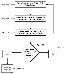
Now referring to FIG. 4, where an exemplary method of assessing lesion
quality is shown. The method includes exchanging cryogenic ablation energy
with a
region of tissue such that the region of tissue is ablated and/or cooled (Step
100). For
example, medical device 10 may be positioned proximate the region of tissue to
ablated. A cryogenic fluid may then be circulated towards the thermally
conductive
region 16 where it delivers cryogenic energy to the target tissue region. The
cryogenic energy may be exchanged with a region of tissue for a time period
of, for
example, 1-5 minutes. Alternatively, cryogenic ablation energy may be
exchanged
for a time period of, two minutes, followed by a two minute thaw period, where
no
cryogenic energy is exchaneed, followed by an additional minute of cryogenic
energy
being exchanged. In an exemplary embodiment, the temperature of the cryogenic
:A 02814985 2013-04-17
WO 2012/057911
PCT/US2011/049632
7
ablation element and/or contacted tissue is reduced to approximately -55 C to -
60 C.
The cryogenic energy may be exchanged with the region of tissue by any of the
medical device embodiments discussed above and with any tissue region, for
example, the atrial valve or vasculature.
Following the exchange of cryogenic energy, the reactance or resistance of the
treated tissue region may be measured by the device 10 and/or the console 24
(Step
102). For example, as a current is induced between electrodes (26, 28) and/or
electrodes (32, 34) or electrodes 40, the opposition of the ablated tissue
region to a
change in current, known as reactance, is measured. As the tissue is ablated,
the
reactance decreases as the opposition to the current decreases. Optionally,
the
reactance of the tissue adjacent the treated tissue region may also be
measured to
prevent unwanted tissue from being ablated. The reactance may be measured at
one
or more excitation frequencies, for example, 10kHz, 470kHz, and 1MHz. By
measuring the reactances at one or more excitation frequencies, the magnitude
of the
percentage of reduction in reactance for each time period at each frequency
may be
measured. For example, at higher frequencies, for example, 1MHz, the
destruction of
the cellular membrane may be detected in the form of a change in reactance and
compared to a change in reactance at lower frequencies. The change in the
reactance
of the cryogenically treated tissue region may then be correlated to determine
and
assess the transmurality of the tissue region (Step 104). As used herein, the
term
"transmurality" means the depth or distance a lesion or ablated tissue passes
through
the wall of the tissue region. For example, tissue treated with cryogenic
energy for
five minutes exhibits a larger decrease in reactance, which can be correlated
to the
destruction of cellular membranes and to tissue transmurality.
Further, at particular frequencies the correlation between reactance and
transmurality may be stronger than that of measurement of impedance, thus
allowing
for an accurate and real-time assessment of the quality of the cryogenic
lesion.
Similarly, the time rate of change in reactance or resistance measured at
particular
frequencies may be correlated to the depth of a lesion because the time rate
of change
of resistance during the treatment procedure, for example, may correspond to
how
quickly the tissue freezes. The measured ablated tissue region transmurality
may then
be compared to a predetermined ablated tissue region transmurality or
reactance
:A 02814985 2013-04-17
WO 2012/057911
PCT/1JS2011/049632
8
threshold. (Step 106). If the desired transmurality is achieved, (e.g., the
treatment
transmurality threshold is reached), treatment may be modified or stopped, for
example, by terminating the delivery of coolant to the thermally conductive
region 16.
(Step 108) If the desired transmurality has not been achieved, cryogenic
ablation
energy may be delivered for an additional time period (Step 110). These
methods may
be performed by the one or more programmed processors, controllers, or other
components of the console 24 in an automated process, resulting in the
generation of
an indication of the analysis or comparison to the user or physician.
Now referring to FIG. 5, where another method of assessing the quality of a
lesion includes exchanging cryogenic ablation energy with a tissue region. The
method includes pretreating the tissue region with cryogenic ablation energy
for any
time period, such as, for example, approximately five minutes by the method
discussed above. (Step 200). Optionally, either a fluoroscopic or a non-
fluoroscopic
navigation system may be used to track one or more of electrodes (for example,
electrodes 40, electrodes 26 and 28 and/or electrodes 32 and 34) and a
reference
electrode (not shown) disposed on the medical device 10 or the second medical
device
36, such that the location of the medical device 10 or the second medical
device 36
may be graphically displayed during the pretreatment of the tissue region.
The reactance of the ablated tissue region may then be measured at a plurality
of frequencies, simultaneously with or sequentially after the pretreatment
with the
medical device 10 and/or the console 24 (Step 202). The time rate of change of
the
measured reactance may also be measured during the pretreatment to determine
when
the tissue region is covered with ice. The measured reactance may be compared
to a
predetermined ablated, treated, or cooled tissue region reactance or
transmurality
threshold, which may be selected prior to the treatment. (Step 204). For
example, the
medical device 10 may include a particular reactance or transmurality
threshold, for
example, a 60-90% decrease in reactance of treated tissue as compared to the
reactance of untreated tissue may be indicative of a quality lesion, which may
be
device specific and correlated to a particular transmurality. In particular, a
baseline
reactance of untreated tissue may be defined before or measured during the
thermal
treatment of the tissue region. The baseline reactance measurement may then be
:A 02814985 2013-04-17
WO 2012/057911
PCT/US2011/049632
9
compared to the measured reactance to determine the percent decrease in the
reactance of the treated tissue.
The measured reactances over at the plurality of frequencies may then be
correlated to determine and assess the lesion depth, transmurality, or
continuity. (Step
206). For example, lesion quality may be assessed by calculating a tissue
transmurality based on the compared measured reactance. Alternatively, the
reactance measurements recorded at each of the plurality of applied RF
frequencies
may be compared and correlated to tissue transmurality. If the desired
transmurality
is achieved (Step 208), for example, the treatment transmurality threshold is
reached
and treatment may be modified or stopped. (Step 210). If the desired
transmurality is
not achieved, the tissue region may be treated with additional cryogenic
energy and
the method may recycle. (Step 212). Alternatively, RF ablation energy may be
delivered to the cryogenically pretreated tissue region immediately following
the
delivery of cryogenic energy while the reactance is measured. For example, the
reactance may be remeasured and correlated to tissue transmurality after the
RF
ablation energy is transmitted to the tissue region.
Additionally, the correlated transmurality may be used to determine if a
contiguous lesion was successfully created. For example, gaps in a lesion may
be
detected by measuring the reactance at one or more frequencies. In particular,
the
measurement of reactance at higher frequencies may be more sensitive to slight
changes in reactance to aid in identifying lesion gaps. If there is no change
in the
measured reactance, a lesion may not have been created at a desired location.
In
particular, a plurality of reactance measurements may be made at a variety of
different
locations at a particular treatment region. As such, the measured reactance at
each of
many locations can be correlated to determine a lesion's shape, quality, and
transmurality.
Additionally, the measured reactance and/or correlated tissue quality or
transmurality data may be displayed numerically and/or graphically on a
display or
the console 24 during the procedure. For example, the determined transmurality
or
continuity data may be graphically displayed and treatment may be modified
based on
the displayed transmurality. As such, gaps may be detected in a lesion and
displayed
for the physician. Optionally, the tissue quality, reactance, and/or
transmurality data
:A 02814985 2013-04-17
WO 2012/057911
PCT/IJS2011/049632
may be recorded and stored remotely in a database. For example, previously
recorded
data may be compared to current data to assess treatment efficacy and monitor
patient
progress. As such, it is contemplated that treatment models may be created
based on
historical and present reactance, quality, and tissue transmurality data.
5 The delivery of RF energy for measuring reactance may include unipolar
and/or bipolar RI: modalities. For example, a current may be induced and a
voltage
applied between two adjacent electrodes 40 on the second medical device 36
such that
RF energy is transmitted between them. Alternatively, when power is delivered
to
medical device 10 or the second medical device 36, RF energy may be
transmitted
10 from electrodes (26, 28) and/or electrodes (32, 34) or electrodes 40 to
the back plate.
Referring now to FIG. 6a and FIG. 6b, exemplary results of performing the
method described with respect to FIG. 5 is shown as applied to bovine
ventricular
tissue for five separately created lesions. In particular, the graph in FIG.
6a shows a
comparison of the lesion depth percentage for tissue measured with bipolar RF
energy
alone ("control") versus tissue pretreated with cryogenic energy for five
minutes at -
60 C then measured with bipolar RF energy ("cryo"). The lesion depth
percentage as
used herein is the depth of the lesion divided by the tissue thickness
multiplied by one
hundred, e.2. a 60% lesion has a depth at 60% of the overall tissue depth;
100% is a
completely transmural lesion. The table in FIG. 6b compares and tabulates,
among
other things, the measured reactance of the "control" to the "cryo" treated
tissues and
the associated two-sided P-values, as well as the percent lesion depth
comparison.
As shown in FIGS 6a and 6h, while there is about a two-fold increase in the
percent lesion depth shown in the "cryo" tissue compared to the "control"
tissue, the
magnitude of the measured impedance of the "control" and "cryo" tissues are
virtually
the same (20.5D. for "control" compared to 18.4E2 for "cryo"), with a P-value
of
0.09. Because the P-value is greater than 0.05, the hypothesis that these two
value are
statistically different cannot be accepted. This is so because, as discussed
above, once
the tissue is necrotic the impedance does not change. Therefore, comparing
changes
in impedance alone provides no information as to the percent change in the
legion
depth, because no statistically significant change in impedance is observed.
Similarly,
the measured resistances of the "control" and "cryo" tissues were virtually
the same
:A 02814985 2013-04-17
WO 2012/057911
PCT/US2011/049632
11
(18.8f2 for "control" compared to 18.01 for "cryo"), with P-value of 0.4. As
such,
changes in resistance do not show a significant correlation to changes in
lesion depth.
Significantly, however, the measured reactance of the "cryo" tissue showed
about a two-fold decrease when compared to the measured reactance of the
"control"
tissue (-8. in for "control" compared to -3.9 for "cryo"), with a P-value less
than
0.001. Specifically, the results indicate that when the percent lesion depth
is about
100% greater in the "cryo" tissue compared to the "control" tissue, the
measured
reactance of the "cryo" tissue is about 100% less compared to the "control"
tissue.
Thus, the change in reactance when compared to the change in the lesion depth
of the
"control" and "cryo" tissues are substantially inversely proportional, such
that the
change in reactance may be correlated to lesion depth.
Referring now to FIGS. 7a and 7b, exemplary results of performing the
method described with respect to FIG. 5 are shown as applied to bovine
ventricular
tissue for five distinct lesions. In this case, the "cryo" tissue is measured
with
unipolar RF energy instead of bipolar RF energy as shown in FIGS. 6a and 6b.
Similar to the results shown in FIG. 6a and 6b, the change in reactance
between the
"control" and "cryo" tissues is substantially inversely proportional to the
change in
the lesion depth. Also, the measured reactance of both "cryo" and "control"
lesions
were significantly lower than the measured reactance of both "cryo" and
"control"
lesions created with bipolar RF energy. As such, it is further contemplated
that the
measuring and correlating of tissue reactance may be used to distinguish
unipolar RF
ablated regions from bipolar RF ablated regions.
Any of the above methods may be performed not only to distinguish currently
treated tissue, but also to identify pretreated tissue or tissue treated or
ablated by other
modalities. For example, the measured changes in reactance may be used to
identify
and assess the quality, transmurality, and continuity of lesions created by RF
ablation,
ultrasound ablation, light ablation, for example, infrared, laser, or visible
light
energies, chemical ablation, radiation, microwave ablation, electromagnetic
radiation,
irreversible electroporation, among other ablation modalities. As such, the
measured
reactance not only provides information as to lesion depth, but also as to the
identity
of a lesion previously created or to identify gaps in a created lesion. It is
further
contemplated that in addition to measuring reactance, other measurements that
detect
:A 02814985 2013-04-17
WO 2012/057911
PCT/US2011/049632
12
the change in the cell membrane thickness may be used to determine lesion
quality
and transmurality. For example, electroporation may be used to determine
lesion
quality and transmurality.
It will be appreciated by persons skilled in the art that the present
invention is
not limited to what has been particularly shown and described herein above. In
addition, unless mention was made above to the contrary, it should be noted
that all of
the accompanying drawings are not to scale. A variety of modifications and
variations are possible in light of the above teachings without departing from
the
scope and spirit of the invention, which is limited only by the following
claims.