Note: Descriptions are shown in the official language in which they were submitted.
CA 02816674 2013-05-01
DESCRIPTION
Title of the Invention: Blue-colored gold nanoparticles for immunological
measurement,
process for production of same, and measurement method using same
Technical Field
[0001]
The present present invention relates to blue-colored gold nanoparticles and a
colloidal solution of blue-colored gold nanoparticles, each having a highly
vivid color
developing property and at the same time, having stable durability and
excellent
distinguishability and useful as a labeling agent for immunological
measurement or a
protein stain. The present invention also relates to a method for producing
the blue-
colored gold nanoparticles of the present invention, and a test kit and a
measuring method
using the blue-colored gold nanoparticles. Moreover, the present invention
relates to a
labeling substance for immunological measurement in which the blue-colored
gold
nanoparticles of the present invention is used as a labeling substance in an
immunological
measurement system.
Background Art
[0002]
In recent years, immunochromatographic strip type immunoassay has become
more important as a simple in-vitro diagnostic kit or portable diagnostic
device for
detecting an antigen in a sample solution by making use of the specific
reactivity which an
antibody has. In particular, simple multiplex analysis tools based on
immunochromatography for analyzing the presence or absence of infection with
pathogens such as influenza virus or bacteria have been under research and
development.
[0003]
Colloidal metal particles or latex particles have generally been used as an
insoluble carrier to be used in an immunological measurement method. Latex
particles
need a cumbersome production step such as modification of a chemical
functional group
in order to firmly support a substance such as protein to be labeled.
Therefore, colloidal
gold particles capable of easily supporting a substance to be labeled and are
produced
conveniently at a low cost have been preferably used.
Although immunochromatographic test drugs which have labeled an antibody
with an insoluble carrier have been used generally since they are easy to
operate and need
only a short time for the test, a line which can be observed when the test
result is positive
is not clear since they have generally lower sensitivity in comparison with
EIA.
1
CA 02816674 2013-05-01
In order to overcome such a problem, various metal colloids having a higher
sensitivity than conventional colloidal metal particles already put into
practical use and are
suitable as labeling agents for immunological measurement or protein staining
agents are
developed.
[0004]
Patent Document 1 provides colloidal metal particles having an average
particle
size of from 50 to 150 nm obtained by supporting platinum on the surface of
colloidal
metal particles (average particle size: from 30 to 100 nm) since colloidal
platinum
particles do not develop color sufficiently due to small average particle size
and are not
suited for practical use in immunochromatography. The colloidal metal
particles are
prepared by reducing chloroauric acid in a solvent to form colloidal gold
particles and
then reducing chloroplatinic acid in the presence of the resulting colloidal
gold particles
(refer to Patent Document 1).
[0005]
Patent Document 2 provides colloidal metal particles obtained by improving the
above-mentioned colloidal metal particles and therefore having higher
sensitivity.
Namely, colloidal metal particles (average particle size: 30 to 100 nm) having
platinum
supported thereon which has an average particle size of 5 nm are provided.
They are
prepared in a production method wherein by adjusting, to a predetermined
range, the
amount of a reducing agent added in preparing colloidal metal particles in a
medium and
the amount of a reducing agent added in reducing and supporting platinum on
the colloidal
metal particles and wherein the medium does not substantially contain a
protective colloid
forming agent. Examples of such protective colloid forming agent include water
soluble
high-molecular substances such as PVA, PVP, and gelatin, surfactants, and high-
molecular
chelating agents (refer to Patent Document 2).
[0006]
As another method for improving the sensitivity in immunological and
immunocytological diagnostic test, a method of coating gold sol ultrafine
particles with an
alkanethiol (derivative) to impart the gold sol surface with certain
hydrophobic-
hydrophilic balance so as to prevent aggregation which is caused by a salt by
which non-
specific interaction between the gold sol surface and an exogenous protein
(refer to Patent
Document 3) is minimized is provided.
[0007]
On the other hand, in vitro diagnostics for pregnancy diagnosis, red-colored
spherical colloidal gold particles already put on the market have been
improved to exhibit
higher sensitivity. Colloidal gold is required to have a particle size suited
for an intended
2
CA 02816674 2013-05-01
use; have a sharp particle size distribution; and have a uniformly spherical
shape so that a
production process of it is under development.
Patent Document 4 includes a nucleus formation stage of adding a first
reducing
agent (citrate) to a solution of a first gold salt to form colloidal nucleus
particles (average
particle size: from 12 to 17 nm) and a growth stage of simultaneously adding,
to the
solution of the colloidal nucleus particles, a second gold salt and a second
reducing agent
(ascorbate) to grow a colloidal nucleus. This growth stage is conducted at
least once.
The average particle size of the colloidal gold particles is 17 nm or greater
and less than
55 nm in the first growth stage; 55 nm or greater and less than 110 nm in the
second
growth stage; and from 110 to 220 nm in the third growth stage. The standard
deviation
of the particle diameter is within 10% (refer to Patent document 4).
[0008]
In the case of testing only one item such as a pregnancy test kit for finding
whether pregnancy or not, it is only necessary to use one labeling agent in
visual judgment.
Recently, a multiplex test should be conducted when it is necessary to
identify a causative
virus as in a virus test in cold-like infections or respiratory infections.
Thus, various test
systems have been developed with a view to easing the burden of patients and
health care
workers.
[0009]
For example, although there is a known lateral flow type immunoassay capable
of
detecting a plurality of viruses (rotavirus, calcivirus, coronavirus,
adenovirus, enterovirus,
and the like) by using one test tool, the assay has the problem that a
plurality of detection
lines tend to lead to erroneous visual judgment.
[0010]
In the test of a virus in respiratory infections by using
immunochromatography, a
testing method including pretreating a specimen such as nasal discharge,
sputum or a swab
from the nasal cavity with a specimen treatment solution to prepare a test
sample suited
for the test of a plurality of respiratory infections and analyzing respective
portions of the
resulting test sample by using a plurality of test tools such as a first test
tool (for example,
testing an influenza virus infection) and a second test tool (for example,
testing an
adenovirus infection or an RS virus infection) (refer to Patent Document 5)
has been
developed.
[0011]
Further, a measurement method including immunochromatography having a
high ability of judging with labeled antibody particles having an arbitrary
color and
capable of simultaneously measuring two or more measurement objects by using
two or
more labeled antibody particles has been developed. More specifically, hCG and
LH are
3
CA 02816674 2013-05-01
measured simultaneously by using a luminescent dye such as TRITC (absorption
maximum: about 550 nm, red) and FITC (absorption maximum: about 500 nm,
orange)
(refer to Patent Document 6).
[0012]
When multiplex tests are conducted simultaneously through visual judgment by
using one test tool and labeling agents or protein staining agents used are of
the same
color or similarcolor, there is a possibility of causing misjudgment or wrong
diagnosis.
In order to prevent misjudgment or wrong diagnosis by visual judgment, it is
desired to
conduct visual judgment by using labeling agents or protein staining agents of
highly
distinguishable colors.
[0013]
When two colors are present, their distinguishability differs with the colors
used
in combination. Since a red color and a blue color can be highly distinguished
from each
other by visually view, they are used for various distinguishing purposes as
can be seen in
indications for distinguishing between male and female or indications for
distinguishing
between hot water (red) and water (blue). Colloidal gold particles which have
been
conventionally put into practical use are red-colored spherical particles. If
blue-colored
colloidal gold particles different in color, in other words, highly
distinguishable from red
color are used as a labeling agent or protein staining agent, misjudgment or
wrong
diagnosis through visual judgment is presumed to decrease markedly. However,
blue-
colored colloidal gold particles have not yet been put into practical use.
[0014]
In patent Documents 7 to 9, metal nanoparticles having light absorption
wavelength properties varied by changing the size, pattern, structure/shape or
the like of
metal nanoparticles are described.
According to Patent Documents 7 and 8, blue-colored gold nanoparticles have a
structure/shape of gold nanoshells, nanorods, nanotubes, or nanoprism
particles; the gold
nanoparticles are produced by (1) adding a reducing agent to a yellow-colored
silver
nanoparticle solution (containing a protecting agent such as
polyvinylpyrrolidone or
ethylene glycol) and then, refluxing the resulting mixture at about 100 C, (2)
pouring a
gold salt solution in the reaction mixture thus refluxed to react them, and
(3) after cooling
to normal temperature, the reaction mixture is filtered through a 0.2 m
microfilter; and
the gold nanoparticles thus obtained are made of gold (gold nanoshell) only at
the surface
layer thereof According to these documents, gold nanorods, gold nanotubes, or
gold
nanoprisms are obtained by using a surfactant such as
hexadecyltrimethylammonium
bromide (bromide) (C6TAB) in formation of gold nanoparticles. These documents
do not
include a definite description on the size of the particles. They include a
description on
4
CA 02816674 2013-05-01
the use of them as pigment for cosmetics but do not include a description on
the use of
them as a labeling agent or protein staining agent in immunoassay (refer to
Patent
Documents 7 and 8).
[0015]
Patent Document 9 describes rod-like gold nanoparticles obtained by reducing a
gold ion with a reducing agent (an amine) in an aqueous solution containing
C16TAB (a
surfactant of an ammonium salt). The aspect ratio (long axis/short axis) of
the gold
nanoparticles can be controlled by regulating a mixing ratio of the amine and
the
ammonium salt used in combination. By doing so, gold nanorods having an aspect
ratio
of from 2 to 11 and an absorption wavelength peak area of from 658 to 1200 nm
are
obtained. According to the description, these gold nanorods can be used as a
test drug
(refer to Patent Document 9).
Since the gold nanoparticles thus obtained contain C16TAB as a surfactant,
they
are not suited for directly supporting (modifying) with protein such as
detection antibody.
Since it needs a cumbersome operation such as removal or substitution of the
surfactant, it
is not preferred as a labeling substance for a protein to be used as a test
drug in the
immunological measurement method. In addition, they are not preferred from the
standpoint of handling because C6TAB has toxicity.
[0016]
Non-patent Document 1 describes a colloid of stick-shaped gold nanocrystals
exhibiting a bluish green color. The stick-shaped gold naocrystals have a
complex three-
dimensional structure; have from one to eight protrusions; and have a crystal
size,
including the protrusion, of from 30 to 50 nm (protrusion length of from about
15 to 25
nm and a width of about 8 nm). The three-dimensional branch-shaped gold
nanocrystals
are obtained in a high yield (92%) by reacting an aqueous solution of
chloroauric acid and
an organic acid (HEPES, HEPPSO, PIPES, or the like) which is a Good's buffer
component at room temperature (refer to Non-patent Document 1).
[0017]
However, the colloid of branch-shaped nanocrystals obtained in Non-patent
Document 1 and exhibiting a bluish green color has a crystal size of from 30
to 50 nm,
which is not a desired size. Therefore, even if it is used as an
immunochromatographic
diagnostic agent, insufficient color development prevents smooth visual
judgment.
As can be seen in the related art documents, a colloid of gold nanocrystals
exhibiting a bluish green color is not suited as a labeling carrier of an
immunochromatographic diagnostic agent since the colloidal particle size is as
relatively
small as from about 30 to 50 nm. In addition, so-called multipod-shaped,
branch-shaped,
CA 02816674 2013-05-01
or confeito-shaped ones often use a shape stabilizer and the shape stabilizer
makes it
difficult to achieve direct modification of gold nanoparticles with a protein.
Prior Art Documents
Patent Documents
[0018]
Patent Document 1: JP-A-2003-262638
Patent Document 2: JP-A-2005-233744
Patent Document 3: JP-A-6-116602
Patent Document 4: JP-A-2007-321232
Patent Document 5: JP-A-2008-164403
Patent Document 6: JP-A-10-132817
Patent Document 7: JP-A-2008-545884
Patent Document 8: JP-A-2009-501786
Patent Document 9: JP-A-2006-118036
Non-patent Documents
[0019]
Non-Patent Document 1: Chem. Mater. 2007, 19, 2823-2830
Non-Patent Document 2: Langmuir 2005, 21, 2012-2016
Non-Patent Document 3: J. Phys. Chem. B 2006, 110, 19291-19294
Non-Patent Document 4: Nano Lett. 2006, 6, 683-688
Summary of the Invention
Problems to be solved by the Invention
[0020]
An object of the present invention is to provide blue-colored gold
nanoparticles, a
colloidal solution of blue-colored gold nanoparticles obtained by dispersing
the gold
nanoparticles in a medium, and blue-colored gold nanoparticles exhibiting a
highly vivid
blue color with visually view, excellent in quality stability, storage
stability and
distinguishability, useful as a labeling agent for immunological measurement
or protein
staining agent, and easily distinguishable by a difference in color from a
conventional red
color; and to overcome the problem relating to the production method of the
blue-colored
gold nanoparticles, a test kit having enhanced measurement accuracy by using
the blue-
colored gold nanoparticles, and the measurement method using the test kit.
Blue-colored gold nanoparticles in Non-patent Documents 1 to 3 used above as
reference are not suited as a carrier for immunochromatographic diagnostic
agent because
of the following two problems:
6
CA 02816674 2013-05-01
1. These blue-colored gold nanoparticles have a size which is not suited
for
immunological measurement. The particle size suited for immunochromatography
reagent is from about 40 to 100 nm in terms of average particle size.
According to Non-
patent Document 1, the particle size is about 30 nm.
2. They contain a shape stabilizer. The three-dimensional stick-shaped gold
nanoparticles described in Non-patent Document 2 to 4 contain a shape
stabilizer in order
to control their shape. The shape stabilizer prevents direct modification of
gold
nanoparticles with a protein.
[0021]
When a multiplex test is carried out and conventional red-colored gold
nanoparticles and colored latex particles are used for the simultaneous
multiplex
measurement, it is difficult to select, in immunochromatography, an
immunochromatographic carrier having a pore size suited for both particles
since the gold
nanoparticles and latex particles are different in the particle size (the
latex particles
generally employed have a greater size than the gold nanoparticles).
Therefore, it is
required to use, as a labeling substance, two kinds of colloidal gold
particles which are
different in color; capable of easily supporting a substance to be labeled
such as protein;
and inexpensive.
[0022]
In order to overcome the above-mentioned problems, the inventors of the
present
invention have succeeded in providing blue-colored gold nanoparticles suited
for a carrier
o f an immunochromatographic diagnostic agent, more specifically, blue-colored
gold
nanoparticles usable for a multiplex detection reagent in multiplex detection
by
immunochromatography by increasing the size of the particles in order to make
it suited
for immunological measurement and by selecting a shape stabilizer permitting
direct
modification of gold nanoparticles with a protein.
Means for Solving the Problems
[0023]
The present invention provides blue-colored gold nanoparticles suited for
immunological measurement, permitting easy modification of the gold
nanoparticles with
a protein and at the same time, most suited as a multiplex detection reagent.
Described specifically, the blue-colored gold nanoparticles of the present
invention are composed of organic acid containing a piperazine ring (such as
HEPES)
which is a Good's buffer component, Au (gold) and an organic acid having
reducing
properties (such as ascorbic acid and citric acid); exhibits a blue color when
viewed
visually; and has a confeito-like shape.
7
CA 02816674 2013-05-01
The blue-colored gold nanoparticles of the present invention have an average
particle size of from 20 to 200 nm, preferably from 40 to 180 nm from the
standpoint of
color vividness, stable durability, and stable durability of a colloid,
typically most
preferably from 50 to 120 nm from the various practical standpoints including
marked
distinguishability in a test. The most appropriate range is from 60 to 100 nm.
The blue-
colored gold nanoparticles have a feature of a blue color with visual view in
liquid
wherein the blue-colored gold nanoparticles are dispersed as colloid.
The term "average particle size" as used in the present invention means a
value
determined by including a nucleus protruding portion of blue-colored cold
nanoparticles
which will be described later. In the blue-colored gold nanoparticles of the
present
invention, the nucleus protruding portion has a length of preferably from 5 to
50 nm.
The number of protrusions is four or more per nucleus.
[0024]
In an aqueous colloidal solution containing the blue-colored gold
nanoparticles
according to the present invention, the colloidal gold particles have an
average particle
size of from 20 to 200 nm, preferably from 40 to 180 nm, usually most
preferably from 50
to 120 nm, most appropriately from 60 to 100 nm. Its average particle nucleus
size is
from 20 to 60 nm. The aqueous colloidal solution containing the blue-colored
gold
nanoparticles according to the present invention is characterized by that it
has a maximum
absorption wavelength in a range of from 570 to 750 nm in an ultraviolet
visible
absorption spectrum. By using the gold nanoparticles contained in the aqueous
colloidal
gold solution of the present invention as a labeling substance in
immunochromatography,
detection with a blue color highly distinguishable from a red color is
possible. This
makes it possible to conduct immunochromatography measurement with reducing
wrong
diagnosis cases in the simultaneous multiplex detection. It is to be noted
that the term
colloidal solution of the blue-colored gold nanoparticles according to the
present invention
means a dispersion of fine particles with a nanosize (nm), particularly gold
nanoparticles,
in a solvent such as water. In short, the present invention has succeeded in
providing
blue-colored nanoparticles, a colloidal solution of blue-colored gold
nanoparticles, a
production method thereof, and confeito-shaped blue-colored gold nanoparticles
suited for
immunological measurement; permitting easy modification of blue-colored gold
nanoparticles with a protein; and at the same time, most suited as a multiplex
detection
reagent.
[0025]
The present invention provides blue-colored gold nanoparticles and production
method and using method of them. The gold nanoparticles of the present
invention have
characteristics as follows:
8
CA 02816674 2013-05-01
(a) The first feature of the present invention is blue-colored gold
nanoparticles comprising
gold nanoparticles having an average particle size of from 20 to 200 nm;
(b) The second feature of the present invention is the blue-colored gold
nanoparticles
according to (a), wherein the maximum absorption wavelength in ultraviolet
visible
absorption spectra falls within a range of from 570 to 800 nm;
(c) The third feature of the present invention is the blue-colored gold
nanoparticles
according to (a) or (b), wherein the gold nanoparticles are graft-shaped
particles,
multipod-shaped particles, or confeito-shaped particles having a three-
dimensional
protrusion;
(d) The fourth feature of the present invention is the blue-colored gold
nanoparticles
according to any one of (a) to (c), obtained by growing the periphery of the
nucleus
composed of gold nanoparticles; and
(e) The fifth feature of the present invention is the blue-colored gold
nanoparticles
according to any one of (a) to (d), having an average particle nucleus size of
from 20 to 60
nm, an average particle size of from 50 to 120 nm, four or more protrusions
per nucleus,
and a protrusion length of from 5 to 50 nm.
[0026]
The colloid wherein the gold nanoparticles of the present invention are
dispersed
in a medium such as water has characteristics as follows:
(I) The sixth feature of the present invention is a colloidal solution of blue-
colored gold
nanoparticles, comprising the blue-colored gold nanoparticles as described in
(a); organic
acid containing a piperazine ring which is a Good's buffer component; and an
organic acid
having reducing properties and is dispersed as a colloidal solution.
[0027]
The production methods for specifically gold nanoparticles of the present
invention has characteristics as follows:
(g) The seventh feature of the present invention is a method for producing
blue-colored
gold nanoparticles, comprising a nucleus formation step by reacting organic
acid
containing a piperazine ring which is a Good's buffer component with a
solution of a first
gold salt to form nucleus gold nanoparticles and a growth step by
simultaneously adding
and reacting a solution of a second gold salt and an organic acid having
reducing
properties with a solution of the nucleus gold nanoparticle to grow the
nucleus gold
nanoparticles;
(h) The eighth feature of the present invention is the method for producing
blue-colored
gold nanoparticles according to (g), wherein the growth step is conducted at a
reaction
temperature of 10 C or greater and less than 40 C;
9
CA 02816674 2013-05-01
(i) The ninth feature of the present invention is the method for producing
blue-colored
gold nanoparticles according to (g) or (h), wherein the organic acid in the
growth step has
a concentration of from 0.075 to 0.15 mM;
(j) The tenth feature of the present invention is the method for producing
blue-colored
gold nanoparticles according to (i), wherein the organic acid containing
piperazine ring
which is a Good's buffer component is one or more organic acids selected from
the group
consisting of 244-(2-hydroxyethyl)-1-piperazinyllethanesulfonic acid, 4-(2-
hydroxyethyl)-1-piperazinepropanesulfonic acid, 4-(2-hydroxyethyl)piperazine-1-
(2-
hydroxypropane-3-sulfonie acid), piperazine-1,4-bis(2-ethanesulfonic acid),
34442-
hydroxyethyl)-1-piperazinyl]propanesulfonic acid, and piperazine-1,4-bis(2-
hydroxy-3-
propanesulfonic acid);
(k) The eleventh feature of the present invention is the method for producing
blue-colored
gold nanoparticles according to (g), wherein the organic acid having reducing
properties is
one or more organic acids selected from the group consisting of tartaric acid,
tartrates,
tannic acid, tannates, ascorbic acid, ascorbates, citric acid, and citrates;
and
[0028]
(I) The twelveth feature of the present invention is the method for producing
blue-colored
gold nanoparticles according to (g), wherein in the growth step, the organic
acid
containing a piperazine ring which is a Good's buffer component is used in
combination
with the organic acid having reducing properties.
Next, the present invention, specifically, as the labeling substance for
immunological measurement has characteristics as follows:
(m) The thirteenth feature of the present invention is a labeling substance
for
immunological measurement, comprising the blue-colored gold nanoparticles as
deacribed
in any one of (a) to (e);
(n) The fourteenth feature of the present invention is the labeling substance
for
immunological measurement according to (m), comprising at least two kinds of
gold
nanoparticles different in shape;
(o) The fifteenth feature of the present invention is the labeling substance
for
immunological measurement according to (n), which comprises at least two kinds
of gold
nanoparticles of different shapes which are spherical gold nanoparticles and
graft-shaped,
multipod-shaped, or confeito-shaped gold nanoparticles having a three-
dimensional
protrusion; and
(p) The sisteenth feature of the present invention is an immunological
measurement
method using the blue-colored gold nanoparticles as described in any one of
(a) to (e) as a
labeling substance.
CA 02816674 2013-05-01
The problems of the present invention can be overcome by employing the above-
mentioned constitutions of the present invention.
Effect of the Invention
[0029]
Since the blue-colored gold nanoparticles of the present invention have an
average particle size of from 20 to 200 nm; preferably from 40 to 180 tun;
usually most
preferably from 50 to 120 nm; and most appropriately from 60 to 100 nm, the
blue-
colored gold nanoparticles can provide a particle size which is most suited
for an
immunochromatographic diagnostic agent.
Using in combination with spherical red-colored gold nanoparticles or the like
enables preparation of an immunochromatographic diagnostic agent having two or
more
judgment lines. This prevents wrong diagnosis or misjudgment since visual
judgment
can be made easily and precisely in a multiplex test.
Moreover, the blue-colored gold nanoparticles of the present invention can be
easily modified with a protein so that they enable precise judgment of the
results without
causing deterioration in sensitivity. They are therefore excellent in the
performance as an
immunochromatographic diagnostic agent.
Furthermore, an immunochromatographic diagnostic agent prepared from the
blue-colored gold nanoparticles of the present invention is more inexpensive
than those
prepared from particles obtained by another method.
Brief Description of the Drawings
[0030] [FIG. 1A] FIG lA is a transmission electron microscope image showing
the
shape and rough size of one example of the blue-colored gold nanoparticles of
the present
invention.
[FIG 1B] FIG 1B is a transmission electron microscope image showing the
shape and rough size of another example of the blue-colored gold nanoparticles
of the
present invention.
[FIG 2A] FIG 2A is a transmission electron microscope image of one example
of the blue-colored gold nanoparticles of the present invention before growth.
[FIG 2B] FIG. 2B is a transmission electron microscope image of one example
of the blue-colored gold nanoparticles of the present invention after growth.
[FIG. 3A] FIG 3A is a transmission electron microscope image showing another
example of the blue-colored gold nanoparticles of the present invention before
growth at
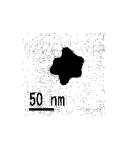
20-fold magnification (the length of the scale bar in the drawing is 50 mm).
11
CA 02816674 2013-05-01
[FIG. 3B] FIG. 3B is a transmission electron microscope image showing the
blue-colored gold nanoparticles of FIG. 3A after growth at 50-fold
magnification (the
length of the scale bar in the drawing is 20 mm).
[FIG 4A] FIG 4A shows the relationship between the wavelength (nm) of
ultraviolet visible absorption spectrum and absorbance in the synthesis of the
blue-colored
gold nanoparticles of the present invention.
[FIG 4B] FIG. 4B shows the relationship between various reaction temperatures
( C) and maximum absorption wavelength in the synthesis of the blue-colored
gold
nanoparticles of the present invention in FIG 4A.
[FIG 5] FIG 5 shows the relationship between the maximum absorption
wavelength (nm) of ultraviolet visible absorption spectrum and the
concentration of
ascorbic acid in the synthesis of the blue-colored gold nanoparticles of the
present
invention.
[FIG 6] FIG. 6 shows the comparison in detection sensitivity when the blue-
colored gold nanoparticles of the present invention are used as an
immunochromatographic reagent.
Mode for Carrying Out the Invention
[0031]
As the blue-colored gold nanoparticles of the present invention, although it
is
ideal to produce, those having a large average particle size in one step, it
is rational to
form particles with a predetermined size first and then, conduct a growth step
to obtain
particles with a larger particle size. The blue-colored gold nanoparticles of
the present
invention are composed of gold nanoparticles having an average particle size
of from 20
to 200 nm. The average particle size of colloidal gold particles of a
colloidal solution
obtained by dispersing the gold nanoparticles of the present invention in a
medium is from
20 to 200 nm; preferably from 40 to 180 nm; usually most preferably from 50 to
120 nm;
and the most appropriate range is from 60 to 100 nm. From various standpoints
in
practical use such as marked distinguishability in a test, the gold
nanoparticles have
preferably a sharp particle size distribution and have a uniform confeito-like
shape. The
average particle size can be determined typically by gravimetric light
scattering
(determined from the precipitation rate of colloidal particles rotated, as in
a sol, at from
14000 to 5530000 xg and treated in an ultracentrifuge). In the present
invention, a
projected area diameter of 100 particles selected at random from a projection
photograph
taken by a transmission electron microscope (TEM, "JEM-2010", product of JEOL,
Ltd.)
is measured and then, based on the average value, an average particle diameter
(average
particle size) is determined.
12
CA 02816674 2013-05-01
[0032]
When the X-axis (for example, size of gold nanoparticles) and the Y axis (for
example, number fraction) are set to make a particle size distribution of the
gold
nanoparticles and a distribution curve of average particles is plotted along
them, the apex
of the distribution curve of the gold nanoparticles of the present invention
substantially
belongs to a particle size ranging from typically from 40 to 120 nm,
preferably from 50 to
110 nm, more preferably from 60 to 100 nm. This reveals that this distribution
curve is
relatively narrow, which means that many nanoparticles have a particle size
approximating
to each other and thus have a uniform particle size. It is expected that the
nanoparticles
exhibit stable and highly reliable behaviors and suppress generation of an
error span due
to foreign matters mixed therein.
Quantitatively, a total weight of the gold nanoparticles belonging to the
range of
from 20 to 200 nm is usually 40% or greater, preferably 60% or greater, more
preferably
80 wt% or greater. The remaining portion is composed of particles which have
remained
without growing, spherical ones, and unreacted residue.
[0033]
The gold nanoparticles of the present invention are so-called confeito-shaped
nanoparticles having a nucleus and a three-dimensional protrusion. Those
having any
average particle size within a range of from 20 to 200 nm can be obtained by
changing the
operation of the production method. In use as labeling particles, those having
an average
particle size falling within a range of from 50 to 120 nm, preferably within a
range of from
about 55 to 100 nm are excellent in enhancing the accuracy of visual judgment
based on a
particular color of the labeling particles.
These confeito-shaped particles have preferably a plurality of three-
dimensional
protrusions. The term average particle size as used herein means a value
determined
including the nucleus protrusion. The gold nanoparticles of the present
invention have
from about 1 to 20 protrusions, preferably from about 4 to 10 protrusions per
nucleus.
The length of each protrusion is typically from about 5 to 50 nm. It is very
difficult to
determine the number or length of these protrusions in advance, because they
depend on
the growth of the nuclei.
[0034]
The gold nanoparticles and colloidal gold nanoparticles having a three-
dimensional protrusion on the nucleus thereof are collectively called graft-
shaped,
multipod-shaped, or confeito-shaped gold nanoparticles and colloidal gold
nanoparticles,
respectively. As so-called gold nanoparticles and colloidal gold
nanoparticles, there may
be various structures having a three-dimensional protrusion and called by a
known name
such as nanocubes, nanorods, nanopods, star-shaped gold nanoparticles, or
graft-shaped
13
CA 02816674 2013-05-01
gold nanoparticles having, as shown in FIGS. lA and 1B, a nucleus from which a
stick-
shaped protrusion has grown three-dimensionally. The colloidal gold
nanoparticles
developing a vivid blue color have a shape or structure analogous to that of a
tetrapod
used for breakwater. Therefore, the term is employed, the colloidal gold
nanoparticles
having one branch grown as a graft are called "monopod" and they may have
various
shapes such as "dipod", "tripod", "tetrapod", and "pentapod" with an increase
in the
number of branches. In the present invention, the number of protrusions per
nucleus is
preferably relatively large so that such shapes are collectively called
"multipod".
The multipod-shaped colloidal gold nanoparticles or confeito-shaped colloidal
gold nanoparticles according to the present invention exhibit a color,
depending on their
spreading manner, in comparison with conventional spherical colloidal gold
particles
exhibiting a red color. This enables a colloidal gold nanoparticle solution to
exhibit
various colors including blue.
[0035]
Described specifically, as gold nanoparticles which are typical blue-colored
gold
nanoparticles of the present invention of graft-shaped, multipod-shaped, or
confeito-
shaped gold nanoparticles having a three-dimensional protrusion, gold
nanoparticles
having a shape as shown in FIG. lA or 1B are shown as one example. These gold
nanoparticles have, at the center portion thereof, a so-called nucleus and a
protrusion or
branch has grown as a graft on the nucleus. Since the growth starting point of
the graft is
in close contact with the nucleus, they look like multipod-shaped gold
nanoparticles or
confeito-shaped gold nanoparticles having a protrusion and a nucleus
integrated with each
other.
Examples of the gold nanoparticles in FIGS. 1A and 1B specifically show the
example of blue-colored gold nanoparticles having a size of about 50 nm. More
specifically, the gold nanoparticles shown in FIGS. lA and 1B have an average
particle
size (DLS) of 66.5 nm and a maximum absorption wavelength of about 610 nm. In
addition, according to the measurement through TEM observation, the gold
nanoparticles
have an average outer diameter of 62.2 nm, an average nucleus diameter of 35.7
nm, an
average protrusion length of 13.2 nm, and a protrusion angle of about 50
degrees. They
have an AR (aspect ratio) of 1 or greater. It is needless to say that the
average outer
diameter, average nucleus diameter, average protrusion length, and protrusion
angle of the
gold nanoparticles of the present invention can be changed arbitrarily in
consideration of a
predetermined product different in color.
In the present invention including Examples, the wavelength was measured in
the
following manner. The wavelength was measured using an ultraviolet visible
absorption
spectrometer (name of the spectrometer: "UV-2550", product of Shimadzu
Corporation).
14
CA 02816674 2013-05-01
It was measured under the following conditions: a quartz cell: 10 mm,
wavelength: from
800 to 200 nm, and a band width: 0.5 nm.
[0036]
The blue-colored gold nanoparticles and blue-colored colloidal gold
nanoparticles
are effective for the development of a multiplex diagnostic reagent. When
there is a
plurality of judgment lines, they can remove the possibility of wrong
diagnosis upon
visual judgment. Gold nanoparticles to be used as an immunological measurement
labeling substance in such a multiplex diagnostic reagent are an immunological
measurement labeling substance characterized by that they are composed of at
least two
kinds of gold nanoparticles different in shapes. More specifically, an
immunological
measurement labeling substance composed of at least two kinds of spherical red-
colored
gold nanoparticles and graft-shaped, multipod-shaped, or confeito-shaped blue-
colored
gold nanoparticles having a three-dimensional protrusion is suitable.
[0037]
The gold nanoparticles of the present invention to be used as an immunological
measurement labeling substance in a multiplex diagnostic reagent include, for
example, a
mixture (which will hereinafter be called "mixture-type gold nanoparticle
labeling
substance") of two kinds or three kinds of gold nanoparticles which are
different in shape,
for example, a mixture of spherical gold nanoparticles and gold nanoparticles
having a
three-dimensional protrusion. In this case, when the grain size distribution
is analyzed
by shape, the mixture type may form a distribution curve with two apexes, that
is, a
particle size distribution curve formed by the spherical gold nanoparticles
and a particle
size distribution formed by the gold nanoparticles having a three-dimensional
protrusion.
It is needless to say that in the case of a mixture of gold nanoparticles
having three kinds
of shapes different from each other, a particle size distribution curve having
three apexes
can be drawn. In the present invention, if the particle size distribution of
at least two
kinds of metal nanoparticles is analyzed without paying attention to a
difference in shape,
the average particle size inevitably falls within a relatively wide range of
from 20 to 220
nm since gold nanoparticles having a relatively small average particle size
and gold
nanoparticles having a relatively large average particle size are present as a
mixture.
Anyway, when each particle size distribution curve forms a sharp peak,
measurement
accuracy can be enhanced, since it means that the amount of predetermined gold
nanoparticles is larger. A detailed example of this mixture-type gold
nanoparticle
labeling substance will be described below.
[0038]
The state of the "mixture-type gold nanoparticle labeling substance" of the
present invention is described in detail. When the mixture-type gold
nanoparticles of the
CA 02816674 2013-05-01
present invention is recognized as two kinds and for example, one are
spherical gold
nanoparticles and the other are gold nanoparticles having a three-dimensional
protrusion, a
mixture is presumed to contain these two kinds of particles at a mass %
ranging from
10:90 to 90:10 with taking into consideration of a detection sensitivity of
label. It means
that when the amount of the spherical gold nanoparticles is 40 mass%, the
amount of the
gold nanoparticles having a three-dimensional protrusion is 60 mass%. It is
needless to
say that the calculation is made with eliminating substances, other than
predetermined
ones, such as unreacted substances, nanoparticles which have remained without
growing,
and impurities.
For example, the spherical gold nanoparticles constituting this mixture-type
gold
nanoparticle labeling substance are relatively large particles having an
average particle
size of from 20 to 220 nm, preferably from 30 to 200 nm, and more preferably
from about
40 to 150 nm. With regard to the gold nanoparticles having a three-dimensional
protrusion, those having an average particle size of from about 20 to 200 nm
may be
present in the mixture. In order to enhance color vividness, color stability
for long hours,
stability of colloid, labeling accuracy, and reliability, the average particle
size is preferably
from 40 to 180 nm; usually most preferably from 50 to 120 nm; and the most
appropriate
range is from 60 to 100 nm.
The mixture-type gold nanoparticle labeling substance can be obtained, for
example, by a simple method of mixing spherical gold nanoparticle labeling
substance
which has been prepared in advance and has a predetermined average particle
size with a
gold nanoparticle labeling substance having a three-dimensional protrusion at
a
predetermined ratio.
[0039]
The immunological measurement labeling substance which is the mixture-type
gold nanoparticle labeling substance of the present invention and composed of
at least two
kinds of gold nanoparticles which are different in shape contains at least two
kinds gold
nanoparticles to be used as a labeling substance constituting a labeling
reagent which
modifies a detector substance having a binding ability with a target substance
in an
immunological measurement system and labels through binding with the target
substance,
wherein
1) the two kinds of gold nanoparticles each have an average particle size of
from
20 to 220 nm, and
2) one of the two kinds of gold nanoparticles is spherical and the other one
has at
least four three-dimensional protrusions.
When such a mixture-type gold nanoparticle labeling substance is used, various
antigens can be discriminated clearly by a difference in color such as red and
blue.
16
CA 02816674 2013-05-01
Therefore, it can ease the burden on the test and simplify the test operation
in the medical
front. As a result, it can markedly improve its usefulness.
[0040]
The colloidal gold particles of the present invention exhibit a blue color
when
they are viewed visually. It means that a colloidal gold solution obtained by
dispersing
colloidal gold particles in a solvent such as water exhibits a blue color or a
color
analogous to a blue color such as bluish green or bluish violet with visually
view. More
specifically, it means that the hue of the solution specified by the Munsell
color system is
from 3P to 1P, 10PB to 1PB, 10B to 1B, 10 BG to 1BG, or 10G to 8G Of these,
the hue
from 10PB to 1PB, from 10B to 1B, or from 10BG to 1BG is preferable in view of
distinguishability from a red color. With regard to the colorimetry, a quartz
cell (light
path length: about 10 mm) used for spectrophotometric measurement is filled
with the
colloidal solution; the color tone of it is confirmed visually on a white
background (white
drawing paper); and then, the color hue is evaluated based on a commercially
available
Munsell book of color.
[0041]
The method for producing gold nanoparticles according to the present invention
includes a nucleus formation stage wherein a first gold salt in an aqueous
solution is
reduced with a first reducing agent into confeito-shaped nucleus gold
nanoparticles and a
growth stage wherein a second gold salt and a second reducing agent are added
simultaneously dropwise to grow the nucleus gold nanoparticles into confeito-
shaped gold
nanoparticles having a greater size. The growth stage may be conducted at
least once.
In order to form confeito-shaped gold nanoparticles having a longer protrusion
in
the growth stage, a mixture of the second reducing agent and the first
reducing agent,
namely, organic acid containing a piperazine ring which is a Good's buffer
component is
used.
The amount of the first reducing gent used in combination with the second
reducing agent is almost equal to that of the second reducing agent, depending
on the
concentration of the second reducing agent to be used in the growth stage.
Namely, the
concentration of the first reducing agent for use is adjusted to be within a
range of from
0.01 to 100 mM in an aqueous solution for growing the nucleus gold
nanoparticles in the
growth stage.
[0042]
In order to analyze the behavior of the chemical species of the blue-colored
gold
nanoparticles, as one mode, particles corresponding to the nucleus particles
before the
growth reaction are called "Particle 1" and a solution of "Particle 1" is
prepared by mixing
0.43 mM AuC14 and 39.0 mM HEPES. Particles corresponding to particles which
have
17
CA 02816674 2013-05-01
grown as a result of the growing reaction are called "Particle 2" and a
solution of "Particle
2" is prepared by mixing 0.05 mM AuC14, 0.82 mM HEPES, and 0.10 mM ascorbic
acid.
The behavior of the resulting solutions is analyzed.
An example of increasing the particle size of the present invention without
changing a peak wavelength is described specifically based on FIGS. 2A and 2B.
In the
absorption spectrum of "Particle 1" of FIG. 2A, the inventors of the present
invention have
achieved in the present invention the growth of the particle size into
"Particle 2" of FIG
2B without changing the peak wavelength of the ultraviolet visible absorption
spectrum.
In FIGS. 2A and 2B, the peak wavelength means a range from about 570 to 630
nm.
[0043]
Examples of the first gold salt to be used in the nucleus formation stage of
the
present invention include salts such as chloroauric acid, gold tribromide,
gold trifiuoride,
gold triiodide, gold tricyanide, gold monochloride, gold monoiodide, gold
monofluoride,
gold monocyanide, hydroxy gold oxide, gold trisnitrate, and gold nitrate,
hydrates thereof,
and a solution of gold in aqua regia. Gold salts are not limited to the above-
mentioned
ones but any substance capable of forming the first gold salt in an aqueous
solution can be
used.
[0044]
As the first reducing agent to be used in the nucleus formation stage of the
present invention, organic acid containing a piperazine ring which is a Good's
buffer
component can be used. Examples include, but not limited to, 244-(2-
hydroxyethyl)-1-
piperazinyl]ethanesulfonic acid (which will hereinafter be abbreviated as
"HEPES"), 4-(2-
hydroxyethyl)-1-piperazinepropanesulfonic acid (which will hereinafter be
abbreviated as
"HEPPS"), 4-(2-hydroxyethyl)piperazine-1-(2-hydroxypropane-3-sulfonic acid)
(which
will hereinafter be abbreviated as "HEPPSO"), piperazine-1,4-bis(2-
ethanesulfonic acid)
(which will hereinafter be abbreviated as "PIPES"), 3-[4-(2-hydroxyethyl)-1-
piperazinyl]propanesulfonie acid (which will hereinafter be abbreviated as
"EPPS"), and
piperazine-1,4-bis(2-hydroxy-3-propanesulfonic acid) (which will hereinafter
be
abbreviated as "POPSO"). As the reducing agent, HEPES, HEPPSO, and PIPES are
preferable. As the reducing agent, HEPES is more preferable. A mixture of them
may
be used as needed.
[0045]
As the second gold salt to be used in the growth stage of the present
invention,
the gold salts which are described as examples as the first gold salt to be
used in the
nucleus formation stage can be used. The second gold salt and the first gold
salt may be
the same or different. Chloroauric acid can be used preferably as the first
gold salt and
the second gold salt.
18
CA 02816674 2013-05-01
[0046]
As the second reducing agent to be used in the growth stage of the present
invention, organic acids having reducing properties such as ascorbic acid and
derivatives
thereof, citric acid and derivatives thereof oc-hydroxycarboxylic acids such
as D(L)-malic
acid, D(L)-tartaric acid, tartronic acid, and mucic acid, lactic acid, tannic
acid, and
reducing sugar can be used. Of these, ascorbic acid and derivatives thereof
and citric
acid and derivatives thereof are preferred, of which ascorbic acid and
derivatives thereof
are most preferred. A mixture of them can also be used.
[0047]
As the ascorbic acid and derivatives thereof, those having reducing properties
such as ascorbic acid (salts thereof), isomers or analogues thereof and
derivatives thereof
can be used. Examples include L (or D)-ascorbic acid, isoascorbic acid,
erythorbic acid,
scorbamic acid, dehydroisoascorbic acid, deoxyascorbic acid, halogenated
deoxyascorbic
acids such as chlorodeoxyascorbic acid, alkyl ester ascorbates such as ethyl
ascorbate;
alkali metal salts of ascorbic acid such as sodium ascorbate, and alkaline
earth metal salts
of ascorbic acid such as calcium ascorbate. Of these, L (or D)-ascorbic acid
(salts
thereof) and isoascorbic acid are particularly preferable. Mixtures of them
can also be
used as needed.
[0048]
As citric acid and derivatives thereof those having reducing properties such
as
citric acid (salts thereof), isomers or analogues thereof and derivatives
thereof can be used.
Examples include citric acid, isocitric acid, citric anhydride, isocitric
anhydride, alkali
metal salts such as sodium citrate and potassium citrate, ammonium salts such
as
ammonium citrate, alkaline earth metal salts such as calcium citrate, and
alkyl citrates
such as methyl citrate and ethyl citrate. Of these, citric acid and sodium
citrate are
particularly preferable. Mixtures of them can also be used as needed.
[0049]
The reaction temperature in the nucleus formation stage of the present
invention
is from 0 to 40 C, preferably from 10 to 30 C (room temperature), more
preferably from
15 to 25 C. The reaction is conducted from 30 minutes to 5 hours. The reaction
temperatures exceeding 40 C increase the number of spherical particles and
reduce the
yield. The reaction temperature reduced to even less than 0 C does not
increase the yield
and is therefore technically useless, not economical, and wasteful.
The concentration of the first reducing agent to be used in the nucleus
formation
stage is from 1 to 150 mM, preferably from 30 to 100 mM in an aqueous solution
in which
the nucleus gold nanoparticles are formed in the nucleus formation stage. When
concentrations are greater than 150 mM, the concentrations exceed the
necessary
19
CA 02816674 2013-05-01
concentration and become technically useless, uneconomical, and wasteful. When
concentrations are less than 1 mM, the function of the reducing agent is too
weak so that
they are not sufficient for the nucleus formation reaction.
The concentration of the first gold salt to be used in the nucleus formation
stage is
from 0.1 to 100 mM, preferably from 1 to 50 mM and more preferably from 5 to
25 mM
in an aqueous solution in which the nucleus gold nanoparticles are formed in
the nucleus
formation stage.
The term "mM" as used herein means mmol/L.
The reaction is conducted so that in the nucleus formation stage, the
concentration of gold in the colloidal gold solution obtained by reacting the
first reducing
agent having the above-mentioned concentration range with the first gold salt
having the
above-mentioned concentration range falls within a range of from 0.1 to 100
mM.
[0050]
The reaction temperature in the growth stage of the present invention is from
0 to
40 C, preferably from 10 to 30 C (room temperature), more preferably from 15
to 25 C.
The reaction is conducted for from 1 to 10 hours. At the reaction temperatures
exceeding
40 C, the particles tend to become spherical ones, leading to a decrease in
yield. At the
same time, the maximum absorption wavelength of the ultraviolet visible
absorption
spectrum is below 570 nm and is thus shifted to a shorter wavelength side. The
reaction
temperatures reduced to be less than 0 C have no effect and are useless.
[0051]
When a rational synthesis process of the gold nanoparticles of the present
invention was extensively investigated with the standpoint of reducing the
amount of
unreduced chloroauric acid, the results will be described based on FIG 4A and
FIG 4B.
It has been found as a result of studying the relationship between the amount
of the
unreduced chloroauric acid and the reaction temperature or reaction rate that
the
nanoparticles show behavior to become more bluish by setting the reaction
temperatures
to be low temperatures. FIG. 4A and FIG. 4B have revealed that the reaction
temperature
set at from about 10 to 35 C is most suitable.
Similarly, when explanation is described based on FIG 4A and FIG 4B., FIG. 4A
shows the studying results of the wavelength (nm) at varied reaction
temperatures: 10 C,
20 C, 30 C, and 40 C. When reaction temperatures are set at 40 C or greater, a
tendency to shift from a blue color to a red color can be observed. When the
so-called
reaction temperature is increased, the colloidal gold nanoparticles tend to be
more reddish.
On the other hand, when the reaction temperature is decreased, the colloidal
gold
nanoparticles tend to be more bluish. More specifically, as can be found from
FIG 4B,
colloidal gold particles having a maximum absorption wavelength of about 600
nm can
CA 02816674 2013-05-01
easily be obtained by setting the reaction temperature at from about 10 to 30
C, most
suitably from 15 to 25 C.
[0052]
The concentration of the second reducing agent such as ascorbic acid or
derivative thereof to be used in the growth stage of the present invention can
be set at from
0.01 to 100 mM, preferably from 1 to 50 mM and more preferably from 5 to 25 mM
in an
aqueous solution in which the nucleus gold nanoparticles are grown in the
growth stage.
FIG 5 shows the measurement results of a change in the maximum absorption
wavelength of ultraviolet visible absorption spectrum of the colloidal gold
particle
suspensions obtained by changing the using amount of ascorbic acid in the
growth stage as
described in Example 4. The weight concentration of the aqueous solution of
ascorbic
acid added in the growth stage is plotted along the abscissa of FIG 5. With
consideration
of the most suited amount of ascorbic acid or derivative thereof in the growth
stage, it has
revealed that as shown in FIG. 5, the aqueous solution of ascorbic acid to be
added can be
used at a relatively wide concentration range of from 0.02 to 0.07 (mass%) in
order to
develop the blue color of colloidal gold. However, from the standpoint of the
relationship with blue color wavelength, the most suited condition of
concentration of
ascorbic acid in the whole aqueous solution in which nucleus gold
nanoparticles are grown
in the growth stage is from 0.075 to 0.15 mM. According to the finding of the
inventors
of the present invention, this range is technically critical.
[0053]
The second gold salt to be used in the growth stage of the present invention
can
be used at a concentration of from 0.1 to 100 mM, preferably from 0.2 mM to 20
mM in
an aqueous solution in which nucleus gold nanoparticles are grown in the
growth stage.
The second reducing agent to be used in the growth stage of the present
invention
can be added in an amount of from 5 to 500 times, more preferably from 25 to
250 times
per mole concentration of the nucleus gold nanoparticles added. The second
gold salt to
be used in the growth stage of the present invention is added in an amount of
from 0.1 to
times, more preferably from 0.5 to 5 times per mole concentration of the
nucleus gold
nanoparticles added.
The second gold salt and the second reducing agent are simultaneously added
dropwise to the colloidal gold solution, which has been synthesized in the
nucleus growth
stage, at a rate of from 0.1 to 3.0 ml/min, preferably from 0.3 to 1.5 ml/min,
and
particularly preferably from 0.5 to 1.0 mL/min.
[0054]
The immunological measurement method according to the present invention is a
measurement method based on an immunologically specific binding reaction
derived from
21
CA 02816674 2013-05-01
the affinity which a biological molecule has. For example, immunostaining,
agglutination, ELISA, and immunochromatography are known. As such binding
derived
from affinity, antigen-antibody binding is typical and is used widely in the
immunological
measurement method. Not only such binding, but also sugar-lectin binding,
hormone-
receptor binding, enzyme-inhibitor binding, nucleic acid-complementary nucleic
acid
binding, or binding of nucleic acid and protein having a binding ability
thereto can also be
used. As immune response or immunological reaction, usable are, for example, a
sandwich assay in which a sandwich type composite, for example, "solid-phase
antibody¨
antigen-labeled antibody (labeling reagent)" is formed to trap and detect the
antigen or a
competitive assay using, as a principle, a competitive reaction of a solid-
phased antigen
and a free antigen in a specimen to a predetermined amount of a labeled
antibody (labeling
reagent) added into the reaction system. Of these, a most convenient assay
making use
of a sandwich reaction between an antigen and an antibody is an
immunochromatographic
assay using chromatography. Immunochromatographic assay is used generally
because
its operation is easy, needs only short detection time, and facilitates visual
judgment.
[0055]
Excellence of the blue-colored gold nanoparticles of the present invention in
detection sensitivity when it is used for various immunochromatographic
reagents is
described based on FIG 6. FIG. 6 shows the measurement results of color
intensity by
using an immunochromatographic reader in a test similar to
immunochromatographic
detection of Influenza B virus as described in Example 8. "Particle 1" is a
system using,
as a labeling substance, a blue-colored colloidal gold particle suspension
formed only by
the nucleus formation stage of Example 1 and "Particle 2" is a system using,
as a labeling
substance, a blue-colored colloidal gold particle suspension formed by the
nucleus
formation stage and the growth stage of Example 1. As an antigen, an aqueous
solution
containing 60 lag/m1 of an antigen was used after dilution to 1400 times in
case of
"Particle 1" and after dilution to 2400 times in case of "Particle 2",
respectively. When
"Particle 1" (antigen dilution ratio: 1400 times) and "Particle 2" (antigen
dilution ratio:
2400 times) are compared, it has revealed that color is vivid in "Particle 2",
which is
presumed to occur since the surface area of "Particle 2" is wider. It is
impossible to
identify the exact reason because there are various reasons. However, the blue-
colored
gold nanoparticles of the present invention are excellent in detection
sensitivity and have
an effect of markedly improving the accuracy of visual judgment using an
immunochromatographic reagent.
[0056]
In the immunological measurement method of the present invention, a sample
(specimen) containing a detection object is, for example, mainly a biological
sample such
22
CA 02816674 2013-05-01
as blood, serum, plasma, urea, saliva, spinal fluid, sweat, tear, amniotic
fluid, discharge
from the nipple, nasal discharge, sputum, swab from the nasal cavity or
pharynx, skin
exudate, and extract from the tissue, cell, or feces.
[0057]
The detection object in the present invention is not particularly limited as
long as
there is a substance specifically binding to it, for example, a substance
specifically binding
as in a reaction between an antigen and antibody or a nucleic acid and a
nucleic acid
complementary thereto or as long as such a substance can be prepared. The
detection
object may be a complete antigen which itself has antigenicity or may be a
hapten
(incomplete antigen) which itself has no antigenicity but can have
antigenicity by the
chemical modification. It is only necessary that a substance specifically
binding to the
detection object exists or can be prepared. It may be a monoclonal antibody or
a
polyclonal antibody.
Examples of the detection object in the present invention include peptide
hormones (growth hormone (GH), adrenocorticotropic hormone (ACTH), melanocyte
stimulating hormone (MSH), prolactin, thyroid stimulating hormone (TSH),
luteinizing
hormone (LH), follicle-stimulating hormone (FSH), pituitary hormone, calcium
metabolism regulating hormone, renal hormone, gut hormone, vasoactive hormone,
placental hormones such as human chorionic gonadotropin hormone (hCG),
prostatic acid
phosphatase (PAP), prostate specific antigen (PSA), alkali phosphatase,
transaminase,
trypsin, pepsinogen, a-fetoprotein (AFP), tumor specific substances such as
carcinoembryonic antigen (CEA), serum protein components such as
immunoglobulin G
(IgG), rheumatism factors, serotonin, Urokinase, ferritin, substance P,
estrogens such as
estrone, fecal occult blood, syphilitic antibody, influenza virus, adenovirus,
RS virus,
rotavirus, HBs antigen, HBs antibody, bacterial antigens such as chlamydial
antigen and
Streptococcus pyogens antigen, natural or synthetic progestational hormone,
androgens
such as testosterone, adrenocortical hormones such as cortisol, cholesterol,
bile acid,
cardiotonie steroid and the other steroids such as sapogenin, epinephrine,
dopamine,
physiologically active alkaloids, amino-containing psychotropic agents, low
molecular
weight peptides such as TRH, thyroid hormones such as diiodothyronine,
prostaglandins,
vitamins, antibiotics such as penicillin, DNA, RNA, oligonucleotide,
polynucleotide,
amplified products thereof, other in-vivo components, drugs to be administered
in vivo
and metabolites thereof, foods such as pork, beef, chicken, and egg, and food
extracts
containing them. Of these detection objects, viruses are preferable and
influenza virus,
adenovirus, and RS virus are more preferable.
[0058]
23
CA 02816674 2013-05-01
The most suited specimen in the present invention is a nasal discharge, a swab
from the nasal cavity or pharynx, or sputum. By diluting such a specimen with
a
developing solution in advance, an antigen (virus: mainly, influenza virus,
adenovirus, RS
virus) collected from respiratory disease patients can be detected exactly as
a detection
target.
[0059]
The developing solution for immunochromatography to be used in the present
invention is prepared typically by using water as a solvent and adding thereto
a buffer, a
salt, a blocking agent, and a nonionic surfactant. There is no particular
limitation for the
adding order and they may be added simultaneously. When the developing
solution is
used, a mixture of a sample to be detected (target sample) and the developing
solution may
be supplied/added dropwise onto a sample pad (sample addition portion) for
developing.
Depending on the sample, the sample to be detected may be supplied/added
dropwise onto
a sample pad (sample addition portion) at first, followed by supply/dropwise
addition of
the developing solution onto the sample pad (sample addition portion) to
develop the
sample.
[0060]
The buffer to be used for the immunochromatographie developing solution in the
present invention is not particularly limited as long as it has action (buffer
action) which is
not influenced fatally by a change in the concentration due to the addition of
the sample,
evaporation or dilution of the sample, or mixing of some foreign maters from
the outside.
Examples of the buffer in the present invention include good buffers such as
acetate buffer (acetic acid + sodium acetate), phosphate buffer (phosphoric
acid + sodium
phosphate), citrate buffer (citric acid + sodium citrate), borate buffer, tris
HCL buffer
(tris(hydroxylmethyl)aminomethane + hydrochloric acid), TE buffer (tris +
ethylenediaminetetraacetic acid), TAE buffer (tris + acetic acid +
ethylenediaminetetraacetic acid), TBE buffer (tris + boric acid +
ethylenediaminetetraacetic acid), and HEPES buffer (244-(2-hydroxyethyl)-1-
piperazinyl]ethanesulfonic acid). Of these, acetate buffer, phosphate buffer,
and tris HC1
buffer are preferable and tris HC1 buffer are more preferable.
[0061]
The salt to be used for the immunochromatographic developing solution of the
present invention is not particularly limited as long as it is a salt obtained
by a reaction
between an acid and a base. Examples include sodium chloride and potassium
chloride.
Of these, sodium chloride is preferable.
[0062]
24
CA 02816674 2013-05-01
Examples of the nonionic surfactant to be used for the immunochromatographic
developing solution of the present invention include polyoxyethylene alkyl
ethers,
polyoxyethylene/polyoxypropylene alkyl ethers, polyoxyethylene sorbitan fatty
acid esters
("Tween" series, trade name, product of Sigma Aldrich), polyoxyethylene p-t-
octylphenyl
ethers ("Triton" series, trade name; product of Sigma Aldrich),
polyoxyethylene p-t-
nonylphenyl ethers ("Triton N" series, trade name; product of Sigma Aldrich),
alkyl
polyglycosides, fatty acid diethanolamides, and alkyl monoglyceryl ethers etc.
These
nonionic surfactants may be used either singly or as a mixture of two or more
of them.
[0063]
It is possible and effective to incorporate, in the immunochromatographic
developing solution of the present invention, one or more additives known to
suppress a
side reaction due to biological affinity or suppress a nonspecific reaction,
for example, as
an accelerator of an antigen antibody reaction or a blocking agent for
repressing a non-
specific reaction, proteins (such as bovine serum albumin, gelatin, and
casein), high
molecular compounds (such as polyethylene glycol, methyl cellulose,
polyvinylpyrrolidone, polyvinyl alcohol, and dextran), ionic surfactants or
polyanions
(such as dextran sulfuric acid, heparin, polystyrene sulfonic acid, and
chondroitin sulfuric
acid), or antibiotics. Incorporation of them does not interfere with the
effects of the
present invention. It is also possible and effective to retain, on a transfer
pathway of a
mobile phase on a chromatographic medium constituting a stationary phase, one
or more
of proteins, high molecular compounds, ionic surfactants or polyanions, or
antibiotics for
accelerating an antigen antibody reaction or repressing a non-specific
reaction. Retention
of them does not interfere with the effects of the present invention.
[0064]
In an immunochromatographic device for detecting a detection target in a
specimen, the structure and operation/detection method of it are known. It
usually
comprises (1) a sample addition site, (2) a labeling substance retention site,
(3) a
chromatographic medium, (4) a detection site (which is also called "judgment
portion"),
(5) an absorption site, and (6) a backing sheet.
A specimen sample obtained by diluting a specimen in advance is added dropwise
by using a developing solution to a sample pad of a conventional
immunochromatographic
device and developed on an immunochromatographic medium in the direction of an
absorption site to cause an antigen-antibody reaction. Based on this reaction,
assay such
as identification, determination, or the like of a detection target in the
specimen can be
conducted.
[0065]
The immunochromatographic device will be described.
CA 02816674 2013-05-01
The sample addition site (1) is made of a porous sheet such as glass filter
paper
which permits rapid absorption of a sample but has a weak retention power so
that it
enables prompt transfer of the sample to a reaction site.
[0066]
The labeling substance retention site (2) retains a labeling reagent obtained
by
labeling a reagent component with a labeling component. Examples of the
labeling
component include colloidal metal particles, latex particles, enzymes, and
fluorescent
compounds. Of these, colloidal metal particles are most suitable. The
colloidal
particles of the blue-colored gold nanoparticles of the present invention are
used as the
labeling component. The reagent component is a particle or a molecule having
an ability
of recognizing an analyte, preferably a monoclonal antibody or a polyclonal
antibody, or a
fragment thereof (second reagent).
[0067]
The chromatographic medium (3) has the detection site (4) on a membrane
carrier.
The membrane carrier is not particularly limited as long as it can absorb and
transfer a
sample specimen through capillary action. For example, it can be selected from
the
group consisting of nitrocellulose, cellulose acetate, nylons, polyether
sulfone, polyvinyl
alcohol, polyesters, glass fibers, polyolefins, and celluloses, and artificial
polymers made
of mixed fibers thereof.
[0068]
At the detection site (4), a monoclonal antibody or a polyclonal antibody, or
a
fragment thereof (first reagent) is supported and fixed on a nitrocellulose
sheet.
The absorption site (5) is made of a material having an ability to rapidly
absorb
an excess sample, for example, glass filter paper etc.
The backing sheet (6) is a base material. By applying or attaching an adhesive
or an adhesive tape to one side of the sheet, the sheet has adhesiveness on
one side and
some or all of the sample addition site (1), the labeling substance retention
site (2), the
chromatographic medium (3), the detection site (4), and the absorption site
(5) are adhered
closely. The backing sheet (6) is not particularly limited as a base material
as long as it is
made impermeable or moisture impermeable to the sample solution by the
adhesive.
[0069]
Either one or both of the reagent component (first reagent) to be used for the
detection site (4) and the reagent component (second reagent) to be used for
the labeling
reagent may be a monoclonal antibody or a polyclonal antibody. It is
preferable that the
reagent component (second reagent) to be used for the labeling reagent is a
monoclonal
antibody having high specificity from the standpoint of measurement
sensitivity or the like.
26
CA 02816674 2013-05-01
The reagent component (first reagent) to be used for the detection site (4)
may be either a
monoclonal antibody or a polyclonal antibody.
[0070]
The monoclonal antibody or polyclonal antibody, or a fragment thereof is known
and is available. It can be prepared in a known manner. Examples of antibody
producing animals include human, mouse, rat, rabbit, goat etc. As an
immunoglobulin,
any of IgG, IgM, IgA, IgE, and IgD may be used.
The monoclonal antibody can be obtained by the conventional method. Splenic
cells and myeloma cells of mice immunized with an antigen (for example,
influenza A
virus) are hybridized. A hybridoma that produces a target antibody is selected
and a
monoclonal antibody produced therefrom is obtained. Refer to, for example, the
method
announced by Kohler and Milstein (Nature, 256 (1975), 495-497).
The polyclonal antibody can be obtained in a usual manner by isolating a
target
antibody from an anti-serum obtained by immunizing a producing animal (such as
human,
mouse, rat, rabbit, goat, horse etc.) with an antigen (for example, influenza
A virus).
[0071]
Although it is described in Examples of the present invention that a mouse
derived anti-influenza A monoclonal antibody is used as the reagent component
(second
reagent) to be used for the labeling reagent and a mouse anti-influenza A
monoclonal
antibody is used as the reagent component (first reagent) to be used for the
detection site
(4), the reagent components are not limited to them. A mouse derived anti-
influenza A
polyclonal antibody can also be used.
[0072]
The following is the outline of the judgment principle.
1. A predetermined amount (usually from 0.1 to 2 ml) of a specimen
sample
(specimen diluted with a developing solution) is added dropwise onto the
sample pad (1).
When the specimen sample is added dropwise, it is absorbed quickly in the
sample pad (1)
but the resulting pad starts moving immediately together with the sample. When
the
sample pad (1) is impregnated with an immunochromatography reagent
composition, the
immunochromatography reagent composition is dissolved in the water content of
the
specimen sample and starts moving together with the specimen sample.
2. The specimen sample firstly moves to the labeling substance retention site
(2).
When the specimen sample passes through the site, the labeling reagent (second
reagent)
retained on the labeling substance retention site (2) is dissolved in the
water of the sample
and moves together with the sample.
[0073]
27
CA 02816674 2013-05-01
3. Next, the labeling reagent dissolved in the water of the specimen
sample
passes through the detection site (4) on the chromatographic medium (3). Here,
a non-
specific binding reaction is suppressed by the immunochromatography reagent
composition dissolved in the specimen sample. When the specimen sample
contains a
detection target (for example, antigen), specific reaction and binding occurs
with being
sandwiched between the antibody supported and fixed on the detection site (4)
and the
labeling reagent due to the antigen-antibody specific binding reaction,
resulting in
coloring of the detection site (4). When the specimen sample does not contain
a
detection target (for example, antigen), the labeling reagent dissolved in the
water of the
sample, even if the sample passes through the detection site (4) on the
chromatographic
medium (3), a specific binding reaction does not occur. Therefore, the
detection site (4)
is not colored.
4. Lastly, the water of the sample moves to the absorption site (5).
Thus, the presence or absence of a detection target (for example, an antigen)
in
the specimen sample can be exactly judged.
[0074]
The present invention will hereinafter be described specifically by Examples
and
Comparative Examples. However, the present invention is not limited to or by
these
Examples.
(i) Measurement of average particle size
Although an average particle size can be determined by gravimetric light
scattering (determined from a precipitation rate of colloidal particles in a
sol state rotated
at from 14000 to 5530000 xg and treated in an ultracentrifuge), in the present
invention,
the average particle size is calculated using dynamic light scattering (DLS)
analyzer
"Zetasizer Nano ZS" (trade name; product of Malvern Instruments). It is also
possible to
measure a projected area diameter of 100 particles selected randomly from a
projection
photograph taken by a transmission electron microscope (TEM, "JEM-2010",
product of
JEOL, Ltd.) and calculate, based on the average value, an average particle
diameter
(average particle size). An average nucleus size is calculated similarly from
an average
value of the projected area diameter of 100 particles selected randomly from a
TEM
projection photograph and an average protrusion length (average length of
graft) is
calculated by dividing a difference between the average particle size and the
average
nucleus size by 2.
[0075] [Example 1]
In this Example, confeito-shaped colloidal nucleus was formed by reducing
chloroauric acid serving as the first gold salt with HEPES serving as the
first reducing
agent in the nucleus formation stage. Then, in the growth stage, chloroauric
acid serving
28
CA 02816674 2013-05-01
as the second gold salt and L-ascorbic acid serving as the second reducing
agent were
simultaneously added dropwise to form confeito-shaped colloidal gold having a
particle
size greater.
[0076] [Nucleus formation stage]
To a 10-ml glass container with a lid, 10 ml of 4x10⁻² mol/L HEPES pH 7.8 was
charged and it was retained in a temperature-controlled bath until the liquid
temperature
became 25 C. Separately, 0.7 g (1.6x10⁻² mol) of chloroauric acid tetrahydrate
was
dissolved in 100 ml of ultrapure water. The resulting solution was retained on
ice until
the liquid temperature became 4 C. When the liquid temperature of each of the
aqueous
solution of HEPES and the aqueous solution of chloroauric acid became stable,
0.3 ml of
the aqueous solution of chloroauric acid was added dropwise to the aqueous
solution of
HEPES. The reaction mixture was allowed to stand for one hour in the 25 C
temperature-controlled bath. As a result, colloidal gold nanoparticles having
an average
particle size including the protrusion of about 43 nm and having a
substantially confeito
shape, graft shape, or multipod shape (having from 1 to 9 protrusions) were
prepared.
The yield per unit volume (0.1 ml) of the colloidal solution was about 91%.
The residue
is presumed to include spherical particles, unreacted particles, or the like.
[0077] [Growth stage]
The colloidal nucleus (5 ml) prepared by the above-mentioned process and
having a gold concentration of 4.0x10⁻⁴ mol/L was charged in a 500 ml three-
necked flask
and stirred in a temperature-controlled bath until the liquid temperature
became 20 C.
After becoming the liquid temperature stable, an aqueous solution of
chloroauric acid
obtained by dissolving 1.5x10⁻² g (4.0x10⁻⁵ mol) of chloroauric acid
tetrahydrate in 116 ml
of ultrapure water and 116 ml of an aqueous solution of L-ascorbic acid
obtained by
dissolving 4.2x10⁻² g (2.4x10⁻⁴ mol) of L-ascorbic acid in 116 ml of ultrapure
water were
simultaneously added dropwise at a rate of 1.0 ml/min. They were reacted for 2
hours
with stirring to conduct the growth stage. After completion of the dropwise
addition, the
three-necked flask was taken out from the temperature-controlled bath and
allowed to
stand overnight in a refrigerator. The gold nanoparticles thus obtained had an
average
particle size (DLS) of about 66.5 nm. TEM observation showed that gold
nanoparticles
had an average nucleus size of about 35.7 nm; an average protrusion length of
13.2 nm;
four or more protrusions on average; a protrusion angle of about 50 degrees;
and an AR of
1 or greater. The colloidal gold solution thus obtained was blue (measured
visually based
on the Munsell color system: hue of approximately 5B) and had a maximum
absorption
wavelength of 610 nm.
[0078] [Example 2]
29
CA 02816674 2013-05-01
This Example was conducted in order to synthesize confeito-shaped colloidal
gold having a longer protrusion.
The colloidal nucleus (5 ml) formed in the nucleus formation stage of Example
1
and having a gold concentration of 4.0x10⁴ mol/L was charged in a 500 ml three-
necked
flask and stirred in a temperature-controlled bath until the liquid
temperature became 20 C.
After becoming the liquid temperature stable, an aqueous solution of
chloroauric acid
obtained by dissolving 1.5x10⁻² g (4.0x10⁻⁵ mol) of chloroauric acid
tetrahydrate in 116 ml
of ultrapure water and 116 ml of an aqueous solution of L-ascorbic acid and
HEPES
obtained by dissolving 4.2x10⁻² g (2.4x10⁻⁴ mol) of L-ascorbic acid and 0.11 g
(4.0x10⁻³
mol) in 116 ml of water were simultaneously added dropwise at a rate of 1.0
ml/min. They
were reacted for 2 hours with stirring. Thus, the growth stage was conducted.
After
completion of the dropwise addition, the three-necked flask was taken out from
the
temperature-controlled bath and allowed to stand overnight in a refrigerator.
The gold nanoparticles thus obtained had an average particle size (DLS)
including the protrusion of about 98 nm. It was presumed from the results of
TEM
observation that a larger amount of confeito-shaped colloidal gold having a
longer
protrusion was formed. The confeito-shaped colloidal gold thus formed had an
average
nucleus size of about 65.7 nm, an average length of the protrusion (graft)
thus grown of
about 16.7 nm, 4 or more protrusions, a protrusion angle of about 50 degrees,
and an AR
of 1 or greater. The colloidal gold solution thus obtained was bluish green
(measured
visually based on the Munsell color system: hue of approximately 8B0) and had
a
maximum absorption wavelength of 641 nm.
[0079] [Example 3]
In a similar manner to Example 1 except that the liquid temperature in the
growth
stage was changed to 10 C, colloidal gold was synthesized. The maximum
absorption
wavelength of the colloidal gold solution thus obtained is shown in Table 1.
In a 500 ml three-necked flask, 5 ml of 4.3x10⁻⁴ mol/L colloidal nucleus
formed
in the nucleus formation stage of Example 1 was charged, followed by stirring
in a
temperature-controlled bath until the growth temperature, namely, the liquid
temperature
became 10 C. After becoming the liquid temperature stable, an aqueous solution
of
chloroauric acid obtained by dissolving 1.7x10⁻² g (4.2x10⁻⁵ mol) of
chloroauric acid
tetrahydrate in 116 ml of ultrapure water and 116 ml of an aqueous solution of
L-ascorbic
acid obtained by dissolving 4.2x10⁻² g (2.4x10⁻⁴ mol) of L-ascorbic acid in
116 ml of
water were simultaneously added dropwise at a rate of 1.0 ml/min. They were
reacted for
2 hours with stirring. Thus, the growth stage was conducted. After completion
of the
dropwise addition, the three-necked flask was taken out from the temperature-
controlled
bath and allowed to stand overnight in a refrigerator.
CA 02816674 2013-05-01
The gold nanoparticles thus obtained had an average particle size (DLS),
including the protrusion, of about 67 nm. TEM observation showed that the gold
nanoparticles thus obtained had an average nucleus size of about 51.0 nm, an
average
length of the grown protrusion (graft) of about 8.0 nm, four or more
protrusions, a
protrusion angle of about 50 degrees, and an AR of 1 or greater. The colloidal
gold
solution thus obtained was blue (measured visually based on the Munsell color
system:
color hue of approximately 5PB) and had a maximum absorption wavelength of 587
nm.
[0080] [Example 4]
In a similar manner to Example 1 except that the liquid temperature in the
growth
stage was changed to 30 C, substantially confeito-shaped, graph-shaped, or
multipod-
shaped (with from 2 to 4 protrusions) colloidal gold nanoparticles having
three-
dimensional protrusions were synthesized.
The gold nanoparticles thus obtained had an average particle size (DLS),
including the protrusion, of about 60.5 nm. TEM observation showed that the
gold
nanoparticles thus obtained had an average length of the grown protrusion
(graft) of about
7.5 nm, four or more protrusions on average, a protrusion angle of about 50
degrees, and
an AR of 1 or greater. The colloidal gold solution thus obtained had a maximum
absorption wavelength of 586.5 nm.
The results are shown in Table 1.
[0081] [Comparative Example 1]
In a similar manner to Example 1 except the liquid temperature in the growth
stage was changed to 40 C, colloidal gold was synthesized. The maximum
absorption
wavelength of the colloidal gold solution thus obtained is shown in Table 1.
The gold nanoparticles obtained by changing the temperature in the growth
stage
to 40 C had an average particle size (DLS) including the protrusion of about
53 nm.
TEM observation showed that the gold nanoparticles thus obtained had an
average nucleus
size of 45 nm, an average length of the grown protrusion (graft) of about 4
nm, four or
more protrusions on average, and a protrusion angle of about 10 degrees. The
colloidal
gold particles thus obtained were multi-pod shaped (having from 2 to 4
protrusions) with
slightly rounded three-dimensional protrusions. The remaining portion is
presumed to
contain spherical particles and unreacted particles. The colloidal gold
solution thus
obtained was reddish (measured visually based on the Munsell color system: hue
of
approximately 1ORP) and had a maximum absorption wavelength of 530 nm.
[0082]
[Comparative Example 2]
In a similar manner to Example 1 except that the amount of ascorbic acid in
the
growth stage was changed to 2.1x10⁻² g (1.2x10⁻⁴ mol), colloidal gold was
synthesized.
31
CA 02816674 2013-05-01
The maximum absorption wavelength of the colloidal gold solution thus obtained
is
shown in Table 1.
To a 500 ml three-necked flask, 5 ml of 4.3x10⁻⁴ mol/L colloidal nucleus
formed
in the nucleus formation stage of Example 1 was charged, followed by stirring
in a
temperature-controlled bath until the growth temperature, namely, the liquid
temperature
became 30 C. When the liquid temperature became stable, an aqueous solution of
chloroauric acid obtained by dissolving 1.7x10⁻² g (4.2x10⁻⁵ mol) of
chloroauric acid
tetrahydrate in 116 ml of ultrapure water and 116 ml of an aqueous solution of
L-ascorbic
acid obtained by dissolving 2.1x10⁻² g (1.2x10⁻⁴ mol) of L-ascorbic acid in
116 ml of
ultrapure water were simultaneously added dropwise at a rate of 1.0 ml/min.
They were
reacted for 2 hours with stirring. Thus, the growth stage was conducted. After
completion of the dropwise addition, the three-necked flask was taken out from
the
temperature-controlled bath and allowed to stand overnight in a refrigerator.
The gold nanoparticles thus obtained had an average particle size including
the
protrusion of about 48 nm.
The colloidal gold solution thus obtained had a maximum absorption wavelength
of 536.3 nm and was reddish.
[0083] [Comparative Example 3]
In a similar manner to Example 1 except that the amount of ascorbic acid in
the
growth stage was changed to 8.4x10⁻² g (4.8x10⁻⁴ mol), colloidal gold was
synthesized.
The maximum absorption wavelength of the colloidal gold solution thus obtained
is
shown in Table 1.
The colloidal gold obtained by changing the growth stage temperature to 30 C
had an average nucleus diameter of 60.2 nm and an average particle size of
70.2 nm. The
colloidal gold solution thus obtained had a maximum absorption wavelength of
550.0 nm
and was orangish.
[0084] [Example 5]
In a similar manner to Example 2 except for the use of HEPPSO instead of
HEPES, colloidal gold particles having an average particle size of about 72 nm
were
obtained. The colloidal gold solution thus obtained exhibited a blue color
(measured
visually based on the Munsell color system: hue of approximately 1B) and had a
maximum absorption wavelength of 632 nm.
[0085] [Example 6]
In a similar manner to Example 2 except for the use of PIPES instead of HEPES,
colloidal gold particles having an average particle size of about 81 nm were
prepared.
The colloidal gold solution thus obtained exhibited a blue color (measured
visually based
32
CA 02816674 2013-05-01
on the Munsell color system: hue of approximately 3B) and had a maximum
absorption
wavelength of 626 nm.
[0086] [Example 7]
In a similar manner to Example 2 except that ascorbic acid to be used in the
growth stage was replaced by 4.7x10⁻² g (2.4x10⁻⁴ mol) of sodium L-ascorbate
and
HEPES was used in an amount of 0.22 g (8.0 x 10⁻³ mol), a colloidal gold
solution was
synthesized.
The colloidal gold particles thus obtained had an average particle size (DLS)
including the protrusion of about 82 nm, an average nucleus size of about 48
nm, an
average length of the protrusion (graft) thus grown of about 20 nm, four or
more
protrusions on average, a protrusion angle of about 50 degrees, and an AR of 1
or greater.
The colloidal gold solution thus obtained was exhibited a dark blue color
(measured
visually based on the Munsell color system: hue of approximately 5PB) and had
a slightly
high maximum absorption wavelength of 752 nm.
Measurement results of Examples 1 to 7 and Comparative Examples 1 to 3 are
shown collectively in Table 1.
[0087]
[Table 1]
LiquidAmount of HEPES Maximum
Amount of L-ascorbic
temperature in (or substitute) in absorption
acid (or substitute) in
the growth the growth stage wavelength
the growth stage (g)
stage ( C) (g) (nm)
Example 1 20 4.2x10⁻² 0 610
Example 2 20 4.2x10⁻² 0.11 641
Example 3 10 4.2x10⁻² 0 587
Example 4 30 4.2x10⁻² 0 586.5
Example 5 20 4.2x10⁻² (HEPPSO) 0.11 632
Example 6 20 4.2x10⁻² (PIPES) 0.11 626
Example 7 20 4.7x10² (LAANa) 0.22 752
Comp. Ex. 1 40 4.2x10⁻²
530
Comp. Ex. 2 30 2.1x10⁻² 0 536.3
Comp. Ex. 3 30 8.4x10⁻²
0 550
[0088]
LAANa in the above table means L-ascorbic acid.
[0089]
A colloidal gold solution was synthesized in a similar manner to Example 1
except for the use of citric acid instead of ascorbic acid in the growth
stage.
The colloidal gold particles thus obtained had an average particle size (DLS)
including the protrusion and an average nucleus size on the same level as
those of the
33
CA 02816674 2013-05-01
colloidal gold particles obtained in each Example. They had an average length,
average
number, and an angle of the grown protrusion (graft) on the same level as
those of the
colloidal gold particles obtained in each Example. They had Ar of 1 or
greater. Thus,
the colloidal gold solution thus obtained was on the same level as that
obtained in each
Example. This suggests that an organic acid other than the organic acid having
reducing
properties such as ascorbic acid or derivative thereof, or citric acid or
derivative thereof
can be used as the second reducing agent to be used in the growth stage of the
present
invention. For example, it is presumed that a colloidal gold solution obtained
using
D(L)-malic acid, D(L)-tartaric acid, lactic acid, tannic acid, or reducing
sugar has
properties within a predetermined range satisfying the object of the present
invention,
although there is a little different from the colloidal gold solution obtained
according to
the present invention. A predetermined colloidal gold solution can be obtained
according
to the above-mentioned method such as that described in Example 7 by using an
inorganic
or organic salt of each of the above-mentioned acids.
[0090]
Although the effectiveness of the present invention will hereinafter be
described
by Tests, the present invention is not limited to or by it.
'rest of virus detection by immunochromatography>
[Example 8]
[0091] 1. Preparation of reaction site on chromatographic medium
Anti-influenza A virus monoclonal antibody diluted to the concentration of 1.0
mg/mL with a phosphate buffer (pH 7.4) containing 5 wt% isopropyl alcohol was
applied
to the developing direction upstream side (Table 2: line 1) of a
nitrocellulose membrane
("HF120"; product of Millipore) and an anti-influenza B virus monoclonal
antibody was
applied to the downstream side (Table 2: line 2) of the anti-influenza A
monoclonal
antibody by using an antibody applicator (product of BioDot), followed by
drying at 50 C
for 30 minutes. After drying, it was dried overnight at room temperature and a
reaction
site was prepared on a chromatographic medium.
[0092]
2. Preparation of Labeling substance solution 1
To 0.5 mL of the blue colloidal gold suspension obtained in Example 1, 0.1 mL
of
an anti-influenza B virus monoclonal antibody diluted to a concentration of
0.1 mg/mL
with a phosphate buffer (pH 7.4) was added and the resulting mixture was
allowed to
stand at room temperature for 10 minutes. Then, 0.1 mL of a phosphate buffer
(pH 7.4)
containing a 10 wt% bovine serum albumin was added. After thorough stirring,
the
reaction mixture was centrifuged at 8000 xg for 15 minutes. The supernatant
was
34
CA 02816674 2013-05-01
..
,
removed and then, 0.1 mL of a phosphate buffer (pH 7.4) containing 1 wt%
bovine serum
albumin was added to obtain Labeling substance solution 1.
[0093]
3. Preparation of Labeling substance solution 2
To 0.5 mL of a colloidal gold suspension "LC-40" (product of Tanaka Kikinzoku
Kogyo: average particle size of 40 nm) was added 0.1 mL of an anti-influenza A
virus
monoclonal antibody diluted to a concentration of 0.1 mg/mL with a phosphate
buffer (pH
7.4) and the resulting mixture was allowed to stand at room temperature for 10
minutes.
Then, 0.1 mL of a phosphate buffer (pH 7.4) containing a 10 wt% bovine serum
albumin
was added. After thorough stirring, the reaction mixture was centrifuged at
8000 xg for
15 minutes. The supernatant was removed and then, 0.1 mL of a phosphate buffer
(pH
7.4) containing 1 wt% bovine serum albumin was added to obtain Labeling
substance
solution 2.
[0094]
4. Preparation of chromatographic medium
Labeling substance solutions 1 and 2 prepared above were added uniformly to a
pad made of glass fibers and then, dried in a vacuum drier to obtain a
detection reagent
retention member. Then, the chromatographic medium thus prepared, the
detection
reagent retention member, a sample pad to be used for a sample addition
portion, and an
absorption pad for absorbing the developed sample and insoluble carrier were
laminated
on a base material made of a backing sheet. Finally, the laminate was cut into
a
chromatographic medium having a width of 5 mm.
[0095]
5. Measurement
By using the chromatographic medium thus prepared, presence or absence of
Influenza A virus (Table 2: antigen A) and Influenza B virus (Table 2: antigen
B) in a
sample was analyzed by the following method. Namely, a developing solution
composed
of 0.5% Tween 20, 0.6% polyvinylpyrrolidone (PVP) K-90 (molecular weight:
360000),
and tris buffer solution (pH 8.0) containing 1.0% bovine serum albumin and 150
mM
sodium chloride was used as a negative specimen sample. To the resulting
developing
solution, inactivated Influenza A virus and/or Influenza B virus having a
protein
concentration of 25 ng/mL was added to obtain a positive specimen sample. The
negative specimen sample and the positive specimen sample, each 1501.1, were
placed
and developed on the sample pad of the chromatographic medium. Fifteen minutes
later,
visual judgment was conducted. The specimen sample from which a luminescence
signal was clearly found from the test lines (lines 1 and 2) at the reaction
sites was rated as
"+"; the specimen sample from which a luminescence signal was found, though it
had a
very pale color was rated as " "; and the specimen sample from which no
luminescence
signal was found was rated as "-". The results of Example 5 are shown in Table
2.
[0096] [Table 2]
Antigen A + Only Antigen
Only Antigen
Negative specimen
Antigen B A
Line 1 None Red Red None
Line 2 None Blue None Blue
[0097]
By using the confeito-shaped colloidal gold particles of the present invention
in
combination with conventionally used colloidal metal particles such as
spherical colloidal
gold particles as a labeling agent for immunological assay, particularly,
immunochromatographic assay, two different detection targets contained in a
biological
sample were detected clearly with high sensitivity as luminescence signals
from the test
lines (lines 1 and 2) at the reaction sites, respectively, without
misidentification.
Industrial Applicability
[0098]
Colloidal gold particles of the present invention exhibit a blue color, have
no
toxicity because they do not contain a protective colloid forming agent or
ammonium salt,
and contain gold good for health. Therefore, they can be used as pigments,
cosmetics,
labeling agents for immunological measurement, cytochemical markers, or
protein
staining agents. In particular, the above-mentioned colloidal gold particles
characterized
by:
(1) having from 4 to 20 protrusions on a spherical nucleus of the colloidal
gold particles,
and
(2) having an average particle size of from 20 to 200 nm, and capable of
labeling and
distinguishing a detection target by a visible blue color can be used as a
labeling agent for
immunological measurement in an immunochromatography test having two or more
color
lines.
Although the present invention is described in detail or referring to some
specific
embodiments, it is apparent for those skilled in the art that various changes
or
modifications can be given without departing from the scope of the present
invention.
The present application is based on Japanese Patent Application (Japanese
Patent Application No. 2010-248463) filed on November 5, 2010.
CA 2816674 2017-06-12 36