Note: Descriptions are shown in the official language in which they were submitted.
CA 02816863 2013-06-19
CLOSURE DEVICE, DEPLOYMENT APPARATUS, AND
METHOD OF DEPLOYING A CLOSURE DEVICE
BACKGROUND OF THE INVENTION
1. Field of Invention
[0002] The present invention relates generally to medical devices and
methods for sealing
and closing passages formed through tissue. More specifically, the present
invention relates to
devices for sealing or closing an opening formed through biological tissue
comprising a distal or
outside margin or surface, and a proximal or inside margin or surface (i.e., a
wall thickness), and to
apparatuses and methods for delivering such devices, to control (or prevent or
stop) bleeding (or the
flow of other biological fluid or tissue). The openings comprise punctures,
incisions, or other
openings formed through biological tissue such as blood vessels or organs.
2. Description of Prior Art
[0003] Access to arterial and venous vascular systems is necessary for
intravascular surgical
procedures such as cardiac catheterizations and interventional procedures such
as percutaneous
transluminal coronary angioplasty or stenting. These intravascular surgical
procedures generally are
performed by inserting a hollow needle through a patient's skin
(percutaneously) and any intervening
tissue into the vascular system, e.g., an artery such as a femoral artery. A
guide wire may then be
passed through the needle lumen into the patient's blood vessel. Once the
guide wire is in place the
needle may be removed, leaving the guide wire in place. An introducer sheath
may be advanced over
the guide wire into the vessel, e.g., in conjunction with or subsequent to a
dilator. A catheter or other
device utilizing the percutaneous opening may then be advanced through a lumen
of the introducer
sheath and over the guide wire into the desired intravascular position.
[0004] Upon completing the intravascular procedure, the catheter,
introducer sheath, guide
wire and other medical device components may be removed, leaving an opening in
the blood vessel
wall (the so-called puncture site, or arteriotomy) and the proximal tissue
tract through which blood
can flow to the outside (bleeding). External pressure (manual
CA 02816863 2013-05-02
WO 2012/061486 PCT/US2011/058936
compression) may be applied to the percutaneous puncture site until clotting
and wound
sealing occur. This procedure, however, may be expensive and time consuming,
requiring as
much as an hour of a physician's or nurse's time. It is also uncomfortable for
the patient, and
requires that the patient remain immobilized in the operating room,
catheterization laboratory,
or holding area. In addition, a risk of hematoma exists from bleeding before
hemostasis
occurs.
[0005] Once the bleeding has stopped, an elastic bandage (pressure bandage)
or sandbag
is often placed over the site of the puncture; this exerts pressure so as to
prevent the blood
clot from being washed away by the pressure in the blood vessel which can
easily happen,
especially in the case of an arterial puncture. This pressure bandage or
sandbag must remain
in place for some time, varying from clinic to clinic from 8 to 24 hours.
During the period of
time that the pressure bandage is in place, the patient must remain resting in
bed. After
removing the pressure bandage, the patient can become mobile again. This
usually means, in
practice, that following a percutaneous arterial procedure, the patient must
stay in the hospital
for a prolonged period of time, often overnight.
[0006] This external pressure procedure (manual compression) is associated
with quite a
few complications which are inherent in the technique. Intense bleeding can
occur in addition
to pseudo-aneurysms (whereby a passage exists, via the puncture site. between
the lumen of
the blood vessel and a clot situated around the blood vessel (hematoma),
arteriovenous
fistulas (passages between the arterial and venous systems of blood vessels)
and
retroperitoneal hematomas can also arise. Neighboring nerves can also become
compressed or
traumatized from direct pressure or profuse bleeding, resulting in pain,
sensation disturbances
or even paralysis of the groups of muscles which are innervated by these
nerves. These
complications arise in approximately 1 - 3 % of all procedures. Surgical
intervention is
sometimes necessary whereby the hematoma is relieved and the puncture site is
sutured over
(and, if required, any fistula is sealed).
[0007] Various apparatuses and devices have been suggested and are being
used for
percutaneously sealing a vascular puncture by occluding or approximating the
margins
(edges) of the puncture site (These apparatuses and devices should be known to
those skilled
in the art, all of which need not be specifically referenced herein). These
apparatuses and
devices relate to closure devices that must be manually deployed via a
deployment
instrument. See, e.g., U.S. Patent No. 5,676,689, issued to Kensey et al. With
respect to the
prior art, the efficacy of vascular closure depends strongly on the user's
ability to position the
2 1768546.2
11/2/20 II
CA 02816863 2013-05-02
WO 2012/061486 PCT/US2011/058936
closure means accurately with respect to the puncture site while the procedure
is performed
blindly. The manual deployment means of such vascular closure devices
(characterized by
multiple user-performed steps and device manipulations) necessitates the user
to develop a
highly subjective -feel" or -tactile technique" to reliably position the
closure device correctly.
[0008] This requirement of tactile manipulation coupled with the many user-
induced
procedural steps, difficulty of use, long learning curves, and low precision
(of the prior art
devices) has lead to a slow adoption rate for vascular closure devices among
cardiac
catheterization laboratories. As a result, the benefits to the patient
(comfort and improved
medical outcome) and to the institution (enhanced throughput and decreased
costs) are
compromised.
SUMMARY OF THE INVENTION
[00091 It is therefore a principal object and an advantage of the present
invention to
provide deployment devices or instruments (that are used to deploy closure
implants) that
offer improved ease-of-use as compared with the current devices (as discussed
supra). i.e.,
that: (1) minimize tactile manipulation, (2) minimize user-induced procedural
steps, (3)
minimize user training time to learn how to effectively use the deployment
devices or
instruments, (4) increase closure precision, and (5) increase the typical
user's desire to use
such deployment devices or instruments. More specifically, it is a principal
object and an
advantage of the present invention to provide deployment devices or
instruments with
automated functionality.
[0010] It is another object and advantage of the present invention to
provide a closure
device that provides a better, more effective seal on a repeatable basis, as
compared with the
first generation closure devices described supra.
[0011] It is a further object and advantage of the present invention to
provide a closure
device that dissolves (biodegrades) in vivo, allowing for future arterial
access, i.e. 're-sticks'.
[0012] It is another object and advantage of the present invention to
provide a closure
device that is operable to lock in place, to stabilize the closure implant
(the device) across the
vessel wall, i.e., where the implant construct compresses the vessel wall and
then is held in
place (locked) such that it is immoveable. One of the risks of bleeding in
existing devices is
that they don't provide a closure construct which is resistant to dislodgement
due to
physiologic motion (hip flexion, etc.). Hence, a locked (or stable) device in
accordance with
an embodiment of the present invention would allow for a more secure early
ambulation of
the patient.
3 1768546.2
11/2/2011
CA 02816863 2013-05-02
WO 2012/061486 PCT/US2011/058936
[0013] In accordance with the foregoing objects an advantages, an
embodiment of the
present invention provides medical devices and methods for sealing and closing
passages
formed through tissue that overcome the problems of the prior art. More
specifically, devices
for sealing or closing an opening formed through biological tissue comprising
a distal or
outside margin or surface, and a proximal or inside margin or surface (i.e., a
wall thickness),
and apparatuses and methods for delivering such devices, to control (or
prevent or stop)
bleeding (or the flow of other biological fluid or tissue), are provided. The
openings
comprise punctures, incisions, or other openings formed through biological
tissue such as
blood vessels or organs.
[0014] In accordance with an embodiment of the present invention, a closure
device is
provided for sealing openings formed through biological tissue of various
sizes (e.g.,
openings formed as a result of small percutanous puncture procedures such as
diagnostic
catheterization or coronary angioplasty or stenting, and openings formed as a
result of large
percutaneous puncture procedures such as mitral valve repair techniques).
[0015] In accordance with an embodiment of the present invention, a closure
device for
sealing an opening formed through biological tissue is provided which
comprises a footplate,
a plug, and a wire in a pre-deployed closure device deployment configuration
and position.
[0016] In accordance with an embodiment of the present invention, a closure
device for
sealing an opening formed through biological tissue is provided which
comprises a footplate,
a plug, and a wire in a post-deployed closure device deployment configuration
and position.
[0017] In accordance with an embodiment of the present invention, the
footplate
comprises a monolithic structure, i.e., fabricated as a single structure (wire
form) comprising
a distal portion of the wire. The distal portion of the wire that comprises
the footplate
comprises a looped or elliptically shaped distal portion of the wire. The
monolithic
embodiment of the footplate is operable to plastically deform.
[0018] In accordance with an embodiment of the present invention, the
footplate
comprises a structure which is separate from and permanently fixed to the
wire. The footplate
portion comprises a stamped or machined plate portion. In this embodiment, a
portion,
preferably a distal portion, of the wire can be welded to the footplate. This
welded
embodiment of the footplate is operable to plastically deform. Alternatively,
a portion,
preferably a distal portion, of the wire is attached to the footplate either
by a ball-and-socket
mechanism/configuration, or hingedly attached to the footplate by a hinge
mechanism.
4 1768546 2
11/2/2011
CA 02816863 2013-05-02
WO 2012/061486 PCT/US2011/058936
[0019] In accordance with an embodiment of the present invention, the
footplate is
separate from and may be hingedly attached to the wire, such as the ball-and-
socket
mechanism mentioned supra.
[0020] In accordance with an embodiment of the present invention, the wire
is attached
to the footplate by a ball-and-socket configuration whereby the ball is
integral to, and coaxial
with, the wire, and whereby the diameter of the ball (sphere) is greater than
the diameter of
the wire. Further, whereby the ball is co-located with the distal end of the
wire. The ball may
be formed on the distal end of the wire by a method such as melting (making
the wire
material molten to flow into a ball, or spherical shape and then allowing the
ball to cool and
solidify) where the heating source may be, e.g., a laser or an induction-type
heating means. or
other heating source. Alternatively, the ball-shaped end may be a separate
spherically-shaped
part (such as a solid sphere with a through-hole) which is attachable to the
distal end of the
wire by such means as, e.g.. crimping, rotary swaging, laser weldin2, or
other acceptable
means.
[0021] In accordance with an embodiment of the present invention, the
footplate and the
wire (including a separate spherically-shaped part as the ball-end) comprise a
biocompatible
and biocorrodible metal.
[0022] In accordance with an embodiment of the present invention, the
footplate and the
wire (including a separate spherically-shaped ball-end) comprise a
biocompatible and
biocon-odible metal comprising magnesium.
[0023] In accordance with an embodiment of the present invention, the
footplate and the
wire (including a separate spherically-shaped ball-end) comprise a
biocompatible and
biocorrodible metal comprising a magnesium alloy (e.g., Mg 9980A, Mg 9990A, Mg
9995A,
AM100A, AZ63A. AZ91A, AZ91B, AZ91C, AZ92A, AZ81A, EK30A, EK41A, EZ33A,
HK31A, HZ32A, K IA, ZE41A, ZH62A, ZK51A, ZK61A, AZ31B, AZ31C, AZ61A, AZ80A,
HM31A, M1A, ZK21A, ZK60A, (P)ZK60B, HM21 A. ZE10A, TA54A, WE54, WE43, ZW3,
AZM, AZ80, AZ31, ZM21, ZK60, and the like).
[0024] In accordance with an embodiment of the present invention, the
footplate and the
wire (including a separate spherically-shaped ball-end) comprise a
biocompatible and
biocon-odible metal comprising a magnesium alloy comprising magnesium and a
rare earth
metal.
[0025] In accordance with an embodiment of the present invention, the
footplate and the
wire (including a separate spherically-shaped ball-end) comprise a
biocompatible and
I76546.2 11/2/2011
CA 02816863 2013-05-02
WO 2012/061486 PCT/US2011/058936
biocorrodible metal comprising, a magnesium alloy comprising magnesium and at
least one
rare earth metal, wherein the rare earth metal is selected from the group
consisting of
scandium, lanthanum, cerium, praseodymium, neodymium, promethium, samarium,
europium. gadolinium, terbium, dysprosium, holmium, erbium, thulium, ytterbium
and
lutetium, among others.
[0026] In accordance with an embodiment of the present invention, the
footplate
comprises a bioabsorbable polymer and the wire (including a separate
spherically-shaped
ball-end) comprises a biocompatible and biocorrodible metal, including the
biocompatible
and biocorrodible metals discussed supra.
[0027] In accordance with an embodiment of the present invention, the
footplate
comprises a bioabsorbable polymer (e.g., Poly-L-Lactic Acid (PLLA), Poly-
Lactic-Co-
Glycolic Acid (PLGA), and Poly-Glycolic Acid (PGA), and the like), and the
wire may
comprise a biocompatible and biocorrodible metal as disclosed supra.
[0028] In accordance with an embodiment of the present invention, the
footplate may
comprise a biocompatible and biocorrdible metal and the wire may comprise a
bioabsorbable
polymer, including the biocompatible and biocorrodible metals and
bioabsorbable polymers
as disclosed supra.
[0029] Embodiments of the footplate can be fabricated by using a number of
manufacturing techniques. These include, but are not limited to, molding,
extruding,
machining, stamping, casting, forging, laser cutting and/or processing,
laminating, adhesively
fixing, welding, combinations thereof, among others, with effectiveness, as
needed or desired.
[0030] In accordance with an embodiment of the present invention, the wire
comprises a
tensile element.
[0031] In accordance with an embodiment of the present invention, the wire
comprises a
tensile element, wherein the tensile element comprises a multifilament.
[0032] In accordance with an embodiment of the present invention, the wire
comprises a
tensile element, wherein the tensile element comprises a multifilament,
wherein the
multifilament comprises a multifilament braided section.
[0033] In accordance with an embodiment of the present invention, the wire
comprises a
tensile element, wherein the tensile element comprises a monofilament.
[0034] In accordance with an embodiment of the present invention, the
footplate and the
wire may both comprise a bioabsorbable polymer, including the bioabsorbable
polymers as
disclosed supra.
6 l7646.2
11/2/2011
CA 02816863 2013-05-02
WO 2012/061486
PCT/US2011/058936
[0035] In accordance with an embodiment of the present invention, the plug
comprises a
bioabsorbable polymer, including the bioabsorbable polymers as disclosed
supra.
[0036] In accordance with an embodiment of the present invention, the plug
comprises a
biocompatible and biocon-odible metal, including the biocompatible and
biocorrodible metals
as disclosed supra.
[0037] In accordance with an embodiment of the present invention, the plug
is conically-
shaped and comprises a distal portion and a proximal portion, wherein a
diameter of the
plug's distal portion is smaller than a diameter of the plug's proximal
portion.
[0038] Embodiments of the plug can be fabricated by using a number of
manufacturing
techniques. These include, but are not limited to, molding, extruding,
machining, deep
drawing, casting, forging, laser cutting and/or processing, laminating,
adhesively fixing,
welding, combinations thereof, among others, with effectiveness, as needed or
desired.
[0039] In accordance with an embodiment of the present invention, the
closure device is
biodegradable.
[0040] In accordance with an embodiment of the present invention, the
footplate is
formulated to biodegrade in viva at a rate greater than the plug such that the
footplate
completely degrades prior to the complete degradation of the plug.
[0041] In accordance with an embodiment of the present invention, a
deployment device
or instrument that is easy to use, that minimizes the need for tactile
manipulation, provides
for a minimal number of user-induced procedural steps, requires a minimal
amount of user
training time (-short learning curve") to learn how to effectively use the
deployment device or
instrument, and which has high precision, all of which leads to an increase in
the typical
user's desire to use such a deployment device or instrument, is provided. More
specifically,
in accordance with an embodiment of the present invention, a deployment device
or
instrument with automated functionality for deploying closure devices of an
embodiment of
the present invention is provided.
[0042] In accordance with an embodiment of the present invention, a
deployment device
or instrument that utilizes a housing, at least one first bias or elastic
member (e.g., coil spring,
leaf spring, constant force spring, or other member or mechanism capable of
storing and
releasing kinetic energy), a first moveable/slidable element, and a first
release mechanism
(e.g., pin release, hook-and-shoulder release, cam-action release, toggle
release, or other
mechanism capable of releasing a component or components under a spring load),
is
provided.
7 1768546.2 I
112/2011
CA 02816863 2013-05-02
WO 2012/061486
PCT/US2011/058936
[0043] In accordance with an embodiment of the present invention, a
deployment device
or instrument that utilizes a housing, at least one second bias or elastic
member (e.g.. coil
spring, leaf spring, constant force spring, or other member or mechanism
capable of storing
and releasing kinetic energy), a second moveable/slidable element, and a
second release
mechanism (e.g., pin release, hook-and-shoulder release, toggle release, or
other mechanism
capable of releasing a component or components under a spring load), is
provided.
[0044] In accordance with an embodiment of the present invention, a
deployment device
or instrument manufactured primarily of thermoplastic parts is provided, which
is disposable
immediately after the vascular closure device of an embodiment of the present
invention has
been deployed. The deployment device of an embodiment of the present invention
manufactured primarily of thermoplastic parts can offer a cost effective means
(via
inexpensive materials) to close an opening formed in biological tissue.
[0045] In accordance with an embodiment of the present invention, a system
for sealing
an opening formed through biological tissue (such as a percutaneously formed
puncture
comprising an opening formed in a wall of a blood vessel) comprising a closure
device for
sealing the opening and a deployment device for deploying the closure device
into the
opening to seal the opening, to control (or prevent or stop) bleeding (or the
flow of other
biological fluid or tissue), is provided. The percutaneously formed puncture
further
comprises a tissue tract contiguous with the opening formed in the wall of the
blood vessel,
which extends through the tissue to the surface of the skin overlying the
blood vessel. The
closure device comprises a plug, a wire, and a footplate, as described supra.
The deployment
device comprises: distal C-tubes comprising an outer distal C-tube and an
inner distal C-tube
housed within the outer distal C-tube, a skin flange assembly (a portion of
which is coaxial
with the longitudinal axis of the wire), a housing shell, a control housing,
proximal tubes
comprising an outer proximal tube and an inner proximal tube, a push tube, a
slide barrel
assembly comprising a slide barrel and a cut-off lever, a bias member
comprising a plurality
of lateral constant force springs, a second bias member comprising an upper
constant force
spring and a lower constant force spring, a wire ferrule comprising an
elongated U-shaped
structure wherein the U-shaped structure comprises a closed proximal end and
an open distal
end, and a squeeze lever handle assembly comprising a squeeze lever handle, a
button held
within a retainer portion of the squeeze lever handle, wherein the button is
slidable within the
retainer portion, and a link.
8 1768546.2
11/2/2011
CA 02816863 2013-05-02
WO 2012/061486 PCT/US2011/058936
[0046] In accordance with an embodiment of the present invention, in a pre-
deployed
closure device deployment configuration and position. the footplate resides
within the distal
end of the outer distal C-tube. The proximal end of the footplate abuts the
distal end of the
inner distal C-tube. The plug is proximal to the footplate, and resides along
the longitudinal
axis of the wire within the distal portion of the outer proximal tube and is
distally adjacent to
the push tube. The wire extends proximally from the proximal end of the
footplate through
the inner distal C-tube, through an axial hole in the plug, and through the
push tube, and
attaches to an inner portion of the proximal closed end of the wire ferrule.
[0047] In accordance with an embodiment of the present invention, the
distal C-tubes are
concentrically nested together forming a main conduit area therethrough. The
main conduit
area is operable to serve as a blood marking passageway. The outer distal C-
tube and an
inner distal C-tube each comprise a side hole which are concentrically lined
up with one
another and are operable to serve as an atmospheric exit for proximal blood
flow from the
blood vessel through the blood marking passageway. The outer distal C-tube
includes an
inlet hole towards the outer distal C-tube's distal end. This inlet hole
serves as an entrance to
the blood marking passageway and is preferably located towards the proximal
end of the
footplate's pre-deployed closure device deployment position. This allows for
an indication
that the entire footplate is within the blood vessel. The proximal blood flow
through the
blood marking passageway is due to a lower pressure at the atmospheric exit
than at the inlet
hole.
[0048] In accordance with an embodiment of the present invention, the main
conduit area
is operable to serve as a deployment area for deploying the plug. The distal C-
tubes are
operable to locally expand and disassociate creating an irreversible un-nested
condition to
allow passage of the plug into the post-vascular deployment configuration and
position,
wherein the plug comprises a proximal diameter which is larger than an inner
diameter of the
main conduit area.
[0049] In accordance with an embodiment of the present invention, the
distal C-tubes are
operable to independently slide coaxially with the longitudinal axis of the
wire.
[0050] In accordance with an embodiment of the present invention, the
deployment
device further comprises a guide wire lumen comprising a proximal guide wire
exit and a
distal guide wire entrance for insertion of a guide wire. Upon insertion of
the guide wire, the
guide wire extends percutaneously in a proximal direction from the lumen of a
blood vessel
through the percutaneously formed puncture and to the distal guide wire
entrance. From the
9 1765.546.2
11/2/2011
CA 02816863 2013-05-02
WO 2012/061486 PCT/US2011/058936
distal guide wire entrance, the guide wire extends proximally through the
guide wire lumen to
the proximal guide wire exit wherein the guide wire proximally exits from the
guide wire
lumen.
[0051] In accordance with an embodiment of the present invention, the skin
flange
assembly comprises a distal end and a proximal end, and is operable to
distally slide along the
longitudinal axis of the control housing. The proximal portion slides along
the outside
portion of the control housing and the distal portion slides along the outside
portion of the
distal C-tubes.
[0052] In accordance with an embodiment of the present invention, the
control housing is
partially housed by the skin flange assembly.
[0053] In accordance with an embodiment of the present invention, the
proximal tubes
are housed within the control housing and are operable to independently slide
along the
longitudinal axis of the wire.
[0054] In accordance with an embodiment of the present invention, the slide
barrel is
generally distal to the position where the proximal portion of the wire
attaches to the wire
ferrule within the control housing. The slide barrel assembly is operable to
distally slide
along the longitudinal axis of the wire.
[0055] In accordance with an embodiment of the present invention, the push
tube is
operable to push the plug into a post-deployed closure device deployment
configuration and
position. The push tube resides within the proximal tubes. A distal end of the
push tube is
adjacent to the plug. (Alternatively, the distal end of the push tube can be
adjacent to an
insert, which is adjacent to the plug). A proximal end of the push tube
partially stretches
through the slide barrel assembly. is distal to a proximal portion of a slide
barrel assembly,
and is underneath a cut-off lever. The proximal end of the push tube can be
nested within an
alignment key. The push tube is operable to distally slide along the
longitudinal axis of the
wire, and is operable to push the plug through the main conduit area.
[0056] In accordance with an embodiment of the present invention, the cut-
off lever
comprises a proximal portion that is hingedly attached by a hinge pin
mechanism to the slide
barrel. The cut-off lever is operable to move (hingedly movable) about the
hinge pin
mechanism in a perpendicular direction such that its distal end rotates up and
away from the
longitudinal axis of the wire.
[0057] In accordance with an embodiment of the present invention, the
lateral constant
force springs reside partially within the skin flange assembly and comprise a
left lateral
1768546.2 11/212011
CA 02816863 2013-05-02
WO 2012/061486 PCT/US2011/058936
constant force spring and a right lateral constant force spring. The left
lateral constant force
spring and the right lateral constant force spring each comprises a flat
portion and a roll
spring portion. The roll spring portions of the lateral constant force springs
reside at a lateral
outside distal portion of the control housing (within the distal end of the
skin flange
assembly). A proximal end of the flat portion of the left lateral constant
force spring resides
within the left inside proximal portion of the skin flange assembly and is
attached to the
inside proximal portion of the skin flange assembly by an acceptable
attachment means (e.g.,
screw), and extends distally along a left outside portion of the control
housing to the roll
spring portion of the left lateral constant force spring. A proximal end of
the flat portion of
the right lateral constant force spring resides within the right inside
proximal portion of the
skin flange assembly and is attached to the inside proximal portion of the
skin flange
assembly by an acceptable attachment means (e.g., screw), and extends distally
along a right
outside portion of the control housing to the roll spring portion of the right
lateral constant
force spring.
[0058] In accordance with an embodiment of the present invention, the
lateral constant
force springs are operable to move the skin flange portion in a distal
direction by a constant
distal force.
[0059] In accordance with an embodiment of the present invention, the
lateral constant
force springs are operable to apply a constant distal force to an outside
surface of the skin,
just proximal to the percutaneous puncture.
[0060] In accordance with an embodiment of the present invention, the
lateral constant
force springs are operable to apply a constant tensile proximal force to the
wire wherein the
constant tensile proximal force seats the footplate against an inside wall of
the blood vessel.
A datum is created at a point where the footplate is seated.
[0061] In accordance with an embodiment of the present invention, the
deployment
device further comprises a rotary damping system partially residing within the
skin flange
assembly and along an outside portion of the control housing. The rotary
damping system is
operable to partially resist, and not fully negate. the constant distal force
created by the lateral
constant force springs on the skin flange assembly.
[0062] In accordance with an embodiment of the present invention, the upper
and lower
constant force springs partially reside within the skin flange assembly,
wherein the upper
constant force spring and lower constant force spring each comprises a flat
portion and a roll
spring portion. A proximal end of the lower flat spring portion of the lower
constant force
11 17ô546.2
11/2/2011
CA 02816863 2013-05-02
WO 2012/061486
PCT/US2011/058936
spring is attached (by an acceptable fastening means, e.g., a screw) to a
lower portion of the
slide barrel, and distally extends along a lower outside portion of the
control housing to the
lower roll spring portion. The lower roll spring portion resides at a lower
distal outside
portion of the control housing (within the distal portion of the skin flange
assembly). A
proximal end of the upper flat spring portion of the upper constant force
spring is attached (by
an acceptable fastening means, e.g., a screw) to an upper portion of the slide
barrel, and
distally extends along an upper outside portion of the control housing to the
upper roll spring
portion. The upper roll spring portion resides at an upper distal outside
portion of the control
housing (within the distal portion of the skin flange assembly).
[0063] In accordance with an embodiment of the present invention, the upper
constant
force spring and the lower constant force spring are operable to move the
slide barrel
assembly in a distal direction by a constant distal force. The slide barrel
assembly is operable
to push the push tube in a distal direction by the constant distal force
applied by the upper and
lower constant force springs to the slide barrel. The plug is pushed
percutaneously into the
percutaneous puncture and into a post-deployed closure device deployment
configuration and
position (e.g., within the opening formed in the wall of the blood vessel),
wherein the post-
vascular closure device deployment position is controlled by the creation of
the datum with
the wire and the footplate in order to seal the opening formed in the wall of
the blood vessel.
Thus, this opening through biological tissue (e.g.. formed in the wall of the
blood vessel)
comprising a distal or outside margin or surface, and a proximal or inside
margin or surface
(i.e., a wall thickness), provides a -platform" for which the closure device
of an embodiment
of the present invention is useful.
[0064] In accordance with an embodiment of the present invention, the wire
ferrule
resides within the proximal tubes and is operable to longitudinally slide
along the longitudinal
axis of the control housing.
[00651 In accordance with an embodiment of the present invention, the
squeeze lever
handle of the squeeze lever handle assembly is removably attached to the
proximal end of the
skin flange assembly by lateral upper hook-shaped ends. The lateral upper hook-
shaped ends
comprise a left upper hook-shaped end and a right upper hook-shaped end. The
link of the
squeeze lever handle assembly comprises an upper hook-shaped portion and a
lower portion.
The upper hook-shaped portion of the link is removably attachable to a lower
hinge pin
mechanism of the slide barrel and the lower portion of the link is attached to
the squeeze
lever handle by a hinge pin mechanism.
12 176854.6.2
H/2/2011
CA 02816863 2013-05-02
WO 2012/061486 PCT/US2011/058936
[0066] The deployment device can be formed from a number of suitably
durable
materials. In one embodiment, the deployment device is formed from a
combination of
suitable plastic (such as thermoplastic), and metal. In modified embodiments,
other suitable
plastics, metals, alloys, ceramics, or combinations thereof, among others, may
be effectively
utilized, as needed or desired. Suitable surface coatings or finishes may be
applied, as
required or desired.
[0067] Embodiments of the deployment device can be fabricated by using a
number of
manufacturing techniques. These include, but are not limited to; molding,
extruding,
machining, stamping, casting, forging, laser cutting and/or processing,
laminating, adhesively
fixing, welding, combinations thereof, among others, with effectiveness, as
needed or desired.
[0068] In accordance with an embodiment of the present invention, a method
of actuating
a deployment device for purposes of automatically deploying a closure device
is provided.
The method employs a user-induced first squeezing action which creates
automatic actuation
of a first release mechanism (e.g., a hook-and-shoulder release) and
simultaneously, at least
one first elastic member is allowed to impart kinetic energy on a first
moveable/slidable
element.
[0069] In accordance with an embodiment of the present invention, a method
of actuating
a deployment device for purposes of automatically deploying a closure device
is provided.
The method employs a second user-induced squeezing action which creates
automatic
actuation of a second release mechanism (e.g., a hook-and-shoulder release)
and
simultaneously, at least one second elastic member is allowed to impart
kinetic energy on a
second movable/slidable element.
[0070] In accordance with an embodiment of the present invention, a method
of
deploying a closure device of an embodiment of the present invention to
control (or prevent
or stop) bleeding (or the flow of other biological fluid or tissue) by sealing
or closing
openings formed through biological tissue such as percutaneously formed
punctures,
incisions, or other openings, such as in blood vessels (e.g., an artery such
as the femoral
artery), organs, and the like, is provided. For example, this method can be
performed at the
conclusion of a diagnostic or therapeutic intravascular surgical procedure.
[0071] In accordance with an embodiment of the present invention, a closure
device for
sealing an opening formed through biological tissue comprising a plug, a rigid
wire
comprising a plastically deformable portion configurable between an
unrestrained position
and a restrained position relative to the plug, and a footplate attached to
the wire is provided.
13 1768546.2
11/2/2011
CA 02816863 2013-05-02
WO 2012/061486
PCT/US2011/058936
[0072] At least one of the plug, wire, and footplate can be at least
partially formed of a
biocorrodible metal. The biocorrodible metal can comprise magnesium or a
magnesium
alloy. The magnesium alloy can comprise AZ31.
[0073] The plug of the closure device can comprise a first portion having a
first
dimension and a second portion having a second dimension that is greater than
the first
dimension. The footplate can be positioned distally to the first portion of
the plug and the
deformable portion can be positioned proximally to the second portion of the
plug.
[0074] The plug can comprise a distal surface and a proximal surface,
wherein an area of
the plug's distal surface is smaller than an area of the plug's proximal
surface. The wire of
the closure device can be in the restrained position, and contain a
plastically deformed bend
that is positioned in secure engagement with the proximal surface of the plug.
The wire can
comprise a longitudinal axis, and the plastically deformed bend can be bent at
about a 30 to
90 degree angle from the longitudinal axis.
[0075] The plug can also be substantially t-shaped, substantially conically-
shaped, or
substantially bugle-shaped. The plug can include a passageway through which
the wire
extends, and the plug can be movable along the wire.
[0076] The footplate of the closure device can be a substantially looped
distal portion of
the wire. The footplate can comprise an elongated plate portion attached to a
distal end of the
wire, and an aperture formed therethrough. The footplate can comprise an
elongated plate
portion comprising a socket, wherein a distal end of the wire is captured by
the socket. The
distal end of the wire can be substantially spherically shaped. The footplate
can comprise a
longitudinally shaped plate portion that is hingedly attached to the distal
end of the wire.
[0077] The wire of the closure device can be a tensile element selected
from the group
consisting of a monofilament and a multifilament.
[0078] The footplate and plug can be biodegradable. The footplate can be
operable, or
adapted, to biodegrade at a rate greater than that of the plug, such that the
footplate
completely biodegrades prior to the complete biodegradation of the plug.
[0079] In accordance with an embodiment of the present invention, a closure
device
deployment device comprising (a) a housing extending along a longitudinal
axis, (b) at least
one bias member adapted to exert a bias force, (c) a first sliding member
connected to the bias
member so that the bias force is applied to the sliding member, and (d) a bias
member release
mechanism moveable between a first position and a second position so that the
first sliding
member is constrained with respect to the housing when the bias member release
mechanism
14 1768546.2
11/2/2011
CA 02816863 2013-05-02
WO 2012/061486 PCT/US2011/058936
is in the first position is provided. Also, the bias member release mechanism
is moveable
between a first position and a second position so that the first sliding
member is slidable
along the direction of the longitudinal axis when the bias member release
mechanism is in the
second position, wherein the bias force actuates the first sliding member to
slide along the
direction of the longitudinal axis when the bias member release mechanism is
in the second
position. The closure device can be a footplate extending along an elongated
plane.
[0080] The deployment device can further comprise at least a first distal C-
tube
interconnected to the elongated housing. The first distal C-tube can comprise
a pivot point
adapted to actuate the footplate from an elongated planar position parallel to
the longitudinal
axis to an elongated planar position substantially perpendicular to the
longitudinal axis.
[0081] The deployment device can further comprise a second distal C-tube
interconnected to the housing, wherein the first distal C-tube is
concentrically housed within
the second distal C-tube forming a main conduit area therethrough. The closure
device can
comprise a plug, wherein the plug is movable through the main conduit area.
The outer distal
C-tube can further comprise an elongated guidewire lumen attached thereto. The
second
distal C-tube can comprise an inlet aperture that is operable, or adapted, to
allow biological
fluid from the biological tissue to proximally flow into the main conduit
area.
[0082] Each of the first and second distal C-tubes can comprise an outlet
aperture which
are concentrically aligned and are operable, or adapted, to serve as an
atmospheric exit for the
proximal flow of the biological fluid. Each of the inner and outer distal C-
tubes can be
adapted to locally expand and disassociate from one another to allow the
movement of the
plug through the main conduit area. Each of the inner and outer distal C-tubes
can be adapted
to independently coaxially slide along the longitudinal axis.
[0083] The at least one bias member can comprise a lateral constant force
spring
comprising a distal portion and a proximal portion. The first sliding member
can comprise a
skin flange assembly, wherein the proximal end of the lateral constant force
spring is
interconnected to the skin flange assembly. The lateral constant force spring
can be adapted
to displace the skin flange assembly in a distal direction when the bias
member release
mechanism is in the second position. The skin flange assembly can further
comprise at least
one proximal portion, wherein at least one proximal portion of the skin flange
assembly
further comprises a proximal end including the bias member release point,
wherein the bias
member release point further comprises an undercut portion. The bias member
release
mechanism can further comprise a handle interconnected to the housing
comprising at least
15 176854.6.2
11/2/2011
CA 02816863 2013-05-02
WO 2012/061486 PCT/US2011/058936
one hooked shaped end, the at least one hooked shaped end is configured to
selectively
engage the undercut portion.
[0084] The at least one bias member can comprise a constant force spring
comprising a
proximal end and a distal end selected from the group consisting of an upper
constant force
spring and a lower constant force spring. The first sliding member can
comprise a slide
barrel, wherein the proximal end of the constant force spring is
interconnected to the slide
barrel. The slide barrel can further comprise a bottom portion including the
bias member
release point, wherein the bias member release point can further comprise a
hinge pin. The
bias member release mechanism can further comprise a squeeze lever handle
assembly
interconnected to the housing comprising a link having a hooked shaped end,
the hooked
shaped end being configured to selectively disengage from the hinge pin.
[0085] In accordance with an embodiment of the present invention, a closure
device
comprising a rigid plastically deformable wire extending along a longitudinal
axis and having
a proximal portion and a distal end, a footplate extending along an elongated
plane and
located at the distal end of the wire pivotable between a first position where
the elongated
plane is at least substantially parallel to the longitudinal axis and a second
position where the
elongated plane is not substantially parallel to the longitudinal axis, and a
substantially rigid
plug adapted to move along the wire from the proximal portion to the distal
end to a position
adjacent to the footplate in the second position is provided. The footplate
can be unitary with
the wire, and the footplate and wire can be constructed as separate pieces.
[0086] In accordance with an embodiment of the present invention, a closure
device for
sealing an opening formed through biological tissue comprising a plug, a wire,
and a
footplate, wherein at least one of the plug, the wire, and the footplate is at
least partially
formed of a biocorrodible metal, is provided. The biocorrodible metal can
compirse
magnesium or a magnesium alloy. The magnesium alloy can comprise AZ31. The
plug can
be at least partially formed of a first magnesium alloy and the footplate can
be at least
partially formed of a second magnesium alloy, wherein the first magnesium
alloy and the
second magnesium alloy are different magnesium alloys.
[0087] In accordance with an embodiment of the present invention, a closure
device for
sealing an opening formed through biological tissue comprising a plug, a wire,
a footplate,
and a connection mechanism adapted to connect the wire and the footplate
together. The
connection mechanism comprises a substantially spherically shaped ball
portion, and a socket
portion adapted to capture the ball portion. The ball portion can be connected
to the footplate
16 I76i.4o.2
11/2/2011
CA 02816863 2014-08-25
and the socket portion can be connected to the wire. Alternatively, the ball
portion can be connected
to the wire and the socket portion can be connected to the footplate. The
footplate can be rotatable
with respect to the wire about at least a first and a second axis. The
footplate can be rotatable with
respect to the wire about more than two axes.
[0088] In accordance with an embodiment of the present invention, a
closure device
deployment device comprising a housing extending along a longitudinal axis, a
first distal Ctube
interconnected to the housing, a second distal C-tube interconnected to the
housing, wherein the first
distal C-tube is concentrically housed within the second distal C-tube forming
a main conduit area
therethrough, and each of the first and second distal C-tubes are adapted to
independently coaxially
slide along the longitudinal axis. The closure device can comprise a plug,
wherein the plug is
movable through the main conduit area. Each of the inner and outer distal C-
tubes can be adapted to
locally expand and disassociate from one another to allow the movement of the
plug through the
main conduit area.
[0088a] In accordance with one aspect then, there is provided a closure
device deployment
device comprising: an elongated housing extending along a longitudinal axis; a
sliding member
comprising a first portion connected to the inside of the housing, and
slidable within the housing
between a first position and a second position in the proximal direction along
the longitudinal axis
upon the application of a first force in the proximal direction, and a second
portion extending outside
of the housing and along a plane that is perpendicular to the longitudinal
axis; a sheath assembly
extending along the longitudinal axis comprising a proximal end which is
connected to a distal end of
said first portion of said sliding member, wherein said sheath is moveable in
the proximal direction
along the longitudinal axis upon the application of the first force on said
sliding member; a rigid wire
extending along the longitudinal axis and connected at a proximal end to said
housing; a footplate
connected to a distal end of said wire and extending along a plane that is
parallel to the
longitudinal axis, and structured to be within the distal end of said sheath
assembly when the sliding
17
CA 02816863 2013-06-19
member is in the first position, and outside of the sheath assembly when the
finger pull is in the
second position; a skin flange extending along the longitudinal axis,
connected to the outside of the
housing, and slidable in the distal direction along the housing between a
first position and a second
position; and a skin flange locking feature structured to lock said skin
flange in the first position,
wherein said sliding member is structured to release said skin flange from
said skin flange locking
feature per proximal movement of said sliding member allowing said skin flange
to slide in the distal
direction.
[008813] In accordance with another aspect, there is provided a closure
device deployment
device comprising: an elongated housing extending along a longitudinal axis; a
first sliding member
comprising a first portion connected to the inside of the housing, and
slidable within the housing
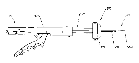
between a first position and a second position in the distal direction along
the longitudinal axis upon
the application of a first force in the distal direction, and a second portion
extending outside of the
housing and along a plane that is perpendicular to the longitudinal axis; a
rigid wire extending along
the longitudinal axis and connected at a proximal end to said housing; a push
tube extending along
the longitudinal axis over the wire and positioned distally to the portion of
the first sliding member
that is within the housing, wherein said push tube is movable over the wire in
the distal direction
from a first position to a second position along the longitudinal axis upon
the application of the first
force on said first sliding member; a plug comprising a proximal end and a
distal end and positioned
distally to said push tube over said wire, wherein said plug is movable over
the wire in the distal
direction from a first position to a second position along the longitudinal
axis upon the application of
the first force through said push tube by said first sliding member; and a
force conduit mechanism
positioned over said wire between the first sliding member and said push tube
within the housing,
and structured to maintain the distal first force ultimately applied to the
plug at or below a
predetermined force.
17a
CA 02816863 2013-06-19
BRIEF DESCRIPTION OF THE DRAWINGS
[0089] The present invention will be more fully understood and
appreciated by reading the
following Detailed Description in conjunction with the accompanying drawings,
in which:
[0090] Figs. la - lp are perspective views of footplates according to
embodiments of the
present invention.
[0091] Fig. 2a shows a fully assembled right side perspective view of the
deployment
device, according to an embodiment of the present invention.
[0092] Fig. 2b is a partially exposed right side perspective view of the
deployment device,
according to an embodiment of the present invention.
[0093] Fig. 3a is a partially exposed right side perspective view of the
deployment device,
according to an embodiment of the present invention.
[0094] Fig. 3b is a magnified window view of a portion of the deployment
device of Fig. 3a,
according to an embodiment of the present invention.
[0095] Fig. 4a is a perspective view of a distal portion of the of the
deployment device,
according to an embodiment of the present invention.
[0096] Fig. 4b is a magnified window view of a portion of the deployment
device of Fig. 4a,
according to an embodiment of the present invention.
[0097] Figs. 5a - 5f are perspective views of the plug, according to an
embodiment of the
present invention.
17b
CA 02816863 2013-05-02
WO 2012/061486
PCT/US2011/058936
[0098] Fig. 6a is a partially exposed right side perspective view of the
deployment
device, according to an embodiment of the present invention.
[0099] Fig. 6b is a magnified window view of a portion of the deployment
device of Fig.
6a, according to an embodiment of the present invention.
[00100] Fig. 6c is a partially exposed right side perspective view of the
deployment
device, according to an embodiment of the present invention.
[00101] Fig. 7a is a partially exposed top side perspective view of the
deployment device,
according to an embodiment of the present invention.
[00102] Fig. 7b is a magnified window view of a portion of the deployment
device of Fig.
7a, according to an embodiment of the present invention.
[00103] Fig. 7c is a magnified window view of a portion of the deployment
device of Fig.
7a, according to an embodiment of the present invention.
[00104] Fig. 8 is a perspective view of a partially exposed distal portion
of the deployment
device, according to an embodiment of the present invention.
[00105] Fig. 9a is a perspective view of a distal portion of the deployment
device,
according to an embodiment of the present invention.
[00106] Fig. 9b is a magnified window view of a portion of the deployment
device of Fig.
9a, according to an embodiment of the present invention.
[00107] Fig. 9c is a cutaway perspective view of the distal end of the
deployment device,
according to an embodiment of the present invention.
[00108] Fie. 9d is a perspective view showing the local expansion of a
portion of the
distal C-Tubes while allowing passage of the plug therethrough, according to
an embodiment
of the present invention.
[00109] Fig. 10a is a right side perspective view of a partially exposed
section of the
deployment device, according to an embodiment of the present invention.
[00110] Fig. 10b is a magnified window view of a portion of the deployment
device of
Fig. 10a, according to an embodiment of the present invention.
[00111] Fig. 11 is a right side perspective view of a partially exposed
section of the
deployment device, according to an embodiment of the present invention.
[00112] Fig. 12 is a right side perspective view of a partially exposed
section of the
deployment device, according to an embodiment of the present invention.
[00113] Fig. 13 is a right side perspective view of a partially exposed
section of the
deployment device, according to an embodiment of the present invention.
18 176546.2
11/2/2011
CA 02816863 2013-05-02
WO 2012/061486
PCT/US2011/058936
[00114] Fig. 14 is a right side perspective view of a partially exposed
section of the
deployment device, according to an embodiment of the present invention.
[00115] Fig. 15a is a partially exposed right side perspective view of the
deployment
device, according to an embodiment of the present invention.
[00116] Fig. 15b is a magnified window view of a portion of the deployment
device of
Fig. 15a, according to an embodiment of the present invention.
[00117] Fig. 16 is a right side perspective view of a partially exposed
section of the
deployment device, according to an embodiment of the present invention.
[00118] Fig. 17a is a partially exposed right side perspective view of the
deployment
device, according to an embodiment of the present invention.
[00119] Fig. 17b is a right side perspective view of a partially exposed
section of the
deployment device, according to an embodiment of the present invention.
[00120] Fig. 18 is a partially exposed rear side perspective view of the
deployment device,
according to an embodiment of the present invention.
[00121] Fig. 19 is a right side perspective view of a partially exposed
section of the
deployment device, according to an embodiment of the present invention.
[00122] Fig. 20 is a top side perspective view of a partially exposed
section of the
deployment device, according to an embodiment of the present invention.
[00123] Fig. 21a is a right side cross-sectional view of a partially
exposed section of the
deployment device, according to an embodiment of the present invention.
[00124] Fig. 2 lb is a top side cross-sectional view of a partially exposed
section of the
deployment device, according, to an embodiment of the present invention.
[00125] Figs. 21c and 21d are magnified window views of portions of the
deployment
device of Fig. 21a, according to an embodiment of the present invention.
[00126] Fig. 22a is a right side cross-sectional view of a partially
exposed section of the
deployment device, according, to an embodiment of the present invention.
[00127] Fig. 22b is a top side cross-sectional view of a partially exposed
section of the
deployment device, according to an embodiment of the present invention.
[00128] Figs. 22c and 22d are magnified window views of portions of the
deployment
device of Fig. 22b, according to embodiments of the present invention.
[00129] Fig. 22e is a magnified window view of a portion of the deployment
device of
Fig. 22a, according to embodiments of the present invention.
19 1768546 2
11/2/2011
CA 02816863 2013-05-02
WO 2012/061486 PCT/US2011/058936
[00130] Fig. 23a is a right side cross-sectional view of a partially
exposed section of the
deployment device, according to an embodiment of the present invention.
[00131] Fig. 23b is a top side cross-sectional view of a partially exposed
section of the
deployment device, according to an embodiment of the present invention.
[00132] Figs. 23c and 23d are magnified window views of portions of the
deployment
device of Fig. 23b, according to embodiments of the present invention.
[00133] Fig. 23e is a magnified window view of a portion of the deployment
device of
Fig. 23a, according to embodiments of the present invention.
[00134] Fig. 24a is a right side cross-sectional view of a partially
exposed section of the
deployment device, according to an embodiment of the present invention.
[00135] Fig. 24b is a top side cross-sectional view of a partially exposed
section of the
deployment device, according to an embodiment of the present invention.
[00136] Figs. 24c ¨ 24f are magnified window views of portions of the
deployment device
of Fig. 24b, according to embodiments of the present invention.
[00137] Fig. 25a is a right side cross-sectional view of a partially
exposed section of the
deployment device, according to an embodiment of the present invention.
[00138] Fig. 25b is a top side cross-sectional view of a partially exposed
section of the
deployment device, according to an embodiment of the present invention.
[00139] Figs. 25c ¨ 25d are magnified window views of portions of the
deployment
device of Fig. 25b. according to embodiments of the present invention.
[00140] Fig. 26a is a right side cross-sectional view of a partially
exposed section of the
deployment device, according to an embodiment of the present invention.
[00141] Fig. 26b is a top side cross-sectional view of a partially exposed
section of the
deployment device, according to an embodiment of the present invention.
[00142] Figs. 26d and 26f are magnified window views of portions of the
deployment
device of Fig. 26a, according to embodiments of the present invention.
[00143] Fig. 26g is a magnified vertical cross-sectional view through a
portion of the
deployment device of Fig. 26a. according to embodiments of the present
invention.
[00144] Figs. 26c and 26e are magnified window views of portions of the
deployment
device of Fig. 26b, according to embodiments of the present invention.
[00145] Fig. 27 is a left side perspective view of a partially exposed
section of the
deployment device, according to an embodiment of the present invention.
20 1768546.2
11/2/2011
CA 02816863 2013-05-02
WO 2012/061486
PCT/US2011/058936
[00146] Fig. 28a is a right side cross-sectional view of a partially
exposed section of the
deployment device, according to an embodiment of the present invention.
[00147] Fig. 28b is an under side perspective view of the deployment
device, according to
an embodiment of the present invention.
[00148] Fig. 29a is a right side cross-sectional view of a partially
exposed section of the
deployment device, according to an embodiment of the present invention.
[00149] Fig. 29b is a top side cross-sectional view of a partially exposed
section of the
deployment device, according to an embodiment of the present invention.
[00150] Figs. 29d, 29e and 29f are magnified window views of portions of
the deployment
device of Fig. 29a, according to embodiments of the present invention.
[00151] Fig. 29c is a magnified vertical cross-sectional view through a
portion of the
deployment device of Fig. 29a, according to embodiments of the present
invention.
[00152] Fig. 30a is a right side cross-sectional view of a partially
exposed section of the
deployment device, according to an embodiment of the present invention.
[00153] Figs. 30b ¨ 30c are magnified window views of portions of the
deployment
device of Fig. 30a, according to embodiments of the present invention.
[00154] Figs. 31 ¨ 41 show the sequential steps in the use of the
deployment device to
deploy the closure device to seal an opening formed through a blood vessel,
according to an
embodiment of the present invention.
[00155] Figs. 42-43 show a closure device in a sealing relationship with
the opening
formed through a blood vessel (i.e., post-closure device deployment
configuration and
position), according to an embodiment of the present invention.
[00156] Figs. 44 a-b show a bottom perspective view and top perspective
view of a
footplate, according to an alternative embodiment of the present invention.
[00157] Fig. 45 shows a ball-shaped end on a distal tip of wire connected
at a wire ball
socket of a footplate, according to an alternative embodiment of the present
invention.
[00158] Figs. 46 a-d show various views of a wire assembly, according to an
alternative
embodiment of the present invention.
[00159] Figs. 47 a-c show various views of a sheath assembly, according to
an alternative
embodiment of the present invention.
[00160] Fig. 48 shows the finger pull/sheath interface, according to an
alternative
embodiment of the present invention.
21 17o546.2
11/2/2011
CA 02816863 2013-05-02
WO 2012/061486 PCT/US2011/058936
[00161] Fig. 49 shows the retracted sheath and exposed footplate, according
to an
alternative embodiment of the present invention.
[00162] Figs. 50 a-b show various views of the plug, according to an
alternative
embodiment of the present invention.
[00163] Fig. 51 shows a longitudinal section view through the locking pin,
housing, finger
pull, and skin flange, according to an alternative embodiment of the present
invention.
[00164] Figs. 52 a-b show removal of the locking pin from the deployment
device,
according to an alternative embodiment of the present invention.
[00165] Figs. 53 a-b show a right side perspective view of the deployment
device and a
magnified window portion view of the deployment device, according to an
alternative
embodiment of the present invention.
[00166] Figs. 54 and 55 show magnified views of a portion of the inside of
the skin flange
and housing, according to an alternative embodiment of the present invention.
[00167] Figs. 56a-b show various views of the thumb crutch, according to an
alternative
embodiment of the present invention.
[00168] Figs. 57 and 58 a-b show' the thumb crutch and its interrelation to
other
components on the proximal end of the deployment device, according to an
alternative
embodiment of the present invention.
[00169] Fig. 59 shows the distal movement functionality of the thumb crutch
with respect
to portions of the deployment device, according to an alternative embodiment
of the present
invention.
[00170] Fig. 60 shows detail of the push tube locking lever in a pre and
post lock
positions. according to an alternative embodiment of the present invention.
[00171] Fig. 61 shows the skin flange, and other distal portions of the
deployment device,
in use and in a nearly fully deployed position, according to an alternative
embodiment of the
present invention.
[00172] Fig. 62 shows the plug and footplate in a deployed position with
respect to a
relatively thin arterial wall (as compared with Fig. 63). according to an
alternative
embodiment of the present invention.
[00173] Fig. 63 shows the plug and footplate in a deployed position with
respect to a
relatively thick arterial wall (as compared with Fig. 62), according to an
alternative
embodiment of the present invention.
22 1768546.2
11/2/2011
CA 02816863 2013-05-02
WO 2012/061486 PCT/US2011/058936
[00174] Figs. 64 a-c show a push tube assembly. and various magnified
sections of the
assembly, according to an alternative embodiment of the present invention.
[00175] Fig. 65 shows a cutter lever assembly, according to an alternative
embodiment of
the present invention.
[00176] Figs. 66a-c show a cutter tube assembly, and various magnified
portion views of
the assembly, according to an alternative embodiment of the present invention.
[00177] Fig. 67 shows the cutter tube assembly bending over the wire and
shearing it
apart, according to an alternative embodiment of the present invention.
[00178] Fig. 68 shows portions of the skin flange and the housing damped
via viscous
shearing with a film of high viscosity biocompatible lubricant, according to
an alternative
embodiment of the present invention.
[00179] Figs. 69 a-b shows the maintenance of a constant pressure angle
throughout the
rotation of the cutter lever assembly, according to an alternative embodiment
of the present
invention.
[00180] Fig. 70 shows a left side perspective view and magnified window
view of the skin
flange assembly. according to an alternative embodiment of the present
invention.
[00181] Fig. 71 shows an exploded view of the deployment device, according
to an
alternative embodiment of the present invention.
[00182] Fig. 72 shows a left side and right side view of the finger pull,
according to an
alternative embodiment of the present invention.
[00183] Fig. 73 shows a left side and right side view of the left half of
the housing,
according to an alternative embodiment of the present invention.
[00184] Fig. 74 shows a left side and right side view of the right half of
the housing,
according to an alternative embodiment of the present invention.
DETAILED DESC'RIP TION
[00185] Reference will now be made in detail to the present preferred
embodiments of the
invention, examples of which are illustrated in the accompanying drawings.
[00186] In accordance with an embodiment of the present invention, closure
device 100
comprising a footplate 110 (the footplate may include any of the embodiments
of the
footplate, as discussed infra), a plug 111, and a wire 120 is provided and can
be used to seal
or close an opening formed through biological tissue, such as a pet-
cutaneously formed
puncture (the puncture comprises the opening formed through the wall of the
blood vessel
and a tissue tract contiguous with the opening formed through the biological
tissue, which
23 1768546 2
11/2/2011
CA 02816863 2013-05-02
WO 2012/061486 PCT/US2011/058936
extends through the tissue and to skin overlying the blood vessel), an
incision, or some other
type of opening formed through biological tissue, such as a blood vessel,
organ, or the like, to
control (or prevent or stop) bleeding (or the flow of other biological fluid
or tissue). For
example, the closure device 100 of an embodiment of the present invention can
be used to
seal an arteriotomy, which is an opening or incision in an artery, such as the
femoral artery,
and is formed in conjunction with a percutaneously formed puncture (an open
tissue tract
through the skin and tissue just above the blood vessel) by a clinician during
a diagnostic or
therapeutic intravascular surgical procedure.
[00187] In accordance with an embodiment of the present invention, the
closure device
100 may be in a pre-deployed closure device deployment configuration and
position or in a
post-vascular closure device deployment configuration and position. A pre-
deployed closure
device deployment configuration and position includes a configuration and
position where the
closure device 100 resides within a deployment device 200 of an embodiment of
the present
invention (which is used to deploy the closure device 100 into, e.g., an
opening in the wall of
a blood vessel, to seal the blood vessel to stop blood from flowing through
the opening). A
post-deployed closure device deployment configuration and position includes a
configuration
and position where the closure device 100 resides within and through the
opening in the wall
of the blood vessel.
[001881 The closure device 100, the pre- and post-deployed closure device
deployment
configurations and positions, the deployment device 200, and the method of
deploying the
closure device 100 to seal an opening in the wall of a blood vessel, with
reference to the
figures, is more fully described infra.
[00189] Referring now to the drawings where like numbers refer to like
parts throughout,
Fig. la shows a footplate 110 according, to an embodiment of the present
invention. This
embodiment shows a footplate 110 in a pre-deployed closure device deployment
configuration and position, wherein the footplate 110 is within a distal end
of a deployment
device 200 (not shown). The footplate 110 comprises a unitary length of a
distal portion of
the wire 120 (monolithic structure) bent into an elongated configuration
presenting an
elongated U-shaped loop 30. The elongated U-shaped loop 30 comprises an open
proximal
end 101, a closed distal end 102, and a pair of longitudinally laterally
spaced extending legs
31, 32. The closed distal end 102 and pair of longitudinally laterally spaced
extending legs
31, 32 are substantially coplanar in a common plane and substantially parallel
to the
longitudinal axis of the control housing 210 of the deployment device 200. The
closed distal
24 1768546.2
11/2/2011
CA 02816863 2013-05-02
WO 2012/061486 PCT/US2011/058936
end 102 of the elongated U-shaped loop 30 defines a longitudinal distal end of
the bent wire
elongated configuration. The pair of longitudinally-extending laterally spaced
legs 31, 32 of
the elongated U-shaped loop comprises a free leg 31, having a free proximal
end located at
the open proximal end of the elongated U-shaped loop 101, and a connecting leg
32. A
helically shaped connecting portion 33 connects to the wire 120. The helically
shaped
connecting portion 33 is operable to permanently (plastically) deform at a
bending region.
The wire 120 is axial to a longitudinal axis of the control housing 210. The
wire 120 is
proximal to the footplate 110, and the helically shaped connecting portion 33
extends
between a joining leg 34 (which is substantially coplanar with the
longitudinally-extending
laterally spaced legs 31, 32) and the wire 120 at the open proximal end 101 of
the elongated
U-shaped loop 30.
[00190] Turning to Fig. lb, the footplate 110 according to an embodiment of
the present
invention is illustrated. This embodiment shows the footplate 110 (of Fig. I
a) in a post-
deployed closure device deployment configuration and position, wherein a
portion of the
footplate 110 is seated against an inside wall of a blood vessel (e.g., an
artery, not shown)
under a percutaneous puncture therein (not shown). The helically shaped
connecting portion
33 comprises a bending region, wherein the bending region is permanently
(plastically)
deformed. The closed end 102 and pair of longitudinally laterally spaced
extending legs 31,
32 remain substantially coplanar in a common plane. and are substantially
perpendicular to a
longitudinal axis of the puncture and substantially parallel to a plane of the
inside wall of the
blood vessel. The wire 120 extends proximally from the helically shaped
connecting portion
33 through the opening in the wall of the blood vessel to the tissue tract
wherein the wire 120
is axial to the longitudinal axis of the puncture (prior to being cut and bent
by the deployment
device 200).
[00191] Turning to Fig. 1 c, a footplate according to an embodiment of the
present
invention is illustrated. This embodiment shows a footplate 110' in a pre-
deployed closure
device deployment configuration and position, wherein the footplate 110' is
within the distal
end of a deployment device 200 (not shown). The footplate 110' comprises a
unitary length of
a distal portion of the wire 120 (monolithic structure) bent into an elongated
configuration
presenting an elongated U-shaped loop 30'. The elongated U-shaped loop 30'
comprises an
open distal end 102', a closed proximal end 101', and a pair of longitudinally
laterally spaced
extending legs 31', 32'. The closed proximal end 101' of the elongated U-
shaped loop 30'
defines a longitudinal proximal end of the bent wire elongated configuration.
The pair of
25 1768546.2
11/2/2011
CA 02816863 2013-05-02
WO 2012/061486 PCT/1JS2011/058936
longitudinally-extending laterally spaced legs of the elongated U-shaped loop
30' comprise a
free leg 31' having a free distal end located at the open distal end 102' of
the elongated U-
shaped loop 30, and a connecting leg 32'. An arcuately-curved connecting
portion 33', and a
medial leg 34' comprising a bending region are shown. The arcuately-curved
connecting
portion 33' extends between the connecting leg 32' and the medial leg 34' at
the open distal
end 102', defining a longitudinal distal end of the bent wire configuration.
The elongated U-
shaped loop 30' and the arcuately-curved connecting portion 33' are
substantially coplanar in
a common plane and axial to a longitudinal axis of the control housing 210 of
the deployment
device 200. The arcuately-curved connecting portion 33' medially curves toward
the
connecting leg 32' to the medial leg 34', in between the free leg 31' and the
connecting leg
32'. Each of the free leg 31' and the connecting leg 32' is secured to a
distal portion of the
medial leg by a spot weld 35. The spot weld 35 may comprise an electron beam
spot weld or
a laser spot weld. The distal end of the free leg ends at a point where the
free leg is spot
welded to the medial leg 34' (but could be longer or shorter). The medial leg
34' extends
proximally toward and under (but could extend over) the closed proximal end
101', and
extends beyond the closed proximal end 101'at an angle from the common plane
to the wire
120, wherein the wire 120 is proximal to the footplate 110'.
[00192] Turning to Fig. ld. the footplate 110' according to an embodiment
of the present
invention is illustrated. This embodiment shows the footplate 110' of Fig. lc
in a post-
deployed closure device deployment configuration and position, wherein a
portion of the
footplate 110' is seated against an inside wall of a blood vessel (e.g., an
artery, not shown)
under a percutaneous puncture therein (not shown). The bending region
(preferably at the
proximal margin of the spot weld 35 in the medial leg 34') is permanently
(plastically)
deformed. The elongated U-shaped loop 30' and the arcuately-curved connecting
portion 33'
remain substantially coplanar in a common plane, and are substantially
perpendicular to a
longitudinal axis of the puncture and substantially parallel to a plane of the
inside wall of the
blood vessel. The wire 120 extends proximally from the bending region through
the
opening in the wall of the blood vessel and inside the tissue tract wherein
the wire 120 is axial
to the longitudinal axis of the puncture (prior to being cut and bent by the
deployment device
200).
[00193] Turning to Figs. le-lf, a footplate 710 according to an embodiment
of the present
invention is shown. These embodiments of the footplate are similar to the
footplates
illustrated in Figs. lc- Id, respectively, except for free leg 731 and spot
weld 735. As shown
26 1768546.2
11/2/2011
CA 02816863 2013-05-02
WO 2012/061486 PCT/US2011/058936
in Fig. le and Fib. g If, free leg 731 comprises a hooked distal end, and the
spot weld 735 only
secures medial leg 734 to connecting leg 732. An elongated U-shaped loop 730,
a proximal
1101 and distal end 1102, and an acuately-curved connecting portion 733 are
also shown.
[00194] Turning to Fig. lg, a footplate 810 according to an embodiment of
the present
invention is shown. This embodiment shows a footplate 810 in a pre-deployed
closure device
deployment configuration and position, wherein the footplate 810 is within a
distal end of a
deployment device 200 (not shown). The footplate 810 is a longitudinally
shaped block or bar
837. The bar 837 comprises longitudinal aperture 840, a top arcuately-shaped
surface 838, a
bottom arcuately-shaped surface (not shown) (alternatively, the top and bottom
surfaces can
be substantially planar). a peripheral side surface 839. a proximal end 2101
and a distal end
2102. The wire 120 is connected to the bar 837 by a flat or coined distal end
836 (preferably
welded), which is connected to the distal portion 2102 of the top arcuately-
shaped surface 838
of the bar 837 (could also be connected to the bottom arcuately-shaped
surface). The coined
distal end 836 proximally extends to a medial portion 834, which proximally
extends through
the aperture 840 and under (but could extend over) the proximal end 2101.
[00195] Turning to Fig. lh, the footplate 810 according to an embodiment of
the present
invention is illustrated. This embodiment shows the footplate 810 of Fig. lg
in a post-
deployed closure device deployment configuration and position, wherein a
portion of the
footplate 810 is seated against an inside wall of a blood vessel (e.g., an
artery, not shown)
under a percutaneous puncture therein (not shown). The medial portion 834
comprises a
bending region wherein the bending region is permanently (plastically)
deformed. The
medial portion 834 proximally extends through the aperture 840. The wire 120
extends
proximally from the medial portion 834 through the opening in the wall of the
blood vessel to
the tissue tract, wherein the wire 120 is coaxial to the longitudinal axis of
the puncture (not
shown) (prior to being cut and bent by the deployment device 200).
[00196] Turning to Figs. li-lj, a footplate 1010 according to an embodiment
of the
present invention is shown. These embodiments of the footplate are similar to
the footplates
illustrated in Figs. lg-I h. respectively, except for the distal end 1036 of
the wire 120. The
distal end 1036 is neither flattened nor coined (but is left as the same
circular cross-section as
the remainder of the wire). The wire 120 is affixed (preferably welded) on
both sides along
the longitudinal interface between the distal portion of the wire 120 and the
arcuately-shaped
top surface 1038 at the distal end 4102 of the bar 1037 of the footplate 1010.
A proximal end
27 176546.2
11/2/2011
CA 02816863 2013-05-02
WO 2012/061486 PCT/US2011/058936
4101, a medial portion 1034, an aperture 1040, and a peripheral side surface
1039 are also
shown.
[00197] Turning to Fig. lk, a top perspective view of footplate 910
according to an
embodiment of the present invention is shown. This embodiment shows a
footplate 910 in a
pre-deployed closure device deployment configuration and position, wherein the
footplate
910 is within a distal end of a deployment device 200 (not shown). The
footplate 910
comprises a longitudinally shaped block or bar 937. The bar 937 comprises a
top
substantially planar surface 938, a bottom substantially planar surface (not
shown), a
peripheral side surface 939, a proximal end 3101 and a distal end 3102. The
wire 120 is
connected to the bar 937 by a ball-shaped end 936, which is connected to a
socket 941 of the
top planar surface 938 of the bar 937 (could also be connected to the bottom
planar surface).
The socket 941 is shaped like a "C" to allow for the actuation of the
footplate 910, as shown
in Fig. 11. A portion of the wire 120 may sit in an arcuately-shaped depressed
section 942 of
the top surface 938 of the bar 937.
[00198] Turning to Fig. 11, a bottom perspective view of the footplate 910
according to an
embodiment of the present invention is shown. This embodiment shows the
footplate 910 of
Fig. Ii in a post-deployed closure device deployment configuration and
position, wherein a
portion of the footplate 910 is seated against an inside wall of a blood
vessel (e.g., an artery,
not shown) under a percutaneous puncture therein (not shown). There is no
bending region in
this embodiment of the footplate 910. The bar 937 is operable to rotate
pursuant to the ball
936 and socket 941 configuration/mechanism. This establishes a rotation point
with the
proximal end 3101 rotating down and in the distal direction and the distal end
3102 rotating
up (could alternatively rotate in the opposite direction with an alternative
configuration) and
in the proximal direction about the established rotation point. Fig. 11 shows
the footplate 910
in its fully actuated or rotated position. The wire 120 extends proximally
from the ball 936
through the opening in the wall of the blood vessel to the tissue tract,
wherein the wire 120 is
axial to the longitudinal axis of the puncture (not shown) (prior to being cut
and bent by the
deployment device 200). A bottom substantially planar surface 943 is shown,
which may
further comprise a protruding section comprising the bottom portion of the
depressed section
942 of the top surface 938 of the bar 937.
[00199] Turning to Fig. lm, a top perspective view of footplate 1110
according to an
embodiment of the present invention is shown. This embodiment shows a
footplate 1110 in a
pre-deployed closure device deployment configuration and position, wherein the
footplate
28 1768546 2
11/2/2011
CA 02816863 2013-05-02
WO 2012/061486 PCT/US2011/058936
1110 is within a distal end of a deployment device 200 (not shown). The
footplate 1110
comprises a longitudinally shaped block or bar 1137. The bar 1137 is "y-
shaped" and
comprises a top substantially planar surface 1138, a bottom substantially
planar surface 1143
(not shown), a peripheral side surface 1139, a proximal end 5101, a distal end
5102 and two
proximally extending substantially coplanar legs 1144 and 1145 (not shown)
which are
separated by a slot 1146. The wire 120 is connected to the bar 1137 by a hinge
mechanism
comprising a hooked shaped end 1136, which is connected to a half-circled
portion 1141 that
is attached to the top substantially planar surface 1138 of the bar 1137
(could also be
connected to the bottom planar surface).
[00200] Turning to Fig. In, a bottom perspective view of footplate 1110
according to an
embodiment of the present invention is shown. This embodiment shows the
footplate 1110 of
Fig. 1m in a post-deployed closure device deployment configuration and
position, wherein a
portion of the footplate 1110 is seated against an inside wall of a blood
vessel (e.g., an artery,
not shown) under a percutaneous puncture therein (not shown). There is no
bending region in
this embodiment of the footplate 1110. The bar 1137 is operable to rotate
pursuant to the
hinge mechanism, which establishes a rotation point, with the proximal end
5101 rotating
down and in the distal direction and the distal end 5102 rotating up and in
the proximal
direction, about the established rotation point (could alternatively rotate in
the opposite
direction with an alternative configuration). Fig. 1 n shows the footplate
1110 in its fully
actuated or rotated position. The wire 120 extends proximally through the
opening in the
wall of the blood vessel to the tissue tract, wherein the wire 120 is axial to
the longitudinal
axis of the puncture (not shown) (prior to being cut and bent by the
deployment device 200).
A bottom substantially planar surface 1143 is also shown.
[00201] Turning to Figs. lo -1p, a footplate 1210 according to an
embodiment of the
present invention is shown. These embodiments of the footplate are similar to
the footplate
embodiments illustrated in Figs. 1m -In, respectively, except for the hook-
shaped distal end
1236 of the wire 120 and its attachment through an aperture 1241 and slot
1246. The hooked-
shaped distal end 1236 is attached to the bar 1237 through an aperture 1241.
The hooked-
shaped distal end 1236 stretches through the aperture 1241 from the bottom
substantially
planar surface 1243 to the top substantially planar surface 1238, and then
hooks through the
slot 1246 thereby securing the footplate 1210 to the wire 120. A bar 1237, a
distal end 6102
and a proximal end 6101, a top substantially planar surface 1238, a bottom
substantially
29 l76546.2 I
I/2/2011
CA 02816863 2013-05-02
WO 2012/061486 PCT/US2011/058936
planar surface 1243, a peripheral side surface 1239, two proximally extending
substantially
coplanar legs 1244 and 1245, a slot 1246, and a wire 120, are also shown.
[00202] In accordance with an embodiment of the present invention, the
deployment
device 200 with the closure device 100 of an embodiment of the present
invention is
described below with reference to the figures. References regarding the
footplate are
specifically made to footplate 110', as an example of a footplate that may be
used. However,
it is to be understood that any footplate embodiment including those
referenced supra, may be
used in place of footplate 110'.
[00203] Turning to Figs. 2a-4b, right side views (Fig. 2a shows a fully
assembled
deployment device 200, and Figs. 2b, 3 and 4 are partially exposed views of
the deployment
device 200, i.e., missing parts to reveal other parts of the deployment device
200) of a
deployment device 200, with a proximal end 10 and a distal end 20, according
to an
embodiment of the present invention is illustrated. In accordance with an
embodiment of the
present invention, prior to deployment into a vessel that requires sealing,
the footplate 110' is
located at the distal end 20 of a deployment device 200 and resides within an
outer distal C-
tube 201, while the remainder of the wire 120 is located proximally to the
distal footplate
110' within the deployment device 200 ending at a wire ferrule 250 (see Fig.
7a, which is
described infra). The footplate 110' is in an axial position relative to the
longitudinal axis of
the control housing 210 of the deployment device 200. The footplate's 110'
proximal end
101 abuts the distal end of an inner distal C-tube 202, as described infra
(see Figs. 4a-4b).
[00204] Turning to Figs. 5a-f, a plug 111 according to an embodiment of the
present
invention is illustrated. These embodiments show a plug 111 that is conically-
shaped and
comprises a distal portion (or end) 104 and a proximal portion (or end) 103,
wherein a
diameter of the plug's distal portion 104 is smaller than a diameter of the
plug's proximal
portion 103. The diameter of the plug 111 at its largest point is greater than
the diameter of
the main conduit area 205 of the deployment device, as discussed infra.
Turning to Fig. 5a,
a right side view of the plug 111 is shown. Turning to Fig. 5b, a top side
view of the plug 111
is shown. Turning to Fig. Sc. a left side inverted view of the plug 111 is
shown. Turning to
5d, a front side view of the plug 111 with a lumen 105 is shown. Turning to
Fig. 5e, a bottom
side view of the plug 111 is shown. Turning to Fig. 5f, a rear side view of
the plug Ill with a
lumen 105 is shown. One or more "cutouts" or "cavities" may be provided in the
distal end
of the plug to allow nesting of the plug 111 with the footplate 110' and wire
120, according to
an embodiment of the present invention. Also, one or more -cutouts" or -
cavities" may be
30 176S546.2
1112/2011
CA 02816863 2013-05-02
WO 2012/061486 PCT/US2011/058936
provided in the proximal end of the plug 111 to allow the insert 112 (see Fig.
10b) in the
distal end of a push tube 212 to maintain rotational control of the plug I 1 1
with respect to the
footplate 110', according to an embodiment of the present invention.
Embodiments of the
present invention contemplate a plug 111 with various combinations of -
cutouts" or without
any -cutouts".
[00205] Turning to Figs. 6a-6c, a partially exposed right side view of a
deployment device
200 in accordance with an embodiment of the present invention is illustrated.
In accordance
with an embodiment of the present invention, prior to deployment into a vessel
that requires
sealing (i.e., a pre-deployed closure device deployment configuration and
position), the plug
111 is located proximally to the footplate 110' and along the longitudinal
axis of the wire
120. The plug 111 is distally adjacent to the push tube 212, inside a distal
portion of an outer
proximal tube 211 (which is inside a control housing 210) of the deployment
device 200.
[00206] Turning to Figs. 7a-7c, a partially exposed top view of the
deployment device
according to an embodiment of the present invention is illustrated. This
embodiment shows
the location of the wire 120 within the deployment device 200. The wire 120
stretches from
the footplate 110' through a longitudinally axial hole 105 (not shown) in the
plug 111 in a
distal to proximal direction. The wire 120 stretches from the footplate 110'
through an inner
distal C-tube 202 (not shown), and a sheer tube 224 (within the push tube
212), to the inner
proximal end of a wire ferrule 250.
[00207] In accordance with an embodiment of the present invention, a pre-
deployed
closure device deployment configuration (default configuration) of the
deployment device
200 of an embodiment of the present invention will be described generally from
its distal end
20 to its proximal end 10, infra. Generally, in appropriate figures,
acceptable fastening means
(e.g., screws) are labeled with the number 226 and washers are labeled with
the number 214.
The method of use of the deployment device 200, and the closure device 100 in
its post-
deployed closure device deployment configuration and position, will be
described infra.
[00208] Turning back to Figs. 2a-2b, these embodiments show a deployment
device 200
comprising an outer distal C-tube 201. The outer distal C-tube's 201 distal
end comprises a
narrowed nose or tip portion 203. This nose portion 203 of the outer distal C-
tube 201 is the
portion of the deployment device which houses the footplate 110' (not shown).
The
remainder of the outer distal C-tube 201 houses an inner distal C-tube 202
(see Fig. 8),
comprising a longitudinal opening 204 in its bottom portion, and the wire 120.
These distal
31 1768546.2
11/2/2011
CA 02816863 2013-05-02
WO 2012/061486 PCT/US2011/058936
C-tubes are concentrically nested together forming a main conduit area 205,
which is
described infra.
[00209] Turning to Figs. 8, 9a-9c, distal views of the deployment device
200 according to
an embodiment of the present invention are illustrated. Fig. 9b shows the
entrance ¨ inlet
hole 406 - to a main conduit area 205 (see Fig. 9c) formed by the outer distal
C-tube 201 and
inner distal C-tube 202, which serves as a blood marking passageway. The inlet
hole 406
resides toward the outer distal C-tube's 201 distal end. Further, the outer
distal C-tube 201
and inner distal C-tube 202 each contains a side hole 206 (an atmospheric
exit) which are
concentrically aligned with one another. The side hole 206 is proximal to the
footplate 110'
and distal to the plug 111 (not shown). The side hole 206 is operable to serve
as an
atmospheric exit for proximal blood flow flowing from the blood vessel and
into the inlet
hole 406, and through the blood marking passageway 205. This proximal blood
flow that
exits the side hole 206 indicates that the footplate and distal portion of the
deployment device
200 have entered the blood vessel (not shown, which is described infra). The
main conduit
area 205 additionally is operable to serve as a deployment area for deploying
the plug Ill,
wherein the distal C-tubes are operable to locally expand and disassociate
creating an
irreversible un-nested condition to allow passage of the plug 111 into a post-
vascular
deployment configuration and position (see Fig. 9d).
[002101 In accordance with an embodiment of the present invention, Figs. 9a
& 9b are
views of the distal portion of the deployment device 200 and Fig. 9c is a cut-
away view of the
distal portion of the deployment device 200, which shows the nested
configuration of the
outer distal C-tube 201 (including the guidewire lumen 300) and the inner
distal C-tube 202,
which together form the blood marking passageway 205, and a passageway in
which the wire
120 nests, according to an embodiment of the present invention. The outer
distal C-tube 201
comprises a guide wire lumen 300 (see Fig. 9c) comprising a proximal guide
wire exit 207
(see Fig. 9a) and a distal guide wire entrance 301 (see Figs. 9b & 9c) for
insertion of a guide
wire (not shown). The proximal guide wire exit 207 is proximal to the
footplate 110' and
distal to the plug's 111 pre-deployed closure device deployment position. The
distal guide
wire entrance 301 is located at the most distal point (at the distal nose
portion 203) of the
deployment device 200.
[00211] In accordance with an embodiment of the present invention, the
outer distal C-
tube 201 and inner distal C-tube 202 can move independently of one another in
the
longitudinal direction, i.e., the distal C-tubes are operable to independently
slide along the
32 176846.2
11/2/2011
CA 02816863 2013-05-02
WO 2012/061486
PCT/US2011/058936
longitudinal axis of the wire 120 (e.g., to allow and to assist in the
actuation of the footplate
110 to a substantially petpendicular position relative to the longitudinal
axis of the control
housing 210 once inside the lumen of the artery, as will be discussed infra).
[00212] Turning to Fig. 10a ¨ 10b, a partially exposed right side view of
the deployment
device 200 in accordance with an embodiment of the present inventions is
illustrated. This
embodiment shows that the proximal ends of the outer distal C-tube 201 and
inner distal C-
tube 202 end just within an outer proximal tube 211 (the inner distal C-tube
202 ends at a
retainer ring 325 (see Fig. 11, which is described infra) and slightly more
proximally than the
outer distal C-tube 201, which ends at a ring retainer 324 (see Fig. 11). The
outer proximal
tube 211 is surrounded by a control housing 210, which is in turn partially
surrounded by a
skin flange assembly 222 (not shown) comprising a distal portion 221 and a
proximal portion
303 (see Fig. 2b). The skin flange assembly 222 (not shown) is operable to
distally slide
along a longitudinal axis of the wire 120, and along an outside portion of the
control housing
210 and an outside portion of the distal C-tubes. The plug 111 is distally
adjacent to an insert
112 and the distal end of a push tube 212, which mainly resides directly
within an inner
proximal tube 213 (which resides within the outer proximal tube 211. etc.)
which stretches in
the proximal direction to about the proximal end 10 of the deployment device
200 (see Figs.
6a ¨ 6b). The proximal tubes are operable to independently slide along the
longitudinal axis
of the wire 120.
[00213] Turning to Figs. 11-13, partially exposed right side views of the
deployment
device 200 of an embodiment of the present invention are illustrated. This
embodiment
shows the push tube 212, which resides within the proximal tubes (211, 213)
and is
surrounded by a ring retainer 325 at its distal end, and is cradled by an
alignment key 326 at
its proximal end. The push tube 212 extends proximally from the push tube
insert 112
(which is affixed to the distal end of the push tube 212 by an appropriate
means such as a
weld) through a washer 214 (which is welded to the push tube 212 and whose
proximal
surface is adjacent to the distal end of a slide barrel 215), and protrudes
through the main
body of the slide barrel 215 such that its most proximal tip is approximately
adjacent to the
most proximal end of the slide barrel. At its distal end on the top, the push
tube 212 has an
opening 219 (which is a slot) that extends in a proximal direction from a
point just slightly
proximal of the distal tip of the push tube 212. Concentrically contained
within the push tube
212 is a shear tube 224 which extends in a proximal direction from the push
tube insert's 112
angled proximal surface 350 (see Fig. 30c) back to its most proximal end
(slightly proximal
33 17654o.2
11/2/2011
CA 02816863 2013-05-02
WO 2012/061486 PCT/US2011/058936
of the most proximal end of the push tube 212). The proximal end of the shear
tube 224 has a
cap 216 affixed to it. The push tube 212 and the shear tube 224 are operable
to distally slide
along the longitudinal axis of the wire 120. The push tube 212 is operable to
push the plug
111 through the main conduit area 205 into its post-deployed closure device
deployment
configuration and position as is discussed infra. The shear tube 224 (in
conjunction with the
push tube insert 112) is operable to both bend and shear-off the wire 120 into
its post-vascular
closure deployment configuration. as described infra.
[00214] Turning to Figs. 14-16. a partially exposed right side view of the
deployment
device 200 (shown in the default position) according to an embodiment of the
present
invention is illustrated. In accordance with an embodiment of the present
invention, constant
force springs comprising a plurality of lateral constant force springs,
comprising a left lateral
constant force spring 125 and a right lateral constant force spring 125, are
provided. The
lateral constant force springs 125 (left and right) each comprise a flat
portion 227 and a roll
spring portion 228. The roll spring portion 228 of each of the lateral
constant force springs
125 (left and right) resides (nests) partially within the distal portion of
the control housing
210, on the left and right side respectively (see Fig. 15a) and is covered by
(contained within)
the distal portion 221 of the skin flange assembly 222 (see Fig. 14). The flat
portion 227 of
the lateral constant force springs 125 (left and right) stretches flatly along
the outside of the
control housing 210 (on the left and right sides respectively) in a proximal
direction from the
roll spring portion 228, to the inside proximal portion 303 of the right and
left sides
(respectively) of the skin flange assembly 222 where they are fastened by an
acceptable
fastening means 226 (see Fig. 14). The lateral constant force springs are
operable to move the
skin flange assembly 222 in a distal direction by a constant distal force. The
lateral constant
force springs are also operable to apply a constant distal force to an outside
surface of a
patient's skin. Further, the lateral constant force springs are operable to
apply a constant
tensile proximal force to the wire 120. This constant tensile proximal force
seats the
footplate 110' against an inside wall 403 (not shown) of a blood vessel,
wherein a datum is
created at a point where the footplate 110' is seated, as discussed infra.
[00215] In accordance with an embodiment of the present invention, constant
force
springs comprising an upper and lower constant force spring 135, each
comprising a flat
portion 230 and a roll spring portion 229, are provided. The roll spring
portions 229 of the
upper and lower constant force springs 135 reside (nest) on the outside (on
the top and
bottom) of the distal end of the control housing 210, and are covered
(contained within) the
34 1768546.2 I
1/2/20 II
CA 02816863 2013-05-02
WO 2012/061486 PCT/US2011/058936
distal portion 221 of the skin flange assembly 222 (see Fig. 14). The flat
portions 230 of the
upper and lower constant force springs 135 extend proximally from the
respective roll spring
portions 229 (within the distal portion of the control housing 210) and
stretch flatly along the
outside of the control housing 210 (on the top and bottom respectively), and
are fastened by
an acceptable fastening means (e.g., a screw 226 and washer 214) to about the
middle portion
(top and bottom, respectively) of the slide barrel 215 (see Fig. 16). The
upper and lower
constant force springs 135 are operable to move the slide barrel 215 in a
distal direction by
application of a constant distal force to the slide barrel 215. The slide
barrel 215 is operable
to advance the push tube 212 in a distal direction by the constant distal
force applied by the
upper and lower constant force springs 135 to the slide barrel 215, wherein
the plug 111 is
pushed percutaneously into a percutaneous puncture (see Fig. 39) and into a
post-deployed
closure device deployment configuration and position. This post-deployed
closure device
deployment configuration and position is controlled by the creation of the
datum (as
discussed infra) with the wire 120 and the footplate 110', in order to seal
the opening in the
wall of the blood vessel.
[00216] Turning to Figs. 17a, 17b, and 18, the proximal end 10 (partially
exposed right
rear side and rear view) of the deployment device 200 according to an
embodiment of the
present invention is illustrated. This embodiment shows a squeeze lever handle
assembly 231
of the deployment device 200. The squeeze lever handle assembly 231 comprises
a squeeze
lever handle 232, a button 233 held within a retainer plate 234 of the squeeze
lever handle
232, and a link 235. The button 233 is slidable within the retainer portion
234. The link 235
is removably attached at its proximal end (by an upwardly hook-shaped portion
or C-feature
266, not shown) to the bottom part (by a hinge pin 256, not shown) of the
slide barrel 215
(which transfers mechanical energy to, and creates distal movement of, the
slide barrel, upon
the squeezing of the squeeze lever handle 232, described infra). The link 235
is attached at
the other end (lower portion) to the squeeze lever handle 232 by a hinge pin
mechanism 236.
The squeeze lever handle 232 is removably attached to the proximal portion 303
of the skin
flange assembly 222 (on both the left and right sides of the device) by
lateral upper hook-
shaped ends 237.
[00217] Turning to Fig. 19, an exposed right side view of components
interlocated in the
proximal portion of the deployment device 200 according to an embodiment of
the present
invention is illustrated. This embodiment shows a slide barrel assembly
comprising a slide
barrel 215, and a cut-off lever 218 that comprises a proximal portion which is
hingedly
35 176S.546.2
11/2/2011
CA 02816863 2013-05-02
WO 2012/061486
PCT/US2011/058936
attached by a hinge pin mechanism 238 to the slide barrel 215. A distal
portion of the cut-off
lever 218 is hingedly movable about the hinge pin mechanism 238 in a
perpendicular
direction away from the longitudinal axis of the wire 120 (not shown). The
slide barrel 215 is
distal to where the proximal end of the wire 120 (not shown) attaches to the
wire ferrule 250
and is contained within the control housing 210 (not shown). The slide barrel
assembly is
operable to distally slide along the longitudinal axis of the wire 120 (not
shown).
[00218] Turning to Fig. 20, an exposed top view of components interlocated
in the
proximal portion of the deployment device 200 according to an embodiment of
the present
invention is illustrated. This embodiment shows the wire ferrule 250, which
comprises an
elongated U-shaped structure. The elongated U-shaped structure comprises a
closed proximal
end 251 and an open distal end 252. The wire ferrule 250 resides within the
inner proximal
tube 213 (not shown) and is operable to longitudinally slide along the
longitudinal axis of the
wire 120 (not shown). Protruding through the right-side proximal end of the
wire ferrule 250
is a release shaft 239 that extends distally to about the proximal end of the
slide barrel 215.
Also shown, at the most proximal end 10 of the deployment device 200 is a
proximal control
housing cap 240, that has two laterally spaced cap fingers (right cap finger
241, left cap finger
242) extending from the proximal control housing cap's 240 distal inner
surface.
[00219] In accordance with an embodiment of the present invention, at the
conclusion of a
diagnostic or therapeutic intravascular surgical procedure, a closure device
100 of an
embodiment of the present invention is deployed by a deployment device 200 of
an
embodiment of the present invention to control (or stop or prevent) the
bleeding by plugging
or sealing the arteriotomy (the method of deployment is described, infra).
[00220] In accordance with an embodiment of the present invention,
following a
intravascular surgical procedure, a guide wire 299 (as shown in Fig. 31a) is
preferably left in
the site of the arteriotomy (vessel wall is shown by number 401) after the
operating cannula is
removed by the clinician. (Alternatively, a new guide wire 299 may be inserted
into the
arteriotomy). This guide wire 299 extends distally from its exposed portion
307 (outside the
patient's body), to its unexposed portion 306 (inside the patient's body),
i.e., through the skin
puncture 397 of the patient's skin 399, through the tissue tract 407, through
the arteriotomy
405, and into the lumen 404 of the blood vessel 400, as described supra.
[00221] In accordance with an embodiment of the present invention, a method
of sealing
an opening (an arteriotomy) formed in the wall 401 of a blood vessel 400
(e.g., an artery such
as the femoral artery, where the opening in the wall of the blood vessel was
percutaneously
36 1768546.2
11/2/2011
CA 02816863 2013-05-02
WO 2012/061486 PCT/US2011/058936
formed in conjunction with a tissue tract contiguous with the opening and
extending
proximally through subcutaneous tissue and through the surface of the skin
overlying the
blood vessel (the percutaneous puncture) (see Fig. 31a), by a clinician during
a diagnostic or
therapeutic intravascular surgical procedure, will now be described in a
series of motions.
That is, those motions/actions initiated by the user, and those motions which
occur passively
within the assemblies of both the closure device 100 and the deployment device
200. The
method comprises providing a system comprising a closure device 100 for
sealing an opening
(an arteriotomy) formed in the wall 401 of a blood vessel 400 (see Fig. 43),
and a deployment
device 200 (see Fig. 2a) for deploying the closure device 100 into the opening
(the arteriotmy)
formed in the wall 401 of a blood vessel 400, to seal the opening 405.
[00222] Embodiments of the methods of the present invention, with are
described and
illustrated herein, are not limited to the sequence of motions/actions
described, nor are they
necessarily limited to the practice of all of the motions set forth. Other
sequences of motions,
or less than all of the motions, or simultaneous occurrence of the motions,
may be utilized in
practicing the embodiments of the invention.
[00223] Figs. 31a ¨ 43 show the functionality of the distal portion 20 of
the deployment
device 200 and the closure device 100 (including the guide wire 299, as
described supra) with
respect to a patient's anatomy and the incisional architecture of the
percutaneously formed
puncture created prior to a vascular closure procedure, i.e. skin puncture,
tissue tract,
arteriotomy, etc., as described infra, in accordance with an embodiment of the
present
invention.
[00224] Turning to Fig. 31a, prior to the beginning of the use of the
deployment device
200, the guide wire 299 is in place, i.e., an unexposed portion 306 of the
guide wire 299
extends from the patient's skin 399, in a distal direction through the skin
puncture 397 and
the tissue tract 407, to a position inside the lumen of the blood vessel 404;
and an exposed
portion 307 (contiguous with the unexposed portion 306) of the guide wire 299
extends in a
proximal direction from the patient's skin such that it is outside the
patient's body.
[00225] Turning to Figs. 3Ib & 32, at the beginning of the deployment of
the closure
device 100 by the deployment device 200, the proximal tip 305 of the guide
wire 299 (which
is in a pre-existing position partially inside the patient's body and
partially outside the
patient's body, as described supra) is inserted into the guide wire entrance
301 (in a proximal
direction). The guide wire 299 is further advanced proximally until the
proximal end 305 of
the guide wire 299 travels through the guide wire exit 207. Once the proximal
end 305 of the
37 17686.2
11/2/2011
CA 02816863 2013-05-02
WO 2012/061486 PCT/1JS2011/058936
guide wire 299 has protruded through the guide wire exit 207 and is exposed
outside the
device, the guide wire 299 is grasped and pulled by the user to remove any
slack in the guide
wire 299 without changing the position of the guide wire 299 inside the
patient's body. The
deployment device 200 may then be advanced in a distal direction, over the
guide wire 299
such that the distal end 20 of deployment device 200 passes through the skin
puncture 397 (at
an angle of less than 90' relative to the plane of the surface of a patient's
skin 399), continues
moving distally through the length of the percutaneously formed puncture,
i.e., through the
tissue tract 407 (extending through the subcutaneous tissue 409 overlying the
vessel 400), and
through the arteriotomy 405 into the lumen 404 of the blood vessel 400, until
the distal end
20 of the deployment device 200 (comprising the footplate 110' and the distal
ends of the
distal C-tubes) are intralumenal (inside lumen 404 of the blood vessel 400).
[00226] In accordance with an embodiment of the present invention, once
inside the
vessel 400, owing to the positive arterial blood pressure, blood flows into
the main conduit
area 205 (which acts as a blood marking passageway) via the inlet hole 406 and
then
proximally to the side hole 206, where blood droplets 408 can be observed
("blood marking")
(see Fig. 32). Such visual observation of proximal blood flow is an
affirmative indication to
the user that the footplate 110' is positioned inside the vessel 400. The
distal end 20 of the
deployment device 200 is then preferably advanced a few millimeters more to
make sure that
the footplate 110' is completely within the lumen 404 of the blood vessel 400,
and that the
clinician is not observing false blood marking. The guide wire 299 is then
completely
removed by the user (by pulling it in the proximal direction through the
proximal guide wire
exit 207) and then disposed of in a proper medical waste container, while the
deployment
device 200 is held in place by the user. (See Fig. 33 ¨ guide wire 299 has
been removed,
while the distal end 20 of the deployment device 200 remains within the lumen
404 of the
blood vessel 400, i.e., the default position).
[00227] Figs. 21a ¨ 30c relate to the deployment of the closure device 100
by the
deployment device 200 in accordance with an embodiment of the present
invention. These
figures show the action and automatic functionality of the deployment device
200 as well as
depict the sequential displacements (movements) of the various parts within
the assembly of
the deployment device 200. Figs. 21a¨ 30c are shown primarily as section views
to enable a
better understanding of the relative movements of the individual parts within
the assembly of
the deployment device 200 (without showing the percutaneous puncture, blood
vessel, etc.),
according to an embodiment of the present invention. In these figures, an
axial center-line is
38 1768546.2
11/2/2011
CA 02816863 2013-05-02
WO 2012/061486
PCT/US2011/058936
indicated which is coincident with the longitudinal center-line of the wire
120. These figures
also provide "windows" for close-up views of the inner workings of specific
portions (shown
by a dashed line and circle) of the device 200. Parts of the deployment device
such as the
inner proximal tube 213, the outer proximal tube 211. wire ferrule 250, the
slide barrel 215,
cut-off lever 218. squeeze lever handle 232, link 235 and button 233, right
and left side lateral
constant force springs 125 (comprising the roll spring portion 228 and the
flat portion 227),
lateral upper hook-shaped ends 237, upper and lower constant force springs 135
(comprising
roll spring portions 229 and the flat portions 230), the outer distal C-tube
201, the inner distal
C-Tube 202, the footplate 110' (monolithic footplate embodiment shown), the
plug 111,
sheer tube 224, and the nose portion 203 of the outer distal C-tube 201, are
shown.
[00228] As described supra, Figs. 33 ¨ 43 show the relative movements of
the individual
parts of the distal portion 20 of the deployment device 200 and the closure
device 100, with
respect to the patient's anatomy and the architecture of the percutaneous
passageway, i.e.,
skin puncture, subcutaneous tissue tract, arteriotomy, and blood vessel,
according to an
embodiment of the present invention.
[00229] Turning to Figs. 21a ¨ 21d, a deployment device 200 is shown in its
default
position, prior to the squeeze lever handle 232 being squeezed by the user.
The squeeze lever
handle 232 is in the fully open (un-squeezed) position. The squeeze lever
handle 232 is
hingedly attached to the control housing 210 via cylindrical features 271 that
extend from
both sides of the control housing 210 and are coaxial with the through-holes
272 in the distal
ears of the squeeze lever handle 232. A link 235 is hingedly attached to both
the squeeze
lever handle 232 and the slide barrel 215. This link 235 is a coupling element
that transmits
force from the squeeze lever handle 232 to the slide baiTel 215. At this step,
the inner
proximal tube 213 and outer proximal tube 211 are in their fully distal
positions. The lateral
upper hook-shaped ends 237 of the squeeze lever handle 232 are engaged with
the hooked
features 255 of the proximal end 222 of the skin flange assembly 222. The
inner distal C-
tube 202 (not shown) and outer distal C-tube 201 are in their fully distal
positions. The
footplate 110 (not shown) is housed in the outer distal C-tube 201. The closed
end of the
footplate 101' is in frictional contact with the under-cut feature 208 at the
distal end of the
inner distal C-tube 202 (see Fig. 22e).
[00230] In accordance with an embodiment of the present invention, after
the guide wire
299 is removed from the deployment device 200, the distal end 20 of the
deployment device
200 (in which the footplate 110' resides) is within the lumen of the vessel
(see Fig. 33) prior
39 1768546.2
11/2/2011
CA 02816863 2013-05-02
WO 2012/061486 PCT/US2011/058936
to the squeeze lever handle 232 being squeezed. The description of this
forthcoming
squeezing action is detailed in a series of successive steps for a better
understanding of how
the deployment device 200 operates, infra, as shown in Figs. 22a ¨ 26g.
However, in a
preferred embodiment, this squeezing motion/action occurs all in one squeezing
motion/action.
[00231] Turning to Figs. 22a ¨ 22e, the squeeze lever handle 232 is
squeezed such that the
slide barrel 215 is moved proximally via the link 235 which is hingedly
attached to both the
squeeze lever handle 232 and the slide barrel 215. The slide barrel's 215
squeeze finger catch
tabs 217 are in frictional contact with the outer proximal tube's 211 catch
tabs 220 such that
the outer proximal tube 211 is pulled proximally. The outer distal C-tube 201
is slid
proximally with respect to the inner distal C-tube 202, thus exposing the
footplate 110 on the
inside of the lumen 404 of the vessel 400. (See also Fig. 34, showing the
exposure of the
footplate 110' within the lumen of the blood vessel.)
[00232] Turning to Fig. 23a ¨ 23e, the squeeze lever handle 232 is further
squeezed such
that the slide barrel 215 moves further proximally via the link 235 (which is
hingedly attached
to both the squeeze lever handle 232 and the slide barrel 215) (see Fig. 23a).
Proximal
movement of the slide barrel's 215 squeeze finger catch tabs 217 (engaged with
the outer
proximal tube's 211 catch tabs 220) (see Fig. 23d) results in further proximal
movement of
the outer proximal tube 211. This further proximal movement of the outer
proximal tube 211
creates engagement of the outer proximal tube's push tabs 223 with the distal
surface 253 of
the wire ferrule 250 (see Fig. 23c). The proximal movement of the wire ferrule
250 translates
into proximal movement and force (tensile load) applied to the wire 120. This
force actuates
the footplate 110' (which has a stable pivot/hinge point provided by the
undercut feature 208
on the distal end of the inner distal C-tube 202) to a substantially
perpendicular position
relative to the longitudinal axis of the control housing 210 inside the vessel
400 (see Figs. 23e
& 35). The embodiments of the footplate that are related to the monolithic
footplate and the
footplate comprising more than one part that are permanently fixed to each
other, and
permanently deform (plastically deform) due to this applied tensile load. The
embodiments
of the footplate related to the hinge and ball-and-socket mechanisms, do not
plastically
deform, but rotate into the actuated position due to the applied tensile load.
At the end of the
proximal travel of the wire ferrule 250, the proximal snap finger 243 of the
outer proximal
tube 211 locks with the snap feature 260 of the control housing 210 (see Fig.
23c). Further,
40 1768546.2
11/2/2011
CA 02816863 2013-05-02
WO 2012/061486 PCT/US2011/058936
the wire ferrule's proximal snap finger 254 engages with the cap finger 242
such that the wire
ferrule 250 is locked in its fully proximal position (see Fig. 23c).
[00233] Turning to Figs. 24a ¨ 24f, the squeeze lever handle 232 is further
squeezed such
that the slide barrel 215 moves further proximally via the link 235 (which is
hingedly attached
to both the squeeze lever handle 232 and the slide barrel 215) (see Fig. 24a).
As the slide
barrel 215 moves proximally, the outside radiused portion 270 of the squeeze
fingers 269 at
the distal portion of the slide barrel 215 come into frictional contact with a
reduced-width
region 273 in the cut-out 261 in the top portion of the control housing 210
(see Fig. 24e). The
two squeeze fingers 269 are squeezed together (elastically deformed, each in
an inward
direction) until there is complete disengagement of the squeeze finger catch
tabs 217 from the
catch tabs 220 of the outer proximal tube 211 (see Fig. 24f). This proximal
movement of the
slide barrel 215 creates contact of the proximal end of the slide barrel 244
with the release
shaft 239 (see Fig. 24d). Consequently, the release shaft 239 is moved
proximally such that
the release shaft's proximal end 245 comes into frictional contact with the
radiused feature
246 of the cap finger, right 241. The cap finger, right 241 is elastically
deformed in an
outward direction such that the distal end of the cap finger, right 241
becomes disengaged
from the proximal end 262 of the inner proximal tube 213 (see Fig. 24c).
[00234] Turning to Figs. 25a ¨ 25d, the squeeze lever handle 232 is further
squeezed such
that the slide barrel 215 is moved further proximally via the link 235 (which
is hingedly-
attached to both the squeeze lever handle 232 and the slide barrel 215) (see
Fig. 25a). At this
point in the actuation process, the slide barrel is engaged with neither the
inner nor the outer
proximal tube. Rather, this step simply offsets the sequence timing of the
relative movement
of the inner proximal tube 213 and the outer proximal tube 211. At the end of
this step, the
proximal surfaces of the push features 248 on the proximal end 244 of the
slide barrel 215 are
in frictional contact with the distal surfaces of the catch tabs 263 of the
inner proximal tube
213.
[00235] Turning to Fig. 26a ¨ 26g, the squeeze lever handle 232 is further
squeezed such
that the slide barrel 215 moves further proximally via the link 235 (which is
hingedly attached
to both the squeeze lever handle 232 and the slide barrel 215) (see Fig. 26a).
At the end of
this squeezing motion, the squeeze lever handle 232 is disallowed from being
further
squeezed owing to a box-shaped feature 265 protruding upwards from the slide
button 233
(which is slidably attached to the squeeze lever handle 232) coming into
frictional contact
with the underside of the control housing 210 (see Figs. 26d & 26g). During
the squeezing
41 1768546.2
11/2/2011
CA 02816863 2013-05-02
WO 2012/061486 PCT/US2011/058936
motion, the proximal surfaces of the push features on the proximal end 248 of
the slide barrel
215 push the inner proximal tube (via the distal surfaces of the catch tabs
263 of the inner
proximal tube 213) in a proximal direction (see Fig. 26c) to the inner
proximal tube's 213 full
and final proximal position. At the end of this step, the snap fingers 264 of
the inner
proximal tube 213 are locked with the catch features 259 of the control
housing 210 (see Fig.
26e). Also at the end of this step, the snap fingers 249 of the squeeze lever
handle 232 have
snapped into the primary undercut features 258 on the outside of the control
housing 210 (see
Fig. 26g). At this point, the inner distal C-tube 202 has been moved in the
proximal direction
(to its fully-most proximal position) such that it is completely detached from
the footplate
110', leaving the footplate 110' completely exposed within the lumen 404 of
the blood vessel
400 (see also Fig. 36). This squeezing action also disconnects the lateral
upper hook-shaped
ends 237 of the squeeze lever handle 232 (on both the left and right sides of
the device) from
the hook features 255 on the proximal end of the skin flange assembly 222,
thereby releasing
the skin flange assembly 222, which moves in the distal direction until the
distal surface 209
of the distal portion 221 of the skin flange assembly 222 contacts the outside
surface of the
patient's skin 399 (see also, Fig. 38). The distal movement of the skin flange
assembly 222 is
due to a constant distal force created by the lateral constant force springs
125 (on the left and
right sides of the control housing 210). As the skin flange assembly 222 is
moving in a distal
direction, but prior to the distal surface 209 of the distal portion 221
coming into contact with
the outside surface of the patient's skin 399, the user may vertically orient
the deployment
device 200 to a substantially perpendicular position with respect to the plane
of the surface of
the patient's skin 399 (see Fig. 37). This vertical orientation of the
deployment device 200
creates a planar relationship between the distal surface 209 of the distal
portion 221, and the
outside of the patient's skin 399 such that an approximately even contact
pressure exists
between the planar interface of the distal surface 209 of the distal portion
221 of the skin
flange assembly 222, and the outside of the patient's skin 399 (see Fig. 38).
A rotary
damping system 225 (see Fig. 27), which comprises a rack and pinion
configuration, may be
provided to provide a force to the skin flange portion in opposition to the
distal force exerted
by the lateral constant force springs, which partially resists, but does not
fully negate, the
constant distal force. This rotary damping system 225 serves to maintain an
appropriately
low velocity of the skin flange which offers two benefits; (1) it allows the
user time to
vertically orient the deployment device 200 (as discussed supra) and, (2) it
minimizes the
impact force at the moment that the distal surface 209 comes into contact with
the outside of
42 1768546.2
11/2/2011
CA 02816863 2013-05-02
WO 2012/061486 PCT/US2011/058936
the patient's skin 399. Once the distal surface 209 of the distal portion 221
of the skin flange
assembly 222 is in contact with the skin 399 of a patient, it applies a
constant distal force to
the skin 399 which, in turn, creates a tensile proximal force in the wire 120,
which seats the
footplate 110' against the inside of the vessel wall 403. A datum is created
at the point where
the footplate is seated (see also. Fig. 38). At this point, the distal ends of
both the outer distal
C-tube 201 and the inner distal C-tube 202 have been moved in the proximal
direction to a
position proximal (outside) of the outside surface 402 of the blood vessel
wall 401 of the
blood vessel 400 (see Fig. 38).
[00236] Turning successively to Figs. 28a -28b, the slide button 233 is
slid in a distal
direction, which allows the squeeze lever handle 232 to be free for further
squeezing in the
next step. When the slide button 233 has been actuated (slid distally), the
box-shaped feature
265 is placed in a distal position such that it is free (from mechanical
interference) to enter a
rectangularly-shaped opening 273 in the bottom side of the control housing
210. The
entrance of the box-shaped feature 265 into the rectangularly-shaped opening
273 does not
occur until the next step during further squeezing of the squeeze lever handle
232.
[00237] Turning to Fig. 29a ¨ 29f. the squeeze lever handle 232 is further
squeezed a final
time, to its fully-most squeezable position. In accordance with the distal
movement of the
slide button 233 (as described supra), the box-shaped feature 265 (protruding
upwardly from
the slide button 233), is allowed to protrude into the rectangulary--shaped
opening 273 in the
bottom of the control housing 210, during the final squeeze, thus allowing the
squeeze lever
handle 232 (to which the slide button 233 is slidably attached) to come to its
final, fully-most
squeezed position (see Figs. 29a & 29d). At the end of this step, the snap
fingers 249 of the
squeeze lever handle 232 snap into the secondary undercut features 275 on the
outside of the
control housing, 210 (see Fig. 29c). This final squeeze releases the slide
barrel 215 at the
lower hinge pin 256 from the C-feature 266 on the proximal end of the link
235. The C-
feature 266 is stripped from the lower hinge pin 256 via cam-action of the
centrally located
cam features 267 of the link 235 with the underside (outside surface) 257 of
the control
housing 210 (see Fig. 29e). Immediately upon disassociation of the link 235
from the slide
barrel 215, the slide barrel 215 moves distally under the force of the upper
and lower constant
force springs 135 (see Fig. 29a). As the slide barrel 215 moves in a distal
direction, so does
the push tube 212, the push tube insert 112, and the plug 111. The plug 111
moves over the
wire 120, while remaining concentric with the wire 120, and rotationally
aligned with the
wire 120 and the footplate 110'. When the distal end 104 of the plug 111 comes
into
43 176546.2
11/2/2011
CA 02816863 2013-05-02
WO 2012/061486 PCT/US2011/058936
proximity of the proximal margin 113 of the footplate 110', motion ceases (see
Fig. 29f). In
accordance with an embodiment with the present invention, the distal C-tubes
locally expand
and disassociate creating an irreversible un-nested condition that allows
passage of the plug
111 into the post-vascular deployment configuration and position, wherein the
plug 111
comprises a proximal diameter which is larger than an inner diameter of the
main conduit
area 205. The distal C-tubes remain disassociated (un-nested) from one another
after the plug
111 has traveled (proximal-to-distal) through the longitudinal length of the
distal C-tubes (see
Fig. 9d). At the end of the distal movement of the slide barrel 215, the cut-
off lever 218 flips
up as a result if its distal, underside portion coming into contact with the
ramp features 277 on
the top side of the control housing 210 (see Fig. 29a).
[00238] As shown in Fig. 39, at the end of this step, the plug 111 has
entered the
arteriotomy 405, and the plug 111 and the footplate 110' are in their final
positions relative to
one another, and the vessel wall 401 (a post-deployed closure device
deployment
configuration and position, as described infra). The post-deployed closure
device deployment
position (in the distal-proximal direction) is controlled by the datum that
was created, as
discussed supra.
[00239] Turning to Figs. 30a ¨ 30c, the distal portion of the cut-off lever
218 is pulled up
in a direction perpendicularly away from the longitudinal axis of the wire, by
the user.
During the cut-off procedure, the cut-off lever 218 rotates about a hinge pin
238, co-located
with a through-hole 274 at a proximal margin of a proximal extension on the
slide barrel 215.
The contact surface 310 at the underside of the cut-off lever 218 comes into
frictional contact
with the most proximal surface 315 of the cap 240 at the proximal end of the
shear tube 224.
The shear tube 224 is driven in a distal direction owing to the cam-action
imparted by the
contact surface 310 of the cut-off lever 218. As the shear tube 224 is
displaced distally over
the static (stationary) wire 120, the angled, distal end 312 of the shear tube
224 is placed in
high contact force with the angled proximal surface 350 of the push tube
insert 112 (which is
resisting the distally directed force being applied to the shear tube 224). A
scissor-type
shearing force is applied to the wire 120 at a position just slightly proximal
of the proximal
end 103 of the plug 111, as the angled distal surface 312 of the shear tube
224 slides over
(and past) the angled proximal surface 350 of the push tube insert 112. When
the ultimate
shear strength of the wire 120 has been exceeded, the wire material fails
(disassociates).
Simultaneously, the short remaining wire section that is left protruding
proximally from the
proximal end 103 of the plug 111, is bent in the direction of the movement of
the shear tube
44 1768546 2
11/2/2011
CA 02816863 2013-05-02
WO 2012/061486 PCT/US2011/058936
224 (see Figs. 30c & 41). The bend that is created in the wire is sufficient
to lock the relative
positions of the plug 111 and the footplate 110' in order to provide a stable
and secure final
implant construct. Details of the cut off system are shown in Fig. 40 (the pre-
cut/pre-bent
configuration) ¨ including the shear tube 224 and wire 120. The cutting and
bending of the
wire 120 by the shear tube 224 (the post-cut/post-bent configuration) is shown
in Fig. 30c and
41. The deployment device 200 may then be removed from the percutaneous
puncture and
disposed of in a proper medical waste container.
[00240] Turning to Figs. 42 & 43, the closure device's 100 post-deployed
closure device
deployment configuration and position will now be described. This
configuration and
position can include any of the various embodiments of the footplate as
described supra. The
discussion of the closure device's 100 post-deployed closure device deployment
configuration
and position, however, will specifically refer to footplate 110' (with plug
111 and wire 120),
as an example of this configuration and position with brief references to some
of the other
footplate embodiments.
[00241] In accordance with an embodiment of the present invention, during
the method of
deploying the closure device 100 of an embodiment of the present invention as
described
supra, the plug 111 is pushed through the main conduit area 205 and over the
proximal
portion of the wire 120 as the footplate 110' rests against the inner wall 403
of the vessel 400
in its post-deployed closure device deployment configuration and position.
Additionally, the
plug 1 1 1 is pushed percutaneously into the puncture, down through the tissue
tract and into
the arteriotomy. The plug's 111 distal portion 104 extends through the vessel
wall over the
distal portion of the wire 120 and into contact with the footplate 110' at the
proximal leg 34',
at about the common plane established by the elongated U-shaped loop 30' and
the arcuately-
curved connecting portion 33'. (In the closure device embodiment comprising
footplate 110,
for example, the distal portion 104 of the plug 111 pinches (traps) part of
the artery wall at the
margin of the arteriotomy 405 (drawing this part of the artery wall and
holding it) as it nests
itself within the U-shaped looped portion 30 of the footplate 110, where the
distal end 104 of
the plug 111 can reside slightly distal of the inside surface of the vessel
wall 403 (within the
lumen 404 of the blood vessel 400)). The portion of the footplate 110' that is
seated against
the inside wall 403 of the artery comprises the elongated U-shaped loop 30'.
The wire 120 of
the footplate 110' extends through the axial hole 105 in the plug I 1 1 in a
proximal direction,
where the wire 120 is bent at an acute angle in a direction away from a
longitudinal axis of
the plug's axial hole 105 at the proximal end 103 of the plug 111. The
proximal portion 103
45 1768546 2 I
112/2011
CA 02816863 2013-05-02
WO 2012/061486 PCT/US2011/058936
of the plug 111 resides outside the wall 401 of the artery in the tissue
tract. Alternatively, the
entire plug may reside within the arterial wall. Generally, the diameter of
the proximal
portion 103 of the plug 111 is larger than the opening in the wall of the
blood vessel (the
arteriotomy 405) at the radial interface between the arteriotomy 405 and the
proximal portion
103 of the plug 111. In this post-deployed closure device deployment
configuration and
position, the closure device's 100 seal is formed by the radial interface of
the plug 111 and
the arteriotomy 405. (In the closure device embodiment comprising footplate
110, for
example, the vessel wall tissue that was drawn into the looped portion (and
supported by the
footplate 110) can also help form the seal of the closure device 100.) The
mechanism of
retention (locking) of the closure device 100 comprises the portion of the
wire 120, which is
proximal to the plug 111, that was cut and bent (by the action of the cut-off
lever 218 of the
deployment device 200, as described supra) to secure the plug 111 and
footplate 110'
together in conjunction with the footplate's 110' substantially parallel
configuration with
respect to the inside wall 403 of the blood vessel. This mechanism of
retention allows the
footplate 110' to resist passage back through the arteriotomy 405, in a
proximal direction.
Likewise, this mechanism of retention aids in preventing the plug 111 from
migrating
(passing) completely through the arteriotomy 405, in a distal direction. Hence
the closure
device 100 (the final implant construct) is stable, i.e. locked, as to resist
dislodgement in vivo
in either the distal or proximal direction.
[00242] The same basic post-deployed closure device deployment
configuration and
position can be established with any of the embodiments of the footplate, as
described supra.
For instance, the portion of the footplate that can be seated against the
inside wall of the
blood vessel (and is in contact with the distal portion 104 of the plug 111)
comprises, for
example; the elongated U-shaped loop 730 for footplate 710 (see Fig_ If),
where the plug's
1 1 I distal portion 104 extends through the vessel wall over the distal
portion of the wire 120
and into contact with the footplate 710 at the proximal leg 734, at about the
common plane
established by the elongated U-shaped loop 730 and the arcuately-curved
connecting portion
33. In the embodiments where the footplate is represented by a longitudinally
shaped bar
(e.g., footplates 810, 910, 1010, 1110, and 1210) either the top or bottom
surface of the
footplate is seated against the inside wall of the blood vessel. For example,
the bottom
arcuately-shaped surfaces of footplates 810 and 1010 (see Figs. 12. 1k li, and
1j) are seated
against the inside wall of the blood vessel; or the substantially planar top
surfaces ( 938 and
1138) of footplates 910 and 1110 respectively (see Figs. lk, 11, 1m. and 1n)
are seated
46 1768546.2
11/2/2011
CA 02816863 2013-05-02
WO 2012/061486 PCT/US2011/058936
against the inside wall of the blood vessel; or the bottom substantially
planar surface 1243 of
footplate 1210 (see Figs. lo and 1p) is seated against the inside wall of the
blood vessel.
[00243] The following description relates to alternative embodiments of the
closure device
implant and the closure device delivery device (or deployment device) of the
present
invention, parts of which may be substituted with parts previously discussed.
The alternative
embodiments include alternate and sometimes additional components, however,
some of the
alternative embodiments function in a similar manner. Thus, much of the
discussion set forth
above with respect to previous embodiments with respect to functionality, and
the discussion
set forth above with respect to some of the basic parts of the implant and the
deployment
device, apply to the alternative embodiments discussed below.
[00244] As previously discussed with respect to embodiments of the closure
device
implant, the closure device implant includes a footplate, a wire, and a plug.
Turning to Figs.
44a-b, a bottom perspective view and top perspective view of a footplate 910'
are shown,
respectively. Figs. 44a-b show footplate 910' in a pre-deployed closure device
deployment
configuration and position, wherein the footplate 910' is within a distal end
of a deployment
device 200' (not shown). The footplate 910' comprises a longitudinally shaped
block or bar
937'. The bar 937' comprises a proximal end 3101' and a distal end 3102'. A
wire channel
942' and a wire ball socket 941' are also shown. Turning now to Figure 61, the
proximal end
includes a transom 6203, a ramp-like feature which acts much like a ski-tip
such that as the
wire and footplate are retracted proximally during deployment, the transom
feature contacts
the inside surface of the artery wall causing the footplate to articulate to
an approximate right
angle to the wire. Turing back now to Figure 44, the wire channel 942' is made
up of a wire
channel wall 6204, a semi-circular, pipe-like feature the diameter of which
closely conforms
to the wire. The wire channel 942' can be set on an axis that is six degrees
from the primary
axis of the footplate thereby precluding a footplate-to-wire orientation which
may prevent the
transom 6203 from reliably articulating the footplate relative to the wire
during deployment as
described supra. A ball lock 6205 feature precludes the wire ball-shaped end
936' from
dislocating from the footplate's wire ball socket 941' (see also Fig. 45)
while the footplate is
in its default configuration, that is, parked inside the sheath. A ball lock
core 6206 feature
maintains an approximately consistent wall thickness (for effective corrosion
and
manufacturability).
[00245] As shown in Fig. 45, a ball-shaped end 936' on a distal tip of wire
120' is
connected at the wire ball socket 941' of the footplate 910' (see also Figs.
46a-c). The ball-
47 17o4.2
11/2/2011
CA 02816863 2013-05-02
WO 2012/061486 PCT/US2011/058936
shaped end 936' is actually positioned inside the footplate 910' at the wire
ball socket 941'.
In a pre-deployed closure device deployment configuration and position, wire
120' stretches
proximally from the wire ball socket 941', through the wire channel 942'
within a distal tip
203' of a sheath assembly, and through the sheath 201'. The wire 120' can be
comprised of a
magnesium alloy wire 6301 section and a stainless steel tube 6302 section the
outside
diameters of which are identical and consistent along their length. The two
sections are
connected via a joint 6303, as depicted in Figure 46, consisting of a smaller
diameter step in
the wire 6301 section inserted into the tube 6302 section and joined via
adhesive, swaging,
crimping, welding or other technique. The very proximal end of the tube 6302
is terminated
with a ferrule 11.3 which can be crimped in place via a crimping tool and is
accurately and
precisely located so as to properly locate the plug once deployed to the
footplate. The crimp
strength is improved via the use of a solid wire insert 11.4 to prevent
crushing of the tube
6302 at the location of the ferrule 11.3.
[00246] As shown in Figs. 47a-c, various views of portions of the sheath
assembly
according to an alternative embodiment are shown. The sheath assembly includes
one or
more of the following: a sheath 201', sheath extension 42, sheath loft 43,
distal tip 203',
guide wire entrance 301', blood inlet holes 406', guide wire lumen 300', guide
wire exit 207',
and a blood exit hole 206'. As shown in Fig. 47b, the sheath is comprised of a
dual lumen
extrusion, the larger lumen of which is slit longitudinally and then heat-
formed to fold over
itself thereby minimizing its diameter. The smaller lumen is intended for the
insertion of a
guidewire and its outside diameter limits the minimum folded diameter of the
larger lumen.
[00247] As shown in Fig. 48, the sheath assembly 201' is connected at its
proximal end to
the finger pull 232' via the interface of the sheath extension 42 and the
sheath assembly
socket 16. During use of the deployment device 200' as further described
infra, when the
finger pull 232' travels proximally it moves the sheath assembly 201' with it.
The finger pull
232' also retracts the distal tip 203' of sheath assembly 201' away from the
footplate 910',
thereby exposing the footplate 910' (see Fig. 49).
[00248] Turning to Figs. 50a-b, a plug 111' according to an alternative
embodiment of the
present invention is shown. The plug 111' comprises a proximal plug barrel
portion 103', a
wire boss 6401, and a distal plug cone portion 104'. The plug Ill' also
comprises a wire
channel 105'.
[00249] Additional parts of the deployment device (and implant) of an
alternative
embodiment of the present invention will now be discussed with reference to
the functionality
48 1768S46.2
11/2/2011
CA 02816863 2013-05-02
WO 2012/061486 PCT/US2011/058936
of the discussed parts. Fig. 71 shows an exploded view of the deployment
device of an
alternative embodiment of the present invention. The deployment device
includes one or
more of the following: left portion and right portion of the skin flange 222',
left portion and
right portion of the housing 210', thumb crutch 7, cutter lever assembly 218',
push tube
assembly 212', plug 111', wire 120', sheath 201', cutter tube 224', footplate
910', femile
11.3, wire insert 11.4, locking lever spring 56, elastomeric spring 6201.
locking pin 12, finger
pull 232', and skin flange spring 13.
[00250] In general, the deployment device is operated by a one-hand, non-
handed, single
squeezing motion to accomplish three chief functions: (i) sheath retraction
and footplate
exposure within the artery as described supra, (ii) plug deployment, and (iii)
wire bend-over
and cut-off. This single squeezing motion operates in sequence two levers: the
finger pull
232' and the thumb crutch 7, the distance between the two in the default
configuration can be
designed to accommodate the 95 percentile adult female grip axis. Turning to
Fig. 51, a
longitudinal section view through the locking pin 12 is shown. One
longitudinal half of the
skin flange 222', and one longitudinal half of the housing 210', are also
shown (both of which
are made up of two longitudinal halves). The locking pin 12 is held in place
by the pin
retention feature 151. which abuts the locking pin retention undercut feature
152, and by the
tension of the skin flange spring (13) (see also Fig. 48) pulling on the skin
flange 222'.
[00251] As shown in Fig. 52a-b. rotation of the locking pin 12 about 90
degrees by the
user, causes the pin retention feature 151 to be oriented concentric with the
locking pin
retention feature relief 153 feature. enabling the locking pin 12 to be
removed from the
deployment device by pulling the locking pin 12 straight up (away) from the
deployment
device. Once the locking pin 12 is removed from the deployment device, the
finger pull 232'
is free to move in a proximal direction. Proximal movement of the finger pull
232' triggers
three events: (I) skin flange 222' release; (2) thumb crutch 7 unlock, and (3)
footplate
exposure 910'.
[00252] Turning to Fig. 53a, a right side perspective view of the
deployment device 200'
is shown, according to an alternative embodiment of the present invention.
Fig. 53b shows a
magnified window view of a portion of the deployment device of Fig. 53a,
according to an
embodiment of the present invention. As shown, a portion of the finger pull
232' is
positioned inside the housing 210'. and is guided by the finger pull guide
rails 37. Once the
locking pin 12 is removed as discussed above, the finger pull 232' is free to
travel proximally
by the user. Thus, at this point, a user may pull the finger pull 232'
proximally until it is
49 1768546.2
11/2/2011
CA 02816863 2013-05-02
WO 2012/061486 PCT/US2011/058936
locked in place. This locking of the finger pull 232' is accomplished when the
trigger feature
17 snaps into position in the skin flange/finger pull locking finger proximal
surface 27 (see
also Figs. 54 and 55). Fig. 72 shows the left half and the right half of the
finger pull 232'.
The sheath assembly socket 16, trigger feature 17, proximal surface finger
pull 18, and sloped
proximal suiface trigger feature 171 are also shown.
[00253] Figs. 54 and 55 show magnified views of a portion of the inside of
the skin flange
and housing, according to an alternative embodiment of the present invention.
The release of
the skin flange 222' from a locked position will now be discussed. The skin
flange 222' can
be unlocked as a consequence of the sloped proximal surface 171 of the trigger
feature 17
displacing the skin flange/finger pull locking finger 28, thereby eliminating
the interference
between the skin flange/finger pull locking finger 28 and the skin flange
locking feature 141.
After the interference is eliminated, the skin flange spring 13 is free to
pull the skin flange
222' distally until stopped by the skin surface of the patient and the counter-
directed force
developed in the wire by the footplate contacting the inside surface of the
artery. The skin
flange spring 13 (see also Fig. 48) can be a constant force variety thus
applying a consistent
force through the skin flange 222' to patient's body causing the delivery
device and thereby
the footplate to retract in a proximal direction until the footplate is
positioned along the inside
surface of the artery with a pre-determined force (e.g. 2 lbf). Turning now to
Fig. 68, the skin
flange linear travel, by virtue of the skin flange spring 13, along its and
the housing's guide
rail features, can be damped via viscous shearing with a film of high
viscosity biocompatible
lubricant coated onto mating sliding surfaces on both the skin flange 222' and
the housing
210'. Fig. 73 shows a left side and right side view of the left half of the
housing 210'. The
sloped surface, thumb crutch locking finger 21, thumb crutch guide rails 25,
skin flange
finger pull locking finger 28 are also shown.
[00254] Fig. 61 shows the skin flange 222' in a nearly fully deployed
position. The chin
feature 6501 on the distal end of the skin flange 222' displaces and reorients
the plane of the
patient's skin 950 such that it is substantially perpendicular to the sheath
201'. This
reorientation of the plane of the patient's skin 950 is aided by the upper and
lower saw-tooth-
like gripping features 6502 (see also Fig. 70). These saw-tooth-like gripping
features 6502
create engagement (i.e. friction) with the patients skin 950 and prevent
tangential slippage of
the skin flange 222' with the patient's skin 950 allowing reliable
reorientation of the patient's
skin 950 and prevention of side loads being applied to the sheath 201'.
50 1768546.2
11/2/2011
CA 02816863 2013-05-02
WO 2012/061486 PCT/US2011/058936
[00255] Figs. 56a-b show various views of the thumb crutch 7, according to
an
embodiment of the present invention. The thumb crutch 7 includes one or more
of the
following: a thumb ring 71, guide rails 72, spring yoke 73, cutter lever pin
surface 74, locking
slot proximal surface 75, and a locking slot 76.
[00256] Turning to Figs. 57 and 58 a-b, these figures show the thumb crutch
7 and its
interrelation to other components on the proximal end of the deployment device
200',
according to an alternative embodiment of the present invention. A portion of
the thumb
crutch 7 is assembled inside the housing 210', with the thumb crutch guide
rails 72 positioned
inside the thumb crutch guide slots 25. The thumb crutch 7 is locked in a
starting position via
the interference between the proximal surface of the thumb crutch locking slot
75 and the
thumb crutch locking finger distal surface 23. As the finger pull 232' is
retracted proximally,
the proximal surface of the finger pull 18 comes into contact with the sloped
surface of the
thumb crutch locking finger 21, displacing the thumb crutch locking finger 22
laterally. This
lateral displacement of the thumb crutch locking finger 22 eliminates the
interference between
the proximal surface of the thumb crutch locking slot 75 and thumb crutch
locking finger
distal surface 23, thereby unlocking the thumb crutch 7 for distal
displacement by the user.
[00257] Turning to Fig. 59, the distal movement functionality of the thumb
crutch 7 is
shown with respect to portions of the deployment device, according to an
alternative
embodiment of the present invention. The thumb crutch 7 is moved distally,
driving the plug
111' and push tube assembly 5 distally via the compression force of the spring
tube 6201
acting on the push tabs 52. In a preferred embodiment, the spring tube 6201 is
an elastomeric
spring, which acts as a force conduit. The spring tube 6201 preferably
prevents the user from
ever applying more than a certain amount of force (e.g., 4 lbs.). Due to this
compliant nature
of the spring tube 6201 as it extrudes over the push tabs 52, a constant force
is applied to the
push tabs 52 regardless of the amount of resistance acting against the push
tube assembly 5
from friction, tissue tract, and/or any variations in arterial wall thickness.
[00258] Thus, the system comprised of the spring tube 6201 and the push
tube tabs 52 acts
as an automatic clutch which (i) seats the plug to the footplate with a pre-
determined force
(e.g. 4 lbf) over a range of arterial wall thicknesses (e.g., 1 mm) to achieve
an adequate blood
seal, and (ii) precludes stressing the footplate socket-to-wire ball joint and
the tube-to-wire
joint beyond a pre-determined force (e.g. 4 lbf) to prevent joint failure. As
the plug 111' is
driven distally through the sheath 201', the sheath 201' is uncurled by the
conical portion of
the plug 104' (similarly to what is shown in Fig. 9d). The plug 111' travels
along the wire
51 1768546 2
11/2/2011
CA 02816863 2013-05-02
WO 2012/061486 PCT/US2011/058936
extension 120' and passes through the distal tip 203' of the sheath 201'. The
plug 111'
continues to travel distally until it comes either into contact with the top
surface of the
footplate 910', meets sufficient resistance from a thin arterial wall (see
Fig. 62), or it meets
sufficient resistance from thickened hardened arterial wall (see Fig. 63). In
either case, the
plug I I I' has met sufficient resistance to prevent further distal travel.
However, the thumb
crutch 7 is able to continue to move distally. This causes the tube spring
6201 to deform over
the push tabs 52, which allows the thumb crutch continued distal movement.
[00259] Figs. 64 a-c show a push tube assembly, and various magnified
sections of the
assembly, according to an alternative embodiment of the present invention.
Push tube
assembly includes one or more of the following: a push tube 212', push tabs
52, push tab
proximal surface 51, push tube locking lever 58, push tube lock lever lateral
surface 59, pivot
block 53, anti-rotation screw 55, lever axle 54, locking lever spring 56, stop
pin 50, push tube
insert 112', push tube insert sloped surface 350', and cutter tube exit 57.
The preferred
method of fastening the push tube insert 112' to the push tube is laser
welding.
[00260] Push tube locking lever 58 is designed to lock the push tube 212'
in position ¨ as
the thumb crutch 7 is driven distally, the push tube locking lever lateral
surface 59 comes into
contact with the locking lever boss 333. As shown in Fig. 60, the push tube
locking lever 58
is maintained in the proper position relative to the thumb crutch 7 via a
wound compression
spring 56 wound over the push tube 212' acting between a vertical wall feature
6202 located
on the pivot block 53 and the push tube locking lever 58. Continued distal
movement of the
push tube 212', combined with the interface between the locking lever boss 333
and the push
tube locking lever lateral surface 59, results in the push tube locking lever
58 cocking
proximally and effectively locking the push tube assembly into position
relative to the
housing (see Fig. 60). Fig. 74 shows a left side and right side view of the
right half of the
housing 210'. The thumb crutch guide rails 25 and locking lever boss 333 are
also shown.
[00261] Fig. 65 shows a cutter lever assembly 218', according to an
alternative
embodiment of the present invention. Cutter lever assembly 218' includes one
or more of the
following: a cutter lever 81, a cutter lever pin 82, a cutter tube drive
surface 83, and a pivot
hole 84.
[00262] Figs. 66a-c show a cutter tube assembly, and various magnified
portion views of
the assembly, according to an alternative embodiment of the present invention.
Cutter tube
assembly includes one or more of the following: a cutter tube 224', a cutter
tube cap 216', a
cutter tube cap proximal surface 62, and a cutter tube assembly distal surface
63.
52 1768546.2
11/2/2011
CA 02816863 2014-08-25
[00263] The thumb crutch 7 is designed to displace the cutter tube 224'
forward distally.
Continued distal movement of the thumb crutch 7, combined with the push tube
212' being
locked into position, results in the cutter lever assembly 218' being rotated
forward via the
engagement of the cutter lever pin 82 and the cutter lever pin surface 74. As
depicted in Fig.
69, due to the profile of the cutter lever pin cam profile surface 74, a
constant pressure angle
(e.g., approximately 340) is maintained throughout the rotation of the cutter
lever assembly
218', resulting in cutter lever displacement greater than the thumb crutch
displacement, thus
achieving the 90th percentile female grip axis as measured between the thumb
crutch and the
finger pull.
[00264] The cutter lever assembly 218' drives the cutter tube 224' forward.
Due to the
interface of the cutter tube drive surface 83 and the cutter tube cap proximal
surface 62,
rotation of the cutter lever assembly 218' results in the cutter tube 224'
being driven distally.
At this point, the cutter tube assembly bends over wire 120' and then shears
it apart (see Fig.
67). As the cutter tube assembly is driven distally, the cutter tube assembly
distal surface 63
bends and then shears the wire 120' due to the interface between the push tube
insert sloped
surface 350' and the cutter tube assembly distal surface 63. The sloped
surface angle is
selected (e.g. 30 degrees) to minimize possible wire 120' lengthening during
wire bend-over
and wire cut-of, that is, between the time the wire is contacted by the cutter
tube distal surface
63 and the wire is cut by the insert sloped surface 350'.
[00265] At this stage, the closure device implant has been inserted, seated
and sheared
from the deployment device 200'. The deployment device 200' is then
automatically retracted
from the patient by the action of the skin flange 222' now allowed to continue
its linear travel
via the action of the skin flange spring 13 until it reaches the end of its
travel and the device is
then discarded.
[00266] While the invention is susceptible to various modifications, and
alternative forms,
specific examples thereof have been shown in the drawings and are herein
described in detail.
It should be understood, however, that the invention is not to be limited to
the particular
forms or methods disclosed, but to the contrary, the invention is to cover all
modifications,
equivalents and alternatives falling within the scope of the appended claims.
53