Note: Descriptions are shown in the official language in which they were submitted.
MODIFIED IMMUNE-MODULATING PARTICLES
CROSS REFERENCE TO-RELATED-APPLICATIONS-
.
[0001] This-- application- claims-priority-to-US-ProvisionaLA,pplication-
Nes7-64141,341-6----
:
and -61/413,0-18; both filed November 42-, 20-11- and-both, of-which are-
incorporatod- by
reference herein intheirentireties
BACKGROUND OF INVENTION
[0002] Inflammatory diseases and disorders are conditions in which an
abnormal or
otherwise deregulated inflammatory response contributes to the etiology or
severity of
disease. Examples include autoimmune diseases such as rheumatoid arthritis,
multiple
sclerosis, and diabetes, infectious diseases such as tuberculosis and various
forms of
meningitis and encephalitis including West Nile Virus encephalitis and other
disorders
include atherosclerosis and ischemic reperfusion.
[0003] Many of these diseases are characterized by a mononuclear cell
infiltration at a
site of tissue injury or other insult. Examples of mononuclear cells that have
been observed in
these infiltrations include lymphocytes, especially T lymphocytes, and cells
of the
mononuclear phagocyte system (MPS cells) such as monocytes, macrophages,
dendritic cells,
nnicroglial cells and others.
[0004] Many of the cells observed in the mononuclear cell infiltrates are
suspected of
having a role in these abnormal inflammatory responses. For example, in
diseases such as
multiple sclerosis, CD4+ T cells are known to play a central role in the
pathologic
autoimmune response. At an earlier time point in T cell activation, dendritic
cells and other
MPS cells may be responsible for activation of CD4+ T cells. MPS cells could
also contribute
to inflammation through phagocytosis although in at least some inflammatory
diseases it is
not clear whether such cells would be capable of this in the absence of CD4- T
cells.
[0005] Peripheral blood monocytes may be classified into one of two
groups according to
the expression or not of certain cell surface molecules. In particular, human
"resident
monocytes" or "mature monocytes" are understood to have a CD I eCD16-
phenotype (the
mouse counterpart is CX3CR111CCR2-GrI). Another group of cells, the
"inflammatory
CA 2817755 2018-06-07
monocytes" or "immature monocytes" are understood to have a CD14+CD16-
phenotype (the
mouse counterpart is CX3CRII0CCR2'Grl '). (Geissmann F. et al. 2003 Immunity
19: 71-82)
100061 Importantly, while the latter are understood to be "inflammatory" in
the sense that
they arc observed to migrate into inflamed tissue from bone marrow derived
peripheral blood
cells, these cells have not been shown to cause inflammation either directly
or through the
action of other cells. Further, the various MPS cells that may be formed when
these cells
differentiate have also not been shown to cause inflammation.
100071 Conventional clinical strategies for general long-term
immunosuppression in
disorders associated with an undesired immune response are based on the long-
term
administration of broad acting immunosuppressive drugs, for example, signal 1
blockers such
as cyclosporin A (CsA), FK506 (tacrolimus) and corticosteroids. Long-term use
of high doses
of these drugs can have toxic side-effects. Moreover, even in those patients
that are able to
tolerate these drugs, the requirement for life-long immunosuppressive drug
therapy carries a
significant risk of severe side effects, including tumors, serious infections,
nephrotoxicity and
metabolic disorders.
100081 Methods of inducing antigen-specific tolerance have been developed,
including
cell coupling of an antigen or peptide. For example, in one method, peptide
induced cell
coupled tolerance involved collection, separation and treatment of peripheral
blood cells with
disease specific autoantigens and the ethylene carbodimide (ECDI) coupling
reagent under
sterile conditions, and subsequent re-infusion into the donor/patient. This
process is costly
and must be conducted under closely monitored conditions by skilled
practitioners and is
limited in the number of centers that can conduct the procedure The use of red
blood cells as
the donor cell type expands the potential source to include allogencic donors
thus increasing
the supply of source cells dramatically and potentially expanding the delivery
of this therapy
to any setting certified for blood transfusion. These approaches have
significant limitations in
terms of supply of source cells and necessity for tissue type matching to
minimize immune
response to the donor cells. In addition the local treatment of the cells to
couple autoantigens
via EDCI presents a significant quality control issue. Furthermore, these
approaches also
require at least some knowledge of the pathological antigen for which immune
tolerance is
sought.
[00091 Recently, peptide-coupled particles have been described which
eliminates the
requirement for a supply of source cells and circumvents the tissue-typing
requirement of the
prior approaches, See WO 2010/085509 rincorporated¨by-- ference¨herein--itt--
its--entirety.
However, these approaches still rely on antigen-specific immune tolerance.
2
CA 2817755 2018-06-07
CA 0281 755 2013-05-10
WO 2012/065153 PCT/US2011/060537
[0010] Antigen-specific tolerance is generally not ideal because specific
antigens/eptitopes are generally not known in human diseases. Furthermore,
antigens can
vary from subject to subject in order for an antigen specific approach to be
effective,
therefore it would be necessary to determine which antigens each individual
patient would
recognize, or it would require coupling a library of possible peptides to the
particles prior to
administration. The synthesis and individual coupling of these peptides is
both time
consuming and expensive. Therefore, a need exists for a therapy which solves
both of these
problems thereby eliminating the need to for a source of tissue matched cells
and at the same
time eliminating the need to synthesize and couple large panels of peptides.
SUMMARY OF THE INVENTION
[00111 The current invention involves the surprising finding that modified
particles alone,
that is, without a peptide coupled thereto, are effective in ameliorating the
inflammatory
immune response in patients in need thereof Surprisingly, all that is
necessary to dampen an
inflammatory immune response, and treat inflammatory disease is the
administration of
carboxylated particles, without the need for coupling peptide(s) thereto.
[0012] In a one embodiment, the current invention provides a pharmaceutical
composition comprising carboxylated particles. In a further embodiment, the
carboxylated
particles arc free from attached peptide or antigenic moieties. In some
embodiments, the
carboxylated particles are polystyrene particles. In other embodiments, the
carboxylated
particles are diamond particles. In still other embodiments, the carboxylated
particles are
poly(1 acti c-co-glycolic acid) (PLGA) particles.
[0013] In one embodiment, the pharmaceutical composition containing the
carboxylated
particles induces immune tolerance when administered to a subject in need
thereof In a
further embodiment, the pharmaceutical composition containing the carboxylated
particles
ameliorates an inflammatory immune response when administered to a subject in
need
thereof
[0014] In one embodiment, the carboxylated particles comprising the
pharmaceutical
formulation of the current invention have a diameter of about 0.1 gm to about
10 gm. In a
further embodiment, the carboxylated particles have a diameter of about 0.3 gm
to about 5
gm. In yet a further embodiment the carboxylated particles have a diameter of
about 0.5 gm
3
CA 028177552013-05-10
WO 2012/065153 PCT/US2011/060537
to about 3 gm. In still a further embodiment, the carboxylated particles have
a diameter of
about 0.5 gm.
[00151 In one
embodiment, the current invention provides a method of reducing the
duration or severity of an inflammatory immune response in a subject
comprising
administering to the subject a pharmaceutical composition comprising
carboxylated particles.
In a further embodiment, the carboxylated particles are free from attached
peptide or
antigenic moieties. In some embodiments, the carboxylated particles are
polystyrene
particles. In other embodiments, the carboxylated particles are diamond
particles. In still
other embodiments, the carboxylated particles are poly(lactic-co-glycolic
acid) (PLGA)
particles.
[00161 In one
embodiment, the method of the invention induces immune tolerance when
administered to a subject in need thereof. In a further embodiment, the method
ameliorates
an inflammatory immune response when administered to a subject in need
thereof.
[0017] In one
embodiment, the method of the invention utilizes carboxylated particles
comprising those having a diameter of about 0.1 lam to about 10 gm. In a
further
embodiment, the carboxylated particles have a diameter of about 0.3 gm to
about 5 pm. In
yet a further embodiment the carboxylated particles have a diameter of about
0.5 gm to about
3 gm. In still a further embodiment, the carboxylated particles have a
diameter of about 0.5
gm.
[00181 In one
embodiment, the subject has an autoimmune disorder. In a further
embodiment the autoimmune disorder is multiple sclerosis, scleroderma, type-I
diabetes,
rheumatoid arthritis, thyroiditis, systemic lupus erythmatosis, Reynauud's
syndrome,
Sjorgen's syndrome, autoimmune uveitis, autoimmine myocarditis, or Crohn's
disease. In a
particular embodiment, the autoimmune disease is multiple sclerosis
[00191 In
another embodiment, the subject has an allergic disorder. In a further
embodiment, the allergic disorder is eczema, asthma, allergic rhinitis or skin
hypersensitivity.
[00201 In
another embodiment, the subject is a transplant recipient. In still another
embodiment, the subject has suffered a cardiac infarction. In still another
embodiment, the
patient has ischemic reperfusion. In still another embodiment, the patient has
atherosclerosis.
[00211 In one
embodiment, the method includes administering the carboxylated particles
by any suitable means. In one embodiment, the composition is administered
orally, nasally,
intravenously, intramuscularly, ocularly, transdermally, or subcutaneously. In
a particular
embodiment, the carboxylated particles are administered nasally. In still
another
embodiment, the particles are administered intravenously.
4
CA 028177552013-05-10
WO 2012/065153 PCT/US2011/060537
[0022] In one embodiment, the current invention provides a method of
treating a bacterial
or viral infection in a subject comprising administering to the subject a
pharmaceutical
composition comprising carboxylated particles. In a further embodiment, the
carboxylated
particles are free from attached peptide or antigenic moieties. In some
embodiments, the
carboxylated particles are polystyrene particles. In other embodiments, the
carboxylated
particles are diamond particles. In still other embodiments, the carboxylated
particles are
poly(lactic-co-glycolic acid) (PLGA) particles.
100231 In one embodiment, the method of the invention induces immune
tolerance when
administered to a subject with a bacterial or viral infection. In a further
embodiment, the
method ameliorates or dampens an inflammatory immune response when
administered to a
subject with a bacterial or viral infection.
[0024] In one embodiment, the methods of treating a bacterial or viral
infection of the
invention utilizes carboxylated particles comprising having a diameter of
about 0.1 gm to
about 10 gm. In a further embodiment, the carboxylated particles have a
diameter of about
0.3 gm to about 5 gm. In yet a further embodiment the carboxylated particles
have a
diameter of about 0.5 gm to about 3 gm. In still a further embodiment, the
carboxylated
particles have a diameter of about 0.5 gm.
[0025] In one embodiment, the subject has a viral infection. In a further
embodiment, the
viral infection is a herpes virus infection, a hepatitis virus infection, a
west nile virus
infection, a flavivirus, an influenza infection, a rhinovirus infection, a
papillomavirus
infection, a or parainfluenza virus infection. In a further embodiment, the
viral infection
infects the central nervous system of said subject. In still a further
embodiment, the viral
infection causes viral encephalitis or viral meningitis.
[0026] In one embodiment, the subject has a bacterial infection. In a
further embodiment,
the bacterial infection infects the central nervous system of said subject. In
still a further
embodiment, the bacterial infection causes sepsis bacterial encephalitis or
bacterial
meningitis.
BRIEF DESCRIPTION OF THE DRAWINGS
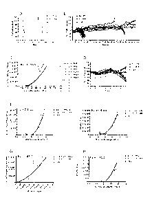
[0027] Figure 1 shows (A) the percent survival of mice after high dose or
low dose
infection with WNV; (B) weight loss associated with high dose infection of
mice with WNV;
(C) the viral titers in the brain of mice that succumb to infection; (D)
weight loss in mice
infected with high and low dose WNV through days 0-7 post infection; (E) the
viral titers in
the brains of mice infected with high and low dose WNV at day 7 post infection
and the
CA 028177552013-05-10
WO 2012/065153 PCT/US2011/060537
correlation between percentage of weight loss on day 7 and viral titer; (F)
the correlation
between percentage weight loss and the presence of CD45+ leukocytes in the
brain of mice at
7 days post infection with high and low dose WNV; (G) the correlation between
viral titer in
the brain and the presence of CD45 leukocytes in the brain of mice at 7 days
post infection
with high and low dose WNV; (H) the correlation between percentage weight loss
and the
presence of CD45111 macrophages in the brain of mice at 7 days post infection
with high and
low dose WNV.
[0028] Figure 2 shows the correlation between percentage of weight loss and
the
presence of (A) CD45intCD11b+ immigrant microglia; (C) CD3+ T cells; (D)
CD1lbhi Ly6G+
neutrophils and (E) NK1.1 'CD1 1 bl 1- natural killer cells while the numbers
of CD45lo resident
microglia (B) remained unchanged after 7 days post infection with high dose or
low dose
WN V; (F) shows the correlation between weight loss and virus titer in the
brain at the time of
sacrifice of mice infected with low dose WNV; (G) shows the correlation of
leukocyte
infiltration and percentage weight loss in mice infected with low dose WNV and
(H) shows
that there was no correlation between virus titer and leukocyte infiltration
after low dose
WNV infection.
[0029] Figure 3 (A) shows the long term survival of mice treated with
carboxylated
polystyrene beads in PBS at day 6 after infection with high dose WNV; (B)
shows that
treating low dose WNV-infected mice with carboxylated polystyrene beads
beginning at day
6 post infection is ineffective at prolonging survival of mice; (C) shows that
treating low dose
WNV-infected mice with carboxylated polystyrene beads beginning at day 6 post
infection is
ineffective at preventing weight loss in mice compared to control mice; (D)
shows that
treatment of low dose WNV-infected mice is effective at prolonging survival of
mice when
the beads are administered upon weight loss in the mice. (E-G) shows weight
loss recorded
in these mice up to 20 days pi.
[0030] Figure 4 (A-D) are examples of treating low dose WNV-infected mice
with
carboxylated polystyrene beads upon weight loss in the mice the mice in (A-B)
only require
bead treatment for 5 days and weight remains stable and they go on to survive
without further
bead treatment, whereas the mice in (C-D) begin to lose weight again when bead
treatment is
ceased after 5 days, so treatment resumes until weight restabalizes (E) shows
the infiltration
of CD45+CD1 lb+ macrophages into the brains of low dose WNV-infected mice at 9
days
post infection, that either lost weight or did not lose weight and were
treated with either PBS
or carboxylated beads at day 8 post infection; (F) is a graphical
representation of the types of
cells found infiltrating the brain of WNV-infected mice at 9 days post
infection, that either
6
CA 028177552013-05-10
WO 2012/065153 PCT/US2011/060537
lost weight or did not lose weight and were treated with either PBS or
carboxylated beads at
day 8 post infection.
[00311 Figure 5 shows (A) the difference in survival of mice treated with
carboxylated
polystyrene beads, naked polystyrene beads or PBS after low dose infection
with WNV; (B)
shows the difference in percent weight loss in mice treated with carboxylated
beads, naked
beads or PBS after low dose infection with WNV; (C,D) shows the difference in
percent
weight loss between carboxylated bead treatment and naked bead treatment in
mice after low
dose infection with WNV; (E,O) shows the localization of FITC-conjugated
carboxylated
beads or naked beads on day 7 in mice infected with high dose WNV on day 0 and
FITC-
carboxylated beads, FITC-naked beads or PBS on day 6. (E-G) are blood from 3
separate
PBS-treated mice, (H-J) are blood from 3 separate naked polystyrene bead-
treated mice, and
(L-N) are blood from 3 separate carboxylated polystyrene bead-treated mice,
showing that
more of the plain beads remain in the blood than carboxylated beads.
[00321 Figure 6 shows (A-C) the lack of infiltration of FITC-conjugated
polystyrene
beads in the brains of mice infected and treated as in Figure E-0; (D-E) shows
the reduction
in infiltration of various leukocytes, macrophages and microglia into the
brains of WNV-
infected mice treated with carboxylated polystyrene beads or naked polystyrene
beads as in
Figure 5 (E-0).
[00331 Figure 7 shows (A) the association of FITC-conjugated polystyrene
carboxylated
beads and FITC-conjugated naked polystyrene beads in the spleen with CD45+
leukocytes
(A,B,F) within CD1 lb (C,G), CD 1 lc (D,H) Ly6c+ (E,I) cells; (J-R) shows the
types of
cells that take up FITC-conjugated carboxylated beads and FITC-conjugated
naked beads
[00341 Figure 8 shows the ability of FITC-conjugated polystyrene
carboxylated beads or
FITC-conjugated naked polystyrene beads to be taken up by and increase the
numbers of
CD11b+ CD I lca monocytes (A) and CD1 lb CD1 lc (B) or CD1lba CD1 1 (C)
dendritic
cells in the spleen after infection with high dose WNV.
[00351 Figure 9 shows (A-D) the ability of FITC-conjugated carboxylated
polystyrene
beads or FITC conjugated naked polystyrene beads to be taken up by and
increase the
numbers of CD19+ B cell and CD3+ T cell subsets in the spleen after infection
with high dose
WNV.
[00361 Figure 10 shows (A-L) the ability of FITC-conjugated carboxylated
polystyrene
beads or FITC conjugated naked polystyrene beads to be taken up by CD11b+
(C,G), CD1 lc+
(D,H), and Ly6c+ (E,I) cells, specifically, within CD11b+ CD 11c- monocytes
(J) and CD11b+
7
CA 028177552013-05-10
WO 2012/065153 PCT/US2011/060537
CD11c+ (K) or CD11b- CD1 1c+ (L) dendritic cells, in the liver after infection
with high dose
WNV.
[0037] Figure 11 shows (A-G) the ability of FITC-conjugated carboxylated
polystyrene
beads or FITC conjugated naked polystyrene beads to be taken up by CD1 lb
,(C,F), CD1 1 c+
and Ly6C+ (D,G) cells, in the bone marrow after infection with high dose WNV.
[0038] Figure 12 shows (A) the percent survival of low dose WNV-infected
mice treated
with high dose or low dose carboxylated polystyrene beads of different sizes;
(B) shows the
percent survival of low dose WNV-infect mice treated with FITC-conjugated
carboxylated
beads, naked FITC-conjugated beads, carboxylated PLGA spheres or naked PLGA
spheres;
(C) shows the infiltration/activation of various monocyte populations in the
brain of mice
infected with low dose WNV and treated with carboxylated FITC-beads,
carboxylated-FITC
PLGA spheres, or carboxylated nanodiamonds.
[0039] Figure 13 shows (A) the percent survival and (B) weight loss in wild-
type and T
cell deficient mice infected with high or low dose WNV; (C) the correlation
between weight
loss and viral titers in the brains of wild-type and T cell deficient mice
infected with high or
low dose WNV; (D) weight loss (E) and immune cell infiltration into the brains
of wild type
and T cell deficient mice infected with high dose WNV at day 8 post infection;
(F) percent
survival (G) and weight loss of wild-type and T cell deficient mice infected
with high or low
dose WNV and treated with carboxylated beads or PBS upon significant weight
loss.
DETAILED DESCRIPTION OF THE INVENTION
[0040] The present inventors have surprisingly found that when carboxylated
particles,
such as carboxylated polystyrene, PLGA, or diamond particles of a certain
size, are
administered to subjects, inflammatory immune responses are ameliorated.
Additionally, the
present inventors have also surprisingly found that these same carboxylated
particles, when
administered to subjects with active viral or bacterial infections,
particularly those infecting
the central nervous system prolong, lead to a dramatic decrease in symptoms of
these
infections and prolonged survival. These, particles, therefore, may be useful
in the treatment
of any disease or condition characterized by an excessive inflammatory immune
response,
such as autoimmune diseases, as well as in the treatment of bacterial and
viral infections.
[0041] "Particle" as used herein refer to any non-tissue derived minute
composition of
matter, it may be a sphere or sphere-like entity or bead. The term "particle"
and the term
"bead" may be used interchangeably. Additionally, the term "particle" may be
used to
encompass beads and spheres.
8
CA 0281 755 2013-05-10
WO 2012/065153 PCT/US2011/060537
[0042] "Carboxylated particles" or "carboxylated beads" or "carboxylated
spheres"
includes any particle that has been modified to contain a carboxyl group on
its surface. In
some embodiments the addition of the carboxyl group enhances
phagocyte/monocyte uptake
of the particles from circulation, for instance through the interaction with
scavenger receptors
such as MARCO.
[0043] "Antigenic moiety" as used herein refers to any moiety, for example
a peptide,
that is recognized by the host's immune system. Examples of antigenic moieties
include, but
are not limited to, autoantigens and/or bacterial or viral proteins, peptides
or components.
Without being bound by theory, while the carboxylated beads themselves may be
recognized
by the immune system, the carboxylated beads with nothing more attached
thereto are not
considered an "antigenic moiety" for the purposes of the invention.
[00441 "Naked beads" or "naked particles" or "naked spheres" as used herein
refers to
beads, particles or spheres that have not been carboxylated.
[0045] The particle may have any particle shape or conformation. However,
in some
embodiments it is preferred to use particles that are less likely to clump in
vivo. Examples of
particles within these embodiments are those that have a spherical shape.
[0046] It is not necessary that each particle be uniform in size, although
the particles must
generally be of a size sufficient to trigger phagocytosis in an antigen
presenting cell or other
MPS cell. Preferable, the particles are microscopic or nanoscopic in size, in
order to enhance
solubility, avoid possible complications caused by aggregation in vivo and to
facilitate
pinocytosis. Particle size can be a factor for uptake from the interstitial
space into areas of
lymphocyte maturation. A particle having a diameter of from about 0.1 gm to
about 10 gm is
capable of triggering phagocytosis. Thus in one embodiment, the particle has a
diameter
within these limits. In another embodiment, the particle has a diameter of
about 0.3 Inn to
about 5 gm. In still another embodiment, the particle has a diameter of about
0.5 gm to about
3 gm. In preferred embodiment the particle has a size of about 0.5 gm. The
particles in a
composition need not be of uniform diameter. By way of example, a
pharmaceutical
formulation may contain a plurality of particles, some of which are about 0.5
gm, while
others are about 1.0 gm. Any mixture of particle sizes within these given
ranges will be
useful.
[0047] In some embodiments, the particle is non-metallic. In these
embodiments the
particle may be formed from a polymer. In a preferred embodiment, the particle
is
biodegradable in an individual. In this embodiment, the particles can be
provided to an
9
CA 028177552013-05-10
WO 2012/065153 PCT/US2011/060537
individual across multiple doses without there being an accumulation of
particles in the
individual. Examples of suitable particles include polystyrene particles, PLGA
particles, and
diamond particles.
[0048] Preferably the particle surface is composed of a material that
minimizes non-
specific or unwanted biological interactions. Interactions between the
particle surface and the
interstitium may be a factor that plays a role in lymphatic uptake. The
particle surface may be
coated with a material to prevent or decrease non-specific interactions.
Steric stabilization by
coating particles with hydrophilic layers such as poly(ethylene glycol) (PEG)
and its
copolymers such as PLURONICS (including copolymers of poly(ethylene glycol)-bl-
poly(propylene glycol)-bl-poly(ethylene glycol)) may reduce the non-specific
interactions
with proteins of the interstitium as demonstrated by improved lymphatic uptake
following
subcutaneous injections. All of these facts point to the significance of the
physical properties
of the particles in terms of lymphatic uptake. Biodegradable polymers may be
used to make
all or some of the polymers and/or particles and/or layers. Biodegradable
polymers may
undergo degradation, for example, by a result of functional groups reacting
with the water in
the solution. The term "degradation" as used herein refers to becoming
soluble, either by
reduction of molecular weight or by conversion of hydrophobic groups to
hydrophilic groups.
Polymers with ester groups are generally subject to spontaneous hydrolysis,
e.g., polylactides
and polyglycolides.
[0049] Particles of the present invention may also contain additional
components. For
example, carriers may have imaging agents incorporated or conjugated to the
carrier. An
example of a carrier nanosphere having an imaging agent that is currently
commercially
available is the Kodak X-sight nanospheres. Inorganic quantum-confined
luminescent
nanocrystals, known as quantum dots (QDs), have emerged as ideal donors in
FRET
applications: their high quantum yield and tunable size-dependent Stokes
Shifts permit
different sizes to emit from blue to infrared when excited at a single
ultraviolet wavelength.
(Bruchez, et al., Science, 1998, 281, 2013; Niemeyer, C. M Angew. Chem. Int.
Ed. 2003, 42,
5796; Waggoner, A. Methods Enzymol. 1995, 246, 362; Brus, L. E. J. Chem. Phys.
1993, 79,
5566). Quantum dots, such as hybrid organic/inorganic quantum dots based on a
class of
polymers known as dendrimers, may used in biological labeling, imaging, and
optical
biosensing systems. (Lemon, et al., J. Am. Chem. Soc. 2000, 122, 12886).
Unlike the
traditional synthesis of inorganic quantum dots, the synthesis of these hybrid
quantum dot
nanoparticles does not require high temperatures or highly toxic, unstable
reagents. (Etienne,
et al., Appl. Phys. Lett. 87, 181913, 2005).
CA 028177552013-05-10
WO 2012/065153 PCT/US2011/060537
[0050] Particles can be formed from a wide range of materials. The particle
is preferably
composed of a material suitable for biological use. For example, particles may
be composed
of glass, silica, polyesters of hydroxy carboxylic acids, polyanhydrides of
dicarboxylic acids,
or copolymers of hydroxy carboxylic acids and dicarboxylic acids. More
generally, the
carrier particles may be composed of polyesters of straight chain or branched,
substituted or
unsubstituted, saturated or unsaturated, linear or cross-linked, alkanyl,
haloalkyl, thioalkyl,
aminoalkyl, aryl, aralkyl, alkenyl, aralkenyl, heteroaryl, or alkoxy hydroxy
acids, or
polyanhydrides of straight chain or branched, substituted or unsubstituted,
saturated or
unsaturated, linear or cross-linked, alkanyl, haloalkyl, thioalkyl,
aminoalkyl, aryl, aralkyl,
alkenyl, aralkenyl, heteroaryl, or alkoxy dicarboxylic acids. Additionally,
carrier particles can
be quantum dots, or composed of quantum dots, such as quantum dot polystyrene
particles
(Joumaa et al. (2006) Langmuir 22: 1810-6). Carrier particles including
mixtures of ester and
anhydride bonds (e.g., copolymers of glycolic and sebacie acid) may also be
employed. For
example, carrier particles may comprise materials including polyglycolic acid
polymers
(PGA), polylactic acid polymers (PLA), polysebacic acid polymers (PSA),
poly(lactic-co-
glycolic) acid copolymers (PLGA), [rho]oly(lactic-co-sebacic) acid copolymers
(PLSA),
poly(glycolic-co-sebacic) acid copolymers (PGSA), etc. Other biocompatible,
biodegradable
polymers useful in the present invention include polymers or copolymers of
caprolactones,
carbonates, amides, amino acids, orthoesters, acetals, cyanoacrylates and
degradable
urethanes, as well as copolymers of these with straight chain or branched,
substituted or
unsubstituted, alkanyl, haloalkyl, thioalkyl, aminoalkyl, alkenyl, or aromatic
hydroxy- or di-
carboxylic acids. In addition, the biologically important amino acids with
reactive side chain
groups, such as lysine, arginine, aspartic acid, glutamic acid, serine,
threonine, tyrosine and
cysteine, or their enantiomers, may be included in copolymers with any of the
aforementioned materials to provide reactive groups for conjugating to antigen
peptides and
proteins or conjugating moieties. Biodegradable materials suitable for the
present invention
include diamond, PLA, PGA, and PLGA polymers. Biocompatible but non-
biodegradable
materials may also be used in the carrier particles of the invention. For
example, non-
biodegradable polymers of acrylates, ethylene-vinyl acetates, acyl substituted
cellulose
acetates, non-degradable urethanes, styrenes, vinyl chlorides, vinyl
fluorides, vinyl
imidazoles, chlorosulphonated olefins, ethylene oxide, vinyl alcohols, TEFLON
(DuPont,
Wilmington, Del.), and nylons may be employed.
[0051] Suitable beads which are currently available commercially include
polystyrene
beads such as FluoSpheres (Molecular Probes, Eugene, Oreg.).
11
CA 028177552013-05-10
WO 2012/065153 PCT/US2011/060537
[0052] Physical properties are also related to a nanoparticle's usefulness
after uptake and
retention in areas having immature lymphocytes. These include mechanical
properties such as
rigidity or rubberiness. Some embodiments are based on a rubbery core, e.g., a
poly(propylene sulfide) (PPS) core with an overlayer, e.g., a hydrophilic
overlayer, as in
PEG, as in the PPS-PEG system recently developed and characterized for
systemic (but not
targeted or immune) delivery. The rubbery core is in contrast to a
substantially rigid core as
in a polystyrene or metal nanoparticle system. The term rubbery refers to
certain resilient
materials besides natural or synthetic rubbers, with rubbery being a term
familiar to those in
the polymer arts. For example, cross-linked PPS can be used to form a
hydrophobic rubbery
core. PPS is a polymer that degrades under oxidative conditions to
polysulphoxide and finally
polysulphonc, transitioning from a hydrophobic rubber to a hydrophilic, water-
soluble
polymer. Other sulphide polymers may be adapted for use, with the term
sulphide polymer
referring to a polymer with a sulphur in the backbone of the mer. Other
rubbery polymers that
may be used are polyesters with glass transition temperature under hydrated
conditions that is
less than about 37 C. A hydrophobic core can be advantageously used with a
hydrophilic
overlayer since the core and overlayer will tend not to mingle, so that the
overlayer tends to
sterically expand away from the core. A core refers to a particle that has a
layer on it. A layer
refers to a material covering at least a portion of the core. A layer may be
adsorbed or
covalently bound. A particle or core may be solid or hollow. Rubbery
hydrophobic cores are
advantageous over rigid hydrophobic cores, such as crystalline or glassy (as
in the case of
polystyrene) cores, in that higher loadings of hydrophobic drugs can be
carried by the
particles with the rubbery hydrophobic cores.
[0053] Another physical property is the surface's hydrophilicity. A
hydrophilic material
may have a solubility in water of at least 1 gram per liter when it is
uncrosslinked. Steric
stabilization of particles with hydrophilic polymers can improve uptake from
the interstitium
by reducing non-specific interactions; however, the particles' increased
stealth nature can also
reduce internalization by phagocytic cells in areas having immature
lymphocytes. The
challenge of balancing these competing features has been met, however, and
this application
documents the creation of nanoparticles for effective lymphatic delivery to
DCs and other
APCs in lymph nodes. Some embodiments include a hydrophilic component, e.g., a
layer of
hydrophilic material. Examples of suitable hydrophilic materials are one or
more of
polyalkylene oxides, polyethylene oxides, polysaccharides, polyacrylic acids,
and polyethers.
The molecular weight of polymers in a layer can be adjusted to provide a
useful degree of
steric hindrance in vivo, e.g., from about 1,000 to about 100,000 or even
more; artisans will
12
CA 028177552013-05-10
WO 2012/065153 PCT/US2011/060537
immediately appreciate that all the ranges and values within the explicitly
stated ranges are
contemplated, e.g., between 10,000 and 50,000.
[0054] The nanoparticles may incorporate functional groups for further
reaction.
Functional groups for further reaction include electrophiles or nucleophiles;
these are
convenient for reacting with other molecules. Examples of nucleophiles are
primary amines,
thiols, and hydroxyls. Examples of electrophiles are succinimidyl esters,
aldehydes,
isocyanates, and maleimides.
[0055] The particles of the current invention can be given in any dose
effective to
dampen the inflammatory immune response in a subject in need thereof or to
treat a bacterial
or viral infection in a subject in need thereof. In certain embodiments, about
102 to about
1020 particles are provided to the individual. In a further embodiment between
about 103 to
about 1015 particles are provided. In yet a further embodiment, between about
106 to about
1012 particles are provided. In still a further embodiment between about 108
to about 1010
particles are provided. In one embodiment the preferred dose is 0.1%
solids/ml. Therefore,
for 0.5 gm beads, a preferred dose is approximately 4 x 10⁹ beads, for 0.05um
beads, a
preferred dose is approximately 4 x 10¹² beads, for 3um beads, a preferred
dose is 2 x 101
beads. However, any dose that is effective in treating the particular
condition to be treated is
encompassed by the current invention.
[0056] The invention is useful for treatment of immune related disorders
such as
autoimmune disease, transplant rejection and allergic reactions. Substitution
of a synthetic,
biocompatible particle system to induce immune tolerance could lead to ease of
manufacturing, broad availability of therapeutic agents, increase uniformity
between samples,
increase the number of potential treatment sites and dramatically reduce the
potential for
allergic responses to a carrier cell.
[0057] As used herein, the term "immune response" includes T cell mediated
and/or B
cell mediated immune responses. Exemplary immune responses include T cell
responses,
e.g., cytokine production and cellular cytotoxicity. In addition, the term
immune response
includes immune responses that are indirectly affected by T cell activation,
e.g., antibody
production (humoral responses) and activation of cytokinc responsive cells,
e.g.,
macrophages. Immune cells involved in the immune response include lymphocytes,
such as B
cells and T cells (CD4 CD8', Thl and Th2 cells); antigen presenting cells
(e.g., professional
antigen presenting cells such as dendritic cells, macrophages, B lymphocytes,
Langerhans
cells, and nonprofessional antigen presenting cells such as keratinocytes,
endothelial cells,
13
CA 028177552013-05-10
WO 2012/065153 PCT/US2011/060537
astrocytes, fibroblasts, oligodendrocytes); natural killer cells; myeloid
cells, such as
macrophages, eosinophils, mast cells, basophils, and granulocytes. In some
embodiments,
the modified particles of the present invention are effective to reduce
inflammatory cell
trafficking to the site of inflammation.
[0058] As used herein, the term "anergy," "tolerance," or "antigen-specific
tolerance"
refers to insensitivity of T cells to T cell receptor-mediated stimulation.
Such insensitivity is
generally antigen- specific and persists after exposure to the antigenic
peptide has ceased. For
example, anergy in T cells is characterized by lack of cytokine production,
e.g., IL-2. T-cell
anergy occurs when T cells are exposed to antigen and receive a first signal
(a T cell receptor
or CD-3 mediated signal) in the absence of a second signal (a costimulatory
signal). Under
these conditions, re-exposure of the cells to the same antigen (even if re-
exposure occurs in
the presence of a costimulatory molecule) results in failure to produce
cytokines and
subsequently failure to proliferate. Thus, a failure to produce cytokines
prevents proliferation.
Anergic T cells can, however, proliferate if cultured with cytokines (e.g., IL-
2). For example,
T cell anergy can also be observed by the lack of IL-2 production by T
lymphocytes as
measured by ELISA or by a proliferation assay using an indicator cell line.
Alternatively, a
reporter gene construct can be used. For example, anergic T cells fail to
initiate DL-2 gene
transcription induced by a heterologous promoter under the control of the 5'
IL-2 gene
enhancer or by a multimer of the API sequence that can be found within the
enhancer (Kang
et al. 1992 Science. 257:1134).
[0059] As used herein, the term "immunological tolerance" refers to methods
performed
on a proportion of treated subjects in comparison with untreated subjects
where: a) a
decreased level of a specific immunological response (thought to be mediated
at least in part
by antigen-specific effector T lymphocytes, B lymphocytes, antibody, or their
equivalents);
b) a delay in the onset or progression of a specific immunological response;
or c) a reduced
risk of the onset or progression of a specific immunological response.
"Specific"
immunological tolerance occurs when immunological tolerance is preferentially
invoked
against certain antigens in comparison with others. "Non-Specific"
immunological tolerance
occurs when immunological tolerance is invoked indiscriminately against
antigens which
lead to an inflammatory immune response. "Quasi-Specific" immunological
tolerance occurs
when immunological tolerance is invoked semi-discriminately against antigens
which lead to
a pathogenic immune response but not to others which lead to a protective
immune response.
[0060] A proxy for tolerogenic activity is the ability of a particle to
stimulate the
production of an appropriate cytokine at the target site. The immunoregulatory
cytokine
14
CA 0281 755 2013-05-10
WO 2012/065153 PCT/US2011/060537
released by T suppressor cells at the target site is thought to be TGF-I3
(Miller et al., Proc.
Natl. Acad. Sci. USA 89:421, 1992). Other factors that may be produced during
tolerance are
the cytokines IL-4 and IL-10, and the mediator PGE. In contrast, lymphocytes
in tissues
undergoing active immune destruction secrete cytokines such as IL-1, 1L-2, IL-
6, and IFNy.
Hence, the efficacy of a modified particle can be evaluated by measuring its
ability to
stimulate the appropriate type of cytokines.
[0061] With this in mind, a rapid screening test for modified particles,
effective mucosal
binding components, effective combinations, or effective modes and schedules
of mucosal
administration can be conducted using animal model systems. Animals are
treated at a
mucosal surface with the test particle composition, and at some time are
challenged with
administration of the disease causing antigen or an infectious agent. Spleen
cells are isolated,
and cultured in vitro in the presence of the disease causing antigen or an
antigent derived
from the infectious gent at a concentration of about 50 gg/mL. Cytokinc
secretion into the
medium can be quantitated by standard immunoassay.
[0062] The ability of the particles to suppress the activity of cells can
be determined
using cells isolated from an animal immunized with the modified particles, or
by creating a
cell line responsive to a disease causing antigen or viral antigen target
antigen (Ben-Nun et
al., Eur. J. Immunol. 11:195, 1981). In one variation of this experiment, the
suppressor cell
population is mildly irradiated (about 1000 to 1250 rads) to prevent
proliferation, the
suppressors are co-cultured with the responder cells, and then tritiated
thymidine
incorporation (or MTT) is used to quantitate the proliferative activity of the
responders. In
another variation, the suppressor cell population and the responder cell
population are
cultured in the upper and lower levels of a dual chamber transwell culture
system (Costar,
Cambridge Mass.), which permits the populations to coincubate within 1 mm of
each other,
separated by a polycarbonate membrane (WO 93/16724). In this approach,
irradiation of the
suppressor cell population is unnecessary, since the proliferative activity of
the responders
can be measured separately.
[0063] The effectiveness of compositions and modes of administration for
treatment of
specific disease can also be elaborated in a corresponding animal disease
model. The ability
of the treatment to diminish or delay the symptomatology of the disease is
monitored at the
level of circulating biochemical and immunological hallmarks of the disease,
immunohistology of the affected tissue, and gross clinical features as
appropriate for the
CA 028177552013-05-10
WO 2012/065153 PCT/US2011/060537
model being employed. Non-limiting examples of animal models that can be used
for testing
are included in the following section.
[0064] The invention contemplates modulation of tolerance by modulating TH1
response,
TH2 response, TH17 response, or a combination of these responses. Modulating
TH1
response encompasses changing expression of, e.g., interferon-gamma.
Modulating TH2
response encompasses changing expression of, e.g., any combination of IL-4, IL-
5, IL-10,
and IL-13. Typically an increase (decrease) in TH2 response will comprise an
increase
(decrease) in expression of at least one of IL-4, IL-5, IL-10, or IL-13; more
typically an
increase (decrease) in TH2 response will comprise an increase in expression of
at least two of
IL-4, IL-5, IL-10, or IL-13, most typically an increase (decrease) in TH2
response will
comprise an increase in at least three of 1L-4, IL-5, IL-10, or 1L-13, while
ideally an increase
(decrease) in TH2 response will comprise an increase (decrease) in expression
of all of IL-4,
IL-5, IL-10, and IL-13. Modulating TH17 encompasses changing expression of,
e.g., TGF-
beta, IL-6, IL-21 and IL-23, and effects levels of IL-17, IL-21 and IL-22.
[0065] Tolerance to autoantigens and autoimmune disease is achieved by a
variety of
mechanisms including negative selection of self-reactive T cells in the thymus
and
mechanisms of peripheral tolerance for those autoreactive T cells that escape
thymic deletion
and are found in the periphery. Examples of mechanisms that provide peripheral
T cell
tolerance include "ignorance" of self antigens, anergy or unresponsiveness to
autoantigen,
cytokine immune deviation, and activation-induced cell death of self- reactive
T cells. In
addition, regulatory T cells have been shown to be involved in mediating
peripheral
tolerance. See, for example, Walker et al. (2002) Nat. Rev. Immunol. 2: 11-19;
Shevach et al.
(2001) Immunol. Rev. 182:58-67. In some situations, peripheral tolerance to an
autoantigen is
lost (or broken) and an autoimmune response ensues. For example, in an animal
model for
EAE, activation of antigen presenting cells (APCs) through TLR innate immune
receptors
was shown to break self-tolerance and result in the induction of EAE (Waldner
et al. (2004) J.
Clin. Invest. 113:990-997).
[0066] Accordingly, in some embodiments, the invention provides methods for
increasing antigen presentation while suppressing or reducing TLR7/8, TLR9,
and/or TLR
7/8/9 dependent cell stimulation. As described herein, administration of
particular modified
particles results in antigen presentation by DCs or APCs while suppressing the
TLR 7/8,
TLR9, and/ot TLR7/8/9 dependent cell responses associated with
immunostimulatory
polynucleotides. Such suppression may include decreased levels of one or more
TLR-
associated cytokines.
16
CA 028177552013-05-10
WO 2012/065153 PCT/US2011/060537
[0067] As discussed above this invention provides novel compounds that have
biological
properties useful for the treatment of Mac-1 and LFA-1 mediated disorders.
[0068] Accordingly, in another aspect of the present invention,
pharmaceutical
compositions are provided, which comprise the carboxylated particles and
optionally
comprise a pharmaceutically acceptable carrier. In certain embodiments, these
compositions
optionally further comprise one or more additional therapeutic agents.
Alternatively, the
modified particles of the current invention may be administered to a patient
in need thereof in
combination with the administration of one or more other therapeutic agents.
For example,
additional therapeutic agents for conjoint administration or inclusion in a
pharmaceutical
composition with a compound of this invention may be an approved anti-
inflammatory agent,
or it may be any one of a number of agents undergoing approval in the Food and
Drug
Administration that ultimately obtain approval for the treatment of any
disorder characterized
by an uncontrolled inflammatory immune response or a bacterial or viral
infection. It will
also be appreciated that certain of the modified particles of present
invention can exist in free
form for treatment, or where appropriate, as a pharmaceutically acceptable
derivative thereof.
[0069] The pharmaceutical compositions of the present invention
additionally comprise a
pharmaceutically acceptable carrier, which, as used herein, includes any and
all solvents,
diluents, or other liquid vehicle, dispersion or suspension aids, surface
active agents, isotonic
agents, thickening or emulsifying agents, preservatives, solid binders,
lubricants and the like,
as suited to the particular dosage form desired. Remington's Pharmaceutical
Sciences,
Sixteenth Edition, E. W. Martin (Mack Publishing Co., Easton, Pa., 1980)
discloses various
carriers used in formulating pharmaceutical compositions and known techniques
for the
preparation thereof. Except insofar as any conventional carrier medium is
incompatible with
the compounds of the invention, such as by producing any undesirable
biological effect or
otherwise interacting in a deleterious manner with any other component(s) of
the
pharmaceutical composition, its use is contemplated to be within the scope of
this invention.
Some examples of materials which can serve as pharmaceutically acceptable
carriers include,
but are not limited to, sugars such as lactose, glucose and sucrose; starches
such as corn
starch and potato starch; cellulose and its derivatives such as sodium
carboxymethyl
cellulose, ethyl cellulose and cellulose acetate; powdered tragacanth; malt;
gelatine; talc;
excipients such as cocoa butter and suppository waxes; oils such as peanut
oil, cottonseed oil;
safflower oil, sesame oil; olive oil; corn oil and soybean oil; glycols; such
as propylene
glycol; esters such as ethyl oleate and ethyl laurate; agar; buffering agents
such as magnesium
hydroxide and aluminum hydroxide; alginic acid; pyrogenfree water; isotonic
saline; Ringer's
17
CA 0281 755 2013-05-10
WO 2012/065153 PCT/US2011/060537
solution; ethyl alcohol, and phosphate buffer solutions, as well as other non-
toxic compatible
lubricants such as sodium lauryl sulfate and magnesium stearate, as well as
coloring agents,
releasing agents, coating agents, sweetening, flavoring and perfuming agents,
preservatives
and antioxidants can also be present in the composition, according to the
judgment of the
formulator.
[00701 Liquid dosage forms for oral administration include, but are not
limited to,
pharmaceutically acceptable emulsions, microemulsions, solutions, suspensions,
syrups and
elixirs. In addition to the active compounds, the liquid dosage forms may
contain inert
diluents commonly used in the art such as, for example, water or other
solvents, solubilizing
agents and emulsifiers such as ethyl alcohol, isopropyl alcohol, ethyl
carbonate, ethyl acetate,
benzyl alcohol, benzyl benzoate, propylene glycol, 1,3-butylene glycol,
dimethylformamide,
oils (in particular, cottonseed, groundnut, corn, germ, olive, castor, and
sesame oils),
glycerol, tetrahydrofurfuryl alcohol, polyethylene glycols and fatty acid
esters of sorbitan,
and mixtures thereof. Besides inert diluents, the oral compositions can also
include adjuvants
such as wetting agents, emulsifying and suspending agents, sweetening,
flavoring, and
perfuming agents.
[0071] Injectable preparations, for example, sterile injectable aqueous or
oleaginous
suspensions may be formulated according to the known art using suitable
dispersing or
wetting agents and suspending agents. The sterile injectable preparation may
also be a sterile
injectable solution, suspension or emulsion in a nontoxic parenterally
acceptable diluent or
solvent, for example, as a solution in 1,3-butanediol. Among the acceptable
vehicles and
solvents that may be employed are water, Ringer's solution, U.S.P. and
isotonic sodium
chloride solution. In addition, sterile, fixed oils are conventionally
employed as a solvent or
suspending medium. For this purpose any bland fixed oil can be employed
including
synthetic mono-or diglyccrides. In addition, fatty acids such as oleic acid
are used in the
preparation of injectables.
[0072] The injectable formulations can be sterilized, for example, by
filtration through a
bacterial-retaining filter, or by incorporating sterilizing agents in the form
of sterile solid
compositions which can be dissolved or dispersed in sterile water or other
sterile injectable
medium prior to use.
[0073] In order to prolong the effect of a drug, it is often desirable to
slow the absorption
of the drug from subcutaneous or intramuscular injection. This may be
accomplished by the
use of a liquid suspension or crystalline or amorphous material with poor
water solubility.
The rate of absorption of the drug then depends upon its rate of dissolution
that, in turn, may
18
CA 028177552013-05-10
WO 2012/065153 PCT/US2011/060537
depend upon crystal size and crystalline form. Alternatively, delayed
absorption of a
parenterally administered drug form is accomplished by dissolving or
suspending the drug in
an oil vehicle. Injectable depot forms are made by forming microencapsule
matrices of the
drug in biodegradable polymers such as polylactide-polyglycolide. Depending
upon the ratio
of drug to polymer and the nature of the particular polymer employed, the rate
of drug release
can be controlled. Examples of other biodegradable polymers include
(poly(orthoesters) and
poly(anhydrides). Depot injectable formulations are also prepared by
entrapping the drug in
liposomes or microemulsions which are compatible with body tissues.
100741 Solid dosage forms for oral administration include capsules,
tablets, pills,
powders, and granules. In such solid dosage forms, the modified particles are
mixed with at
least one inert, pharmaceutically acceptable excipient or carrier such as
sodium citrate or
dicalcium phosphate and/or a) fillers or extenders such as starches, lactose,
sucrose, glucose,
mannitol, and silicic acid, h) binders such as, for example,
carboxymethylcellulose, alginates,
gelatin, polyvinylpyrrolidinone, sucrose, and acacia, c) humectants such as
glycerol, d)
disintegrating agents such as agar-agar, calcium carbonate, potato or tapioca
starch, alginic
acid, certain silicates, and sodium carbonate, e) solution retarding agents
such as paraffin, f)
absorption accelerators such as quaternary ammonium compounds, g) wetting
agents such as,
for example, cetyl alcohol and glycerol monostearate, h) absorbents such as
kaolin and
bentonite clay, and i) lubricants such as talc, calcium stearate, magnesium
stearate, solid
polyethylene glycols, sodium lauryl sulfate, and mixtures thereof. In the case
of capsules,
tablets and pills, the dosage form may also comprise buffering agents.
[0075] Solid compositions of a similar type may also be employed as fillers
in soft and
hard-filled gelatin capsules using such excipients as lactose or milk sugar as
well as high
molecular weight polyethylene glycols and the like. The solid dosage forms of
tablets,
dragees, capsules, pills, and granules can be prepared with coatings and
shells such as enteric
coatings and other coatings well known in the pharmaceutical formulating art.
They may
optionally contain opacifying agents and can also be of a composition that
they release the
active ingredient(s) only, or preferentially, in a certain part of the
intestinal tract, optionally,
in a delayed manner. Examples of embedding compositions that can be used
include
polymeric substances and waxes. Solid compositions of a similar type may also
be employed
as fillers in soft and hard-filled gelatin capsules using such excipients as
lactose or milk sugar
as well as high molecular weight polyethylene glycols and the like.
[0076] The modified particles can also be in micro-encapsulated form with
one or more
excipients as noted above. The solid dosage forms of tablets, dragees,
capsules, pills, and
19
CA 028177552013-05-10
WO 2012/065153 PCT/US2011/060537
granules can be prepared with coatings and shells such as enteric coatings,
release controlling
coatings and other coatings well known in the pharmaceutical formulating art.
In such solid
dosage forms the active compound may be admixed with at least one inert
diluent such as
sucrose, lactose and starch. Such dosage forms may also comprise, as in normal
practice,
additional substances other than inert diluents, e.g., tableting lubricants
and other tableting
aids such as magnesium stearate and microcrystalline cellulose. In the case of
capsules,
tablets and pills, the dosage forms may also comprise buffering agents. They
may optionally
contain opacifying agents and can also be of a composition that they release
the modified
particles only, or preferentially, in a certain part of the intestinal tract,
optionally, in a delayed
manner. Examples of embedding compositions which can be used include polymeric
substances and waxes.
[00771 The present invention encompasses pharmaceutically acceptable topical
formulations of the inventive modified particles. The term "pharmaceutically
acceptable
topical formulation", as used herein, means any foimulation which is
pharmaceutically
acceptable for intradermal administration of modified microparticles of the
invention by
application of the formulation to the epidermis. In certain embodiments of the
invention, the
topical formulation comprises a carrier system. Pharmaceutically effective
carriers include,
but are not limited to, solvents (e.g., alcohols, poly alcohols, water),
creams, lotions,
ointments, oils, plasters, liposomes, powders, emulsions, microemulsions, and
buffered
solutions (e.g., hypotonic or buffered saline) or any other carrier known in
the art for
topically administering pharmaceuticals. A more complete listing of art-known
carriers is
provided by reference texts that are standard in the art, for example,
Remington's
Pharmaceutical Sciences, 16th Edition, 1980 and 17th Edition, 1985, both
published by Mack
Publishing Company, Easton, Pa., the disclosures of which are incorporated
herein by
reference in their entireties. In certain other embodiments, the topical
formulations of the
invention may comprise excipients. Any pharmaceutically acceptable excipient
known in the
art may be used to prepare the inventive pharmaceutically acceptable topical
formulations.
Examples of excipients that can be included in the topical formulations of the
invention
include, but are not limited to, preservatives, antioxidants, moisturizers,
emollients, buffering
agents, solubilizing agents, other penetration agents, skin protectants,
surfactants, and
propellants, and/or additional therapeutic agents used in combination to the
modified
particles. Suitable preservatives include, but are not limited to, alcohols,
quaternary amines,
organic acids, parabens, and phenols. Suitable antioxidants include, but are
not limited to,
ascorbic acid and its esters, sodium bisulfite, butylated hydroxytoluene,
butylated
CA 0281 755 2013-05-10
WO 2012/065153 PCT/US2011/060537
hydroxyanisole, tocopherols, and chelating agents like EDTA and citric acid.
Suitable
moisturizers include, but are not limited to, glycerine, sorbitol,
polyethylene glycols, urea,
and propylene glycol. Suitable buffering agents for use with the invention
include, but are not
limited to, citric, hydrochloric, and lactic acid buffers. Suitable
solubilizing agents include,
but are not limited to, quaternary ammonium chlorides, cyclodextrins, benzyl
benzoate,
lecithin, and polysorbates. Suitable skin protectants that can be used in the
topical
formulations of the invention include, but are not limited to, vitamin E oil,
allatoin,
dimethicone, glycerin, petrolatum, and zinc oxide.
[0078] In certain embodiments, the pharmaceutically acceptable topical
formulations of
the invention comprise at least the modified particles of the invention and a
penetration
enhancing agent. The choice of topical formulation will depend or several
factors, including
the condition to be treated, the physicochemical characteristics of the
inventive compound
and other excipients present, their stability in the formulation, available
manufacturing
equipment, and costs constraints. As used herein the term "penetration
enhancing agent"
means an agent capable of transporting a pharmacologically active compound
through the
stratum comeum and into the epidermis or dermis, preferably, with little or no
systemic
absorption. A wide variety of compounds have been evaluated as to their
effectiveness in
enhancing the rate of penetration of drugs through the skin. See, for example,
Percutaneous
Penetration Enhancers, Maibach H. I. and Smith H. E. (eds.), CRC Press, Inc.,
Boca Raton,
Fla. (1995), which surveys the use and testing of various skin penetration
enhancers, and
Buyuktimkin et al., Chemical Means of Transdermal Drug Permeation Enhancement
in
Transdermal and Topical Drug Delivery Systems, Gosh T. K., Pfister W. R., Yum
S. I.
(Eds.), Interpharm Press Inc., Buffalo Grove, Ill. (1997). In certain
exemplary embodiments,
penetration agents for use with the invention include, but are not limited to,
triglycerides
(e.g., soybean oil), aloe compositions (e.g., aloe-vera gel), ethyl alcohol,
isopropyl alcohol,
octolyphenylpolyethylene glycol, oleic acid, polyethylene glycol 400,
propylene glycol, N-
decylmethylsulfoxide, fatty acid esters (e.g., isopropyl myristate, methyl
laurate, glycerol
monooleate, and propylene glycol monooleate) and N-methylpyrrolidone.
[00791 In certain embodiments, the compositions may be in the form of
ointments, pastes,
creams, lotions, gels, powders, solutions, sprays, inhalants or patches. In
certain exemplary
embodiments, formulations of the compositions according to the invention are
creams, which
may further contain saturated or unsaturated fatty acids such as stearic acid,
palmitic acid,
oleic acid, palmito-oleic acid, cetyl or oleyl alcohols, steatic acid being
particularly preferred.
Creams of the invention may also contain a non-ionic surfactant, for example,
polyoxy-40-
2 1
CA 028177552013-05-10
WO 2012/065153 PCT/US2011/060537
stearate. In certain embodiments, the active component is admixed under
sterile conditions
with a pharmaceutically acceptable carrier and any needed preservatives or
buffers as may be
required. Ophthalmic formulation, eardrops, and eye drops are also
contemplated as being
within the scope of this invention. Additionally, the present invention
contemplates the use of
transdermal patches, which have the added advantage of providing controlled
delivery of a
compound to the body. Such dosage forms are made by dissolving or dispensing
the
compound in the proper medium. As discussed above, penetration enhancing
agents can also
be used to increase the flux of the compound across the skin. The rate can be
controlled by
either providing a rate controlling membrane or by dispersing the compound in
a polymer
matrix or gel.
[00801 The modified particles can be administered by aerosol. This is
accomplished by
preparing an aqueous aerosol, liposomal preparation or solid particles
containing the
modified particles. A nonaqueous (e.g., fluorocarbon propellant) suspension
could be used.
[0081] Ordinarily, an aqueous aerosol is made by formulating an aqueous
solution or
suspension of the agent together with conventional pharmaceutically acceptable
carriers and
stabilizers. The carriers and stabilizers vary with the requirements of the
particular
compound, but typically include nonionic surfactants (Tweens, Pluronics, or
polyethylene
glycol), innocuous proteins like serum albumin, sorbitan esters, oleic acid,
lecithin, amino
acids such as glycine, buffers, salts, sugars or sugar alcohols. Aerosols
generally are prepared
from isotonic solutions.
[00821 It will also be appreciated that the modified particles and
pharmaceutical
compositions of the present invention can be formulated and employed in
combination
therapies, that is, the compounds and pharmaceutical compositions can be
formulated with or
administered concurrently with, prior to, or subsequent to, one or more other
desired
therapeutics or medical procedures. The particular combination of therapies
(therapeutics or
procedures) to employ in a combination regimen will take into account
compatibility of the
desired therapeutics and/or procedures and the desired therapeutic effect to
be achieved. It
will also be appreciated that the therapies employed may achieve a desired
effect for the same
disorder (for example, an inventive compound may be administered concurrently
with
another anti-inflammatory agent), or they may achieve different effects (e.g.,
control of any
adverse effects).
[0083] In certain embodiments, the pharmaceutical compositions containing
the modified
particles of the present invention further comprise one or more additional
therapeutically
active ingredients (e.g., anti-inflammatory and/or palliative). For purposes
of the invention,
22
CA 028177552013-05-10
WO 2012/065153 PCT/US2011/060537
the term "Palliative" refers to treatment that is focused on the relief of
symptoms of a disease
and/or side effects of a therapeutic regimen, but is not curative. For
example, palliative
treatment encompasses painkillers, antinausea medications and anti-sickness
drugs.
[0084] The invention provides methods of regulating an immune response in
an
individual, preferably a mammal, more preferably a human, comprising
administering to the
individual the modified particles described herein. Methods of
immunoregulation provided
by the invention include those that suppress and/or inhibit an innate immune
response or an
adaptive immune response, including, but not limited to, an immune response
stimulated by
immunostimulatory polypeptides or viral or bacterial components.
[0085] The modified particles are administered in an amount sufficient to
regulate an
immune response. As described herein, regulation of an immune response may be
humoral
and/or cellular, and is measured using standard techniques in the art and as
described herein.
[0086] In certain embodiments, the individual suffers from a disorder
associated with
unwanted immune activation, such as allergic disease or condition, allergy and
asthma. An
individual having an allergic disease or asthma is an individual with a
recognizable symptom
of an existing allergic disease or asthma.
[0087] In certain embodiments, the individual suffers from a disorder
associated with
unwanted immune activation, such as atherosclerosis, ischemie reperfusion
injury, and
myocardial infarction.
[0088] In certain embodiments, the individual suffers from a disorder
associated with
unwanted immune activation, such as autoimmune disease and inflammatory
disease. An
individual having an autoimmune disease or inflammatory disease is an
individual with a
recognizable symptom of an existing autoimmune disease or inflammatory
disease.
[0089] Autoimmune diseases can be divided in two broad categories: organ-
specific and
systemic. Autoimmune diseases include, without limitation, rheumatoid
arthritis (RA),
systemic lupus erythematosus (SLE), type I diabetes mellitus, type II diabetes
mellitus,
multiple sclerosis (MS), immune- mediated infertility such as premature
ovarian failure,
scleroderma, Sjogren's disease, vitiligo, alopecia (baldness), polyglandular
failure, Grave's
disease, hypothyroidism, polymyositis, pemphigus vulgaris, pemphigus
foliaceus,
inflammatory bowel disease including Crohn's disease and ulcerative colitis,
autoimmune
hepatitis including that associated with hepatitis B virus (HBV) and hepatitis
C virus (HCV),
hypopituitarism, graft-versus-host disease (GvHD), myocarditis, Addison's
disease,
autoimmune skin diseases, uveitis, pernicious anemia, and hypoparathyroidism.
23
CA 028177552013-05-10
WO 2012/065153 PCT/US2011/060537
[0090] Autoimmune diseases may also include, without limitation,
Hashimoto's
thyroiditis, Type I and Type II autoimmune polyglandular syndromes,
paraneoplastic
pemphigus, bullus pemphigoid, dermatitis herpetiformis, linear IgA disease,
epidermolysis
bullosa acquisita, erythema nodosa, pemphigoid gestationis, cicatricial
pemphigoid, mixed
essential cryoglobulinemia, chronic bullous disease of childhood, hemolytic
anemia,
thrombocytopenic purpura, Goodpasture's syndrome, autoimmune neutropenia,
myasthenia
gravis, Eaton-Lambert myasthenic syndrome, stiff-man syndrome, acute
disseminated
encephalomyelitis, Guillain-Barre syndrome, chronic inflammatory demyelinating
polyradiculoneuropathy, multifocal motor neuropathy with conduction block,
chronic
ncuropathy with monoclonal gammopathy, opsonoclonus-myoclonus syndrome,
cerebellar
degeneration, encephalomyelitis, retinopathy, primary biliary sclerosis,
sclerosing
cholangitis, gluten-sensitive enteropathy, ankylosing spondylitis, reactive
arthritides,
polymyositisidermatomyositi s, mixed connective tissue disease, B ech et' s
syndrome,
psoriasis, polyarteritis nodosa, allergic anguitis and granulomatosis (Churg-
Strauss disease),
polyangiitis overlap syndrome, hypersensitivity vasculitis, Wegener's
granulomatosis,
temporal arteritis, Takayasu's arteritis, Kawasaki's disease, isolated
vasculitis of the central
nervous system, thromboangiutis obliterans, sarcoidosis, glomerulonephritis,
and cryopathies.
These conditions are well known in the medical arts and are described, for
example, in
Harrison's Principles of Internal Medicine, 14th ed., Fauci A S et al., eds.,
New York:
McGraw-Hill, 1998.
[0091] Animal models for the study of autoimmune disease are known in the
art. For
example, animal models which appear most similar to human autoimmune disease
include
animal strains which spontaneously develop a high incidence of the particular
disease.
Examples of such models include, but are not limited to, the nonobeses
diabetic (NOD)
mouse, which develops a disease similar to type 1 diabetes, and lupus-like
disease prone
animals, such as New Zealand hybrid, MRL-FasiPr and BXSB mice. Animal models
in which
an autoimmune disease has been induced include, but are not limited to,
experimental
autoimmune encephalomyelitis (EAE), which is a model for multiple sclerosis,
collagen-
induced arthritis (CIA), which is a model for rheumatoid arthritis, and
experimental
autoimmune uveitis (EAU), which is a model for uveitis. Animal models for
autoimmune
disease have also been created by genetic manipulation and include, for
example, IL-2/IL-10
knockout mice for inflammatory bowel disease, Fas or Fas ligand knockout for
SLE, and IL-1
receptor antagonist knockout for rheumatoid arthritis.
24
CA 028177552013-05-10
WO 2012/065153 PCT/US2011/060537
[0092] In certain embodiments, the individual suffers from a bacterial or
viral infection.
An individual having a bacterial or viral infection is an individual with a
recognizable
symptom of an existing bacterial or viral infection.
[0093] A non-limiting list of viral infections treatable with the modified
particles of the
current invention includes herpes virus infections, hepatitis virus
infections, west nile virus
infections, flavivrus infections, influenza virus infections, rhinovirus
infections,
papillomavirus infections, paromyxovirus infections, parainfluenza virus
infections, and
retrovirus infections. Preferred viruses are those viruses that infect the
central nervous
system of the subject. Most preferred viruses are those that cause
encephalitis or meningitis.
[0094] A non-limiting list of bacterial infections treatable with the
modified particles of
the current invention include staphlococcus infections, streptococcus
infections,
mycobacterial infections, bacillus infections, Salmonella infections, Vibrio
infections,
spirochete infections, and Neisseria infections. Preferred are bacteria that
infect the central
nervous system of the subject. Most preferred are bacteria that cause
encephalitis or
meningitis.
[0095] In some embodiments, the invention relates to uses of compositions
of this
invention prior to the onset of disease. In other embodiments, the invention
relates to uses of
the compositions of this invention to inhibit ongoing disease. In some
embodiments, the
invention relates to ameliorating disease in a subject. By ameliorating
disease in a subject is
meant to include treating, preventing or suppressing the disease in the
subject.
[0096] In some embodiments, the invention relates to preventing the relapse
of disease.
For example, an unwanted immune response can occur at one region of a peptide
(such as an
antigenic determinant). Relapse of a disease associated with an unwanted
immune response
can occur by having an immune response attack at a different region of the
peptide. Since the
carboxylated particles of the current invention arc free from attached
peptides or antigenic
moieties, the particles will be effective against multiple epitopes. T-cell
responses in some
immune response disorders, including MS and other TH1 /17-mediated autoimmune
diseases,
can be dynamic and evolve during the course of relapsing-remitting and/or
chronic-
progressive disease. The dynamic nature of the T-cell repertoire has
implications for
treatment of certain diseases, since the target may change as the disease
progresses.
Previously, pre-existing knowledge of the pattern of responses was necessary
to predict the
progression of disease. The present invention provides compositions that can
prevent the
effect of dynamic changing disease, a function of "epitope spreading." A known
model for
relapse is an immune reaction to proteolipid protein (PLP) as a model for
multiple sclerosis
CA 028177552013-05-10
WO 2012/065153 PCT/US2011/060537
(MS). Initial immune response can occur by a response to PLP139_1515.
Subsequent disease
onset can occur by a relapse immune response to PLP151-171.
[0097] Certain embodiments of this invention relate to treatment of
pathological
conditions relating to an unwanted hypersensitivity. The hypersensitivity can
be any one of
types I, II, III, and IV. Immediate (type I) hypersensitivity. The frequency
of administration
will typically correspond with the timing of allergen exposure. Suitable
animal models are
known in the art (for example, Gundel et al., Am. Rev. Respir. Dis. 146:369,
1992, Wada et
al., J. Med. Chem. 39, 2055, 1996; and WO 96/35418).
[00981 Other embodiments of this invention relate to transplantation. This
refers to the
transfer of a tissue sample or graft from a donor individual to a recipient
individual, and is
frequently performed on human recipients who need the tissue in order to
restore a
physiological function provided by the tissue. Tissues that are transplanted
include (but are
not limited to) whole organs such as kidney, liver, heart, lung; organ
components such as skin
grafts and the cornea of the eye; and cell suspensions such as bone marrow
cells and cultures
of cells selected and expanded from bone marrow or circulating blood, and
whole blood
transfusions.
[0099] A serious potential complication of any transplantation ensues from
antigenic
differences between the host recipient and the engrafted tissue. Depending on
the nature and
degree of the difference, there may be a risk of an immunological assault of
the graft by the
host, or of the host by the graft, or both, may occur. The extent of the risk
is determined by
following the response pattern in a population of similarly treated subjects
with a similar
phenotype, and correlating the various possible contributing factors according
to well
accepted clinical procedures. The immunological assault may be the result of a
preexisting
immunological response (such as preformed antibody), or one that is initiated
about the time
of transplantation (such as the generation of TH cells). Antibody, T helper
(TH) cells, or
cytotoxic T (Tc) cells may be involved in any combination with each other and
with various
effector molecules and cells. However, the antigens which are involved in the
immune
response are generally not known, therefore posing difficulties in designing
antigen-specific
therapies or inducing antigen-specific tolerance. The modified particles of
the current
invention are particularly useful in preventing the rejection of organs
because no attached
peptides or antigens need to be conjugated to the modified particles in order
for the particles
to be effective in inducing tolerance or ameliorate an inflammatory immune
response.
[00100] Certain embodiments of the invention relate to decreasing the risk of
host versus
graft disease, leading to rejection of the tissue graft by the recipient. The
treatment may be
26
CA 028177552013-05-10
WO 2012/065153 PCT/US2011/060537
performed to prevent or reduce the effect of a hyperacute, acute, or chronic
rejection
response. Treatment is preferentially initiated sufficiently far in advance of
the transplant so
that tolerance will be in place when the graft is installed; but where this is
not possible,
treatment can be initiated simultaneously with or following the transplant.
Regardless of the
time of initiation, treatment will generally continue at regular intervals for
at least the first
month following transplant. Follow-up doses may not be required if a
sufficient
accommodation of the graft occurs, but can be resumed if there is any evidence
of rejection
or inflammation of the graft. Of course, the tolerization procedures of this
invention may be
combined with other forms of immunosuppression to achieve an even lower level
of risk.
EXAMPLES
[00101] The following examples are provided to further illustrate the
advantages and
features of the invention, but are not intended to limit the scope of this
disclosure.
Materials and Methods
Virus propagation
[00102] As previously described (Getts et al., J Neurochem. 103 :1019, 2007),
West Nile
Virus (Sarafend strain) was derived from the brains of neonatal mice and used
to infect
confluent vero cell monolayers, at a multiplicity of infection of 5 plaque
forming units (PFU)
per cell. Cells were incubated with virus for 40 hours at 37 C, after which
they were frozen.
Flasks were then thawed and the virus-rich supernatant clarified by
centrifugation, after
which aliquots were stored at ¨70 C until use.
Mice and Infection
[00103] Eight to twelve-week old female C57BL/6 (CD45.2) and congenic B6.SJL-
PtprcaPep3b/BoyJ (CD45.1) mice were obtained from the Animal Resources Center,
Western
Australia. C57BL/6-7.2fms-EGFP transgenic (CD45.2) mice were obtained from the
Transgenic Animal Resources Center Queensland, Australia. All procedures were
performed
with permission of the University of Sydney Animal Ethics Committee. All
animals were
housed under class 11 biohazard conditions in hepa-filter top cages. Food and
water was
provided ad libitum.
[00104] High dose intra-nasal infection was conducted as previously described
with 6x10⁴
PFU WNV in sterile phosphate-buffered saline (PBS; Gibco BRL, California,
USA). For the
low dose infection, mice were inoculated with 6x10³ PFU WNV. Mock infection
was
27
CA 028177552013-05-10
WO 2012/065153 PCT/US2011/060537
conducted by inoculating mice with sterile PBS only. (Getts et al., J
Neurochem. 103: 1019,
2007, Wacher et al., J Virol. 81: 860, 2007).
[00105] Mice were weighed twice daily following infection. Mice were
sacrificed under
anaesthesia by cardiac perfusion using 40m1 of ice cold PBS. For histology,
mice were
further perfused with 20m1 of 4% paraformaldehyde (Sigma Aldrich, St Louis,
USA) in PBS.
Plaque assay to determine viral titre
[00106] To determine titres of live virus in tissue samples, a plaque assay
using virus-
susceptible BHK cells (kindly donated by Guna Kapuriah, John Curtin Medical
School,
Canberra, Australia) was employed. As previously described (Getts et al., J
Neurochem. 103:
1019, 2007), tissue samples were dissected from animals and disassociated with
a power
homogeniser (Tissue Tearor, Biospec, Bartles, OK, USA). Briefly, five-fold
dilutions of
clarified homogenates were prepared in Roswell Park Memorial Institute 1640
media (RPMI;
CSL Biosciences), and used to infect confluent BHK cells in 6-well plates (1 x
106 cells
seeded overnight in 2mL RPMI).
[00107] Cells were incubated for 1 hour at 37 C, after which the inoculums
were removed
by aspiration. Wells were overlaid with 3 ml of 1.5% (w/v) low-gelling Agarose
II (Amresco,
Solon, OH, USA) in 2X Minimum Essential Medium (MEM; GibcoBRL, Grand Island,
NY,
USA). Cells were incubated for a further 3 days at 37 C, after which they were
fixed with 3
ml of 10% formalin (Co.) for 2 hr prior to agarose plug removal. A 3% crystal
violet
(Hopkins and Williams, Essex, England) dye solution in 20% methanol (Fronine,
Riverstone,
NSW, Australia) was used to stain fixed cells. Plaques were counted using a
colony counter
(JUL S. A., Barcelona, Spain), and the final PFU per gram (tissue) was
determined by
factoring the number of plaques, the inoculum volume and sample dilution.
Generation of chimeric mice
[00108] As previously described (Getts et al., J Neurochem. 103: 1019,
2007), six- to
eight-week old B6.SJL-PtprcaPep3b/BoyJ (CD45.1) mice were irradiated with one
dose of
950 rads. Twelve hours later, mice were reconstituted with 107 bone marrow
cells from
C57BL/6-7.2fins-EGFP donors. Mice were given sulfamethoxazole (Sigma Aldrich)
and
trimethoprim (Sigma Aldrich) in the drinking water for 10 days following
irradiation. Mice
were infected with WN V six weeks after irradiation, as described above.
Chimerism was
28
CA 0281 755 2013-05-10
WO 2012/065153 PCT/US2011/060537
checked using flow cytometry and was invariably found to be 96-99% of donor
origin as
previously demonstrated (Getts et al., J Neurochem. 103: 1019, 2007).
Intravenous injection of beads
[00109] 0.5, 0.05, and 3ium FITC or Bright Blue (BB) Flouresbrite naked and
carboxylated
polystyrene beads were obtained from Polyscience, NY, USA. Poly(lactic-co-
glycolic acid)
(PLGA) naked and carboxylate beads were obtained from Phosphorex, MA, USA.
[00110] Beads were diluted in sterile PBS, and 300 p.1 was intravenously
injected into the
tail veins of infected or mock-infected mice. For high dose survival studies
(6x10⁴ PFU
WNV i.n.), mice were given one dose of beads each day beginning on day 6 post
infection If
required, mice were given further injections each day until weight had
restabilized. For low
dose survival studies (6x10³ PFU WN V i.n.), mice were either injected with
beads from d6
p.i., or when significant weight loss was recorded (4-6% of total body
weight). Mice were
injected with 300u1 of beads once a day for 5 days, or until weight had
restabilized. For tissue
collection, 6x10⁴ or 6x10³-infected mice were injected with 30041 of beads on
either d6 p.i.,
or when significant weight loss was recorded, and culled when required. Organs
were
harvested for immunohistochemistry, flow cytometry, plaque assay, and cytokine
analysis, as
described below.
Inununohistology
[00111] Fluoresence immunohistochemistry (IHC) was conducted on brain, spleen,
liver,
lungs and kidneys collected from C57BL/6 and cFMS-EGFP (into B6.SJL-
PtpreaPep3b/BoyJ)
chimeras. Following perfusion, organs were fixed in 4% paraformaldehyde at 4 C
for 4 hours
and then immersed in 30% sucrose overnight, before being frozen in Optimum
Cutting
Temperature Compound (OCT; Tissue-Tek, Tokyo Japan). Eight-micron tissue
sections were
cut on a cryostat microtome and air-dried. Fluorescence IHC was performed as
previously
described, (Getts et al, J Exp Med. 29: 2319, 2007), with the addition of a
tyramide-based
amplification system (TSA kit; Perkin Elmer, Belgium), used according to the
manufacturer's
instructions. Tissue sections were counter-stained with DAPI-anti fade
(Vector) prior to
visualisation.
29
CA 0281 755 2013-05-10
WO 2012/065153 PCT/US2011/060537
Microscope and image acquisition
[0100] Images were acquired on an Olympus BX-51 microscope (Olympus, Japan),
using a
DP-70 camera and DP manager 2.2.1 software (Olympus).
Isolation of leukocytes from the brain and liver
[0101] As previously described (Getts et al, J Exp Med. 29: 2319, 2007)
leukocytes were
obtained from the brains of PBS-perfused mice by digesting brains for 60
minutes at 37 C in
PBS with deoxy-ribonuclease (0.005 g/ml; Sigma Aldrich) and collagenase IV
(0.05 g/ml;
Sigma Aldrich). Digestion was stopped with 10% FCS, and the homogenate was
passed
through a 70itm nylon cell strainer (Becton Dickinson, NJ, USA). The pellet,
obtained after
minutes centrifugation at 340xg, was resuspended in 30% Percoll (Amersham,
Norway)
and layered over 80% Percoll. Leukocytes were collected from the 30%/80%
interface after
centrifugation at 1140xg for 25 minutes at room temperature. The same protocol
is also used
to derive leukocytes from the liver, with the tissue weighed before
processing.
Isolation of leukocytes from the spleen, blood and bone marrow
[0102] For flow cytometric analysis, the right femur was dissected out and
bone marrow cells
flushed out using PBS loaded syringes. For bone marrow precursor isolation,
femurs and
tibias from at least 4 mice were utilized. The cellular suspension achieved
after flushing was
filtered through a 70iam cell strainer and centrifuged for 5 mins at 340g. Red
blood cells in
the resulting pellet were lysed in NH4C1-based red cell lysis buffer (BD Pharm
Lyselm; BD
Pharmingen), before centrifugation for 5 mins at 340xg. In the case of
peripheral blood,
blood was collected via cardiac puncture and immediately transferred into
citrate buffer
(mMol, Sigma Alrich). The resulting suspension was layered over 70% Percoll
and
centrifuged at 1140xg for 20 minutes at room temperature with the brake off
The interface
was collected and the cells washed once in PBS, centrifuged at 340xg. For the
isolation of
splenic leukocytes, spleens were passed through a 70701am cell strainer and
centrifuged for 5
mins at 340g. Red blood cells in the resulting pellet were lysed in NH4C1-
based red cell lysis
buffer (BD Pharna LyseTM; BD Pharmingen), before centrifugation for 5 mins at
340xg.
CA 028177552013-05-10
WO 2012/065153 PCT/US2011/060537
Flow cytometry
[0103] Cells collected (as described above) from the brain, liver, blood, and
bone marrow
were washed in PBS, and blocked with anti-CDI6/CD32 antibody (Biolegend).
Viable cells
were counted using trypan blue exclusion, which routinely showed >95% cell
viability.
[0104] Cell surface molecule expression was measured and cell sorts carried
out on a FACS
ARIA (Becton Dickinson), equipped with an Argon ion and HeNe laser. Viable
populations
were gated by forward and side scatter and identified fluorescent populations
determined by
forward-gating thereafter. Sorting was carried out using specific fluorescent
and scatter
parameters identifying the population of interest. Sorting stringencies was
set to purity to
achieve >98% purity for bone marrow populations.
[0105] Acquired FACS data files were analysed using the flow cytometry
program, Flow Jo
(FlowJo, Ashland, OR, USA). Quantification of cell populations of interest
were calculated
based on flow cytometry percentages at analysis and absolute cell counts from
each organ.
Adoptive transfer
[0106] Both Ly6Chi/cFMS-EGFP /CD1 1 b ' and Ly6C16/Cfms-EGFP /CD 1 b
populations
were sorted from day 6 intranasally WNV-infected mice. Ly6Ch1 BM was labeled
with 10
mM cell tracker orange (CMTMR [5-(and -6)-(((4-chloromethyl) benzoyl) amino)
tetramethylrhodamine)-mixed isomers; Invitrogen). Sorted cells were
centrifuged and raised
in 1 ml PBS containing 10 i_.tM CMTMR. Cells were stained for 10 min before
the reaction
was stopped with 10% FCS. Cells were washed in 50 ml PBS at least three times.
Labeled
CMTMR Ly6Chi BM cells were then mixed 1:1 with Ly6C16 cells, which, except for
the
addition of CMTMR, had been treated similarly to Ly6Ch1 populations. A total
of 2 x 10⁶ BM
cells were i.v. injected into either day 6.5 WNV-infected or mock-infected
Ly5.1-057BL/6
congenic mice. This was immediately followed by an intravenous injection of
300u1 BB
polystyrene beads. 12 h later, brains, spleens and livers were harvested as
described above.
The presence of donor cells was investigated using flow cytometry, both using
cFMS-EGFP
and CMTMR¨cell tracker orange.
Multiplex ELISA
[0107] Multiplexed plate ELISAs were performed according to the manufacturers
instructions (Quansys Biosciences, Logan, Utah, USA). Briefly, brain, spleen,
and liver tissue
were homogenized in PBS, clarified by a 1000xg spin, and stored at -20 C until
the assay was
31
CA 0281 755 2013-05-10
WO 2012/065153 PCT/US2011/060537
performed. Serum samples were also used. Thawed samples and standards were
diluted in the
provided buffer, and 30 1 of each were plated in each well that contains 16
spots each
containing a capture antibody for a particular soluble protein. Plates were
then incubated for
1 hour on an orbital shaker at 120r.p.m. Plates were washed 3 times, and 30u1
of detection
antibody was added to each well and incubated for another hour. After washing
3 times,
strepavidin-HRP was added and incubated for a further 15 minutes. Plates were
then washed
6 times, and substrate mix was added. Plates were immediately read on a CCD
imager
(Kodak, Rochester NY, USA). Plate images were analysed using Quansys Q-view
software
(Quansys Biosciences).
Induction and evaluation of Experimental Autoinunune Encephalitis (EAE)
[01081 C57BL/6 mice were injected sub-cutaneously with emulsion containing
0.1mg MOG
Peptide (MEVGWYRSPFSRVVHLYRNGK; Auspep, Parkvi lie, Victoria, Australia; >95%
HPLC purified) and Complete Freund's adjuvant containing 2mg/mL Mycobacterium
tuberculosis (Sigma Aldrich). Two days later, mice were administered 500,u1
Pertussis toxin
(Sigma Aldrich) i.p. Mice were monitored for disease progression, and graded
on the
following scale: 1, limp tail and/or weakness of 1 hind limb; 2, weakness in
more than one
limb, gait disturbance; 3, paralysis in 1 limb; 4, paralysis in more than one
limb,
incontinence: 5, moribund.
Induction of thioglycolate-induced peritonfitis
[01091 The induction of peritonitis was performed by the injection of lml
thioglycolate (4%
(w/v); Sigma Alrich) dissolved in PBS. Intraperitoneal lavage was performed on
mice
sacrificed by cervical dislocation. Breifly, 5 ml of peritoneal lavage buffer
(PLB; PBS
containing 0.5mM EDTA (Fronine) with heparin (Sigma Aldrich) 9.9 units/m1) was
injected
into the peritoneum. The peritoneum was gently massaged before the PLB was
aspirated into
a 10m1 syringe. This process was repeated twice. The lavage was then raised to
50m1 in PLB
and spun at 340xg for 5 minutes. Cells were prepared for flow cytometry as
described above.
Statistics
[01101 Graphs were made and computerized statistical analysis was performed in
GraphPad
Prism, and InStat, respectively (both programs from GraphPad software, San
Diego, CA,
32
CA 028177552013-05-10
WO 2012/065153 PCT/US2011/060537
USA). Depending on the data, an unpaired, two-tailed Student t-test, or one
way ANOVA
with a Tukey-Kramer post test was performed, with P < 0.05 considered to be
significant.
For correlation analysis between parameters such as weight loss, infiltration,
and virus
titre, a non-linear regression (curve fit) was used, with a second order
polynomial (Y=A +
B*X + C*XA2).
Example 1
Characterization of the high and low does models of FINV encephalitis
[0111] High dose infection of C57BL/6 mice with 6x10⁴ PFU WNV i.n. results in
100%
mortality on d7 p.i. (Figure 1A). Macrophages and immigrant microglia, which
we have
previously shown to derive from circulating Ly6ch1 monocyte precursors (Getts
et al, J Exp
Med. 29: 2319, 2007), infiltrate the WNV-infected brain from d5 p.i. T cells,
NK cells, and
neutrophils also enter the CNS from this timepoint onwards, with the peak of
infiltration seen
on d7 p.i. (Figure 1F-H, Figure 2A-D) Brain virus titre also exponentially
increases from d5
p.i., reaching high levels on d7 p.i. (Figure 1C), and body weight of infected
mice
significantly decreases from d5 p.i. until death (Figure 1B).
[0112] Inoculation of mice with 10-fold less virus, i.e. 6x10³ PFU i.n.,
produces sub-lethal
outcomes. Some variability occurs in mortality rate when comparing independent
experiments, which ranges between 40-60% (Figure 1A); however, a strong
correlation
between the percentage of weight loss and virus titre/infiltration has been
consistently shown
(Figure 1B-H, Figure 2A-E). The daily monitoring of mouse weight has proven to
be a
reliable method to predict the outcome of infection in individuals, and is
indicative of when
mice require intervention in order to survive infection (Figure 1B).
[0113] Mice that do not lose weight after inoculation with 6x10³ WNV do not
succumb to
infection, and are immune to subsequent reinfection with 6x10⁴ (Figure 1A-B).
These mice
do not show any symptoms of illness upon initial infection or high dose
reinfection, including
weight loss. Conversely, mice that lose a significant amount of weight
compared to the
normal fluctuations of mock-infected controls (usually >4% of body weight in a
24 hour time
period) continue to lose weight, typically for 2-3 days before death occurs.
In this model of
encephalitis, the majority of mice that succumb to infection begin to lose
weight between d6
and dl 1 post infection, and death occurs before d16 p.i. These mice show high
titres of virus
in the brain at TOD, comparable to d7 mice infected with 6x10⁴ (Figure 1C).
33
CA 0281 755 2013-05-10
WO 2012/065153 PCT/US2011/060537
[01141 Flow cytometry comparing leukocyte infiltration in the brains of 6x10⁴
and 6x10³-
infected mice has revealed that percentage weight loss on d7 p.i. strongly
correlates with the
infiltration of total leukocytes (Figure 1F), macrophages (Figure 1H),
immigrant microglia
(Figure 2A), T cells (Figure 2C), and NK cells (Figure 2E), with some
correlation shown
between weight loss and neutrophil immigration (2D). Not surprisingly, numbers
of CD4510
microglia remained relatively unchanged in all mice, as this population mainly
comprises the
resident microglia of the brain (Figure 2B).
[01151 To further investigate the low dose model of infection, 6x10³-infected
mice were
weighed daily and sacraficed on dO-d14, with brains harvested for flow
cytometry and plaque
assay. Some correlation was shown between percentage weight loss at time of
sacrifice and
virus titre (Figure 2F), or leukocyte infiltration (Figure 2G). However, a
comparison between
virus titre and leukocyte infiltration revealed that some mice had high titres
of virus without
significant infiltration (Figure 2H), and while some of these mice had also
lost a significant
amount of weight, others had not (Figure 2F). Because mice have to be
sacraficed to
determine virus titres in the brain, it is unclear whether these mice that had
virus in the brain
without significant weight loss or leukocyte infiltration would clear the
virus without
showing weight loss or infiltration, or if the high virus titre seen precedes
infiltration of the
brain/weight loss.
Example 2
Carboxylated bead treatment significantly improves survival in the high and
low dose models
of WNV encephalitis
[01161 Fluorescent beads have been used frequently in the literature to follow
the trafficking
of monocyte subsets in various in vivo models of disease. However, these
studies overlook
the potential influence that these theoretically "inert" beads may have on
monocyte function,
and as a result, the outcome of disease.
[01171 In an attempt to track monocytes during WNV infection, we used both
naked, and
carboxylated polystyrene beads, and found an unexpected alteration on the
course of disease.
The following data reveal that both naked and carboxylated polystyrene beads
significantly
reduce infiltration of the brain by immune cells, and promote long-term
survival of infected
mice.
[01181 Injection of 6 x 10⁴ WNV-infected mice with 4.41x10⁹ O.5jsm
carboxylated
polystyrene beads in 300 1 PBS on d6 p.i. i.v. resulted in 10% long-term
survival of mice in
34
CA 0281 755 2013-05-10
WO 2012/065153 PCT/US2011/060537
this lethal model of disease (Figure 3A). To see if survival outcomes could be
improved, we
also injected this same concentration of beads into mice infected with the
lower dose of 6x10³
on d6 p.i. However, this strategy reduced survival of treated mice as compared
to PBS-treated
controls (Figure 3B). These results emphasize that beads need to be
administered
therapeutically i.e. when mice initially show significant weight loss, which
we have shown to
be indicative of high virus titre and infiltration of the brain by leukocyte
populations (see
Figure 1,2).
[0119] Therefore we adopted a therapeutic approach to bead administration in
the low dose
model of infection. Mice were weighed daily (Figure 3E-G), and 4.41x10⁹
carboxylated
polystyrene beads were administered if significant weight loss was detected in
a 24-hour time
period (usually >4% of total body weight, as compared to the normal
fluctuations of mock-
infected controls). Using this strategy, we were able to increase survival of
mice that would
usually continue to lose weight and die without intervention, by 60% (40-80%
in independent
experiments) (Figure 3D). As expected, all mice that lost weight and were
treated with PBS
continued to lose weight and died 2-4 days later (Figure 3E, G). Mice that did
not lose a
significant amount of weight at any time showed similar stability in weight as
PBS-infected
controls (Figure 3E-F).
[0120] Mice were treated with carboxylated beads for 5 consecutive days upon
the initial
detection of significant weight loss. In some mice, 5 days of bead treatment
was sufficient
and weight remained stabilized after bead injection ceased and these mice went
on to survive
infection without further intervention (Figure 4A-B). However, some mice began
to lose
weight again when bead treatment ceased at 5 days, so treatment was
recommenced until
weight restabalized, and these mice also survived long-term (Figure 4C-D).
[0121] Flow cytometry was conducted on d9 p.i. on brains of 6 x 10³ WNV-
infected mice
that were either treated with carboxylated polystyrene beads or PBS when
significant weight
loss was detected on d8 p.i. Mice that had not lost weight were also bead- or
PBS-treated on
d8 as controls for the experiment. As shown in Figures 4D-L, mice that did not
lose weight
on d8 p.i. but were bead- (Figure 4H-I) or PBS-treated (Figure 4D-E) showed no
infiltration
of the brain by monocyte-derived macrophages or immigrant microglia, T cells,
NK cells or
neutrophils, with the main population isolated being the CD451wiffi CD11b+
resident
microglia. In mice that did lose weight on d8 p.i. and were treated with PBS,
a massive
infiltration of the brain was evident on d9 p.i. (Figure 4F-G), with the main
population being
the CD45hi inflammatory monocyte-derived macrophages (Figure 4L). However, in
the mice
that lost weight and were treated with beads on d8 p.i., a significant
infiltrate was still
CA 028177552013-05-10
WO 2012/065153 PCT/US2011/060537
present, but was greatly reduced in comparison to PBS-treated mice (Figure 4J-
L). Flow
cytometry of 103-infected mice treated with bead or PBS upon weight loss needs
to be
repeated to show that reductions in infiltration occur when mice are bead-
treated across days
6 -14 p.i.
Example 3
Carboxylation of beads is critical for significant improvement in survival and
changes in
leukocyte populations in WNV-inftcted mice
[0122] At this stage of investigation, it was unclear whether the
carboxylation of beads was
critical for the improvements seen in survival and reductions in leukocyte
trafficking to the
brain. 6 x 10³ WNV-infected mice were injected with 4.41x10⁹ 0.5 rim naked or
carboxylated
beads in 300 1 PBS iv., or PBS only, when significant weight loss was
recorded. Mice
injected with carboxylated beads showed a significant improvement in survival
of 60%,
whereas naked bead-treated mice showed a much smaller improvement of 25%
(Figure 5A).
Mice that did not lose weight were not treated and survived infection without
intervention,
whereas mice that lost weight and were treated with PBS all died by d14 p.i.
(Figure 5B-D).
Weight loss was monitored in these mice up to d20 p.i.; mice that did not lose
a significant
amount of weight showed a similar pattern of stability as PBS-infected
controls (data not
shown), whereas mice that were treated with either carboxylated or naked beads
(Figure 5B-
C) eventually stabilized and survived, or continued to lose weight and died 2-
4 days later. All
mice that lost weight and were treated with PBS (B, D) continued to lose
weight and died 2-5
days later.
[0123] To investigate the changes in leukocyte populations and determine which
cells could
be found in association with either carboxylated or naked beads, flow
cytometry was
conducted on mice infected with 6x10⁴PFU WNV. Mice were injected with 4.41x10⁹
0.5rim
carboxylated F1TC-beads or naked F1TC-bcads, delivered i.v. in 300u1 PBS, or
PBS alone, on
d6 p.i. Mice were sacraficed on d7 p.i., and blood, brain, bone marrow, liver
and spleen was
collected for flow cytometry.
[0124] "Free" beads, as well as beads in association with CD45+ leukocytes,
could be
detected by flow cytometry (Ex/Em Max 441/486nm). As expected, beads could not
be
detected in the blood of PBS-treated controls (Figure 5E-G), but could be seen
in naked
(Figure 5H-J) and carboxylated (Figure L-M) bead-treated mice. Beads appeared
to be
cleared more effectively when they were carboxylated, and of those that
remained in the
36
CA 028177552013-05-10
WO 2012/065153 PCT/US2011/060537
circulation, many were in association with CD45+ leukocytes (Figure 50), as
compared to
naked beads (Figure 5K), which were primarily "free". This data suggests that
the
carboxylation somehow promotes the uptake of beads from the circulation by
leukocytes as
compared to naked beads, some of which still remain "free" in the circulation
24 hours post
injection.
[0125] Flow cytometry of the brain revealed some significant differences in
the reduction of
leukocyte populations in the brains of mice treated with either carboxylated
or naked beads.
Carboxylated or naked beads could not be detected in the brain on d7 p.i., 1
day post-
treatment (Figure 6A-C), which suggests that cells taking up the beads did not
enter the
infected CNS. The total number of leukocytes in the brains of both naked and
carboxylated
bead-treated mice was significantly reduced as compared to PBS-treated
controls, as well as
total macrophage and microglial populations (Figure 6D). However, only
carboxylated beads
reduced the numbers of T cells and NK cells in the brains of these mice. Both
Ly6ch1 and
Ly6ckiliiii
populations of macrophages were significantly reduced by both bead treatments,
with no changes seen in the small population of Ly6c- cells. As for CD45int
activated
microglia, reductions in the Ly6ch1 subset were only seen with carboxylated
bead treatment,
but reductions in Ly6cwint and Ly6c- subsets were seen with both bead
treatments. No
changes were seen in the small subset of Ly6ch1 CD451 resting microglia,
however both bead
treatments reduced Ly6c10/int and Ly6c- subsets significantly.
[0126] Flow cytometry of the spleen revealed that many beads were taken up by
leukocytes
in this organ, with some interesting differences in populations between
treatments. As shown
in Figure 7A-I, bead+ cells were primarily CD11b-', CD11c-' and Ly6c
[0127] Further analysis showed that three main subsets of interest were found
to take up
beads in the spleen ¨ the CD1 lb CD11c- monocytes (Figure 7J, M, P), the CD I
lb-, CD 11c
"myeloid" dendritic cells, primarily Ly6c1 /- (Figure 7K, N, Q), and the CD11b-
, CDI 1c
dendritic cells, primarily Ly6c1'- (Figure 7L, 0, R).
[0128] Increases in these 3 populations of cells were also apparent in the
spleen of
carboxylated bead-treated mice. Increases in total CD11b+, CD11c- monocytes,
as well as
total Ly6c1ntIlil, bead + Ly6cinth1, and bead- Ly6cin"1 subsets were found in
carboxylated-treated
mice (Figure 8A). Total Ly6c410, bead+ Ly6c-110 and bead- Ly6c-/k were also
found to increase
with carboxylated bead treatment. These data suggest that monocytes may
traffick to the
spleen as a result of carboxylated bead treatment, and not only bead- but also
bead- cells are
recruited.
37
CA 028177552013-05-10
WO 2012/065153 PCT/US2011/060537
[0129] Of the CD 1 lb+ CD1 1 c+ dendritic cells in the spleen, the Ly6c-/10
population was found
to take up both types of beads (Figure 8B). However, only carboxylated beads
increased the
total numbers of CD1 lb+ CD11c+ dendritic cells in the spleen, and of these,
the Ly6c-/I
population. As there was no increase in bead- Ly6c-10 dendritic cells, it is
apparent that only
bead Ly6c-'1 dendritic cells traffic to the spleen after the uptake of
carboxylated beads. It is
possible that these cells are in fact derived from Ly6ch1 inflammatory
monocytes that take up
beads in the circulation, divert to the spleen instead of trafficking to the
brain, where they
down regulate Ly6c expression, and upregulate CD lie expression as they
differentiate into
dendritic cells.
[0130] Of the CD1 lb- CD1 1c+ dendritic cell populations in the spleen, Ly6c1
/- cells took up
both bead types (Figure 8C). With carboxylated bead treatment increases were
seen in total
numbers of CD11b- CD1 1c dendritic cell populations, more specifically Ly6c101-
cells.
Increases were seen in both bead- and bead- cells of this population,
suggesting that not only
bead + but also bead- cells increase in the spleen. This population may
comprise of splenic
resident DC, non-myeloid DC recruited from the periphery, or may also
potentially consist of
inflammatory monocytes that have taken up beads, down regulated CD1 lb and
Ly6c and
upregulated CD1 lc expression.
[0131] Beads were also found in association with a small percentage of B cells
in the spleen,
however only carboxylated beads significantly increased the number of B cells
in the spleen
(Figure 9A-D). The only other significant increase seen in the spleen was in
CD8+ T cells
while numbers of CD4+ T cells, NK cells, and neutrophils remained unchanged.
[0132] Beads could also be detected in the liver, primarily in association
with CD1 lb, CD1 1 c
and Ly6c-expressing cells (Figure 10A-I). As for the spleen, CD1 lb + CD1 1c-
monocytes
(Figure 10J), CD 1 lb CD 1 lc+ (Figure 10K) and CD1 lb- CD 1 lc + (Figure 1
OL) dendritic cells
were found to take up beads in the liver. However, there was no significant
increases or
decreases in any leukocyte populations of the liver. A small number of beads
were also
detected in the bone marrow, in a small population of CD11b CD11c Ly6c
leukocytes
(Figure 1 1A-G). However, there were no significant increases or decreases
found in any
leukocyte populations of the bone marrow.
Example 4
[0133] 6 x 10³ WNV-infected mice were injected with a low dose of 0.1% or a
high dose of
0.5% beads of 0.5, 0.05, or 3i_tm carboxylated polystyrene beads in 300,u1 PBS
i.v., or PBS
only, when significant weight loss was recorded. Mice that were treated with a
low dose of
38
CA 028177552013-05-10
WO 2012/065153 PCT/US2011/060537
0.5, 0.05, or 3 m showed similar improvements in survival, of approximately 40-
50%
(Figure 12A). However, high dose treatment of mice appeared to be detrimental
to survival,
with a much smaller improvement of 20%. A significant number of the mice
treated with the
high dose of beads showed atypical symptoms of illness, and it could be
speculated that the
build up of such a high dose of beads in the vital organs over a number of
days was
detrimental to the mouse. This time course will be repeated, with the
potential for including
other lower doses of beads. Flow cytometry will also be conducted in 6x10⁴ WNV-
infected
mice treated on d6 p.i. with different sizes of beads.
[0134] These data highlight the fact that carboxylated polystyrene beads may
not broken
down in vivo, and thus may have limited therapeutic applicability. We
therefore obtained
0.5 m biodegradable poly(lactic-co-glycolic acid) (PLGA) spheres that were
either
carboxylated or naked (however the naked spheres also contain a small amount
of carboxyl
groups). 6 x 10³ WNV-infected mice were injected with 4.41x10⁹ 0.5pm naked or
carboxylated polystyrene beads or PLGA spheres when significant weight loss
was detected.
As can be seen in Figure 12B, to date, PLGA naked and carboxylated spheres
showed similar
improvements in survival as carboxylated polystyrene beads.
[0135] To see if carboxylated PLGA spheres injected on d6 p.i. reduced
infiltration of the d7
brain in 6x1 4 WNV-infected mice, we compared PBS, 0.511m carboxylated beads,
0.5 m
carboxylated PLGA spheres, and 0.5 m carboxylated diamond particle treatment
of mice
with 4.41x1 9 particles delivered i.v. Brains, blood, spleen and liver were
processed for flow
cytometry. Analysis of this experiment is ongoing, however preliminary
analysis of the brains
of these mice show that all three particles successfully reduce infiltration
of the brain by
immune cells (Figure 12C). Increases in total leukocytes in the spleen were
also observed,
consistent with the results we have seen in carboxylated bead treated mice.
Example 5
Bead treatment in T-cell deficient mice
[01361 C57BL/6 wildtype (WT) and RAG (T cell-deficient) mice were infected
with 6x10⁴ or
6x10³ PFU WNV. WT mice infected with the high dose of 6x10⁴ showed 100%
mortality on
d7 p.i., while RAG mice infected with the same dose began to die on d8 p.i.,
with some mice
living up to d13 p.i. WT mice infected with the lower dose of 6x10³ showed 60%
mortality
by d8 p.i., but 40% long-term survival after this time point. RAG mice
infected with this
39
CA 028177552013-05-10
WO 2012/065153 PCT/US2011/060537
same dose did not begin to die until d14 p.i., however by d16 all RAG mice
were dead
(Figure 13A-B). These data suggest either a direct or indirect role for T
cells in the
immunopathology of WNV encephalitis, as T cell deficient mice survive for
longer than
RAGS. However, it also highlights the point that T cells are critical to
control viral infection,
as all of the RAG mice infected with the low dose succumb to disease whereas
40% of the
WT mice survive with immunity.
[0137] The weight loss of these mice was recorded daily, and it was shown that
weight loss
precedes death in both RAGS and WT mice (Figure 13B). However, it was observed
that WT
only live for a maximum of 2-3 days after initial weight loss, while RAGS
could live up to 5
days after weight loss had begun. Plaque assay of brains harvested from RAG
and WT mice
infected with either 6x10⁴ or 6x10³ revealed that percentage weight loss
strongly correlated
with virus titre (Figure 12C). In general, RAG mice showed greater weight loss
at time of
death and higher titres of virus than WT mice. It appears that this may be
attributed to the fact
that they were able to survive for longer than WT mice after initial weight
loss, and thus virus
continued to increase in the brain and the mice continued to lose weight until
time of death,
as opposed to directly being a result of not being able to control virus
without T cells. This
hypothesis is supported by the flow cytometry of the brain (Figure 13D-E) in
which d7 WT
and d7/d8 RAG mice brains were compared. RAG mice show a significant reduction
in
infiltration of the brain by macrophages, microglia, T cells (as they are
deficient) and
neutrophils, which does not increase between d7 and d8 p.i. This may explain
why these
animals survive longer than WT mice after initial weight loss if
immunopathology is the main
contributor to death of these animals.
[01381 RAG and WT mice were also infected with 6x10³ PFU WNV and were weighed
daily
in order to test the efficacy of carboxylated beads in the absence of T cells.
Upon significant
weight loss (>4% in 24 hours) mice were treated with 4.41x10⁹ carboxylated
beads or PBS
i.v. delivered in a 300u1 volume. There was no significant improvement in
survival of RAG
mice by bead injection (Figure 1 2F). RAG mice continued to lose weight after
bead or PBS
injection and died 2-5 days later (Figure 12G). This experiment needs to be
repeated again to
confirm results.
Example 6
Bead treatment in EA E
[0139] C57BL/6 mice will be primed with MOG and CFA adjuvant. At the time of
disease
symptom development, as determined by changes in gait, posture and other
activities,
carboxylated particles will be administered intravenously, either very 8
hours, every 16
hours, every day or every 48 hours. The severity of disease will be determined
by changes in
disease score. Infusion of particles will prevent migration of monocytes into
the brain and
subsequent T cell priming resulting in significant reductions in disease
scores.
Example 7
Bead treatment in Atherosclerosis and neointimal smooth muscle cell
proliferation
[0140] APO/E deficient mice will be fed a high fat diet. From 4 weeks of age,
carboxylated
particles will be administered intravenously, every 8 hours, every 16 hours,
every 24 hours,
every 48 hours, every 72 hours, once weekly, or once monthly. The severity of
disease will
be determined by changes in arterial histology. Infusion of particles will
prevent migration of
monocytes into the arterial wall and prevent subsequent immune sequelae
critical to drive
smooth muscle proliferation and intimal plaque formation.
[0141] While specific embodiments of the invention have been described and
illustrated,
such embodiments should be considered illustrative of the invention only and
not as limiting
the invention as construed in accordance with the accompanying claims.
41
Date Recue/Date Received 2020-08-24