Note: Descriptions are shown in the official language in which they were submitted.
CA 02818660 2013-06-11
1
ELECTROSURGICAL INSTRUMENT
Technical Field
This invention relates to an electrosurgical instrument for the treatment of
tissue. Such instruments are commonly used for the vaporisation and/or
coagulation
of tissue in surgical intervention, most commonly in "keyhole" or minimally
invasive
surgery, but also in "open" surgery.
Background to the Invention
There is a frequent requirement during a surgical procedure for suction in
order to remove matter from the surgical site, whether it is tissue debris,
smoke, fluid,
gas bubbles or other unwanted matter that interfere with the procedure or
obscure the
surgeon's view of the surgical site. US Patents 6,210,405 & 6,482,202 describe
examples of this type of surgical instrument, and it is the object of the
present
invention to provide an improvement to such suction instruments.
Summary of Invention
Accordingly, from one aspect an electrosurgical instrument is provided for the
treatment of tissue, the instrument comprising an instrument shaft having a
longitudinal axis, a suction lumen extending at least partially along the
instrument
shaft, and an electrode assembly at one end of the shaft, the electrode
assembly
comprising at least one tissue treatment electrode, the tissue treatment
electrode
having a distal end face with at least a transverse portion oriented at a 90
degree angle
to the longitudinal axis, and at least one side face with at least a radial
portion offset
but parallel to the longitudinal axis, the tissue treatment electrode
including a plurality
of apertures in communication with the suction lumen, at least one aperture
being
present in the transverse portion of the distal end face so as to be capable
of aspirating
material in the region of the distal end face of the electrode, and at least
one aperture
being present in the radial portion of the side face so as to be capable of
aspirating
material in the region of the side face of the electrode.
The instrument of the present invention can provide suction both through the
end face of the tissue treatment electrode and through the side face thereof
This
means that whatever the orientation of the instrument tip, the surgeon can
achieve the
CA 02818660 2013-06-11
2
effective evacuation of debris from the surgical site. Furthermore, the
provision of
different suction apertures means that should one aperture become obscured,
either by
tissue or by debris too large to pass through the aperture, other apertures
are still
available to provide effective suction.
According to one convenient arrangement, the tissue treatment electrode is
substantially cylindrical with a side face that is circular in cross-section.
The tissue
treatment electrode is preferably provided with a plurality of apertures
disposed
around the side face of the tissue treatment electrode. This means that there
is suction
available around a large area of the instrument tip, preferably throughout 360
degrees.
to Conveniently,
the apertures are equally spaced around the side face of the tissue
treatment electrode.
Conveniently, the end face of the tissue treatment electrode is substantially
curved, typically in the form of a hemisphere. The end face of the tissue
treatment
electrode is conveniently provided with a single aperture located at the
centre of the
end face. In this way, the end face of the tissue treatment electrode has an
effective
surface are for the treatment of tissue, but still provides suction to
evacuate tissue or
other debris from the surgical site.
Preferably, there is a discrete transition between the end face and the side
face
of the electrode. In this way, the user of the surgical instrument is able to
select either
the end face or the side face as the tissue treatment area of the electrode,
as required.
Thus, the user may orient the electrode relative to the tissue to be treated
such that
only the end face is in contact with the tissue. Conversely, the user may
orient the
electrode relative to the tissue to be treated such that only the side face is
in contact
with the tissue. Either way, the user has more control over the tissue
treatment effect
of the electrode, as compared with prior art designs where the side and end
faces
merge one into the other. Conveniently, the transition is an annular elbow
having an
angle between the side and end faces of at least 30 degrees.
According to a preferred arrangement, the electrode assembly includes a return
electrode separated from the tissue treatment electrode by an insulator
therebetween.
This allows the use of the electrode assembly as a bipolar system, with the
advantages
known to be associated with bipolar systems. The return electrode is
preferably
axially set back with respect to the tissue treatment electrode.
In addition, an embodiment of the invention also provides an electrosurgical
CA 02818660 2013-06-11
3
instrument, comprising an instrument shaft, a suction lumen extending at least
partially along the length of the instrument shaft, and an electrode assembly
located at
one end of the instrument shaft. The
electrode assembly comprises a cupola-shaped
tissue treatment electrode having at least one side wall and an end face, the
at least
one side wall having one or more suction apertures located therein, and the
end face
having at least one suction aperture located therein, the suction apertures
being in
fluid communication with the suction lumen for the aspiration of material in
the
region of said suction apertures.
The cupola-shaped tissue treatment electrode provided with suction apertures
on the end face and the side wall provides for convenient and effective
evacuation of
debris from the surgical site. In particular, the provision of suction
apertures on
different faces of the cupola-shaped tissue treatment electrode means that
blockage of
all of the apertures is unlikely, and aspiration should therefore be
maintained even if
the suction apertures are blocked on at least one face of the cupola.
In one embodiment a plurality of suction apertures are provided in the at
least
one side wall of the cupola-shaped tissue treatment electrode. In particular,
preferably
the plurality of suction apertures are substantially equiangularly arranged
around the
at least one side wall of the cupola-shaped tissue treatment electrode.
Providing
multiple suction apertures increases aspiration and prevents the chance of all
apertures
being blocked, if only temporarily.
In an embodiment a single suction aperture is provided in the end face of the
cupola-shaped tissue treatment electrode, and preferably the single suction
aperture is
substantially centrally located in the end face of the cupola-shaped tissue
treatment
electrode.
In addition, in an embodiment the end face of the cupola-shaped tissue
treatment electrode may be angled with respect to the side wall. More
preferably the
end face of the tissue treatment electrode may also be curved in shape, and
even more
preferably dome-shaped. Where the electrode is dome shaped and a single
suction
aperture is provided then the aperture is at the apex of the dome.
Conveniently the cupola-shaped tissue treatment electrode is substantially
circular in cross-section, the cross-section being transverse to the
instrument shaft.
This provides for a cylindrical cupola, preferably with a dome shaped end
face.
The electrosurgical instrument may further comprise a return electrode axially
CA 02818660 2013-06-11
4
separated from the tissue treatment electrode along the instrument shaft. The
return
electrode provides a return current path for electrical current. The return
electrode is
conveniently substantially cylindrical in shape.
The electrode can be part of an electrosurgical system including an
electrosurgical instrument and an electrosurgical generator, the
electrosurgical
instrument including an instrument shaft having a longitudinal axis, a suction
lumen
extending at least partially along the instrument shaft, and an electrode
assembly at
one end of the shaft, the electrode assembly comprising at least one tissue
treatment
electrode and at least one return electrode, each of the electrodes being
electrically
insulated one from another by means of one or more insulation members, the
tissue
treatment electrode having an end face and at least one side face, the tissue
treatment
electrode including a plurality of apertures in communication with the suction
lumen,
at least one aperture being present in the end face and at least one aperture
being
present in the side face.
In a first arrangement, the generator and electrosurgical instrument are such
that the instrument is designed to be operated in a conductive fluid, with the
conductive fluid completing the current path between the electrodes. This
means that
the system operates to perform what is known as "underwater" electrosurgery,
in
which the conductive site is immersed in a conductive fluid such as saline,
and the
electrodes operate immersed in said conductive fluid. An example of this type
of
electrosurgical system is given in our earlier US patent 6,004,319. The power
and
voltage setting used by the generator are such that the conductive fluid
surrounding
the electrodes is vaporised when the electrosurgical instrument is operated in
its
cutting mode. The provision of suction apertures, not only in the end face of
the
tissue treatment electrode but also in the side face thereof, allows for
fluid, to be
evacuated from the surgical site, helping to ensure that the fluid is
replenished with a
fresh supply of conductive fluid. Other debris, such as vaporised tissue
particles, gas
bubbles, and other matter can be evacuated from the surgical site through the
apertures, to help maintain the visibility of the surgical site.
Alternatively, the generator and electrosurgical instrument are such that the
instrument is designed to be operated in a dry-field environment, with the
electrodes
being in direct contact with the tissue to be treated, and with the tissue
completing the
current path therebetween. An example of this type of electrosurgical system
is given
CA 02818660 2013-06-11
in our earlier US patent US 6,832,998. The power and voltage settings used by
the
generator are generally lower than in underwater electrosurgical systems, as
the
electrodes contact the tissue directly and there is no need to form a pocket
of
vaporised saline surrounding the electrode. In this instance, the suction
apertures are
5 used for the evacuation of tissue particles or smoke particles, both of
which can
obscure the field of view if not removed from the surgical site.
Description of the Drawings
The invention will now be further described, by way of example only, with
reference to the accompanying drawings, in which:
Figure 1 is a schematic diagram of an electrosurgical system using an
electrosurgical instrument in accordance with the present invention,
Figure 2 is a perspective view of the tip of an electrosurgical instrument in
accordance with the present invention and capable of being used in the system
of
Figure 1, and
Figure 3 is a cut-away perspective view of the tip of Figure 2.
Description of Embodiments
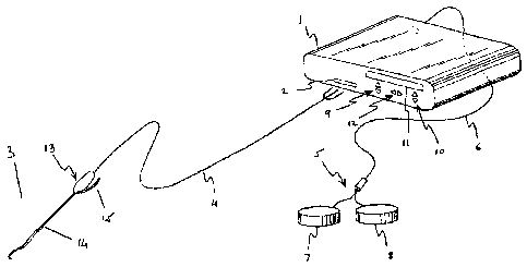
Referring to the drawings, Figure 1 shows electrosurgical apparatus including
a generator 1 having an output socket 2 providing a radio frequency (RF)
output, via a
connection cord 4, for an electrosurgical instrument 3. Activation of the
generator 1
may be performed from the instrument 3 via a handswitch (not shown) on the
instrument 3, or by means of a footswitch unit 5 connected separately to the
rear of
the generator 1 by a footswitch connection cord 6. In the illustrated
embodiment, the
footswitch unit 5 has two footswitches 7 and 8 for selecting a desiccation
mode and a
vaporisation mode of the generator 1 respectively. The generator front panel
has push
buttons 9 and 10 for respectively setting desiccation and vaporisation power
levels,
which are indicated in a display 11. Push buttons 12 are provided as an
alternative
means for selection between the desiccation and vaporisation modes.
The electrosurgical instrument 3 comprises a housing 13 with an elongate shaft
14, and tissue treatment electrodes at the distal end of the shaft, as will be
described
below. A movable handle 15 associated with the housing can be actuated to
cause the
shaft to bend. This instrument is particularly suited to the treatment of the
hip joint,
CA 02818660 2013-06-11
6
where a relatively long shaft with articulation capability is needed to access
the area to
the treated.
Figures 2 & 3 show a tissue treatment electrode 16 comprising a hemispherical
end face 17 and a cylindrical side face 18. The electrode is typically formed
from
tungsten (or an alloy of tungsten and platinum), and can be formed from a
single
integral component or from two components welded one to the other. Whether
formed from one or two components, the end face 17 meets the side face 18 at a
discrete transition 30, in the form of an elbow portion at which the end face
17 and
side face 18 meet at an angle of at least 30 degrees. That is, the end face 17
and the
side face 18 with the discrete transition 30 therebetween together form a
cupola-
shaped electrode, with the side face 18 forming the side walls of the cupola,
and the
end face 17 forming the cap or "roof' of the cupola. The end face 17 is
provided with
a single aperture 19 located at the centre thereof; while the side face 18 is
provided
with a plurality of apertures 20 spaced at equal distances around its
circumference.
The tissue treatment electrode is located on a ceramic component 21, and held
in
place by a c-clip 22. A split ring retainer 23 is then placed over the ceramic
component 21, and the end of the shaft 14 is laser-welded to the split ring
23. The
ceramic component 21 is hollow so as to form a suction lumen 24, and also to
accommodate a lead 25 to supply RF energy to the tissue treatment electrode
16.
The majority of the shaft 14 is covered by an insulating sheath 26, but a
portion is left uncovered in order to form a return electrode 27. The return
electrode
27 is separated from the tissue treatment electrode 16 by a portion of the
ceramic
component, forming an insulator 28. In use the instrument 3 is introduced into
the
body of a patient and manoeuvred into position, typically adjacent a hip
joint. RF
energy is supplied to the tissue treatment electrode 16, which is used to
vaporise or
coagulate tissue depending on the type of RF energy supplied from the
generator 1.
The instrument is typically used submerged in an electrically conductive
fluid, such as
normal saline, with the RF energy flowing from the tissue treatment electrode
16,
through the conductive liquid to the return electrode 27, with any tissue
entering the
region of the tissue treatment electrode 16 being vaporised or coagulated
depending
on the circumstances.
The suction lumen 24 is connected to a source of suction, and the apertures 19
and 20 are in communication with the suction lumen 24. In this way, any tissue
CA 02818660 2013-06-11
7
fragments, bubbles or other debris in the vicinity of the tissue treatment
electrode 16
will be drawn into the suction lumen via either the aperture 19 in the end
face 17 or
the multiple apertures 20 in the side face 18. The provision of apertures in
both the
end face 17 and the side face 18 allows for debris to be removed from the
vicinity of
15 As previously mentioned, the instrument 3 is primarily designed to be
operated
in a conductive fluid such as saline, with the fluid completing the circuit
between the
electrodes. However, the instrument 3 can also be used as a dry-field
instrument, in
which case the user must ensure that the electrodes are placed in contact with
the
tissue to be treated. In this way, the current flows from the tissue treatment
electrode,
Alternative embodiments will be envisaged by those skilled in the art without
departing from the scope of the present invention. For example, the
electrosurgical
instrument can also be used for delivering a blended power output. This is
achieved
by automatically alternating the output of the RF generator 1 between the
coagulation
with a consequent reduction in both bubble formation and the risk of tissue
charring.