Note: Descriptions are shown in the official language in which they were submitted.
CA 02819250 2016-12-16
METHODS FOR DIAGNOSING AND TREATING EYE-LENGTII RELATED
DISORDERS
10
FIELD OF THE INVENTION
The invention relates to methods for detecting and treating eye-length related
disorders, including myopia. In addition, the invention relates to certain
haplotypes
associated with eye-length related disorders.
BACKGROUND
In a process termed emmetropization, the growth of eye length is regulated by
visual
experience to match the eye's optics and to compensate for genetic variation
in corneal/lens
curvature and power. High acuity photopic vision and, thus, the signals that
guide
emmetropization are initiated by light absorption in photopigments found in
the long
wavelength (L) and middle-wavelength (M) sensitive cone photoreceptors.
Changes in the
pattern of light and dark in the retinal image that characterize blurred
versus sharply focused
images are monitored by a biological process to stop eye growth when the
correct length for
coordinated plano (neutral) optics is reached. However, in myopic individuals,
the relative
axial length of the eye to overall eye size continues to increase during
development, past a
CA 02819250 2013 05 27
WO 2012/097213
PCT/US2012/021185
length that provides near-optimal focusing of distant objects, leading to
increasingly
pronounced myopia.
The rate of incidence of myopia is increasing at alarming rates in many
regions of the
world. Until recently, excessive reading during childhood was believed to be
the only
identifiable environmental or behavioral factor linked to the occurrence of
myopia, although
genetic factors were suspected. Limiting reading (and encouraging more outdoor
activity)
are presently the only practical techniques for preventing excessive eye
lengthening in
children, and corrective lenses, including glasses and contact lenses,
represent the primary
means for ameliorating eye-length related disorders, including myopia. While
these
measures optically correct the refractive errors associated with eye-length
related disorders
they do not address the underlying cause which is excessive growth of eye
length.
Thus, there remains a need for methods of detecting a susceptibility to an eye-
length
related disorder, and treatments for such individuals that would prevent
excessive eye
lengthening.
SUMMARY OF THE INVENTION
The invention provides a method for determining the myopic potential of a
patient
comprising: testing a biological sample obtained from the patient to determine
the L:M opsin
gene haplotype of the patient; and correlating the haplotype with a predicted
spherical
equivalent refraction. In another aspect, the method further comprises the
steps of:
determining the L:M cone ratio in an eye of the patient; and correlating the
L:M opsin gene
haplotype and the L:M cone ratio with a predicted spherical equivalent
refraction.
The invention also provides a method for diagnosing susceptibility of a
patient to an
eye-length related disorder, the method comprising: testing a biological
sample obtained from
the patient to determine the L:M opsin gene haplotype of the patient; and
correlating the
haplotype with a predicted spherical equivalent refraction; wherein the
patient is susceptible
to an eye-length related disorder if the predicted spherical equivalent
refractive error
(measured in diopters) has a negative power. In one aspect, the method further
comprises
the steps of: determining the L:M cone ratio in an eye of the patient; and
correlating the L:M
opsin gene haplotype and the L:M cone ratio with a predicted spherical
equivalent refraction.
2
CA 02819250 2013 05 27
WO 2012/097213
PCT/US2012/021185
The invention further provides a method for diagnosing susceptibility of a
patient to
an eye-length related disorder, the method comprising testing a biological
sample obtained
from a patient for a particular combination of amino acids encoded by the
patient's L opsin
gene or M opsin gene, wherein the patient is susceptible to an eye-length
related disorder if
one of the amino acid combinations shown in Table 1 is present.
In addition, the invention provides a method of treating an eye-length related
disorder
comprising: testing a biological sample obtained from the patient to determine
the L:M opsin
gene haplotype of the patient; determining the L:M cone ratio in an eye of the
patient;
correlating the haplotype and the L:M cone ratio with a predicted spherical
equivalent
.. refraction; providing the patient with a therapeutic device comprising a
wavelength-
dependent filter if the patient's predicted spherical equivalent refractive
error has a negative
power..
In one aspect, the wavelengths filtered by the wavelength-dependent filter are
selected
based on the L:M opsin gene haplotype and the L:M cone ratio of the patient.
In another aspect, a therapeutic device used in a method of the invention is a
pair of
spectacles comprising blur-inducing lenses. In certain aspects, the blur-
inducing lenses
induce blurring by one or more of: small bumps or depressions in one or both
surfaces of the
lenses; inclusions within the lenses of a material different from the lens
material;
incorporation of higher-level aberrations in the lenses; providing an
increased correlation
between the activities of neighboring cone photoreceptors by one or both
lenses; and coatings
or films applied to one or both surfaces of the lenses to produce diffusive or
diffractive blur.
In yet another aspect, a therapeutic device used in a method of the invention
comprises blur-inducing contact lenses. In certain aspects, the blur-inducing
contact lenses
induce blurring by one or more of: inclusions within the lenses of a material
different from
the lens material; incorporation of higher-level aberrations in the lenses;
and coatings or films
applied to one or both surfaces of the lenses that produce blur by diffusion,
diffraction or
light scattering.
In certain aspects, the L:M opsin gene haplotype identified in a method of the
invention is one of haplotypes 1 to 13 as set forth in Table 1.
3
CA 02819250 2013 05 27
WO 2012/097213
PCT/US2012/021185
The invention also provides a microarray for determining susceptibility of a
patient to
an eye-length related disorder comprising a set of allele specific
oligonucleotides capable of
identifying at least one of haplotypes 1 to 13 as set forth in Table 1.
The invention further provides kits for determining whether a patient is
susceptible to
an eye-length related disorder. In one aspect, a kit of the invention
comprises: at least one
pair of oligonucleotides that can identify at least one of haplotypes 1 to 13
as set forth in
Table 1; and instructions for use. In another aspect, a kit of the invention
comprises an assay
for detecting at least one of haplotypes 1 to 13 as set forth in Table 1.
In another aspect, the present invention provides methods for limiting
introduction of
refractive error in a subject's eye caused by exposure to display screens,
comprising the
subject wearing a therapeutic optical device that comprises a wavelength-
dependent filter
capable of preferentially blocking red light emanating from the display screen
prior to entry
into the subject's eye, thereby limiting introduction of refractive error in
the subject's eye.
In a further aspect, the present invention provides methods for limiting
development
of an eye-length related disorder in a subject, comprising the subject wearing
a therapeutic
optical device that comprises a wavelength-dependent filter capable of
preferentially blocking
red light emanating from a display screen prior to entry into the subject's
eye, thereby
limiting development of an eye-length related disorder in the subject. In one
embodiment, the
eye-length related disorder comprises myopia.
Specific preferred embodiments of the invention will become evident from the
following more detailed description of certain preferred embodiments and the
claims.
BRIEF DESCRIPTION OF THE FIGURES
Figure 1. Averaged adaptive optics retinal images of the cone mosaic of
participants with
LIAVA variants (B, C, D) compared with a normal control (A). For subjects
shown in B, C,
& D cones expressing the LIAVA variant had a low reflectance compared to
normal cones
and appear as dark area in the mosaic. There was large variability in the
proportion of cones
expressing the LIAVA variant. B, C & D have low, medium and high proportions
of cones
expressing the myopia-genic variant which correlates with axial length (E) and
also with
refractive error.
4
CA 02819250 2013 05 27
WO 2012/097213
PCT/US2012/021185
Figure 2. Association between axial length and cone ratio for different ethnic
groups. There
was a high positive correlation between L:M cone ratio and axial length (and
incidence of
myopia) across ethnic groups.
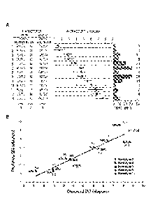
Figure 3. (A) Myopic potential of 13 different L/M photopigment haplotypes
from 159
males, arranged in order of increasing myopic potential. The number of
individuals with each
haplotype is given at the right. Haplotype designations use the single letter
amino acid code:
M = methionine, I = isoleucine; S = serine, V = valine, A = alanine, and L =
leucine. Average
SER is the mean spherical equivalent refraction calculated for the most myopic
half of the
subjects for each haplotype, 1 SEM. (B) Predicted versus observed spherical
equivalent
refraction (SER) for 11 subjects with haplotypes corresponding to those
described in (A). The
L:M cone ratio was estimated for each subject and is expressed as the
percentage of L plus M
cones that are L.
Figure 4. (A) Myopic shift produced by exposure to the red light for 2 hours
per day. Axial
lengths were measured for each subject before the onset of the experimental
procedure.
Subsequently, each subject played a black and white video game for 2 hours per
day while
wearing goggles with the right lens untinted and the left lens tinted so that
the L cones are
activated much more than M cones. (B) Normalized axial length measurements as
a function
of time for 20 eyes wearing the experimental lens and (C) for 20 fellow eyes
that served as
the controls for each experimental eye. Black lines with error bars represent
the averages for
all eyes (error bars 2 SEM). The experimental lenses significantly reduced
the rate of eye
growth of myopic children. (D) Growth rate of eyes wearing the experimental
lens are to the
left, and for eyes wearing the control lens are to the right.
DETAILED DESCRIPTION OF THE INVENTION
The particulars shown herein are by way of example and for purposes of
illustrative
discussion of the preferred embodiments of the present invention only and are
presented in
5
CA 02819250 2013 05 27
WO 2012/097213
PCT/US2012/021185
the cause of providing what is believed to be the most useful and readily
understood
description of the principles and conceptual aspects of various embodiments of
the invention.
In this regard, no attempt is made to show structural details of the invention
in more detail
than is necessary for the fundamental understanding of the invention, the
description taken
with the drawings and/or examples making apparent to those skilled in the art
how the several
forms of the invention may be embodied in practice.
The following definitions and explanations are meant and intended to be
controlling
in any future construction unless clearly and unambiguously modified in the
following
examples or when application of the meaning renders any construction
meaningless or
essentially meaningless. In cases where the construction of the term would
render it
meaningless or essentially meaningless, the definition should be taken from
Webster's
Dictionary, 3rd Edition or a dictionary known to those of skill in the art,
such as the Oxford
Dictionary of Biochemistry and Molecular Biology (Ed. Anthony Smith, Oxford
University
Press, Oxford, 2004).
As used herein and unless otherwise indicated, the terms "a" and "an" are
taken to
mean "one", "at least one" or "one or more". Unless otherwise required by
context, singular
terms used herein shall include pluralities and plural terms shall include the
singular.
In certain embodiments, the invention provides methods that can be used to
determine
the benefit of a preventative treatment for an eye-length disorder and to
determine the
appropriate prescription of characteristics of preventative optics for a
patient who is identified
as having a susceptibility to an eye-length related disorder. As discussed
herein, such
preventative optics include spectral characteristics and/or dispersive
properties that can
prevent eye-length growth, which if left uncontrolled would lead to an eye-
length related
disorder.
In one embodiment, the invention provides a method for diagnosing
susceptibility of a
patient to an eye-length related disorder, the method comprising: testing a
biological sample
obtained from the patient to determine the patient's L:M opsin gene haplotype,
and
correlating the haplotype with a predicted spherical equivalent refraction;
wherein the patient
is susceptible to an eye-length related disorder if the predicted spherical
equivalent refraction
is a negative diopter.
6
CA 02819250 2013 05 27
WO 2012/097213
PCT/US2012/021185
As used herein, the term "correlating" refers to the step of using the
combination of
information about a patient's cone ratio and opsin haplotype in order to
determine the
susceptibility of the patient to an eye-length related disorder as shown and
discussed herein.
As used herein, the phrase "eye-length related disorder" includes, but is not
limited to,
myopia.
In one embodiment, the L:M opsin gene haplotype is determined by identifying
the
nucleotide sequence of a patient's DNA to determine the patient's Xq28 opsin
gene locus
haplotype. The haploytpe can be determined by identifying the nucleotide
sequence of exons
2, 3, and 4 of the OPN1LW and OPN1MW genes. As discussed herein, the
haplotypes are
created by the amino acids encoded by codons 65, 111, 116, 153, 171, 178, 180,
230, 233,
and 236 of the OPN1LW and OPN1MW genes. In a particular embodiment, the
haplotype is
determined by the amino acids encoded by codons 153, 171, 178, and 180 in exon
3 and
codon 236 in exon 4. In a preferred embodiment, the L:M opsin gene haplotype
is one of the
13 haplotypes shown in Table 1, which are shown herein for the first time as
being associated
with myopia (see Examples and Figure 3A). Thus, if a patient has one of the 13
haplotypes
identified in Table 1, that patient is diagnosed as being susceptible to an
eye-length related
disorder. In particular, a patient having one of the haplotypes shown in Table
1 is diagnosed
as being susceptible to myopia. In one embodiment, a patient is diagnosed as
being
susceptible to myopia if one of the variant amino acid combinations shown in
Table 1
associated with the L-opsin gene is identified in the patient. In another
embodiment, a
patient is diagnosed as being susceptible to myopia if one of the variant
amino acid
combinations shown in Table 1 associated with the M-opsin gene is identified
in the patient.
TABLE 1
Myopia Haplotypes
L-OPSIN M-OPSIN
Codons Codons
153 171 178 180 236 153 171 178 180
1 M I I S M M V V A
2 M V I S M M V V A
3 L V I S M M V V A
4 M V I S M M V I A
5 L V I A M M V I A
7
CA 02819250 2013 05 27
WO 2012/097213
PCT/US2012/021185
6 M V I A M M V I A
7 L V I S M L/M V I A
8 L V I S M M V I A
9 L I I S M M V V A
M V V A V M V I A
11 M V I S V M V V A
12 L V I S M L V I S
13 L V I A M L/M V I A
In another embodiment, the invention provides a method for diagnosing
susceptibility
of a patient to an eye-length related disorder, the method comprising: testing
a biological
5 sample
obtained from the patient to determine the patient's L:M opsin gene haplotype,
determining the L:M cone ratio in an eye of the patient, and correlating the
L:M opsin gene
haplotype and the L:M cone ratio with a predicted spherical equivalent
refraction, wherein
the patient is susceptible to an eye-length related disorder if the predicted
spherical equivalent
refractive error is a negative power (in diopters).
10 The L:M
cone ratio can be determined using methods known to those of skill in the
art. For example, adaptive optics retinal imaging can be used as described
herein, or an
electroretinogram (ERG) (such as a flicker photometric ERG) and individualized
cone
spectra can be used. The L:M cone ratio measurement can also involve genetics,
as described
for example in Neitz and Neitz, J. Vis. 2:531-42, 2002. Another non-limiting
example of
measuring L:M cone ratio includes wide-field color multifocal ERG as described
in
Kuchenbecker et al., Vis. Neurosci. 25(3):301-6, 2008. Another non-limiting
example of
measuring L:M cone ratio includes measuring the ratio of red-to-green light
perceived to
have the minimum flicker using psychophysical heterochromatic flicker
photometry as
described in Gunther and Dobkins Vision Research 42:1367-1378, 2002.
In one embodiment, the invention provides a method for determining the myopic
potential of a patient comprising testing a biological sample obtained from
the patient to
determine the patient's L:M opsin gene haplotype.
As used herein, the term "myopic potential" refers to the predicted spherical
equivalent refraction associated with an L:M opsin haplotype, which correlates
with the
predicted degree of myopia that the patient has or is likely to have. In
particular, the myopic
8
CA 02819250 2013 05 27
WO 2012/097213
PCT/US2012/021185
potential refers to a certain spherical equivalent refraction predicted based
on the patient's
particular L:M opsin gene haplotype, as shown, for example, in Figure 3A.
In a particular embodiment, myopic potential can be more specifically
determined by
measuring the patient's L:M cone ratio, and correlating the ratio with the
spherical equivalent
refraction predicted for the particular L:M opsin gene haplotype. For example,
as discussed
in the examples below, the L:M cone ratio can be determined for a patient that
has a certain
L:M opsin haplotype, such as a haplotype shown in Table 1. The L:M cone ratio
is
determined, and a calculation is made to arrive at the more specific predicted
myopic
potential. For instance, if a person had haplotype 8 (Figure 3A), their myopic
potential is -4.5
diopters. If that person had a 1:1 cone ratio they would be expected to have
the full -4.5
diopters of refractive error. However, if he had nearly 100 percent L cones he
would be
expected to be nearly emmetropic. 75% L cones falls midway between a 1:1 cone
ratio
(50%L) and 100 % L so a person with haplotype 8 and 75% L cones would be
predicted to
have 50% of the SER (or -4.5/2 = -2.25 diopters).
As used herein, the phrase "susceptibility to an eye-length related disorder"
refers to
the high likelihood of developing an eye-length related disorder, such as
myopia, when a
certain L:M opsin gene haplotype is present. In one embodiment, a patient is
considered
susceptible to an eye-length related disorder if one of the haplotypes shown
in Table 1 is
present, which are listed in order of increasing myopic potential.
After identifying a patient that is susceptible to an eye-length related
disorder and/or
has a myopic potential associated with a negative diopter as described herein,
an eye care
provider can prescribe a treatment protocol and/or suggest certain behaviors
intended to treat
or reduce the myopic potential of the patient. For example, a patient may be
treated with a
therapeutic device (as described herein, for example) or be given
pharmacological
intervention. In addition or instead of such treatments, a patient may be told
to limit exposure
to red light or green light (depending on the patient's particular L:M
variants) limit reading at
a young age and spending more time doing activities outdoors.
The term "biological sample" as used herein includes, but is not limited to,
blood,
saliva, cells from buccal swabbing, biopsies of skin, amniotic fluid, various
other tissues and
the like. Methods for purifying or partially purifying nucleic acids from a
biological sample
9
CA 02819250 2013 05 27
WO 2012/097213
PCT/US2012/021185
for use in diagnostic assays are well known in the art. The nucleic acid can
be, for example,
genomic DNA, RNA, or cDNA. Genomic DNA can be isolated, for example, from
peripheral blood leukocytes using QIAamp DNA Blood Maxi Kits (Qiagen,
Valencia, CA).
In another embodiment, the invention provides a method for diagnosing Bornholm
Eye Disease (BED) in a patient, the method comprising obtaining a biological
sample from
the patient and identifying the nucleotide sequence of the patient's L and M
opsin genes,
wherein the patient is diagnosed as having BED if the patient has a normal
opsin gene and a
variant opsin gene. In a preferred embodiment, the variant opsin gene
comprises Leucine at
amino acid position 153 (L153), Valine at position 171 (V171), Alanine at 174
(A174),
Valine at 178 (V178), and Alanine at 180 (A180) ("LVAVA") or Leucine at amino
acid
position 153 (L153), Isoleucine at position 171 (1171), Alanine at 174 (A174),
Valine at 178
(V178), and Alanine at 180 (A180) ("LIAVA") in either the L or M opsin gene.
In another
embodiment, the second gene has the combination of Methionine, Valine, Valine,
Valine, and
Alanine at amino acids at positions 153, 171, 174, 178, and 180 ("MVVVA").
The diagnostic methods of the invention involve the use of standard molecular
biology methods, including in one non-limiting embodiment the polymerase chain
reaction
(PCR), to determine the L:M opsin gene haplotype of a patient. There are
currently a variety
of molecular biological methods available that allow examination of the DNA
sequences of
the L and M opsin genes. For example, gene fragments may be amplified using
the
polymerase chain reaction (PCR). The genes can be separately and selectively
amplified as
described previously (Neitz et al., Vision Research 35: 2395-2407, 1995).
Amplified gene fragments will preferably be subjected to one or more of the
following procedures that provide information about the DNA sequence:
1) Direct DNA sequence of the PCR products as described previously (J. Neitz,
M.
Neitz and Grishok, supra, 1995).
2) Restriction digestion analysis (described previously in J. Neitz, M. Neitz
and
Grishok, supra, 1995).
3) Single strand conformation polymorphism or other similar procedures. The
amplified DNA fragment is fluorescently or radioactively end labeled,
denatured into single
CA 02819250 2016-12-16
strands, and the strands are separated electrophoretically. Based On the
mobility of the strands
in the electric field, information about the DNA sequence can be deduced.
In another embodiment, the invention provides a method of treating an eye-
length
related disorder comprising: testing a biological sample obtained from a
patient to determine
the L:M opsin gene haplotype of the patient; determining the L:M cone ratio in
an eye of the
patient; correlating the haplotype and the L:M cone ratio with a predicted
spherical
equivalent refraction; providing the patient with a therapeutic device
comprising a
wavelength-dependent filter if the patient's predicted spherical equivalent
refraction is a
negative diopter. In one embodiment, the L:M opsin gene haplotype is one of
haplotypes 1 to
13 as set forth in Table 1.
As discussed in International Patent Application Publication No. WO
2010/075319,
genetic
variation in opsin genes affects the absorbance characteristics of the opsin
photoreceptor
protein. Thus, the wavelength-dependent filter utilized in a method of the
invention is
intended to filter light prior to entry into the eye in order to adjust the
effective absorbance
spectrum of variant opsin photoreceptor proteins. In patients having a
defective M
photoreceptor protein, caused by a variant M-opsin gene, that absorbs less
light than the
normal M photoreceptor protein, the wavelength-dependent filter may
preferentially block
red light. On the other hand, in patients having a defective L photoreceptor
protein, caused
by a variant M-opsin gene, that absorbs less light than the normal L
photoreceptor protein,
the wavelength-dependent filter may preferentially block green light.
In certain embodiments, the particular wavelength-dependent filter utilized in
a
method of the invention can be selected based on the patient's L:M opsin gene
haplotype,
which identifies specific photoreceptor variants and/or the patient's L:M cone
ratio, which
identifies the number of L photoreceptors relative to M photoreceptors present
in the patient's
eye. Based on the particular L:M opsin gene haplotype and/or the L:M ratio, a
filter can be
designed to block and/or transmit very specific wavelengths to restore
relative absorption
characteristics of the defective photoreceptor proteins. Thus, the invention
further provides
methods for customizing a therapeutic device for a particular patient based on
the L:M opsin
gene haplotype and/or the L:M cone ratio of the patient. For example, if the
patient had opsin
variants associated with more active red (M) cones, the filter could be
designed to block red
11
CA 02819250 2013 05 27
WO 2012/097213
PCT/US2012/021185
light; whereas if the patient had opsin variants associated with more active
green (L) cones,
the filter could be designed to block green light.
In certain embodiments, the therapeutic device comprises blur-inducing lenses,
for
example as described in International Patent Application Publication No. WO
2010/075319.
In one embodiment, the device is a pair of spectacles comprising blur-inducing
lenses, where
the blur is designed to reduce the relative activities between neighboring
cone photoreceptors
in the retina which has been shown herein to result in signals that stimulate
the eye to grow in
length abnormally. The blur-inducing lenses can be made to induce blurring,
for example, by
one or more of: small bumps or depressions in one or both surfaces of the
lenses; inclusions
within the lenses of a material different from the lens material;
incorporation of higher-level
aberrations in the lenses; and coatings or films that induce blur by light
scatter, diffusion or
diffraction applied to one or both surfaces of the lenses.
In yet another embodiment, the therapeutic device comprises blur-inducing
contact
lenses. The blur-inducing contact lenses can be made to induce blurring, for
example, by one
or more of: inclusions within the lenses of a material different from the lens
material;
incorporation of higher-level aberrations in the lenses; providing progressive
negative
corrections in one or both lenses from the center of the lens to the bottom of
the lenses; and
coatings or films that induce blur by light scatter, diffusion or diffraction
applied to one or
both surfaces of the lenses.
In one further aspect, the present invention provides methods for limiting
introduction
of refractive error in a subject's eye caused by exposure to display screens,
comprising the
subject wearing a therapeutic optical device that comprises a wavelength-
dependent filter
capable of preferentially blocking red light emanating from the display screen
prior to entry
into the subject's eye, thereby limiting introduction of refractive error in
the subject's eye.
In a still further aspect, the present invention provides methods for limiting
development of an eye-length related disorder in a subject, comprising the
subject wearing a
therapeutic optical device that comprises a wavelength-dependent filter
capable of
preferentially blocking red light emanating from a display screen prior to
entry into the
subject's eye, thereby limiting development of an eye-length related disorder
in the subject.
In one embodiment, the eye-length related disorder comprises myopia.
12
CA 02819250 2013 05 27
WO 2012/097213
PCT/US2012/021185
These methods can be used to limit damage to the eye caused by excessive
exposure
to red-light from a screen display. In various non-limiting embodiments, the
screen display
may be a computer monitor, a tablet monitor, a television screen, a handheld
device screen, a
video game screen, a head-mounted display screen, and a movie theater screen.
As used herein, "limiting" means one or more of (a) reducing the incidence of
introduction of refractive error in a subject's eye and/or reducing the
incidence of eye-length
related disorders developing in treated subjects; (b) reducing the severity of
subsequently
developed refractive error in a subject's eye and/or reducing the severity of
a subsequently
developed eye-length related disorder in the subject; and/or (c) limiting or
preventing
development of symptoms characteristic of refractive error in a subject's eye
and/or an eye-
length related disorder.
In each of these further aspects, the therapeutic optical device may further
comprise a
blur-inducing lens, including but not limited to those disclosed in WO
2010/075319 and as
disclosed above. In one embodiment, the blur-inducing lens comprises a
holographic diffuser
applied to the lens surface, for example, as described in the examples below.
The holographic
diffuser can be used, for example, to spread the incident light rays from the
display over a
desired angle to produce a slight blur and thus reduce activity differences
between adjacent
cones. In any of these embodiments, the therapeutic optical device may be of
any suitable
type, including but not limited to glasses/spectacles and contact lenses.
Any suitable subject may be treated in these aspects, including children 21
years of
age or younger, preferably between the ages of 3-21, 3-20, 3-19, or 3-18. In
another
embodiment that can be combined with any of the above embodiments, wherein the
subject is
susceptible to an eye-length related disorder, such as myopia. This embodiment
may
comprise treating any subject at risk as discussed in any of the preceding
disclosure. In one
particular embodiment, the subject is susceptible to an eye-length related
disorder if the
subject has an L:M opsin gene haplotype as set forth in Table 1.
In one embodiment, the invention provides kits that can be used, for example,
for eye-
length related disorder diagnosis. In certain embodiments, a kit of the
invention comprises a
set of haplotype specific oligonucleotides to identify the presence or absence
of L:M opsin
13
CA 02819250 2013 05 27
WO 2012/097213
PCT/US2012/021185
gene haplotypes, such as those identified in Table 1. For example, a kit
comprises: a set of
primer pairs for amplifying portions of exons 3 and 4 associated with the
haplotypes
described herein, such as 1,2, 3,4, 5, 6, 7, 8, 9, 10, 11, 12, or 13 of the
haplotypes listed in
Table 1; a set of probes that can hybridize to portions of exons 3 and 4
associated with the
haplotypes described herein; and/or a microanay, such as a SNP chip. Primers
and probes
can be readily and easily designed by those skilled in the art by reference to
a sequence
associated with the portions of exons 3 and 4 associated with the haplotypes
described herein.
Microarrays can also be easily and readily designed with oligonucleotides of
the invention
that correspond to the portions of exons 3 and 4 associated with the
haplotypes described
herein. Alternatively, analysis could be done using a mass spectrometry
instrument (for
example, a MassArrayTM instrument) that allows genotyping at known polymorphic
sites
using specially designed PCR primers followed by mass spectrometry. This
technique is
suited to diagnosis of conditions such as axial length disorders described
here whose genetic
underpinnings are well understood. A MassArrayTM primer extension process
detects
sequence differences at the single nucleotide level. An initial round of PCR
amplifies from
genomic DNA a short length of DNA surrounding the SNP. This is followed by
single-base
extensions of a primer that anneals directly adjacent to the SNP. The primer
is extended
dependent upon the template sequence, resulting in an allele-specific
difference in mass
between extension products. This mass difference allows differentiation
between SNP alleles
.. using MALDI TOF mass spectrometry.
In another embodiment, the invention provides a mouse model of an eye-length
related disorder as described in the Examples herein, which comprises a
variant green (L)
photopigment protein associated with myopia. The invention further provides a
mouse model
that expresses variant red (M) and normal or variant green (L) photopigment
proteins,
.. wherein a variant protein has an amino acid sequence associated with
myopia. Such mice can
be generated as described, for example, in the Methods provided herein. Such
mice have
been generated using the method described herein, wherein the heterozygous
mice of the
method comprise the red and green photopigment proteins. In certain
embodiments, a mouse
model of the invention can be used to test eye-length related disorder
intervention, such as
pharmacological or genetic intervention.
14
CA 02819250 2016-12-16
In certain embodiments, the present invention provides a machine readable
storage
medium, comprising a set of instructions for causing a diagnostic device to
measure a
patient's L:M cone ratio or L:M opsin gene haplotype. In other embodiments,
the invention
provides a machine readable storage medium that comprises instructions for
causing a
processor to execute automated method steps for correlating a patient's L:M
opsin gene
haplotype and L:M cone ratio to determine an appropriate prescription of
characteristics of
preventative optics for a patient who is identified as having a susceptibility
to an eye-length
related disorder. As used herein the term "computer readable storage medium"
includes
magnetic disks, optical disks, organic memory, and any other volatile (e.g.,
Random Access
.. Memory ("RAM")) or non-volatile (e.g., Read-Only Memory ("ROM")) mass
storage system
readable by the CPU. The computer readable medium includes cooperating or
interconnected
computer readable medium, which exist exclusively on the processing system or
be
distributed among multiple interconnected processing systems that may be local
or remote to
the processing system. As used herein, "diagnostic device" means a device
capable of
carrying out the L:M cone ratio measurements or L:M opsin gene haplotype
determination to
carry out the methods of invention, including but not limited to a microarray
reader or a mass
spectrometer.
Those of skill in the art, in light of the present disclosure, will appreciate
that obvious
modifications of the embodiments disclosed herein can be made without
departing from the
spirit and scope of the invention. All of the embodiments disclosed herein can
be made and
executed without undue experimentation in light of the present disclosure. The
full scope of
the invention is set out in the disclosure and equivalent embodiments thereof.
The
specification should not be construed to unduly narrow the full scope of
protection to which
the present invention is entitled.
EXAMPLES
CA 02819250 2013 05 27
WO 2012/097213
PCT/US2012/021185
The following examples, including the experiments conducted and results
achieved
are provided for illustrative purposes only and are not to be construed as
limiting the
invention.
Example 1
Mutant OPN1LW and OPN1MW menes in Bornholm Eve Disease
The first identified high-grade myopia locus was localized to chromosome Xq28
and
designated MYP1 (M. Schwartz, M. Haim, D. Skarsholm, Clinical Genetics 38, 281
(October, 1990)). The phenotype is also known as the Bornholm Eye Disease
(BED), and is
an X-linked cone dysfunction syndrome with myopia, astigmatism and optic nerve
changes
(T. L. Young et al., Archives of Ophthalmology 122, 897 (June, 2004); U.
Radhakrishna et
al., Investigative Ophthalmology & Visual Science supplement (abstract #3814)
(2005); M.
Michaelides et al., Ophthalmology 112, 1448 (2005)). Part of the phenotype of
BED with X-
linked cone dysfunction syndrome is an abnormal cone electroretinogram (ERG).
The
OPN1LW and OPN1MW genes reside at Xq28 and encode cone photopigments
responsible
for the initial events that generate the cone ERG.
The L and M cone opsin genes were evaluated as candidates for the BED
phenotype.
The two unrelated X-linked myopia/cone dysfunction families described by Young
et al. (T.
L. Young et al., Archives of Ophthalmology 122, 897 (June, 2004)) have color
vision
deficiencies which are caused by the absence of an OPN1MW gene in either of
the first two
positions in the cone opsin gene array in the original BED (M. Schwartz, M.
Haim, D.
Skarsholm, Clinical Genetics 38, 281 (October, 1990)) family and by the
absence of an intact
OPN1LW gene in the case of the Minnesota (MN) family. In a third family,
residing in India,
the affected males (U. Radhakrishna et al., Investigative Ophthalmology &
Visual Science
.. supplement (abstract #3814) (2005)) have normal color vision. The first
gene in the X-
chromosome opsin array was selectively amplified and individual exons from
affected and
unaffected males in the MN, BED1, and Indian families were directly sequenced.
The opsin
genes downstream of the first gene were also selectively amplified, and the
exons were
directly sequenced. For all affected males in the MN family, the first
position (5'-most) opsin
gene in the array encoded an M opsin with an unusual combination of amino
acids specified
16
CA 02819250 2013 05 27
WO 2012/097213
PCT/US2012/021185
by the dimorphic codons in exon 3. This combination was Leucine at amino acid
position 153
(L153), Valine at position 171 (V171), Alanine at 174 (A174), Valine at 178
(V178), and
Alanine at 180 (A180), henceforth abbreviated "LVAVA." The second gene in the
array
encoded a combination of amino acids at these positions ("MVVVA") typically
found in M
opsins in individuals with no vision abnormalities.
The affected members of the second, unrelated BED family (BED1) reported by
Young et al. (T. L. Young et al., Archives of Ophthalmology 122, 897 (June,
2004)) and the
Indian family (U. Radhakrishna et al., Investigative Ophthalmology & Visual
Science
supplement (abstract #3814) (2005)) were also found to have the LVAVA
combination, but
in the L opsin. In both of these latter families, the downstream genes in
affected males
encoded variants that are typical of individuals with normal vision.
Unaffected males in the
BED families did not have an LVAVA variant. As a control experiment, 261
OPN1MW
genes and 320 OPN1LW from males with no serious vision abnormality were
sequenced.
None of the genes specified the LVAVA combination.
Affected males in five additional families (M. Michaelides et al.,
Ophthalmology 112,
1448 (2005); M. McClements, M. Neitz, A. Moore, D. M. Hunt, Invest Ophthalmol
Vis Sci,
ARVO E (2010)) and one other unrelated individual with the BED phenotype were
found to
have either the LVAVA combination or a similar combination, in which
isoleucine is present
at position 171 (I171) instead of valine. This combination is designated
"LIAVA" and was
previously shown to cause photoreceptors to be non-functional in adults (J.
Carroll, M. Neitz,
H. Hofer, J. Neitz, D. R. Williams, Proceedings of the National Academy of
Sciences of the
United States of America 101, 8461 (2004); M. Neitz et al., Visual
Neuroscience 21, 205
(2004); M. A. Crognale et al., Visual Neuroscience 21, 197 (2004)). Affected
members of a
seventh family reported to have X-linked cone dysfunction syndrome were found
to have a
mutation that replaces the cysteine normally found at position 203 with
arginine (C203R) in
both the L and M opsins (M. Michaelides et al., Ophthalmology 112, 1448
(2005)), a
mutation known to render the opsin non-functional (M. Michaelides et al.,
Ophthalmology
112, 1448 (2005); J. Winderickx et al., Nature Genetics 1, 251 (1992); J.
Nathans et al.,
Science 245, 831 (1989)).
17
CA 02819250 2013 05 27
WO 2012/097213
PCT/US2012/021185
Cone phenotype of BED opsin mutation in mice with a targeted gene replacement
Although the LIAVA and C203R mutations found in some of the families have been
previously documented to cause cone photoreceptor malfunction, the LVAVA amino
acid
combination found in many BED families and its impact on cone function and
viability was
never identified. Individuals with LVAVA encoded in their only expressed X-
linked cone
pigment gene have cone dystrophy indicating that cones expressing this
haplotype function
abnormally and eventually degenerate. To verify the abnormal cone function
associated with
LVAVA, a mouse line was created in which exons 2 through 6 of the mouse M
opsin gene
were replaced with a cDNA containing exons 2-6 of a human L opsin gene that
specified the
LVAVA combination. A control mouse line was also created that was identical in
the
structure of the X-chromosome opsin gene replacement except that it specified
the
combination LIAIS, which is commonly found in individuals with normal vision.
The mice
were tested using ON-OFF ERG using an L cone isolating stimulus. The ERG
amplitudes
were reduced in mice with the LVAVA mutation compared to control mice,
consistent with
the abnormal ERG findings in the BED patients (T. L. Young et al., Archives of
Ophthalmology 122, 897 (June, 2004)). The ERG-a-wave, the component most
associated
with photoreceptor function, was reduced in amplitude by half in the LVAVA
mouse
compared to the control mouse.
Cone ratio and the severity of the BED phenotype
In the case of individuals with the LIAVA or C203R mutation, both of which
render
cones expressing them non-functional, a single cone type absorbing in the
middle-to-long
wavelengths is left, accounting for their color vision defects. In the case of
individuals with
the LVAVA mutations and a color vision defect, cones containing the LVAVA
opsin
function, but the first two genes in array encode the same opsin type, L for
the BED1 family,
and M for the MN family. In contrast, in the Indian family, L cones express
the abnormally
functioning LVAVA photopigments, but a normal M opsin is expressed in a
separate cone
subpopulation and the individuals with BED myopia in this family have normal
color vision.
Usually, only the first two genes in the X-chromosome opsin gene array are
expressed. However, the BED/X-linked high myopia patients have one X-linked
opsin gene
18
CA 02819250 2013 05 27
WO 2012/097213
PCT/US2012/021185
with a mutation that causes cone photoreceptor malfunction and second normal
gene. Each
of the first two opsin genes from the array is expressed in its own submosaic
of cones with
the two being randomly interspersed. Each of the mutations found to be
associated with
BED/X-linked high myopia produces a more debilitating vision disorder (cone
dystrophy in
the cases of LVAVA) or one in which L and M cone function is absent entirely
in adults
(blue cone monochromacy in the case of LIAVA and C203R) when it is the only
L/M opsin
expressed in an individual's retina. What appears to rescue the high myopia
patients from the
more debilitating retinal phenotype is the presence of a normal X-chromosome
pigment gene
expressed in a submosaic of cones. However, having the interspersed normal and
mutant
cones appears to be responsible for the high myopia.
There is widespread variability in L:M cone ratio in the normal population. A
similar
variation in cone ratio was found among the LIAVA BED subjects (Figure 1). It
is clear
from the adaptive optics (AO) images that the mutations associated with BED
disrupt the
cone mosaic, most likely impairing the ability of the eye to extract reliable
information about
the presence of sharply focused, fine-grained images from comparisons of
activity among
neighboring cones and thus interferes with emmetropization. Imaging of three
individuals
showed a dramatic illustration of how the degree of cone mosaic disruption
correlated with
axial length and the severity of myopia (Figure 1E).
In the LVAVA BED patients, the mutant cones are functional, but the difference
in
response between normal and mutant cones is larger than would be produced by
two normal
cones, one on the light side and one on the dark side of a sharply focused
dark-light edge in
an image. In adulthood, cones containing an opsin with the LIAVA combination
are
completely non-functional (J. Carroll, M. Neitz, H. Hofer, J. Neitz, D. R.
Williams,
Proceedings of the National Academy of Sciences of the United States of
America 101, 8461
(2004); M. Neitz et al., Visual Neuroscience 21, 205 (2004; M. A. Crognale et
al., Visual
Neuroscience 21, 197 (2004); however, there is evidence that they function to
some degree in
childhood (L. Mizrahi-Meissonnier, S. Merin, E. Banin, D. Sharon,
Investigative
Ophthalmology and Visual Science (March 20, 2010, 2010)).
Here, for the first time, the complete etiology for a form of myopia (i.e.,
Bornholm
Eye Disease) was determined.
19
CA 02819250 2013 05 27
WO 2012/097213
PCT/US2012/021185
Example 2
Opsin Mutations and Haplotypes Associated with Myopia
Among humans with normal color vision, there is tremendous variation in the
amino
acid sequences of the L and M opsins that has arisen via unequal homologous
recombination
(J. Nathans, T. P. Piantanida, R. L. Eddy, T. B. Shows, D. S. Hogness, Science
232, 203
(1986); M. Drummond-Borg, S. S. Deeb, A. G. Motulsky, Proceedings of the
National
Academy of Sciences of the United States of America 86, 983 (1989); B. C.
Verrelli, S. A.
Tishkoff, American Journal of Human Genetics 75, 363 (2004)). For example, in
the control
sample described above, there were 34 different L opsin sequences in 320
subjects, and 17
different M opsin sequences in 261 subjects. The ratio of L to M cones also
varies widely
among humans. For example, among Caucasian males with normal color vision, the
ratio of
L: M cones ranges from 1.1:1 to 19:1, with an average of 2.7:1 (J. Carroll, M.
Neitz, J. Neitz,
Journal of Vision 2, 531(2002); H. Hofer, J. Carroll, J. Neitz, M. Neitz, D.
R. Williams,
Journal of Neuroscience 25, 9669 (Oct, 2005)).
To determine if a biased L:M cone ratio would be protective against myopia,
the
mean axial length versus the mean L:M cone ratios for three ethnic groups were
plotted. L:M
cone ratios were estimated previously from ERGs and genetics for males self-
reported to be
Caucasian (n = 86) (H. Hofer, J. Carroll, J. Neitz, M. Neitz, D. R. Williams,
Journal of
Neuroscience 25, 9669 (Oct, 2005); J. Carroll, C. McMahon, M. Neitz, J. Neitz,
Journal of
the Optical Society of America A 17, 499 (Mar, 2000)) and African (n = 28) (C.
McMahon, J.
Carroll, S. Awua, J. Neitz, M. Neitz, Journal of Vision 8, 1 (2008)). The L:M
ratio for a
sample of 5 unrelated Japanese males (n = 5) was also determined. The values
ranged from
48.13% L to 38% L cones, with an average of 43.4% L cones corresponding to a
mean ratio
of 0.8L:1M. Even for this small sample the results indicated a statistically
significant
difference (p <0.0001; Mann Whitney U) in the mean L:M cone ratio for
Caucasian males
versus Japanese males (Figure 2). The mean axial length data were from Twelker
et al. (J.
D. Twelker et al., Optometry and Vision Science 86, 918 (2009)) for boys age
12 at their last
birthday in the ethnic categories White, African American, and Asian. The L:M
cone ratio
CA 02819250 2013 05 27
WO 2012/097213
PCT/US2012/021185
bias was strongly negatively associated with axial length (R2 = 0.99), and
thus with
susceptibility to myopia.
Variation in the coding sequences of the OPN1LW and OPN1MW genes was then
evaluated as candidates for causing myopia. Subjects were 336 self-reported
Caucasian
males, age 21 years or older, all of whom were confirmed to have normal color
vision. Axial
lengths and corneal curvatures were measured using the Zeiss IOL master
without
cycloplegia, and their spherical equivalent refraction (SER) were calculated
using an equation
described in Methods below. An opsin gene haplotype was determined for each
subject by
selectively amplifying and sequencing exons 2, 3 and 4 of the OPN1LW and
OPN1MW
genes. Haplotypes were created using the amino acids encoded by codons 65,
111, 116, 153,
171, 178, 180, 230, 233, and 236 of the OPN1LW and OPN1MW genes. Complete
haplotypes were obtained for 303 subjects. Haplotypes were identified as the
combination of
amino acids at the variant positions encoded by exons 2, 3 and 4. Over 50%, or
159 males,
belonged to 13 haplotype groups with at least 3 subjects per group (see Figure
3A). Within
each of the 13 haplotype groups there was no variation at codons 65, 111, 116,
230, or 233 in
either gene or in codon 236 in OPN1MW genes.
Within each haplotype, it was expected that subjects varied in cone ratio, and
subjects
with a highly biased L:M cone ratio would be protected from the myopia-genic
action of the
haplotype. The average SER for each haplotype was calculated as the mean SER
for the
most myopic half of the subjects within the haplotype. The most-myopic 50%
from each
group were considered, based on the premise that these individuals would have
more nearly
equal L:M cone ratios and be a more accurate reflection of potential for each
haplotype to
cause myopia. The haplotypes were arranged in order of myopic potential with
haplotype
number 1 having the least potential for causing myopia, and haplotype number
13 having the
greatest, and the myopic potential increased from an average SER of -1 to -9
diopters (Figure
3A). A one-way analysis of variance was used to test for an association
between haplotype
and spherical equivalent refraction (SER); there was a highly significant
association (p <
0.0001).
The L:M cone ratio of eleven of the subjects from Figure 3A was estimated
using
flicker photometric ERG and individualized cone spectra (J. Carroll, C.
McMahon, M. Neitz,
J. Neitz, Journal of the Optical Society of America A 17, 499 (Mar, 2000)).
For each of the
21
CA 02819250 2013 05 27
WO 2012/097213
PCT/US2012/021185
11 subjects, the predicted SER was calculated by taking the mean SER for the
haplotype
group from Figure 3A, and scaling it according to the percentage of L plus M
cones that
were L cones for each subject. For example, if a person had haplotype 8
(Figure 3A), their
myopic potential was -4.5 diopters. If that person had a 1:1 cone ratio they
would be
expected to have the full -4.5 diopters of refractive error. However, if he
had nearly 100
percent L cones he would be expected to be nearly emmetropic. 75% L cones
falls midway
between a 1:1 cone ratio (50%L) and 100 % L so a person with haplotype 8 and
75% L cones
would be predicted to have 50% of the SER (or -4.5/2 = -2.25 diopters).
The SER for each subject was compared to the SER predicted by the combined
haplotype and cone ratio data (Figure 3B). The correlation coefficient (R2)
was 0.86,
suggesting that 86% of the SER could be predicted by knowing the Xq28 opsin
gene locus
haplotype and the L:M cone ratios for each subject in this sample. L:M cone
ratio is also
encoded by genetic variation in the X-linked opsin gene array.
Example 3
Red Content of Video Games Causes Increased Refractive Error
The potential for the red content of video games to contribute to myopia was
evaluated as follows. Baseline axial length measurements were obtained for
seven 18 year
old subjects, and at 2 week intervals thereafter, for 2 months during which
time each subject
played a video game for 1 hour per day while wearing special goggles. The
video game was
in black and white, and while playing the game, subjects viewed the computer
monitor
through a pair of goggles in which the right lens was clear so that the L and
M cones were
nearly equally activated, and the left lens was tinted such that only the L
but not the M cones
were activated. The data plotted in Figure 4A shows the trend line of a
significant myopic
shift (p=0.0076) in the left eye that viewed the red video games relative to
the right control
eye of the subjects. The increased axial length of eye exposed to the red
relative to black and
white video game corresponds to an increase in refractive error of 1/3 of a
diopter per year.
Example 4
22
CA 02819250 2013 05 27
WO 2012/097213
PCT/US2012/021185
Glasses that control the spectral distribution of light reaching the retina
can prevent
Myopia
The ability of modified eyeglasses to influence the growth of the axial length
of the
eye when routinely worn by children was evaluated. Both lenses of the study
eyeglasses had
the optimal correction for each subject as determined by the participant's
optometrist. One
lens in each pair of glasses was the experimental lens, which was tinted and
had a
holographic diffuser applied to the surface. The tint removed red light and
the diffuser spread
the incident light rays over an angle of 0.5 degree to produce a slight blur
to reduce activity
differences between adjacent cones. The other lens in each pair of glasses was
a control lens
that was tinted with a neutral filter that equally activated L and M cones,
and was chosen so
that both eyes were exposed to the same light intensity. The dominant eye was
identified for
each subject, and for the first 3 month period, all subjects wore the
experimental lens on the
dominant eye. Subjects were offered the opportunity to re-enroll after 3
months, and those
who chose to re-enroll wore the experimental lens over the non-dominant eye
during the
second 3 month period.
Before participants began wearing the experimental glasses, the axial lengths
of both
eyes were measured using the Zeiss IOL Master, which has been established
previously to
produce accurate and reproducible measurements in children, with a standard
deviation
between repeated measures of axial length in children of 0.019 (A. Carkeet,
S. M. Saw, G.
Gazzard, W. Tang, D. T. Tan, Optometry and Vision Science 81, 829 (2004); J.
Gwiazda et
al., Investigative Ophthalmology & Visual Science 44, 1492 (2003)). Each axial
length
measurement plotted in Figure 4 was the average of twenty measurements for
each eye.
General baseline characteristics of the thirteen subjects enrolled in the
study are given in
Table 2. Spherical equivalent refraction (SER) was determined for both eyes at
the
beginning of the study, and axial length measurements were determined for each
eye the day
that the children received their modified eye glasses. The values given were
the average of
twenty measurements for each eye. The last column indicates which eye had the
experimental
versus control lens (OS left eye, OD right eye).
Table 2
23
CA 02819250 2013-05-27
WO 2012/097213 PCT/US2012/021185
Subiect tender Age .._ ,
SER SER. .Ax iai Length Axial Length E.xperimentali
ID No. pO) (OS) (Thriz) (OD)
(rm.) (OS) Control
001 P 12 -1.50 -1.50 25.18 25.27
OD/OS
002 F 13 -3.75 -3_25 24.76 24.95
OS/OD
003 F 14 -7.875 -8.125 27.53 27.84 OD1Os
004 M 11 -3.25 -3..25 23.96 23.93
OS/OD
OSIOD
005 F 9 -2.75 -3.00 24.80 24.66
()DOS
OSIOD
006 F 11 -1.50 -1.375 '.)2,0"-), 23.08
ODIOS
ODIOS
007 M 8 -1.50 -1.50 25.25 .. 25,17
OS/OD
OD/OS
008 M 13 -1.75 -1.50 24.39 24.54
0D
008 F 10 -2.1.25 -2.125 25.88 25.98
ODIOS
010 , 11 -1.375 -1.375 24 15 24.10
OS/OD
011 M 8 -1.50 -1.50 24.08 24.09
00/03
03./OD
012 M 11 -1.125 -1.25 23.21 23)46
OD/OS
OS/OD
013 NI 12 -4.00 -3.75 25,85 25.77
ODPOS
All participants completed the study with the dominant eye as the experimental
eye
and the other eye as an internal control. Seven participants re-enrolled to
complete the study
a second time, but with the experimental lens on the non-dominant eye. Which
lens, and
therefore, which eye, had the experimental versus control lens is listed in
Table 2. Initial,
spherical equivalent refraction (SER) was measured by cycloplegic
autorefraction to
determine eligibility for the study, and it ranged from a minimum of -1.00 to
a maximum of -
8.50 diopters. Baseline axial lengths ranged from 22.93 to 27.53 millimeters
(mm) for the
right eye (OD) and from 23.08 to 27.84 mm for the left eye (OS).
Axial length growth was the primary outcome measure used to evaluate the
effect of
the experimental lens versus the control lens over the course of three months.
The relative
growth of axial length was determined for the twenty eyes wearing the
experimental lens and
for the twenty eyes wearing the control lens. Growth curves for each of the
twenty trials
demonstrated the dramatic difference in the experimental versus the control
group (Figure
4B and C). Growth curves for eyes that wore the experimental lens clustered
around
baseline representing a reduction in elongation of the eye, whereas growth
curves for eyes
24
CA 02819250 2013 05 27
WO 2012/097213
PCT/US2012/021185
that wore the control lens deviated toward positive growth, representing
continued elongation
of the eye. Sixteen of the twenty trials followed this growth pattern, where
the experimental
lens reduced growth and the control lens had continued growth. Overall,
normalized
differences in axial length between the control and experimental eyes were
evaluated by a
.. paired 2-sample t test. Absolute difference in growth between the two eyes
reached statistical
significance by day 30, as a group. Individually, the date where the growth
difference
between the two eyes reached significance ranged from day 30 to day 75.
The rate of axial elongation for eyes wearing experimental versus control
lenses was
also evaluated (Figure 4D). The average axial length growth rate in the eyes
wearing the
experimental lens was 0.063 0.33 p.m/day (mean SE), whereas the average
axial length
growth rate in the eyes wearing the control lens was 1.43 0.24 p.m/day. Again,
sixteen of the
twenty trials resulted in reduced rate of axial elongation for the eye wearing
the experimental
lens versus the eye wearing the control lens. Reduction in the overall growth
rate in the
experimental group relative to the control group was statistically significant
(p = 0.0019,
Figure 4D).
On average, the eyes wearing the experimental lens grew nearly ten times
slower than
eyes wearing the control lens.
CA 02819250 2013 05 27
WO 2012/097213
PCT/US2012/021185
Methods
Color Vision Testing: Participants were screened for the presence of an
inherited red-green
color vision deficiency using the Nagel anomaloscope and the Richmond HRR 2004
edition
pseudoisochromatic plate test.
Determination of L:M cone ratio: L:M cone ratios were estimated using flicker
photometry and genetics as previously described (H. Hofer, J. Carroll, J.
Neitz, M. Neitz, D.
R. Williams, Journal of Neuroscience 25, 9669 (Oct, 2005); J. Carroll, C.
McMahon, M.
Neitz, J. Neitz, Journal of the Optical Society of America A 17, 499 (Mar,
2000)).
Adaptive Optics Imaging: Images of retinas were obtained using adaptive optics
as
described previously (J. Carroll, M. Neitz, H. Hofer, J. Neitz, D. R.
Williams, Proceedings of
the National Academy of Sciences of the United States of America 101, 8461
(2004); J.
Carroll et al., Proceedings of the National Academy of Sciences of the United
States of
America 106, 20948 (2009); J. Carroll et al., Proc. Natl. Acad. Sci. USA
submitted (2010)).
Axial length, corneal curvature, and spherical equivalent refraction (SER):
Axial
lengths and corneal curvatures for both eyes will be measured for each subject
using the Zeiss
IOL Master, and the predicted spherical equivalent refraction (SERs) were
calculated using a
formula derived from a linear regression of a dataset of actual SERs, axial
lengths (AL) and
corneal curvatures (CC) from a group of 400 male subjects. The formula is: SER
= -(AL *
2.03 + 0.94 * CC) + 88.58, where the value for AL was the average of 20
measurements per
eye, and CC was the average of two different methods of measuring corneal
curvature.
Measurements were made for both eyes.
26
CA 02819250 2013 05 27
WO 2012/097213
PCT/US2012/021185
Genetic analysis: DNA was isolated from whole blood or from buccal swabs use
the
PureGene kit. The polymerase chain reaction was used to selectively amplify
the OPN1LW
and the OPN1MW genes, and exons 2, 3, and 4 were directly sequenced as
previously
described (M. Neitz et al., Visual Neuroscience 21, 205 (2004)). Quantitative
real time PCR
was performed on a DNA sample from each subject to estimate the relative
number of
OPN1LW and OPN1MW genes using previously described assays (M. Neitz, J. Neitz,
Color
Research & Application 26, S239 (2001)).
Human Subjects Research: All human subjects research was conducted under IRB
approved protocols at the Medical College of Wisconsin and followed the tenets
of the
Declaration of Helsinki.
Knock-in/Knock-out mouse constructs. The targeting vector was designed to
replace the
endogenous mouse OPN1MW gene on the X-chromosome with a human L opsin cDNA.
The 5' homology arm was 11,917 bp in length extends from nucleotide position
71,366,218
which is upstream of the OPN1MW gene on the mouse X-chromosome through codon
65 of
exon 2 of the mouse OPN1MW gene (nucleotide position 71,378,135 July 2007
version of
mouse genome assembly). Site directed mutagenesis (QuickChange Kit,
Stratagene) was
used to alter mouse codons 58, 62, and 65 to encode the same amino acids as
the
corresponding codons in human OPN1LW. Amino acids 58 and 62 do not vary among
human OPN1LWs but codons 65 does, and in our construct this codon specifies
threonine
(T65). Mouse codon 58 was changed from ACC to GTC, mouse codon 62 was changed
from
CTT to TTT, and mouse codon 65 was changed from GTT to ACT. A human cDNA
segment from plasmid hs7 (M. Drummond-Borg, S. S. Deeb, A. G. Motulsky,
Proceedings of
the National Academy of Sciences of the United States of America 86, 983
(1989)) extending
from codon 66 through the polyadenylation signal (nucleotide 1679 in plasmid
hs7 plus 142
base pairs of the polylinker from hs7 was ligated in frame to the 5' homology
arm. A PGK-
NE0 cassette flanked by lox P sites was ligated downstream of the human cDNA
fragment,
and downstream of that was ligated the 3' homology arm extending from mouse X-
chromosome nucleotide 71,389,460 to 71,392,250. The 3' homology arm
corresponds to a
27
CA 02819250 2013 05 27
WO 2012/097213
PCT/US2012/021185
2823 base pair segment within intron 5 of the mouse OPN1MW gene. All vectors
were
confirmed by direct sequencing of the complete vector. Creation of the final
vector used and
of the knock-in/knock-out mice was done by Ozgene Inc. The targeting
constructs were
electroporated into embryonic stem cells, and Neomycin resistant cells were
screened by
Southern Hybridization for correctly targeted events and confirmed by
sequencing. Mice
showing germline transmission of the correctly targeted locus were mated to
Cre mice to
delete the PGK Neo cassette. Animals were screened by PCR for the final
altered locus, and
confirmed by direct sequencing. Upon receiving founder mice from Ozgene,
genomic DNA
from each mouse was sequenced to confirm the presence of the correctly
targeted locus.
Gene expression at the targeted locus was controlled by the endogenous mouse
regulatory DNA sequences, and the N terminal tail of the encoded opsin
corresponded to that
encoded by mouse exon 1. The portion of the N terminus encoded by exon 1
differed from
human in that amino acids 4 thru 8 were deleted and the sequence differed at 7
other
positions as follows: threonine instead of alanine at positon 11, glutamic
acid instead of
arginine at positon 13, glutamine instead of histidine at position 14,
threonine in place of
proline at position 15, leucine instead of glutamine at position 16, histidine
instead of serine
at position 18, and lysine instead of arginine at position 37. Human L opsins
vary at amino
acid positions 65, 111 and 116 encoded by exon 2 and 230, 233 and 236 encoded
by exon 4.
The targeted locus specified T65, 1116, S116, 1230, A233 and M236. Two
versions of the
targeted locus were constructed regarding the amino acid sequence specified by
exon 3. The
control locus specified L153, 1171, A174, 1178, S180 (LIAIS) which corresponds
to the
sequence found in chimpanzee L opsins, and mutant under study specified L153,
V171,
A174, V178, A180 (LVAVA).
Mouse ERGs: Mice were anesthetized with ketamine/xylazine and kept on a
warming table
throughout the experiment. The recording electrode was placed on the cornea,
the reference
electrode was placed under the lid and the ground electrode was touching the
tongue. ON-
OFF ERGs (alternating 30s ON 30s OFF) were performed using 525 nm LED stimuli
at 5
different light intensities (0.3 log intensity steps) controlled by pulse
width modulation. The
525 nm lights produce responses mediated by human L cone opsin encoded by the
transgenes
28
CA 02819250 2013 05 27
WO 2012/097213
PCT/US2012/021185
but not endogenous mouse UV opsin. Recording was performed under light adapted
conditions in which rods were saturated.
It should be understood that the foregoing disclosure emphasizes certain
specific
embodiments of the invention and that all modifications or alternatives
equivalent thereto are
within the spirit and scope of the invention as set forth in the appended
claims.
29