Note: Descriptions are shown in the official language in which they were submitted.
CA 02819616 2013-05-31
WO 2012/074788
PCT/US2011/061370
1
METHODS FOR TREATING DISEASES OF THE RETINA
By Inventors: Jyotirmoy Kusari, Sheila X. Zhou, Mingting Tian,
Edwin Padillo, Sandhya Rao, Daniel W. Gil, and Larry A. Wheeler
CROSS REFERENCE TO RELATED APPLICATIONS
This application claims the benefit of U.S. Provisional Application No.
61/419,660, filed December 3, 2010, which is incorporated herein by reference
in its
entirety.
METHODS FOR TREATING DISEASES OF THE RETINA
Disclosed herein are methods of treating diseases affecting the retina by
administering to a patient in need of such treatment clozapine, n-desmethyl
clozapine,
olanzapine, certain metabolites of olanzapine and various other compounds as
set forth
herein below.
SUMMARY OF THE INVENTION
The present invention discloses a method of treating a retinal disorder which
is
caused or aggravated by oxidative stress, the method comprising administering
to a
patient in need thereof a compound selected from the group consisting of
S
_____________________________________ = 110 zS 11 410
N.c /
*
-N
-N
\
\
(I) r-N
, lo 14. CH, ,and
cniEsci
N¨
CI am
N *
H ; or a pharmaceutically acceptable salt thereof.
In another embodiment, the retinal disorder sought to be treated is selected
from
the group consisting of wet and dry age related macular degeneration,
retinitis
pigmentosa, Stargardt's disease cone dystrophy and pattern dystrophy of the
retinal
CA 02819616 2013-05-31
WO 2012/074788
PCT/US2011/061370
2
pigmented epithelium, macular edema, retinal detachment, retinal trauma,
retinal tumors
and retinal diseases associated with said tumors, congenital hypertrophy of
the retinal
pigmented epithelium, acute posterior multifocal placoid pigment
epitheliopathy, and
acute retinal pigment epithelitis.
In another embodiment, the retinal disorder is selected from the group
consisting
of wet and dry age related macular degeneration retinitis pigmentosa,
Stargardt's
disease cone dystrophy and pattern dystrophy of the retinal pigmented
epithelium,
congenital hypertrophy of the retinal pigmented epithelium, acute posterior
multifocal
placoid pigment epitheliopathy, and acute retinal pigment epithelitis.
In another embodiment, the compound that is administered is administered
orally.
In another embodiment, the compound that is administered is administered by
injecting it into the eye.
In another embodiment, the compound that is administered is administered
topically to the eye.
BRIEF DESCRIPTION OF THE FIGURES
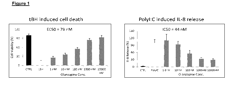
Figure 1 shows that olanzapine significantly protects in a dose responsive
fashion retinal pigmented epithelium (RPE) cells from oxidative stress (tBH)
induced
apoptosis (A) & inhibits Poly I:C induced IL-8 secretion from RPE cells (B).
Error
bars,SEM
Figure 2 shows that olanzapine, its metabolites, and clozapine protect RPEs
from tBH induced cell death. Error bars, SEM
Figure 3 shows that olanzapine significantly protects superior retinal
thickness of
blue light exposed rats. Drug treatment was started 2 days before blue light
exposure.
Animals received olanzapine IP injection once per day for 3 days and the last
dosing
was 1 hour before blue light exposure. Rats were dark adapted for 24 hours
before
they were exposed to blue light with lux intensity of ¨6-7 k for 4 hours.
Right after
the blue light exposure, the rats were dark adapted again for another 3 days
before
returning to normal room light (12-hour light/ 12¨ hour dark). Optical
coherence
tomography (OCT) was used to evaluate the retinal thickness change caused by
blue
light at 7-10 days post blue light exposure. Error bars, SEM. OS = Left Eye,
OD = Right
Eye.
Figure 4 shows that olanzapine significantly protects retinal a- and b-waves
of
blue light exposed rats. Drug treatment and dark adaptation were similar as
described in
CA 02819616 2013-05-31
WO 2012/074788
PCT/US2011/061370
3
Figure 3. Electroretinograms were recorded at 7-10 days post blue light
exposure. Error
bars, SEM. BL = Blue Light.
Figure 5 shows that olanzapine significantly protects outer nuclear layer of
blue
light exposed rats. Drug treatment and dark adaptation were similar as
described in
Figure 3. H&E staining was performed 2-3 wks after blue light exposure. B-L =
Blue
Light. ONH = Optic nerve head.
Figure 6 shows that olanzapine significantly protects retinal rhodopsin loss
caused by blue light exposure. Drug treatment and dark adaptation were similar
as
described in Figure 3. lmmuno-histochemistry study was performed 2-3 wks after
blue
light exposure. B-L = Blue Light. ONH = Optic nerve head.
Figure 7 shows that olanzapine significantly protects RPE65 loss and partially
inhibits GFAP upregulation. Drug treatment and dark adaptation were similar as
described in Figure 3. lmmuno-histochemistry study was performed 2-3 wks after
blue
light exposure. B-L = Blue Light. ONH = Optic nerve head.
Figure 8 shows that intravitreal delivery of olanzapine significantly prevents
ERG
alteration of blue light exposed rats. For intravitreal injection (IVT),
animals received
olanzapine 1 hour before blue light exposure. Water was used as a parallel
control
vehicle during IVT injection. Rats were dark adapted for 24 hours before they
were
exposed to blue light with lux intensity of -6-7 k for 4 hours. Right after
the blue light
exposure, the rats were dark adapted again for another 3 days before returning
to
normal room light (12-hour light/ 12- hour dark). CTRL = Naïve Control, Veh =
Water,
0.04, 0.2, or 1 ug of olanzapine/eye. Left Panel = Scattered plots of data
from each eye
and Right panel = Bar diagrams of average results. Electroretinograms were
recorded at
7-10 days post blue light exposure. Error bars, SEM.
Figure 9 shows that topical ocular dosing of olanzapine significantly protects
retinal a- and b-wave signals of blue light exposed SD rats. For topical
administration,
the drug was given 24 hours (BID) and one hour (QI) before blue light
exposure. Water
was used as a parallel control vehicle during topical administration. Rats
were dark
adapted for 24 hours before they were exposed to blue light with lux intensity
of -6-7
k for 4 hours. Right after the blue light exposure, the rats were dark adapted
again for
another 3 days before returning to normal room light (12-hour light/ 12- hour
dark).
Electroretinogramsmere recorded at 7-10 days post blue light exposure. Error
bars,
SEM.
CA 02819616 2013-05-31
WO 2012/074788
PCT/US2011/061370
4
Figure 10 shows that olanzapine significantly attenuates hyperoxia induced
retinal neovascularization. Litters of newborn mice and their dams were placed
in a 75%
oxygen chamber from P7 to P12. The chamber contained enough food and water for
5
days and was opened only to allow drug administration to the animals. The mice
were
returned to room air with normal oxygen content on P12. Olanzapine in water or
VEH
(water) was administered once daily by gavage beginning on P10 and continuing
through P16. Retinal neovascularization was evaluated on P17 after 5 days of
exposure
of the animals to room air. Error bars, SEM.
Figure 11 shows that olanzapine significantly inhibits laser induced choroidal
neovascularization in rats. Error bars, SEM.
DETAILED DESCRIPTION OF THE INVENTION
Conditions of the Retina
The compound of the invention may be used to treat diseases of the retina. By
"diseases of the retina," the applicants mean any condition of the retina or
the tissues
that surround it which are caused or aggravated by oxidative stress. These
include
macular degeneration, diabetic retinopathy, choroidal neovascular membrane,
macular
edema (also referred to as cystoid macular edema and macular swelling),
epiretinal
membrane (macular pucker), macular hole, retinitis pigmentosa, macular
dystrophies
(such as Stargardt's juvenile macular degeneration, Best's vitelliform
dystrophy, cone
dystrophies, and pattern dystrophy of the retinal pigmented epithelium),
retinal
detachment, retinal trauma, retinal tumors and retinal diseases associated
with them,
congenital hypertrophy of the retinal pigmented epithelium, acute posterior
multifocal
placoid pigment epitheliopathy, and acute retinal pigment epithelitis.
Macular degeneration, also referred to as age-related macular degeneration, is
the most common cause of vision loss in the United States in those 50 or
older, and its
prevalence increases with age. AMD is classified as either wet (neovascular)
or dry
(non-neovascular). The dry form of the disease is most common. It occurs when
the
central retina has become distorted, pigmented, or most commonly, thinned, a
process
associated with atrophy of the retinal pigment epithelium and loss of macular
photoreceptors. The result is central geographic atrophy. The wet form of the
disease
is responsible for most severe loss of vision. The wet form is usually
associated with
aging, but other diseases that can cause wet macular degeneration include
severe
myopia and some intraocular infections such as histoplasmosis, which may be
CA 02819616 2013-05-31
WO 2012/074788
PCT/US2011/061370
exacerbated in individuals with AIDS. The wet form is characterized by
abnormal blood
vessels growing through the retinal pigment epithelium, resulting in
hemorrhage,
exudation, scarring, or retinal detachment.
Retinopathy associated with diabetes is a leading cause of blindness in type 1
5 diabetes, and is also common in type 2 diabetes. The degree of
retinopathy depends on
the duration of the diabetes, and generally begins to occur ten or more years
after onset
of diabetes. Diabetic retinopathy may be classified as (1) non-proliferative
or
background retinopathy, characterized by increased capillary permeability,
edema,
hemorrhage, microaneurysms, and exudates; or 2) proliferative retinopathy,
characterized by neovascularization extending from the retina to the vitreous,
scarring,
fibrous tissue formation, and potential for retinal detachment. Diabetic
retinopathy is
believed to be caused, at least in part, by the development of glycosylated
proteins due
to high blood glucose. Glycosylated proteins generate free radicals, resulting
in
oxidative tissue damage and depletion of cellular reactive oxygen species
(ROS)
scavengers, such as glutathione.
In choroidal neovascular membrane, abnormal blood vessels stemming from the
choroid grow up through the retinal layers. The fragile new vessels break
easily, causing
blood and fluid to pool within the layers of the retina.
In macular edema, which can occur as a result of disease, injury or surgery,
fluid
collects within the layers of the maoula, causing blurred, distorted central
vision.
Epiretinal membrane is a cellophane-like membrane that forms over the macula,
affecting the central vision by causing blur and distortion. As it progresses,
the traction
of the membrane on the macula may cause swelling. The disease is seen most
often in
people over 75 years of age.
Retinitis pigmentosa is a retinal degeneration characterized by night
blindness
and progressive loss of peripheral vision, eventually leading to total
blindness;
ophthalmoscopic changes include dark mosaic-like retinal pigmentaion,
attenuation of
the retinal vessels, waxy pallor of the optic disc, and in the advanced forms,
macular
degeneration. In some cases there can be a lack of pigmentation Retinitis
pigmentosa
can be associated to degenerative opacity of the vitreous body, and cataract.
Macular dystrophy is a term applied to a heterogeneous group of diseases that
collectively are the cause of severe visual loss in a large number of people.
A common
characteristic of macular dystrophy is a progressive loss of central vision
resulting from
the degeneration of photoreceptor cells in the retinal macula. In many forms
of macular
CA 02819616 2013-05-31
WO 2012/074788
PCT/US2011/061370
6
dystrophy, the end stage of the disease results in legal blindness. More than
20 typ'es
of macular dystrophy are known. Some of these are, for example, age-related
macular
dystrophy, Stargardt-like dominant macular dystrophy, recessive Stargardt's
disease,
atypical vitelliform macular dystrophy (VMD1), Usher Syndrome Type 1B,
autosomal
dominant neovascular inflammatory vitreoretinopathy, familial exudative
vitreoretinopathy, and Best's macular dystrophy (also known as hereditary
macular
dystrophy or Best's vitelliform macular dystrophy (VMD2).
Stargardt-like dominant macular dystrophy (also called autosomal dominant
macular atrophy) is a juvenile-onset macular degeneration. Patients afflicted
with this
disease generally have normal vision as young children, but during childhood,
visual
loss begins, which rapidly progresses to legal blindness. Clinically it is
characterized by
the presence of an atrophic macular lesion with sharp borders and is often
associated
with yellow fundus flecks.
Best's macular dystrophy is an inherited autosomal dominant macular dystrophy
of unknown biochemical cause. The disease has an age of onset that can range
from
childhood to after 40. Clinical symptoms include, at early stages, an abnormal
accumulation of the yellowish material lipofuscin in the retinal pigmented
epithelium
(RPE) underlying the macula. This gives rise to a characteristic "egg yolk"
appearance
of the RPE and gradual loss of visual acuity. With increasing age, the RPE
becomes
more and more disorganized, as the lipofuscin accumulations disperse and
scarring and
neovascularization take place. These changes are accompanied by further loss
of
vision.
The pathological features seen in Stargardt-like dominant macular dystrophy
and
Best's macular dystrophy are in many ways similar to the features seen in age-
related
macular dystrophy (AMD), the leading cause of blindness in older patients in
the
developed world.
Retinal detachment occurs when the sensory layers of the retina become
separated from their underlying supporting tissue of retinal pigment
epithelium and the
choroid. Generally, retinal detachment is caused by a retinal tear or the
presence of
vitreous traction, either of which may occur spontaneously or may be due to
trauma.
Retinal detachment may also result from pathology, such as retinopathy of
prematurity
in premature infants or diabetic retinopathy in diabetic individuals. Symptoms
of retinal
detachment are painless and sudden segmental or total visual loss in one eye.
When
there is a tear, or when there is traction causing separation of the retina
from its
CA 02819616 2013-05-31
WO 2012/074788
PCT/US2011/061370
7
underlying structures, the liquid vitreous passes through the opening and into
the
subretinal space, inducing further exudation in the subretinal space. The
retina
gradually separates and detaches from the underlying retinal pigment
epithelium. This
deprives the outer retina of its normal supply of oxygen and nutrients from
the choroid.
With time, retinal detachment also results in loss of vision, due to loss of
photoreceptor
cells located in the outer part of the retina.
By "treat," the applicants mean to deal with medically. The term includes
administering the compound of the invention to alleviate symptoms of a retinal
disease,
such as the decrease in visual acuity that accompanies macular degeneration,
as well
as to address the physiological changes associated with the disease, such as
the
abnormal blood vessel growth that accompanies that condition.
Compounds of the invention
Methods of the inventions treat retinal disease by administering to a patient
in
need of such treatment clozapine, olanzapine, or certain metabolites of
olanzapine.
The compounds are well known.
Clozapine has been prescribed since the 1970s as an antipsychotic. It has the
following structure:
CI 411 N
EN)
H3C
Its chemical name is 2-chloro-11-(4-methylpiperazin-1-yI)-5H-
dibenzo[b,e][1,4]diazepine. It may be synthesized in various ways. One way is
as
follows:
=
CA 02819616 2013-05-31
WO 2012/074788 PCT/US2011/061370
8
NH2 CI Cu
0 ¨DP- NH
0 H3C¨N NH
= \__/
CI NO2 CI NO2
OCH3 OCH3
NH NH
H2 / Raney. Ni POCL3
0 0 0
CI NO2 CI NH2
CI
N =
¨N
N¨
H3C
Another way is as follows:
CA 02819616 2013-05-31
WO 2012/074788
PCT/US2011/061370
9
1. (CH3)30K
N 2. CI¨H2C = NO2
CI
HN
= N
H3C¨Nr¨\NH
CI
_______________________________________________________ No-
N¨
S¨CH2 NO
ci N
¨N
H3C
N-desmethyl clozapine, which has the structure
cro\I
N-
CI fillk
Wir N *
can be obtained from Tocris.
Olanzapine is another well known antipyschotic drug. It has the following
structure:
CA 02819616 2013-05-31
WO 2012/074788 PCT/US2011/061370
Its chemical name is 2-methyl-4-(4-methylpiperazin-1-y1)-10H-
benzo[b]thieno[2,3-
,
e][1,4]diazepine. Elli Lilly and Company markets the drug under the trade name
Zyprexa. One way of making olanzapine is as follows. It is disclosed in U.S.
Patent No.
5 5,631,250, the contents of which are incorporated herein by reference:
NC
(NO
s N. Au 1
0:1µ.
c-.3 4 . =
do-(lID
4.1aH or;k0H
'NH '
NO2 NC.
;012 2
SACI HO
reflux CH
= N S 3
H -
(yo
cm) oo.
relux
CHa
* H 6
N s CH
Another way is as follows. It is disclosed in U.S. Patent Application
Publication No.
2006/035887, the contents of which are incorporated herein by reference:
CA 02819616 2013-05-31
WO 2012/074788
PCT/US2011/061370
11
mi2
(NH
H µ N
relux ,..,..)
0- > . 4.,= = . !ipoota , ii
.
=W
Cl-
rH,Iiii
SO-. NaOH
HCHO. Ista0Ac. ' :: CO
/
. "el
AcOH, NaBH4 or T': 15 '
2µ
HCHO. HCOOH. / /ii CIA.o: Ao0 H
,relux
,
=,,,,.. N....C..14S
N
*
='._)......
. . ' / \
ti.
11,:
Olanzapine is metabolized to the following compounds, both of which may also
be used in the method of the invention:
H
..............S.....õ...õN Illni
-N
(--)N+
/ \
-0 CH3
Metabolite A
=
CA 02819616 2013-05-31
WO 2012/074788
PCT/US2011/061370
12
-N
HN
Metabolite B
Olanzapine-N-oxide (Metabolite A) and N-demethyl olanzapine (Metabolite B)
can be obtained from Toronto Research Chemical.
In one embodiment, one administers the compounds of the invention as
pharmaceutically acceptable salts. A pharmaceutically acceptable salt is any
salt of the
parent compound that is suitable for administration to an animal or human. A
pharmaceutically acceptable salt also refers to any salt which may form in
vivo as a
result of administration of an acid, another salt, or a prodrug which is
converted into an
acid or salt. A salt comprises one or more ionic forms of the compound, such
as a
conjugate acid or base, associated with one or more corresponding counter-
ions. Salts
can form from or incorporate one or more deprotonated acidic groups (e.g.
carboxylic
acids), one or more protonated basic groups (e.g. amines), or both (e.g.
zwitterions).
Pharmaceutically acceptable salts of acidic functional groups may be derived
from organic or inorganic bases. The salt may comprise a mono or polyvalent
ion. Of
particular interest are the inorganic ions, lithium, sodium, potassium,
calcium, and
magnesium. Organic salts may be made with amines, particularly ammonium salts
such
as mono-, di-, and tri-alkyl amines or ethanol amines. For a review on
suitable salts,
see Handbook of Pharmaceutical Salts: Properties, Selection, and Use by Stahl
and
Wermuth (Wiley-VCH, 2002).
CA 02819616 2013-05-31
WO 2012/074788
PCT/US2011/061370
13
Formulation and administration
The compound of the invention may be administered via either the oral,
transdermal (e.g.. through the use of a patch), intranasal, sublingual,
rectal, or
parenteral routes. In one preferred embodiment, the compound is delivered by
injecting
it into the eye.
In one embodiment the compound is administered at doses ranging from about
0.25 mg up to about 1500 mg per day; in another embodiment the compound is
administered at doses of 0.25 to about 300 mg per day in single or divided
doses; in
another embodiment the compound is administered at doses of 0.01 mg to about
10 mg
per kg of body weight per day, although variations will necessarily occur
depending
upon the weight and condition of the subject being treated and the particular
route of
administration chosen, as well as the individual's responses to the treatment,
the
formulation chosen, and the length of time the patient is treated. In some
instances,
doses less than 0.25 mg per day may be adequate, while in other cases still
larger
doses may be employed without causing any harmful side effects, provided that
such
larger doses are first divided into several small doses for administration
throughout the
day.
The active compounds can be administered alone or in combination with
pharmaceutically acceptable carriers or diluents by any of the several routes
previously
indicated. More particularly, the active compounds can be administered in a
wide
variety of different dosage forms, e.g., they may be combined with various
pharmaceutically acceptable inert carriers in the form of tablets, capsules,
solutions,
suspensions, and the like. Such carriers include solid diluents or fillers,
sterile aqueous
media and various non-toxic organic solvents. In addition, oral pharmaceutical
compositions can be suitably sweetened and/or flavored. In general, the active
compounds are present in such dosage forms at concentration levels ranging
from
about 5.0% to about 70% by weight.
In one embodiment of the invention, clozapine may be delivered topically to
the
eye or by injection into the eye. Suitable formulations for this purpose
include liquids,
suspensions, emulsions, and the like. Topical formulations of ophthalmic drug
products
are well known in the art and described in, for example, U.S. Patent
Application
Publication No. 20050059583; No. 20050277584; No. 20070015690; and No.
20070015691; and U.S. Patent Nos. 5,474,979 and 6,582,718, the disclosures of
all
which are incorporated herein by reference.
=
CA 02819616 2013-05-31
WO 2012/074788
PCT/US2011/061370
14
In one embodiment, the clozapine formulation is administered as an eye drop;
in
a typical administration, 25 to 50 pl of the formulation is administered to
the eye. Such
formulations may be administered once, twice, three times, four times, or
more, daily.
EXAMPLES
The invention is illustrated by the following examples.
ARPE-19 cells from ATCC were grown in DEME/F12 medium supplemented with
10% Fetal Bovine Serum (FBS) and split into 96-well plates at a density of
10,000 cells
per well. Cells were incubated overnight in 0.1%FBS medium before addition of
compotind/drug. Stocks of compounds of interest were made at different
concentrations
and added to ARPE-19 cells in 0.1% FBS medium for one hour.
Cell Viability Assay. After one hour of compound/drug pretreatment, cells were
incubated with 0.3 mM tert-butyl hydroperoxide (tBH) for 3 hours followed by
three times
of washing and replacement with fresh drug and medium (0.1% FBS medium) for
overnight. Cell viability was determined by cell proliferation assay kit
(Millipore, 2210).
Error bars, SEM. n = 3. The results are shown in Figs. 1, 2 & 12.
IL-8 Assay After one hour of drug pretreatment, cells were incubated with
1ug/m1
polyl:C for 24 hours. The cell culture supernatant was used to measure IL-8
production
with IL-8 ELISA Kit (R&D Systems, D8000C). Error bars, SEM. n = 3. The results
are
shown in Figure 1.
Blue Light Study Four to five month old Sprague-Dawley (SD) male rats were
used in the following study. Drug treatment was started 2 days before blue
light
exposure. Animals received olanzapine IP injection once per day for 3 days and
the last
dosing was 1 hour before blue light exposure.
For intravitreal injection (IVT), animals received olanzapine (having the
structure shown
below) 1 hour before blue light exposure. For topical administration, the drug
was given
24 hours (BID) and one hour (QI) before blue light exposure. Water was used
for
parallel control vehicle IP/IVT injection or topical administration. Rats were
dark adapted
for 24 hours before they were exposed to blue light with lux intensity of ¨6-7
k for 4
hours. Right after the blue light exposure, the rats were dark adapted again
for another
3 days before returning to normal room light (12-hour light/ 12¨ hour dark).
Optical
coherence tomography (OCT) was used to evaluate the retinal thickness change
caused by blue light at 7-10 days post blue light exposure. Error bars, SEM.
(Figure 3).
CA 02819616 2013-05-31
WO 2012/074788
PCT/US2011/061370
Electroretinography (ERG) Assay: Bilateral Flash Electroretinograms were
recorded in SD rats using the Espion E2electroretinography system. On the day
of ERG,
Animals were dark adapted for at least 30 minutes. Their eyes were dilated
with
Tropicamide HCI ( 1%) and Ak-dilate (10%). Prior to ERG recording, the animal
was
5 anesthetized with intramuscular injection of 40mg/m1 Ketamine HCL and
12mg/m1
Xylazine HCL, and placed onto heated platform. The ground needle was placed in
the
skin under the arm and the reference needle was place in the skin above the
head.
Retinae were stimulated using 1 cd.s/m2 flash for an average of 10 traces at
0.1 Hz.
Recording filter was set at 300 Hz. ERG responses were analyzed using Espion
E2 ,and
10 Microsoft excel program . The b-wave amplitude was measured from the
trough of the
a-wave to the peak of the b-wave, and the a-wave was measured as the
difference in
amplitude between recording at onset and the trough of the negative
deflection. Error
bars, SEM. (Figures 4, 8, and 9)
Rat Ocular Tissue Processing & H&E staining: Sprague-Dawley male rats 2-3
15 weeks after blue light exposure were euthanized with CO2 and orbits
enucleated. Eyes
were fixed in Davidson's fixative overnight at room temperature and
transferred to 70%
ethanol for 24 hrs. Further tissue processing was done by serial dehydration
in 80%,
95% & 100% alcohol and Propar, followed by paraffin embedding. Whole rat eyes
were
transversely cut in the vertical meridian proceeding from nasal to temporal
side, using a
Microtome (RM2255; Leica Microsystems). Using optic nerve head as the
landmark, a
total of 45 serial sections with 5 microns/section were collected on 15 glass
slides. Slide
#s 1,5,10 & 15 were diparaffinized and sequentially stained using hematoxylin
(nucleus)
and eosin (cytoplasm) as per standard protocol to compare photoreceptor/RPE
lesion
between experimental groups. The rest of the slides were used to determine
expressions of Rhodopsin, RPE65 and GFAP using the specific antibodies by
standard
immune-histochemistry techniques. The results are shown in Figures 5-7.
Oxygen-induced retinopathy: Oxygen-induced retinopathy (01R)/Hyperoxia was
induced in C57B6 mice using the protocol reported by Smith et al. (Smith LE,
WeSolowski E, McLellan A, et al. Oxygen-induced retinopathy in the mouse.
Invest
Ophthalmol Vis Sci. 1994;35(1):101-111). Litters of newborn mice and their
dams were
placed in a 75% oxygen chamber from P7 to P12. The chamber contained enough
food
and water for 5 days and was opened only to allow drug administration to the
animals.
The mice were returned to room air with normal oxygen content on P12.
Olanzapine in
water or VEH (water) was administered once daily by gavage beginning on P10
and
CA 02819616 2013-05-31
WO 2012/074788
PCT/US2011/061370
16 =
continuing through P16. Retinal neovascularization was evaluated on P17 after
5 days
of exposure of the animals to room air.
Retinal angiography and quantification. Retinal neovascularization was
evaluated by angiography in mice subjected to OIR as described previously
(Smith LE,
Wesolowski E, McLellan A, et al. Oxygen-induced retinopathy in the mouse.
Invest
Ophthalmol Vis Sci. 1994;35(1):101-111). P17 mice were deeply anesthetized and
then
were perfused through the left ventricle with 1 mL of PBS containing 50 mg of
high¨
molecular-weight (2000 kDa) fluorescein-dextran (Sigma, St. Louis, MO). Eyes
were
enucleated and fixed in 4% paraformaldehyde for 24 hours. After removal of the
lens,
the retina was dissected and wholemounted with glycerol-gelatin.
Quantification of
retinal neovascularization was performed as described previously (Chen J,
Connor KM,
Aderman CM, Smith LE. Erythropoietin deficiency decreases vascular stability
in mice. J
Clin Invest. 2008;118(2):526-533). Images of retinal whole-mounts taken at 4x
magnification on an epifluorescence microscope (Olympus, Center Valley, PA)
were
imported into Adobe Photoshop 7.0 software (Adobe Systems, Mountain View, CA)
and
merged to produce an image of the entire retina. Neovascularization was
quantified as
described previously (Bai Y, Ma JX, Guo J, et al. Muller cell-derived VEGF is
a
significant contributor to retinal neovascularization. J Pathol.
2009;219(4):446-454). The
Photoshop freehand tool was used to outline areas of neovascular tuft
formation, and
the area of neovascularization (in pixels) was expressed as a percentage of
the area of
the whole retina (inpixels). To avoid bias, quantification of
neovascularization was
performed by an observer masked to the animal treatment. The results are shown
in
Figure 10.
Chotoidal Neovasculatization (CNV) Assay. Brown Norway rats (Charles Rivers),
weighing 250 ¨ 300 grams each were used in the study. Drug treatment was
started 2
days prior to Laser treatment. Olanzapine was given i.p. (1mg/kg) once a day
for 3 days
(day -2, -1 & 0). The last dose on day 0 was delivered about 1 hr before laser
treatment.
Phosphate buffered saline (PBS) was used for parallel control vehicle IP
injection. On
day zero of the laser procedure, rat eyes (pupil) were dilated with
Tropicamide HCI (
1%) and Ak-dilate (10%). Rats were then anesthetized with intrasmuscular
injection of
40mg/mIKetamine HCL and 12mg/mIXylazine HCL. The fundo's was visualized using
a microscope slide coverslip with Refresh Liquigel eye drops as an optical
coupling
agent. For each animal, 6 laser spots in each eye (360 mW power, 0.07 sec.
duration,
50 pm spot size) were made with argon laser (Coherent Inc.; Santa Clara, Ca.)
CA 02819616 2013-05-31
WO 2012/074788
PCT/US2011/061370
17
concentrically at approximately equal distances around the optic disc, between
every 2
retinal vessels. A single 5-ul volume of the drug (olanzapine) or PBS was
injected into
the vitreous cavity at day 1, 4 & 6 post-laser photocoagulation procedure. At
11 days
after laser treatment, animals were sacrificed by CO2 exposure and CNV
formation was
assayed as described previously. Briefly, eyes were enucleated and fixed in
10%
Formalin solution for 1 hour. Eyes were rinsed in PBS, 5 min x 2. or kept in
PBS
overnight. The eye was thoroughly cleaned and cut in half in a Petri dish
leaving the
eye cup and attached retina in place. The eye cup-retina was washed in PBS,
and
retina was separated and removed from the choroid. The eye cup-choriod was
incubated overnight in Isolectin 164 conjugate (10 ug/ml) in PBS / 0.5 %
Triton X 100.
Eye cup/Choroid was washed in PBS, 20 min x 3 and cut in four locations and
flat
mounted using aqueous mounting media. The area of fluorescence was quantified
using
Metamorph image analysis software (RPI, Natick, MA). The results are shown in
Figure
11.
The experiments establish that the compounds of the invention protect RPEs
from
oxidative stress (as summarized, below) and the diseases that that such stress
causes.
COMPOUND PROTECTION (%) AT 10 UM
Olanzapine 116
Metabolite A 103
Metabolite B 129
Clozapine 99
n-desmethly clozapine 77
Each and every reference disclosed in the specification, whether non-patent
(e.g., scientific, journal references) or patent (granted patents or published
patent
applications) is incorporated herein by reference in its entirety for all
purposes.
The foregoing descriptions details specific methods and compositions that can
be
employed to practice the present invention, and represents the best mode
comtemplated. It should not be construed as limiting the overall scope hereof;
rather,
the ambit of the present invention is to be governed only by the lawful
construction of
the appended claims.