Note: Descriptions are shown in the official language in which they were submitted.
CA 02820957 2013-07-11
LIQUID CHROMATOGRAPHY WITH TANDEM MASS SPECTROMETRY OF
ESTRONE, ESTRADIOL AND FREE THYROXINE
FIELD OF INVENTION
The presently disclosed subject matter relates to methods and systems for the
analysis of
biomarkers. In certain embodiments, the biomarkers are endogenous to human
subjects such that
the measurement may be used for clinical diagnosis.
BACKGROUND
Biomarkers, such as hormones, vitamins, metabolites, can be used for the
clinical
diagnosis of multiple disorders and as endogenous biomarkers in endocrinology.
For example, the
measurement of estrogen compounds, such as estrone and estradiol can be used
to evaluate
ovarian function and to evaluate excess or diminished estrogen levels in a
patient. Also,
measurement of thyroxine can be used to quantify thyroid function.
Requirements for the clinical diagnostic testing of endogenous biomarkers in
endocrinology may include highly sensitive and specific assays, the ability to
analyze small
sample volumes (e.g., pediatric sample volumes can be limited to less than
about 200 pt), and the
ability to screen for multiple analytes to accurately diagnose a disease
state, e.g., an endocrine
disorder. Historically, radioimmunoassay (RIA) and enzyme-linked immunoassay
(ELISA)
methods have been used in such clinical diagnostic testing. Immunoassay
methods (IA), such as
RIA and EIA, however, may suffer from low throughput, antibody cross-
reactivity, which can
require extra preparation for specificity, and poor scalability. Also, the
analysis of endogenous
biomarkers by RIA may require multiple serial dilutions for the analysis of
each individual
marker, which can lead to the need to make multiple adjustments to normalize
sample volumes
and/or the need for multiple separate tests. Also, immunoassay tesing is not
particularly
conducive to the analysis of multiple biomarkers in each sample. The analysis
for multiple
analytes in a single assay can allow for using samples of reduced size which
results in assays of
increased sensitivity and efficiency per sample.
An important class of hormones are the steroid hormones, such as testosterone
and
estrogens. Testosterone develops and maintains the male secondary sex
characteristics, and
1
CA 02820957 2013-07-11
WO 2007/139956
PCT/US2007/012525
promotes growth and development of sperm. Estrogen is the term used for a
group of
hormones of which there are three principle forms, estrone, estradiol, and
estriol.
For example, relatively small variations in estrogen levels may be clinically
significant. Generally, the level of estrogen in post-menopausal women, adult
males, and
prepubescent children is < 10 pg/mL. Elevated estrogen levels in children may
lead to
precocious puberty (and short stature). In post-menopausal women, low estrogen
levels may
require replacement, where as levels greater than 5 pg/mL may be prognostic
for certain
cancers. In adult males, elevated estrogen levels may be indicative of certain
disease states
(testicular cancer). In adult females, reduced or elevated levels may also be
indicative of
certain cancers (e.g., ovarian cancer). A level of serum estrogen of 15 pg/mL
is clinically
different from 10pg/mL and thus, measurement of estrogen compounds (e.g.,
estradiol and
estrone) requires an LLOQ of 1-5 pg/mL irrespective of sample type, patient
age, gender and
diet.
Another important class of hormones are the thyroid hormones. Thyroxine (T4)
and
triiodothyronine (T3) are examples of thyroid hormones. T4 and T3 enter cells
and bind to
intracellular receptors. T4 and T3 are important in regulation of a number of
factors
including growth and development, carbohydrate metabolism, oxygen consumption,
and
protein synthesis. T4 acts as a prohormone, as the bulk of T3 present in blood
is produced by
monodeiodination of T4 by intracellular enzymes. Thyroid hormone
concentrations in blood
are essential tests for the assessment of thyroid function.
Thus, there is a need to develop analytical techniques that can be used for
the
measurement of endogenous biomarkers, and for methods that provide more
sensitivity and
higher throughput than RIA. Until recently, however, only GC-MS or LC-MS/MS
with
derivatization has been successful for small sample volumes. Thus, there is a
need in the art
for LC-MS/MS techniques for the analysis of endogenous biomarkers for clinical
diagnosis in
endocrinology capable of providing detection limits at acceptable levels,
without the need for
the cumbersome derivatization processes.
SUMMARY
In some embodiments, the presently disclosed subject matter provides methods
and
systems for the quantitative analysis of endocrine biomarkers in a test
sample. The
quantification of such markers may, in certain embodiments, be used for
clinical diagnosis in
endocrinology. For example, in some embodiments, the methods and systems of
the present
invention may be used for the quantitative analysis of total levels of certain
hormones,
2
CA 02820957 2013-07-11
WO 2007/139956
PCT/US2007/012525
including steroid hormones, such as estrone and estradiol, and their
metabolites, such as
estrone sulfate. In other embodiments, the methods and systems of the present
invention
provide for the quantitative analysis of biomarkers that can be difficult to
detect in their
active state. For example, the systems and methods of the present invention
may be used to
quantify free (i.e., not bound to protein) serum hormones, such as free
thyroxine (T4) in
biological samples. Or, in other embodiments, the systems and methods of the
present
invention may be used to quantify free triiodothyronine (T3) or testosterone.
In an
embodiment, the methods and systems of the present invention allow for
measuresment of
such hormones without the need for derivatation processes.
In some embodiments, the biomarkers of interest are estradiol and/or estrone.
Thus,
in one embodiment, the present invention comprises a method for determining
the presence
or amount of estradiol in a sample by tandem mass spectrometry, comprising:
(a) generating
a dehydrated precursor ion of the estradiol; (b) generating one or more
fragment ions of the
precursor ion; and (c) detecting the presence or amount of one or more of the
ions generated
in step (a) or (b) or both, and relating the detected ions to the presence or
amount of the
estradiol in the sample. In an embodiment, the sample comprises a mixture of
estradiol and
estrone.
In other embodiments, the biomarker comprises free thyroxine (T4) or
triiodothyronine (T3). In certain embodiments, the present invention provides
a high-
throughput assay for free thyroxine (T4). Thus, in one embodiment, the present
invention
comprises a method for determining the presence or amount of free thyroxine in
a plurality of
samples by tandem mass spectrometry, comprising: (a) dialyzing the plurality
of samples to
separate the free thyroxine from the protein-bound thyroxine in the samples;
(b) generating a
precursor ion of the thyroxine; (b) generating one or more fragment ions of
the thyroxine; and
(c) detecting the presence or amount of one or more of the ions generated in
step (b) or (c) or
both, and relating the detected ions to the presence or amount of the free
thyroxine in the
plurality of samples.
In some embodiments, the methods and systems of the present invention comprise
liquid chromatography (LC) methods in combination with other analytical
techniques as a
means to measure such biomarkers with high sensitivity and high throughput. In
certain
embodiments, the present invention comprises quantitative liquid
chromatography tandem
mass spectrometry (LC-MS/MS) analysis of endocrine biomarkers in a test
sample. In some
embodiments, two-dimensional or tandem LC is used. The method may include, in
alternate
3
CA 02820957 2013-07-11
emboiments, liquid-liquid extractions, dialysis, sample dilution, and/or
sample dehydration steps prior to
analysis by tandem mass spectrometry.
Accordingly, embodiments of the present invention may provide methods for the
quantitative
LC-MS/MS and 2D-LC-MS/MS analysis of hormones, including steroid hormones,
such as estrone and
estradiol. Additionally or alternatively, embodiments of the present invention
may provide methods for the
quantitative determination of a free (i.e., non-protein bound) hormone or
metabolite using dialysis in
combination with LC-MS/MS analysis for hormones that in biological samples,
may be predominantly protein-
bound. Such hormones may include free thyroxine (T4), free triiodothyronine
(3), or free testosterone.
In a broad aspect, the present invention provides a method for determining an
amount of estradiol in a
sample comprising: (a) providing a sample comprising estradiol and
dehydroepiandrosterone (DHEA); (b)
chromatographically separating the estradiol from DHEA and other components in
the sample using two-
dimensional chromatography; (c) generating a dehydrated precursor ion of the
estradiol in the sample; (d)
generating one or more fragment ions of the dehydrated precursor ion; and (e)
detecting an amount of one or
more of the ions generated in steps (c) and (d), and correlating said amounts
to an amount of estradiol in the
sample.
In another broad aspect, the present invention provides a method for
determining an amount of free
thyroxine in a sample, the method comprising: providing a sample comprising
free thyroxine and protein-
bound thyroxine; dialyzing the sample; partially purifying the solution using
dilution or liquid-liquid
extraction; chromatographically separating the free thyroxine from other
components in the sample; and
analyzing the chromatographically separated free thyroxine by mass
spectrometry to determine the amount of
free thyroxine in the sample.
Certain objects of the present invention, having been stated hereinabove, will
become further evident
as the description proceeds when taken in connection with the accompanying
figures and examples as
described herein below.
BRIEF DESCRIPTION OF THE DRAWINGS
Having thus described the invention in general terms, reference will now be
made to the
accompanying drawings, which are not necessarily drawn to scale.
FIG. I shows a flow chart of a method for quantitative analysis of a biomarker
of interest in
accordance with one embodiment of the present invention.
FIG. 2 shows dehydration of estradiol and the effect on mass spectrometry (MS)
analysis in
accordance with an embodiment of the present invention.
FIG. 3 shows potential isobaric interferences for measurement of estrone and
estradiol due to
dehydration of dehydroepiandrosterone (DHEA) in accordance with one embodiment
of the present invention.
FIG. 4 shows an example of heart-cutting from a primary separation gradient to
remove compounds
that comprise isobaric interference in the analysis of estrone and estradiol
in accordance with one embodiment
of the present invention.
4
CA 02820957 2013-07-11
FIG. 5 shows a method for the quantification of estrone and estradiol in
accordance with an
embodiment of the present invention.
FIG. 6 shows a method for the quantification of free thyroxine (T4) in
accordance with an
embodiment of the present invention.
FIG. 7 shows a system for quantitative analysis of a metabolite in accordance
with one embodiment of
the present invention (Panel A), and a system for multiplex analysis (Panel B)
in accordance with alternate
embodiments of the present invention.
4a
CA 02820957 2013-07-11
WO 2007/139956 PCT/US2007/012525
FIG. 8 shows a LC-MS/MS chromatogram of estrone sulfate at a limit of
quantification
of 100 pg/mL in accordance with one embodiment of the present invention.
FIG. 9 shows a LC-MS/MS chromatogram of free thyroxine at a limit of
quantification
of 2 pg/mL in accordance with one embodiment of the present invention_
FIG. 10 shows a 2D-LC-MS/MS chromatogram of 25-hydroxyvitamin D2 at a limit of
quantification of 1 ng/mL in accordance with one embodiment of the present
invention.
FIG. 11 shows a 2D-LC-MS/MS chromatogram of 25-hydroxyvitamin D3 at a limit of
quantification of 1 ng/mL in accordance with one embodiment of the present
invention.
FIG. 12 shows a 2D-LC-MS/MS chromatogram of estrone at a limit of
quantification
of 2.5 pg/mL in accordance with one embodiment of the present invention.
FIG. 13 shows a 2D-LC-MS/MS chromatogram of estradiol at a limit of
quantification
of 1 pg/mL in accordance with one embodiment of the present invention.
FIG. 14 shows a LC-MS/MS chromatogram of estrone sulfate at an upper limit of
quantification of 50 ng/mL in accordance with one embodiment of the present
invention.
FIG. 15 shows a LC-MS/MS chromatogram of free thyroxine at an upper limit of
quantification of 100 pg/dL in accordance with one embodiment of the present
invention.
FIG. 16 shows a 2D-LC-MS/MS chromatogram of 25-hydroxyvitamin D2 at an upper
limit of quantification of 250 ng/mL in accordance with one embodiment of the
present
invention.
FIG. 17 shows a 2D-LC-MS/MS chromatogram of 25-hydroxyvitamin D3 at an upper
limit of quantification of 250 ng/mL in accordance with one embodiment of the
present
invention.
FIG. 18 shows a 2D-LC-MS/MS chromatogram of estrone at an upper limit of
quantification of 500 pg/mL in accordance with one embodiment of the present
invention.
FIG. 19 shows a 2D-LC-MS/MS chromatogram of estradiol at an upper limit of
quantification of 500 pg/mL in accordance with one embodiment of the present
invention.
FIG. 20 shows a calibration curve obtained by LC-MS/MS for estrone sulfate in
accordance with one embodiment of the present invention.
FIG. 21 shows a calibration curve obtained by LC-MS/MS for free thyroxine in
accordance with one embodiment of the present invention.
FIG. 22 shows a calibration curve obtained by 2D-LC-MS/MS for 25-
hydroxyvitamin
D2 in accordance with one embodiment of the present invention.
5
CA 02820957 2013-07-11
WO 2007/139956 PCT/US2007/012525
FIG. 23 shows a calibration curve obtained by 2D-LC-MS/MS for 25-
hydroxyvitamin
D3 in accordance with one embodiment of the present invention.
FIG. 24 shows a calibration curve obtained by 2D-LC-MS/MS for estrone in
accordance with one embodiment of the present invention.
FIG. 25 shows a calibration curve obtained by 2D-LC-MS/MS for estradiol in
accordance with one embodiment of the present invention.
FIG. 26 shows cross-validation data for LC-MS/MS as compared to
radioimmunoassay
(RIA) for estrone sulfate in accordance with one embodiment of the present
invention.
FIG. 27 shows cross-validation data for LC-MS/MS as compared to immunoassay
(IA)
for free thyroxine in accordance with one embodiment of the present invention.
FIG. 28 shows cross-validation data for 2D-LC-MS/MS as compared to a
competitive
binding protein assay (CBP) (Panel A) or immunoassay (IA) (Panel B) for total
25-
hydroxyvitamin D (25-hydroxyvitamin D2+D3) in accordance with alternate
embodiments of
the present invention.
FIG. 29 shows cross-validation data for 2D-LC-MS/MS as compared to RIA for
Estrone in accordance with one embodiment of the present invention.
FIG. 30 shows cross-validation data for 2D-LC-MS/MS as compared to RIA for
Estradiol in accordance with one embodiment of the present invention.
FIG. 31 shows a comparison of Estradiol (E2) cross-validation of LC-MS/MS with
derivatization to 2D-LC-MS/MS without derivatization in accordance with an
embodiment of
the present invention.
FIG. 32 shows the measured concentration (pg/mL) of free thyroxine vs.
dialysis time
(hours). The squares (m) show dialysis losses and the diamonds (*) show
effective dialysis
for free thyroxine using 96-well equilibrium dialysis plates in accordance
with one
embodiment of the present invention.
DETAILED DESCRIPTION
The presently disclosed subject matter now will be described more fully
hereinafter
with reference to the accompanying description and drawings, in which some,
but not all
embodiments of the presently disclosed subject matter are shown. The presently
disclosed
subject matter can be embodied in many different forms and should not be
construed as
limited to the embodiments set forth herein; rather, these embodiments are
provided so that this
disclosure will satisfy applicable legal requirements. Like numbers refer to
like elements
throughout.
6
CA 02820957 2013-07-11
Many modifications and other embodiments of the presently disclosed subject
matter set forth herein
will come to mind to one skilled in the art to which the presently disclosed
subject matter pertains having the
benefit of the teachings presented in the foregoing descriptions and the
associated drawings. Therefore, it is to
be understood that the presently disclosed subject matter is not to be limited
to the specific embodiments
disclosed. Although specific terms are employed herein, they are used in a
generic and descriptive sense only
and not for purposes of limitation. The disclosure utilizes the abbreviations
shown below.
ABBREVIATIONS
APCI ¨ atmospheric pressure chemical ionization
CBP = competitive binding protein
El = Estrone
E2 = I 73-Estradiol or Estradiol
FT4 = Free Thryoxine
HTLC = high turbulence (throughput) liquid chromatography
HPLC = high performance liquid chromatography
LLE = liquid-liquid extraction
LOQ = limits of quantification
LLOQ = lower limit of quantification
IA = immunoassay
ELISA = enzyme linked immunoassay
RIA = radioimmunoassay
SST = system suitability test
ULOQ = upper limit of quantification
2D-LC-MS/MS = two-dimensional liquid chromatography hyphenated to
tandem mass
spectrometry
(LC)-LC-MS/MS = two-dimensional liquid chromatography tandem
hyphenated to mass
spectrometry
(LC)-MS/MS = liquid chromatography hyphenated to tandem mass
spectrometry
Definitions
While the following terms are believed to be well understood by one of
ordinary skill in the art, the
following definitions are set forth to facilitate explanation of the presently
disclosed subject matter. Other
definitions are found throughout the specification. Unless otherwise defined,
all technical and scientific terms
used herein have the same meaning as commonly understood by one of ordinary
skill in the art to which this
presently described subject matter belongs.
7
CA 02820957 2013-07-11
Notwithstanding that the numerical ranges and parameters setting forth the
broad scope
of the invention are approximations, the numerical values set forth in the
specific examples are
reported as precisely as possible. Any numerical value, however, inherently
contains certain
errors necessarily resulting from the standard deviation found in their
respective testing
measurements. Moreover, all ranges disclosed herein are to be understood to
encompass any and
all subranges subsumed therein. For example, a stated range of "Ito 10" should
be considered to
include any and all subranges between (and inclusive of) the minimum value of
1 and the
maximum value of 10; that is, all subranges beginning with a minimum value of
1 or more, e.g. 1
to 6.1, and ending with a maximum value of 10 or less, e.g., 5.5 to 10.
The terms "a", "an", and "the" refer to "one or more" when used in this
application,
including the claims. Thus, for example, reference to "a cell" includes a
plurality of such cells,
unless the context clearly is to the contrary (e.g., a plurality of cells),
and so forth. As used herein,
the term "biomarker" is any biomolecule that may provide biological
information about the
physiological state of an organism. In certain embodiments, the presence or
absence of the
biomarker may be informative. In other embodiments, the level of the biomarker
may be
informative. A biomarker may be a hormone, such as an estrogen (e.g.,
estradiol, estrone),
testosterone, thyroxine (T4), triiodothyronine (T3), or a metabolite of a
hormone (estrogen
sulfate). A biomarker may also be a vitamin or a metabolite of a vitamin. For
example, in one
embodiment, the measured biomarker may comprise a vitamin D compound such as
25-
hydroxyvitamin D2 and 25-hydroxyvitamin D3.
As used herein, the terms "purify" or "separate" or derivations thereof do not
necessarily
refer to the removal of all materials other than the analyte(s) of interest
from a sample matrix.
Instead, in some embodiments, the terms "purify" or "separate" refer to a
procedure that enriches
the amount of one or more analytes of interest relative to one or more other
components present
in the sample matrix. In some embodiments, a "purification" or "separation"
procedure can be
used to remove one or more components of a sample that could interfere with
the detection of the
analyte, for example, one or more components that could interfere with
detection of an analyte by
mass spectrometry.
As used herein, "derivatizing" means reacting two molecules to form a new
molecule.
Derivatizing agents may include isothiocyanate groups, dansyl groups, dinitro-
fluorophenyl
groups, nitrophenoxycarbonyl groups, and/or phthalaldehyde groups.
8
CA 02820957 2013-07-11
WO 2007/139956
PCT/US2007/012525
As used herein, "chromatography" refers to a process in which a chemical
mixture
carried by a liquid or gas is separated into components as a result of
differential distribution
of the chemical entities as they flow around or over a stationary liquid or
solid phase.
= As used herein, "liquid chromatography" (LC) means a process of selective
retardation of one or more components of a fluid solution as the fluid
uniformly percolates
through a column of a finely divided substance, or through capillary
passageways. The
retardation results from the distribution of the components of the mixture
between one or
more stationary phases and the bulk fluid, (i.e., mobile phase), as this fluid
moves relative to
the stationary phase(s). "Liquid chromatography" includes reverse phase liquid
chromatography (RF'LC), high performance liquid chromatography (HPLC) and high
turbulence liquid chromatography (HTLC).
As used herein, the term "HPLC" or "high performance liquid chromatography"
refers
to liquid chromatography in which the degree of separation is increased by
forcing the mobile
phase under pressure through a stationary phase, typically a densely packed
column.
The chromatographic column typically includes a medium (i.e., a packing
material) to
facilitate separation of chemical moieties (i.e., fractionation). The medium
may include
minute particles. The particles include a bonded surface that interacts with
the various
chemical moieties to facilitate separation of the chemical moieties such as
the biomarker
analytes quantified in the experiments herein. One suitable bonded surface is
a hydrophobic
bonded surface such as an alkyl bonded surface. Alkyl bonded surfaces may
include 0-4, C-
8, or 0-18 bonded alkyl groups, preferably C-18 bonded groups. The
chromatographic
column includes an inlet port for receiving a sample and an outlet port for
discharging an
effluent that includes the fractionated sample. In the method, the sample (or
pre-purified
sample) may be applied to the column at the inlet port, eluted with a solvent
or solvent
mixture, and discharged at the outlet port. Different solvent modes may be
selected for
eluting different analytes of interest. For example, liquid chromatography may
be performed
using a gradient mode, an isocratic mode, or a polytyptic (i.e. mixed) mode.
In one
embodiment, HPLC may performed on a multiplexed analytical HPLC system with a
C18
solid phase using isocratic separation with water:methanol as the mobile
phase.
As used herein, the term "analytical column" refers to a chromatography column
having sufficient chromatographic plates to effect a separation of the
components of a test
sample matrix. Preferably, the components eluted from the analytical column
are separated in
such a way to allow the presence or amount of an analyte(s) of interest to be
determined. In
9
CA 02820957 2013-07-11
WO 2007/139956
PCT/1JS2007/012525
some embodiments, the analytical column comprises particles having an average
diameter of
about 5 p.m. In some embodiments, the analytical column is a functionalized
silica or
polymer-silica hybrid, or a polymeric particle or monolithic silica stationary
phase, such as a
phenyl-hexyl functionalized analytical column.
Analytical columns can be distinguished from "extraction columns," which
typically
- are used to separate or extract retained materials from non-retained
materials to obtained a
"purified" sample for further purification or analysis. In some embodiments,
the extraction
column is a functionalized silica or polymer-silica hybrid or polymeric
particle or monlithic
silica stationary phase, such as a Poroshell SBC-18 column.
The term "heart-cutting" refers to the selection of a region of interest in a
chromatogram and subjecting the analytes eluting within that region of
interest to a second
separation, e.g., a separation in a second dimension.
The term "electron ionization" as used herein refers to methods in which an
analyte of
interest in a gaseous or vapor phase interacts with a flow of electrons.
Impact of the electrons
with the analyte produces analyte ions, which may then be subjected to a mass
spectrometry
technique.
The term "chemical ionization" as used herein refers to methods in which a
reagent
gas (e.g. ammonia) is subjected to electron impact, and analyte ions are
formed by the
interaction of reagent gas ions and analyte molecules.
The term "field desorption" as used herein refers to methods in which a non-
volatile
test sample is placed on an ionization surface, and an intense electric field
is used to generate
analyte ions.
The term "matrix-assisted laser desorption ionization," or "MALDI" as used
herein
refers to methods in which a non-volatile sample is exposed to laser
irradiation, which desorbs
and ionizes analytes in the sample by various ionization pathways, including
photo-ionization,
protonation, deprotonation, and cluster decay_ For MALDI, the sample is mixed
with an
energy-absorbing matrix, which facilitates desorption of analyte molecules.
The term "surface enhanced laser desorption ionization," or "SELDI" as used
herein
refers to another method in which a non-volatile sample is exposed to laser
irradiation, which
desorbs and ionizes analytes in the sample by various ionization pathways,
including photo-
ionization, protonation, deprotonation, and cluster decay. For SELDI, the
sample is typically
bound to a surface that preferentially retains one or more analytes of
interest. As in MALDI,
this process may also employ an energy-absorbing material to facilitate
ionization.
CA 02820957 2013-07-11
WO 2007/139956 PCT/US2007/012525
The term "electrospray ionization," or "ESI," as used herein refers to methods
in which
a solution is passed along a short length of capillary tube, to the end of
which is applied a
high positive or negative electric potential. Upon reaching the end of the
tube, the solution
may be vaporized (nebulized) into a jet or spray of very small droplets of
solution in solvent
vapor. This mist of droplet can flow through an evaporation chamber which is
heated
slightly to prevent condensation and to evaporate solvent. As the droplets get
smaller the
electrical surface charge density increases until such time that the natural
repulsion between
like charges causes ions as well as neutral molecules to be released.
The term "Atmospheric Pressure Chemical Ionization," or "APCI," as used herein
refers to mass spectroscopy methods that are similar to ESI, however, APCI
produces ions
by ion-molecule reactions that occur within a plasma at atmospheric pressure.
The plasma is
maintained by an electric discharge between the spray capillary and a counter
electrode:
Then, ions are typically extracted into a mass analyzer by use of a set of
differentially
pumped skimmer stages. A counterflow of dry and preheated N2 gas may be used
to
improve removal of solvent. The gas-phase ionization in APCI can be more
effective than
ESI for analyzing less-polar species.
The term "Atmospheric Pressure Photoionization" ("APPI") as used herein refers
to
the form of mass spectroscopy where the mechanism for the photoionization of
molecule M
is photon absorption and electron ejection to form the molecular M+. Because
the photon
energy typically is just above the ionization potential, the molecular ion is
less susceptible to
dissociation. In many cases it may be possible to analyze samples without the
need for
chromatography, thus saving significant time and expense. In the presence of
water vapor or
protic solvents, the molecular ion can extract H to form MH+. This tends to
occur if M has a
high proton affinity. This does not affect quantitation accuracy because the
sum of M+ and
MH+ is constant. Drug compounds in protic solvents are usually observed as
MH+, whereas
nonpolar compounds such as naphthalene or testosterone usually form M+ (see
e.g., Robb et
al., 2000, Anal. Chem. 72(15): 3653-3659).
The term "inductively coupled plasma" as used herein refers to methods in
which a
sample is interacted with a partially ionized gas at a sufficiently high
temperature to atomize
and ionize most elements.
The term "ionization" and "ionizing" as used herein refers to the process of
generating an analyte ion having a net electrical charge equal to one or more
electron units_
11
CA 02820957 2013-07-11
WO 2007/139956 PCT/US2007/012525
Negative ions are those ions having a net negative charge of one or more
electron units, while
positive ions are those ions having a net positive charge of one or more
electron units.
The term "desorption" as used herein refers to the removal of an analyte from
a surface
and/or the entry of an analyte into a gaseous phase.
As used herein, the term "immunoassay" (IA) refers to a method for measuring
the
amount of an analyte of interest by quantifying the binding, or the inhibition
of binding, of a
substance to an antibody. Where an enzyme is used to detect the amount of
binding of the
substance (e.g. antigen) to an antibody, the assay is an enzyme-linked
immunoassay (ELISA).
As used herein, the term "radioimmunoassay" (RIA) refers to a method for
measuring the
amount of an analyte of interest by quantifying the binding, or the
inhibition, of binding, of a
radiolabled substance to an antibody.
As used herein, the term "hemolysed" refers to the rupturing of the red blood
cell
membrane, which results in the release of hemoglobin and other cellular
contents into the
plasma or serum and the term "lipemic" refers to an excess of fats or lipids
in blood.
Analysis of Biomarkers by LC-MS/MS
Thus, embodiments of the present invention relate to methods and systems for
the
quantitative analysis of endogenous biomarkers for clinical diagnosis. The
present invention
may be embodied in a variety of ways.
In one embodiment, the present invention comprises a method for determining
the
presence or amount of at least one biomarker of interest in a biological
sample, the method
comprising: providing a biological sample believed to contain at least one
biomarker of
interest; chromatographically separating the at least one biomarker of
interest from other
components in the sample; and analyzing the chromatographically separated at
least one
biomarker of interest by mass spectrometry to determine the presence or amount
of the at
least one biomarker of interest in the sample.
In an embodiment, the at least one biomarker comprises a steroid hormone or a
thyroid hormone. For example, in one embodiment, the at least one biomarker
comprises
estradiol and estrone. Or, the at least one biomarker may comprise free
thyroxine (T4) or
triiodothyronine (T3).
In cetain embodiments, the chromatography may comprise high performance liquid
chromatography (HPLC). In an embodiment, the chromatography may comprises
extraction
and/or analytical liquid chromatography.
12
CA 02820957 2013-07-11
WO 2007/139956 PCT/US2007/012525
In an embodiment, the method may comprise purifying the biomarker of interest
prior
to chromatography. For example, the sample may be partially purfied by at
least one of
liquid-liquid extraction. Also, the method may comprise the step of diluting
the sample into a
solvent or solvents used for LS and/or MS.
In some embodiments, the method may comprise the use of two liquid
chromatography steps. For example, in certain embodiments, the method for
determining the
presence or amount of one or more biomarkers in a test sample may comprise the
steps of: (a)
providing a sample suspected of containing one or more biomarkers of interest;
(b) partially
purifying the one or more biomarkers of interest from other components in the
sample by at
least one of liquid-liquid extraction or by diluting the sample; (c)
transferring the extracted
one or more hormones or metabolites onto an extraction column (i.e., on-line
or off-line); (d)
transferring the one or More biomarkers of interest from the extraction column
onto an
analytical column and chromatographically separating the one or more
biomarkers of interest
from other components in the sample; and (e) analyzing the chromatographically
separated
biomarkers of interest by mass spectrometry to determine the presence or
amount of the one or
more biomarkers in the test sample.
In certain embodiments, the present invention comprises methods for measuring
at
least one of estradiol and/or estrone in a sample. In certain embodiments, the
estradiol is
dehydrated to reduce the complexity of the MS/MS spectrum, such that the
sensitivity of
estradiol detection is increased. For example, in one embodiment, the present
invention
comprises a method for determining the presence or amount of estradiol in a
sample by
tandem mass spectrometry, comprising: (a) generating a dehydrated precursor
ion of the
estradiol; (b) generating one or more fragment ions of the precursor ion; and
(c) detecting the
presence or amount of one or more of the ions generated in step (a) or (h) or
both, and
relating the detected ions to the presence or amount of the estradiol in the
sample.
In an embodiment, the sample may be subjected to a purification step prior to
ionization.
For example, in certain embodiments, the purification step may comprises
chromatography.
As discussed herein, in certain embodiments, the chromatography comprises high
performance liquid chromatography (HPLC). The LC step may comprise one LC
separation,
or multiple LC separations. In one embodiment, the chromatographic separation
comprises
extraction and analytical liquid chromatography. Additionally or
alternatively, high
13
CA 02820957 2013-07-11
WO 2007/139956 PCT/US2007/012525
turbulence liquid chromatography (HTLC) (also known as high throughput liquid
chromatography) may be used.
The purification may comprise steps in addition to HPLC or other types of
chromatographic separation techniques. In alternate embodiments, the method
may comprise
at least one of liquid-liquid extraction or dilution. In one embodiment, the
sample is diluted
into a solvent or solvent mixture that may be used for LC and/or MS (e.g., LC-
MS/MS or 2D-
LC-MS/MS).
In an embodiment, the treatment of estradiol to form a dehydrated form of the
compound reduces the molecular weight of the estradiol by about 18 mass units.
Thus, in an
embodiment, the precursor ion has a mass/charge ratio (m/z) of about 255.2.
Also, in an
embodiment, treatment of estradiol to form a dehydrated form of the compound
reduces the
complexity of the mass spectrum. Thus, in a embodiment the fragment ions
comprise ions
having a mass/charge ratio (rn/z) of about 159.0 and 133Ø By reducing the
complexity of
the spectrum, the sensitivity of the procedure may be increased. The method
may comprise
detection of estradiol over a range of from a LOQ of about 1 pg/ml to an ULOQ
of about 500
pg/mL as a single assay (i.e., as a linear assay without multiple dilution of
the samples).
Also, the method may comprise detection of estrone over a range of from a LOQ
of about 2.5
pg/mL to and ULOQ of about 500 pg/mL as a single assay (i.e., as a linear
assay without
multiple dilution of the samples).
Also, since the spectrum of the estradiol is simplified, the analysis may
further
comprise a determination of the amount of other estrogens, such as estrone, in
the sample.
The sample may only require heating for a relatively brief period of time to
form the
dehydrated estradiol. For example, the sample may be heated within the range
of 300 C to
1000 C. In an embodiment, the sample is heated in the interface where the
sample is
transferred to the mass spectrometer. In alternate embodiments, the heating
step is done for
less than 1 second, or less than 100 milliseconds (msec), or less than 10
msec, or less than 1
msec, or less than 0.1 msec, or less than 0.01 msec, or less than 0.001 msec.
In other embodiments, the present invention comprises methods for determining
the
presence or amount of a free thyroxine in a sample or a plurality of samples.
In an
embodiment, the present invention may comprise a method for determining the
presence or
amount of free thyroxine in a plurality of samples by tandem mass
spectrometry, comprising:
(a) dialyzing the plurality of samples to separate the free thyroxine from the
protein-bound
thyroxine in the samples; (b) generating a precursor ion of the thyroxine in
each sample; (b)
14
CA 02820957 2013-07-11
WO 2007/139956 PCT/US2007/012525
generating one or more fragment ions of the thyroxine in each sample; and (c)
detecting the
presence or amount of one or more of the ions generated in step (b) or (c) or
both in each
sample, and relating the detected ions to the presence or amount of the free
thyroxine in the
plurality of samples.
In an embodiment, the method may comprise detection of thyroxine over a range
of
from a LLOQ of about 2.0 pg/mL to an ULOQ of about 100 pg/mL as a single assay
(i.e.,
without dilution of the samples). In an embodiment, and as described in more
detail herein,
the dialysing step may comprise the use of a buffer, and wherein the buffer
comprises and
sufficient salts such that the buffer is isotonic.
In an embodiment, the sample may be subjected to a purification step prior to
ionization.
For example, in certain embodiments, the purification step may comprises
chromatography.
As discussed herein, in certain embodiments, the chromatography comprises high
performance liquid chromatography (HPLC). The LC step may comprise one LC
separation,
or multiple LC separations. In one embodiment, the chromatographic separation
comprises
extraction and analytical liquid chromatography. Additionally or
alternatively, high
turbulence liquid chromatography (H'TLC) may be used.
The purification may comprise steps in addition to HPLC or other types of
chromatographic separation techniques. In alternate embodiment, the
purification may
comprise at least one of liquid-liquid extraction or dilution. In alternate
embodiment, the
sample may diluted in a solvent or solvents used for LC or MS, rather than
undergoing LLE.
In other embodiments, the present invention comprises a system for determining
the
presence or amount of one or more biomarkers in a sample. In. an embodiment,
the system
for determining the presence or amount of one or more biomarkers in a sample
may comprise
a station for chromatographically separating the one or more biomarkers from
other
components in the sample. For example, in some embodiments, the present
invention may
comprise system for determining the presence or amount of at least one
biomarker of interest
in a sample, the system comprising: a station for providing a sample believed
to contain at
least one biomarker of interest; a station for chromatographically separating
the at least one
biomarker of interest from other components in the sample; and a station for
analyzing the
chromatographically separated at least one biomarker of interest by mass
spectrometry to
determine the presence or amount of the one or more biomarkers in the sample.
In an
embodiment, the system may comprise a station for partially purifying the at
least one
CA 02820957 2013-07-11
WO 2007/139956 PCT/US2007/012525
biomarker of interest from other components in the sample.. In an embodiment,
the mass
spectrometry is operated in an atmospheric pressure chemical ionization (APCI)
mode. In an
embodiment, the system may further comprise a station for dialyzing a
plurality of samples as
a means to separate the at least one biomarker of interest that is bound to
proteins in the
sample from the portion of the biomarker of interest that is free in solution
(i.e., "free"). Also
in certain embodiments, at least one of the stations is automated and/or
controlled by a
computer. For example, as described herein, in certain embodiments, at least
some of the
steps are automated such that little to no manual intervention is required.
In one embodiment, the station for chromatographic separation comprises at
least one
apparatus to perform liquid chromatography (LC). In one embodiment, the
station for liquid
chromatography comprises a column for extraction chromatography. Additionally
or
alternatively, the station for liquid chromatography comprises a column for
analytical
chromatography. In certain embodiments, the column for extraction
chromatography and
analytical chromatography comprise a single station or single column. For
example, in one
embodiment, liquid chromatography is used to purify the biomarker of interest
from other
components in the sample that co-purify with the biomarker of interest after
extraction or
dilution of the sample.
The system may also include a station for analyzing the chromatographically
separated one or more biomarkers of interest by mass spectrometry to determine
the presence
or amount of the one or more biomarkers in the test sample. In certain
embodiments, tandem
mass spectrometry is used (MS/MS). For example, in certain embodiments, the
station for
tandem mass spectrometry comprises an Applied Biosystems API4000 or API5000 or
therm
quantum or Agilent 7000 triple quadrupole mass spectrometer.
The system may also comprise a station for extracting the one or more hormones
or
metabolites from the test sample and/or diluting the sample. In an embodiment,
the station
for extraction comprises a station for liquid-liquid extraction. The station
for liquid-liquid
extraction may comprise equipment and reagents for addition of solvents to the
sample and
removal of waste fractions. In some cases a isotopically-labeled internal
standard is used to
standardize losses of the biomarker that may occur during the procedures.
Thus, the station
for liquid-liquid extraction may comprise a hood or other safety features
required for working
with solvents.
Additionally, the system may comprise a station for dialyzing sample as a
means to
separate the free hormone or metabolite from a sample that comprises free and
protein-bound
16
CA 02820957 2013-07-11
WO 2007/139956
PCT/1JS2007/012525
hormone or metabolite for measurement. The station for dialysis may comprise
equipment'
for aliquoting samples into dialysis chambers. Also, the station for dialysis
may comprise a
mixing chamber to effect dialysis of the free analyte (e.g., free homone) from
the sample.
In embodiments of the methods and systems of the present invention, the
biomarker is
a hormone or a metabolite. The methods and systems of the present invention
may be used to
measure the amount of either total and/or free biomarkers of intersest in
serum. In an
embodiment, the hormone may comprise a steroid hormone. Or, the hormone may
comprise
a thyroid hormone. Or, the hormone may comprise a protein or peptide hormone.
For
example, in alternate embodiments, the steroid hormone may comprise an
estrogen,
androgen, mineralcorticoid, or glucocorticoid hormone. In certain embodiments,
the
hormone may comprise at least one of estrone or estradiol. In other
embodiments, the
hormone may comprise an estrogen metabolite. For example, the hormone may
comprise
estrone sulfate and/or glucoronidated and sulphated metabolites of estradiol,
estrone or
estriol. Or, other steroid hormones or steroid hormone metabolites may be
measured. For
example, the hormone may comprise testosterone. Or, non-steroid hormones may
be
measured. For example, in certain embodiments, the methods and systems may be
used to
measure a thyroid hormone, such as free thyroxine (T4) or triiodothyronine
(T3). Or, pre-
hormones (such as 25 hdroxyvitamin D) may be measured. For examples, the
methods and
systems of the present invention may be used to measure vitamins or other
metabolites. In
some embodiments, the metabolite may comprise a vitamin D compound such as 25-
hydroxyvitamin D2 or 25-hydroxyvitamin D3, 1,25 dihydroxyvitamin D2 and 1,25
dihydroxyvitamin D3. In yet other embodiments, the methods and systems of the
present
invention may be used to measure a non-hormone compound.
In certain embodiments, the test samples suitable for analysis by the methods
and
systems of the present invention can include any liquid sample that can
contain one or more
target analytes of interest. In an embodiment, the biomarker is endogenous to
a subject. For
example, in some embodiments, the test sample comprises a biological sample.
As used
herein, the term "biological sample" refers to a sample obtained from a
biological source,
including, but not limited to, an animal, a cell culture, an organ culture,
and the like. Suitable
samples include blood, plasma, serum, urine, saliva, tear, cerebrospinal
fluid, organ, hair,
muscle, or other tissue sample.
As used herein, a subject may comprise an animal. Thus, in some embodiments,
the
biological sample is obtained from a mammalian animal, including, but not
limited to a dog, a
17
CA 02820957 2013-07-11
WO 2007/139956 PCT/US2007/012525
cat, a horse, a rat, a monkey, and the like. In some embodiments, the
biological sample is
obtained from a human subject. In some embodiments, the subject is a patient,
that is, a living
person presenting themselves in a clinical setting for diagnosis, prognosis,
or treatment of a
disease or condition. In some embodiments, the test sample is not a biological
sample, but
comprises a non-biologicial sample, e.g., obtained during the manufacture or
laboratory
analysis of a synthetic steroid, which can be analyzed to determine the
composition and/or
yield of the manufacturing and/or analysis process.
A variety of methods may be used to extract the biomarker of interest from the
sample. In certain embodiments, extracting the one or more hormones or
metabolites from
the test sample comprises a liquid-liquid extraction procedure. For example,
for the analysis
of estrone and estradiol in serum, a hexane:ethyl acetate is used for
extraction. For example,
in one embodiment, a 9:1 hexane:ethyl acetate solution may be used.
In certain embodiments, purifying the at least one biomarker of interest from
the test
sample may also comprise the use of a liquid chromatography extraction column.
In one
embodiment, the column is on-line. In an embodiment, purification of the
biomarker of
interest using a extraction column may comprises the steps of: (i)
transferring the test sample
on an extraction column; and (ii) eluting the biomarker of interest from the
extraction
column.
In certain embodiments, the methods and systems of the present invention may
comprise multiple liquid chromatography steps. Thus, in certain embodiments, a
two-
dimensional liquid chromatography (LC) procedure is used. For example, in one
embodiment, the method and systems of the present invention may comprise
transferring the
biomarker of interest from the LC extraction column to an analytical column.
In one
embodiment, the transferring of the at least one biomarker of interest from
the extraction
column to an analytical column is done by a heart-cutting technique. In
another embodiment,
the biomarker of interest is transferred from the extraction column to an
analytical column by
a chromatofocusing technique. Alternatively, the biomarker of interest is
transferred from the
extraction column to an analytical column by a column switching technique.
These transfer
steps may be done manually, or may be part of an on-line system.
Various columns comprising stationary phases and mobile phases that may be
used
for extraction or analytical liquid chromatography are described herein. The
column used for
extraction liquid chromatography may be varied depending on the biomarker of
interest. In
some embodiments, the extraction column is a functionalized silica or polymer-
silica hybrid
18
CA 02820957 2013-07-11
WO 2007/139956 PCT/US2007/012525
or polymeric particle or monlithic silica stationary phase, such as a
Poroshell SBC-18
column. The column used for analytical liquid chromatography may be varied
depending on
the biomarker of interest and/or the column that was used for the extraction
liquid
chromatography step. For example, in certain embodiments, the analytical
column comprises
particles having an average diameter of about 5 Rm. In some embodiments, the
analytical
column is a functionalized silica or polymer-silica hybrid, or a polymeric
particle or
monolithic silica stationary phase, such as a phenyl-hexyl functionalized
analytical column.
A variety of methods may be used to quantify the at least one biomarker of
interest
once the biomarker of interest has been substantially purified (i.e.,
substantially separated
away from other components that may have been present in the sample). In some
embodiments, mass spectrometry is used to quantify the at least one biomarker
of interest. In
certain embodiments, the mass spectrometer may comprise a tandem mass
spectrometer
(MS/MS). For example, in one embodiment of the methods and systems of the
present
invention, the tandem MS/MS spectrometry comprises a triple quadrupole tandem
mass
spectrometer.
The tandem MS/MS may be operated in a variety of modes. In one embodiment, the
tandem MS/MS spectrometer is operated in an atmospheric pressure chemical
ionization
(APCI) mode. In some embodiments, the quantification of the analytes and
internal standards
is performed in the selected reaction monitoring mode (SRM).
Thus, embodiments of the present invention comprise methods and systems for
applying liquid chromatography and mass spectrometry as a means to separate a
biomarker
analyte of interest from other components that may be present in a biological
sample. In certain
embodiments, two liquid chromatography (LC) steps are used in tandem. Also,
the method
may comprise an off-line liquid-liquid extraction and/or sample dilution step
as a means to
partially purify the sample prior to liquid chromatography. In some
embodiments, tandem
mass spectrometry is used to quantify the analyte of interest. The methods and
systems may be
used for clinical diagnosis.
The systems and methods of the present invention may, in certain embodiments,
provide for a multiplexed assay. For example, certain embodiments of the
present invention
may comprise a multiplexed liquid chromatography tandem mass spectrometry (LC-
MS/MS)
or two-dimensional or tandem liquid chromatography-tandem mass spectrometry
(LC)-LC-
MS/MS) methods for the quantitative analysis of one or more analytes,
including steroid
19
CA 02820957 2013-07-11
WO 2007/139956 PCT/US2007/012525
hormones, such as estrone and estradiol and/or thyroid hormones, such as free
thyroxine (T4)
or triiodothyroine (T3) in biological samples.
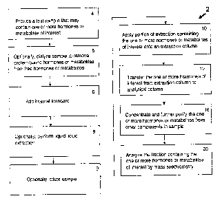
An example of a method (2) of the present invention is shown in FIG. I. Thus,
in an
embodiment, the method may include a step of providing a biological sample,
for example, a
serum sample believed to contain one or more analytes of interest (4). In some
embodiments,
an appropriate internal standard is added to the sample (6). For example, in
some
embodiments of the presently disclosed method for analyzing estrone and
estradiol in serum
samples, deuterated D4-estrone and D5-estradiol are added as internal
standards. Or, C3-
estrone and C13-estradiol stable labeled isotopes may be used. Or, for
thyroxine, a deuterated
or C13 derviative may be used. For example, in one embodiment, Thyroxine Ring-
13C6 may
be used. In yet other embodiments, structural analogus of the biomarker of
interest may be
used. For example, such structural analogues may comprise compounds wherein a
first
chemical group is replaced with a second chemical group. In general, the
groups are of
similar chemical reactivity, but different mass, as for example, the
replacement of a methyl (-
CI13) group with an ethyl (¨CH2CH3) group.
In some embodiments, the analytes of interests are partially purified by
liquid-liquid
extraction of the sample (8). Or, the sample may be diluted (9) in a solvent
that can be used
for LC or MS in subsequent purification steps.
In an embodiment, the liquid-liquid extraction is used to concentrate and
partially
purify the analyte. For example, for estradiol/estrone analysis, the liquid
extraction may be
used to remove conjugated estrogens, such as sulfated and glucoronidated
estrogens. Also,
the liquid extraction may remove lipids and/or fibrinogen from the samples. In
some
embodiments, estrone and estradiol can be extracted from a serum sample with
an organic
solvent that can separate estrone and estradiol from conjugated estrogens. For
example, in an
embodiment, an alkane mixed with a more polar solvent is used. For example, in
certain
embodiments, hexane is mixed with a more polar solvent. In an embodiment, the
polar
solvent comprises ethyl acetate or a similar solvent. In an embodiment, 9:1
hexane:ethyl
.acetate is used.
Or, other solvents may be used. As is known in the art, the solvents employed
may be
optimized to separate the analyte of interest from the sample. For example,
the solvents used
to extract estrone and estradiol from serum may not be the same solvent or
solvent mix as
used to extract estrone and estradiol from urine. Or, the solvents used to
extract estrone and
estradiol from serum may not be the same solvent or solvent mix as used to
extract thyroxine
CA 02820957 2013-07-11
WO 2007/139956 PCT/US2007/012525
(T4), triiodothyronine (T3), or vitamin-D compounds from serum. For example,
in certain
embodiments, acetonitrile is used for liquid extraction of vitamin-D
compounds, and ethyl
acetate:hexane:methanol is used for extraction of T4.
Certain biomolecules may have a propensity to nonspecifically bind to proteins
or
other biomolecules. For example, thyroid hormones can non-specifically bind to
proteins
such as serum albumin, sex hormone binding globulin, and the like. For
determination of
free thyroxine (T4), the sample may be treated to separate the free thyroxine
from thyroxine
that is bound to proteins in the biological sample (e.g., serum).
In one embodiment, the sample may initially be dialyzed to separate the free
hormone
or metabolite from a mixture of free and protein-bound hormone or metabolite
(5). In certain
embodiments, multiple samples may be processed concerrently. For example, the
dialysis
may be performed using a multiwell dialysis plate which allows for the
dialysis of multiple
samples at one time. In certain embodiments 96 well plates are used. In this
way, multiple
samples are processed to comprise a high throughput assay.
For example, samples of serum that may contain free thyroxine and protein-
bound
thyroxine may be introduced into the individual sample chambers which are on
one side of
the membrane and a buffer solution introduced into the diluent chambers on the
other side of
the membrane from the sample. The 96 well plate is then positioned vertically
and rotated to
facilitate transfer of the free thyroxine across the membrane.
The dialysis buffer may, in certain embodiments, be isotonic and contain
gelatin. The
gelatin may be used over a range of concentrations depending upon the nature
of the
membranes and hardware used for dialysis. In alternate embodients, the gelatin
may be in a
range of from about 0.1 to 10 mg/mL. In an embodiment, the gelatin is at about
1 mg,/mL. In
certain embodiments, the buffer used for dialysis comprises multiple
endogenous salts to
provide a buffer that is isotonic with the serum sample to thereby negate any
potential
dilution effects and/or disruptions to the ratio of bound thyroxine to free
thyroxine in the
sample. Also, gelatin may be include to prevent adsorptive losses of free
thyroixine onto the
dialysis membrane or the sample chamber. Gelatin may act as a carrier on the
dialysate side
of the 96-well plate to ensure free thyroxine remains in the dialysate
solution. Gelatin does
not bind free thyroxine and thus, does not affect the ratio of bound throxine
to free thyroxine
in the sample on the sample side of the membrane.
For the analysis of free thyroxine, a liquid extraction step may be performed
after the
dialysis. The liquid extraction may be designed to remove residual salts
and/or other
21
CA 02820957 2013-07-11
WO 2007/139956 PCT/US2007/012525
=
additives which are used in the dialysis solution and/or remain from the
sample, but that may
interfere with the MS analysis. Thus, in one embodiment, the dialysate
comprising free
thyroxine is extracted with 71.25:23.75:5 ethyl acetate:hexane:methanol. In
another
embodiment, the dialysate may be diluted with a solution of methanol
containing a stable
labeled internal standard and directly injected onto the LC-MS/MS system for
analysis.
Where the sample is extracted, the internal standard addition may include a
protein to
prevent the free thyroxin from sticking to the walls of the sample container.
Addition of
protein (e.g., bovine serum albumin) can minimize loss in extraction and
recovery for liquid-
liquid extraction. Where extraction is not performed, the internal standard
may be added in
methanol or a similar solvent used for LC.
As is known in the art, in some embodiments, the organic extract may be
transferred to
a fresh tube and then back-washed. For example, in an embodiment where the
analyte of
interest is estradiol and/or estrone, the solvent may be back-washed with
aqueous sodium
hydroxide (pH of about 12) to further purify the sample. Or, for extraction of
other
biomarkers, back-extraction may employ other solvents. The back-wash may, in
certain
embodiments, remove additional lipids or interfering analytes from the sample.
The extract supernatant may then be evaporated and the sample reconstituted.
For
example, for analysis of estradiol and/or estrone, the sample may be
reconstituted in 70:30
water:methanol. Or, for analysis of thyroxine, the solvent used for liquid-
liquid extraction
may be evaporated and the sample reconstituted in 50:50 water:methanol.
Still referring to FIG. 1, the method may further include liquid
chromatography as a
means to separate the analyte of interest from other components in the sample.
In an
embodiment, two liquid chromatography steps are used. For example, the method
may
comprise a first extraction column liquid chromatography (10), transfer of the
biomarker of
interest to a second analytical column (12), and an analytical column liquid
chromatography
(16). In other embodiments, only one liquid chromatography step is used.
The first extraction liquid chromatography column may, in certain embodiments,
comprise a step whereby the analytes of interest are separated from a majority
of
contaminants. Thus, in certain embodiments, the first column provides the
majority of
selectivity for the procedure. The second analytical liquid chromatography
column may, in
certain embodiments, comprise a step whereby the analytes of interest are
concentrated, to
thereby increase sensitivity for analysis by mass spectrometry (MS).
22
CA 02820957 2013-07-11
WO 2007/139956
PCT/US2007/012525
For example, the reconstituted extract may be applied onto a high performance
liquid
chromatography (HPLC) system, wherein the analytes are eluted using an
isocratic separation
through an extraction column. In certain embodiments, the mobile phase that is
used
comprises a gradient. For example, in an embodiment for the separation of
estradiol and
estxone from other components in serum, the stationary phase comprises a
Poroshell
300SBC-18 column. Thus, the inventors have found that surprisingly, a
stationary phase
designed for large molecules such as proteins may be used to separate smaller
molecules such
as estrone and estradiol. The mobile phase may comprise methanol and water.
Depending upon the biomarker of interest, a variety of analytical columns
known in
the art may be used as needed to provide good purification. In cetain
embodiments, the
analytical column may comprise particles having an average diameter of about 5
um. In
some embodiments, the analytical column is a functionalized silica or polymer-
silica hybrid,
or a polymeric particle or monolithic silica stationary phase, such as a
phenyl-hexyl
functionalized analytical column.
For example, in one embodiment, estrone and estradiol are separated from
isobaric
substances by separation using a Poroshell 300SBC10 column (7.5 mm by 2.1mm)
with 5
micron particle size using a gradient separation using methanol and water for
elution at 1 rnL
per minute flow rate. Estrone and estradiol are transferred from the
extraction column after
2.5 minutes and chromatofocused onto a phenyl-hexyl column (50mm by 2.1 mm)
with 5
micron particles using water for 45 seconds. The transferred and purified
analytes are
chromatographed using an accelerated gradient employing methanol and water to
improve
sensitivity prior to introduction into the mass spectrometer interface and
subsequent
detection.
For liquid chromatography of thyroxin, a single liquid chromatography step may
be
used. Thus, for liquid chromatography of thyroxin, a phenyl hexyl column (50mm
by 2.1
mm) with 5 micron particle size may be used. Thus, following either: (a)
liquid-liquid
extraction, evaporation and reconstitution; or (b) post-dialysis sample
dilution with internal
standard solution; samples are injected onto the liquid chromatography column.
The
transferred analyte and internal standard are chromatographed using a methanol
:water
gradient separation at 1 mL per minute. To enable further sensitivity gains, a
post-separation
additional flow of 90:10 methanol:water containing ammonium carbonate (1 mM)
is
introduced at 200 microliters per minute prior to introduction into the mass
spectrometer
(MS) electrospray interface.
23
CA 02820957 2013-07-11
WO 2007/139956
PCT/US2007/012525
If two liquid chromatography steps are employed, the eluted analytes may be
transferred to the analytical column in a manner such that the sample is
concentrated upon
application to the analytical column. In some embodiments, the eluted analytes
are
transferred to the analytical column via a heart-cutting technique. In some
embodiments, a
chromato-focusing procedure is used to transfer and focus the analytes on the
analytical
column. Also in some embodiments, a column-switching procedure is used to
transfer the
analytes to the analytical column. The analytes may then be separated on the
analytical
column (16) and the fraction containing the analyte of interest is eluted. In
an embodiment,
the second column in run in a manner to maximize throughput, and to provide
the sample in a
reduced volume.
The separated analytes are then introduced into a mass spectrometer (MS)
system
(20). In some embodiments, a tandem MS/1\4S system is used. As is known by
those of skill
in the art, in tandem MS spectrometry, the precursor ion is selected following
ionization, and
that precursor ion is subjected to fragmentation to generate product (i.e.,
fragment) ions,
whereby one or more product ions are selected for detection.. A sample may
therefore be
analyzed for both estradiol and estrone since the compounds have different
precursor and
product ions in tandem mass spectrometric methodologies (i.e., different
transitions).
The analyte of interest may then be quantified based upon the amount of the
characteristic transitions measured by tandem MS. In some embodiments, the
tandem mass
spectrometer comprises a triple quadrupole mass spectrometer. In some
embodiments, the
tandem mass spectrometer is operated in a positive ion Atmospheric Pressure
Chemical
Ionization (APCD mode. In some embodiments, the quantification of the analytes
and internal
-standards is performed in the selected reaction monitoring mode (SRM). Or,
other methods of
ionization such as the use of inductively coupled plasma, or MALDI, or SELL)!,
ES!, or APP!
may be used for ionization.
In some embodiments, the back-calculated amount of each analyte in each sample
may
determined by comparison of unknown sample response or response ratio when
employing
internal standardization to calibration curves generated by spiking a known
amount of purified
analyte material into a standard test sample, e.g., charcoal stripped human
serum. In one =
embodiment, calibrators are prepared at known concentrations and analyzed as
per the
biomarker methodology to generate a response or response ratio when employing
internal
= standardization versus concentration calibration curve.
24
CA 02820957 2013-07-11
WO 2007/139956
PCT/1JS2007/012525
In one embodiment, the sample may be treated so as to chemically modify the
analyte
of interest to allow for improved detection in the MS system. For example, in
one
embodiment, a sample being analyzed for estrone and/or estradiol may be heated
to the extent
that the estradiol loses a molecule of water thereby converting the estradiol
to a dehydrated
form of the compound (FIG. 2, Panels A and B, respectively). This conversion
can reduce the
number of major product ion peaks seen for estradiol from about 60 to 3 (FIG.
2, panels C and
D). For MS analysis, the sensitivity of the analysis is generally inversely
proportional to the
number of product ion peaks. Thus, with fewer peaks, the sensitivity of
detection using tandem
mass spectrometry is increased. For example, as illustrated in FIG. 2,
estradiol may be
qualified by measuring the transition from the precursor ion at a mass to
charge (m/z) 255.3
0.5 mass units to the two product (fragment) ions at a mass to charge (m/z) of
159.0 0.5 mass
units and 133.0 0.5.
In alternate embodiments, the sensitivity obtained for measurement of
estradiol is
increased more than 10 fold, or more than 20 fold, or more than 50 fold, or
more than 100 fold,
or more than 150 fold, or more than 200 fold, or more than 500 fold, or more
than 1,000 fold.
For example, in alternate embodiments, the sensitivity is increased by about 5-
1,000 fold, or a
by about 20-500 fold, or by about 50-150 fold, or by about 100 fold.
The temperature for heating the sample may, in alternate embodiments range
from
300 C to about 1000 C and includes all ranges therein.. In an embodiment, the
dehydration
step is performed within the interface of the mass spectrometer employed in
APCI or
electrospray mode at 500 degrees C 100 degrees. In an embodiment, the sample
is heated for
several microseconds at the interface for dehydration to occur. In alternate
embodiments, the
heating step is done for less than 1 second, or less than 100 milliseconds
(msec), or less than
10 msec, or less than 1 msec, or less than 0.1 msec, or less than 0.01 msec,
or less than 0.001
msec.
In an embodiment, the tandem liquid chromatograhy (LC) steps help reduce
isobaric
interferences. For example, in one embodiment, there are 24 potential isobaric
interferences in
estradiol (transition in/z 255 -> 159, 133), and 16 potential interferences
for estrone (transition
m/z 273 -> 159, 133). For example, dehydroepiandrosterone (DHEA) undergoes
thermal
dehydration forming MH-H20]+ and MH-2H20j+ (FIG. 3). There may be DHEA
concentrations that are about 300-1,500 times the levels of estrone and
estradiol in healthy
patients. Thus, the M+2 isotopic overlap of dehydrated DHEA may become an
isobaric
interference. Heart cutting from the primary separation using isocratic or
gradient separation
= 25
CA 02820957 2013-07-11
WO 2007/139956
PCT/US2007/012525
resolves most isobaric interferences (FIG. 4). Thus, as shown in FIG. 4, heart
cutting combined
with chromatofocusing may be used to separate estradiol (E2) and estrone (Fl)
from all but one .
potential isobaric contaminant which is separated within the analytical
(second) liquid
chromatography separation dimension.
An example of a method for measuring estradiol and estrone is provided in FIG.
5. For
example, in an embodiment, a method (40) of measuring estrone and estradiol
comprise
providing a sample believed to contain at least one of estrone and estradiol
(44). The method
may also comprise adding an internal standard of D4-estrone and D5-estradiol
to the sample
(46).
Also, the method may optionally comprise partial purification of the estrone
and
estradiol by liquid-liquid extraction of the estrone and/or estradiol from the
serum with 9:1
hexane-ethyl acetate (48). Or, the sample may be diluted (50) as a means to
improve sensitivity
in subsequent purification and/or analysis steps (e.g. LC and/or MS).
After initial purification by liquid-liquid extraction or dilution, the sample
may be
further purified by liquid chromatography. Thus, in one embodiment, the
solvent is evaporated
and the extracted estrone/estradiol is reconstituted in 30:70 methanol water
for application to a
liquid chromatography extraction column (52). The estradiol/estrone may be
eluted from the
extraction column. For example, in alternate embodiments, the
estradiol/estrone may be eluted
by heart cutting, chromatofocusing or column switching. Next, the fraction
containing the
estrone/estradiol may, in certain embodiments, be applied to an analytical LC
column (54).
The fraction containing the estrone/estradiol may then be transferred to the
LC-MS/MS
interface to undergo ionization and dehydration of the estradiol (60) prior to
MS/MS detection
in SRM mode (62).= In an embodiment, heating the estradiol removes a molecule
of water, and
changes the resultant MS/MS profile such that it comprises only three major
product ions.
Thus, the methods provide the ability to quantify estrone and/or estradiol at
physiologically relevant levels. As discussed herein, the difference between a
serum level of
10 pg/mL and i5 pg/mL may be clinically relevant. In one embodiment, the
method is able to
measure estrone and/or estradiol at levels of about 2.5pg/mL and 1 pg/mL
respectively.
An example of a method for measuring free thyroxine (T4) (70) is provided in
FIG. 6.
In an embodiment, the method may comprise providing a sample that includes
thyroxine (both
free and protein-bound) (74). The method may also comprise dialyzing the
sample (76) to
separate the free thyroxine from the protein bound thyroxine. Also, the method
may comprise
, adding an internal standard such as 6C13-thyroxine (78) to allow for the
measured amount of
26
CA 02820957 2013-07-11
WO 2007/139956
PCT/US2007/012525
thyroxine to be correlated to the actual amount present in the sample (i.e.,
to quantify the
amount lost during the extraction and measurement procedures).
The method may also comprise an optional step whereby the free thyroxine
present in
the dialysate is extracted by liquid-liquid extraction (80). Alternatively,
the sample may be
diluted into the solvent used for LC-MS/MS as a means to reduce interference
from non-T4 or
non-T3 analytes (81). At this point, the solvent used for extraction may be
evaporated, and the
extracted thyroxine reconstituted in 50:50 methanol:water for application to
an LC column
(82). The free thyroxine may then be eluted from the column (84) and then
quantified by
MS/MS (86).
Thus, the methods provide the ability to quantify free thyroxine at
physiologically
relevant levels. The difference between a serum level of 8 pg/mL and 12 pg/mL
T4 may be
clinically relevant. The method is able to measure free thyroxine (T4) at
levels of 2 pg/mL
Systems for Quantification of Endogenous Biomarkers
FIG. 7A shows an embodiment of a system of the present invention. As shown in
FIG. 7, the system may comprise a station for aliquoting a sample (104) that
may comprise a
biomarker of interest into sampling containers. In one embodiment, the sample
is aliquoted
into a container or containers to facilitate liquid-liquid extraction or
sample dilution. The
station for aliquoting may comprise receptacles to discard the portion of the
biological
sample that is not used in the analysis.
Alternatively or additionally, the sample may be aliquoted into a container
for
dialysis. As described above, the container for dialysis may comprise a multi-
well plate.
Thus, in addition to the station for aliquoting, the system may comprise a
station for dialysis
(106). The station for dialysis may comprise a rotator oven, multi-chamber
pipettes for
sample transfer, as well as receptacles to discard the portion of the
biological sample that is
not used in the analysis.
The system may further comprise a station for adding an internal standard to
the
sample (108). In an embodiment, the internal standard comprises the biomarker
of interest
labeled with a non-natural isotope. Thus, the station for adding an internal
standard may
comprise safety features to facilitate adding an isotopically labeled internal
standard solutions
to the sample. The system may also, in some embodiments, comprise a station
(110) for
liquid-liquid extraction and/or dilution of the sample.
The system may also comprise a station for liquid chromatography (LC) of the
sample. As described herein, in an embodiment, the station for liquid
chromatography may
27
CA 02820957 2013-07-11
WO 2007/139956
PCT/US2007/012525
comprise an extraction liquid chromatography column (112). The station for
liquid
chromatography may comprise a column comprising the stationary phase, as well
as
containers or receptacles comprising solvents that are used as the mobile
phase. In an
embodiment, the mobile phase comprises a gradient of methanol and water,
acetonitrile and
water, or other miscible solvents with aqueous volatile buffer solutions.
Thus, in one
embodiment, the station may comprise the appropriate lines and valves to
adjust the amounts
of individual solvents being applied to the column or columns. Also, the
station may
comprise a means to remove and discard those fractions from the LC that do not
comprise the
biomarker of interest. In an embodiment, the fractions that do not contain the
biomarker of
1.0 interest are continuously removed from the column and sent to a waste
receptacle for
decontamination and to be discarded.
A variety of extraction LC systems may be used. For example, in the embodiment
where the system is being used to measure estrone or estradiol, a Poroshell
300SBC18
extraction column with a phenyl hexyl analytical column, with mobile phases
comprising a
gradient of methanol and water are used. Or, for measurement of thyroxine, a
phenyl hexyl
column, with a mobile phase of methanol:water is used with post-column
addition of a
methanol:water solution containing arnmonium carbonate. Or, for vitamin D
metabolites, a
Fluophase WP extraction column, with a mobile phase of methanol:water is used
and an
Extent C18 analytical column is used with a mobile phase of methanol:water is
used.
The system may also comprise an analytical LC column (114). The analytical
column
may facilitate further purification and concentration of the biomarker of
interest as may be
required for further characterization and quantfication.
Also, the system may comprise a station for characterization and
quantification of the
biomarker of interest. In one embodiment, the system may comprise a station
for mass
spectrometry (MS) of the biomarker. In an embodiment, the station for mass
spectrometry
comprises a station for tandem mass spectrometry (MS/MS). Also, the station
for
characterization and quantification may comprise a computer and software for
analysis of the
MS/MS results. In an embodiment, the analysis comprises both identification
and
quantification of the biomarker of interest.
In some embodiments, one or more of the purification or separation steps can
be
preformed "on-line." As used herein, the term "on-line" refers to purification
or separation
steps that are performed in such a way that the test sample is disposed, e.g.,
injected, into a
system in which the various components of the system are operationally
connected and, in
28
CA 02820957 2013-07-11
WO 2007/139956
PCT/US2007/012525
some embodiments, in fluid communication with one another. The on-line system
may
comprise an autosampler for removing aliquots of the sample from one container
and
transferring such aliquots into another container. For example, an autosampler
may be used
to transfer the sample after extraction onto an LC extraction column.
Additionally or
alternatively, the on-line system may comprise one or more injection ports for
injecting the
fractions isolated from the LC extraction columns onto the LC analytical
column.
Additionally or alternatively, the on-line system may comprise one or more
injection ports for
injecting the LC purified sample into the MS system. Thus, the on-line system
may comprise
one or more columns, including but not limited to, an extraction column,
including an HTLC
extraction column, and in some embodiments, an analytical column. Additionally
or
alternatively, the system may comprise a detection system, e.g., a mass
spectrometer system.
The on-line system may also comprise one or more pumps; one or more valves;
and necessary
plumbing. In such "on-line" systems, the test sample and/or analytes of
interest can be
passed from one component of the system to another without exiting the system,
e.g., without
having to be collected and then disposed into another component of the system.
In some embodiments; the on-line purification or separation method can be
automated. In such embodiments, the steps can be performed without the need
for operator
intervention once the process is set-up and initiated. For example, in one
embodiment, the
system, or portions of the system may be controlled by a computer or computers
(102). Thus,
in certain embodiments, the present invention may comprise software for
controlling the
various components of the system, including pumps, valves, autosamplers, and
the like. Such
software can be used to optimize the extraction process through the precise
timing of sample
and solute additions and flow rate.
Although some or all of the steps in the method and the stations comprising
the
system may be on-line, in certain embodiments, some or all of the steps may be
performed
"off-line." In contrast to the term "on-line", the term "off-line" refers to a
purification,
separation, or extraction procedure that is performed separately from previous
and/or
subsequent purification or separation steps and/or analysis steps. In such off-
line procedures,
the analytes of interests typically are separated, for example, on an
extraction column or by
liquid/liquid extraction, from the other components in the sample matrix and
then collected for
subsequent introduction into another chromatographic or detector system. Off-
line
procedures typically require manual intervention on the part of the operator.
29
CA 02820957 2013-07-11
WO 2007/139956
PCT/US2007/012525
Liquid chromatography may, in certain embodiments, comprise high turbulence
liquid
chromatography or high throughput liquid chromatography (HTLC). See, e.g.,
Zimmer et al.,
J. Chromatogr. A 854:23-35 (1999); see also, U.S. Pat. Nos. 5,968,367;
5,919,368;
5,795,469; and 5,772,874. Traditional HPLC analysis relies on column packings
in which
laminar flow of the sample through the column is the basis for separation of
the analyte of
interest from the sample. In such columns, separation is a diffusional
process. Turbulent flow,
such as that provided by HTLC columns and methods, may enhance the rate of
mass transfer,
improving the separation characteristics provided. In some embodiments, high
turbulence
liquid chromatography (HTLC), alone or in combination with one or more
purification
methods, may be used to purify the biomarker of interest prior to mass
spectrometry. In such
embodiments, samples may be extracted using an HTLC extraction cartridge which
captures
the analyte, then eluted and chromatographed on a second HTLC column or onto
an
analytical HPLC column prior to ionization. Because the steps involved in
these
chromatography procedures can be linked in an automated fashion, the
requirement for
operator involvement during the purification of the analyte can be minimized.
Also, in some
embodiments, the use of a high turbulence liquid chromatography sample
preparation method
can eliminate the need for other sample preparation methods including liquid-
liquid
extraction. Thus, in some embodiments, the test sample, e.g., a biological
fluid, can be
disposed, e.g., injected, directly onto a high turbulence liquid
chromatography system.
For example, in a typical high turbulence or turbulent liquid chromatography
system,
the sample may be injected directly onto a narrow (e.g., 0.5mm to 2mrn
internal diameter by
20 to 50mm long) column packed with large (e.g., > 25 micron) particles. When
a flow rate
(e.g., 3-500 mL per minute) is applied to the column, the relatively narrow
width of the
column causes an increase in the velocity of the mobile phase. The large
particles present in
the column can prevent the increased velocity from causing back pressure and
promote the
formation of vacillating eddies between the particles, thereby creating
turbulence within the
column.
In high turbulence liquid chromatography, the analyte molecules may bind
quickly to
the particles and typically do not spread out, or diffuse, along the length of
the column. This
lessened longitudinal diffusion typically provides better, and more rapid,
separation of the
analytes of interest from the sample matrix. Further, the turbulence within
the column
reduces the friction on molecules that typically occurs as they travel past
the particles. For
example, in traditional HPLC, the molecules traveling closest to the particle
move along the
CA 02820957 2013-07-11
WO 2007/139956
PCT/US2007/012525
column more slowly than those flowing through the center of the path between
the particles.
This difference in flow rate causes the analyte molecules to spread out along
the length of the
column. When turbulence is introduced into a column, the friction on the
molecules from the
= particle is negligible, reducing longitudinal diffusion.
.5 The methods and systems of the present invention may use mass
spectrometry to detect
and quantify the biomarker of interest. The terms "mass spectrometry" or "MS"
as used herein
generally refer to methods of filtering, detecting, and measuring ions based
on their mass-to-
charge ratio, or "m/z." In MS techniques, one or more molecules of interest
are ionized, and
the ions are subsequently introduced into a mass spectrometer where, due to a
combination of
electric fields, the ions follow a path in space that is dependent upon mass
("m") and charge
In certain embodiments, the mass spectrometer uses a "quadrupole" system. In a
"quadrupole" or "quadrupole ion trap" mass spectrometer, ions in an
oscillating radio
frequency (RF) field experience a force proportional to the direct current
(DC) potential
applied between electrodes, the amplitude of the RF signal, and rn/z. The
voltage and
amplitude can be selected so that only ions having a particular rri/z travel
the length of the
quadrupole, while all other ions are deflected. Thus, quadrupole instruments
can act as both a
"mass filter" and as a "mass detector" for the ions injected into the
instrument.
In certain embodiments, tandem mass spectrometry is used. See, e.g., U.S. Pat.
No.
6,107,623, entitled "Methods and Apparatus for Tandem Mass Spectrometry,"
which is
hereby incorporated by reference in its entirety. Further, the selectivity of
the MS technique
can be enhanced by using "tandem mass spectrometry," or "MS/MS." Tandem mass
spectrometry (MS/MS) is the name given to a group of mass spectrometric
methods wherein
"parent or precursor" ions generated from a sample are fragmented to yield one
or more
"fragment or product" ions, which are subsequently mass analyzed by a second
MS
procedure. MS/MS methods are useful for the analysis of complex mixtures,
especially
biological samples, in part because the selectivity of MS/MS can minimize the
need for
extensive sample clean-up prior to analysis. In an example of an MS/MS method,
precursor
ions are generated from a sample and passed through a first mass filter to
select those ions
having a particular mass-to-charge ratio. These ions are then fragmented,
typically by
collisions with neutral gas molecules in a suitable ion containment device, to
yield product
(fragment) ions, the mass spectrum of which is recorded by a electon
multiplier detector. The
product ion spectra so produced are indicative of the structure of the
precursor ion, and the
31
CA 02820957 2013-07-11
WO 2007/139956
PCT/US2007/012525
two stages of mass filtering can eliminate ions from interfering species
present in the
conventional mass spectrum of a complex mixture.
In an embodiment, the methods and systems of the present invention use a
triple
quadrupole MS/MS (see e.g., Yost, Enke in Ch. 8 of Tandem Mass Spectrometry,
Ed.
McLafferty, pub. John Wiley and Sons, 1983). Triple quadrupole MS/MS
instruments
typically consist of two quadrupole mass filters separated by a fragmentation
means. In one
embodiment, the instrument may comprise a quadrupole mass filter operated in
the RF only
mode as an ion containment or transmission device. In an embodiment, the
quadropole may
further comprise a collision gas at a pressure of between 1 and 10 millitorr.
Many other
types of "hybrid" tandem mass spectrometers are also known, and can be used in
the
methods and systems of the present invention including various combinations of
magnetic
sector analyzers and quadrupole filters. These hybrid instruments often
comprise high
resolution magnetic sector analyzers (i.e., analyzers comprising both magnetic
and
electrostatic sectors arranged in a double-focusing combination) as either or
both of the mass
filters. Use of high resolution mass filters may be highly effective in
reducing chemical noise
to very low levels.
For the methods and systems of the present invention, ions can be produced
using a
variety of methods including, but not limited to, electron ionization,
chemical ionization, fast
atom bombardment, field desorption, and matrix-assisted laser desorption
ionization
("MALDI"), surface enhanced laser desorption ionization ("SELDI"), photon
ionization,
electrospray ionization, and inductively coupled plasma.
In those embodiments, such as MS/MS, where precursor ions are isolated for
further
fragmentation, collision-induced dissociation ("CID") may be used to generate
the fragment
ions for further detection. In CID, precursor ions gain energy through
collisions with an inert
gas, and subsequently fragment by a process referred to as "unimolecular
decomposition."
Sufficient energy must be deposited in the precursor ion so that certain bonds
within the ion
can be broken due to increased vibrational energy.
In some embodiments, to attain the required analytical selectivity and
sensitivity, the
presently disclosed 2D-LC-MS/MS methods include multiplexed sample preparation
procedures. For example, in certain embodiments dialysis of the sample is
performed using a
96 well plate having a dialysis membrane in each well or multiple sample tubes
(FIG. 7B).
Additionally or alternatively, the multiplex system may comprise staggered
multiplexed LC
and MS sample inlet systems. Also, the methods and systems of the present
invention may
32
=
CA 02820957 2013-07-11
WO 2007/139956
PCT/US2007/012525
comprise multiple column switching protocols, and/or heart-cutting (LC-LC or
2D-LC)
techniques, and/or LC separations prior to MS detection. In some embodiments,
the methods
and systems of the present invention may include a multiplexed two-dimensional
liquid
chromatographic system coupled with a tandem mass spectrometer (MS/MS) system,
for
example a triple quadrupole MS/MS system. Such embodiments provide for
staggered,
parallel sample input into the MS system.
Thus, as shown in FIG. 7B, four samples (132 A-D) may each be applied to
individual
extraction columns (134 A-D). Once the samples have each run through the
extraction column,
they may each be transferred directly (e.g., by column switching) to a second
set of analytical
columns (136 A-14 As each sample elutes from the analytical column, it may be
transferred
(138) to the mass spectrometer (140) for identification and quantification.
A plurality of analytes can be analyzed simultaneously or sequentially by the
presently disclosed LC-MS/MS and 2D-LC-MS/MS methods. Exemplary analytes
amenable
to analysis by the presently disclosed methods include, but are not limited
to, steroid hormones,
such as estradiol, estrone, and metabolites, such as estrone sulfate. In other
embodiments,
thyroid hormones, such as free thyroxine (T4) and triiodothyronine (T3) can be
measured. In
the other embodiments, metabolites, such as 25-Hydroxyvitamin D2, 25-
Hydroxyvitamin D3,
may be measured. One of ordinary skill in the art would recognize after a
review of the
presently disclosed subject matter that other similar analytes could be
analyzed by the
methods and systems disclosed herein. Thus, in alternate embodiments, the
methods and
systems may be used to quantify steroid hormones, protein and peptide
hormones, peptide
and protein biomarkers, drugs of abuse and therapeutic drugs. For example,
optimization of
key parameters for each analyte can be performed using a modular method
development
strategy to provide highly tuned bioanalytical assays. Thus, certain steps may
be varied
depending upon the analyte being measured as disclosed herein.
Also, embodiments of the methods and systems of the present invention may
provide
greater sensitivity than the sensitivities previously attainable for many of
the analytes being
measured. For example, through using this optimization procedure, an LOQ of
about 1
picogram per milliliter (pg/mL), or less than 5 pg/mL, or less that 10 pg/mL,
or less than 25
pg/mL is attained for the analysis of at least one of estradiol, estrone or
free thyroxine
without the cumbersome derivatization processes historically required for LC-
MS/MS
analyses of steroids. Importantly, the low levels of detection allow for the
analysis of small
sample volumes, for example 100 ;IL, 200 AL, 500 p.L, or less than 1 mL, which
can be
33
CA 02820957 2013-07-11
WO 2007/139956
PCT/US2007/012525
necessary to analyze pediatric sample volumes. Thus, the presently disclosed
LC-MS/MS
and (LC)-LC-MS/MS methods can be used to measure levels of steroid hormones,
such as
estrone and estradiol, or other hormones or metabolites (e.g., free thyroxine,
vitamin D
metabolites and the like) in serum or plasma samples from children, women, and
men.
Embodiments of the present invention may provide certain advantages. In
certain
embodiments, the methods and systems of the present invention may provide
greater
sensitivity than the sensitivities previously attainable for many of the
analytes being
measured.
Also, embodiments of the methods and systems of the present invention may
provide
for rapid throughput that has previously not been attainable for many of the
analytes being
measured. For example, using the methods and systems of the present invention,
multiple
samples may be analysed for free thyroxine using 96 well plates and a
multiplex system of
four LC-MS/MS systems, significantly increasing the throughput.
As another advantage, the specificity and sensitivity provided by the methods
and
systems of the present invention may allow for the analysis of analytes from a
variety of
biological materials. For example, the 2D-LC-MS/MS methods of the present
invention can
be applied to the quantification of analytes of interest in complex sample
biological matrices,
including, but not limited to, blood, serum, plasma, urine, saliva, and the
like. Thus, the
methods and systems of the present invention are suitable for clinical
applications and/or
clinical trials.
As additional potential advantages, in certain embodiments, the systems and
methods
of the present invention provide approaches for addressing isobaric
interferences, varied
sample content, including hemolysed and lipemic samples, while attaining low
pg/mL limits of
quantification (LOQ) of the target analytes. Accordingly, embodiments of the
methods and
systems of the present invention may provide for the quantitative, sensitive,
and specific
detection of clinical biomarkers used in the clinical diagnosis of endocrine
disorders.
Validation of LC-MS/MS and 2D-LC-MS/MS Assays for Endogenous Biomarkers
A general strategy for the validation of the presently disclosed LC-MS/MS and
2D-LC-
MS/MS methods for endogenous biomarkers is provided in Scheme 1. Thus, Scheme
1 shows
the different tests that were used to validate the procedures. Matrix
specificity testing was
performed by analyzing 6 different lots of charcoal stripped matrix in
quadruplicate for the
presence of residual analyte, absence of analyte enables the charcoal stripped
matrix to be
spiked with known concentrations of target analytes to generate calibration
curves. Internal
34
CA 02820957 2013-07-11
WO 2007/139956
PCT/US2007/012525
standard specificity was performed by spiking the stable labeled internal
standard into analyte-
free charcoal stripped matrix and measuring for the presence of analyte in
quadruplicate.
Absence of unlabeled analyte confirms the purity of internal standard
materials. Endogenous
(hormones) and exogenous (drugs) are spiked into analyte free matrix to
confirm the selectivity
of the method.
Accuracy and precision was determined using 6 replicates per level in spiked
charcoal
stripped serum at the LLOQ, 2 levels within the analytical range and the ULOQ
in 3 different
batches. Precision was determined using 6 replicates in 3 separate runs of
pooled matrix
samples at concentrations of approximately 3 to 10 times the LLOQ, the mid
point of the
analytical range and approximately 80% of the ULOQ. Accuracy was determined in
pooled
matrix samples using spike and recovery (standard addition) at 3 different
concentrations
throughout the analytical range using 4 replicates per level.
Linearity was confirmed using multi-level calibrators over 5 separate runs.
Sample
mixing experiments were also undertaken mixing pooled matrix samples with
fortified stripped
matrix samples to ensure the assays were free of matrix interferences in
quadruplicate.
Recovery was undertaken using both spiked stripped matrix and pooled matrix
samples in
quadruplicate as r.onfmnation of linearity and also further proof that the
assay was free of
matrix effects. The effect of matrix content on measurement was also tested
following post-
column infusion, addition of lipemia and hemolysis content, alternate sample
types (e.g. serum
and or plasma) and sample draw-tubes in quadruplicate. Sample stability was
undertaken using
both spiked stripped matrix samples and pooled matrix samples at storage
conditions expected
from sample collection to final analysis: Each condition was compared against
baseline
samples drawn and frozen at ¨70 C for comparison and analyzed in quadruplicate
at each
concentration.
Inter-assay comparison was performed using at least 50 samples representing
physiological range during comparison of LC-MS/MS and LC-MS/MS assays with
alternate
techniques. Reference range generation and/or transference was undertaken
using guidance
from the National Committee on Clinical Laboratory Standards (NCCLS).
Scheme 1. Bioanalytical Validation Strategy
Specificity Testing: Matrix, Internal Standard,
Endogenous/Exogenous Analytes
Accuracy and Precision: Stripped Matrix: LLOQ, Mid levels (x2) ULOQ;
Pooled Matrix: 3x LLOQ, mid and 80% of ULOQ;
Spike and Recovery at 3 levels
CA 02820957 2013-07-11
WO 2007/139956
PCT/US2007/012525
Scheme 1. Bioanalytical Validation Strategy
Linearity: Stripped Matrix: 7-10 point duplicate curves; 5
batches;
Pooled Matrix: Sample dilution (mixing) at 4 levels
Recovery: Stripped and Pooled Matrix: Spike and recovery
at 3 levels
Ionization Effect Stripped and Pooled Matrix: Post-column
infusion, post-extraction
spiking, Heparin and EDTA anticoagulants,
Lipemic and Hemolysis additives
Sample Stability: Stripped and Pooled Matrix: Pre and Post
processing stability
compared against -70 C baseline samples,
Whole blood stability and stock solutions
Inter-Assay Comparison: Pooled Matrix samples: At least 50 samples
spanning normal and
abnormal range
Reference Range: Transference based upon 90% CI for at least 20
samples per range
=
Representative LC-MS/MS and 2D-LC-MS/MS chromatograms of selected analytes at
the limit of quantification (LOQ) obtained by using the LC-MS/MS and 2D-LC-
MS/MS
methods of the present invention are shown in FIGS. 8-13. In these figures the
X axis is time,
and the Y axis corresponds to the amount of material (i.e., the response).
Thus, it can be seen
that estrone sulfate was detected at 100 pg/mL (FIG. 8); free thyroxine was
detected at 2
pg/mL (FIG. 9); 25-hydroxyvitamin D2 was detected at 1 ng/mL (FIG. 10); 25-
=
hydroxyvitamin D3 was detected at 1 ng,/mL (FIG. 11); estrone was detected at
2.5 pg/mL
(FIG. 12); and estradiol was detected at 1 pg/mL (FIG. 13).
Similarly, representative LC-MS/MS and 2D-LC-MS/MS chromatograms of selected
analytes at the upper limit of quantification (ULOQ) (the level of analyte
above which the
assay is outside of linear range) obtained by using the LC-MS/MS and 2D-LC-
MS/MS
methods and systems of the present invention are shown in FIGS. 14-19. Thus,
it can be seen
that estrone sulfate has an ULOQ of 50 ng,/mL (FIG. 14); free thyroxine has an
ULOQ of 100
pg/mL (FIG. 15); 25-hydroxyvitamin D2 has an ULOQ of 250 ng/mL (FIG. 16); 25-
hydroxyvitamin D3 has an ULOQ of 250 ng/mL (FIG. 17); estrone has an ULOQ of
500
pg/mL (FIG. 18); and estradiol has an ULOQ of 500 pg/mL (FIG. 19).
Representative LC-MS/MS and 2D-LC-MS/MS calibration curves for selected
.analytes are shown in Figures 20-25. FIG. 20 shows a calibration curve for
estrone sulfate
where it is seen that the assay is linear over a 1000-fold range. Calibration
curves for
thyroxine (FIG. 21), 25-hydroxyvitamin D2 (FIG. 22), 25-hydroxyvitamin D3
(FIG. 23),
estrone (FIG. 24) and estradiol (FIG. 25) also show linearity over at 100 to
250 fold range. In
these figures, the X axis is the concentration, and the Y axis is the ratio of
the analyte to an
internal standard response.
36
CA 02820957 2013-07-11
WO 2007/139956
PCT/US2007/012525
Representative inter-assay comparison results for the presently disclosed LC-
MS/MS
and 2D-LC-MS/MS methods versus RIA, CBP, or IA methods are shown in Figures 26-
30.
Thus, it can be seen that using LC-MS/MS provides excellent correlation with
RIA for
measurement of estrone sulfate (FIG. 26); good correlation but a bias (slope
offset) with IA
for measurement of free thyroxine (FIG. 27), excellent correlation but a bias
(slope offset)
for measurement of total 25-hydroxyvitamin D by CBP and average correlation
with IA (FIG.
28, panels A and B, respectively); excellent correlation with RIA for
measurement of estrone
(FIG. 29); and excellent correlation with RIA for measurement of estradiol
(FIG. 30). Also,
FIG. 31 shows good correlation between the 2D-LC-MS/MS assays described herein
for
estradiol with an alternate LC-MS/MS strategy involving derivatization.
Excellent correlation
is observed for samples within the higher analytical range of the
derivatization assay
(10pg/mL LLOQ).
FIG. 32 shows results of dialysis losses for thyroxine. The squares (a) show
dialysis
losses and the diamonds (4.) show effective dialysis for free thyroxine using
96-well
equilibrium dialysis plates in accordance with one embodiment of the present
invention. This
indicates that during the dialysis experiments, free T4 does not degrade or
bind to the 96-well
plate apparatus (i.e. losses). Further, FIG.31 indicates that dialysis is
complete after
approximately 16 hours.
Data showing the validation bias due to ionization effect and recovery for
selected
analytes are provided in Tables 1 through 7, below. Data showing the accuracy
and precision
of the presently disclosed LC-MS/MS and 2D-LC-MS/MS methods are provided in
Tables 8
through 13. As known by those in the art, acceptable values based on the FDA
and CLIA
regulations are < 20% bias or imprecision (% CV) at the LLOQ and < 15% over
the
remainder of the assay. See e.g., FDA Guidance: 1.1 Guidance for Industry,
Bioanalytical
Method Validation, FDA, May 2001, BP, and CLIA Regulation: 42 CFR 493.1253
Standard:
Establishment and verification of performance specifications. Subart K,
Quality System for
Non-waived Testing. Thus, as used herein, "acceptable" or "good" indicates
that the assay or
aspect of the method being measured meets the NCCLS, FDA and CLIA critieria.
Each of the presently disclosed LC-MS/MS and 2D-LC-MS/MS methods was
evaluated for specificity against multiple steroids and/or other potential
interferences for a
total of up to 60 different analytes (see Examples 1 and 2) at excessive
concentrations, for
example, 100 p.g/dL, to ensure accurate measurement in each assay independent
of clinical
levels of endogenous and therapeutic agents, such as steroids, which either
cross-react in RIA
37
CA 02820957 2013-07-11
WO 2007/139956
PCT/US2007/012525
assays or are not discriminated using the specificity provided by MS/MS
detection.
Additionally, analyte stability was evaluated for all conditions expected from
original patient
sampling to final result. Proven stability includes sample shipment (-20 C),
sample
processing (20 C; > 3 freeze/thaw cycles) and post processing (20 C and
autosampler at 10 C)
to ensure accurate and precise determination of analytes, such as hormone
steroids, derived
from patient samples(see Examples 1 and 2).
Table 1
Estrone Sulfate
Validation Bias (%)
Serum -3.8-2.6
Lipemia -0.2
Hemolysis 0.7
Recovery 85.0 ¨ 97.1
Table 2
Free Thyroxine
Validation Bias (%)
Serum -6.9--4.8
Lipemia 1.0
Hemolysis 12.8
Recovery 90.5 ¨ 95.8
Table 3
25-Hydroxyvitamin D2
Validation Bias (%)
Serum -18-2.2
= Lipemia -5.0
Hemolysis -5.4
Recovery 93.2 ¨ 100.5
Table 4
25-Hydroxyvitamin D3
Validation Bias (%)
Serum -8.8-11.9
Lipemia -0.3
Hemolysis -1.6
Recovery 88.8 ¨ 94.1
38
CA 02820957 2013-07-11
WO 2007/139956
PCT/1JS2007/012525
Table 5
Estrone
Validation Bias (%)
Serum -11.9-7.8
Lipemia 8.3
Hemolysis 2.8
Recovery 97.2- 106.9
Table 6
=
Estradiol
Validation Bias (%)
Serum -4.5-4.0
Lipemia -5.9
Hemolysis -2.9
Recovery 90.0 - 95.7
Table 7
Estrone Sulfate
Accuracy (%) Precision (%)
Conc. (ng/dL) 10 150 2500 5000 10 150 2500 5000
Inter -13.2 -5.5 5.4 0.5 9.4 5.5 5.7 7.9 _
Table 8
Free Thyroxine
Accuracy ( /(!) Precision (%)
Conc. (nit:IL) 0.2 5 10 0.2 5 10
Intra - 1 -3.4 - -7.0 3.0 5.0 6.2
5.5
Infra - 2 0.1 -1.2 -0.3 5.1 5.2
2.2
Intra - 3 -0.4 0.6 3.7 4.4 4.1
5.5
Inter -1.3 -2.5 2.1 4.8 6.0
4.8
Table 9
25-Hydroxyvitamin D2
Accuracy (%) Precision (%)
Conc. (ng/mL) 1.0 2.5 1.00 250 1.0 2.5
100 250
Intra - 1 6.3 -8.1 -2.0 -0.4 4.6 5.6
0.9 2.2
Intra - 2 13.9 8.6 -2.2 1.7 3.3 6.0
2.6 4.5
Infra - 3 8.7 4.2 -0.1 4.2 6.8 5.2 4.4
3.7
Inter 11.3 1.6 -1.4 1.3 6.9 8.9
3.0 3.9
39
CA 02820957 2013-07-11
WO 2007/139956
PCT/1JS2007/012525
Table JO
25-Hydroxyvitamin D3
Accuracy (%_)
Precision (%)
Conc. (ng/mL) 1.0 2.5 1.00 250 1.0 2.5 100
250
Intra - 1 -3.9 -4.0 -2.8 -0.4 5.8 5.7
4.6 2.1
Intra - 2 6.2 4.8 -1.4 1.5 11.2 4.6 3.9
5.5
Intra - 3 5.9 3.7 -2.1 -0.4 3.0 9.9 3.0
3.9
Inter 2.7 - 1.5 -2.1 0.3 8.5 7.8 3.7
3.9
Table 11
Estrone
Accuracy (%)
Precision (%)
Conc. (pWmL) 2.5 250 500 2.5 250
500
Intra - 1 12.4 1.1 -1.4 2.9 5.5
3.3
Intra - 2 -0.3 9.2 4.0 7.3 2.8
2.4
Intra - 3 3.6 6.3 5.1 6.3 3.8
1.6
Inter 4.8 5.5 2.6 7.4 5.1
3.7
Table 12
Estradiol
Accuracy (%)
Precision (%)
Conc. (pg/mL) 1.0 2.5 1.00 250 1.0 2.5 100
250
Intra - 1 -7.1 1.1 -1.1 0.4 4.7 4.8 2.8
1.2
Intra - 2 -7.4 1.1 6.4 6.5 4.4 6.3 1.6
4.3
Intra - 3 4.7 5.6 0.6 ' 1.4 4.7 5.6 2.6
3.5
Inter -3.3 2.7 1.9 2.8 7.4 5.2 3.9
4.1
=
EXAMPLES
Additional data from the analytical validation and standard operating
procedures for the
presently disclosed method for the quantification of estrone and estradiol by
liquid-liquid
extraction and 2D-LC-MSTMS, or free thyroxine by dialysis, an optional liquid-
liquid
extraction, and LC-MS/MS are set forth in the following Examples.
The following Examples have been included to provide guidance to one of
ordinary
skill in the art for practicing representative embodiments of the presently
disclosed subject
matter. In light of the present disclosure and the general level of skill in
the art, those of skill
can appreciate that the following Examples are intended to be exemplary only
and that
numerous changes, modifications, and alterations can be employed without
departing from
the scope of the presently disclosed subject matter.
CA 02820957 2013-07-11
WO 2007/139956
PCT/US2007/012525
Example 1: 2D-LC-MS/MS Analysis for Estrone, Estradiol, and Estrone Sulfate
Estrone and estradiol were validated to 2.5 pg/mL and 1 pg/mL, respectively,
from a
1-mL serum sample. Sensitivity and selectivity were generated using heat
assisted and guided
fragmentation of estradiol to optimize sensitivity. Analytical specificity was
generated via 2D
LC using gradients in both LC dimensions, heart-cutting and chromato-focusing
prior to
MS/MS detection. Optimum selectivity was generated following titration of
buffer pH and
chromatography to enable separation from co-extracted isobaric endogenous
interferences,
while retaining necessary sensitivity (SIN >20) for accurate quantification
quantification in
estrone sulfate analysis using LC-MS/MS.
More particularly, estrone (El) and estradiol (E2) were measured by two-
dimensional
(2D) liquid chromatography with tandem mass spectrometry detection (2D-LC-
MS/MS) after
liquid-liquid extraction (LLE). Deuterated D4-Estrone and D5-Estradiol were
added as
internal standards to serum aliquots. Estrone and Estradiol were extracted
from 1 mL serum
samples with 8 mL of 9:1 Hexane:Ethyl Acetate. The organic extract was
transferred to a
fresh tube and back washed with 1 mL aqueous sodium hydroxide solution (pH of
about 12),
then evaporated and reconstituted in 70:30 Water:Methanol.
Duplicate sets of stripped serum calibrators were analyzed in each batch. All
injections were made in singlicate. All samples are injected onto the AREA TX4
or
Transcend TX4 HTLC system (Thermo Fisher, Franklin, Massachusetts, United
States of
America), where the analytes were first eluted using a separation gradient
through an
extraction column. Analytes of interest were then transferred onto the
analytical column,
where chromatographic separation was continued via a gradient. An API5000
triple
quadrupole mass spectrometer (MDS-SCIEX, Concord, Ontario, Canada), operating
in positive
ion Atmospheric Pressure Chemical Ionization (APCI) mode was used for
detection.
Quantification of analyte and internal standard was performed in selected
reaction
monitoring mode (SRN!). The back-calculated amount of each analyte in each
sample was
determined from duplicate calibration curves generated by spiking known
amounts of
purified estrone and estradiol into charcoal stripped human serum.
Measurement of estrone and estradiol was used to evaluate ovarian function and
to
evaluate excess or diminished estrogen levels. Analysis of estrone and
estradiol by 2D-LC-
MS/MS detection was developed to measure levels in serum or plasma samples
from
children, women and men.
41
CA 02820957 2013-07-11
WO 2007/139956
PCT/1JS2007/012525
The lower limit of detection using the default sample aliquot of lmL was 1.0
pg/rnL
for Estradiol and 2.5 pg/mL for Estrone.
Definitions
APCI - Atmospheric Pressure Chemical Ionization] LLE - Liquid-Liquid
EXtraction; SST -
System Suitability Test; 2D -LC-MS/MS - Two-dimensional liquid chromatography
with
tandem mass spectrometry detection; El ¨ Estrone; E2 - 17P-Estradiol or
Estradiol; HTLC -
High Throughput Liquid Chromatography
Specimens
A recommended sample is 1.5 mL serum or plasma. Separate within one hour.
Store
and ship frozen in plastic vial.
Adult: 1.5mL serum or plasma
Pediatric: 1.5mL serum or plasma
Minimum: lmL serum or plasma _
Draw into red top vacutainer tube. Allow clotting to occur for 20 minutes at
room temperature
(or until clot has retracted). Spin and remove serum to labeled plastic vial.
Freeze immediately.
Storage is for short term storage (2 weeks): Frozen (5-20 C); for long term
storage (6 months):
Frozen (5-20 C). Shipping of specimens is:frozen (5-20 C) - on dry ice.
Generally, samples drawn
into SST tubes are unacceptable for this procedure.
Equipment & Materials
The following materials were used: Standard manual pipetting devices; 1.2m1
MBlock
Polypropylene 96 Well Collection Plate (SPE Ware, Inc. Product No. SPE0210);
Heat Sealing
Foil (SPE Ware, Inc. Product No. AB-0589); Mechanical Shaker, Eberbach Inc.;
Rotary
Evaporator (rotovap), Speed Vac SC 200, Savant (or equivalent); Class A
Volumetric Glass
Containers, various sizes; API 5000 Tandem Mass Spectrometer, Sciex, (Toronto,
Canada);
Turbo VTm Ion Source with APCI probe, Sciex, (Toronto, Canada); Aria TX4 HTLC
System,
Cohesive Technologies, (MA, USA) consisting of 4 each: 1100 Series Quaternary
Pump, 1100
Series Binary Pump, 1100 Series Vacuum Degasser; HTS Twin PAL System
Autosampler,
CTC Analytics AG (Switzerland); Luna Phenyl-Hexyl Analytical Column, 50 X 2.0
mm, 511m
particle size, Phenomenex, (USA) Product No. 00B-4257-B0; Poroshell 300SB-C18
Column,
2.1 x 75mm, 51.tm particle size, Agilent Inc., (USA) Product No. 660750-902;
16mm Flange
Caps, Stockwell Scientific, (AZ, USA) Product No. 8558; Vortex Mixer, VWR or
equivalent;
Combi-thermo Heat Sealer, Abgene Inc., Product No. AB-0559; 96-Well Centrifuge
5804R
Eppendorf or equivalent; 16x100 mm borosilicate glass tubes; Thermo Hot pocket
column
42
CA 02820957 2013-07-11
WO 2007/139956
PCT/US2007/012525
oven, Thermo Electron Corp. (USA) Product No. 92016-150; Analyst Version 1.4
or greater.
Applied Biosystems, (CA, USA); Aria OS Version 1.4 or greater, Cohesive
Technologies
(MA, USA).
Reagents =
The following reagents were used. Water ¨ Type H, Millipore MilliQ or
equivalent;
Charcoal Stripped Human Serum (Bioreclamatio-n, Inc); Ethyl alcohol USP (AAPER
Alcohol, USA); Acetonitrile HPLC grade (EM Science AX0142-1); Methanol
(Reagent
A.C.S., Fisher Scientific); Isopropanol HPLC Grade (Fisher Scientific, Catalog
#A451-4);
170-Estradiol, Sigma-Aldrich, (USA) Product # E8875 or USP, (USA) Product
#1250008;
Estrone, Sigma-Aldrich, (USA) Product # E1274 or USP, (USA) Product #1255001;
1713-
Estradio1-2,4,16,16,17-D5, CDN Isotopes, (USA) Product # D-5403; Estrone-
2,4,16,16-D4,
CDN Isotopes, (USA) Product # D-3650; Acetone HPLC Grade (Burdick & Jackson,
Catalog
#AH010-4); Total Estrogen Stock Solution.(10u.g/mL in ethanol) prepared by
serial dilution.
The following solvents were used as the mobile phases for liquid
chromatography.
Eluting and Loading Pump A Mobile Phase (90% Water and 10% Methanol); Loading
Pump
B Mobile Phase (90% Methanol and 10% Water); and Loading Pump C Mobile Phase
(60:30:10 Acetonitrile: Isopropanol: Acetone).
Internal Standard Solution (1 ng/mL Ds-Estradiol and 2 ng/mL D4-Estrone) was
prepared in 99:1 wateracetonitrile solution. The reconstitution solution (for
reconstituting
the sample after liquid-liquid extraction) was 70:30 Millipore Water:
Methanol. Two needle
wash solutions were used: Needle Wash Solution I (Aqueous 1% Formic Acid); and
Needle
Wash Solution 2 (70:30 Acetonitrile: IN Ammonium Hydroxide).
Total Estrogen Stock Solution (1 Oug/mL) - 51.1g/mL estrone and 511g/mL
estradiol
was used to prepare intermediate stock solutions for preparation of
calibrators. To prepare
standards, 0.5mL of Total Estrogen Stock (10p.g/mL) was diluted to 100mL with
stripped
human serum to yield 25 ng/mL El E2 solution, and 2mL of the 25ng/mL solution
was
diluted to 100tnL in charcoal stripped human serum to yield a 0.5 ng/mL E1E2
solution. The
diluted stocks (0.5 ng/mL and 25 ng/mL) were stored at -20 C. Calibration
Standards were
then prepared as shown in the following table. All standards were prepared in
charcoal
stripped human serum. The standard curve back-fit data should be 80-120% at
LLOQ, 85 ¨
115% at other concentrations of expected curve values.
43
CA 02820957 2013-07-11
WO 2007/139956
PCT/US2007/012525
Standard Preparation Procedure
Standard Stock Stock Final Final
Standard Solution.
Concentration Volume Volume Concentration
Number Concentration
(Pginil-) (mL) (mL) (pg/mL)
(ng/mL)
SI 1.0 0.5 1 500 1.0
S2 2.5 0.5 2.5 500 2.5
S3 5.0 0.5 5 500 5.0
S4 10 0.5 10 500 10
S5 25 25 0.5 500 25
S6 50 25 1 500 50
S7 100 25 2 500 100
S8 200 25 4 500 200
S9 350 25 7 500 350
S10 500 25 10 500 500
Quality Control
Control pools are prepared in human serum and introduced into use according to
the
analytically robust procedures. Each run contained duplicates of four control
pools, each
with a nominal target value.
Quality Control Concentrations
Control Target concentration (pg/mL)
Name Estrone Estradiol
Pool 1 10 10
Pool 2 25 25
-
Pool 3 115 115
Pool 4 300 300
Assay Procedure
The assay was performed as follows. Pipetted 1.0 ml standard, control or
patient into
the tube with an Eppendorf pipette (or equivalent). Using an Eppendorf Plus
repeating
pipette with 5 ml tip (or equivalent), added 50 p.1 Internal Standard Solution
to each tube
except double blanks. Mix all samples on multi-vortexer for 30 seconds. Added
8 mL 9:1
Hexane: Ethyl Acetate extraction solvent to all tubes and shook on mechanical
shaker for 10
minutes. Removed tubes from shaker and spun down all samples in centrifuge up
to 2000
rpm. Labeled a separate set of I 6x100 mm test tubes, and add 1 mL redistilled
water and 1
drop of IN NaOH to each. Froze aqueous layer of extracted samples and poured
into the
44
CA 02820957 2013-07-11
WO 2007/139956
PCIATS2007/012525
tubes containing the water and NaOH. Capped and shook on mechanical shaker for
10
minutes. Removed from shaker and spin down all samples in centrifuge up to
2000rpm.
Froze aqueous layer of extracted samples and poured into new tubes. Placed all
tubes into a
Rotovap (or equivalent) and allow solvent to evaporate for 45 to 60 minutes.
Once there was
no trace of solvent left in the tubes, remove from Rotovap and reconstitute
each with 120 L
E1E2 Reconstitution solution. Covered tubes with parafilm and mixed on
multivortexer, 4 x
30 seconds.
Using an Eppendorf pipette, or equivalent, transfered the reconstituted tubes
to a 96-
well plate. Placed a heat-sealing foil over the plate and seadl the plate with
the heated plate
sealer. Centrifuged plate at 3700 rpm at 10 C for 10 minutes and placed 96-
well plate in LC-
MS/MS Autosampler.
Liquid chromatography procedures
After liquid-liquid extraction and reconstitution in 100 gL, 80 uL of
extracted sample
was injected into the HTLC system using methanol:water in the mobile phase.
The HTLC
system is logically divided into two functions: (1) first dimension
extraction/separation using
a highly selective LC column using a binary gradient; and (2) second dimension
separation
using an sharper binary gradient of methanol and water and a 5 Rrn reverse
phase analytical
column. In this example, a Poroshell 300SBC18 75 x 2.1, 5 gm column was used
for
extraction.
In the extraction mode of the HTLC system, the sample was first pumped through
the
extraction column at a 1 ml/min flow rate using the HTLC loading pump. This
separation
ensures optimized separation of isobaric interferences and the passage of
unwanted
coextracted analytes to waste.
After the first dimension separation step, the flow rate was reduced to
0.5mL/min and
combined with 0.5mL/min of water during transfer and chromatofocussing onto
the second
dimension phenyl hexyl column. Such FIPLC columns are commercially available
(e.g.,
Thermo Hypersil Phenyl hexyl, Phenomenex Luna Phenyl Hexyl)). In the
analytical mode of
the HTLC, after the sample was chromatofocussed onto the analytical column. A
binary
gradient from 0% to 90% methanol at I mL/min was used, resulting in the
separation and
increase in peak concentration of estrone and estradiol from other analytes
contained in the
sample. The separated sample was then transferred to the MS/MS for
quantitation. The 2D-
LC method is summarized below. Estrone, estradiol, D4-estrone and D5-estradiol
elute from
the second column at approximately 5.2 minutes (-F or - 1 minute).
CA 02820957 2013-07-11
WO 2007/139956
PCT/US2007/012525
Estrone and Estradiol 2D-LC Method
STEP Start Step Loading Flow Rate Eluting Flow Rate
Eluting
Time Duration Pump** (mL/minute) Pump** (mL/minute) Pump
(minutes) (seconds) (%B) (%B)
Gradient
Type
1 0.00 20 20 0.7 0.0 1.0
Isocratic
2 0.33 124 36 0.7 0.0 1.0
Isocratic
3* 2.40 42 41 _ 0.5 0.0 0.5
Isocratic
4 3.10 30 100 1.0 23 1.0
Gradient
160 40 100 1.0 43 1.0 Gradient
6 4.27 70 100 1.0 70 1.0
Gradient
7 5.43 - 41 20 1.0 90 1.0
Gradient
8 6.12 45 20 0.7 100 1.0
Gradient
9 6.87 - 10 20 0.7 0.0 1.0
Isocratic
5 'Step 3: Transfer of eluent from the extraction column at 0.5mL/minute
and chromatofocus with
0.5mL/Mminute of millipore water provided by the eluting pump.
**Loading and Eluting Buffers: Loading Pump Buffer A 90:10 Millipore Water
Methanol; Loading
Pump Buffer B 10:90 Millipore Water: Methanol; Eluting Pump Buffer A Millipore
Water; Eluting Pump
Buffer B 10:90 Millipore Water: Methanol
Mass Spectrometry
The flow of liquid solvent from the HTLC system entered the heated nebulizer
(APCI) interface of the MS/MS analyzer. The solvent/analyte mixture was first
converted to
vapor in the heated tubing of the interface. The analytes, contained in the
nebulized solvent,
were ionized and a positive charge added by the corona discharge needle of the
interface,
which applies a large voltage to the nebulized solvent/analyte mixture. During
heating and
ionization of estradiol (E2), the selected interface heater settings of 500 C
enable dehydration
of estradiol to the dehydrated moiety at tniz 255.3 that enables the
sensitivity gains observed
within the underivatized assay described here. The ions passed through the
orifice of the
instrument and entered the first quadrupole. Quadrupoles 1 and 3 (Q1 and Q3)
were the mass
filters, allowing selection of ions based on their mass to charge ratio (m/z).
Quadrupole 2
(Q2) was the collision cell, where ions were fragmented.
The first quadrupole of the MS/MS (Q1) selected for molecules with the mass to
charge ratio of estrone/estradiol (271.3/255.3 0.5 miz or mass units). Ions
with these m/z
passed to the collision chamber (Q2), while ions with any other rniz collided
with the sides of
the quadrupole and were fragmented. Ions entering Q2 collided with neutral gas
molecules
and fragment. This process is called Collisionally Activated Dissociation
(CAD). The CAD
gas used in this example was nitrogen resulting in the generation of fragment
(products). The
46
CA 02820957 2013-07-11
WO 2007/139956
PCT/US2007/012525
fragment ions generated were passed into quadrupole 3 (Q3), where the two
fragment ions of
estradiol to be measured (ink 159.0 1Ø5 m/z and 133.0 0.5 m/z or mass
units) and 2 ions
for estrone (m/z 159.0 0.5m/z and 133.0 +-0.5 m/z or mass units) were
selected for, while
other ions were screened out. The selected fragment ions were collected by the
detector. The
same process was carried out for an internal standards, which were 5-
deuterated estradiol and
4-deuterated estrone molecules. Thus, the selected MS/MS transitions (nominal
masses)
measured were as follows: Estradiol m/z 255 to 159 and 133; Estrone m/z 271 to
159 and 133;
D5- Estradiol nth 260 to 161; and D4-Estrone m/z 275 to 161.
Selected MS/MS parameters were as follows: Dwell time: 100 msec for each
estradiol
and estrone transition, and 50 msec for each internal standard transition;
Unit mass resolution
in both resolving g quadrupoles (Q1 and Q3); Curtain Gas: 10; CADGas: 6; NC:
A; Temp:
500 C; GS1: 20; GS2: 0; CE: 27 for estradiol and internal standards and 35 for
estrone and
internal standards.
As ions collide with the detector, they produce a pulse of electrons. The
pulse was
converted to a digital signal, which was counted to provide an ion count. The
acquired data
was relayed to the computer, which plotted counts of the ions collected vs.
time. Areas of the
chromatohgraphic peaks generated were computer-measured, response factors
(ratio of
analyte to internal standard responses) were generated from calibration
materials spiked into
stripped matrix calibrators at known concentrations, and estradiol and estrone
concentrations
in unknown samples were thereby quantitated by back-calculating the area
response ratios of
analyte to internal standards against the constructed calibration curves.
The HTLC system can be operated with 1 to 4 channels in parallel, with each
channel
incorporating 1 or more columns. Given that a single assay requires about 3-6
minutes
minutes to traverse the column or columns, by staggering the start time on
each column, a 4-
fold multiplexed system can inject four times as many test samples into the
MS/MS
instrument than with a single column. Thus, a set of 1000 samples may be
assayed for estrone
and estradiol in 1 day using HTLC 4 channel muliplexing, as opposed to 4 days
when using a
single channel system. Furthermore, following transfer of samples to the
autosampler, no
further operator handling of samples is required, as the HTLC may be computer-
controlled to
perform the subsequent purification and analysis steps in a fully on-line
configuration.
Calculations
Calibration curves are constructed using the software system (Analyst) that
controls
the mass spectrometer. Calibration curves are generated by assigning the known
47
CA 02820957 2013-07-11
WO 2007/139956
PCT/US2007/012525
concentrations to calibrators to generate a response ratio of analyte to
internal standard versus
concentration of analyte added [FIG. 20 ¨ 26]. The concentrations of unknown
samples are
automatically calculated by comparing the response ratio of analyte to
internal standard
observed in measuring unknown samples to the calibration curve generated
above. Samples
that have an initial value greater than 500 pg/mL are diluted and re-
extracted/analyzed to
provide a result within the linear range of the assay.
Assay Performance Characteristics
A quantitative bioanalytical method for the determination of estrone and
estradiol in
human serum using LLE and 2D LC-MS/MS detection was developed and validated.
The
assay accuracy and precision was shown to be specific for the analysis of
estrone and
estradiol.
Spiked standard samples at nine (estrone) and ten (estradiol) concentrations
were used
to generate a weighted (1/x) linear regression calibration curve, which
covered the range from
2.5 to 500 pg,/mL for estrone and 1 to 500 pg/mL for estradiol. Average
inaccuracies and
imprecision < 20% at the LLOQ and <15% throughout the remainder of the range
were
observed. The correlation coefficients of the curves were greater than 0.98.
Sample dilution
was evaluated for both estrone and estradiol. Acceptable bias was observed for
all samples
within the analytical ranges of both assays.
Selectivity
Blank Matrix Interference
Quadruplicate injections of stripped serum were injected to determine the
degree of
blank matrix interference for each analyte. Matrix responses were <20% of the
mean LLOQ
for estrone in all 6 lots tested and <20% of the mean LLOQ for estradiol in 5
of 6 lots tested.
Anticoagulant Effect on Matrix.
The effect of EDTA and heparin as anticoagulants was tested by drawing four
healthy
volunteers using red top serum collection tubes and vacutainers containing
Sodium Heparin
and EDTA anticoagulants. The results of the heparin and EDTA tubes were
compared to the
results from the serum collection tubes. Variation of sample type and
anticoagulant exhibited
a bias < 15% for measurement of estrone and estradiol. Thus, plasma samples
collected with
Heparin and EDTA are acceptable specimen types.
48
CA 02820957 2013-07-11
WO 2007/139956 PCT/US2007/012525
Anticoagulant Effect on Estrone and Estradiol Measurement
Concentration (pg/mL)
Sample Serum Heparin EDTA
32.200 27.953 28.590
E 20.213 19.387 20.425
strone
162.481 151.423 156.020
17.925 17.841 16.521
Mean 58.205 54.151 55.389
Mean Matrix Effect (%) NA -6.96 -4.84
4 4 4
22.475 21.226 20.300
40.975 42.657 42.183
Estradiol
181.435 184.180 193.202
13.833 13.635 13.092
Mean 64.680 65.425 67.194
Mean Matrix Effect (%) NA 1.15 3.89
4 4 4
=
Effect of Lipemia and Hemolysis in the Matrix.
The effect of lipernia and hemolysis on the quantitative result was determined
by
spiking pooled patient serum with 5% by volume of lipid solution or lysed red
blood cells.
The samples were run in quadruplicate and the results compared to the results
of the pool
before contamination. Bias was < 15% following addition of lipemic or
hemolyzed material
was observed for both estrone and estradiol. Thus, lipemic and hemolyzed
samples may be
processed using this assay.
Internal Standard Interference.
Internal standards (D4-estrone and Ds-estradiol) working solution was spiked
into
charcoal stripped serum and tested in quadruplicate to evaluate the presence
of unlabeled
analyte. Internal standards (D4-estrone and D5-estradiol) interference
responses were less
than 20% of the mean LLOQ response for both estrone and estradiol.
Matrix Effect.
The matrix effect was calculated at low, mid and high level concentrations.
The
matrix effect for the internal standard was measured at a single
concentration. A minimum of
4 samples per QC level were analyzed for both analytes to determine matrix
effect on
quantitative result. Matrix effects were less than 15% for all analytes
(estrone, estradiol and
D4-estrone and D5-estradiol internal standards) tested when comparing water
and pooled =
samples.
49
CA 02820957 2013-07-11
WO 2007/139956 PCT/US2007/012525
Effect of Lipemia and Hemolysis on Estrone and Estradiol ,
Concentration (pg/mL)
Sample Serum Lipemic Hemolyzed
118.286 118.877 113.430
108.600 120.709 114.765
Estradiol
108.772 120.159 114.246
113.406 126.448 119.172
Mean 112.266 121.548 115.403
Mean Matrix Effect (%) NA 8.27 2.79
n 4 4 4
108.159 97.741 114.344
' 108.131 103.348 96.851
Estrone
105.072 98.358 104.242
102.476 99.496 96.149
Mean 105.960 99.736 102.897
Mean Matrix Effect (%) NA -5.87 -2.89
n 4 4 4
Estrone Matrix Effect
Sample Matrix Millipore Water Pooled Human
Serum
Post-Column Anal yte Internal Standard Anal yte Internal
Standard
Ratio Ratio
Infusion Level Response (cps) Response (cps) Response
(cps) Response (cps)
13704.285 13282.465 1.032 12977.458 14357.420
0.904
11983.207 12280.880 0.976 12726.749 15154.084
0.840
Low Infusion
13447.370 14129.109 0.952 13866.153 14011.588
0.990
12912.166 13543.690 0.953 12798.316 14614.364
0.876
MARR per Conc. NA NA 0.978 NA NA 0.902
% Change NA NA NA NA NA -7.76
34680.714 12444.598 2.787 37110.060 13962.366
2.658
34353.842 11496.604 2.988 35363.380 13567.318
2.607
Mid Infusion
38284.618 13119.045 2.918 37102.053 14459.360
2.566
36175.359 12448.411 2.906 37599.353 15799.305
2.380
MARR per Conc. NA NA 2.900 NA NA 2.553
% Change NA NA NA NA NA -11.98
123737.835 11465.898 10.792 132777.319 14635.791 9.072
109476.758 11070.122 9.889 123422.614 14005.516
8.812
High Infusion
130085.379 12808.997 10.156 142380.844 14729.975 9.666
124071.128 11506.322 10.783 133162.192 13750.007 9.685
MARR per Conc. NA NA 10.405 NA NA
9.309
% Change NA NA NA NA NA -10.54
=
=
CA 02820957 2013-07-11
WO 2007/139956 PCT/US2007/012525
Estradiol Matrix Effect
Sample Matrix Millipore Water Low
Pooled Human Serum
Post-Column Analyte Internal Standard Analyte Internal
Standard
Ratio Ratio
Infusion Level Response (cps) Response
(cps) Response (cps) Response (cps)
9289.430 15134.202 0.614 10756.094 16273.794 0.661
9142.882 14970.197 0.611 9623.939
15742.986 0.611
Low Infusion
9178.822 14245.804 0.644 10323.741 15672.658 0.659
9115.187 14769.048 0.617
10657.311 16270.173 = 0.655
MA RR per Conc. NA NA 0.622 NA NA
0.646
% Change NA NA NA NA NA
4.02
32753.289 14367.449 2.280 34773.500 15118.142 2.300
33992.565 14615.840 2.326 33326.110 14131.532 2.358
Mid Infusion
34038.400 14387.971 2.366 34970.558 14653.828 2.386
33191.028 13699.972 2.423 33447.218 14509.296 2.305
MA RR per Conc. NA NA 2.348 NA NA
2.338 .
% Change NA NA NA NA NA -
0.47
123906.209 13814.999 8.969 147602.652 15918.071 9.273
129674.464 13636.345 9.509 140981.287 16008.385 8.807
High Infusion
126576.356 13173.822 9.608 136601.192 15250.626 8.957
128043.106 13263.078 9.654 149499.158 16614.989 8.998
MA RR per Conc. NA NA 9.435 NA NA
9.009
% Change NA NA NA NA NA -
4.52
For both the estrone and estradiol matrix effect tables, matrix effect =
[(Mean Analyte to Internal standard
ratio in pooled serum)/ Mean Analyte to Internal standard ratio in water)]-1,
expressed as a percentage
A. Intra-assay Precision
The intra-assay precision of the analytical method was calculated for three
assays
using patient pools (Pool 1 at 10 pg/mL, pool 2 at 25 pg/mL, Pool 3 at 115
pg/mL, and Pool 4
at 300 pg/mL, all concentrations are approximate). The following tables show
the data for
these pools, as well as, the data from quality controls made from spiked
charcoal stripped
human serum at four concentrations (1.0 pg/mL, 2.5 pg/mL, 250 pg/mL, and 500
pg/mL).
51
CA 02820957 2013-07-11
WO 2007/139956
PCT/US2007/012525
Estrone Itetra-assay Precision
Intra-Assay %CV
Assay No. Pool 1 Pool 2 Pool 3 Pool 4 1.0 2.5 250
500
1
4.50 5.03 4.42 2.69 15.35 2.91 5.52 3.30
2 5.21 4.71 3.64 4.30 19.56 7.16
2.76 2.39
3 3.47 2.91 1.87 2.95 29.91 6.30
3.81 1.63
Estradiol Intra-assay Precision
Intra-Assay %CV
Assay No. Pool 1 Pool 2 Pool 3 Pool 4 1.0 2.5 250
500
1 2.27 3.43 1.80 3.23 4.72 4.77
2.81 1.15
2 4.31 3.04 1.34 2.56 4.44 6.26
1.60 4.33
3 5.59 1.35 3.64 4.86 4.70 3.92
2.61 3.46
Reproducibility
The inter-assay precision was calculated from the overall data from the
precision
assays for each of the QC samples. As shown in the inter-assay tables, the
method has good
inter-assay precision.
Estrone Inter-Assay Precision
Pool 1 Pool 2 Pool 3 Pool 4 1.0 2.5 250 500
Average 10.34 26.16 116.91 309.26 1.22 2.62 263.79 512.80
%CV 4.88 4.59 4.65 5.37 29.23 7.39
5.08 3.67
18 18 18 18 18 18 18 18
Estradiol Inter-Assay Precision
Pool 1 Pool 2 Pool 3 Pool 4 1.0 2.5 250 500
Average 10.83 23.94 114.18 293.45 0.97 2.57 254.82 513.87
%CV 4.36 3.53 . 3.33 4.86 7.39 5.22
3.94 4.10
18 18 18 18 18 18 18 18
Accuracy
The inter-assay accuracy was determined by calculating the percent bias for
samples
of known concentrations. Stripped human serum was spiked to 1.0 pg/mL, 2.5
pg/mL, 250
pg/mL, and 500 pg/mL and then assayed 6 times in three different runs. As
shown in the
intra-assay tables, the method has good inter-assay accuracy.
52
CA 02820957 2013-07-11
WO 2007/139956
PCT/US2007/012525
Estrone Inter-assay Accuracy Data
System Precision 1.0 pg/mL 2.5 pg/mL 250 pg/mL 500 pg/mL
Expected Result 1.0 2.5 250 500
Average Result 1.22 2.62 263.79 512.80
% Bias 22.13 4.79 5.51 2.56
=
Estradiol Inter-assay Accuracy Data
System Precision 1.0 pg/mL 2.5 pg/mL 250 pg/mL 500 pg/mL
Expected Result 1.0 2.5 250 500
Average Result 0.97 2.57 254.82 513.87
% Bias -3.28 2.70 1.93 2.77
Spike and Recovery
A patient pool and stripped human serum calibrator were spiked with Estrone
and
Estradiol. Recovery was performed by comparing the measured results of samples
spiked
with 50, 200 and 400 pg/mL of standard material against expected values.
Samples were
analyzed in quadruplicate. Both estrone and estradiol exhibited recoveries
>85% and <115%.
Specificity
Specificity was tested by adding 1
of the steroids listed below to 1 mL (equivalent
to 10011g /dL) of water before extraction and injection. Where specificity is
being further
evaluated, lower concentrations of analytes are used (i.e. I 0Ong/mL or
lOng/mL).
Acceptability Criteria: Response less than the LLOQ at the appropriate
retention time in
excess of physiologically significant amounts of potential interfering
analytes_
It was found that estrone analysis is not affected by the presence of
circulating
hormones or drugs at physiological concentrations in the testing. The liquid-
liquid extraction
step employed is known to exclude extraction of estrone sulfate during sample
processing,
thus, it is apparent that the estrone sulfate material tested contains estrone
as an impurity at
approximately 0.03% and thus, the assay is considered specific for the
measurement of
estrone. Also, estradiol analysis was not affected by the presence of the
circulating hormones
and drugs tested at relevant physiological levels.
53'
CA 02820957 2013-07-11
WO 2007/139956 PCT/US2007/012525
\ Estrone and Estradiol Spike and Recovery -
Concentration Added (pg/mL)
0.000 50.000 200.000 400.000
Concentration Measured (pg/mL)
10.716 61.395 199.547 394.156
Estrone Stripped . 11.578 58.841 198.228
423.264
Serum 12.133 65.149 216.259 400.486
10.381 59.695 206.891 431.507
Mean 11.202 61.270 205.231 412.353
Expected Conc. NA 61.202 211.202 411.202
Recovery (%) NA 100.1 97.2 100.3
n 4 4 4 4
24.513 83.123 234.620 434.374
Estrone Pooled 27.357 82.876 228.706 431.772
Serum 27.574 81.036 230.123 434.208
25.478 78.861 231.405 433.010
Mean 26.231 81.474 231.214 433.341
Expected Conc. NA 76.231 226.231 426.231
Recovery (%) NA 106.9 102.2 101.7
n 4 4 4 4
10.029 54.400 197.858 362.120
Estradiol Stripped 9.950 56.014 197.599 373.907
Serum 10.542 59.706 200.453 373.998
9.757 56.920 204.747 366.630
Mean 10.070 56.760 200.164 369.164
Expected Conc. NA 60.070 210.070 410.070
Recovery (%) NA 94.5 95.3 90.0
n 4 4 4 4
21.627 67.816 207.658 375.399
Estradiol Pooled 23.183 70.755 202.194 390.105
Serum 24.317 70.378 . 206.603 411.400
23.876 71.583 221.472 400.773
Mean 23.251 70.133 209.482 394.419
Expected Conc. NA 73.251 223.251 423.251
Recovery (%) NA 95.7 93.8 93.2
n 4 4 4 4
54
CA 02820957 2013-07-11
WO 2007/139956
PCT/US2007/012525
Estrone Hormone Specificity
Measured Relative Response (%)
Amount added
Steroid / Concentration (LLOQ of 2.5pg/mL =
(pgmL)
(pg/m L) 0.00025%)
Dihydrotestosterone 1000000 6.234 0.00062
A ndrostene diol 1000000 0.000 0.00000
5-androsten-3, 11,17-trione 1000000 0.000 0.00000
Androstenedione 1000000 0.000 0.00000
17a-Methyltestosterone 1000000 0.000 0.00000
Cortisone 1000000 0.000 0.00000
Epi testosterone 1000000 0.000 0.00000
Dehydroepiandrostenedi one 1000000 0.000 0.00000
Dexamethasone 1000000 0.000 0.00000
5a-androstan-3b, 17b-diol 1000000 0.000 0.00000
5b-androstan-3a, 17b-diol 1000000 0.000 0.00000
Epiandrosterone 1000000 0.000 0.00000
17a-Hydroxyprogesterone 1000000 0.000 0.00000
11-Desoxycortisol 1000000 0.000 0.00000
Prednisone 1000000 0.000 0.00000
Estriol 1000000 7.741 0.00077
Corticosterone 1000000 0.385 0.00004
A ndrosterone 1000000 0.000 0.00000
Prednisolone 1000000 0.000 0.00000
17-H ydroxypregn enol one 1000000 0.000 0.00000
Progesterone 1000000 0.000 0.00000
20a-Hydroxy-progesterone 1000000 0.000. 0.00000
20b-Hydroxy-progesterone 1000000 0.000 0.00000
Bey lomethasone ' 1000000 0.000 0.00000
Triamcinolone Acetonide 1000000 0.000 0.00000
Fluticasone Propionate 1000000 0.000 0.00000
Pregnanetriol 1000000 2.385 0.00024
Tctrahydrocortisol 1000000 0.000 0.00000
Tetrahydrocortisone 1000000 0.000 0.00000
Pregnenolone sulphate 1000000 0.000 0.00000
Ethinyl Estradiol 100000 0.845 0.00085
Budesonide 1000000 0.000 0.00000
Pregnanediol 1000000 1.828 0.00018
Desoxycorticosterone 1000000 0.951 0.00010
Cortisol 1000000 0.000 0.00000
21-Desoxycortisol 1000000 0.000 0.00000
Pregnenolone 1000000 0.000 0.00000
A ndrenosterone 1000000 0.000 0.00000
A ldosterone 1000000 0.000 0.00000
Dihydroandrosterone 1000000 0.000 0.00000
1 la Hydroxy-Progesterone 1000000 0.000 0.00000
Testosterone 1000000 0.000 0.00000
=
Estrone-3-Sulfate 1000000 339.301 0.03393
CA 02820957 2013-07-11
WO 2007/139956 PCT/US2007/012525
Estradiol Hormone Specificity
Measured Relative Response (%)
Amount added
Steroid( pg/ )
Concentration (LLOQ of lpg/mL --4---
m L
(pg/rat-) 0.00010%)
Dihydrotestosterone 1000000 0.000 0.00000
'
A ndrostenedi ol 100000 2.763 0.00276
5-androsten-3, 11,17-tri one 1000000 0.000 0.00000
Androstenedione 1000000 0.000 0.00000
17a-Methyltestosterone 1000000 0.000 0.00000
Cortisone 1000000 0.000 0.00000
Epitestosterone 1000000 0.000 0.00000
Dehydroepiandrostenedione 1000000 0.000 0.00000
Dexamethasone 1000000 0.000 0.00000
5a-androstan-3b, 17b-diol 1000000 0.000 0.00000
5b-androstan-3a, 17b-diol 1000000 0.000 0.00000 =
Epiandrosterone 1000000 0.000 0.00000
17a-Hydroxyprogesterone 1000000 0.000 0.00000
11-Desoxycortisol lop000cs 1.739 0.00017
Prednisone 1000000 0.000 0.00000
Estriol 10000 2.937 0.02937
Corticosterone 1000000 0.000 0.00000
Androsterone 1000000 0.000 0.00000
Prednisolone 1000000 0.000 0.00000
17-H ydroxypregnenol one 1000000 4.560 0.00046
Progesterone 1000000 0.000 0.00000
20a-Hydroxy-progesterone 1000000 0.000 0.00000
20b-Hydroxy-progesterone 1000000 0.000 0.00000
Beclomethas one 1000000 0.000 0.00000
Triamcinolone Acetonide 1000000 0.611 0.00006
Fluticasone Propionate 1000000 0.000 0.00000
Pregnanetriol 1000000 1.434 0.00014
Tetrahydrocortisol 1000000 9.511 0.00095
Tetrahydrocortisone 1000000 0.000 0.00000
Pregnenol one sulphate 1000000 0.000 0.00000
Ethinyl Estradiol 100000 0.937 0.00094
Budesonide 1000000 0.417 0.00004
Pregnanediol 1000000 0.000 0.00000
Desoxycorticosterone 1000000 0.000 0.00000
Cortisol 1000000 0.000 om0000
21-Desoxyc ortisol 1000000 0.000 0.00000
Pregnenolone 1000000 0.000 0.00000
A ndrenosterone 1000000 0.000 0.00000
Aldosterone 1000000 0.000 0.00000
Dihydroandrosterone 1000000 0.000 0.00000
lla Hydroxy-Progesterone 1000000 0.000 0.00000
Testosterone 1000000 0.000 0.00000
Estrone-3-Sul fate 100000 0.000 0.00000
56
CA 02820957 2013-07-11
WO 2007/139956 PCT/1JS2007/012525
Stability
Stability in human serum was demonstrated at the following conditions for the
times
shown below.
Storage Condition Estrone Estradiol
Room Temperature 6 days 6 days
, Refrigerated (4 C) , 48 hours 48 hours
Frozen (-20 C) 33 months 32 months
Freeze Thaw 3 cycles 3 cycles
Whole Blood 48 hours 48 hours
Autosarnpler 72 hours 72 hours
Sensitivity
The lower limit of quantitation was determined to be 1.0pg/mL for estradiol
and
2.5pg/mL for estrone using a sample size of lmL with 801L being injected into
the 2D-LC-
MS/MS system.
Inter-assay comparison
A. Radioimmunoassay Compared to 2D LC-MS/MS.
A minimum of 50 routine samples representing the physiological range were
analyzed
by 2D LC-MS/MS and RIA following extraction/off-line chromatographic
separation for
estrone and estradiol and LC-MS/MS with derivatization for estradiol.
The inter-assay comparison of estrone RIA to LC-LC-MS/MS yielded an average
bias
of 5.46% for samples within the analytical range of both assays. Comparison of
data
throughout the range generated a slope of 0.9005 with a correlation
coefficient of 0.8962;
thus, estrone assay-to-assay cross-validation was successful (FIG. 29). Inter-
assay
comparison of estradiol RIA to LC-LC-MS/MS yielded an average bias of 2.22%
for samples
within the analytical range of both assays. Comparison of data throughout the
range
generated a slope of 1.1392 with a correlation coefficient of 0.9776; thus,
estradiol inter-
assay comparison was successful (FIG. 30). Inter-assay comparison of LC-MS/MS
with
derivatization compared to LC-LC-MS/MS yielded an average bias of ¨0.51%
(combined
data for both Tables below) for samples within the analytical range of both
assays.
Comparison of data throughout the range generated a slope of 0.9844 with a
correlation
coefficient of 0.9926 (FIG. 31).
57
CA 02820957 2013-07-11
WO 2007/139956 PCT/US2007/012525
Estrone Inter-assay Comparison of Radioimmunoassay to 2D LC-MS/MS
Concentration (pg/mL) Concentration (pg/mL)
LC-LC- LC-LC-
Sample # R1A Bias (%) Sample # R1A Bias (%)
MS/MS MS/MS
Sample 12 53 58.387 10.16 Sample 59 68
62.183 -8.55
Sample 14 45 40.046 -11.01 Sample 60 36 49.805
= 38.35
Sample 15 60 49.222 -17.96 Sample 61 130
151.848 16.81
Sample 16 12 11.171 -6.91 Sample 62 76
79.513 4.62
Sample 17 82 67.171 -18.08 Sample 63 73
84.343 15.54
Sample 19 BLQ 8.561 NA Sample 64 102
106.993 4.90
Sample 20 8 12.399 54.99 Sample 66 33
23.136 -29.89
Sample 25 59 52.737 -10.62 Sample 67 100
80.956 -19.04
Sample 31 8 13.281 66.01 Sample 68 17
17.714 4.20
Sample 33 44 43.813 -0.42 Sample 69 62
46.039 -25.74
Sample 38 45 45.408 0.91 Sample 70 41
48.489 18.27
Sample 39 6 13.919 131.98 Sample 71 29
36.534 25.98
Sample 42 52 54.994 5.76 Sample 72 57
47.071 -17.42
Sample 44 49 51.138 4.36 Sample 73 28
39.740 41.93
Sample 46 22 25.341 15.19 Sample 74 25
30.986 23.94
Sample 47 31 35.076 13.15 Sample 75 51
47.044 -7.76
Sample 48 BLQ 15.587 NA Sample 76 42
43.604 3.82
Sample 51 90 76.809 -14.66 Sample 77 9
11.251 25.01
Sample 52 57 57.380 0.67 Sample 78 37
33.795 -8.66
Sample 53 27 28.932 7.16 Sample 79 42
44.081 4.95
=
Sample 54 43 30.371 -29.37 Sample 80 19
21.918 15.36
Sample 55 21 21.693 3.30 Sample 81 88
64.153 -27.10
Sample 56 42 30.132 -28.26 Sample 82 78
75.737 -2.90
Sample 57 27 28.123 4.16 Sample 83 107
90.226 -15.68
Sample 58 48 53.190 10.81 Sample 84 57
51.286 -10.02
Average Bias = 5.46
Bias (%) = (Total LC-LC-MS/MS result - RIA result)/ R1A result, expressed as a
percentage
Samples were selected in sequential order following cross-validation of
Estradiol LC-MS/MS to LC-LC-
MS/MS to coincide with anticipated measurable levels in the Estrone RIA assay.
BLQ = below limit of
quantification.
58
CA 02820957 2013-07-11
WO 2007/139956
PCT/US2007/012525
Estradiol Inter-assay Comparison of Radioimmunoassay to 2D LC-MS/MS
Concentration (pg/mL) Concentration (pg,/mL)
LC-LC- LC-LC-
Sample # RIA Bias (%) Sample # RIA Bias (%)
MS/MS MS/MS
Sample 12 14 13.512 -3.49 Sample 59 44 44.229
0.52
Sample 14 19 11.612 -38.88 Sample 60 26 27.235
4.75
Sample 15 18 9.950 -44.72 Sample 61 223 248.650
11.50
Sample 16 18 17.069 -5.17 Sample 62 134 139.889
4.39
Sample 17 188 203.458 8.22. Sample 63 138 144.874
4.98
Sample 19 25 21.031 -15.88 Sample 64 216 235.982
9.25
Sample 20 10 10.688 6.88 Sample 66 12 7.893 -
34.23
Sample 25 92 149.479 62.48 Sample 67 46 41.866
-8.99
Sample 31 12 8.816 -26.53 Sample 68 21 18.206
-13.30
Sample 33 28 24.187 -13.62 Sample 69 61 70.482
15.54
Sample 38 66 79.085 19.83 Sample 70 57 65.890
15.60
Sample 39 10 8.092 -19.08 Sample 71 27 36.189
34.03
Sample 42 78 82.430 5.68 Sample 72 43 50.018
16.32
Sample 44 74 86.914 17.45 Sample 73 37 40.562
9.63
Sample 46 23 22.228 -3_36 Sample 74 72 61.767
-14.21
Sample 47 28 25.616 -8.51 Sample 75 81 104.333
28.81
Sample 48 17 16.038 -5.66 Sample 76 28 28.832
2.97
Sample 51 152 181.717 19.55 Sample 77 42 47.134
12.22
Sample 52 84 89.387 6.41 Sample 78 33 31.675 -
4.02
Sample 53 42 45.092 7.36 Sample 79 48 55.149
14.89
Sample 54 48 50.658 5.54 Sample 80 15 15.629 4.19
Sample 55 17 12.738 -25.07 Sample 81 65 75.748
16.54
Sample 56 32 30.153 -5.77 Sample 82 79 89.239
12.96
Sample 57 34 33.255 -2.19 Sample 83 35 35.169
0.48
Sample 58 27 25.110 -7.00 Sample 84 21 27.692
31.87
Average Bias = 2.22
Bias (%) = (Total LC-LC-MS/MS result - RIA result)/ RIA result, expressed as a
percentage
Samples were selected in sequential order following cross-validation of
Estradiol LC-MS/MS to LC-LC-
MS/MS to coincide with measurable levels in the Estradiol RIA assay.
59
CA 02820957 2013-07-11
WO 2007/139956
PCTfUS2007/012525
Estradiol Inter-assay Comparison of LC-MS/MS to 2D LC-MS/MS
Concentration (pg/m L) Concentration (pg/mL)
LC- LC-LC- =LC- LC-LC-
Sample # Bias MS/MS MS/MS MS/MS MS/MS as
(%) Sample # Bias (%)
Sample 1 BLQ 5.166 NA Sample 26 BLQ BLQ NA
Sample 2 BLQ 1.754 NA Sample 27 BLQ BLQ NA
Sample 3 BLQ 6.888 NA Sample 28 BLQ BLQ NA
Sample 4 BLQ 1.697 NA Sample 29 BLQ BLQ NA
Sample 5 BLQ 2.488 NA Sample 30 BLQ 6.283 NA
Sample 6 ALQ ALQ NA Sample 31 10.22 8.816 -13.75
Sample 7 BLQ 1.911 NA Sample 32 BLQ 1.110 NA
Sample 8 ' BLQ 8.004 NA Sample 33 23.94 24.187 1.02
Sample 9 BLQ 3.811 NA Sample 34 BLQ 6.641 NA
Sample 10 BLQ 1.115 NA Sample 35 BLQ 8.473 NA
Sample 11 BLQ 3.387 NA Sample 36 BLQ BLQ NA
Sample 12 14.56 13.512 -7.22 Sample 37 BLQ BLQ NA
Sample 13 ALQ ALQ NA Sample 38 79.90 79.085 -1.02
Sample 14 12.06 11.612 -3.73 Sample 39 1 L06
8.092 -26.86
Sample 15 10.04 9.950 -0.86 Sample 40 BLQ BLQ NA
Sample 16 18.56 17.069 -8.01 Sample 41 BLQ
4.344 NA
Sample 17 210.69 203.458 -3.43 Sample 42 78.66 82.430 4.79
Sample 18 BLQ 3.202 NA Sample 43 12.37 11.371 -8.10
Sample 19 21.13 21.031 -0.46 Sample 44 87.28
86.914 -0.42
Sample 20 11.05 10.688 -3.23 Sample 45 BLQ 1.043 NA
Sample 21 BLQ 1.906 NA Sample 46 21.21 22.228 4.80
Sample 22 BLQ BLQ NA Sample 47 25.18 25.616 1.74
Sample 23 BLQ 2.716 NA Sample 48 15.81 16.038 1.43
Sample 24 BLQ BLQ NA Sample 49 BLQ 4.677 NA
Sample 25 143.77 149.479 3.97 Sample 50 ALQ ALQ NA
Average Bias (samples 1-50)= -3.30
Bias (%)=--- (Total LC-LC-MS/MS result - LC-MS/MS result)/ LC-MS/MS result,
expressed as a percentage
BLQ==-- Below limit of quantification (10pg/rnL for LC-MS/MS, lpg/m L for LC-
LC-MS/MS)
ALQ --= Above limit of quantification (1000pg,/mLfor LC-MS/MS, 500pg/mL for LC-
LC-MS/MS)
=
CA 02820957 2013-07-11
WO 2007/139956
PCT/US2007/012525
Estradiol Cross-Validation of LC-MS/MS to 2D LC-MSIMS Continued
Concentration (pg/mL) Concentration
(pg,/mL)
LC- LC-LC-LC- LC-LC-
Sample # Bi
MS/MS MS/MS MS/MS MS/MS as (%) Sample # Bias (%)
Sample 51 187.11 181.717 -2.88 Sample 76 28.66
28.832 0.59
Sample 52 = 91.41 89.387 -2.21 Sample 77 50.36
47.134 -6.41
, Sample 53 47.76 45.092 -5.59 Sample 78 34.31
31.675 -7.68
Sample 54 53.73 50.658 -5.71 Sample 79 54.11
55.149 1.91
Sample 55 14.32 12.738 -11.07 Sample 80 15.43
15.629 1.28
Sample 56 26.19 30.153 15.14 Sample 81 75.17
75.748 0.78
Sample 57 34.38 33.255 -3.28 Sample 82 92.92
89.239 -3.96
Sample 58 27.69 25.110 -9.30 Sample 83 34.64
35.169 1.52
Sample 59 41.44 44.229 6.73 Sample 84 28.92
27.692 -4.23
Sample 60 27.36 27.235 -0.46 Sample 85 107.78
105.278 -2.32
Sample 61 260.11 248.650 -4.41 Sample 86 77.28
80.854 4.62
Sample 62 142.53 139.889 -1.85 Sample 87 29.52
36.282 22.92
Sample 63 142.95 144.874 1.34 Sample 88 61.28
60.413 -1.42
Sample 64 236.23 235.982 -0.11 Sample 89 56.01
58.909 5.18
Sample 65 ALQ ALQ NA Sample 90 49.78 49.144
-1.27
Sample 66 12.14 7.893 -34.99 Sample 91 56.81
65.677 15.62
Sample 67 44.69 41.866 -6.31 Sample 92 102.40
104.305 1.86
Sample 68 21.81 18.206 -16.51 Sample 93 39.18
34.476 -12.00
Sample 69 69.20 70.482 1.85 Sample 94 58.75
63.487 8.06
Sample 70 60.36 65.890 9.16 Sample 95 66.04
79.660 20.63
Sample 71 50.51 36.189 -28.35 Sample 96 20.45
20.995 2.65
Sample 72 47.80 50.018 4.65 Sample 97 49.59
47.858 -3.49
Sample 73 41.42 40.562 -2.08 Sample 98 12.34
'14.914 20.88
Sample 74 58.87 61.767 4.92 Sample 99 40.30
49.841 23.69
Sample 75 100.37 104.333 3.95 Sample 100 93.42
108.173 15.79
Average Bias (samples 51-100) = 0.36
Bias (%)= (Total LC-LC-MS/MS result - LC-MS/MS result)/ LC-MS/MS result,
expressed as a percentage
BLQ = Below limit of quantification (10pg/mL for LC-MS/MS, lpg/mL for LC-LC-
MS/MS)
ALQ = Above limit of quantification (1000pg/mLfor LC-MS/MS, 500pg/mL for LC-LC-
MS/MS)
61
CA 02820957 2013-07-11
WO 2007/139956
PCT/US2007/012525
Reference Interval
A. Reference Range Sample Groups and Results
Reference range transfer for estrone, estradiol and total estrogens was
evaluated using
NCCLS guidance (see references). Transfer of the reference range was
established using the
samples listed below.
Normal Patient Serum Reference Sample Groups
Children Adult Males Adult Females Post-menopausal
Females
Sample Number 50 25 25 50
'Children samples will include 25 boys <10 years old and 25 girls <9 years
old.
B. Reference Interval of Patient Test Results
Estrone Reference Ranges
Reference Population Reference Range (pg/mL)
Adult Female (luteal) 30-100 pg/mL
Adult Female (follicular) 90-160 pg/mL
Adult Male 10-50 pg/mL
=
Prepubertal Children <15 pg/mL
Post-menopausal Female <40 pg/mL
Estradiol Reference Ranges
Reference Population Reference Range (pg/mL)
Adult Female 30-100 pg/mL
Adult Female (follicular) 70-300pg/mL
Adult Male 8-35 pg/mL
Prepubertal Children <15 pg/mL
Post-menopausal Female <15 pg/mL
C. Reference Range Transfer
Guidance provided by NCCLS allows reference range transfer where no more than
2
out of 20 (10%) of samples fall outside the original reference range. A total
of 22 out of 23
normal adult female samples were within range for reference range transfer of
estrone,
estradiol and total estrogens. Adult female reference ranges are transferable.
All normal adult
male samples were within range for reference range transfer of estrone and
estradiol. A total
of 22 out of 23 normal adult male samples were within range for reference
range transfer of
total estrogens. Adult male reference ranges are transferable. All 50 pre-
pubertal reference
samples were within range for reference range transfer for estrone, estradiol
and total
62
CA 02820957 2013-07-11
WO 2007/139956 PCT/US2007/012525
estrogens. Pre-pubertal reference ranges are transferable. A total of 24 out
of 25 normal post-
menopausal female samples were within range for reference range transfer of
estrone and
total estrogens. A total of 23 out of 25 normal post-menopausal female samples
were within
range for reference range transfer of estradiol. Post-menopausal female
reference ranges are
transferable.
Estrone and Estradiol Adult Reference Range Verification
Concentration (pg/mL) Concentration (pg/rn
L)
Adults Estrone Estradiol Total Adults Estrone Estradiol
Total
Female 1 70.883 95.809 166.692 Male 1 30.575
21.764 52.339
Female 2 76.600 127.096 203.696 Male 2 28.002
20.025 48.027
Female 3* 13306.414 14156.104 27462.518 Male 3 32.190 22.324
54.514
Female 4 66.318 60.373 126.691 Male 4 28.509
31.160 59.669
Female 5 65.789 132.145 197.934 Male 5 29.666
19.525 49.191
Female 6 95.624 109.358 204.982 Male 6 40.293
18.999 59.292
Female 7 81.402 114.658 196.060 Male 7 20.721
16.842 37.563
Female 8 45.846 33.105 78.951 Male 8 46.467
19.506 65.973
Female 9 37.735 39.985 77.720 Male 9* 77.912
48.095 126.007
Female 10 30.071 38.714 68.785 Male 10 24.086
20.251 44.337
Female 11 58.483 65.622 124.105 Male 11
30.402 14.473 44.875
Female 12 119.945 345.081 465.026 Male
12 40.782 23.593 64.375
Female 13 112.298 246.219 358.517 Male 13
48.644 34.843 83.487
Female 14 .103_689 51.824 155.513 Male 14 29.029
19.883 48.912
Female 15 86.195 109.157 195.352 Male 15 38.322
34.511 72.833
Female 16 43.546 30.348 73.894 Male 16 26.460
34.060 60.520
Female 17 83.344 96.485 179.829 Male 17
38.597 28.584 67.181
Female 18 31.321 31.050 62.371 Male 18* 71.676
55.531 127.207
Female 19 64.664 158.151 222.815 Male
19 49.944 26.688 76.632
Female 20 48.442 30.094 78.536 Male 20 37.529
24.837 62.366
Female 21 45.145 30.001 75.146 Male 21 33.808
19.570 53.378
Female 22* 46.019 11.005 57.024 Male 22 33.282 .. 25.675
.. 58.957
Female 23 26.042 35.425 61.467 Male 23 18.813
14.025 32.838
Female 24 52.566 64.966 117.532 Male 24 26.792
17.124 43.916
Female 25 44.511 142.452 186.963 Male 25
30.602 33.692 64.294
* Abnormal results using alternate assay, excluded from reference range
calculations.
Estrone Female reference range = 30- 100 pg/mL (luteal), 90-160 pg/mL
(follicular)
Estrone Male reference range ---- 10 - 50 pg/mL.
Estradiol Female reference range = 30-. 100 pg/mL (luteal), 70-300 pg/mL
(follicular)
Estrone Male reference range = 8 -35 pg/mL
Total Estrogens Female reference range = 60- 200 pg/mL (luteal), 160-400 pg/mL
(follicular)
Total Estrogens Male reference range = 20 - 80 pg/mL
63
CA 02820957 2013-07-11
WO 2007/139956
PCT/US2007/012525
Estrone and Estradiol Prepubertal Reference Range Verification
Concentration (pg/mL) Concentration (RAIL)
Children
Estrone Estradiol Total Children Estrone Estradiol Total
Female 1 BLQ BLQ BLQ Male 1 BLQ BLQ BLQ
Female 2 BLQ BLQ BLQ Male 2 BLQ BLQ BLQ
Female 3 BLQ BLQ BLQ Male 3 BLQ BLQ BLQ
Female 4 BLQ BLQ BLQ Male 4 BLQ BLQ BLQ
Female 5 BLQ 1.052 1.052 Male 5 BLQ BLQ
BLQ
Female 6 BLQ BLQ BLQ Male 6 BLQ BLQ BLQ
Female 7 BLQ BLQ BLQ Male 7 BLQ BLQ BLQ
Female 8 BLQ BLQ BLQ Male 8 BLQ BLQ BLQ
Female 9 BLQ BLQ BLQ Male 9 BLQ BLQ BLQ
Female 10 BLQ BLQ BLQ Male 10 BLQ BLQ BLQ
Female 11 BLQ BLQ BLQ Male 11 BLQ BLQ BLQ
Female 12 BLQ BLQ BLQ Male 12 BLQ BLQ BLQ
Female 13 BLQ BLQ BLQ Male 13 BLQ BLQ BLQ
Female 14 BLQ BLQ BLQ Male 14 BLQ BLQ BLQ
Female 15 BLQ 1.718 1.718 Male 15 BLQ BLQ
BLQ
Female 16 BLQ BLQ BLQ Male 16 BLQ BLQ BLQ
Female 17 BLQ BLQ BLQ Male 17 2.577 BLQ
2.577
Female 18 BLQ BLQ BLQ Male 18 BLQ BLQ BLQ
Female 19 BLQ BLQ BLQ Male 19 BLQ BLQ BLQ
Female 20 BLQ BLQ BLQ Male 20 BLQ BLQ BLQ
Female 21 BLQ BLQ BLQ Male 21 BLQ BLQ BLQ
Female 22 BLQ BLQ BLQ Male 22 BLQ BLQ . BLQ
Female 23 BLQ BLQ BLQ Male 23 BLQ BLQ BLQ
Female 24 BLQ BLQ BLQ Male 24 BLQ BLQ BLQ
Female 25 BLQ BLQ BLQ Male 25 BLQ BLQ BLQ
Estrone Pre-pubertal reference range ---= <15 pg/mL
Estradiol Pm-pubertal reference range = < 15 pg/mL
Total Estrogens Pre-pubertal reference range = <25 pg/mL
64
CA 02820957 2013-07-11
WO 2007/139956
PCT/US2007/012525
Estrone and Estradiol Post-menopausal Reference Range Verification
Concentration (pg/m L)
Post Menopausal Estrone Estradiol Total
Estrogens
Post Menopausal Female 1 15.512 6.174 21.686
Post Menopausal Female 2 26.081 14.535 40.616
Post Menopausal Female 3 17.392 5.680 23.072
Post Menopausal Female 4 11.037 4.297 15.334
Post Menopausal Female 5 26.321 12.717 39.038
Post Menopausal Female 6 22.282 4.653 26.935
Post Menopausal Female 7 10.769 5.123 15.892
Post Menopausal Female 8 34.193 14.398 48.591
Post Menopausal Female 9 8.257 4.016 12.273
Post Menopausal Female 10 25.306 11.681 36.987
Post Menopausal Female 11 22.873 10.268 33.141
Post Menopausal Female 12 7.838 14.060 21.898
Post Menopausal Female 13 12.848 3.788 16.636
Post Menopausal Female 14 8.348 21.916 30.264
Post Menopausal Female 15 8.502 3.173 11.675
Post Menopausal Female 16 17.502 11.371 28.873
Post Menopausal Female 17 9.837 5.903 15.740
Post Menopausal Female 18 34.756 14.946 49.702
Post Menopausal Female 19 24.933 6.502 31.435
Post Menopausal Female 20 10.280 6.123 16.403
Post Menopausal Female 21 30.275 19.200 49.475
Post Menopausal Female 22 15.434 4.680 20.114
=
Post Menopausal Female 23 8.889 9.670 18.559
Post Menopausal Female 24 7.437 5.139 12.576
Post Menopausal Female 25 44.117 14.607 58.724
Estrone Post-menopausal female reference range = <40 pg/mL
Estradiol Post-menopausal female reference range = <15 pg/mL
Total Estrogens Post-menopausal female reference range = < 50 pg/mL
Standard Curve Fitting and Reproducibility
The reproducibility of the standard curve was evaluated by comparing the back-
calculated concentrations to the theoretical concentration of the standard in
five analytical
runs using the concentrations listed below. Calibrator Concentrations for
estrone and estradiol
(pg/mL) were as follows: 1; 2.5; 5; 10; 25; 50; 100; 200; 350; and 500.
The reproducibility of the standard curve was evaluated by comparing the back-
calculated concentrations to the actual concentration of the standard in five
analytical runs.
The curve was fit with a straight line with weighted 1/x fit, as established
during method
development. Acceptability Criteria: Imprecision of < 20% at the LLOQ and less
than < 15%
at other concentrations. Correlation coefficient (r) greater than 0.98. It was
found that estrone
calibration curves exhibited mean imprecision <15% for all concentrations
between
CA 02820957 2013-07-11
WO 2007/139956
PCT/US2007/012525
2.5pg,/mL and 500pg/mL. Correlation coefficients were greater than 0.98.
Estradiol
calibration curves exhibited mean imprecision <15% for all concentrations
between
1.0pg/mL and 500pg/mL. Correlation coefficients were greater than 0.98.
Analytical Reportable Range
A. LLOQ (Lower Limit of Quantification)
The lower limit of quantification for estrone using this assay was 2.5 pg/mL
as
determined during evaluation of inaccuracy, imprecision and calibration curve
reproducibility. The lower limit of quantification for estradiol using this
assay was 1.0
pg/mL as determined during evaluation of inaccuracy, imprecision and
calibration curve
reproducibility.
B. ULOQ (Upper Limit of Quantification)
The upper limit of quantification using this assay was 500 pg/mL for both
estrone and
estradiol, as determined during evaluation of inaccuracy, imprecision and
calibration curve
reproducibility.
66
CA 02820957 2013-07-11
WO 2007/139956
PCT/US2007/012525
Estrone Standard Curve Fitting and Reproducibility
Std 1 Std 2 Std 3 Std 4 Std 5 Std 6 Std 7 Std 8 Std 9 Std 10
Actual Concentration (pgitnL)
Batch 1.000 2.500 5.000
10.000 25.000 50.000 100.000 200.000 350.000 500.000
1.925 2.768 5.648 9.711 23.467 46.373 94.259 185.715 349.442 469.565
1
1.390 2.754 4.873 11.121 23.507 47.062 92.564 191.601 366.589 557.980
2 0.461 2.173
4.906 9.681 24.933 52.284 97.879 208.321 373.433 466.693
1.511 2.321 5.407 10.333 24.749 52.497 103.488 200.953 355.922 489.025
0.886 2.613 4.448 10.294 25.019 50.470 97.538 203.637 352.701 495.023
3
1.897 2.896 4.602 9.475 24.639 50.202 99.235 209.807 340.983 501.419
0.110 2.514 5.357 9.913 24.999 50.807 100.802 202.390 354.388 473.363
4
1.146 2.371 5.183 - 9.199 24.605 48.966 101.284 204.313 354.972 509.573
IE 2.728 5.174
9.417 23.121 46.892 98.169 188.782 344.288 500.983
1.204 2.372 5.230 10.185 24.417 54.467 102.299 203.899 363.129 499.448
Mean 1.170 2.551 5.083
9.933 24.346 50.002 98.752 199.942 355.585 496.307
Accuracy (%RE) 17.00 2.04 1.66 -0.67 -2.62 0.00 -1.25
-0.03 1.60 -0.74
Precision (%RSD) 52.09 9.28 7.33 5.71 2.91 5.37 3.49 4.15
2.79 5.29
9 10 10 10 10 10 10 10 10 10
1E = Injection error
Estrone Standard Curve Fitting and Reproducibility Continued
Batch Ko (Y-intercept) K1 (slope) Correlation Coefficient
(R)
1 0.0071 0.0148 0.9973
2 0.0227 0.0126 0.9989
3 0.0168 0.0134 0.9998
4 0.0210 0.0135 0.9996
5 0.0153 0.0136 0.9995
Mean 0.0166 0.0136 0.9990
Precision (%RSD) NA 5.81 0.10
5 5 5
=
67
CA 02820957 2013-07-11
WO 2007/139956
PCT/US2007/012525
Estradiol Standard Curve Fitting and Reproducibility
Std 1 Std 2 Std 3 Std 4 Std 5 Std 6 Std 7 Std 8 Std 9 Std 10
Actual Concentration (pg,/mL)
Batch 1.000
2.500 5.000 10.000 25.000 50.000 100.000 200.000 350.000 500.000
0.957 2.550 5.264 10.062 24.338 48.037 96.655 208.854 349.140 495.339
1
0.939 2.590 4.946 10.258 26.356 48.204 100.102 202.014 362.357 488.038
2 0.948
2.281 5.005 10.065 26.660 51.180 100.076 203.386 361.785 493.088
0.838 2.168 5.524 10.089 25.499 51.815 110.920 228.732 329.844 467.097
0.998 2.550 5.306 10.203 23.766 51.117 100.742 206.641 341.105 516.785
3
0.951 2.500 5.171 9.725 24.066 50.488 98.928 195.879 349.439 490.639
1.083 2.494 5.032 9.294 22.005 44.878 95.496 190.745 348.234 477.884
4
1.073 2.463 5.346 9.807 25.939 51.655 105.187 208.906 365.338 514.143
IE 2.685 4.853 9.247 25.130 47.971 97.174 196.876 349.441 500.232
0.995 2.517 5.178 9.999 25.929 49.433 102.279 203.003 355.375 497.684
Mean 0.976
2.480 5.163 9.875 24.969 49.478 100.756 204.504 351.206 494.093
Accuracy (%RE) -2.42 -0.81 3.25 -1.25 -0.12 -1.04 0.76 2.25
0.34 -1.18
Precision (fKRSD) 7.59 6.06 3.98 3.61 5.73 4.47 4.52 5.05
3.05 3.02
9 10 10 10 10 10 10 10 10 10
1E Injection error
Estradiol Standard Curve Fitting and Reproducibility Continued
Batch Ko (Y-intercept) K1 (slope) Correlation
Coefficient (R)
1 0.0069 0.0234 0.9997
2 0.0167 0.0217 0.9980
3 0.0058 0.0229 0.9997
4 0.0035 0.0223 0.9991
5 0.0014 0.0226 0.9999
Mean 0.0088 0.0226 0.9990
Precision (%RSD) NA 2.83 0.08
5 5 5
5
68
CA 02820957 2013-07-11
WO 2007/139956
PCT/US2007/012525
Example 2: 96-Well Ermilibrium Dialysis Followed by Isotope Dilution LC-MS/MS
for
Free Thyroxine
Development and validation of free thyroxine (FT4) was developed using a high
throughput 96-well based equilibrium dialysis techniques to provide a
generational
improvement in assay performance over historical radioimmunoassays.
Equilibrium dialysis
of free thyroxine was chosen over ultrafiltration during method development.
Significant
variance between ultrafiltration and equilibrium dialysis with RJA detection
was observed at
37'C. Further, filtrate yield (and analytical sensitivity) was limited for
sample types
containing significant lipemic content, resulting in blockage of the
filtration membrane.
Optimization of dialysis parameters (rotator speed, dialysate buffer
composition and dialysis
time) was undertaken using pooled normal calibrators, controls of known free
thyroxine
concentration and spiked dialysis buffer (50% yield of original result when
completely
dialysed). Dialysis losses were evaluated using spiked dialysis buffer.
Referring now to FIG.
31, the measured concentration of free thyroxine versus dialysis time is
shown.
Free thyroxine (FT4) was measured by liquid chromatography with tandem mass
spectrometry detection (LC-MS/MS) after 96-well plate based equilibrium
dialysis and either
sample dilution or liquid-liquid extraction (ED-LLE). Thyroxine Ring-6C13 is
added as
internal standard to post dialysis aliquots. For liquid liquid extraction
protocols, free
thyroxine was extracted from dialysate and calibrator samples with 71.25:
23.75: 5 Ethyl
Acetate:Hexane: Methanol. The organic extract was transferred to a fresh tube
and then
evaporated and reconstituted in 50:50 Water: Methanol. For the sample dilution
preparative
protocols, dialysate duffer without gelatin was prepared and only internal
standard solution in
methanol was added, prior to LC-MS/MS analysis.
Duplicate sets of FT4 calibrators were analyzed in each batch. All injections
were
made in singlicate. All samples were injected onto the ARIA TX4 or Transcend
TX4 system
where the analyte of interest was chromatographed through an analytical column
via a
gradient separation. An MDS-SCIEX API5000 triple quadrupole mass spectrometer,
operating in negative ion electrospray ionization (ESI) mode (Turboionspray)
was used for
detection.
Quantification of analyte and internal standard was performed in selected
reaction
monitoring mode (SRM). The back-calculated amount of each analyte in each
sample was
determined from duplicate calibration curves generated by spiking known
amounts of
purified thyroxine into dialysis buffer or methanol:water solutions.
69
CA 02820957 2013-07-11
WO 2007/139956
PCT/US2007/012525
Measurement of free thyroxine is used to evaluate hyperthyroidism and
hypothyroidism, to differentiate and evaluate disorders involving variance in
levels of
circulating proteins, such as, familial dysalbuminemic hyperthyroximenia,
euthyroid
hypothyroxemias, transthyretin excess and variant thyroxine binding globulin.
Analysis of
free thyroxine by ED-LLE-LC-MS/MS or ED-LC-MS/MS detection was developed to
measure levels in serum samples from children, women and men. The lower limit
of
detection using the default sample aliquot of 2004. is 0.2ng/dL (2pg/mL) for
free thyroxine.
Definitions
T4- Thryroxine, ED ¨ Equilibrium Dialysis, LC-MS/MS ¨ Liquid Chromatography
tandem mass spectrometry detection, ESI- Electrospray Ionization
(Turboionspray), L-
Thyroxine, Sigma-Aldrich, (USA) Product # T2376 or USP, (USA) Product
#044K1436;
Stable Labeled Thyroxine, Tyrosine Ring-13C6, CDN Isotopes, (USA) Product #
CLM-6725-
0.
Specimen Requirements
Recommended: 0.5mL serum or plasma. Separate within one hour. Store and ship
frozen in a plastic vial.
Adult: 0.5mL serum preferred or plasma
Pediatric: 0.5mL serum preferred or plasma
Minimum: 0.5mL serum preferred or plasma
Blood was drawn into red top vacutainer tube and clotting allowed to occur for
20
minutes at room temperature (or until clot had retracted). The sample was then
centrifuged
and the serum transferred to a labeled plastic vial, and immediately frozen.
Storage is short
term storage (2 weeks); frozen (< -20 C). Shipping is Frozen (< -20 C) - on
dry ice.
Equipment & Materials
The following materials were used: Standard manual pipetting devices; Big Shot
II
Hybridization Oven (Rotator, Boelcel Scientific Model No. 230401); 96-Well
Equilibrium
101cD Dialyzer Plates (Harvard Apparatus, Product No. 74-2331); 1.2m1MBlock
Polypropylene 96 Well Collection Plate (SPE Ware, Inc. Product No. SPE0210);
Heat
Sealing Foil (SPE Ware, Inc. Product No. AB-0589); Mechanical Shaker, Eberbach
Inc.;
Rotary Evaporator (rotovap), Speed Vac SC 200, Savant; Volumetric Glass
Bottles for
mobile phases, various sizes; API 5000 Tandem Mass Spectrometer, Sciex,
(Toronto,
CA 02820957 2013-07-11
WO 2007/139956
PCT/US2007/012525
Canada); Turbo IS/TM Ion source with ESI probe, Sciex, (Toronto, Canada); Aria
TX4 HTLC
System, Cohesive Technologies, (MA, USA) consisting of 4 each: 1100 Series
Quaternary
Pump, 1100 Series Binary Pump, 1100 Series Vacuum Degasser; HTS Twin PAL
System
Autosampler, CTC Analytics AG (Switzerland); BETASIL Phenyl-Hexyl Analytical
Column, 50 X 2.1 mm, 51.1m particle size, Thermo Electron Corporation, (USA)
Product No.
73005-052130; Vortex Mixer, VWR or equivalent; Combi-thermo Heat Sealer,
Abgene Inc.,
Product No. AB-0559; 96-Well Centrifuge 5804R Eppendorf or equivalent; Solvent
washed
12x75mm boro silicate glass tubes with polypropylene snap caps; Analyst
Version 1.4 or
greater. Applied Biosystems, (CA, USA); Aria OS Version 1.4 or greater,
Cohesive
Technologies (MA, USA).
Reagents
Water ¨ Type II, Millipore MilliQ or equivalent; Water ¨ Glass Distilled;
Acetonitrile
HPLC Grade (EM Science, Catalog#AX0142-1); Methanol HPLC Grade (Fisher
Scientific,
Catalog#A452-4); Hexane HPLC Grade (Fisher Scientific, Catalog #H302-4); Ethyl
Acetate
OPTIMA Grade (Fisher Scientific, Catalog #E196-4); Ammonium Hydroxide 28.0-
30.0%
(JT Baker, Catalog#9721-04); Formic Acid 88% (Fisher Scientific, Catalog#A118-
500);
lmg/mL stock L-Thyroxine (Sigma, Catalog#T2376) (Dissolve appropriate amount
of L-
Thyroxine powder into the appropriate volume of 25% Ammonium Hydroxide in
Methanol);
Blue Dextran Dye Marker (18/mg/mL) in millipore water. Ammonium Carbonate (100
m1v1)
in millipore water. Extraction solvent for liquid-liquid extraction (71.25%
Ethyl Acetate,
23.75% Hexane, and 5% Methanol).
1 mg/mL stock Tyrosine Ring-13C6 (Cambridge Isotope Labs, Catalog#CLM-6725-0)
Dissolve appropriate amount of Tyrosine Ring-13C6 powder into the appropriate
volume of
25% Ammonium Hydroxide in Methanol).
The following protocol was used to prepare FT4 Dialysis Buffer (with and
without
gelatin). Mix 800 inL of Millipore Water or equivalent, add 1 inL Sodium DL-
Lactate 60%
(w/w) (Sigma-Aldrich, Catalog#L1375), 5.265g Sodium Chloride (EMD Science,
Catalog#SX0420-1), 0.224g Potassium Chloride (EMD Science, Catalog#PX1450-1),
0.180g
Potassium Phosphate (EMD Science, Catalog#PX1565-1), 0.246g Magnesium Sulfate
71120
(Sigma-Aldrich, Catalog#M1880), 0.300g Urea (Sigma-Aldrich, Catalog#1J0631).
Dissolved
0.275g of Calcium Chloride (EMD Science, Catalog#SX0130-1) into 5mL of
Millipore
Water in a separate glass vial and added to the above solution. Next (Step A)
is performed:
The buffer solution was heated to 50 C and while mixing, slowly added 1.0g
Laboratory
71
CA 02820957 2013-07-11
WO 2007/139956
PCT/US2007/012525
Grade Gelatin Type A (Fisher, Catalog#G8-500). The buffer solution was then
allowed to.
Step B: added 5.891g HEPES Sodium Salt (Sigma-Aldrich, Catalog#H7006). In a
separate
glass bottle dissolved 7.190g of HEPES Acid (Sigma-Aldrich, Catalog#H3375)
into 200mL
of Millipore Water. Added this to above solution slowly until the pH reached
7.4 and QS to
5- 1000 mL with Millipore Water. The buffer is then aliquoted into glass
vials and stored
frozen at -20 C for up to 3 months. To prepare FT4 Dialysis Buffer without
gelatin, the
buffer is prepared as above except excluding step A.
To prepare internal standard diluent 1, two Liters Millipore Water or
equivalent are
mixed with 0.200g Calcium Chloride (EMD Science, Catalog#SX0130-1), 16.37g
Sodium
Chloride (EMD Science, Catalog#SX0420-1), 2.0g Sodium Azide (Sigma-Aldrich,
Catalog#S8032), 4.30g Sodium Phosphate Dibasic (EMD Science, Catalog#SX0715),
0.380g
Sodium Phosphate Monobasic (J.T. Baker, Catalog#3818-0), 26mL 10% BSA (Sigma-
Aldrich, Catalog# A3803). The lot is tested to ensure minimal analyte response
from buffer
and stored refrigerated at 4 C for up to 6 months. The internal standard
diluent 2 is methanol.
To prepare an internal standard solution for liquid-liquid extraction
(100pg/mL 13C6-
Thyroxine) added 0.1mL of 10Ong/m1 '3C6-Thyroxine to 100 mL internal standard
diluent 1,
and mixed well. The standard was then aliquoted into 20mL glass scintillation
vials. To
prepare an internal standard solution for diluting and inject (100pg/mL 13C6-
Thyroxine),
added 0.1mL of 10Ong/m113C6-Thyroxine to 100 mL methanol.
The FT4 Reconstitution Solution was 50:50 Millipore Water: Methanol. The
following mobile phases were used: Eluting Pump A Mobile Phase (90% Water and
10%
Methanol); Eluting Pump B Mobile Phase (90% Methanol and 10% Water); Loading
Pump
A Mobile Phase for post-column addition (90% Methanol and 10% Water with 1mM
Ammonium Carbonate).
Two needle wash solutions were used: Needle Wash Solution 1 (Aqueous 1% Formic
Acid), Needle Wash Solution 2(70:30 Acetonitrile: IN Ammonium Hydroxide). The
needle
wash solutions were stored at room temperature for up to 6 months.
Calibration Procedures With Liquid-Liquid Extraction
Duplicate standard curves, as described in this procedure, are included with
each
analytical batch. An L-Thyroxine Stock Solution (10Ong/mL) 10Ong/mIL-Thyroxine
=
stock, made by serially diluting the lmg/mL L-Thyroxine stock in methanol, is
used to
prepare intermediate stock solutions for preparation of calibrators. Dilute
1.0mL of L-
Throxine Stock (10Ong,/mL) to 100mL with FT4 Dialysis Buffer to yield lng/rnL
thyroxine
72
CA 02820957 2013-07-11
WO 2007/139956
PCT/US2007/012525
solution. This stock is stable when stored at -70 C. Next, dilute 10mL of the
ing/mL
solution to 100mL in FT4 Dialysis Buffer to yield a 100pg/mL free thyroxine
solution.
Stable when stored at -70 C. Using these stock solutions, calibration
standards of 0.2, 0.5,
1.0, 2.5, 5.0, and 10 ng/dL were made. All standards were prepared in FT4
Dialysis Buffer
with or without gelatin if performing liquid-liquid extraction. The
calibration standards were
then transferred into appropriately labeled glass vials in lmL aliquots,
capped and stored
frozen at -20 C.
Calibration Procedures With Sample Dilution
Duplicate standard curves, as described in this procedure, are included with
each
analytical batch. An L-Thyroxine Stock Solution (10Ong/mL) ¨ 10Ong/m1 L-
Thyroxine
stock, made by serially diluting the lmg/mL L-Thyroxine stock in methanol, was
used to
prepare intermediate stock solutions for preparation of calibrators. To make
the intermediate
stock solutions, diluted 1.0mL of L-Throxine Stock (10Ong,/mL) to 100mL with
methanol to
yield I ng/mL thyroxine solution. This stock is stable when stored at -70 C.
Next, diluted
10mL of the lng/mL solution to 100mL in methanol to yield a 100pg/mL thyroxine
solution.
- This stock is also stable when stored at -70 C. Using these stock solutions,
calibration
standards of 0.2, 0.5, 1.0, 2.5, 5.0, and 10 ng/dL were made. All standards
were prepared in
1:1 methanol:water. The calibration standards were transferred into
appropriately labeled
glass vials in lmL aliquots, capped and stored frozen at -20 C.
Quality Control
Control pools are prepared in human serum as shown below and introduced into
use
according to analytically robust procedures
Quality Control Concentrations
Control Target concentration (ng/dL)
Name Free Thyroxine
QC 1 1.0
QC 2 1.5
QC 3 2.5
QC 4 8.0
The control data was recorded for each run on Levy-Jennings charts. Points
were
plotted and connected. The control chart is reviewed for shifts or trends. A
warning situation
73
CA 02820957 2013-07-11
WO 2007/139956
PCT/US2007/012525
may exists if one control falls within the +.2 SD and 3 SD interval; all test
results may be
released.
Test Procedure
A. Assay Procedure
Dialysis of samples: Dialysis was performed as follows. Warmed the Big Shot II
hybridization oven to reach 37 C. Capped sample side (blue side) of the
dialysis plate.
Turned the plate over and added 300 pL of FT4 Dialysis Buffer either with or
without gelatin
using an Eppendorf Plus repeating pipette, or equivalent, with a 10mL tip to
the buffer side
(clear side) of the dialysis plate. Added the buffer gently to stop bubble
formation. Capped
the buffer side. Turned the plate over and uncapped the sample side of the
dialysis plate.
Pipetted 10 ul of 18mg/mL blue dextran dye marker solution into each well.
Pipette controls
and patient samples (0.2 mL) into the wells with an Eppendorf pipette (or
equivalent). Added
the patient and QC samples gently to stop bubble formation. Recapped the
sample side of the
dialysis plate, and place the dialysis plate into the oven and turn on the
rotator with the speed
set at 15RPM. Let rotate overnight for 16 hours 1 hour.
After the dialysis procedure was complete, thawed and mix the internal
standard
solution to add to the samples for liquid-liquid extraction. Labeled a solvent
washed 12x75
glass tube for each dialysis sample, duplicate tubes for each standard point,
and four tubes for
the double blanks. After dialysis was complete checked for any trace of blue
dextran in the
buffer side (clear side) of the plate. Note dany membrane leakage and did not
transfer the
dialysate from the wells with faulty membranes. Pipetted 2001.d., of dialysate
from the buffer
side (clear side) of the dialysis plate for each sample into the corresponding
12x75 glass tube.
Pipetted 200 pL of standard into the appropriate labeled tubes. Pipetted 200
RI, of FT4
Dialysis Buffer into the double blanks.
Liquid-liquid Extraction Procedure: Liquid-liquid extraction was done as
follows. 50
p.L of 100pg/mL FT4 Internal Standard I was added to all tubes, except the
double blanks to
which 50 pL of water was added. Tubes were mixed 10 times up and down (e.g.,
on a multi-
tube vortexer) and let stand for 10 minutes.
For extraction, 2mL of extraction solvent was added to all tubes. Tubes were
capped
and mixed on a multi-tube vortexer, 4 times for 1 minute intervals. The tubes
were removed
from the shaker and spun down in centrifuge at 3000rpm for 10 minutes. A
separate set of
12x75 solvent (methanol) washed glass tubes was labeled, and after freezing
the aqueous
74
CA 02820957 2013-07-11
WO 2007/139956
PCT/US2007/012525
layer, the extract was poured into the labeled tubes. The tubes were placed in
a Rotovap to
allow solvent to evaporate for at least 45 minutes.
Once there was no trace of solvent left in the tubes, the samples were
reconstituted
with 100 L reconstitution solution. Tubes were then covered with parafilm and
mixed on
multivortexer, 4 times for 30 second intervals. Using an Eppendorf pipette, a
robotic liquid
handler, or equivalent, the reconstituted sample was transferred from the
12x75 tubes into a
96-well plate. A heat-sealing foil was placed over the plate and the plate
sealed with a heated
plate sealer. The sealed plate was then centrifuged at 3700 rpm (approximately
2000g) at
C for 10 minutes. 80 L was used for injection.
10 Dilute and Inject Procedure: For injection, 50 tL of 100pg/mL FT4
Internal Standard
solution 2 (methanol) was added to all tubes, except the double blanks to
which 50 L of
water was added. Tubes were mixed (e.g. 10 times up and down on a multi-tube
vortexer)
and allowed to stand for 10 minutes. Place a heat-sealing foil over the plate
and seal the plate
with the heated plate sealer. Centrifuge plate at 3700 rpm (approximately
2000g) at 10 C for
10 minutes. Inject 100
LC-MS/MS Procedures: For LC-MS/MS, the 96-well plate is placed in LC-MS/MS
Autosampler and the system filled with LC system reagents. After liquid-liquid
extraction or
sample dilution, 80 to 100 p.L of processed sample was injected into the HTLC
system using
methanol:waterin the mobile phase. The HTLC system comprises two HPLC pumps
per
channel that can be employed in two functions: (1) Post HPLC-column addition
of solvents to
improve sensitivity; and (2) HPLC chromatography using a binary gradient and a
5 um
reverse phase analytical column. In this example a phenyl hexyl column was
used for
chromatography, which had a 5-um particle size. Such HPLC columns are
commercially
available (e.g., Thermo Hypersil Phenyl Hexyl, Luna Phenyl Hexyl). In the
analytical mode
of the HTLC, the sample was first loaded onto the analytical column. A binary
gradient of
from 40% to 90% methanol at lin.1, per minute over 2 minutes was used,
resulting in the
separation of thyroxine and internal standard from matrix interferences and
other analytes
contained in the sample. Ionization efficiency and thus detection limits were
enhanced by
post-column addition of a 90:10 Methanol:water solution containing 1m1µ4
ammonium
carbonate at 200 microliters per minute. The separated sample was then
transferred to the
MS/MS for quantitation. The LC method is summarized in the table below.
Thyroxine and
internal standard elute at approximately 2.5 minutes from the start of the
method (+ or - 0.5
minutes)
CA 02820957 2013-07-11
WO 2007/139956
PCT/US2007/012525
Free Thyroxine LC Method
STEP Start Step Post-column Eluting Flow Rate Eluting
Time Duration Loading pump Pump* (mLiminute)
Pump
(minutes) (seconds) Flow Rate (%o) Gradient
(mL/minute)
Type
1 0.00 40 0.2 40 1.0 Isocratic
2 0.67 , 120 0.2 100 1.0 Gradient
3 2.67 , 25 0.2 100 1.0 Isocratic
4 3.08 , 45 0.2 100 1.2 Isocratic
3.83 30 0.2 40 1.0 Isocratic
*Loading Pump Buffer 10:90 Millipore Water: Methanol with 1 mM Ammonium
Carbonate
Eluting Pump Buffer A Millipore Water
5 Eluting Pump Buffer B 10:90 Millipore Water: Methanol
Mass Spectrometry: The flow of combined liquid solvents from the HTLC entered
the
turboionspray (ESI) interface of the MS/MS analyzer. The solvent/charged
analyte mixture
was first electrosprayed and converted to fine droplets after exiting the
electrospray capillary.
The residual solvent is removed from the charged analytes through a
combination of heating
and nitrogen gas flow to eventually yield gas phase analyte ions. The ions
passed through the
orifice of the instrument and entered the first quadrupole. Quadrupoles 1 and
3 (Q1 and Q3)
were the mass filters, allowing selection of ions based on their mass to
charge ratio (m/z).
Quadrupole 2 (Q2) was the collision cell, where ions were fragmented.
The first quadrupole of the MS/MS (Q1) selected for molecules with the mass to
charge ratio of thyroxine (775.5 1.0 m/z or mass units). Ions with these m/z
passed to the
collision chamber (Q2), while ions with any other m/z collided with the sides
of the
quadrupole and were fragmented. Ions entering Q2 collided with neutral gas
molecules and
fragmented. This process is called Collisionally Activated Dissociation (CAD).
The CAD gas
used in this example was nitrogen resulting in the generation of fragments
(product). The
fragment ions generated were passed into quadrupole 3 (Q3), where the two
fragment ions of
thyroxine to be measured (m/z 574.6+-.1.0 m/z & 126.8+-1.0 m/z or mass units)
were selected
for, while other ions were screened out. The selected fragment ions were
measured by the
detector. The same process was carried out for the internal standard, which
was 13C6-
Thyroxine . Thus, the selected MS/MS transitions (nominal masses) measured
were as
follows: Thyroxine m/z 775 to 574 and 127, "C6-Thyroxine nilz 781 to 580 and
127.
Selected MS/MS parameters were as follows: Dwell time: 100 msec for each
transition, Unit mass resolution in both resolving quadrupoles (Ql and Q3)
Curtain Gas: 20
CADgas: 6, Temp.: 450° C. Gas 1: 80, GS2: 40 CE: -80 for m/z 127
productions, -45
76
CA 02820957 2013-07-11
WO 2007/139956
PCT/US2007/012525
for m/z 574 and 580 product ions. Electrospray voltage = -4500V.
As ions collide with the detector, they produce a pulse of electrons. The
pulse was
converted to a digital signal, which was counted to provide an ion count. The
acquired data
was relayed to the computer, which plotted counts of the ions collected vs.
time.
Calculations
Calibration curves are constructed using the software system (Analyst) that
controls
the mass spectrometer. Calibration curves are generated by assigning the known
concentrations to calibrators to generate a response ratio of analyte to
internal standard versus
concentration of analyte added [FIG. 20¨ 26]. The concentrations of unknown
samples are
automatically calculated by comparing the response ratio of analyte to
internal standard
=
observed in measuring unknown samples to the calibration curve generated
above.
Only results within the measurable range (2-100pg/ml, 0.2-10ng/dL) are valid.
Assay Performance Characteristics
A quantitative bioanalytical method for the determination of free thyroxine
(FT4) in
human serum using ED, LLE or sample dilution, and LC-MS/MS detection was
developed
and validated.
The assay was shown to be specific for the analysis of free thyroxine.
Quantitative
interference was not observed in the FT4 Dialysis Buffer. The matrix effect
for free thyroxine
and its internal standard was within acceptable limits for all post-dialysis
sample types
including pooled serum, plasma containing heparin and EDTA anticoagulants,
hemolyzed
and lipemic samples. Assay specificity was shown for 13C6-Thyroxine internal
standard.
The method was assayed and validated using four analytical batches. Replicates
of
spiked quality control (QC) samples at approximately 1.0, 1.5, 2.5 and
8.0ng/dL were
prepared in pooled serum to determine imprecision for free thyroxine.
Replicates of calibrator
samples at approximately 0.1, 0.2, 5.0 and 10.0ng/dL were prepared in FT4
Dialysis Buffer to
determine inaccuracy and imprecision for free thyroxine. The intra- and inter-
assay
inaccuracy (%bias) and imprecision (%CV) for the calibrators and QC
(imprecision only)
samples were < 20% at the LLOQ and < 15% at all other concentrations. The
analytical range
of the assay was validated between 0.2ng/dL (LLOQ) and 10.0ng/dL (ULOQ) for
free
thyroxine.
Spike and recovery experiments were performed using a post-dialysis serum pool
and
a FT4 Dialysis Buffer Calibrator containing low levels of thyroxine. The
dialysate from the
pool and the calibrator were spiked with pure thyroxine standard material.
Mean recoveries
77
CA 02820957 2013-07-11
WO 2007/139956
PCT/US2007/012525
of between 85% and 115% were observed for 1.0nWdL, 2.0ng/dL and 8.0ng/dL
spikes
respectively.
Hormone specificity interference less than the LLOQ response was observed
during
evaluation of free thyroxine for all analytes tested at relevant circulating
levels.
Free thyroxine whole blood stability has been shown for up to 48 hours at room
temperature. Free thyroxine calibrator stability has been shown for up to 16
hours at room
temperature. Free thyroxine QC sample stability has been shown for up to 24
hours at room
temperature. Free thyroxine calibrator and QC stability has been shown for up
to 14 days
when stored at -20 C. Free thyroxine calibrator and QC stability has been
shown for up to
three freeze/thaw cycles. Free thyroxine stock solution stability has been
shown for up to 10
days frozen at -70 C. Free thyroxine working stock solution stability has been
shown for up
to 6 days when stored refrigerated at 4 C. 13C6-Thyroxine internal standard
solution stability
has been shown for up to 7 days refrigerated at 4 C. Free thyroxine
autosampler stability has
been shown for 24 hours at 10 C.
Cross-validation of samples analyzed by Centaur (Free thyroxine) against ED-
LLE
followed by LC-MS/MS indicates acceptable bias. Comparison using scatter plots
(FIG. 27)
produces acceptable comparison for Centaur and ED-LLE followed by LC-MS/MS for
Free
thyroxine. Reference range transfer was successful for free thyroxine in
adults and pre-
pubertal children ages 2-8 years.
Standard curve fitting and reproducibility was evaluated using spiked standard
samples at six concentrations. The standards were used to generate a weighted
(1/x) linear
regression calibration curve, which covered the range from 0.2 to 10.0ng/dL
for free
thyroxine. Average inaccuracies and imprecision < 20% at the LLOQ and < 15%
throughout the remainder of the range were observed. The correlation
coefficients of the
curves were greater than 0.98 (Tables 29-30).
Selectivity
Blank Matrix Interference.
Quadruplicate injections of FT4 Dialysis Buffer and 1:1 methanol:water were
injected
to determine the degree of blank matrix interference for thyroxine. For each
assay, matrix
analyte responses were less than the mean LLOQ responses.
Anticoagulant Effect on Measurement.
The effect of EDTA and heparin as anticoagulants was tested by drawing healthy
volunteers using red top serum collection tubes and vacutainers containing
sodium heparin
78
CA 02820957 2013-07-11
WO 2007/139956
PCT/US2007/012525
and potassium EDTA anticoagulants. The results of the heparin and EDTA tubes
were
compared to the results from the serum collection tubes. It was found that
variation of
sample type and anticoagulant exhibited a bias < 15% for measurement of free
thyroxine.
Thus, plasma samples collected with heparin and EDTA are acceptable specimen
types.
Anticoagulant Effect on Free Thyroxine Measurement
Concentration (ng/dL)
Sample Serum Heparin
EDTA
273.094* 1.536 1.570
1.746 1.754 1.669
FT4
1.745 1.413 1.553
1.651 1.521 1.527
Mean 1.714 1.556 1.580
Mean Matrix Effect (%) NA -9.22 -7.83
4 4 4
* Dialysis error, excluded from calculations; NA = Not Applicable.
Effect of Lipemia and Hemolysis in the Matrix.
The effect of lipemia and hemolysis on the quantitative result was determined
by
spiking pooled patient serum with either a lipid solution or lysed red blood
cells at a
concentration of 5% by volume. The samples were run in quadruplicate and the
results were
compared to the results of the pool before contamination. A bias < 15%
following addition
of lipemic or hemolyzed material was observed for free thyroxine. Thus,
lipemic and
hemolyzed samples may be processed using this assay.
Effect of Lipemia and Hemolysis on Free Thyroxine Measurement
Concentration (ng,/dL)
Sample Serum Lipemic Hem ol yzed
1.417 1463 2.157*
FT4 1.403 1.385 1.684
1.696 3.021* 1.621
1.285 1.547 1.602
Mean 1.450 1.465 1.636
Mean Matrix Effect (%) NA 1.02 12.79
4 4 4
* = Data point outside 3SD from mean, excluded from calculations.
NA = Not Applicable.
=
79
CA 02820957 2013-07-11
WO 2007/139956
PCT/US2007/012525
Internal Standard Interference.
Internal standard (13C6-Thyroxine) working solution was spiked into FT4
Dialysis
Buffer and tested in quadruplicate to evaluate the presence of unlabeled
analyte. Internal
standard (13C6-Thyroxine) interference response < LLOQ response was observed.
Thus,
'3C6-Thyroxine interference was considered acceptable.
Matrix Effect.
Matrix effect was calculated at low, mid, and high level concentrations.
Matrix effect
for internal standard was measured at a single concentration. A minimum of 4
samples per
QC level were analyzed for FT4 to determine matrix effect on quantitative
result. Matrix
effects were less than 15% for free thyroxine and 13C6-Thyroxine Internal
Standard when
comparing water and dialysate from pooled samples.
Free Thyroxine Matrix Effect
Sample Matrix Millipore Water Post-Dialysis Pooled Serum
Post-Column Analyte Peak IS Peak Analyte Peak IS Baseline
Ratio
Ratio
Infusion Level Height (cps) Height (cps) Height (cps) Height (cps)
24299.421 16100.000 1.509 26033.955 17600.000 1.479
24027.876 15400.000 1.560 25844.532 16800.000 1.538
Low Infusion
25823.905 15900.000 1.624 25012.124 17100.000 1.463
24917.386 15900.000 1.567 24618.647 15900.000 1.548
MA RR per Conc. NA NA 1.565 NA NA
1.507
% Change NA NA NA NA NA
-3.71
173144.290 24400.000 7.096 164420.482 21200.000 7.756
181732.088 23100.000 7.867 164337.907 20800.000 7.901
Mid Infusion
175921.645 23900.000 7.361 155907.875 20200.000 7.718
178237.273 23400.000 7.617 156578.143 20000.000 7.829
MA RR per Conc. NA NA 7.485 NA NA
7.801
% Change NA NA NA NA NA
4.22
303787.094 23000.000 13.208 264941.287 19500.000 13.587
312136.060 21600.000 14.451 266588.958 19000.000 14.031
High Infusion
329631.304 24400.000 13.509 272643.856 19100.000 14.275
311677.872 22300.000 13.977 269085.967 19300.000 13.942
MA RR per Conc. NA NA 13.786 NA NA
13.959
% Change NA NA NA NA NA
1.25
Matrix effect ---- [(Mean Analyte to Internal standard ratio in pooled serum)/
Mean Analyte to
Internal standard ratio in water)]-1, expressed as a percentage
Inaccuracy and Imprecision
A. Intra-assay and Inter-assay Imprecision.
Intra-assay imprecision was calculated with replicate samples of spiked FT4
Buffer
solutions (data from 4 runs) and replicate samples at different concentrations
in post-dialysis
CA 02820957 2013-07-11
WO 2007/139956
PCT/US2007/012525
pooled human serum (data from 4 runs). Inter-assay imprecision was calculated
using data
from each of the assay runs (n > 18). Free thyroxine exhibited intra and inter-
assay
imprecision < 20% at the LLOQ, which was established to be 0.2ng/dL, and < 15%
throughout the remainder of the linear range (0.2 to 10.0 ng,/dL) in both post-
dialysis pooled
serum and FT4 Dialysis Buffer.
B. Intra-assay and Inter-assay Inaccuracy.
Intra-assay inaccuracy was calculated in 4 assay runs with replicates at 4
different
concentrations in FT4 dialysis buffer spiked with known amounts of analyte.
Inter-assay
inaccuracy was calculated using data from each of 4 assay runs (n? 18). Free
thyroxine
exhibited intra and inter-assay inaccuracy < 20% at the LLOQ, which was
established to be
0.2 ng/dL, and < 15% throughout the remainder of the linear range (0.2 to
10.0ng/dL) in
FT4 Dialysis Buffer.
A. Intra-assay Precision
The intra-assay precision of the analytical method was calculated for four
assays
using patient pools (QC 1, QC 2, QC 3, and QC 4). The following tables show
the data for
these pools, as well as, the data from quality controls made from spiked FT4
Dialysis Buffer
at four concentrations (0.1ng/dL, 0.2ng/dL, 5.0ng/dL, and 10.0ng/dL).
Free Thyroxine Intra-assay Precision
Intra-day %CV
Batch Number QC1 QC2 QC3 QC4 0.1 0.2 5 10
1 9.61 4.75
3.89 3.79 153.4 4.98 6.20 5.53
2 12.16 9.55
5.53 4.41 39.65 5.12 5.24 2.19
3 4.96 2.31
5.44 6.42 22.20 4.37 4.05 5.47
=
4 3.92 3.51
4.29 5.89 117.7 2.48 0.53 3.90
B. Reproducibility
The inter-assay precision was calculated from the overall data from the
precision
assays for each of the QC samples. As shown in the table below, the method has
acceptable
inter-assay precision.
Free Thyroxine Inter-Assay Precision
QC 1 QC 2 QC 3 QC 4 0.1 0.2 5.0 10.0 _
Average 1.12 1.52 2.51 7.67 0.19 0.20 4.88 10.18
%CV 9.86 8.41 6.79 6.34 144.44 4.61 5.66 4.67
23 22 21 24 20 19 20 20
0.1ns/di standard failed inter-assay precision requirements, therefore
0.2ng/dL standard determined to be the LLOQ.
Si
CA 02820957 2013-07-11
WO 2007/139956
PCT/US2007/012525
Accuracy
The inter-assay accuracy was determined by calculating the percent bias for
samples
of known concentrations. FT4 Dialysis Buffer was spiked to 0.1ng/dL, 0.2ng/dL,
5.0ng/dL,
and 10.0ng/dL and then assayed a total of at least 18 times in 4 different
runs.
Free Thyroxine Accuracy Data
Concentration 0.1ng/dL 0.2ng/dL 5.0ng/dL 10.0ng/dL
Expected Result 0.1 0.2 5.0 10.0
Average Result 0.102 0.198 4.884 10.184
% Bias 2.22 -1.18 -2.31 , 1.84
0.1ng/dL standard failed Inter-Assay precision, therefore 0.2ng/dL standard
determined to
be the LLOQ.
Spike and Recovery
Spike and Recovery Preparation
A post dialysis low level QC sample and a FT4 dialysis buffer calibrator were
spiked
with thyroxine. Recovery was performed by comparing the measured results of
samples
spiked with 1.0, 2.0, and 8.0ng/dL of standard material against expected
values. Samples
were analyzed in quadruplicate. Free thyroxine assay exhibited recoveries >85%
and
<115%.
Thyroxine Spike and Recovery
Concentration Added (ng/dL)
0.000 1.000 2.000 8.000
Concentration Measured (ng/dL)
0.949 1.885 2.760 8.691
1.034 1.887 2.746 7.761
FT4 Calibrator
0.972 1.873 2.900 8.867
1.030 1.855 2.844 8.666
Mean 0.996 1.875 2.813
8.496
Expected Conc. NA 1.996 2.996 8.996
Recovery (%) NA 93.9 93.9 94.4
4 4 4 4
1.072 2.043 2.657 8.600
1.084 1.963 2.905 8.285
Post-Dialysis Pooled Serum
1.088 1.991 2.898 8.515
1.097 1.992 2.761 8.124
Mean 1.085 1.997 2.805
8.381
Expected Conc. NA 2.085 3.085 9.085
Recovery (%) NA 95.8 90.9 92.2
4 4 4 4
NA Not Applicable
82
CA 02820957 2013-07-11
WO 2007/139956
PCT/US2007/012525
Selectivity
Selectivity was tested by spiking lng of the analytes listed below into lmL
(equivalent to 10Ong/dL) of FT4 dialysis buffer before extraction and
injection.
Acceptability Criteria: Response less than the LLOQ at the appropriate
retention time in
excess of physiologically significant amounts of potential interfering
substance.
The circulating concentrations of the cross reactants above are all at
physiological
concentrations less than 3ng/dL. This concentration would give a response
equal to
0.03ng/dL of FT4 at most in the case of RT3. This is well below the level of
detection for
free thyroxine. Free thyroxine analysis is not affected by the presence of
circulating
hormones or drugs at physiological concentrations in the testing performed.
Interfering
substances are removed through sample purification, chromatography and
selected reaction
monitoring.
Free Thyroxine Hormone Specificity
Measured
Amount addedMean Relative
Steroid Concentration
(ng/dL) (ng/dL) Response (%)
Cross Reactant_T3 100 0.377
Cross Reactant_T3 100 0.336
0.40
Cross Reactant_T3 100 0.519
Cross Reactant_T3 100 0.365
Cross Reactant_RT3 100 1.123
Cross Reactant_RT3 100 1.032
1.07
Cross Reactant_RT3 100 1.082
Cross Reactant_RT3 100 1.025
Cross Reactant_3,5-DIT 100 0.162
Cross Reactant_3,5-DIT 100 0.043
0.09
Cross Reactant_3,5-DIT 100 0.062
Cross Reactant_3,5-DIT 100 0.108
Cross Reactant_3-1T 100 0.104
Cross Reactant_3-IT 100 0.183
al7
Cross Reactant 3-IT 100 0.176
Cross Reactant 3-IT 100 0108
T3 = 3,5, 3'-Triiodo-L-Thyronine; RT3 = 3,3', 5'-Triiodo-L-Thyronine; 3,5-D1T
= 3, 5-Diiodo-L-Thyronine;
3-1T = 3-lodo-L-Thyronine.
83
CA 02820957 2013-07-11
WO 2007/139956
PCT/US2007/012525
Stability
Stability was demonstrated at the following conditions for the listed times.
Storage Condition Free thyroxine
Room Temperature 1 day
Frozen (-20 C) 14 days
Freeze Thaw 3 cycles
Whole Blood 48 hours
Autosampler 24 hours
Refrigerated (4 C) 7 days
Inter-assay Comparison
A. Bayer Centaur Immunoassay Compared to ED-LLE-LC-MS/MS.
A minimum of 25 routine samples representing the physiological range, were
analyzed by ED-LLB-LC-MS/MS and Bayer Centaur for assay-to-assay comparison
(see
FIG. 27).
84
CA 02820957 2013-07-11
WO 2007/139956
PCT/US2007/012525
Free Thyroxine Cross-Validation of Centaur to ED-LLE-LC-MS/MS
Concentration (ng/dL)
Sample # Centaur ED LC-MS/MS Bias (%)
Sample 1 1.16 2.231 92.33
Sample 2 1.29 1.781 38.06
Sample 3 1.31 2.072 . 58.17
Sample 4 0.99 1.265 27.78
Sample 5 1.37 2.128 55.33
Sample 6 1.58 2.274 43.92
Sample 7 0.95 1.325 39.47
Sample 8 1.12 1.272 13.57
Sample 9 0.90 1.191 32.33
Sample 10 1.03 1305 26.70
Sample 11 0.95 1.212 27.58
Sample 12 1.63 2.399 47.18
Sample 13 1.03 1.417 37.57
Sample 14 1.11 1.545 39.19
Sample 15 0.81 1.035 27.78
Sample 16 1.15 1.647 43.22
Sample 17 0.98 1.288 31.43
Sample 18 1.36 2.145 57.72
Sample 19 1.38 1.817 31.67
Sample 20 1.43 2.591 81.19
Sample 21 1.14 1.927 69.04
Sample 22 1.00 1.159 15.90
Sample 23 1.98 3.066 54.85
Sample 24 0.38 0.297 -21.84
Sample 25 0.88 1.052 19.55
Average bias (%) 39.59
Bias (%)=- (ED-LLE-LC-MS/MS result - Centaur result) / Centaur result,
expressed as a percentage.
Inter-assay Comparison of Centaur to LC-MS/MS:
Cross-validation of free thyroxine Centaur analysis to ED-LLE-LC-MS/MS yielded
an average bias of 39.59% for samples within the analytical range. Comparison
of data
throughout the range generated a slope of 1.8145 with a correlation
coefficient of 0.8923
(FIG. 27).
Reference Interval
A. Reference Range Sample Groups
Reference range transfer for free thyroxine was evaluated using NCCLS guidance
(see references). Transfer of the reference range was established using the
sample's listed
below.
=
=
CA 02820957 2013-07-11
WO 2007/139956
PCT/US2007/012525
Normal Patient Serum Reference Sample Groups
Children Adult Males Adult Females
Sample Number 50 25 25
Children samples will include 25 boys <10 years old and 25 girls <9 years old.
B. Reference Interval of Patient Test Results
Reference Intervals
Range (ng/dL)
Premature Infants:
26 ¨ 30 Weeks,
3 ¨ 4 Days 0.4 ¨ 2.8
Full-Term Infants:
3 Days: 2.0 ¨ 4.9
1-11 Months 0.9 ¨ 2.6
Prepubertal Children: 0.8 ¨ 2.2
Pubertal Children and 0.8 ¨ 2.3
Adults:
C. Reference Range Transfer
Guidance provided by NCCLS allows reference range transfer where 2 out of 20
(10%) of samples fall outside the original reference range.
It was found that all normal adult female samples were within range for
reference
range transfer of FT4. Adult female reference ranges are transferable. All
normal adult male
samples were within range for reference range transfer of FT4. Adult male
reference ranges
are transferable. There was one pre-pubertal reference sample that was outside
the normal
reference range, and another sample that was potentially out of range. Both
samples initially
produced high results and upon repeat one sample was still high outside of
normal range,
while the other was within normal range. The reference sample that repeated
within range
was not run a third time, which would be in accordance with sample repeat
requirements, due
to the fact that the pre-pubertal reference range passes with or without this
sample being in
the normal reference range. A total of 48 out of 50 pre-pubertal reference
samples were
within range for reference range transfer of FT4. Pre-pubertal reference
ranges are
transferable. Thus, the free thyroxine reference range rransfer acceptance
criteria were met.
86
CA 02820957 2013-07-11
WO 2007/139956
PCT/US2007/012525
Free Thyroxine Adult Reference Range Verification
Concentration (ng/d1.,) Concentration (ng/dL)
Adults FT4 Result Adults FT4 Result
Female 1 1.039 Male 1 1.474
Female 2 1.074 Male 2 1.566
Female 3 1.311 Male 3 1.631
Female 4 1.383 Male 4 1.210
Female 5 = 0.977 Male 5 1.520
Female 6 1.120 Male 6 1.226
Female 7 1.501 Male 7 1.417
Female 8 1.010 Male 8 1.432
Female 9 1.236 Male 9 1.538
Female 10 1.277 Male 10 1.430
Female 11 1.263 Male 11 1.292
Female 12 1.317 Male 12 1.153
Female 13 1.452 Male 13 1.268
Female 14 0.983 Male 14 1.107
Female 15 1.432 Male 15 1.528
Female 16 1.270 Male 16 1.479
Female 17 1.005 Male 17 1.338
Female 18 1.071 Male 18 1.411
Female 19 1.132 Male 19 1.470
Female 20 1.414 Male 20 1.597
Female 21 1.405 Male 21 1.660
Female 22 1.355 Male 22 1.312
Female 23 1.767 Male 23 1.150
Female 24 1.086 Male 24 1.188
Female 25 1.566 Male 25 1.780
Male and Female samples are from an in house draw and are considered to be a
healthy reference population.
Free thyroxine adult reference range is 0.8 to 2.3nedL.
. .
87
CA 02820957 2013-07-11
WO 2007/139956
PCT/US2007/012525
Free Thyroxine Pre-pubertal Reference Range Verification
Concentration (ng/dL) Concentration (ng/dL)
Children FT4 Result Children FT4 Result
Female 1 1.848 Male 1 1.845
Female 2 1.543 Male 2 1.740
Female 3 1.561 Male 3 1.569
Female 4 1.604 Male 4 1.753
Female 5 1.974 Male 5 1.849
Female 6 1.826 Male 6 2.090
Female 7 1.485 Male 7 1.624
Female 8 1.846 Male 8 1.704
Female 9 1.528 Male 9 2.047
Female 10 1.612 Male 10 2.754 RPT-
2.474
Female 11 1.800 Male 11 1.765
Female 12 1.585 Male 12 2.54 RPT=1.799
=
Female 13 1.812 Male 13 2.167
Female 14 1.801 Male 14 1.304
Female 15 1.471 Male 15 1.656
Female 16 1.853 Male 16 1.860
Female 17 1.617 Male 17 1.063
=
Female 18 1.443 Male 18 1.536
Female 19 1.619 Male 19 1.494
Female 20 1.553 Male 20 1.875
Female 21 1.827 Male 21 1.629
Female 22 1.956 Male 22 1.966
Female 23 1.549 Male 23 1.583
Female 24 1.820 Male 24 1.508
Female 25 1.886 Male 25 1.902
Pre-pubertal children samples were previously tested in Allergy screens and
assumed as
normal. Children ranging from ages 2-8 years for both males and females were
selected. Free
thyroxine pre-pubertal reference range is 0.8 to 2.2ng/dL RPT is the repeat
value for the sample
after being run a second time for verification of initial high result.
Standard Curve Fitting and Reproducibility
The reproducibility of the standard curve was evaluated by comparing the back-
calculated concentrations to the theoretical concentration of the standard in
5 analytical runs.
The calibrator concentrations for free thyroxine (ng/dL) were as follows: 0.1,
0.2, 0.5, 1.0,
2.5, 5.0, and 10Ø
The reproducibility of the standard curve was evaluated, using standards 2-7,
by
comparing the back-calculated concentrations to the actual concentration of
the standard in
five analytical runs. The curve was fit with a straight line with weighted 1/x
fit, as
established during method development. Free thyroxine calibration curves
exhibited mean
imprecision <15% for all concentrations between 0.2ng/dL and 10.0ng/dL.
Correlation
coefficients were greater than 0.98.
88
CA 02820957 2013-07-11
WO 2007/139956
PCT/US2007/012525
-
Analytical Reportable Range
A. LLOQ (Lower Limit of Quantification)
The lower limit of quantification for free thyroxine using this assay was
0.2ng/dL as
determined during evaluation of inaccuracy, imprecision and calibration curve
reproducibility.
B. ULOQ (Upper Limit of Quantification) The upper limit of quantification
using this
assay was 10.0ng/dL for free thyroxine, as determined during evaluation of
inaccuracy,
imprecision and calibration curve reproducibility.
Dye Marker Analysis
A. Dye Marker Correlation
Blue dextran was used as a visual dye marker to indicate membrane leakage
during
dialysis. Various concentrations of blue dextran (18mg/mL, 9mg/mL, and
4.5mg/mL in
water) were added to the sample side of the dialysis plate, and the results
were compared to
the same samples that were run with no dye added to see if the dye had any
effect on dialysis
of FT4
All concentrations of blue dextran tested correlated to the results obtained
when no dye was
used. The highest concentration of blue dextran tested, 18.0mg/mL, will be
used as an
indicator of membrane leakage during the dialysis step. This concentration of
blue dextran
was easily noted when membrane leakage occurred.
89
CA 02820957 2013-07-11
WO 2007/139956 PCT/US2007/012525
Free Thyroxine Standard Curve Fitting and Reproducibility
Std 2 Std 3 Std 4 Std 5 Std 6 Std 7
Actual Concentration (ng/dL)
Batch 0.200 0.500 1.000 2.500 5.000 10.000
0.231 0.489 0.960 2.511 4.824 - 10.642
1
0.190 0.480 0.989 2.536 5.023 9.524
0.181 0.518 0.976 2.501 4.728 10.478
2
0.190 0.516 1.106 2.623 4.881 9.701
0.484* 0.525 0.999 2.451 4.556 10.816
3
0.227 0.473 0.938 2.432 5.070 9.713
0.188 0.509 1.030 2.565 4.936 10.796
4
2.113* 0.498 0.980 2.580 5.007 9.111
0.207 0.508 0.992 2.531 5.061 9.878
0.184 0.549 0.923 2.412 5.228 9.927
Mean 0.200 0.507 0.989 2.514 4.931 10.059
' Accuracy (%RE) -0.12 1.30 -1.07 0.57 -1.37 0.59
Precision (%RSD) 9.83 4.46 5.17 2.68 3.91 5.85
n 8 10 10 10 10 10
* Outliers excluded due to preparative error .
Free Thyroxine Standard Curve Fitting and Reproducibility Continued
Batch Ko (Y-intercept) Ki (slope) Correlation
Coefficient
1 0.0489 0.2840 0.9987
2 0.0457 0.2980 0.9988
3 0.0344 0.3070 0.9978
4 0.0268. 0.3110 0.9974
5 0.0408 0.2880 0.9995
Mean 0.0393 0.2976 0.9984
Precision (%RSD) NA 3.92 0.08
n 5 5 5
NA = Not Applicable.
5 Data calculated using Standards 2-7.
CA 02820957 2013-07-11
WO 2007/139956
PCT/US2007/012525
Free Thyroxine Dye Marker Correlation
Concentration (ng/dL)
Blue Dextran Patient
QC1 QC2 QC3 QC4
Concentration 3
Omg/mL Dye 1.23 1.90 2.49 7.00 0.93
Omg/mL Dye 1.09 1.75 2.71 7.06 N/A
Average
1.16 1.83 2.60 7.03 0.93
Concentration
4.5mg/mL Dye 1.11 1.87 2.53 7.71 0.94
4.5mg/mL Dye 1.14 1.59 2.69 7.53 0.88
Average
Average Bias (%)
1.13 1.73 2.61 7.62 0.91
Concentration
Bias (%) -3.02 -5.23 0.33 8.45 -1.51 -
0.20
9.0mg,/mL Dye 1.14 1.70 2.69 7.09 0.94
9_0mg/mL Dye 1.06 1.70 2.74 6.55 0.99
Average
Average Bias (%)
1.10 1.70 2.72 6.82 0.97
Concentration
Bias (%) -5.39 -6.90 4.48 -2.96 4.37 -
1.28
18.0mg/mL Dye 1.14 1.90 2.79 6.95 0.99
18.0mg/mL Dye 1.16 1.58 2.85 7.45 1.02
Average
Average Bias (%)
1.15 1.74 7.20 1.00
Concentration 2.82
Bias (%) -0.95 -4.52 8.42 2.50 8.48
2.79
N/A = Not Applicable; only 1 duplicate run.
Bias (%) = (Average concentration dye added - Average concentration no dye
added)! Average
concentration no dye added, expressed as a percentage.
Example 3
For dialysis of samples to remove free thyroxine or other free hormones from
hormones that are bound to proteins in the sample, a 200 microliter (AL)
sample and 101.1.L of
an 18 mg/mL dextran blue are added to one side of a 5 or 10 kilodalton
molecular weight cut-
off cellulose dialysis membrane in a 96-well equilibrium dialysis plate and
capped. Then, 300
microliters of a dialyzing buffer (described in Example 2) is added to the
other side of the
plate and the wells are capped. The plate is placed vertically within a
temperature controlled
(37 C) rotating oven and rotated at 15 cycles per minute for 16 hours. The 96-
well plate may
then be removed from the rotating oven and the dialysate buffer side is
uncapped. A sample
of 200 uL may be removed for processing by either liquid liquid extraction
using the isotope
dilution LC-MS/MS method.
In some cases, a liquid extraction step is performed after the dialysis to
remove
residual salts and/or other additives which are used in the dialysis solution
and/or remain
from the sample, but that may interfere with the MS/MS analysis. The dialysate
is extracted
with 71.25:23.75:5 ethyl acetate:hexane:methanol. Alternatively, the dialysate
is diluted
91
CA 02820957 2013-07-11
with a solution of I:I methanol:water containing stable labeled iunternal
standard and directly
injected onto the LC-MS/MS system for analysis.
Example 4: 2D-LC-MS/MS Analysis for 25-1-lydroxyvitamin D2 and 25-1-
lydroxyvitamin D3
25-Hydroxyvitamin D3 (Native) and 25-Hydroxyvitamin D2 (Supplemented) analysis
was validated to I ng/mL using 200- .1L of sample. Optimum analytical
specificity and
sensitivity was generated using 2D LC using gradient separations in both LC
dimensions, heart-
cutting and chromato-focusing prior to MS/MS detection.
Abbreviations for Examples
ALQ: Above limit of quantification; BLQ: Below limit of quantification; CAP:
College of
American Pathologists; CLIA: Clinical Laboratory Improvement Act; CPS: Counts
per scan;
CV: Coefficient of variance; ED: Equilibrium Dialysis; EDTA:
Ethylenediamminotetraacetic
acid; FT4: Free Thyroxine; LE: Injection Error; IS: Internal Standard; LC:
Liquid
Chromatography: LLE: Liquid-Liquid Extraction; LLOQ: Lower Limit of
Quantitation;
MARR: Mean average response ratio; MS/MS: Tandem MS/MS detection; N: Number of
replicates; NA: Not Applicable; QC: Quality Control; R: Correlation
Coefficient; RIA:
Radioimmunoassay; SD: Standard Deviation; ULOQ: Upper Limit of Quantitation
Various modifications and variations to the described embodiments of the
inventions will
be apparent to those skilled in the art without departing from the scope and
spirit of the invention.
92