Note: Descriptions are shown in the official language in which they were submitted.
OPHTHALMIC LENS, SYSTEMS AND METHODS HAVING AT LEAST ONE
ROTATIONALLY ASYMMETRIC DIFFRACTIVE STRUCTURE
CROSS-REFERENCES TO RELATED APPLICATIONS
[0001] This application claims priority to U.S. provisional application No.
61/424,433 filed on December 17, 2010. This application is also a
Continuation-In-Part application of the following U.S. Patent Applications:
Single Microstructure Lens, Systems And Methods, U.S. Patent Application
No.: 12/971,506; Limited Echelette Lens, Systems And Methods, U.S.
Patent Application No.: 12/971,607; and Ophthalmic Lens, Systems And
Methods With Angular Varying Phase Delay, U.S. Patent Application No.:
12/971,889. This application is also related to the following U.S. Patent
Application Nos.: 61/047,699 and 12/109,251, both filed on April 24, 2008;
12/429,155 filed on April 23, 2009; 12/372,573 filed on February 17, 2009;
12/197,249 filed on August 23, 2008; 12/120,201 filed on April 13, 2008,
and 12/771,550 filed on April 30, 2010.
BACKGROUND OF THE INVENTION
Field of the Invention
[0002] This invention relates generally to a system, method and apparatus
for
providing an ophthalmic lens, and more particularly, to a lens, system and
method having at least one rotationally asymmetric diffractive structure.
1
CA 2821968 2018-07-24
CA 02821968 2013-06-14
WO 2012/083143 PCT/1JS2011/065433
Description of the Related Art
[0003] Surgery on the human eye has become commonplace in recent years.
Many
patients pursue eye surgery as an elective procedure, such as to avoid the
use of contacts or glasses. Other patients pursue surgery to correct an
adverse condition in the eye. Such adverse conditions may include, for
example, cataracts or presbyopia, as well as other conditions known to those
skilled in the art that may adversely affect elements of the eye.
[0004] The anatomy and physiology of the human eye is well understood.
Generally
speaking, the structure of the human eye includes an outer layer formed of
two parts, namely the cornea and the sclera. The middle layer of the eye
includes the iris, the choroid, and the ciliary body. The inner layer of the
eye
includes the retina. The eye also includes, physically associated with the
middle layer, a crystalline lens that is contained within an elastic capsule,
also referred to as the lens capsule, or capsular bag. Image formation in the
eye occurs by entry of image-forming light to the eye through the cornea, and
refraction by the cornea and the crystalline lens to focus the image-forming
light on the retina. The retina provides the light sensitive tissue of the
eye.
[0005] Ophthalmic lenses, such as intraocular lenses (10Ls), phakic 10Ls
and
corneal implants may be used to enhance or correct vision, such as to
correct for the aforementioned adverse conditions, including aberrations or
inadequacies that adversely affect the performance of the referenced
2
CA 02821968 2013-06-14
WO 2012/083143 PCT/US2011/065433
structures of the eye. For example, 10Ls are routinely used to replace the
crystalline lens of an eye that is removed during cataract surgery.
[0006] By way of example, an ophthalmic lens in the form of an IOL may be
spheric/
aspheric or toric. Spheric/ aspheric 10Ls may be used for correction of a
myriad of vision problems, while toric 10Ls are typically used specifically
for
astigmatic eye correction. Generally, astigmatism is an optical defect in
which vision is blurred due to the ocular inability to focus a point object
into a
sharply focused image on the retina. This may be due to an irregular, or
toric, curvature of the cornea and/or eye lens. When using an 10L, the
angular orientation of the IOL in the eye is of particular importance since a
toric IOL is intended to be inserted at a specific angle. If the insertion
angle
is not correct and/or maintained, any preoperative astigmatism will not be
fully corrected, and in fact the astigmatic condition may worsen. The
condition caused by this misalignment of the IOL is often referred to as
residual cylinder, or remaining astigmatism.
[0007] More particularly, toric 10Ls are generally to be positioned in the
eye such
that the cylinder axis of the IOL is properly aligned with the cylinder axis
of
the patient's cornea. Thus, ophthalmic lenses, such as 10Ls, are typically
sensitive to cylinder orientation misalignment relative to that to be
corrected,
such as wherein the axis of the toric lens in the eye and the lens for
correction are not accurately aligned. Further, typical toric lenses are
highly
sensitive to a mismatch between the intended postoperative refraction and
the power of the selected lens. Suboptimal lens designs may arise due to
3
CA 02821968 2013-06-14
WO 2012/083143 PCT/US2011/065433
these sensitivities based on measurement errors, unintended changes of
cylinder power and/or axis during or after surgery, or because lenses are
offered only in a number of discrete cylinder increments and/or powers.
[008] Thus, a need exists for a lens apparatus, system and method that
improve
the performance of toric ophthalmic lenses.
SUMMARY OF THE INVENTION
[009] The present invention is and includes at least an ophthalmic lens,
such as an
intraocular lens (I0L), a phakic IOL or a corneal implant, and a system and
method relating to same, having coupled thereto at least one rotationally
asymmetric diffractive structure. The lens of the present invention may
include one or more surface regions having a refractive optical power and/or
a diffractive optical power that together enhance vision.
[0010] More particularly, embodiments of the present invention may include
an optic
having at least a toric portion for correcting astigmatism and having a base
cylinder power, and a rotationally asymmetric, single or limited number of
diffractive echelettes for extending depth of focus. The rotational asymmetry
may be with respect to the shape of the single or limited diffractive
echelette(s) with respect to the optical axis. In other words, rather than
have
a concentric echelette(s) in the shape of a circle, the echelette(s) may be in
the shape of an ellipse, or any other shape that is rotationally asymmetric
with
respect to the optical axis. The rotational asymmetry may also be the result
4
of a variable stepheight along the echelette(s). In standard diffractive 10Ls
the echelette stepheight remains constant, although the stepheight between
echelettes may vary. Here, in a single echelette embodiment for example,
the stepheight may vary tangentially as a function of the rotational angle.
The
extended depth of focus accomplished by the rotational asymmetry disclosed
herein may reduce sensitivity of the optic to at least one of rotation and the
base cylinder power. Additionally, the rotational asymmetry may result in a
differential depth of focus along predetermined meridians.
10011] Systems and methods in accordance with the present invention may
include
any manner of providing an ophthalmic lens having one or more rotationally
asymmetric structures. Such systems and method may include, and/or may
be executed by, for example, hardware, software, and computing systems
and processes.
[0012] Thus, the present invention provides a lens, system and method that
improve
the performance of lenses, and particularly of multifocal and/or toric
ophthalmic lenses.
[0012a] In some embodiments, there is provided an ophthalmic lens for
correcting
vision, comprising: an optic having at least a toric portion for correcting
astigmatism and having a base cylinder power; and fewer than 4 diffractive
echelettes integrated with the lens for extending depth of focus,
characterized
in that the echelettes are rotationally asymmetric and are comprised of step
CA 2821968 2018-07-24
heights that vary as a function of the rotational angle, wherein an extended
depth of focus differs between each meridian of the optic at a range of foci.
[001213] In some embodiments, there is provided a method for making an
ophthalmic
lens that decreases sensitivity of astigmatic correction to errors of cylinder
power selection and lens rotational alignment, comprised of integrating a
diffractive structure with fewer than 4 echelettes to a surface of an
intraocular
lens, for extending a depth of focus, characterized in that the diffractive
structure is rotationally asymmetric and the echelettes are comprised of step
heights that vary as a function of the rotational angle, wherein an extended
depth of focus differs between each meridian of the lens at a range of foci.
BRIEF DESCRIPTION OF THE DRAWINGS
[0013] Understanding of the present invention will be facilitated by
consideration of
the following detailed description of the preferred embodiments of the present
invention taken in conjunction with the accompanying drawings, in which like
numerals refer to like parts, and in which:
5A
CA 2821968 2018-07-24
CA 02821968 2013-06-14
WO 2012/083143 PCT/US2011/065433
[0014] Figure 1 is a schematic illustration of an eye;
[0015] Figure 2 is a schematic illustration of an eye;
[0016] Figure 3 is a schematic illustration of an ophthalmic lens according
to
embodiments of the present invention.
[0017] Figure 3A is a top plan view of an ophthalmic lens according to
embodiments
of the present invention.
[0018] Figure 3B is a side view of an ophthalmic lens according to
embodiments of
the present invention.
[0019] Figure 30 is a top plan view of an ophthalmic lens according to
embodiments
of the present invention.
[0020] Figure 30 is a side view of an ophthalmic lens according to
embodiments of
the present invention.
[0021] Figure 3E is a side view of an ophthalmic lens according to
embodiments of
the present invention.
[0022] Figure 3F is a side view of an ophthalmic lens according to
embodiments of
the present invention.
[0023] Figure 4 shows aspects of a single microstructure lens according to
embodiments of the present invention.
6
CA 02821968 2013-06-14
WO 2012/083143 PCT/US2011/065433
[0024] Figure 4A shows aspects of a lens with a central and peripheral
echelette
according to embodiments of the present invention.
[0025] Figure 5 is a graphical illustration of defocus curves; and
[0026] Figure 6 is a block diagram illustrating a clinical computing
system.
[0027] For illustration purposes, the profile geometries shown in certain
aforementioned figures were not drawn exactly to scale. The heights of the
profiles shown in the figures are generally on the order of about 0.5 pm to
about 8.0 pm although the heights may vary depending on factors such as
the amount of correction needed by the patient, the refractive index of the
lens material and surrounding medium, and the desired phase shift/ delay.
DETAILED DESCRIPTION
[00028] It is to be understood that the figures and descriptions of the
present
invention have been simplified to illustrate elements that are relevant for a
clear understanding of the present invention, while eliminating, for the
purpose of clarity, many other elements found in typical lenses, lens systems
and methods. Those of ordinary skill in the pertinent arts may recognize that
other elements and/or steps are desirable and/or required in implementing
the present invention. However, because such elements and steps are well
known in the art, and because they do not facilitate a better understanding of
the present invention, a discussion of such elements and steps is not
provided herein. The disclosure herein is directed to all such variations and
7
CA 02821968 2013-06-14
WO 2012/083143 PCT/US2011/065433
modifications to such elements and methods known to those skilled in the
pertinent arts.
[00029] The present invention is directed to an ophthalmic lens, such as,
for
example, contact lenses, corneal inlays or onlays, or intraocular lenses
(10Ls) including, for example, phakic 10Ls and piggyback 10Ls) and a
system and method relating to same, having thereupon at least one
rotationally asymmetric diffractive structure. The lens of the present
invention may include one or more surface regions having a refractive optical
power and/or a diffractive optical power that together enhance vision. The
terms "power" or "optical power" are used herein to indicate the ability of a
lens, an optic, an optical surface, or at least a portion of an optical
surface, to
redirect incident light for the purpose of forming a real or virtual focal
point.
Optical power may result from reflection, refraction, diffraction, or some
combination thereof and is generally expressed in units of Diopters. One of
skill in the art will appreciate that the optical power of a surface, lens, or
optic
is generally equal to the reciprocal of the focal length of the surface, lens,
or
optic, when the focal length is expressed in units of meters. Further, as used
herein, the term "refractive optical power" or "refractive power" includes
optical power produced by the refraction of light as it interacts with a
surface,
lens, or optic, and the term "diffractive optical power" or "diffractive
power"
includes optical power resulting from the diffraction of light as it interacts
with
a surface, lens, or optic.
8
CA 02821968 2013-06-14
WO 2012/083143 PCT/US2011/065433
[00030] More particularly, in embodiments of the present invention, an
ophthalmic
lens may include one or a limited number of rotationally asymmetric
diffractive echelettes that provide an extended depth of focus, thereby
producing a corrective lens having decreased sensitivity to alignment errors
and to selection of the proper cylinder power in corrective optics. The
embodiments of the corrective lens, system and method of the present
invention thus provide an improved performance after implantation, such as
by at least decreasing dependence of any residual astigmatism on surgical
skill and postoperative patient healing. The present invention is directed to
ophthalmic lenses, such as 10Ls, phakic 10Ls, contact lenses, spectacle
lenses, and corneal inlays, as well as corneal reshaping procedures and
combinations of the foregoing.
[00031] Figure 1 depicts an eye 22a with a corneal astigmatism. Eye 22a of
Figure 1
includes a cornea 310 having a first curvature 320 on a first meridian, and a
second curvature 330 on a second meridian that is perpendicular to the first
meridian. Although Figure 1 depicts one meridian vertically and another
meridian horizontally, the set of two perpendicular meridians may have any
orientation, that is, may be rotated around the optical axis. The variation in
curvature along the meridians causes two foci to be imaged by the eye. The
distance between the foci represents the astigmatism.
[0032] More specifically, a first focus 340 may be created by first
curvature 320 in
cornea 310, and a second focus 350 may be created by second curvature
330 in cornea 310. Since the first focus 340 and the second focus 350 are
9
CA 02821968 2013-06-14
WO 2012/083143 PCT/US2011/065433
not on the retina 30a, as shown, the foci cannot be on the retina 30a
simultaneously using only spherical correction. Consequently, blurry vision
results.
[0033] A corrective lens may be used to correct for the astigmatism
generated within
the cornea 310 correspondent to the unique foci of first curvature 320 and
second curvature 330. Such a corrective lens may be a toric lens that has a
curvature difference between two perpendicular meridians matched to that of
the cornea (first curvature 320 and second curvature 330), but having an
oppositely signed (+/-) astigmatism.
[0034] Figure 2 illustrates an eye 22b having corrected astigmatism. Eye
22b is
similar to the astigmatic eye 22a discussed above with the addition of IOL 20.
IOL 20 may be toric in design, having a first curvature 420 and a second
curvature 430. In order to substantially correct the astigmatism of eye 22b,
it
is necessary that curvature 420 matches curvature 320, and that curvature
430 matches curvature 330, although partial correction may also be achieved
by having a substantial curvature match in each axis. In addition to matching
the curvatures, the correction lens should be aligned with the cornea in order
to achieve optimal correction. Misalignments in the angle of the 10L, either
by surgical placement or by post surgical movement, may leave some
residual astigmatism as discussed above.
[0035] IOL may comprise of one or more fixation members or haptics which
secure
the IOL in the eye. The haptics may be made of the same material as optic
CA 02821968 2013-06-14
WO 2012/083143 PCT/US2011/065433
and/or integrally formed therewith. Alternatively, one or more haptics may be
formed separately and attached to optic. The haptics may comprise any of a
variety of materials which exhibit sufficient supporting strength and
resilience,
and/or which are substantially biologically inert in an intended in-vivo
environment.
Suitable materials for this purpose include, for example,
polymeric materials such as silicone polymeric materials, acrylic polymeric
materials, hydrogel-forming polymeric materials, such
as
polyhydroxyethylmethacrylate, polyphosphazenes, polyurethanes, and
mixtures thereof and the like. In other exemplary embodiments, ophthalmic
lens may include a positioning means that allows optic to move along optical
axis in response to deformation of the capsular bag and/or in response to the
ciliary muscles of the eye.
[0036] As used herein, the terms "extended focus", "depth of focus" or
"extended
depth of focus" (collectively "EDOF") include a depth of focus of a test lens,
optic, or optical element that exceeds the depth of focus of a reference
optic.
The EDOF may be attributable to a particular feature, structure, or mask,
such as the rotationally asymmetric diffractive element discussed further
herein.
[0037] According to an aspect of the present invention, a corrective lens,
such as
IOL 20, may include a diffractive element designed to extend the depth of
focus. The EDOF element 460 may produce a depth of focus for each
meridian. Further, this depth of focus may indicate a sharp focus for each
meridian at a broader range of foci. As used herein, sharp focus may be a
11
CA 02821968 2013-06-14
WO 2012/083143 PCT/US2011/065433
focus that proves useful for vision, and that may be measured using a point
spread function, defocus curves, a modulation transfer function (MTF), or by
analysis of the Zernike polynomial, as will be understood to those skilled in
the pertinent arts, for example.
[0038] As indicated by an MTF, for example, a retinal image may not suffer
from
astigmatism from any residual uncorrected power as a result of cornea and
toric IOL mismatch or surgically induced astigmatism, if the uncorrected
power is smaller than the depth of focus provided by the EDOF element of
IOL 20. Similarly, the retinal image will not suffer from astigmatism when
rotation of the IOL introduces an astigmatism that is smaller than the depth
of
focus provided by the EDOF element of IOL 20.
[0039] Various techniques for extending the depth of focus of an IOL have
been
proposed. For example, some approaches are based on a bulls-eye
refractive principle, and involve a central zone with a slightly increased
power. For these techniques, the EDOF element is typically independent of
rotation due to a rotational symmetry of the EDOF element. In contrast, the
present invention provides a single or limited number of diffractive EDOF
structures that are rotationally asymmetric.
[0040] The present invention provides a lens that combines a base
rotationally
symmetric or asymmetric, toric, spherical or aspherical lens, with a
rotationally asymmetric diffractive surface structure designed of a single or
limited number of echelettes. This diffractive profile is such that it may be
12
CA 02821968 2013-06-14
WO 2012/083143 PCT/US2011/065433
designed to extend the depth of focus of the postoperative eye. Further, the
IOL of the present invention may preferably provide an extended depth of
focus in all meridians. In some exemplary embodiments, the depth of focus
is differential in that it is greater in some meridian(s) versus others. Fine
tuning the differential extended depth of focus with the optic not only allows
for achievement of the intended refractive outcome, but additionally makes
the positioning of the IOL less critical in-situ.
[0041] In a specific example illustrated in Figures 3-3D, the IOL of the
present
invention may be designed to correct astigmatism. A base refractive lens
390 for use with the present invention may be, for example, the Tecnis lens
offered by Abbott Medical Optics. A single rotationally asymmetric diffractive
structure/ echelette 460a upon the base refractive lens may be, in this
particular example, elliptical in nature, and have a diffractive step height
that
varies along the ellipse. In this exemplary embodiment, the short axis of the
ellipse may have a radius of about 0.663 millimeters, and a step height of
0.4232 A or 1.74 m, and the long axis of the ellipse may have a radius of
about 0.856 millimeters, and a step height of 0.5088 A or 2.05 m. The
stepheight may steadily and gradually change between the short axis from
1.74 pm to the long axis at 2.05 1..tm, or there may be an abrupt change from
the stepheight of the short axis to the stepheight of the long axis at a
location
between the short and long axis.
[0042] While the stepheights, as seen in Figures 3B and 3D smoothly and
gradually
transition from the lower stepheight at the short axis, to the higher
stepheight
13
CA 02821968 2013-06-14
WO 2012/083143 PCT/US2011/065433
at the long axis, other exemplary embodiments include abrupt transitions as
seen in Figure 3E, as well as other types of transitions, such as the
sinusoidal transition seen in Figure 3F.
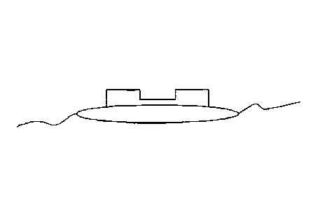
[00043] Figure 4 discloses the general structure of single diffractive
element. Only a
cross section of half of the lens is shown in Figure 4. Profile 100 of the
single
ring surface includes an inner portion or single ring 110, a step or
transition
120, and an outer portion 130. Inner portion 110 extends between a central
location 170 of profile 1 00 and transition 1 20, and outer portion 130
extends
between transition 1 20 and a peripheral location 180 of profile 100. Central
location 170 is typically disposed at the optical axis. Transition 120 is
disposed at a distance of about 1.5 mm from the optical axis, and peripheral
location 180 is disposed at the diameter of the clear aperture of the lens,
here at a distance of about 3.0 mm from the optical axis. In some cases,
transition 1 20 can be disposed at a distance from the optical axis that is
within a range from about 0.5 mm to about 2.0 mm, and peripheral location
180 can be disposed at a distance from the optical axis that is within a range
from about 2.0 to about 3.5 mm, or bigger (for example, for contact lenses,
the ranges would be scaled due to the larger sizes of the contact lens
compared to an 10L).
[00044] As shown in FIG. 4, the surface height or sag (d) from a reference
plane
perpendicular to the optical axis, of each point on the lens profile is
plotted
against the radial distance (r) from the optical axis of the lens. As shown
here, the value of displacement or total sag (d) can have a value within a
14
range from about 0 mm to about 0.07 mm. The total sag can depend on the
refractive shape of the surface and can have a value, for an 10L, of typically
between 0 mm and about 2 mm, or to about minus 2 mm, in cases where the
surface is concave.
[00045] Inner Portion
[00046] Inner portion or echelette 110 includes a center 110a and a
peripheral edge
110b. At center or central section 110a of inner portion 110, the sag (d) of
inner portion 110 is substantially equivalent to the displacement or sag (d)
of
base curve plus offset 160. At peripheral edge 110b, the sag (d) of inner
portion 110 is substantially equivalent to the sag (d) of base curve 140.
Where radial distance (r) is zero, sag (d) of inner portion 110 is equivalent
to
the value of the base curve plus offset 160. The value of sag (d) between
radial distance zero and radial distance at the peripheral edge 110b, for
example at 1.5 mm, gradually and smoothly changes from the value of base
curve plus offset 160 (at r=0) to base curve 140 (at r=1.5 mm) in a parabolic
fashion. As shown here, inner portion 110 can present a parabolic shape, for
example as described in Equation 4a of Cohen, Applied Optics, 31:19, pp.
3750-3754 (1992). In exemplary embodiments where the shape of the inner
portion is asymmetric with respect to the optical axis, as for example in an
ellipse, the peripheral edge 110b of the inner portion 110 may vary between
about 0.5 mm and about 2.0 mm.
CA 2821968 2018-07-24
CA 02821968 2013-06-14
WO 2012/083143 PCT/US2011/065433
[00047] Transition
[00048] At the peripheral edge 110b, where the radial distance (r) is 1.5
mm, the
value of sag (d) steps or changes from the value of base curve 140 to the
value of base curve plus offset 160. Where radial distance (r) corresponds to
transition 120, sag (d) of inner portion 110 is equivalent to the value of the
base curve 140. Relatedly, the displacement of the profile 100 approaches
that of the base curve plus offset 160 as the radial distance increases from a
value of zero to a value of about 1.5 mm. The value of the offset can be
determined along the vertical axis. The offset value may be selected
depending on the amount of phase delay. According to one embodiment, the
inner portion 110 and the outer portion 130 may not end up at the same
vertical height at position 110b/130a. One way to connect these two
endpoints is by using a straight vertical line. As shown here, the diffractive
transition step provides a sharp step in the profile. In some cases the
transition is characterized by a step height having a value within a range
from
about 0.5 microns and about 4 microns. As discussed above, in exemplary
variable stepheight embodiments, the transition may be smooth and gradual
from the low stepheight to the high stepheight, or may be abrupt.
[00049] Outer Portion
[00050] Outer portion 130 includes an inner or central edge 130a and a
peripheral
edge 130b. At inner edge 130a, the sag (d) of outer portion 130 is
substantially equivalent to the sag (d) of base curve plus offset 160. At
16
CA 02821968 2013-06-14
WO 2012/083143 PCT/US2011/065433
peripheral edge 130b, the sag (d) of outer portion 130 remains substantially
equivalent to the sag (d) of base curve plus offset 160. The value of sag (d)
for the outer portion 130 of profile 100 between radial distance 1.5 mm and
radial distance 3.0 mm is equivalent to the value of base curve plus offset
160. The sag of the profile 100 and the base curve plus offset 160 are
approximately equivalent between radial distance values of 1.5 mm and 3.0
mm.
[0051] The limited ring embodiments comprise of adding a limited number of
echelettes to the above detailed single ring microstructure. In general such
limited ring embodiments comprise of a limited number of echelettes that are
either adjacent or non-adjacent to the inner central echelette and may or may
not be separated by a refractive region.
[0052] FIG. 4A provides a graphical representation of a portion of a lens
diffractive
profile with a central echelette and one peripheral adjacent echelette
according to embodiments of the present invention. In FIG. 4A, the height of
the surface relief profile (from a plane perpendicular to the light rays) of
each
point on the echelettes surface is plotted against the distance from the
optical
axis of the lens. The echelettes can have a characteristic optical zone 930
and transition zone 931. Optical zone 930 can have a shape or downward
slope that may be linear when plotted against p as shown in FIG. 4A. When
plotted against radius r, optical zone 930 can have a shape or downward
slope that is parabolic. Central and peripheral echelettes can have a surface
area that is between 1 and 7 mm2. For example, the echelettes may have a
17
CA 02821968 2013-06-14
WO 2012/083143 PCT/US2011/065433
surface area that is 2.3 mm2. An outer (refractive) zone can follow the base
radius with a fixed offset. Exemplary embodiments include peripheral
echelette(s) that are similar in shape (e.g. elliptical) and variable
stepheight
as the central echelette. Of course, this invention includes those
embodiments where the peripheral echelette(s) differ in shape and/or
variable stepheight as compared to the central echelette.
[0053] Although shown in the illustration as associated with the anterior
surface of
lens 390, those skilled in the art will appreciate that the asymmetric
diffractive
structure may alternatively be associated with the posterior surface of lens
390. Further, in preferred embodiments, the diffractive structure 460a may
include a limited number of echelettes.
[0054] Figure 5 shows the predicted defocus curves, in accordance with axis
parameters, for a Tecnis lens, a spherical lens, and two lenses that are
rotationally asymmetric with respect to both shape (ellipse) and stepheight.
In the illustration of Figure 5, the defocus curves are predicted for a 3
millimeter pupil, the horizontal access is the defocus in units of Diopters,
and
the vertical access is the visual acuity in units of logMAR. Figure 5
discloses
that the rotationally asymmetric lenses exhibit an extended depth of focus.
[0055] In an exemplary embodiment, the rotationally asymmetric design has
an
optical performance that depends on the pupil size. For very small pupils,
where the pupil is smaller than the size of the ellipse, the echelette will
act as
a refractive lens, having a very large depth of focus, due to the pinhole
effect.
18
CA 02821968 2013-06-14
WO 2012/083143 PCT/US2011/065433
For higher and medium pupil sizes, where the pupil covers the central
echelette and a part of the outer zone, the lens will act as a
diffractive/refractive lens, with an appropriate phase shift. The size of the
echelette influences the pupil dependence of the lens. As such, the size and
shape of the echelette can be chosen, depending on the pupil sizes of a
specific patient. For example, the pupil sizes of a patient may be measured
in bright light, in dim light, during far vision and during near vision, and
in the
different combinations of light level and accommodative effort. These
different pupil sizes, which may be defined as pupil dynamics, can be used
as input parameters for an optimal design of the single ring or single
echelette design. Additionally, with an elliptical shape, a differential depth
of
focus according to certain meridians may be utilized. That is, for a specific
pupil size, certain parts of the pupil will be inside the wide region of the
elliptically shaped echelette and thus the lens will act as a refractive lens,
while other parts of the pupil will be outside of the elliptically shaped
echelette, and thus the lens will acts as a diffractive with appropriate phase
shift. Accordingly, based on these dynamics an appropriately shaped
asymmetric structure may be designed to suit individual needs.
[0056] It should be appreciated that while the exemplary embodiment
disclosed
herein has rotationally asymmetry in both shape and stepheight, rotational
asymmetry of either shape or stepheight alone may also result in extended
depth of focus. In addition, although an elliptical structure is disclosed, it
is
19
CA 02821968 2013-06-14
WO 2012/083143 PCT/US2011/065433
envisioned that any number of rotationally asymmetric structures will result
in
an extended depth of focus.
[0057] The base refractive lens associated with the EDOF element of the
present
invention may be spherical or aspherical, and/or any type of toric design
indicated to those skilled in the pertinent arts in light of the discussion
herein.
For example, the base refractive IOL may be toric, such as when larger
amounts of corneal cylinder require correction. In such circumstances, the
majority of the corneal cylinder may be corrected by the refractive toric 10L,
and the diffractive element of the present invention may be added merely as
a depth of focus element that makes the base toric IOL less sensitive to
rotation and the choice of optical power. Additionally, the base refractive
lens of the present invention may be combined with an accommodating 10L,
and/or may further be used in combination with any type of ophthalmic lens.
[0058] The rotationally asymmetric diffractive EDOF element of the present
invention may be combined with one or more other diffractive elements
associated with the base refractive lens, such as the multifocal diffractive
zones discussed hereinabove. Thereby, the sensitivity of the multifocal lens
to residual corneal astigmatism may be reduced. As such, the present
EDOF element may be used in conjunction with a bifocal lens or a trifocal
lens. In some embodiments, corrective optics may be provided by phakic
10Ls, which can be used to treat patients while leaving the natural lens in
place. Phakic 10Ls may be angle supported, iris supported, or sulcus
supported. The phakic IOL can be placed over the natural crystalline lens or
CA 02821968 2013-06-14
WO 2012/083143 PCT/US2011/065433
piggy-backed over another 10L. It is also envisioned that the present
invention may be applied to inlays, onlays, accommodating 10Ls, spectacles,
and even laser vision correction.
[0059] Figure 6 is a block diagram illustrating the implementation of the
present
invention in a clinical system 300 comprised of one or more apparatuses that
of capable of assessing the eye's biometry and of performing the calculations
and comparisons set forth in designing the rotationally asymmetric diffractive
EDOF element. The system 300 may include a biometric reader and/or input
301, a processor 302, and a computer readable memory 304 coupled to the
processor 302. The computer readable memory 304 includes therein an
array of ordered values 308 and sequences of instructions 318 which, when
executed by the processor 302, cause the processor 302 to select and/or
design the diffractive structures discussed herein for association with a lens
to be implanted into the eye of the subject presenting the biometric readings
at input 301. The array of ordered values 308 may comprise data used or
obtained from and for use in design methods consistent with embodiments of
the invention. For example, the array of ordered values 308 may comprise
one or more desired refractive outcomes, parameters of an eye model based
on one or more characteristics of at least one eye, and/or data related to an
10L, a set of 10Ls, and one or more rotationally asymmetric echelettes.
[00060] The sequence of instructions 318 may include one or more steps
consistent
with embodiments of the invention. In some embodiments, the sequence of
instructions 318 may include application of calculations, algorithms,
21
CA 02821968 2013-06-14
WO 2012/083143 PCT/US2011/065433
customization, simulation, comparison, remote communications and
networking, and the like.
[0061] The processor 302 may be embodied in a general purpose desktop or
laptop
computer, and/or may comprise hardware and/or software associated with
inputs 301. In certain embodiments, the system 300 may be configured to be
electronically coupled to another device, such as one or more instruments for
obtaining measurements of an eye or a plurality of eyes. Alternatively, the
system 300 may be embodied in a handheld device that may be adapted to
be electronically and/or wirelessly coupled to one or more other devices.
[0062] Although the invention has been described and pictured in an
exemplary
form with a certain degree of particularity, it should be understood that the
present disclosure of the exemplary form has been made by way of example,
and that numerous changes in the details of construction and combination
and arrangement of parts and steps may be made without departing from the
spirit and scope of the invention as set forth in the claims hereinafter.
22