Note: Descriptions are shown in the official language in which they were submitted.
:A 02822612 2013 06 20
WO 2012/092018
PCT/US2011/066115
OPHTHALMIC SURGICAL SYSTEMS HAVING
INTRAOCULAR PRESSURE STABILIZING APPARATUS
Field of Invention
The present invention relates to ophthalmic surgical systems, and more
particularly to ophthalmic surgical systems having intraocular pressure (TOP)
stabilizing
apparatus.
Background of the Invention
The lens of a human eye may develop a cataracteous condition which affects a
patient's vision. Cataracteous lenses may be fragmented and removed using a
surgical
apparatus in a procedure commonly referred to as a lensectomy. Lens
fragmentation can
be achieved using ultrasound in a phacoemulsification lensectomy (also
referred to
simply as "phaco"), laser lensectomy or other procedures. Removal of a
fragmented lens
is typically performed using one or more hand pieces which perform irrigation
and/or
aspiration. In FIG. 1, a hand piece 12 is shown that has a tip 14 that is
inserted through
an incision in the cornea 16 for performing irrigation and aspiration. Such a
hand piece is
typically connected to a surgical console 20 which allows a surgical staff
member to
control irrigation and aspiration as well as various other parameters of the
surgical system
such as those related to ultrasound or laser performance.
The broken lens is removed through an aspiration line 40 that is coupled
between
the hand piece and a vacuum source 46. The distal end of the tip has an
opening that is in
fluid communication with the aspiration line. The distal end of the tip also
typically has a
sleeve which has an opening in fluid communication with an irrigation line 28.
The
irrigation line is typically connected to an irrigation source 30 that can
provide irrigation
fluid to the surgical site.
The lens pieces and irrigation fluid are drawn into the aspiration line
through the
opening of the tip. When performing an irrigation and aspiration procedure, it
is typically
desirable to maintain a suitable, positive intraocular pressure (I0P) within
the eye.
Insufficient or elevated intraoperative IOP may increase the incidence of
complications
during surgery and may also affect the incidence of postoperative
complications.
1
:A 02822612 2013 06 20
WO 2012/092018
PCT/US2011/066115
The fluctuations in pressure during surgery have many causes. Surgical tool
manipulation can cause large pressure increases with long durations. Occlusion
of
aspiration tools and post occlusion surges can cause significant pressure
spikes that have
rise times in the millisecond range. Clearance of these same occlusions can
cause
pressure dips that may lead to momentary chamber collapse and rupture of the
posterior
chamber capsule, resulting in a need for additional surgery.
Summary
Aspects of the present invention are directed to an ophthalmic surgical
apparatus
for use with an eye, comprising a tube adapted to be in fluid communication
with the eye,
a pressure sensor adapted to measure an intraocular pressure value of the eye,
a fluid
reservoir comprising a moveable wall, the fluid reservoir adapted to be in
fluid
communication with the eye through the tube, and at least one processor
coupled to the
pressure sensor to receive the measured intraocular pressure value, the at
least one
processor operable to position the moveable wall in response to a difference
between the
measured intraocular pressure value and a target intraocular pressure value.
In some embodiments, the apparatus further comprises a fluid source adapted to
provide fluid to the eye through the tube. In some embodiments, the apparatus
further
comprises a vacuum source adapted to draw fluid from the eye through the tube.
T he pressure sensor may be disposed within the tube. The pressure sensor may
be a dual sensor, non-invasive pressure sensor.
In some embodiments, the moveable wall comprises a flexible membrane. In
some embodiments, the moveable wall constitutes a wall of an accordion-shaped
container.
The apparatus may comprise a pump fluidly coupled between the fluid source and
the reservoir.
The apparatus may further comprise a voice coil, wherein the at least one
processor is operable to position the moveable wall using the voice coil. In
some
embodiments, the apparatus further comprises a stepper motor, wherein the at
least one
processor is operable to position the moveable wall using the stepper motor.
2
:A 02822612 2013 06 20
WO 2012/092018
PCT/US2011/066115
The apparatus may further comprise a second tube adapted to be in fluid
communication with the eye, a vacuum source adapted to draw fluid from the eye
through the second tube, a second fluid reservoir comprising a second moveable
wall, the
second fluid reservoir adapted to be in fluid communication with the eye
through the
second tube, and the at least one processor coupled to the pressure sensor to
receive the
measured intraocular pressure value, the processor operable to position the
second
moveable wall in response to the difference between the measured intraocular
pressure
value and the target intraocular pressure value.
Brief Description of the Drawings
Illustrative, non-limiting embodiments of the present invention will be
described
by way of example with reference to the accompanying drawings, in which the
same
reference number is used to designate the same or similar components in
different figures,
and in which:
FIG. 1 is a partial schematic illustration of a conventional, surgical
apparatus
including an irrigation line and an aspiration line;
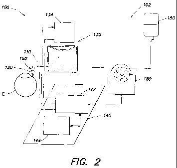
FIG. 2 is partial schematic illustration of an example of a surgical apparatus
according to aspects of the present invention comprising an irrigation line
providing
pressure stabilization;
FIGs. 3A ¨ 3C are schematic illustrations of examples of actuation devices
suitable for use in providing pressure stabilization according to aspects of
the present
invention; and
FIG. 4 is a partial schematic illustration of another example of a surgical
apparatus according to aspects of the present invention comprising an
irrigation line
providing pressure stabiliatoin and an aspiration line providing pressure
stabilization.
Detailed Description
FIG. 2 is a partial schematic illustration of an example of a surgical
apparatus 100
according to aspects of the present invention comprising an irrigation system
102
providing pressure stabilization. System 102 comprises irrigation tube 110, a
pressure
sensor 120, a fluid reservoir 130, and a processor 140 for processing IOP
information.
3
:A 02822612 2013 06 20
WO 2012/092018
PCT/US2011/066115
Irrigation tube 110 is adapted to be in fluid communication with the eye E.
The
tube is connected between a fluid source 150 (e.g., a bottle or other
container of buffered
saline solution) and eye E. The tube is sized and shaped to provide suitable
fluid flow
and fluid pressure in the eye. In the illustrated embodiment, the irrigation
tube is adapted
to be in fluid communication with the eye through a hand piece 160.
Pressure sensor 120 is adapted to measure an intraocular pressure value the
eye E.
Any suitable pressure sensor capable of providing the processor with an IOP
value may
be used. In some embodiments, the pressure sensor is disposed in the fluid
path of the
irrigation system. It will be appreciated that it is generally advantageous
that a sensor in
the fluid path be located proximate the eye, so that the measured value
accurately
represents the IOP. For example, the sensor may be disposed in hand piece 160.
A
sensor to be placed in the fluid path may, for example, be a non-invasive,
dual transducer
device as described in U.S. Patent No. 5,865,764, to Moorehead, titled DEVICE
AND
METHOD FOR NONINVASIVE MEASUREMENT OF INTERNAL PRESSURE
WITHIN BODY CAVITIES, issued February 2, 199, the substance of which is hereby
incorporated by reference.
Fluid reservoir 130 is in fluid communication with the irrigation tube and
comprises a moveable wall 132. The reservoir contains a biocompatible liquid
such as
buffered saline solution that is present in the fluid source. The reservoir
contributes to a
baseline fluid pressure in eye E when the diaphragm is stationary for a
sufficient time to
attain an ambient pressure. However, according to aspects of the present
invention,
actuator 134 moves the movable wall to modify the pressure in the eye in
response to IOP
values measured by sensor 120 during eye surgery. It will be appreciated that,
in addition
to the moveable wall, the remainder of the reservoir is sufficiently rigid
such that a
pressure change in the eye can be attained in response to movement of the
moveable wall.
For example, the moveable wall may comprise a flexible diaphragm in an
otherwise rigid
container or may comprise an accordion-shaped container where opposing walls
are
moved relative to one another.
Processor 140 is coupled to pressure sensor 120 to receive the measured
intraocular pressure value. The processor is operable to position moveable
wall 132 in
response to a difference between the measured intraocular pressure value and a
target
4
:A 02822612 2013 06 20
WO 2012/092018
PCT/US2011/066115
intraocular pressure value. Although in the illustrated embodiment the
processor is
shown as comprising a system processor 144 (e.g., a processor in a
conventional surgical
console (e.g., for receiving using inputs such as pump speed), such as the
processor in the
Stellaris , available from Bausch and Lomb Incorporated, Rochester, NY) and a
chamber
stability processor 142 (e.g, a processor capable of providing signals to and
from sensor,
processor, and actuator in the manner set forth herein), any suitable
processor or
processors may be used to receive and send signals to each of relevant
components.
In some embodiments, a variable-speed infusion pump 180 is fluidly coupled
between fluid source and reservoir. The pump operates to inject fluid into the
reservoir
between actuation events to return the diaphragm to a nominal position thereby
increasing response rate of the system and permitting greater precision in the
response
that occurs when pressure is adjusted.
Although the illustrated embodiment of a pressure stabilizer is shown in
conjunction with an irrigation system, it will be appreciated that the
pressure stabilizer
can be implemented in an aspiration system, for example, as shown in FIG. 4
below. It
will also be appreciated that, although the irrigation tube is adapted to be
in fluid
communication with the eye through a hand piece, the tube can be connected to
another
instrument (not shown) which in turn is in fluid communication with an eye or
the tube
can be configured to be inserted directly into an eye. It will also be
appreciated that a
pressure stabilizer as described herein can be used in apparatus to perform
anterior
segment surgery (e.g., cataract surgery) or posterior segment surgery (e.g.,
vitrealretinal
surgery).
In use during surgery, apparatus 100 provides irrigation to an eye from fluid
source 150 in a conventional manner while measuring IOP using sensor 120. Upon
measurement of a pressure that is outside of a range, processor 140 causes
actuation
device 134 to move a wall of reservoir 130. In response to a measured pressure
that is
too low, the wall is moved inward. Because the fluid in the reservoir is
incompressible
the fluid flows into tube 110 and then into the eye, thereby providing a
compensatory
increase in IOP. It will be appreciated that pump 180 can operate to prevent
all flow
between the reservoir and the fluid source; and in embodiments where the pump
is
omitted a valve (not shown) (e.g., under control of processor 140) can be
provided
:A 02822612 2013 06 20
WO 2012/092018
PCT/US2011/066115
between reservoir 130 and fluid source 150 to control flow between the
reservoir and the
source. In response to a measured pressure that is too high, the wall is moved
outward
drawing fluid form the eye, thereby providing a compensatory decrease in IOP.
It will
be appreciated that pump 180 or the valve can be operated to prevent flow from
coming
from the fluid source.
FIGs. 3A ¨ 3C are schematic illustrations of examples of actuation devices
310,
330, 350 suitable for use in pressure stabilizers according to aspects of the
present
invention. Each actuation device comprises a movement mechanism for moving
movable
wall 132. In FIG. 3A, the actuation device is embodied as a voice coil 312. In
FIG. 38,
the actuation device is embodied as a stepper motor 332 on a lead screw 334.
In FIG. 3C,
the actuation device is embodied as a stepper motor 352 on a cam.
FIG. 4 is partial schematic illustration of an example of a surgical apparatus
according to aspects of the present invention comprising an irrigation system
providing
pressure stabilization and an aspiration system providing pressure
stabilization.
Surgical apparatus 400 comprises an irrigation system 102 as described above
with reference to FIG. 2 and an aspiration system 402 providing pressure
stabilization.
System 402 comprises aspiration tube 410, a pressure sensor 420, a fluid
reservoir 430, a
vacuum source 450, and a processor 440 (comprising vacuum processor 442 and,
in part,
system processor 444) for processing IOP infoimation. In the illustrated
embodiment,
processor 444 receives and/or processes aspiration information and irrigation
information
(e.g., user inputs related to the speed of pumps 480 and 180); however
separate aspiration
and irrigation system processors could be used. To process irrigation
information,
processor 444 operates with processor 442 in the manner of processor 144 as
described
above.
Aspiration tube 410 is adapted to be in fluid communication with the eye E.
The
tube is connected between vacuum source 450 and eye E. The tube is sized and
shaped to
provide a suitable fluid flow and fluid pressure in the eye. In the
illustrated embodiment,
the aspiration line is adapted to be in fluid communication with the eye
through a hand
piece 460. Although the aspiration and irrigation tubes are shown as extending
through
separate hand pieces, in some embodiments both tubes extend through a common
handpiece.
6
:A 02822612 2013 06 20
WO 2012/092018
PCT/US2011/066115
Pressure sensor 120 is adapted to measure an intraocular pressure value of eye
E
as described above. Although the pressure sensor is shown as positioned to
determine
IOP using irrigation fluid in an irrigation tube, in other embodiments, a
pressure sensor
can be positioned to determine IOP using aspiration fluid in an aspiration
tube. It will be
appreciated that using irrigation flow may be advantageous since the
aspiration tube may
be come occluded during removal of a cataract.
Fluid reservoir 430 is in fluid communication with the aspiration tube and
comprises a moveable wall 432. The reservoir contributes to a baseline fluid
pressure in
eye E when the diaphragm is stationary for a sufficient time to attain an
ambient pressure.
However, according to aspects of the present invention, actuator 434 moves the
movable
wall to modify the pressure in the eye in response to IOP values measured by
sensor 420
during eye surgery. Similar to fluid reservoir 130 described above, in
addition to the
moveable wall, the remainder of reservoir 430 is sufficiently rigid such that
a pressure
change in the eye can be attained in response to movement of the moveable
wall. The
moveable wall 432 may comprise a flexible diaphragm.
Processor 440 is coupled to pressure sensor 420 to receive the measured
intraocular pressure value. The processor is operable to position moveable
wall 432 in
response to a difference between the measured intraocular pressure value and a
target
intraocular pressure value. Although in the illustrated embodiment the
processor is
shown as comprising a system processor 444 (e.g., a processor in a
conventional surgical
console, such as the processor in the Stellaris, available from Bausch and
Lomb
Incorporated, Rochester, NY) and a chamber stability processor 442 (e.g., a
processor
capable of providing signals to and from sensor 120 and actuator 434 in the
manner set
forth herein), any suitable processor or processors may be used to receive and
send
signals to each of the relevant components.
Vacuum pump 480 is fluidly coupled between vacuum cassette 450 and reservoir
430. The pump operates to draw fluid from the eye to cassette 450 in a
conventional
manner.
In use during surgery, apparatus 400 provides irrigation to an eye the manner
described above with reference to FIG. 2 while measuring IOP using sensor 120
and
responding when necessary by moving a wall of reservoir 130.
7
:A 02822612 2013 06 20
WO 2012/092018
PCT/US2011/066115
Also during surgery, apparatus 400 aspirates the eye in a conventional manner
while IOP is measured using sensor 120. Upon measurement of a pressure that is
outside
of a range, processor 440 causes actuation device 434 to move a wall of
reservoir 430. In
response to a measured pressure that is too low, the wall is moved inward.
Because the
fluid in the reservoir is incompressible, the fluid flows into tube 410 and
toward the eye,
thereby providing a compensatory increase in IOP. In addition, following
release of an
occlusion, fluid is provided into the aspiration line toward the pump thereby
preventing
severe negative pressure from building up in the aspiration line, thereby
helping to
alleviate what is commonly referred to as post occlusion surge. In response to
a
measured pressure that is too high, the wall is moved outward drawing fluid
faun the eye,
thereby providing a compensatory decrease in IOP.
It will be appreciated that, although the aspiration line is adapted to be in
fluid
communication with the eye through a hand piece, the tube can be connected to
another
instrument (not shown) which in turn is in fluid communication with an eye or
the tube
can be configured to be inserted directly into an eye.
Having thus described the inventive concepts and a number of exemplary
embodiments, it will be apparent to those skilled in the art that the
invention may be
implemented in various ways, and that modifications and improvements will
readily
occur to such persons. Thus, the embodiments are not intended to be limiting
and
presented by way of example only. The invention is limited only as required by
the
following claims and equivalents thereto.
What is claimed is:
8
