Note: Descriptions are shown in the official language in which they were submitted.
CA2823582
WHOLE BLOOD ASSAY FOR MEASURING AMPK ACTIVATION
CROSS REFERENCE TO RELATED APPLICATIONS
This application claims priority to the filing date of United States
Provisional Patent
Application Serial No. 61/430,472 filed January 6,2011.
BACKGROUND
AMPK (adenosine monophosphate kinase) is a heterotrimeric, multisubstrate
kinase
composed of one catalytic (al or a2), one regulatory (131 or p2), and one
AMP/ATP binding
(71, y 2, or y 3) subunit. The C terminus of the p subunit interacts with both
a and 7 subunits,
and current evidence indicates that the p subunit is an obligatory component
of the active
AMPK complex. The exact mechanism by which AMPK is regulated by the energy
status of a
cell is not fully understood. It is thought that when intracellular energy
levels drop (i.e., when
there is a low ATP:AMP ratio), AMP displaces ATP from the y subunit, causing a
conformational change that allows upstream kinases (e.g., LKB1 or CaMKK13) to
phosphorylate and activate the a subunit. Alternatively, AMPK may be
constitutively
phosphorylated, but is quickly dephosphorylated under normal conditions. At
high AMP levels,
however, AMP binding leads to a conformational change shielding activation
site from such
action by phosphatase.
AMPK acts as a sensor of energy status within cells and can be considered a
master
switch of energy metabolism because, upon activation, the enzyme
phosphorylates a number of
downstream protein substrates that have an effect on lipid biosynthesis, fatty
acid oxidation,
glucose uptake, gluconeogenesis and lipogenesis, for example. Phosphorylation
of downstream
targets by AMPK decreases ATP usage by the cell which, in turn, increases the
ATP :AMP ratio
in the cell which, in turn, decreases AMPK activity.
All those properties combine to make AMPK an attractive target in the
treatment of
diabetes, obesity and a variety of other metabolic disorders.
1
CA 2823582 2018-08-23
CA 2823582
SUMMARY
A method of sample analysis is provided. In certain embodiments, the method
comprises: a) labeling cells of a blood sample using an antibody that
specifically binds to
phospho-AMPK or a phosphorylated target thereof, to produce a labeled sample;
and
b) measuring antibody binding by a population of blood cells of the labeled
sample using flow
cytometry. In particular embodiments, the method may further comprise, prior
to the labeling
step: contacting blood with a test agent ex vivo or in vivo; and comparing the
evaluation to
results obtained from a reference sample of blood cells.
Without wishing to be bound to any scientific theory, it is believed that: a)
the effect of
an AMPK-modulatory compound or lifestyle (e.g., diet or exercise) that
modulates AMPK can
be determined by analyzing an organism's blood, and b) blood, which is a
tissue that is not
generally associated with energy production or use, can act as a surrogate for
tissues that are
associated with energy production or use (e.g., liver and muscle, etc.). Thus,
the energy status
of an organism can be evaluated using the organism's blood by flow cytometry
using an
antibody that specifically binds to phospho-AMPK or a phosphorylated target
thereof These
methods do not require an invasive procedure (e.g., a tissue biopsy) and are
faster and more
economical compared to prior approaches (e.g., western blotting).
Various embodiments of the claimed invention relate to a method for
determining the
metabolic health of a subject, comprising: a) contacting permeabilized cells
of a blood sample
from the subject with a fluorescently detectable antibody that specifically
binds to phospho-
adenosine monophosphate kinase (AMPK); b) measuring the amount of the
fluorescently
detectable antibody bound to phospho-AMPK in a plurality of the permeabilized
cells in the
blood sample, wherein the measuring comprises detecting the level of
fluorescence signal on a
single-cell basis, using flow cytometry; and c) calculating a geometric mean
fluorescence value
of the blood sample based on the measurements obtained in step (b), wherein
the geometric
mean fluorescence value provides an indication of the level of AMPK activation
in said
plurality of permeabilized cells that correlates with the metabolic health of
the subject.
Various embodiments of the claimed invention relate to a method of determining
the
effect of a change in lifestyle on adenosine monophosphate kinase (AMPK)
activation in a
subject who has been subjected to the change in lifestyle, comprising: a)
contacting
permeabilized cells of a blood sample from the subject with a fluorescently
detectable antibody
2
CA 2823582 2020-02-24
CA 2823582
that specifically binds to phospho-AMPK; b) measuring the amount of the
fluorescently
detectable antibody bound to phospho-AMPK in a plurality of permeabilized
cells in the blood
sample, wherein the measuring comprises detecting the level of fluorescence
signal on a single-
cell basis, using flow cytometry; c) calculating a geometric mean fluorescence
value of the
blood sample based on the measurements obtained in step (b) , wherein the
geometric mean
fluorescence value provides an indication of the level of AMPK activation in
said plurality of
permeabilized cells that correlates with the metabolic health of the subject;
and d) comparing
said level of AMPK activation to the level of AMPK activation obtained from a
reference
sample of blood cells from the subject before the change in lifestyle, thereby
determining the
effect of said change in lifestyle on AMPK activation of said subject.
Various embodiments of the claimed invention relate to a method for evaluating
the
energy status of a subject, comprising: a) contacting permabilized cells of a
blood sample from
a subject with a fluorescently detectable phosphorylation state-specific
antibody that
specifically binds to a phosphorylated protein that is present in the
permeabilized cells and
whose phosphorylation state in a muscle or liver cell is correlated with an
energy status of the
subject, wherein said phosphorylation state-specific antibody specifically
binds to phospho-
adenosine monophosphate kinase (AMPK); and b) measuring the amount of the
fluorescently
detectable antibody bound to the phosphorylated protein in a plurality of the
permeabilized
cells, wherein the measuring comprises detecting the level of fluorescence
signal which
represent the phosphorylation state of the protein on a single-cell basis
using flow cytometry,
wherein the measured amount of the fluorescently detectable antibody bound to
the
phosphorylated protein in the permeabilized cells provides an indication of
the energy status of
the subject.
Various embodiments of the claimed invention relate to a method for evaluating
the
energy status of a subject, comprising: a) contacting permabilized cells of a
blood sample from
a subject with a fluorescently detectable phosphorylation state-specific
antibody that
specifically binds to a phosphorylated protein that is present in the
permeabilized cells and
whose phosphorylation state in a muscle or liver cell is correlated with an
energy status of the
subject, wherein said phosphorylation state-specific antibody specifically
binds to phospho-
acetyl-CoA carboxylase (ACC); and b) measuring the amount of the fluorescently
detectable
antibody bound to the phosphorylated protein in a plurality of the
permeabilized cells, wherein
2a
CA 2823582 2020-02-24
CA 2823582
the measuring comprises detecting the level of fluorescence signal which
represent the
phosphorylation state of the protein on a single-cell basis using flow
cytometry, wherein the
measured amount of the fluorescently detectable antibody bound to the
phosphorylated protein
in the permeabilized cells provides an indication of the energy status of the
subject.
Various embodiments of the claimed invention relate to a method of evaluating
a
health-related change in lifestyle on the energy status of a subject who has
been exposed to the
health-related change in lifestyle, comprising: a) contacting permeabilized
cells of a blood
sample from the subject with a fluorescently detectable antibody that
specifically binds to a
phosphorylated protein that is present in the permeabilized cells and whose
phosphorylation
state in a muscle or liver cell is correlated with an energy status of the
subject, wherein said
phosphorylation state-specific antibody specifically binds to phospho-
adenosine
monophosphate kinase (AMPK); b) measuring the amount of the fluorescently
detectable
antibody bound to the phosphorylated protein in a plurality of the cells,
wherein the measuring
comprises detecting the level of fluorescence signal which represents the
phosphorylation state
of the protein on a single-cell basis using flow cytometry; and c) comparing
said measured
amount of the fluorescently detectable antibody bound to the phosphorylation
protein in the
cells in the step a) to the measured amount of the fluorescently detectable
antibody obtained
from a reference sample of blood cells from the subject before the change in
lifestyle, thereby
evaluating said change in lifestyle on the energy status of said subject.
Various embodiments of the claimed invention relate to a method of evaluating
a
health-related change in lifestyle on the energy status of a subject who has
been exposed to the
health-related change in lifestyle, comprising: a) contacting permeabilized
cells of a blood
sample from the subject with a fluorescently detectable antibody that
specifically binds to a
phosphorylated protein that is present in the permeabilized cells and whose
phosphorylation
state in a muscle or liver cell is correlated with an energy status of the
subject, wherein said
phosphorylation state-specific antibody specifically binds to phospho-acetyl-
CoA carboxylase
(ACC); b) measuring the amount of the fluorescently detectable antibody bound
to the
phosphorylated protein in a plurality of the cells, wherein the measuring
comprises detecting
the level of fluorescence signal which represents the phosphorylation state of
the protein on a
single-cell basis using flow cytometry; and c) comparing said measured amount
of the
fluorescently detectable antibody bound to the phosphorylation protein in the
cells in the step a)
2b
CA 2823582 2020-02-24
CA 2823582
to the measured amount of the fluorescently detectable antibody obtained from
a reference
sample of blood cells from the subject before the change in lifestyle, thereby
evaluating said
change in lifestyle on the energy status of said subject.
Various embodiments of the claimed invention relate to a method of evaluating
the
energy status of a subject, comprising: a) labeling cells of a blood sample
from the subject
using an antibody that specifically binds to phospho-adenosine monophosphate
kinase
(AMPK), phospho-acetyl-CoA carboxylase or phospho-HMG-CoA reductase, to
produce
labeled cells in the blood sample; and b) measuring the amount of antibodies
bound to
individual labeled cells of a population of labeled cells of said blood sample
using flow
cytometry, thereby obtaining an evaluation of the level of AMPK activation in
said population
of labeled cells, wherein the level of AMPK activation in said population of
labeled blood cells
provides an indication of the energy status of the subject.
BRIEF DESCRIPTION OF THE DRAWINGS
Certain aspects of some embodiments of the invention may be best understood
from the
following detailed description when read in conjunction with the accompanying
drawings. It is
emphasized that, according to common practice, the various features of the
drawings are not to-
scale. On the contrary, the dimensions of the various features are arbitrarily
expanded or
reduced for clarity. Included in the drawings are the following figures:
Fig. 1 schematically illustrates the AMPK pathway.
Figs. 2A and 2B are graphs showing the results of a FACS-based AMPK activation
performed on HepG2 cells.
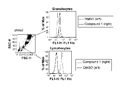
Fig. 3 shows the results of a pAMPK FACS assay using human whole blood. The
2c
CA 2823582 2020-02-24
CA 02823582 2013-07-02
WO 2012/094173
PCT/US2011/066946
results show a 7-fold window for lymphocytes.
Fig. 4 shows the results of a pAMPK FACS assay in human and mouse whole
blood. The protocol was the same as that used for the data shown in Fig. 3.
The results
.. show a robust window in lymphocytes. The control peak is on the left of
each of each
graph, and the peak for compound 2 is on the right.
Fig. 5 shows results of a pAMPK FACS assay using mouse whole blood.
Compound 2, a known modulator of AMPK, is used as the test compound.
Fig. 6 shows results of a FACS assay showing that EC50 data obtained for pACC
correlates with EC50 data for pAMPK.
Fig. 7 is a series of graphs showing that the pAMPK human whole blood FACS
assay is highly specific in that different donors and different antibodies
provide very
similar results.
Fig. 8A-8C. Fig. 8A is a table showing that there is low donor to donor
variability
in pAMPK and pACC stimulation using two AMPK activators. Figs. SB and SC
present
graphs showing an analysis of the data presented in the table of Fig. 8A.
Fig. 9 is a series of graphs showing the effect of incubation time on the EC50
of
Compound 2, a known modulator of AMPK.
Fig. 10 is a scatter plot that shows the correlation between pAMPK assays
using
human white blood cells and HepG2 cells. 200 compounds were tested.
Fig. 11 is a scatter plot showing the correlation between pAMPK and pACC using
human white blood cells.
Fig. 12 is a schematic illustration of a single study in vivo.
Fig. 13 is a series of graphs that show that AMPK phosphorylation after
treatment
by Compound 2, a known modulator of AMPK, is dose-dependent in mouse in viva.
3
CA 02823582 2013-07-02
WO 2012/094173
PCT/US2011/066946
Fig. 14 shows two bar graphs showing that Compound 2, a known modulator of
AMPK, increases AMPK phosphorylation in mouse blood lymphocytes
Fig. 15 shows bar graphs showing the results of a pAMPK FACS assay using
spleen cells and compound 2.
Figs. 16A and 16B are panels of graphs that show that that maximum stimulation
levels of pAMPK are significantly higher in fasted and lean mice compared to
fed and
.. obese mice, while unstimulated levels remain unchanged
Fig. 17A and 17B. Fig. 17A shows an experimental plan whereas Fig. 17B shows
that there is a reasonable correlation between OGTT and PK/PD results.
DEFINITIONS
The term "biological sample" as used herein refers to any sample that contains
or is
made from living material. A biological sample may contain intact cells
obtained from a
multicellular organism. A biological sample may isolated from an individual,
e.g., from a
soft tissue or from a bodily fluid, or from a cell culture that is grown in
vitro. A biological
sample may be made from a soft tissue such as brain, adrenal gland, skin,
lung, spleen,
kidney, liver, spleen, lymph node, bone marrow, bladder stomach, small
intestine, large
intestine at muscle, etc. Bodily fluids include blood, plasma, saliva, mucous,
phlegm,
cerebral spinal fluid, pleural fluid, tears, lactal duct fluid, lymph, sputum,
cerebrospinal
fluid, synovial fluid, urine, amniotic fluid, and semen, etc. Biological
samples also include
cells grown in culture in vitro.
The term "intact cells" includes cells that have been fixed and/or
permeabilized.
Cells that have been lysed and/or sectioned or not intact cells. Western blots
and assays in
which either the proteins of a cell lysate or an antibody are affixed to a
solid support (e.g.,
ELISAs) do not involve intact cells.
The term "blood sample" or grammatical equivalents thereof refer to a sample
of
whole blood or a sub-population of cells in whole blood. Sub-populations of
cells in whole
blood include platelets, red blood cells (erythrocytes), platelets and white
blood cells (i.e.,
peripheral blood leukocytes, which are made up of neutrophils, lymphocites,
eosinophils,
4
CA 02823582 2013-07-02
WO 2012/094173
PCT/US2011/066946
basophils and monocytes). These five types of while blood cells can be further
divided into
two groups, granulocytes (which are also known as polymorphonuclear
leukeocytes and
include neutrophils, eosinophils and basophils) and mononuclear leukocytes
(which include
monocytes and lymphocytes). Lymphocytes can be further divided into T cells, B
cells and
NK cells. Peripheral blood cells are found in the circulating pool of blood
and not
sequestered within the lymphatic system, spleen, liver, or bone marrow. If
blood is first
contacted with an agent and then a sample of the blood is used in an assay,
then a portion
or all of the contacted blood may be used in the assay.
The term "capture agent" refers to an agent that binds a target molecule
through an
interaction that is sufficient to permit the agent to bind and concentrate the
target molecule
from a homogeneous mixture of different molecules. The binding interaction is
typically
mediated by an affinity region of the capture agent. Typical capture agents
include any
moiety that can specifically bind to a target molecule. In certain
embodiments, a
polypeptide, e.g., an antibody, may be employed.
The term "antibody" is used herein to refer to a capture agent that has at
least an
epitope binding domain of an antibody. Types of antibodies include monoclonal
antibodies
and antigen-binding fragments thereof (e.g., Fab, Fv, scFv, and Fd fragments,
chimeric
antibodies, humanized antibodies, single-chain antibodies, etc.) are known and
need not be
described in any further detail.
Capture agents "specifically bind" a target molecule. Accordingly, the term
"capture agent" refers to a molecule or a multi-molecular complex which can
specifically
bind a target molecule, e.g., a phosphorylated polypeptide, with a
dissociation constant
(KD) of less than about 10-6 M (e.g., less than about 10-7M, less than about
10-8M, less than
about 10-9M, less than about 10-10 M, less than about 10-11 M, less than about
1 042 M, to as
low as 111116 M) without significantly binding to other molecules. The term
"specific
binding" refers to the ability of a capture agent to preferentially bind to a
particular target
molecule that is present in a homogeneous mixture of different target
molecule. A specific
binding interaction will discriminate between desirable (e.g., phosphorylated)
and
undesirable (e.g., unphosphorylated) target molecules in a sample, typically
more than
about 10 to 100-fold or more (e.g., more than about 1000- or 10,000-fold).
As used herein, the term "flow cytometry" refers to a method by which the
individual cells of a sample are analyzed by their optical properties (e.g.,
light absorbance,
light scattering and fluorescence properties, etc.) as they pass in a narrow
stream in single
file through a laser beam. Flow cytometry methods include fluorescence
activated cell
5
CA 02823582 2013-07-02
WO 2012/094173
PCT/US2011/066946
sorting (FACS) methods by which a population of cells having particular
optical properties
are separated from other cells.
As used herein, the term "labeling" includes direct and indirect labeling. An
antibody may be fluorescently labeled with a fluorophore or a quantum dot,
many of which
are known.
The term "pre-determined- refers to an element whose identity is known prior
to its
use. An element may be known by name, sequence, molecular weight, its
function, an
amount, optical properties, or any other attribute or identifier.
The term "mixture", as used herein, refers to a combination of elements, e.g.,
cells,
that are interspersed and not in any particular order. A mixture is
homogeneous and not
spatially separated into its different constituents. Examples of mixtures of
elements include
a number of different cells that are present in the same aqueous solution in a
spatially
undressed manner.
"Isolated" or "purified" refers to isolation of a substance (compound,
polynucleotide, protein, polypeptide, poly-peptide composition) such that the
substance
comprises a significant percent (e.g., greater than 2%, greater than 5%,
greater than 10%,
greater than 20%, greater than 50%, or more, usually up to about 90%-100%) of
the sample
in which it resides. A substantially purified component comprises at least
50%, 80%-85%,
or 90-95% of the sample.
The term "assessing" includes any form of measurement, and includes
determining
if an element is present or not. The terms "determining". "measuring",
"evaluating",
"assessing" and "assaying" are used interchangeably and may include
quantitative and/or
qualitative determinations. Assessing may be relative or absolute. "Assessing
the presence
of' includes determining the amount of something present, and/or determining
whether it is
present or absent.
The term -using" has its conventional meaning, and, as such, means employing,
e.g., putting into service, a method or composition to attain an end. For
example, if a
program is used to create a file, a program is executed to make a file, the
file usually being
the output of the program. In another example, if a computer file is used, it
is usually
accessed, read, and the information stored in the file employed to attain an
end. Similarly if
a unique identifier, e.g., a barcode is used, the unique identifier is usually
read to identify,
for example, an object or file associated with the unique identifier.
As used herein, the term "geometric mean" refers to the mean of n numbers
expressed as the n-th root of their product.
6
CA2823582
As used herein, the term -in vivo" refers to the body of a whole living
organism, e.g., a
living mammal.
As used herein, the term "ex vivo" refers to living tissue that has been
removed from the
body of a whole living organism, e.g., a living mammal. A sample of blood that
has been drawn
from a mammal and contains living cells is an example of an ex vivo sample.
As used herein, the term -in vitro" refers to cells that have been grown in
culture.
As used herein, the term "AMPK" or -AMP-activated protein kinase" refers to a
heterotrimeric kinase composed of an alpha catalytic subunit, and non-
catalytic beta and
gamma subunits. AMPK is an important energy-sensing enzyme that monitors
cellular energy
status. In response to cellular metabolic stresses, AMPK is activated and
phosphorylates and
inactivates acetyl -CoA carboxylase (ACC) and beta-hydroxy beta-methylglutaryl-
CoA
reductase (HMGCR), key enzymes involved in regulating de novo biosynthesis of
fatty acid
and cholesterol, as well as other proteins involved in metabolism. AMPK and
its role as an
energy sensor has been reviewed in a variety of publications, including: Kemp
et al (Trends
Biochem. Sei. 1999 24:22-5), Hardie et al (Bioessays. 2001 23:1112-9), Musi et
al (Curr. Drug
Targets Immune Endocr. Metabol. Disord. 2002 2:119-27), Musi et al (Biochem.
Soc. Trans.
2003 31:191-5) and Hardie (Endocrinology. 2003 144:5179-83) and Aschenbach
(Sports Med.
2004 34:91-103).
As used herein, the term -phospho-AMPK" or "p-AMPK" refers to a form of AMPK
in
which the a. subunit has a phosphorylated threonine at position 172.
Phosphorylation at this
position is done by an upstream AMKP kinase (AMPKK). Phosphorylation at this
position
causes the kinase to phosphorylate downstream targets. One downstream target
of phospho-
AMPK is ACC (acetyl-CoA carboxylase), although there are many others.
As used herein, the term -AMPK activation" refers to the phosphorylation state
of
AMPK or a direct target thereof. AMPK may be activated by modulation of a
protein upstream
of AMPK (e.g., the adponectin receptor, the leptin receptor, the a-adrenergic
receptor, or the
insulin receptor etc.) or by AMPK itself. AMPK activation may be determined by
assaying
AMPK itself or a downstream target of AMPK.
An antibody that is specific for phospho-AMPK or a phosphorylated target
thereof
specifically binds the phosphorylated forms of those proteins but not the
unphosphorylated
forms of those proteins.
7
CA 2823582 2018-08-23
CA2823582
Other definitions of terms appear throughout the specification.
DETAILED DESCRIPTION
Before the present invention is described in greater detail, it is to be
understood that this
invention is not limited to particular embodiments described, as such may, of
course, vary. It is
also to be understood that the terminology used herein is for the purpose of
describing
particular embodiments only, and is not intended to be limiting, since the
scope of the present
invention will be limited only by the appended claims.
Where a range of values is provided, it is understood that each intervening
value, to the
tenth of the unit of the lower limit unless the context clearly dictates
otherwise, between the
upper and lower limit of that range and any other stated or intervening value
in that stated range
is encompassed within the invention.
Unless defined otherwise, all technical and scientific terms used herein have
the same
meaning as commonly understood by one of ordinary skill in the art to which
this invention
belongs. Although any methods and materials similar or equivalent to those
described herein
can also be used in the practice or testing of the present invention, the
preferred methods and
materials are now described.
The citation of any publication is for its disclosure prior to the filing date
and should not
be construed as an admission that the present invention is not entitled to
antedate such
publication by virtue of prior invention. Further, the dates of publication
provided may be
different from the actual publication dates which may need to be independently
confirmed.
It must be noted that as used herein and in the appended claims, the singular
forms -a",
-an", and "the" include plural referents unless the context clearly dictates
otherwise. It is
further noted that the claims may be drafted to exclude any optional element.
As such, this
statement is intended to serve as antecedent basis for use of such exclusive
terminology as
-solely," -only" and the like in connection with the recitation of claim
elements, or use of a
-negative" limitation.
8
CA 2823582 2018-08-23
CA 02823582 2013-07-02
WO 2012/094173
PCT/US2011/066946
As will be apparent to those of skill in the art upon reading this disclosure,
each of
the individual embodiments described and illustrated herein has discrete
components and
features which may be readily separated from or combined with the features of
any of the
other several embodiments without departing from the scope or spirit of the
present
invention. Any recited method can be carried out in the order of events
recited or in any
other order which is logically possible.
METHOD Or SAMPLE ANALYSIS
The method described below employs a sample of blood. However, blood is but
one
of many biological samples that can be employed in the method. In other
embodiments,
intact cells from other tissues (e.g., other soft tissues such as liver or
spleen etc.) or cells
grown in tissue culture may be employed. Methods for treating such tissues to
provide a
cell suspension suitable for flow cytometry are known. Once produced, a cell
suspension
may be employed in a similar way to that described below.
In general terms, the subject method involves: a) labeling cells of a blood
sample
using an antibody that specifically binds to phospho-AMPK or a phosphorylated
target
thereof, to produce a labeled sample; and b) measuring antibody binding by a
population of
blood cells of the labeled sample using flow cytometry, thereby obtaining an
evaluation of
AMPK activation in the population of blood cells, Since the results obtained
from blood
correlated well with results obtained from tissues associated with energy
consumption (e.g.,
muscle or liver) the evaluation may of AMPK activation in blood can be
extended to
provided an evaluation of AMPK activation in the subject from which the blood
was
obtained.
While the method may be performed on whole blood, in particular embodiments,
the population of blood cells analyzed may be white blood cells or a sub-
population thereof
(e.g., a lymphocyte population or a granulocyte population). In particular
embodiments, the
blood may contacted with a test agent ex vivo (i.e., using blood drawn from a
subject) or in
vivo (e.g., by administering the test agent to a mammal), and the results from
the assay may
be compared to results obtained from a reference sample of blood cells (e.g.,
blood cells
that have not been in contact with the test agent or with a different amount
of the test agent)
to determine the effect of the compound on AMPK activation in the subject from
which the
blood sample was obtained.
9
CA 02823582 2013-07-02
WO 2012/094173
PCT/US2011/066946
The effect may in certain cases be measured by calculating the difference in
geometric mean fluorescence of the population of blood cells and the geometric
mean
fluorescence of the reference sample of blood cells. As would be apparent, in
certain
embodiments, the contacting may involve administering the test agent to a
subject and then
drawing blood from the subject after a specified period of time. In other
embodiments, the
contacting may involve drawing blood from a subject and then contacting the
agent with
the drawn blood for a specified period of time. The test agent may or may not
be a known
modulator of the AMPK pathway. In particular embodiments, the reference sample
may
contain blood cells obtained from the same individual as the test blood
sample. The
reference sample may or may not have been contacted with the test agent. In
particular
cases. data obtained from the method may be expressed as a graph of the
geometric means
of fluorescence of number of samples, as illustrated in Fig. 2. Such a graph
may show a
time course, or the difference between different doses of a test agent, for
example.
As illustrated in Fig. 1, AMPK acts as a metabolic master switch regulating
several
intracellular systems including the cellular uptake of glucose, the 13-
oxidation of fatty acids
and the biogenesis of glucose transporter 4 (GLUT4) and mitochondria. The
energy-
sensing capability of AMPK can be attributed to its ability to detect and
react to
fluctuations in the AMP:ATP ratio that take place during rest and exercise
(muscle
stimulation). During muscle stimulation, AMP increases while ATP decreases.
which
changes AMPK into a good substrate for activation via an upstream kinase
complex,
AMPKK, or alternatively, where binding of AMP renders activated AMPK that is
phosphorylated at Thr-172 a worse substrate for protein phosphatase 201. AMPKK
is a
complex of three proteins, STE-related adaptor (STRAD), mouse protein 25
(M025), and
LKB1 (a serine/threonine kinase). During a bout of exercise, AMPK activity
increases
while the muscle cell experiences metabolic stress brought about by an extreme
cellular
demand for ATP. Upon activation, AMPK increases cellular energy levels by
inhibiting
anabolic energy consuming pathways (fatty acid synthesis, protein synthesis,
etc.) and
stimulating energy producing, catabolic pathways (fatty acid oxidation,
glucose transport,
etc.).
Triggering the activation of AMPK can be carried out provided that two
conditions
are met. First, the y subunit of AMPK must undergo a conformational change so
as to
expose the active site (Thr-172) on the a subunit, The conformational change
of the y
subunit of AMPK can be accomplished under increased concentrations of AMP.
Increased
concentrations of AMP will give rise to the conformational change on the y
subunit of
CA 02823582 2013-07-02
WO 2012/094173
PCT/US2011/066946
AMPK as two AMP bind the two Bateman domains located on that subunit. It is
this
conformational change brought about by increased concentrations of AMP that
exposes the
active site (Thr-172) on the a subunit. This role of AMP is further
substantiated in
experiments that demonstrate AMPK activation via an AMP analogue 5-amino-4-
imidazolecarboxamide ribotide (ZMP) which is derived from the familiar 5-amino-
4-
imidazolecarboxamide riboside (AICAR). The second condition that must be met
is the
phosphorylation and consequent activation of AMPK on its activating loop at
Thr-172 of
the a subunit brought about by an upstream kinase (AMPKK). The complex formed
between LKB1 (STK 11), mouse protein 25 (M025), and the pseudokinase STE-
related
adaptor protein (STRAD) has of late been identified as the major upstream
kinase
responsible for phosphorylation of AMPK on its activating loop at Thr-172.
Although
AMPK must be phosphorylated by the LKB1/M025/STRAD complex, it can also be
regulated by allosteric modulators which directly increase general AMPK
activity and
modify AMPK to make it a better substrate for AMPKK and a worse substrate for
phosphatases. It has recently been found that 3-phosphoglycerate (glycolysis
intermediate)
acts to further pronounce AMPK activation via AMPKK.
CaMKK has also been identified as an upstream AMPKK. CaMKK phosphorylates
and activates AMPK in an AMP-independent manner, which is triggered instead by
a rise
in the intracellular Ca2+ concentration. The discovery that CaMKK acts as an
AMPKK
indicates that in addition to an increase of the AMP-to-ATP ratio, AMPK may
also be
activated by a rise in the intracellular Ca2+ concentration in response to
nutrients, drugs, or
physiological stimulation.
Some downstream targets of AMPK are illustrated in Fig. 1. As illustrated,
downstream targets of AMPK include proteins that regulate carbohydrate
metabolism (e.g..
GEF, MEF, glycogen synthase, PFK2 and TORC2), lipid metabolism (e.g., HMGCoAR,
ACC, HNF-4, SREBP-1 and HSL), cell growth and apoptosis (eNOS, p53, HrR and
eEF2K) and protein metabolism. One exemplary downstream target of AMPK is
acetyl-
CoA carboxylase (ACC). Acetyl-CoA carboxylase is a biotin-dependent enzyme
that
catalyzes the irreversible carboxylation of acetyl-CoA to produce inalonyl-CoA
through its
two catalytic activities, biotin carboxylase (BC) and carboxyltransferase
(CT). ACC is a
multi-subunit enzyme in the endoplasmic reticulum of most eukaryotes. The most
important function of ACC is to provide the malonyl-CoA substrate for the
biosynthesis of
fatty acids. The activity of ACC can be controlled at the transcriptional
level as well as by
11
CA2823582
small molecule modulators and covalent modification. The human genome contains
the genes
for two different AC Cs: ACACA and ACACB.
Phosphorylation of AMPK can result when the hormones glucagon or epinephrine
bind
to cell surface receptors, but the main cause of phosphorylation is due to a
rise in AMP levels
when the energy status of the cell is low, leading to the activation of the
AMP-activated protein
kinase (AMPK). AMPK is the main kinase regulator of ACC, able to phosphorylate
a number
of serine residues on both isoforms of ACC. On ACC I, AMPK phosphorylates
Ser79, Ser1200,
and Ser1215. On ACC2, AMPK phosphorylates Ser218. Protein kinase A also has
the ability to
phosphorylate ACC, with a much greater ability to phosphorylate ACC2 than
ACC1. However,
the physiological significance of protein kinase A in the regulation of ACC is
currently
unknown. When insulin binds to its receptors on the cellular membrane, it
activates a
phosphatase to dephosphorylate the enzyme; thereby removing the inhibitory
effect.
AMPK and its targets are generally intracellular. As such, the method
generally
involves permeabilizing the blood cells, and then labeling the permeabilized
cells using an
antibody that specifically binds to phospho-AMPK or a phosphorylated target
thereof. While
the exact steps of such intracellular labeling methods may vary greatly, they
generally involve
permeabilizing the cells, labeling the cells using a labeled antibody and then
fixing the stained
cells so that the contents of the cells stay intact during subsequent
manipulations. Exemplary
methods by which cells can be labeled using fluorescent antibodies that are
specific for
intracellular proteins are described in a variety of publications, including:
Lazarus et al
(Cytometry. 1998 32:206-13), Sartor et al (Cytometry. 1994 18:119-22), Gadol
et al
(Cytometry 1994 15:359-70) and Far et al (Cytometry. 1994 15:327-34). Kits for
intracellularly labeling cells for FACS analysis include the INTRACYTETm
intracellular FACS
kit by Orion BioSolutions, Inc (Vista CA), the INTRASURETm or FASTIMMUNETm
kits by
Becton Dickinson (Franklin Lakes, NJ) and the CYTOFIXTm or CYTOPERMTm Plus
kits by
PharMingen (San Diego, CA). Depending on the method employed, the red blood
cell of the
sample may be lysed prior to permeablizing and labeling of the white blood
cells. Such lysis
techniques may be adapted from those commonly employed in blood analysis.
Antibody binding by individual cells of the population of blood cells is
measured using
flow cytometry. Such methods are known and reviewed in a variety of
publications, including
Brown et al (Clin Chem. 2000 46:1221-9), McCoy eta! (Hematol. Oncol. Clin.
North Am.
12
CA 2823582 2018-08-23
CA2823582
2002 16:229-43) and Scheffold J. Clin. Immunol. 2000 20:400-7) and books such
as Carey et al
(Flow Cytoinetry in Clinical Diagnosis, 4th Edition ASCP Press, 2007), Ormerod
(Flow
Cylometry ___ A practical approach 3rd Edition. Oxford University Press,
Oxford, UK 2000),
Ormerod (Flow cytotnetry 2nd Edition. BIOS Scientific Publishers, Oxford, UK
1999) and
___________________ Ormerod (Flow Cytometry A basic introduction 2009
Cytometry Part A 75A, 2009).
In particular cases, the data for a single sample may be processed to provide
the number
of events for each a measurement of fluorescence. As shown in Fig. 3, the data
may in certain
cases be expressed as a single parameter histogram that shows the units of
fluorescence on the
x axis and the cell count on the y axis. The fluorescence may be a log value
and in certain cases
may be the log of an absolute (e.g., raw) or normalized value. The peak of the
histogram
provides an evaluation of AMPK activation in the subject from which the blood
sample was
obtained. The peak of the histogram can be the geometric mean of the
fluorescence values,
however other statistical analysis can be employed to provide a similar
result. Since the various
sub-populations of blood cells (i.e., red blood cells, platelets and white
blood cells which are
composed of neutrophils, lymphocytes, monocytes, eosinophils, and basophils)
are readily
distinguishable using flow cytometry, the data may be analyzed to provide an
evaluation of
AMPK activation in any sub-population of blood cells. In one embodiment, the
data may be
analyzed to provide an evaluation of AMPK activation in lymphocytes. In a
further
embodiments, the blood cells may be labeled with a second antibody, e.g., a
cell surface
.. antibody, and the data may be analyzed to provide an evaluation of AMPK
activation in cells
that are labeled with the second antibody.
The methodology described herein may be generally employed on any suitable
flow
cytometer, examples of which are known on the art and described in, e.g., U.S.
Patents
5,378,633, 5,631,165, 6,524,858, 5,266,269, 5,017,497 and 6,549,876, PCT
publication
.. W099/54494 and as well as published U.S. Patent Applications US20080153170,
20010006787, U520080158561, US20100151472, US20100099074, US20100009364.
US20090269800, US20080241820, U520080182262, U520070196870 and U520080268494.
The antibody used to label the cells should be capable specifically binding to
native
(i.e., folded) phospho-AMPK or a phosphorylated target thereof In particular
cases, antibody
may binds at much reduced (i.e., by a factor of at least 2, 5, 10, 50 or 100)
affinity
13
CA 2823582 2018-08-23
CA 02823582 2013-07-02
WO 2012/094173
PCT/US2011/066946
to the linear (i.e., unfolded, denatured) form of the protein. The structure
to which such an
antibody binds contains may amino acid that are dis-contiguous in the protein.
In other
words, in certain cases binding of such an antibody to a polypeptide may be
dependent
upon the polypeptide being folded into a particular three dimensional
conformation.
Such antibodies may be made by, e.g., immunizing a suitable animal, e.g., a
warm-
blooded animal, in particular a mammal such as a rabbit, mouse, rat, camel,
sheep, cow or
pig or a bird such as a chicken or turkey, with a folded phospho-AMPK or
downstream
target thereof using any of the techniques well known in the art suitable for
generating an
immune response. Procedures for immunizing animals are well known in the art,
and are
described in Harlow (Antibodies: A Laboratory Manual, First Edition (1988)
Cold Spring
Harbor, N.Y.) and Weir (Handbook of Experimental Immunology Vol 4, Blackwell
Scientific Publishers, Oxford, England, 1986). As will be appreciated by one
of ordinary
skill in the art, the immunogen may be admixed with an adjuvant or hapten in
order to
increase the immune response (for example, complete or incomplete Freund's or
lipid A
.. adjuvant), or with a carrier such as keyhole limpet hemocyanin (KLH).
Once a suitable animal has been immunized and an immune response against the
antigen has been established by the animal, antibody producing cells from the
animal are
screened to identify cells that produce antibodies having a desired activity.
In some
embodiments, these methods may employ hybridoma technology in which cells from
the
spleen of the immunized animal are fused with a suitable immortal cell to
produce
hybridoma cells. Supernatants from these hybridoma cells may be screened, and
positive
clones are expanded according to standard procedures (Harlow et al.
Antibodies: A
Laboratory Manual, First Edition (1988) Cold spring Harbor, N.Y.; and Spieker-
Polet et
al., supra).
The antibodies may be screened for binding to phosporylated AMPK or
phosporylated target thereof folded into a native conformation by, e.g., cell
staining to
identify those antibodies that are specific for phosphorylated forms of these
proteins. In
particular embodiments, commercially available antibodies may be screened to
identify a
suitable antibodies.
In alternative embodiments, a phage display antibody may be employed, methods
for the production of which are well known (see, e.g., Scott et al. Science
1990 249: 386;
Devlin et al., Science 1990 249: 404; U.S. Pat. Nos. 5,223,409, 5,733,731,
5,498,530,
5,432,018, 5,338,665, and 5,922,545, for example).
14
CA 02823582 2013-07-02
WO 2012/094173
PCT/US2011/066946
In particular cases, rather than an antibody that is specific for AMPK or a
downstream target thereof, the method described above and below may be
performed using
an antibody that is specific for a phosphorylated AMPK-related kinase (e.g.,
BRSK1,
BRSK2, NUAK1, NUAK2, QIK, QSK, SIK, MARK1, MARK2, MARK3, MARK4,
MELK, or SNARK; Manning et al, Science 2002 298: 1912-1934) which are closely
related to AMPKal and AMPKa2, some of which have also been implicated in
energy
homeostasis (Koh et al PNAS 2010 107: 15541-15546).
In alternative embodiments, a quantitative western blotting method, e.g. ,
using a
capillary-based system such as a Cell Bioscences CB1000 machine, may be
employed.
Alternatively, mass spectrometry or mass cytometry may be employed. In certain
cases, the
assays may be done on a high throughput format, e.g., using 96- or 384-well
plates.
UTILITY
The method described above may be employed to identify compounds that
modulate AMPK activation, to determine whether an administered compound is
having a
desired effect, or to determine an optimal dose of a compound is known to
modulate
AMPK activation, for example. In these embodiments, the measurement obtained
using the
above method may be compared to results obtained from a reference sample of
blood. As
noted above, the test sample of blood may be contacted with a test agent ex
vivo or in vivo.
The reference sample of blood may not have been contacted with the test agent
or may
have been contacted with a different amount of the test agent, for example. In
one
embodiment, both the test and reference samples may have been contacted with
the same
amount of the test compound, but at different times or for different
durations. The test and
reference samples may be obtained from the same subject, or from different
subjects. The
subject may have fasted for at least 8-12 hours, or, in certain cases, the
method may be
performed before, during or after exercise. The method may be coupled with
other medical
tests, such as a cholesterol test (i.e., a lipid panel) or a blood glucose
test to provide an
evaluation of the health of the subject. The target of the test agent may be
upstream of
AMPK, may be AMPK itself, or downstream of AMPK. In some cases, the mechanism
of
action a test agent may be unknown.
In one exemplary embodiment, separate aliquots of blood from the same
individual
are contacted with two or more amounts of a test agent that is known to
modulate AMPK
CA 02823582 2013-07-02
WO 2012/094173
PCT/US2011/066946
activation. The contacted blood may be assayed using the method described
above, and an
effective dose of the test agent may be determined.
In another exemplary embodiment, separate aliquots of blood from the same
individual are contacted with: a) a test agent that is not known to modulate
AMPK
activation and b) a control solution that does not contain the test agent. The
contacted blood
may be assayed using the method described above, and the effect of the
compound on
AMPK activation is be determined.
In these embodiments, the degree to which the test sample and control sample
differ
may be determined by comparing, for example, the geometric mean of the results
obtained
from a test sample to the geometric mean of the results obtained from a
reference sample.
A greater difference between the geometric means indicates that the agent has
a greater
effect on AMPK activation. For example, as illustrated in Fig. 3, the Compound
1 has a
greater effect on AMPK activation in lymphocytes than in granulocytes.
Relative to the
geometric mean fluorescence of a sample that had not been contacted with the
compound, a
compound that modulates AMPK activation may alter the geometric mean
fluorescence by
at least 5%, at least 10%, at least 20%, or at least 50%. In particular
embodiments, the
compoud may lead to a decrease of at least 10%, at least 20%, at least 50%, at
least 70% or
at least 80% in the geometric mean fluorescence. In other embodiments, the
compoud may
lead to an increase of at least 10%, at least 20%, at least 50%, at least 70%,
at least 100%
or at least 200% or at least 500% or more in the geometric mean fluorescence.
As noted above, the test agent may be administered in vivo, in which case, the
contacting may comprise administering the test agent to a subject and then
drawing blood
from the subject after a specified period of time (e.g., from 5 minutes to 1
hr, 1 hr to 12 hr,
12 hr to 24 hr or 24 hr to 1 week or more) prior to analysis by flow
cytometry. In ex vivo
applications, the contacting may comprise drawing blood from a subject and
then
contacting the test agent with the drawn blood for a specified period of time
(e.g.. from 5
minutes to 1 hr, 1 hr to 12 hr, 12 hr to 24 hr or 24 hr to 1 week or more)
prior to analysis by
flow cytometry.
In one in vivo embodiment, one amount of an AMPK modulator may be
administered to a subject and, after a specified period of time, blood may be
drawn from
the subject and assayed using the method described above. Based on the results
of the
assay, a second amount of the AMPK modulator may be administered to the
subject and,
after a specified period of time, blood may be drawn from the subject and
assayed using the
method described above. These steps may be repeated until a desired effect
(e.g., a dose of
16
CA2823582
the AMPK modulator that results in a desired, stable, level of AMPK
activation) is achieved.
This method may be used to optimize the dosage of an AMPK modulator for a
particular
subject.
In general terms the blood used in the assay may be obtained from any
mammalian
subject including the orders carnivore (e.g., dogs and cats), rodentia (e.g.,
mice, guinea pigs,
and rats), and primates (e.g, humans, chimpanzees, and monkeys). In some
embodiments, the
subject may be human. The subject may be healthy or in some cases may have
cancer, an
inflammatory disease or a metabolic disease such as obesity or diabetes. In
particular
embodiments, the subject may have one or more risk factors for metabolic
syndrome, such as,
stress, overweightness, sedentary lifestyle, aging, coronary heart disease,
chronic heart failure,
lipodystrophy, schizophrenia, peripheral artery disease, neurodegenerative
diseases, muscle
atrophy/weakness/myopathy, renal diseases, chronic obstructive pulmonary
disease, age-related
macular degeneration, or rheumatic disease. For example, a subject may have
fasting
hyperglycemia (caused by, e.g., diabetes mellitus type 2 or insulin
resistance), high blood
pressure, central obesity, decreased HDL cholesterol and/or elevated
triglycerides.
In some embodiments, the test agent may be known to affect AMPK activation.
Such
agents are known and include: metformin, cilostazol, 5-aminoimidazole-4-
carboxamide
ribonucleoside (AICAR), 2-dcoxyglucose, thiazolidinediones such as troglitzone
rosiglitazone,
resveratrol, and pioglitazone, as well as a variety of other compounds
described in PCT
publications W008/083124, W009/065131, W009/076631, W009/132136, and
W010/088392. published U.S. patent applications US20100009992, US20090253764,
US20090105293, US20090094709, US20080221088, US20070244202, US20070054965,
US20070015665, US20060287356, US20060134240, US20050038068 and US patent
7,119,205.
In other embodiments, the effect of the test agent on AMPK activation may be
unknown. Such agents may be from any chemical class and in certain cases may
be synthetic,
semi-synthetic, or naturally-occurring inorganic or organic molecules. Test
agents include
those found in large libraries of synthetic or natural compounds. For example,
synthetic
compound libraries are commercially available from Maybridge Chemical Co.
(Trevillet,
Cornwall, UK), ComGenex (South San Francisco, CA), and MicroSource (New
Milford, CT).
Alternatively, libraries of natural compounds in the form
17
CA 2823582 2018-08-23
CA 02823582 2013-07-02
WO 2012/094173
PCT/US2011/066946
of bacterial, fungal, plant and animal extracts are available from Pan Labs
(Bothell, WA) or
are readily producible.
Test agents may be small organic or inorganic compounds having a molecular
weight of more than 50 and less than about 2,500 Da. Test agents may comprise
functional
groups necessary for structural interaction with proteins, particularly
hydrogen bonding,
and may include at least an amine, carbonyl, hydroxyl or carboxyl group, and
may contain
at least two of the functional chemical groups. The candidate agents may
comprise cyclical
carbon or heterocyclic structures and/or aromatic or polyaromatic structures
substituted
with one or more of the above functional groups. Candidate agents are also
found among
biomolecules including peptides, saccharides, fatty acids, steroids, purines,
pyrimidines,
derivatives, structural analogs or combinations thereof.
Test agents may be obtained from a wide variety of sources including libraries
of
synthetic or natural compounds. For example, numerous means are available for
random
and directed synthesis of a wide variety of organic compounds and
biomolecules, including
expression of randomized oligopeptides. Alternatively, libraries of natural
compounds in
the form of bacterial, fungal, plant and animal extracts are available or
readily produced.
Additionally, natural or synthetically produced libraries and compounds are
readily
modified through conventional chemical, physical and biochemical means, and
may be
used to produce combinatorial libraries. Known pharmacological agents may be
subjected
to directed or random chemical modifications, such as acylation, alkylation,
esterification,
amidification, etc. to produce structural analogs. New potential therapeutic
agents may
also be created using methods such as rational drug design or computer
modeling.
Screening may be directed to known pharmacologically active compounds and
chemical analogs thereof, or to new agents with unknown properties such as
those created
through rational drug design.
The subject may contacted with the candidate agent, e.g., the agent is
administered
to the animal by any acceptable route of administration, including, but not
limited to, oral
(e.g., oral gavage), intravenous, intramuscular, intranasal, subcutaneous,
intragastric, etc.,
e.g., any enteral or parenteral route. A single dose is administered, or
multiple doses over a
period of time are administered.
Formulations, including pharmaceutical formulations, comprising an agent
identified by a screening method presented herein, are provided. A formulation
comprises
an effective amount of an agent. An "effective amount" means a dosage
sufficient to
produce a desired result.
18
CA 02823582 2013-07-02
WO 2012/094173
PCT/US2011/066946
Although the dosage used will vary depending on the clinical goals to be
achieved,
a suitable dosage range is one which provides up to about 1 lig to about 1,000
lug or about
10,000 g of an agent that produces a desired result can be administered in a
single dose.
Alternatively, a target dosage of an agent that produces a desired result can
be considered
to be about in the range of about 0.1-1000 M, about 0.5-500 M. about 1-100
M, or
about 5-50 M in a sample of host blood drawn within the first 24-48 hours
after
administration of the agent.
Those of skill will readily appreciate that dose levels can vary as a function
of the
specific compound, the severity of the symptoms and the susceptibility of the
subject to
side effects. Preferred dosages for a given compound are readily determinable
by those of
skill in the art by a variety of means.
In particular embodiments, the subject method may be employed to provide a
read-
out of the metabolic health of a subject in a similar way as a glucose or
cholesterol test.
The subject method may be performed alone, or in combination with other
clinical
techniques (e.g., a physical examination or another blood test). For example,
results
obtained from the subject assay may be combined with other information, e.g,,
information
regarding blood glucose levels, weight, or other proteinaceous blood markers
that indicate
the metabolic health of an individual.
In one exemplary embodiment, prior to the labeling of the cells, the method
may
comprise subjecting a mammal to a change in lifestyle (e.g., a change in diet
or amount of
exercise), and obtaining a blood sample from the mammal that is subsequently
analyzed.
The evaluation may then be compared to results obtained from a reference
sample of blood
cells, thereby determining the effect of the change in lifestyle on AMPK
activation of the
mammal.
In one embodiment, a sample may be collected from a patient at a first
location,
e.g., in a clinical setting such as in a hospital or at a doctor's office, and
the sample may be
forwarded to a second location, e.g., a laboratory where it is processed and
the above-
described method is performed to generate a report. A "report" as described
herein, is an
electronic or tangible document which includes report elements that provide
test results that
may include the geometric mean obtained from the test as well as, for example,
a range of
geometric means that are considered "normal". Once generated, the report may
be
forwarded to another location (which may the same location as the first
location), where it
may be interpreted by a health professional (e.g., a clinician, a laboratory
technician, or a
physician such as an oncologist, surgeon, pathologist), as part of a clinical
diagnosis.
19
CA 02823582 2013-07-02
WO 2012/094173
PCT/US2011/066946
EXAMPLES
The following examples are put forth so as to provide those of ordinary skill
in the
art with a complete disclosure and description of how to make and use the
present
invention, and are not intended to limit the scope of what the inventors
regard as their
invention. Efforts have been made to ensure accuracy with respect to numbers
used (e.g.
amounts, temperature, etc.) but some experimental errors and deviations should
be
accounted for. Unless indicated otherwise, parts are parts by weight,
molecular weight is
weight average molecular weight, temperature is in degrees Centigrade, and
pressure is at
or near atmospheric.
Three different AMPK activators employed in the examples described below.
Compound 1 is described in PCT publication W010/088392, Compound 2 is
described in
PCT publication W009/065131.
Example 1
MEF or C2C12 cells were treated with DMSO or Compound 2 and probed by
western blotting with a Cell Signaling anti-pAMPK antibody. The antibody
exclusively
recognized pAMPK and no other bands are observed. The same antibody was used
for
FACS in subsequent experiments.
HepG2 human liver cancer cells were trypsinized, resuspended in a complete
media
at 2X10⁶ cells per ml and plated in a deep well round-bottom 96-well plate,
100 ul per
well. Compound was added in 1 i1 of DMSO to the cell suspension, mixed and
incubated 1
hr at 37 C. Following the incubation, 900 pl of lyse-fix solution (BD) was
added and the
plate was incubated at 37oC for another 10 min, spun for 5 min and the
supernatant
removed. Cell pellet was resuspended in 250 pl of ice-cold methanol and
incubated for 30
mm at 4oC. Following a transfer to a regular 96-well round-bottom plate, the
cells were
spun down for 5 minutes, washed in 250 pl of PBS containing 2% of FCS (PBS2)
and
resuspended in 100 pl of PBS2 containing 1:100 dilution of pACC antibody
(Millipore).
Suspension was incubated overnight at room temperature on a shaker. The
following
morning, cells were washed once with PBS2 and incubated with secondary goat
anti-rabbit
antibody conjugated to Alexa 488 at 1:200 dilution for 1 hr at room
temperature. After a
singl wash, the cells were sorted on a BD FACS sorter. Quantitation was
performed using
CA 02823582 2013-07-02
WO 2012/094173
PCT/US2011/066946
Flow*, software. Geometric mean of a Alexa 488 signal for live cells gate was
used to plot
the results and determine EC50 in matlab.
Results are shown in Fig. 2. EC5os obtained by FACS strongly correlate with
the in-
cell western ones for the same epitope, validating the FACS-based approach.
Example 2
Whole human blood was aliquoted at 100 I-11 per well into round-bottom plate,
1 ill
of 1 uM Compound 1/DMSO solution or DMSO alone was added, mixed and incubated
for
1 hr at 37oC, then 900 pl of lyse-fix solution (BD) was added. The rest of the
procedure
was performed as described for HepG2 cells above, except pAMPK rabbit antibody
(Cell
Signaling) at 1:100 dilution was used instead of pACC as a primary.
Lymphocytes and
granulocytes were gated as indicated.
Results are shown in Fig. 3. The 4-7 fold greater signal window allows to
create a
robust blood-based assay.
Example 3
Whole human and mouse blood was aliquoted at 100 1.11 per well into round-
bottom
plate, 1 pi of 1 uM Compound 1 DMSO solution or DMSO alone was added, mixed
and
incubated for 1 hr at 37oC, then 900 1_11 of lyse-fix solution (BD) was added.
The rest of the
procedure was performed as described for HepG2 cells above, except pAMPK
rabbit
antibody (Cell Signaling) at 1:100 dilution was used instead of pACC as a
primary. Results
are shown in Fig. 4. The antibody works both using human and rodent blood.
Example 4
FL-1 channel histograms for gated human or mouse lymphocytes and granulocytes
stained for pAMPK were compared. In both species lymphocytes consistently
demonstrated better window than granulocytes. Purified T-cells propagated in
the presence
of IL-2 proved to be poor replacement for the whole blood due to a very narrow
window of
stimulation. As a result, all the follow up experiments used WB lymphocytes to
generate
EC50.
Example 5
EC50 determination for stimulation of AMPK phosphorylation by Compound 2 in
propagated in vitro human T-cells and in human and mouse blood cells. Results
are shown
21
CA 02823582 2013-07-02
WO 2012/094173
PCT/US2011/066946
in Fig. 5. Window for purified T-cells is very narrow due to high background
level of
AMPK phosphorylation, presumably induced by cell stress. EC50 for mouse blood
is
significantly (about 10 fold on average) higher than that of a human. Both
human and
mouse EC50s are significantly higher than the corresponding EC50 for HepG2 (up
to few
hundred fold)
Example 6
Effects of starvation on AMPK phosphorylation in mouse blood. C57B1/6J male
mice were either fasted for 14 hrs or fed normally prior to blood collection.
AMPK
stimulation by the Compound 2 and Compound 1 was performed ex vivo for 1 hr in
50 .1.
Staining procedure was same as for the HepG2, except 1:500 dilution for both
primary and
secondary antibodies was used. Maximum level of AMPK phosphorylation in fasted
animals was consistently higher by almost 2 fold when compared to the fed
ones, while the
background level remained intact.
Example 7
Effects of starvation on ACC phosphorylation by AMPK in mouse blood. All the
observations for AMPK phosphorylation are relevant to pACC staining as well.
Results are
shown in Fig. 6. EC50s for both proteins are identical when same compound is
used, as
expected.
Example 8
Staining using anti-pAMPK rabbit monoclonal and polyclonal antibody is highly
specific and donor ¨ independent. Whole blood samples from two human donors
were used
to stain for pAMPK in the absence (blue profile) and in the presence (red
profile) of 1011M
of Compound 2 that induces AMPK phosphorylation. Both antibodies robustly
detected
similar increase in AMPK phosphorylation upon Compound 2 treatment, confirming
selectivity of staining.
Example 9
EC50 for Compound 2 in human whole blood from two different donors were
identical when using two different antibodies for staining of lymphocytes.
Results are
shown in Fig. 7. Window was significantly better when using monoclonal rabbit
antibody.
22
CA 02823582 2013-07-02
WO 2012/094173
PCT/US2011/066946
Example 10
The curves obtained by staining the blood cells for pAMPK after titrating
compound in whole blood ex vivo are more reproducible when using lymphocyte
gate
rather than the granulocyte one, but EC50 obtained by both methods are
essentially the
same irrespectable of the antibody, cell type or donor.
Example 11
Consistency and stability of the pAMPK assay. Titration curves and EC5Os for
Compound 1 and Compound 2 obtained at different weeks using the blood of the
same
donor. The final EC50s are closely matching each other. Fig. 8A-8C shows that
there is
low donor to donor variability in pAMPK and pACC stimulation using two AMPK
activators.
Example 12
Effects of incubation time with the compound on resulting EC50 in a WB assay.
Human whole blood from the single donor was treated with various amounts of
compound
1 for given time periods at 37 C. The blood was processed according to the
standard
method to detect AMPK phosphorylation by FACS. EC5os were determined by
matlab.
Results are shown in Fig. 9. Very little effect on lymphocytes was observed
(right 2
panels). Granulocytes are extremely sensitive to the incubation time due to a
significant
and steady upward drift in a baseline level of pAMPK detected, even though it
does not
result in significant change in EC50 (left 2 panels). Thus, longer incubation
time
unexpectedly decrease the window and general robustness of the assay. In both
cell types,
Compound 2 demonstrates an extremely fast kinetics of AMPK activation ¨ at 5
min
interval one can see a complete stimulation of the kinase at saturating
concentrations of the
compound. Maximal activation level remains the same, unstimulated levels
increase
marginally.
Example 13
Fig. 10 shows a strong linear correlation between EC5Os obtained by pACC in-
cell-
western (ICW) in HepC2 cells (X axis) and those obtained by pAMPK FACS in
human
whole blood (Y axis). Logarithmic scale for both axis' was used. Majority of
the
compounds demonstrate a significant loss of potency (up to 100 fold) in the
blood
23
CA 02823582 2013-07-02
WO 2012/094173
PCT/US2011/066946
compared to HepG2 cells, presumably due to combination of high protein binding
and high
level of distribution into red blood cells (high VSS) for the compounds
tested.
Example 14
Fig. 11 shows an excellent linear correlation between pACC and pAMPK FACS
EC5Os in human whole blood (Y axis) for the tested compounds. ACC is a
downstream
target of AMPK, and AMPK is the only kinase to target inhibitory
phosphorylation site on
ACC.
pAMPK as a blood biomarker in vivo. Fig. 12 shows an outline of Compound 2's
effect on AMPK activation in vivo study. Fig. 13 shows a one-dimension FACS
plot of
pAMPK staining in mouse blood after a single oral dose of Compound 2. Clear
increase in
pAMPK-positive population of cells is observed at the higher dose of Compound
2
Example 15
pAMPK as a blood biomarker in vivo. Results are shown in Fig. 14. Top,
geometric mean for pAMPK signal in blood-derived lymphocytes of mice treated
with a
single dose of Compound 2 at 5 and 20 mg/kg or with a vehicle control, as
indicated.
Measurements of pAMPK levels from blood samples taken prior to treatment are
shown in
blue and corresponding measurements taken 1 hr post-treatment are in red.
Bottom, ratio of
pAMPK signal geometric mean at 1 hr to that of 0.5 hrs prior to treatment for
the same
group of animals. Clear dose-dependent increase in signal is observed for
blood samples
from animals treated with Compound 2.
Example 16
Fig. 15 shows that a FACS-based pAMPK assay can be used to detect effects of
the
compounds on tissues other than blood. Spleens from the animals treated with
Compound 2
(top) or R043 (bottom) were homogenized into single-cell suspension and the
resulted
samples were processed in the same way as blood and stained for pAMPK. Top,
geometric
mean for samples from vehicle- (right) or Compound 2 - treated (left) animals,
folds over
vehicle control. Unstim, samples processed without any additional treatment.
Stim, samples
treated with 3.2 uM Compound 1 for 5 mm at 37 C before the processing to
determine
maximum AMPK stimulation level for cells in suspension.
Bottom, pAMPK geometric mean for spleen samples from animals treated with
vehicle or Compound 3 at indicated doses and time points after oral dose,
folds over
24
CA2823582
vehicle control at 0.5 hr time point
Conclusion: the same method can be use to assess pAMPK levels in solid tissues
amendable to single-cell suspension processing, such as spleen and liver.
Example 17
Figs. 16A and 1611 shows that maximum stimulation levels of pAMPK are
significantly
higher in fasted and lean mice compared to fed and obese mice, while
unstimulated levels
remain unchanged. Fig. 15A shows that EC50 for any of the compounds tested did
not depend
on satiety. Baseline levels of both AMPK and ACC phosphorylation remained
unchanged in
both groups, while maximum stimulation levels for both were significantly
higher in fasted
group. Fig. 15B top panel shows the pAMPK levels in the blood of obese and
lean mice before
and after ex vivo stimulation with compound 1. The bottom panel of Fig. 15B
shows pAMPK
levels in the splenocytes of obese and lean mice before and after ex vivo
stimulation with
R283.
Fig. 17A shows an experimental plan and Fig. 17B shows that there is a
reasonable
correlation between OGTT and PK/PD results.
The citation of any publication is for its disclosure prior to the filing date
and should not
be construed as an admission that the present invention is not entitled to
antedate such
publication by virtue of prior invention.
While the present invention has been described with reference to the specific
embodiments thereof, it should be understood by those skilled in the art that
various changes
may be made and equivalents may be substituted without departing from the true
spirit and
scope of the invention. In addition, many modifications may be made to adapt a
particular
situation, material, composition of matter, process, process step or steps, to
the objective, spirit
and scope of the present invention. All such modifications are intended to be
within the scope
of the claims appended hereto.
CA 2823582 2018-08-23