Note: Descriptions are shown in the official language in which they were submitted.
CA 02823872 2013-07-04
WO 2012/101511 PCT/1B2012/000124
SYSTEM, METHOD AND DEVICE FOR AUTOMATIC AND
AUTONOMOUS DETERMINATION OF HEMODYNAMIC
AND CARDIAC PARAMETERS USING ULTRASOUND
BACKGROUND AND SUMMARY
100011 The present disclosure relates to an ultrasound system, device and
a method for
determining cardiac and/or hemodynamic parameters, and in particular, to such
a system, device
and method in which the cardiac and/or hemodynamic parameters are determined
in a non-
invasive or minimally invasive manner.
100021 Cardiac monitoring is required for obtaining essential cardiac and
hemodynamic
parameters in monitoring and /or treating a patient. Such cardiac monitoring
may be provided by
different techniques of varying levels of invasiveness for example invasive,
minimally invasive
and non-invasive. For example, angiography, angiogram, cardiac
catheterization, right heart
catheterization, left heart catheterization may be considered invasive or
minimally invasive
cardiac procedures. Alternatively, ultrasound based techniques such as
echocardiogram, may be
considered non-invasive method for determining available cardiac and/or
hemodynamic
parameters.
[0003] Individual cardiac monitoring techniques have their advantages and
disadvantages. Particularly the invasive and/or minimally invasive techniques
may provide
accurate cardiac and/or hemodynamic parameters, however, the level of
invasiveness may
greatly deter individuals and practitioners from undertaking such procedures
unless absolutely
necessary and where no alternative is available.
100041 One alternative to invasive measures and techniques includes the
use of
ultrasongraphy and, in particular, Doppler ultrasound in non-invasive
techniques, such as
echocardiogram. While such ultrasound technology offers and improves upon the
invasive
nature of cardiac monitoring via catheterization, it is limited in that the
accuracy of the
monitored cardiac and/or hemodynamic parameters greatly depends on the skill,
experience and
expertise of the practitioner and/or technician performing the procedure.
Therefore, the
reliability and repeatability of the cardiac and/or hemodynamic parameters are
greatly influenced
and limited by the skill level and acumen of the practitioner performing the
scan. Moreover, due
to the limitation imposed by a practitioner's skill level, current
echocardiograms cannot and do
not provide the same cardiac and/or hemodynamic parameters as that offered by
the invasive
1
CONFIRMATION COPY
CA 02823872 2013-07-04
WO 2012/101511 PCT/1B2012/000124
methods, therefore maintaining the need for the invasive and minimally
invasive procedures.
Accordingly, some cardiac and/or hemodynamic parameters are not available by
non-invasive
means. Such parameters, for example, intra-cardiac pressure, may only be
measured in an
invasive manner, where the Swan-Ganz Catheter provides the gold standard
procedure.
[0005] The system does not require the involvement of a skilled
healthcare giver to
determine cardiac and/or hemodynamic parameters, rather the system, device and
method
according to a preferred embodiment of the present disclosure provide for
automatic,
autonomous determination of cardiac and/or hemodynamic parameters.
[0006] Within the context of this application, the term 'static scanning
area' or
'stationary scanning area' may be used interchangeably to refer to an area
about a subject's torso
that is being scanned with an ultrasound probe, where the ultrasound probe is
essentially
maintained in a static and/or stationary position while performing the scan.
The ultrasound scan
may be performed by a user, physician, caregiver, trained technician, skill
artisan, or performed
by the subject himself, wherein the probe is essentially stationary and/or
static in one location
about the torso. Therefore, the scan is performed essentially without movement
of a probe by the
person performing the ultrasound scan, therein essentially providing for an
ultrasound scan over
a stationary or otherwise static location, for example, along subject's torso,
chest, back side or
the like. The scanned area is a static scanned area over a subject's thoracic
region, for
measuring, obtaining and/or determining hemodynamic and/or cardiac parameters.
Optionally,
the system according to the present disclosure may be utilized to measure,
obtain and/or
determine a plurality of parameters about any scanning area about a subject.
[0007] Within the context of this application, the term 'low resolution
mask detection'
refers to the process of assessing ultrasound reflected data signals with a
low resolution mask
and/or filter, for example, a low resolution edge detection filter.
[0008] Within the context of this application, the term 'high resolution
mask detection'
refers to the process of assessing ultrasound reflected data signals with a
high resolution mask
and/or filter, for example, a high resolution edge detection filter.
[0009] Within the context of this application, the term 'about' when
referring to a value,
refers to plus or minus 10% of the value cited
2
CA 02823872 2013-07-04
WO 2012/101511 PCT/1B2012/000124
[0010] Within the context of this application, the term 'autonomous'
refers to an intrinsic
method, process or calculation carried out or performed independently of and
without the
assistance of a user.
[0011] Within the context of this application, the term 'ultrasound
transducer' may refer
to any type of ultrasound transducer as is known and accepted in the art, for
example, including,
but not limited, to one dimensional and/or two dimensional and/or three
dimensional transducers
comprising a plurality of piezoelectric elements and/or crystals as is known
and accepted in the
art.
[0012] Within the context of this application, the term 'ultrasound
producing elements'
may, for example, include, but are not limited to, Capacitive Micro-machined
Ultrasonic
Transducer (' cMUT'), piezoelectric crystals, ceramics, or the like.
[0013] Within the context of this application the term `Rvir' or virtual
Radius herein
refers to a vessel radius under absolute conditions where the pressure is
equal to about zero.
[0014] Within the context of this application the term `Rdia' is
interchangeable with
diastolic Radius herein refers to vessel radius during the cardiac cycle
diastole.
[0015] Within the context of this application the term `Rsys' is
interchangeable with
systolic Radius herein refers to a vessel radius during the cardiac cycle
systole.
[0016] Within the context of this application the term `Tpp', `tpp, or
pulse pressure time
herein refers to the length of time during cardiac cycle pulse pressure.
[0017] Within the context of this application the term `Tsys' , `tsys',
or systole time
herein refers to the length of time during cardiac cycle systole.
[0018] Within the context of this application the terms `Rp7' and/or,
`P7' herein refers to
vessel radius corresponding to the an ideal blood vessel radius at minimal
pressure.
[0019] Within the context of this application the following shorthand
references are used
to refer to the defined term as commonly understood, known and accepted in the
art as detailed
below: P= Pressure; PP- pulse pressure; Psys ¨ systolic pressure; Pdia ¨
diastolic pressure; 8 ¨
vessel deformation change; p ¨ blood density; c ¨ Pressure wave propagation
velocity; En-
effective Yang modules; Rvir ¨ virtual vessel radius; Rsys/Rs ¨ systolic
radius; Rdia/Rd ¨
diastolic radius; h ¨ vessel wall thickness; Us- systolic blood flow velocity;
Ud ¨ diastolic blood
flow velocity; X. ¨ Yang module coefficient; k¨ constant.
3
CA 02823872 2013-07-04
WO 2012/101511 PCT/1B2012/000124
[0020] Although the foregoing description provides specific examples for
the measuring,
obtaining and/or determination of cardiac and/or hemodynamic parameters based
on ultrasound
scanning of a subject's thoracic region, the system and method of the present
disclosure is not
limited to such application.
[0021] An optional embodiment of the present disclosure provides for a
non-invasive
ultrasound system for automatic and autonomous determination of cardiac and/or
hemodynamic
parameters with or without presenting an image of a scanned area to a user and
wherein the
scanned area is a static area over a subject's thoracic region, for example,
the chest, the system
comprising: an ultrasound probe including a plurality of ultrasound
transducers for scanning the
static area over the chest; and a probe scan engine for controlling the
plurality of ultrasound
transducers and for processing data obtained from the ultrasound transducers
to produce a set of
vessel parameters; and a processor for inferring and/or determining cardiac
and/or hemodynamic
parameters from the vessel parameters.
[0022] An optional embodiment of the present disclosure provides for a
non-invasive
ultrasound probe for automatic and autonomous determination of cardiac
parameters without
presenting an image of a scanned area to a user and wherein the scanned area
is a static area over
the chest, the probe including a single housing comprising a plurality of
ultrasound transducers
for scanning the static area over the chest.
[0023] An optional embodiment of the present disclosure provides for a
non-invasive
method for automatically and autonomously determining cardiac and/or
hemodynamic
parameters of a subject based on a combination of ultrasound and Doppler
ultrasound scan of a
static scanning area over the upper torso, for example, the chest of a
subject, wherein the scan is
performed without presenting an ultrasound image of the static scanning area
to a user and/or
practitioner and/or caregiver, the method comprising: scanning a static
scanning area over the
chest of a subject with an ultrasound probe comprising an array of a plurality
of ultrasound
transducers and autonomously determining at least two vessel parameters for at
least one vessel
within the static scanning area; and further processing the at least two
vessel parameters for at
least one vessel to determine and/or elucidate cardiac and/or hemodynamic
parameters of the
subject.
[0024] Unless otherwise defined, the various embodiment of the present
disclosure may
be provided to an end user in a plurality of formats, platforms, and may be
outputted to at least
4
CA 02823872 2013-07-04
WO 2012/101511 PCT/1B2012/000124
one of a computer readable memory, a computer display, a printout, a computer
on a network or
a user.
[0025] The materials, methods, and examples provided herein are
illustrative only and
not intended to be limiting.
[0026] Implementation of the method and system of the present disclosure
involves
performing or completing certain selected tasks or steps manually,
automatically, or a
combination thereof. Moreover, according to actual instrumentation and
equipment of preferred
embodiments of the method and system of the present disclosure, several
selected steps could be
implemented by hardware or by software on any operating system of any firmware
or a
combination thereof. For example, as hardware, selected steps of the
disclosure could be
implemented as a chip or a circuit. As software, selected steps of the
disclosure could be
implemented as a plurality of software instructions being executed by a
computer using any
suitable operating system. In any case, selected steps of the method and
system of the disclosure
could be described as being performed by a data processor, such as a computing
platform for
executing a plurality of instructions.
BRIEF DESCRIPTION OF THE DRAWINGS
[0027] The embodiments of the present disclosure are herein described, by
way of
example only, with reference to the accompanying drawings. With specific
reference now to the
drawings in detail, it is stressed that the particulars shown are by way of
example and for
purposes of illustrative discussion of the preferred embodiments of the
present disclosure only,
and are presented in order to provide what is believed to be the most useful
and readily
understood description of the principles and conceptual aspects of the
embodiments. The
description taken with the drawings make it apparent to those skilled in the
art how the several
forms of the embodiments of the present disclosure may be embodied in
practice.
[0028] In the drawings:
[0029] FIG. 1A-1C are schematic block diagrams of an exemplary system for
automatic
and autonomous determination of hemodynamic and cardiac parameters using
ultrasound;
[0030] FIG. 2A-2D are schematic block diagrams of an exemplary probe of
the system;
[0031] FIG. 2E-2F are schematic block diagrams of an alternative
embodiment of the
exemplary probe;
CA 02823872 2013-07-04
WO 2012/101511 PCT/1B2012/000124
[0032] FIG. 3 is an exemplary method for automatically and autonomously
determining a
subject's cardiac and/or hemodynamic parameters;
[0033] FIG. 4A-4B are flowcharts of an exemplary methods for automatic
scanning of a
static area over a subject's chest;
[0034] FIG. 4C is a schematic illustrative block diagram depicting the
method of Figure
4A;
[0035] FIG. 5A is a flowchart of an exemplary method for automatic
scanning a static
area over a subject's chest for determining vessel diameter and center;
[0036] FIG. 5B is a schematic illustrative block diagram depicting the
method of Figure
5A;
[0037] FIG. 6A is a flowchart of an exemplary method for automatically
scanning a static
area over a subject's chest for determining vessel parameters;
[0038] FIG. 6B is a schematic illustrative block diagram depicting the
method of Figure
6A;
[0039] FIG. 7 is a flowchart of an exemplary method for determining
hemodynamic and
cardiac parameters automatically based on Doppler ultrasound;
[0040] FIG. 8 is a schematic illustrative diagram of a graph of a velocity
time curve
utilized for automatically determining hemodynamic and cardiac parameters as
depicted in
Figure 7;
[0041] FIG 9. is a flowchart of an exemplary method for determining and
calculation
hemodynamic and cardiac parameters automatically based on Doppler ultrasound;
[0042] FIG. 10 is a flowchart of an exemplary method for automatic
scanning a static
area over a subject's chest for identifying at least one or more vessel
objects;
[0043] FIG. 11 is a flowchart of an exemplary method for automatic
scanning a static
area over a subject's chest for targeting and following a vessel of interest
over time;
[0044] FIG. 12A-B are schematic illustration of the method for targeting
and following a
vessel of interested as described in FIG. 11;
[0045] FIG. 13 is a flowchart of an optional method for processing vessel
objects data for
automatically determining hemodynamic and cardiac parameters based on
ultrasound and
Doppler scans according to an optional embodiment of the present disclosure;
6
CA 02823872 2013-07-04
WO 2012/101511 PCT/1B2012/000124
[0046] FIG. 14 shows an illustrative schematic diagram according to
optional
embodiments of the present disclosure; and
[0047] FIG. 15 is a flowchart of an optional method for processing vessel
objects data for
automatically determining hemodynamic and cardiac parameters based on
ultrasound and
Doppler scans according to an optional embodiment of the present disclosure.
DETAILED DESCRIPTION OF EMBODIMENTS OF THE DISCLOSURE
[0048] The principles and operation of the present disclosure may be
better understood
with reference to the drawings and the accompanying description. The following
reference
labels listed below are used throughout the drawings to refer to objects
having similar function,
meaning, role, or objective.
nl-n8 Ultrasound transducers;
100 Automatic and Autonomous ultrasound system;
102 System Management Processor;
104 User Interface;
110 Ultrasound probe;
112 Scan engine;
114 US transducer array;
115 IR sensor array;
116 Multiplexer;
118 Probe controller;
120 Decision Support System;
800 Velocity time curve;
802a-b Cardiac cycle curve segments;
802a Pulse Pressure segment;
802b Pressure Drop segment;
7
CA 02823872 2013-07-04
WO 2012/101511 PCT/1B2012/000124
804a-d Extremum point, min and max points;
804a, d Diastole point;
804b Maximum velocity;
804c Valve closure;
806 Cardiac cycle sub-segments (i);
Plurality of ultrasound transducers;
n-x Subset of a plurality of ultrasound transducers;
sl Scan lines;
dsl Doppler scan lines;
fdsl Flanking Doppler scan lines;
cl Chord length based scan line;
Vessel of interest;
vc Vessel center;
vr Vessel radius;
An optimal transducer of n transducers.
[0049] Referring now to the drawings, Figure lA shows a schematic block
diagram of a
system 100, according to one embodiment of the present disclosure, comprising
an ultrasound
scanning probe 110 customized and/or specific to system 100, and a system
management
processor 102. System 100 provides for scanning an area on a subject with
ultrasound probe 110
in order to obtain data about the scanned area. The scanned area is a static
scanned area over a
subject's thoracic region, for measuring, obtaining and/or determining
hemodynamic and/or
cardiac parameters. Optionally, the system may be utilized to measure, obtain
and/or determine a
plurality of parameters about any scanning area about a subject.
[0050] Although the foregoing description provides specific examples for
the measuring,
obtaining and/or determination of cardiac and/or hemodynamic parameters based
on ultrasound
scanning of a subject's thoracic region, the system and method of the present
disclosure is not
8
CA 02823872 2013-07-04
WO 2012/101511 PCT/1B2012/000124
limited to such application. System 100 may be utilized to scan any other
region about the
subject's body to determine a plurality of parameters associated with the
scanned area within the
scanning region, for example, including, but not limited to, extremities.
[0051] System management processor 102 is provided with and/or otherwise
coupled
with a user interface 104. System management processor 102 may, for example,
include, but is
not limited to, a computer, personal digital assistant (PDA), mobile computer,
mobile processing
device, mobile communication device, mobile telephone or the like device
comprising a
processor for processing data, managing data flow and/or controlling flow and
processing of
information from a plurality of sources.
[0052] Optionally, at least one or more user interface 104 may be
provided in at least one
of optional forms, for example, including, but not limited to, a display,
keyboard, mouse,
speakers or the like devices known in the art providing for human interface
with system 100 and,
in particular, with system management processor 102.
[0053] Probe 110 is provided in the form of an ultrasound probe that is
controllable with
a scan engine 112. Optionally scan engine 112 communicates with system
management
processor 102. Optionally communication between scan engine 112 and system
management
processor 102 may be provided in at least any one or more forms of wired,
wireless, cellular, or
the like communication methods and/or protocols.
[0054] Probe 110 comprises a plurality of ultrasound transducers.
Optionally, probe 110
comprises at least one ultrasound transducer. Probe 110 may comprises at least
4 or more
ultrasound transducers. Optionally, probe 110 may comprise up to 10 ultrasound
transducers.
Illustratively, probe 110 comprises 8 ultrasound transducers, nl, n2, n3, n4,
n5, n6, n7, and n8.
Probe 110 comprises at least two or more ultrasound transducers, for example
2, or 3, or 4, or 5,
or 6, or 7, or 8 or, 9, or 10 individual ultrasound transducers.
[0055] Optionally, each ultrasound transducer is provided with a
plurality of ultrasound
elements in the form of piezoelectric elements and/or crystals or the like
ultrasound generating
material, as is known and accepted in the art. Optionally, each ultrasound
transducer provided
with probe 110 may, for example, comprise at least 32, and up to about 256,
ultrasound
elements, and more particularly, 48 or more ultrasound elements. For example,
ultrasound
elements may be a piezoelectric element, cMUT or the like ultrasound
generating device. The
ultrasound elements and transducers collectively provide for producing
ultrasound scan-lines (sl)
9
CA 02823872 2013-07-04
WO 2012/101511 PCT/1B2012/000124
over a given scanned area. Optionally probe 110 may comprise at least one or
more of one
dimensional (1D) ultrasound transducers, two dimensional (2D) ultrasound
transducers, and/or
three dimensional (3D) ultrasound transducers, or any combination thereof.
[0056] Individual ultrasound scan-lines may be selectively and
specifically controlled
with scan engine 112. Scan engine 112 provides for controlling probe 110 by
controlling a
plurality of ultrasound transducers and ultrasound elements. A multiplexer 116
(FIG. 2A-2D) is
associated and/or otherwise interfaces, or coupled with the scan engine 112
and probe 110 to
control the individual ultrasound elements and the scan-lines produced
therefrom.
[0057] Probe 110 may, for example, produce a sector scan phased array
ultrasound
and/or Doppler ultrasound signals, or the like signal.
[0058] Probe 110 provides for a non-invasive tool to provide data and/or
information that
may be utilized in evaluating and/or monitoring a scanning area of a subject,
for example, a
human and/or animal. The scanning area is determined by the placement of probe
110 over the
subject, in order to allow the ultrasound elements to generate and/or detect
reflected ultrasound
signal within and/or about the scanning area. Probe 110 may be utilized to
evaluate and/or
monitor the ultrasound signal and/or data provided by and obtained from the
scanned area.
[0059] Optionally, the ultrasound signal and/or data may provide for
abstracting an
ultrasound image and/or a Doppler ultrasound image of the scanned area.
Optionally, an
abstracted ultrasound image may be displayed to a system operator, for
example, a healthcare
provider, doctor, nurse, technician with user interface 104 in the form of an
image.
[0060] The ultrasound signal and /or data provides for abstracting
ultrasound and/or
Doppler data that are not associated with an image and/or a Doppler ultrasound
image of the
scanned area. Optionally, abstracted ultrasound data may be displayed to a
system operator, for
example, with user interface 104 in the form of a display.
[0061] An optional embodiment of the present disclosure is shown in
Figure 1B where
scan engine 112 is coupled or otherwise integrated with probe 110.
[0062] An optional embodiment of the present disclosure is shown in
Figure 1C where
system 100 is further coupled to or otherwise associated with a decision
support System 120.
Optionally, decision support system 120 may be provided in the form of a back
office, call
center, health care provider, ambulatory care, telemedicine center, a medical
decision support
system, a caregiver decision support system, for example, to facilitate
further processing,
CA 02823872 2013-07-04
WO 2012/101511 PCT/1B2012/000124
analysis and/or treatment decision support based on the parameters and data
provided with
system 100 as previously described.
[0063] In an alternative embodiment, system 100 provides for scanning a
static or
constant scan area over a scanning area, for example, over a subject's
thoracic region. System
100 is adept for scanning a static scanning area without displaying an image,
for example, an
ultrasound image, to a system operator and/or subject. System 100 is adapted
to provide for
autonomous and automatic ultrasound and Doppler scanning of a static scanning
area of a
subject.
[0064] Additionally, system 100 provides for determining hemodynamic
and/or cardiac
parameters of a subject from a static scanning area over the subject's
thoracic region, for
example, the chest. An alternative embodiment provides for determining the
hemodynamic
and/or cardiac parameters from a static scanning area over the chest without
displaying to a
system operator any ultrasound signal, however, optionally, an image of the
static scanning area
may be provided to a subject and/or system operator.
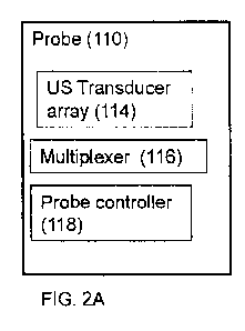
[0065] Figures 2A-2D provide schematic depictions of optional
configurations for probe
110 according to an optional embodiment of the present disclosure comprising
at least two or
more ultrasound transducers in an ultrasound transducer array 114 for
generating and reading
ultrasound signals, a multiplexer 116 for controlling the array of ultrasound
transducers 114 and
a probe controller 118 mediating control and processing of probe 110 with scan
engine 112.
[0066] Figures 2A-2B show an optional configuration of probe 110 wherein
the array of
transducers 114 and multiplexer 116 are provided in a single housing while
probe controller 118
may optionally be disposed in the same housing, as shown in Figure 2A, or in a
separate housing
as shown in Figure 2B, for example, as part of scan engine 112.
[0067] Figure 2C provides an additional depiction of probe 110 as
comprising the array
of ultrasound transducers 114 in a single housing while multiplexer 116 and
probe controller 118
are provided in a separate housing, for example, disposed as part of the scan
engine 112.
[0068] Figure 2D provides an additional optional embodiment, where probe
110 further
comprises at least one or more IR sensor 115. Optionally, IR sensor 115
provides for
determining blood saturation in order to facilitate identifying at least one
or more vessels of
interest within the probe scanning area.
11
CA 02823872 2013-07-04
WO 2012/101511 PCT/1B2012/000124
[0069] Figures 2E-2F provide schematic depictions of probe 110 according
to an optional
embodiment of the present disclosure wherein the ultrasound probe comprises a
plurality of
ultrasound transducers (n), optionally at least two or more transducers. In
particular, there may
be four, six, eight, or ten ultrasound transducers. Illustratively, there are
eight ultrasound
transducers.
[0070] Although the description herewith provides a description of a
probe that
comprises 8 transducers, it is to be understood that the number of transducers
utilized is for
illustrative purpose such that the probe of the present disclosure is not
limited to a predefined
finite number of transducers. While the described functionality may be
described with a probe
comprising eight ultrasound transducers it may equally be realized with a
fewer number of
transducers, therefore, the embodiment of the present disclosure is not
limited to 8 or 4
transducers and rather may be generalized to have a plurality of transducers
comprising at least
two or more ultrasound transducers.
[0071] An optional embodiment of the present disclosure provides for an
ultrasound
probe 110 comprising at least 2 transducers. Optionally, the transducers may
be arranged
relative to one another in a unique manner according to an optional embodiment
of the present
disclosure. Ultrasound probe 110 comprising at least 2 or more ultrasound
transducers, may be
arranged such that 6 transducers are disposed in a hexagonal configuration,
such that each of the
6 transducers is disposed about a vertex of a hexagon and at least 2 or more
transducers may be
disposed along any two chords defined between the six hexagonal vertices.
[0072] Optionally, probe 110 may comprise at least one or more of one
dimensional (1D)
ultrasound transducers, two dimensional (2D) ultrasound transducers, and/or
three dimensional
(3D) ultrasound transducers, or any combination thereof.
[0073] Optionally, probe 110 may comprise a combination of 2D and/or 3D
transducers
individually arranged such that a plurality of two dimensional (2D)
transducers may form a first
arrangement while a plurality of three dimensional (3D) transducers may
produce a second
arrangement about probe 110. For example, probe 110 may comprise 6 transducers
arranged in a
six pointed star formation including a first triangular arrangement including
three (3) 2D
transducers and a second triangular arrangement including three (3) 3D
transducers.
[0074] Optionally, probe 110 may produces at least 128 or more scan-lines
controllable
with scan engine 120.
12
CA 02823872 2013-07-04
WO 2012/101511 PCT/1B2012/000124
[0075] An optional embodiment of probe 110 wherein each transducer (n)
may be
provided with a plurality of ultrasound producing elements, for example,
including, but not
limited to, piezoelectric crystals, piezoelectric ceramic, cMUT, or the like.
For example, each
transducer may comprise from about 32 to about 256 ultrasound piezoelectric
elements.
[0076] Optionally, each of ultrasound transducer (n) utilized with probe
110 may
comprise the same or a different number of piezoelectric elements. For
example, an optional
probe 110 comprising n ultrasound transducers where n=8 (n1 -n8) may all have
about 48
ultrasound elements. For example, an optional probe 110 comprising n
ultrasound transducers
where n=8 {n1...n8} may be configured such that {nl, n3, n4, n7} may be
provided with 64
ultrasound elements, while {n2,n5,n6} may be provided with 32 ultrasound
elements, and {n8}
may be provided with 128 ultrasound elements, or the like arrangement.
[0077] Figure 3 depicts a method for determining hemodynamic and/or
cardiac
parameters with system 100 according to optional embodiments of the present
disclosure, as
shown in Figure 1A-1C. Optionally, the method for measuring, obtaining and/or
determination
of cardiac and/or hemodynamic parameters based on ultrasound scanning of a
subject's thoracic
region with system 100 starts in stage 301 with a combination of an ultrasound
and Doppler
ultrasound scan of a static scanning area about a subject's thoracic region,
utilizing probe 110 to
perform the scan.
[0078] Optionally, the static scanning area is within any region and/or
portion of the
thoracic region, for example, including, but not limited to, the front, back,
right side, left side,
thoracic spine, chest, armpit or the like portion of the thoracic region. In
particular, the static
scanning area is performed over the chest.
[0079] In an alternative embodiment, the static scanning area is such
that once a scanning
area location is determined the probe is placed over the area and the scan is
preformed over the
scan area substantially without gross movement of the probe such that the
probe is maintained in
an essentially constant or static position relative to the surface of scanning
region, for example,
the thoracic region.
[0080] Termination of the combination of ultrasound (FIG. 4A-4C) and
Doppler
ultrasound (FIG. 6A-6B) scans provided with probe 110 initiates stage 302,
where system 100
automatically and autonomously determines at least two vessel properties
associated with at least
one or more vessels detected within the underlying static scanning area. More
detail describing
13
CA 02823872 2013-07-04
WO 2012/101511 PCT/1B2012/000124
the method for determining at least two or more vessel properties is provided
in more detail in
the flowchart of Figure 4A and Figure 5A. The vessel properties, determined
for at least one or
more vessels underlying the static scanning area, comprises vessel radius and
vessel blood
velocity with time stamp.
[0081] Next, in stage 303, system 100 further processes the at least two
or more vessel
parameters to obtain and/or determine a plurality of hemodynamic and cardiac
parameters. The
further processing described in stage 303 is provided with system management
processor 102,
scan engine 112, and decision support system 120. Optionally, further
processing may be
performed with a decision support system 120 based on the at least two vessel
parameters
determined in stage 302.
[0082] In an alternative embodiment, hemodynamic and/or cardiac parameters
may, for
example, include, but are not limited to, stroke volume, stroke volume index,
heart rate, cardiac
output, cardiac index, systolic blood pressure, diastolic blood pressure, mean
arterial pressure,
cardiac power, cardiac index, stroke volume variation, total peripheral
resistance, or the like.
[0083] Optionally, hemodynamic and/or cardiac parameters may be displayed
and/or
presented to a system operator, subject, health care provider, decision
support system, auxiliary
device, communication device or the like, for determining follow up action,
for example,
including, but not limited to, further monitoring, medical intervention, drug
course treatment, or
the like in accordance with standard medical practice and procedures in
relation to the
determined hemodynamic and/or cardiac parameters.
[0084] Figure 4A shows a flowchart depicting, in more detail, stage 302 of
FIG. 3 for the
combination of ultrasound and Doppler ultrasound scans with probe 110
comprising a plurality
of ultrasound transducers and wherein each ultrasound transducers comprises an
array of
ultrasound elements, for example, piezoelectric element and/or crystals. The
various stages
depicted in Figure 4A, particularly stages 404 and 405, are schematically
illustrated in Figure
4C.
[0085] Although the foregoing description describes ultrasound probe 110
with eight
ultrasound transducers, each comprising at least 48 ultrasound elements, the
present disclosure
for a system and method for determining cardiac and hemodynamic parameters is
not limited to
such a probe, as system and method of the present application may be adapted
to work with any
14
CA 02823872 2013-07-04
WO 2012/101511 PCT/1B2012/000124
multi-transducer ultrasound probe capable of producing a plurality of
ultrasound scan-lines, for
example, including, but not limited to, phased array, linear scan.
The method starts with stage 401 where the multi-transducer ultrasound probe
is activated over
the static scanning area producing an ultrasound signal of up to about 5MHz,
and more
particularly, 2.5 MHz.
[0086] The ultrasound signal produced with probe 110 is controlled with
scan engine 112
effectively to generate, transmit, and propagate the required ultrasound
signal to the tissue
underlying probe 110 within the static scanning area. Probe 110 provides a
phased array
ultrasound signal or, optionally, a continuous ultrasound signal. Next, the
generated ultrasound
signals are reflected back to probe 110, and the reflected ultrasound signal
is detected with the
(n) ultrasound elements of probe 110, and the reflected data is optionally
processed in stage 402b
and/or stored for offline processing in stage 402a. Optionally, some data may
be processed
online essentially in real time in stage 402b while other data may be
processed offline in stage
402a. Optionally, initial processing may be provided essentially in real time
in stage 402b and
later completed offline in stage 402a.
[0087] Next, in stage 403, the initial ultrasound scan reflection data
undergoes a low
resolution mask detection, as is known and accepted in the art, to identify at
least one or more
potential vessels of interest underlying the static scanning area, for
example, including, but not
limited to, the aorta, pulmonary artery or the like vessels. If more than one
vessel of interest is
identified, then the method continues with respect to only one such vessel,
where the remaining
identified vessels will be processed in turn, optionally, sequentially or in a
hierarchical manner,
or the like graded manner.
[0088] Illustratively, low resolution mask detection is utilized in stage
403 so as to
optimize the time taken to perform the scan with a plurality of ultrasound
transducers of probe
=
110.
[0089] In the illustrative embodiment, low resolution mask detection is
performed with
each of the ultrasound transducers available with the multi-transducer probe
110. For example,
an optional probe 110 comprising 4 ultrasound transducers, would perform a
scan and low
resolution mask detection of stages 401 and 403 with each of the 4 ultrasound
transducers. For
example, an optional probe 110 comprising at least two or more ultrasound
transducers, for
CA 02823872 2013-07-04
WO 2012/101511 PCT/1B2012/000124
example, (n) ultrasound transducers would perform scan and low resolution mask
detection of
stages 401 and 403 with each of the (n) ultrasound transducers.
[0090] A first vessel of interest is identified according to optional
vessel characteristics
or criteria, for example, including, but not limited to, vessel size, vessel
diameter, fluid dynamics
through the vessel, angle formed between a transducer and vessel, or the like.
For example, the
vessel of interest may be a large vessel, such as the aorta and/or the
pulmonary artery, such that it
is identified based on a diameter of at least about 12 mm and optionally, from
about 12 mm
(millimeters) to about 42 mm (millimeters).
[0091] Next in stage 404, schematically illustrated in Figure 4B,
following low resolution
mask detection, system 100 identifies and/or otherwise determines a subset (n-
x) of the plurality
(n) of the ultrasound transducer from the multi-transducer probe that provide
for the best vessel
criteria. Optionally, stage 404 is provided autonomously and automatically
without the
presentation of ultrasound imagery to a system operator. Vessel criteria may
comprise vessel
size, for example, diameter. More particularly, a vessel having a diameter of
12 mm or more is
considered.
[0092] For example, system 100 utilizing a probe 110 comprising at least
two or more (n)
ultrasound transducers, for example, 8 ultrasound transducers, that may
identify a subset (n-x) of
the at least two or more transducers, for example, 3 or 4 transducers, that
provide data in
accordance with the vessel criteria. For example, the subset of ultrasound
transducers selected
may be selected in accordance with the vessel diameter of the identified
diameter, for example, a
diameter of at least 12 mm.
[0093] Figure 4C provides a schematic non limiting illustration of stages
404 and 405
ending when the subset of ultrasound transducers (n-x) are selected and probe
110 rescans the
static scanning area only with the determined subset of ultrasound transducers
(n-x).
Optionally, system 100 provides for saving both time and computational
resources during the
subset rescan by targeting the identified vessel of interest. Optionally, scan
engine 120
controlling probe 110 provides additional control by controlling individual
ultrasound scan lines.
[0094] More particularly, during low resolution mask detection, the
borders of the vessel
of interest are identified so as to allow scan engine 120 to determine which
of the ultrasound
transducers provided the relevant data based on the size of the detected
vessel having a diameter
of at least about 16 mm (millimeters). Optionally, with the identification of
the vessel borders,
16
CA 02823872 2013-07-04
WO 2012/101511 PCT/1B2012/000124
scan engine 120 activates and/or generates an ultrasound signal utilizing the
ultrasound scan-
lines in the vicinity of the identified subset of transducers to concentrate
the scan area about the
vessel borders.
[0095] For example, scan engine 120 initially utilizing about 128, or
more, scan-lines via
probe 110, identifies that a vessel of interest, based on the size of the
vessel having a diameter of
at least 12 mm, is located between ultrasound scan-lines. Alternatively, scan
engine 120 may
utilize 15-92 scan lines via probe 110. These 15-92 scan lines were,
therefore, identified as
including the borders of a vessel of interest. Accordingly, upon rescan with a
subset of
ultrasound transducers, scan engine 120 activates and/or utilizes ultrasound
scan-lines
surrounding these 15-92 scan-lines, for example, 7-100 ultrasound scan-lines,
so as to encompass
the vessel of interest and account for vessel movement due to blood flow,
stretch, breathing or
the like, while, optionally, simultaneously saving resources during the
subsequent scan and
analysis.
[0096] Next, optional stage 407 is performed, following mask detection of
stage 403 and,
optionally, simultaneously with stage 404, to facilitate identification of a
vessel of interest, if
probe 110 comprises at least one or more infrared (herein referred to as 'IR')
sensors.
Illustratively, at least one or more IR sensors may be disposed with probe 110
to provide for and
determine blood saturation in order to facilitate identifying at least one or
more vessels of
interest. For example, if the vessel characteristics and/or criteria utilize
by the system and
method of the present disclosure comprises blood saturation, the IR sensor may
be utilized in
conjunction with the ultrasound probe.
[0097] Next, in stage 405, schematically illustrated in Figure 4C,
following the
rescanning of the static scanning area with a subset number of ultrasound
transducers (n-x), for
example, n-x of (n) transducers, a high resolution mask detection is
undertaken to reevaluate,
select, determine and/or otherwise identify an optimal transducer (z). The
optimal transducer (z)
from the plurality of (n) ultrasound transducers of probe 110 is selected
based on parameters, for
example, including, but not limited to, angle formed with the vessel of
interest. Optionally, the
optimal transducer (z) is selected based on a transducer to vessel angle of up
to 60 degrees, and
more particularly, an angle of about 45 degrees.
[0098] For example, the optimal ultrasound transducer (z) may be selected
in accordance
with the angle formed with respect to the vessel of interest, having a
diameter of at least 12 mm,
17
CA 02823872 2013-07-04
WO 2012/101511 PCT/1B2012/000124
or more. Optionally, optimal transducer (z) may be selected based on a
transducer to vessel angle
of up to 60 degrees, and more particularly, an angle of about 45 degrees. For
example, system
100 utilizing a probe 110 comprising (n) ultrasound transducers identifies a
subset of (n-x)
transducers where the transducers form an angle of about 55 degrees, a second
transducer forms
an angle of about 25 degrees, and a third transducer forms and angle of about
40 degrees with a
vessel underlying the static scanning area.
[0099] Next, in stage 406, once an optimal transducer (z) from a plurality
of transducers
(n, n-x) is determined in stage 405, the optimal transducer is utilized for
generating a Doppler
ultrasound signal about the static scanning area, as will be depicted in more
detail in Figure 6.
Optionally, the Doppler ultrasound signal may produce an image that may be
presented to a
system operator, however, system 100 may operate autonomously without system
operator
intervention and, therefore, may operate without generating or producing an
image of the static
scanned area.
[00100] Figure 4B shows a flowchart of an optional method according to an
optional
embodiment of the present disclosure, similar to that depicted in Figure 4A,
however, the method
performed such that the optimal transducer may be identified without
performing stages 404 and
405. The method of Figure 4B, therefore, identifies optimal transducer (z)
utilizing a single scan
mask, optionally using a low resolution scan mask a medium resolution scan
mask, and a high
resolution scan mask. The method according to Figure 4B reduces the number of
scans required
to determine the optimal transducer (z) without rescanning a subset of
transducers (n-x), as shown
in Figure 4A.
[0100] Figure 5A shows a flow chart of a method according to the present
disclosure for
determining the center of a vessel of interest (v) within the static scanning
area. The vessels
identified during the ultrasound scan of the static scanning area (Figure 4)
produce an elliptical
vessel surface, however, in order to determine cardiac and/or hemodynamic
parameters
associated with the vessel of interest the elliptical vessel surface must be
converted to a tubular
surface to identify the contour of the vessel of interest (v), that will
thereafter provide for
determining the hemodynamic and/or cardiac parameters, where the center of the
vessel of
interest (vc) and radius (vr) may be determined. The various stages depicted
in Figure 5A are
schematically illustrated in Figure 5B,
18
CA 02823872 2013-07-04
WO 2012/101511 PCT/1B2012/000124
[01011 The method for converting the elliptical vessel surface sensed with
the ultrasound
probe 110 to a tubular vessel surfaces initiates with stage 501 where
ultrasound reflection signals
sensed with the ultrasound elements disposed in probe 110 corresponding to the
ultrasound
reflection from the vessel of interest within the static scanning area are
converted to a plurality of
points corresponding to the ultrasound reflections from the vessel surface.
[0102] Next in stage 502, the plurality of points corresponding to the
surface of the vessel
of interest undergo scan conversion, either low resolution mask detection as
in stage 403 or high
resolution scan conversion as described in stage 406 both of Figure 4A. During
mask detection
the points are plotted on two axes.
[0103] Next, in stage 503, all points are confined within a quadrilateral
about the two
axes, where the quadrilateral structure formed houses and/or encompasses the
surface of the
vessel of interest.
[0104] Next, in stage 504, the coordinates for the quadrilateral center
are identified by
projecting at least two diagonals from each of the quadrilateral corners,
where the center is
determined to be the point of diagonal intersection.
[0105] Next, in stage 505, the quadrilateral center (vc) is utilized to
project a plurality of
chords, optionally, a chord is projected every 0.25 degree to produce 1440
chords about the
quadrilateral center (vc) to intersect the surface of the vessel of interest
(v). Optionally, a plurality
of chords may be projected at least every 0.25 degree, or more, to produce up
to 1440 chords.
[0106] Next, in stage 506, a subset of the projected chords (from stage
505) that intersect
with the reflection coordinates corresponding to the vessel surface (from
stage 501) are selected.
Of the subset of chords, the smallest projected chord is utilized to determine
the radius of the
vessel of interest (vr). Optionally, the radius and center coordinates is
utilized to convert the
elliptical surface to a tubular surface.
[0107] Finally, in stage 507, based on the center (vc) and radius (vr),
coordinates are
utilized to tabulate data that relates the projected chords that intersect
with the vessel surface, to
creating a conversion table including the center coordinates, radius, and
chord to radius ratio.
Optionally, the table may further comprise transducer number, center scan-line
number,
beginning scan-line, end scan-line number, angle and depth. The conversion
table may be
utilized to identify the vessel of interest (v) surface utilizing polar point
conversion.
19
CA 02823872 2013-07-04
WO 2012/101511 PCT/1B2012/000124
[01081 Optionally, determination of the vessel center (vc) and vessel
radius (vr), as
depicted and described with respect to the flowchart of Figure 5A, may be
based on data obtained
from each of (n) ultrasound transducers 114 of probe 110 and based on data
obtained from at least
a subset of transducers (n-x) and based on data from at least the optimal
transducer (z).
[01091 Figure 6A is an exemplary method according to the present
disclosure depicting
the use of the Doppler ultrasound to determine the blood flow velocity of the
vessel of interest.
As previously described in Figure 4A, in stage 406 a Doppler ultrasound signal
is utilized
following a sequence of ultrasound signals and following the determination of
the center and
radius of the vessel of interest, as described in Figure 5A.
[0110] The method for determining the blood flow vector through the vessel
of interest
initiates in stage 601, where a vessel of interest underlying the static
scanning area is identified
and targeted, as described in Figure 4A-4B. Next, in stage 602, and as
described in Figure 5A,
the center and radius of the vessel of interest is identified based on
ultrasound reflection signals.
Next, in stage 603, schematically illustrated in Figure 6B, optionally, the
optimal transducer (z)
of probe 110, identified in stage 406 as described in Figure 4A, is activated
to produce a Doppler
ultrasound signal over the static scanning area, targeting at least one or
more vessel of interest.
The optimal transducer may be activated such that it targets the center of at
least one or more
vessel of interest. The center of at least one or more vessel of interest is
targeted by scan engine
120 to selectively activate a plurality of Doppler ultrasound scan-lines
encompassing the center of
a vessel of interest, as identified in stage 504 of Figure 5A. The Doppler
ultrasound scan-lines
target the center of a vessel of interest by activating a plurality of
flanking Doppler ultrasound
scan-lines. Optionally, the flanking Doppler ultrasound scan-lines (fdsl)
evenly flank the vessel
center (vc) from each side. Optionally, the number of flanking scan-lines that
are activated may
be a function of a subject's data and/or vessel properties for example
including but not limited to
diameter, pulse rate, subject age, or the like. Optionally, the number and
location of activated
flanking scan-lines (fdsl) may be a function of a chord length (c1) or arc-
length running through
the vessel center or the vessel surface, as shown in Figure 6B. Optionally,
the length of the chord
or arc (cl) associated with the center or vessel surface may be predetermined
for example from
about 0.3 mm to about 1.5 mm, and more particularly, 1 mm (one millimeter) or,
optionally, may
be a function of the radius, or the like, vessel properties or subject
parameters.
CA 02823872 2013-07-04
WO 2012/101511 PCT/1B2012/000124
101111 For example a vessel having a diameter of 17 mm may produce 9
flanking
Doppler scan lines comprising one Doppler scan line (dsl) going through the
center and 4
flanking Doppler scan lines (fdsl) on either side of the center, such that
scan lines are separated
by a chord length (c1) of 1 mm.
[0112] For example, at least 2 or more, illustratively 7, Doppler
ultrasound flanking scan-
lines (fdsl), may be utilized to flank a vessel center (vc) where, for
example, 3 flanking Doppler
ultrasound scan-lines (fdsl) are activated on each side of the Doppler
ultrasound scan-line that
corresponds to the vessel center (vc). Such a Doppler scanning scheme provides
for saving
resources in particular time as ultrasound transducers and elements are
selectively activated, and
furthermore provide for real time computational constraints. Flanking the
vessel center further
provides for ensuring that vessel movement due to breathing, stretching and
the like motion are
accounted for while encompassing the center of the vessel blood flow.
Optionally, each flanking
scan-line (fdsl) may be generated at a set interval from one another such that
a first one is fired at
t=0 and the next fdsl is generated at a set time interval t=x. Optionally, the
inter flanking scan
line time interval may, for example, be from about 2ms (millisecond) up to
about 15 ms
(millisecond), and more particularly, about 2.5 ms (millisecond), or any time
interval delay for
example in a resolution of about 0.1 ms (millisecond) from about 2 ms up to
about 15 ms
(millisecond). Optionally, the inter flanking scan lines time may be
controllable and determined
based on vessel parameters, user parameters, physician parameters, or the
like.
[0113] Optionally, the Doppler ultrasound signal generated during stage
603 may be used
to validated and confirm the vessel parameters determined with ultrasound scan
to reconfirm the
radius determined with ultrasound scans, as determined in stage 506 of Figure
5A.
[0114] Optionally, in stage 604 the vessels of interest (v) may be
categorized and/or
identified based on fluid dynamics of the blood flowing through the vessel.
Illustratively, the
vessel of interest may also be identified based on IR saturation data
optionally provided in stage
407 of Figures 4A-4B. Optionally, the vessel of interest (v) may be identified
based on at least
one or both of on fluid dynamics of the blood flowing through the vessel
and/or IR saturation
data. For example, identification of fluid dynamics corresponding to laminar
blood flow is
indicative of the pulmonary artery. For example, identification of fluid
dynamics corresponding
to turbulent blood flow is indicative of the aorta. Fluid dynamics
corresponding to blood flow
velocity is utilized to identify the vessel type. Optionally, the
determination of vessel type is
21
CA 02823872 2013-07-04
WO 2012/101511 PCT/1B2012/000124
based on a threshold blood flow velocity. Optionally, the threshold blood flow
velocity may be
about 60 cm per second (60 cm/sec). For example blood flow below a threshold
velocity of about
60 cm/sec identifies the vessel as the pulmonary artery (PA). For example,
blood flow above a
threshold velocity of about 60 cm/sec identifies the vessel as the aorta (AO).
[0115] Next, in stage 605, the angle formed between the vessels of
interest and the
ultrasound transducer, specifically the optimal transducer (z), is determined.
In one embodiment
of the present disclosure, one vessel to be monitored is selected from the
vessels of interest based
on the angle formed with the ultrasound transducer. Optionally, the angle
formed is from about
20 degrees to about 60 degrees, and more particularly the selected vessel is
selected based on the
vessel that forms an angle closest to 45 degrees.
[0116] Next, in stage 606, the Doppler ultrasound about the individual
flanking Doppler
ultrasound scan lines (fdsl) are activated and monitors the vessel to be
monitored (v), determined
in stage 605, for a given period corresponding for example up to about 6 s
(six seconds) to allot
for measuring a Doppler echocardiogram at least 4 cardiac cycles. Such
monitoring provides for
determining blood flow velocity through the vessel (v).
[0117] Optionally, radius parameters obtained from individual Doppler scan
lines (dsl),
comprising a plurality of flanking scan lines (fdsl), are cross referenced
against the radius
obtained from the ultrasound scan of stage 506. Optionally, parameters
obtained and/or
otherwise determined based on individual Doppler scan lines (dsl), including a
plurality of
flanking Doppler scan lines (fdsl), Figure 6B, may be used to cross reference
and validate vessel
parameters obtained from individual Doppler scan lines (dsl). Optionally, a
Doppler scan lines
(dsl) validation table may for example be determined based on triangulation
calculation relating
to parameters for example including but not limited to dsl length, dsl-angle,
vessel radius, dsl-
angle, cl-chord length distance, or the like. Optionally, the Doppler scan
line validation table
provides for validating vessel radius.
[0118] Next, in stage 607, the two vessel properties including vessel
radius and blood
flow speed each with a time stamp are tabulated and stored in system 100.
Next, in stage 608, the
three vessel properties identified undergo further processing with the system
management
processor 102 to determining a plurality of cardiac and/or hemodynamic
parameters.
[0119] Figure 7 is a flowchart of an exemplary method according to an
optional
embodiment of the present disclosure for determining hemodynamic and cardiac
parameters
22
CA 02823872 2013-07-04
WO 2012/101511 PCT/1B2012/000124
automatically based on Doppler ultrasound parameters, defining an optional
method for further
processing of the Doppler ultrasound parameters and vessel parameters
identified and/or
measured within the static scanning area, as described and depicted herein
above. The method
initiates in stage 700 wherein the Velocity time curve data is tabulate,
and/or optionally presented
in the form of a plotted graph (800 Figure 8), in the form Doppler blood
velocity (s=meter per
second) vs. time curve (t=seconds), as schematically shown in Figure 8. Stage
700 is optionally
performed based on the data measured, tabulated and/or otherwise provided in
stage 606 and 607,
depicted and described in Figure 6A. Optionally, the Velocity time curve data
presented
corresponds to at least one or more cardiac cycles. Optionally, the Velocity-
Time curve data
presented corresponds to at least four or more cardiac cycles, and
alternatively, at least three or
more consecutive cardiac cycles. =
[0120] Optionally, the method in any one or more of its stages may be
performed
manually and/or automatically and/or semi-automatically or in any combination
thereof.
Optionally, the method described may be performed by a user, for example,
including, but not
limited to ,the subject, a healthcare giver, physician, technician or the like
trained individual. The
scan may be performed automatically and autonomously without user intervention
with system
100 and with system management processor 102. Optionally, the Manual and/or
automatic
performance of the method may be provided with a remote system for example
including but not
limited to decision support system 120. Medical decision support system 120
may for example
be provided in the form of a back office and/or call center, and/or health
care provider, and/or
ambulatory care, and/or telemedicine center, and/or a medical decision support
system, and/or a
caregiver decision support system.
[0121] Next, in stage 702, processing of the Velocity-Time curve data is
initiated by
identifying and/or determining at least one or more of the individual cardiac
cycles measured in
stage 606, from at least three or more consecutive cardiac cycles. An
exemplary Velocity-Time
curve 800 is schematically illustrated in Figure 8, showing a cardiac cycle
from point 804a to
804d, optionally defining the diastolic points about the Velocity-Time curve
800.
[0122] Next, in stage 704, the Velocity-Time curve data corresponding to
the individual
cardiac cycles identified in stage 702 is segmented into the plurality of
cardiac cycle segments
802, which are identified within each of the available cardiac cycles, as
shown in Figure 8.
Optionally, at least one or more cardiac cycle segments 802a, b may be
identified. Illustratively,
23
CA 02823872 2013-07-04
WO 2012/101511 PCT/1B2012/000124
at least two cardiac cycle segments are identified within each available
cardiac cycle data, for
example, cardiac cycle segments 802a corresponding to pulse pressure and 802b
corresponding
to pressure drop, as shown in Figure 8.
[0123] Optionally, the cardiac cycle segmentation may be performed
manually by a user,
for example, including, but not limited to, the subject, a physician, a
trained technician, or the
like. Optionally, the number of cardiac cycle segments identified, and/or the
cardiac cycle
segmentation resolution may be controlled based on parameters, for example,
system 100
dependent parameters, parameters independent of system 100, parameters
measured with system
100, external parameters, and/or controllable parameters, or any combination
thereof.
[0124] Optionally, the segmentation is performed based on known cardiac
cycle
landmarks, for example, including but not limited to diastole, systole,
isovolumic contraction,
isovolumic relaxation, A-V valve closure, aortic valve opening, ejection, A-V
valve opening, or
the like.
[0125] In an alternative embodiment, cardiac cycle segmentation and
cardiac landmark
identification may be performed automatically by mathematical manipulation of
the Velocity-
Time curve to identify extrema about the curve and/or within the tabulated
data. Velocity-Time
curve extrema may, for example, include, but is not limited to, identifying
maximum, minimum,
local maximum, local minimum, absolute maximum, absolute minimum, inflection
points,
gradient, slope, supremum, infimum, or the like. An example of such extrema
points 804a-d is
schematically illustrated in Figure 8. For example, point 804a schematically
illustrates the point
corresponding to the start of a cardiac cycle and associated with the diastole
while point 804d
corresponds to the end of a cardiac cycle also associated with the diastole;
point 804b optionally
corresponds to the maximum velocity and the end of the pulse pressure 802a,
during the cardiac
cycle, point 804c corresponds to the inflection point during pressure drop
segment 802b.
[0126] Next, in stage 706, individual cardiac cycle segments are further
subdivided into a
plurality of (i) sub-segments, as schematically illustrated in sub-segments
806 in Figure 8.
Optionally, the resolution and/or the number of sub-segments may be constant,
time based and/or
based on the resolution of the Velocity-Time curve, user defined or the like.
[0127] Next, following segmentation and sub-segmentation during stage
708, a plurality
of vessel associated parameters are determined and/or otherwise calculated
that are optionally
based on vessel parameters, Doppler blood flow parameters measured and/or
otherwise
24
CA 02823872 2013-07-04
WO 2012/101511 PCT/1B2012/000124
determined with system 100 of the present disclosur,e as described
hereinabove. Optionally, the
parameters determined may, for example, include but are not limited to, area,
area under the
Velocity-Time curve, area of cardiac cycle segment, area of cardiac cycle sub-
segment, vessel
volume, blood flow mass, blood flow acceleration, force and pressure, as
depicted in Figure 9
below.
[0128] Next, in stage 710, hemodynamic and cardiac parameters are
determined based on
parameters determined and/or otherwise identified in stage 708 that may, for
example, include but
are not limited to, stroke volume, stroke volume index, heart rate, cardiac
output, cardiac index,
systolic blood pressure, diastolic blood pressure, mean arterial pressure,
cardiac power, cardiac
index, stroke volume variation, total peripheral resistance, or the like.
[0129] EXAMPLE 1: Determination of Diastolic Pressure in Aorta
[0130] The foregoing example is provided for illustrative purpose only and
does not limit
the present application to the description or calculation as described as set
forth.
[0131] Diastolic pressure of the aorta, for example, may be determined in
a non-invasive
manner based on a combination of ultrasound and Doppler scan of a static area
about a subject's
upper torso, for example, a static scanning area about the subject's chest, as
described and
depicted herein above in Figures 1-8. Optionally, the determination below is
performed for
individual cardiac cycles measured with the Doppler ultrasound about the
static scanning area
targeting at least one vessel of interes, for example, the aorta or pulmonary
artery.
[0132] In stage 900, the vessel's cross-section area is determined based
on the vessel
radius as determined according to the method described hereinabove and based
on Equation 1
below, for each sub-segment 806 (i).
2
A = Tor
EQ1.
[0133] Next, in stage 902, the area of at least one, and more
particularly, a plurality of
sub-segments (i), for example, 806 of Figure 8, is determined from the
Velocity-Time curve 800
of Figure 8, or from the corresponding tabulated data, according Equation 2
below. The sub-
segment area of each sub-segment 806 is determined for the full curve 800;
therein accounting for
the full time range of is Doppler scan measurement.
CA 02823872 2013-07-04
WO 2012/101511 PCT/1B2012/000124
(S i+S i_i)(t i-t i_1)
A segment(i) ¨
EQ2. 2
[0134] Next, in stage 904, the blood volume is determined per sub-segment
(i) 806
according to Equation 3.
EQ3. Voi(i)¨ Asegment(i) = A i
[0135] Next, in stage 906, the blood mass per sub-segment (i) 806 is
determined based the
volume determined in Equation 3 and a constant density (p).
in = Voi(i) = p
EQ4. =
[0136] Next, the stage 908, blood acceleration per sub-segment (i) 806 is
determined
based on curve 800 as determined in Equation 5.
(S. - S .
,---1
a = =
(t . ¨t .
EQ5.
[0137] Next, in stage 910, the force per sub-segment (i) 806 is
determined based on
Equation 6.
F = m. = a
EQ6. 1
[0138] Next, in stage 912, the pressure per sub-segment (i) 806
determined based on
Equation 7.
P=
Ai
EQ7.
26
CA 02823872 2013-07-04
WO 2012/101511 PCT/1B2012/000124
[0139] Next, in stage 914, segment pressures (802a, b) are determined for
each segments,
respectively accounting for pulse pressure (Ppulse) segment 802a and the
pressure of pressure drop
(Pdrop) segment 802b, and the total pressure (Psystole) experienced during the
cardiac cycle as
defined from points 804a to 804d. The total pressure and the segment pressure
is determined by
summing the relevant and individual sub-segments 806, for example, as shown by
Equation 8.
Optionally, the total pressure is equivalent to the systolic pressure
(Psystole) of individual cardiac
cycles measured with the Doppler ultrasound scan of the static scanning
according to optional
embodiment of the present disclosure. Optionally, the total pressure may be
determined by
summing pulse pressure (Ppulse) and pressure drop (-Pdrop)
[0140] Next, in stage 916, the segment pressures determined in stages 914
are utilized to
determined the diastolic pressure experienced in the measured vessel of
interest (v), according to
Equation 9 below, of the measured vessel.
systole - p _ pulse '3
diastole
EQ9.
[0141] Now referring to Figures 10-12, describing an optional embodiment
of the present
disclosure provided for determining hemodynamic and cardiac parameters based
on a static
scanning using a combination of ultrasound and Doppler scans.
Figure 10 shows a flow chart describing an optional method according to the
present disclosure
provides for identifying a vessel of interest and obtaining cardiac parameters
associated with the
vessel for further processing to determining cardiac and hemodynamic
parameters.
[0142] First in stage 1001 a plurality of scanned objects are
automatically and
autonomously identified within the static scanning area by the initial
ultrasound scan for example
as previously described in details with respect to FIG. 4.
Next in stage 1002 mask detection and filtering is performed to identify a
first of potential set of
vessel of interest from within the plurality of scanned objects. Optionally
and preferably the
filtering process may for example include at least one and more preferably
plurality of filters
selected from the group including but not limited to rectangular mask
filtering, edge detection,
boundary estimation, object shape and/or size threshold, or the like alone or
any combination
thereof. Optionally edge detection may for example be provided in the form of
Sobel edge
27
CA 02823872 2013-07-04
WO 2012/101511 PCT/1B2012/000124
detection. Optionally Boundary estimate may be provided with polygon filling.
Optionally
object size threshold may for example include removing objects that are too
small based on size
estimation, shape estimation for example remove is not ellipse or circular, or
any combination
thereof.
[0143] Next in stage 1003 the filtered vessel object, most preferably in
the form of ellipse,
ellipsoid, or circle or the like rounded closed formation, identified in stage
1002 undergo a
transformation, for example including but not limited to Random Hough
Transform, to further
identify vessels of interest for example including but not limited to the
aorta and pulmonary
artery. One transformation acceptable for use is the Random Hough Transform
(herein referred
to 'RUT') to identifying parameters associated with the vessel objects
identified. In one
embodiment, the RUT is preformed for an ellipse, as it is assumed that any
vessel of interest
identified in stage 1002 will have an elliptical profile. Utilization of RI-IT
provides for direct
and/or indirect identifying and/or inferring a plurality of parameters
associated with the vessel for
example including but not limited to ellipse center, major axis, and minor
axis and scan angle.
For example as illustrated in Figures 12A-B, showing the axis as well as the
ellipse boundary
points.
[0144] Next in stage 1004 the RUT parameters are utilized to filter for a
subset of vessels
for further scanning to identify the vessel of interests selected from the
aorta (AO) and/or
pulmonary artery (PA). The filtering criteria may comprise a scan angle from
about 20 degrees to
60 degrees and a minor axis of at least 16 mm and up to 40mm (millimeters).
Optionally the
diameter of the vessel of interest is correlated with and/or otherwise
associated as a function of
the minor axis, determined from the RUT processing. Optionally the diameter of
the vessel of
interest may be correlated with and/or otherwise associated as a function of
the major axis,
determined from the RUT processing. Optionally the diameter of the vessel of
interest is
correlated with and/or otherwise associated as a function of both the major
and minor axes,
determined from the RUT processing.
[0145] Next in stage 1005 a further filtering process is performed to
identify at least one
vessel of interest selected form the aorta (AO) or pulmonary artery (PA) by
utilizing a Doppler
scan, to determine the blood velocity flowing through the vessel of interest.
At least one or more
scan line(s) is directed to the vessel center identified by the RHT in stage
1003 for the vessels
subset identified and filtered for in stage 1004. The Doppler scan provides
for determining the
28
CA 02823872 2013-07-04
WO 2012/101511 PCT/1B2012/000124
blood flow velocity through the vessel of interest, most preferably about its
center. Optionally
the Doppler scan is performed from about 1 second to about 3 seconds, and most
preferably for
1.5 seconds per each vessel of interest identified. Optionally , the Doppler
scan is performed for
at least one full cardiac cycle, preferably mapping the blood velocity
fluctuation during the
cardiac cycle. Optionally and preferably the Doppler scan provides to scan and
measure the
maximum blood flow velocity through the vessels center occurring during at
least one cardiac
cycle.
[0146] Finally in stage 1006 at least one or more vessel of interest is
indentified by
selecting the vessel relative to at least one threshold associated with the
vessel(s) of interest, for
example including but not limited to vessel blood flow velocity, and/or vessel
diameter, the like
or any combination thereof. Optionally the threshold values may be determined
based on user
parameters and/or subject parameters, and/or healthcare giver and/or system
parameters.
Optionally threshold values may be determined based on at least one or more
parameter(s) for
example including but not limited to state of health, medical history, age,
medical conditions,
cardiac history, anatomical parameters, atherosclerosis parameters, any
combination thereof or
the like.
[0147] Optionally the blood flow velocity threshold may be about 0.6m/sec
and may be
indicative of the type of vessel being scanned. Optionally monitoring of the
aorta is selected
based on a blood flow velocity threshold that is above an optional threshold
of 0.6m/sec or the
pulmonary artery is selected as having a blood flow velocity below the
threshold of 0.6m/sec..
Optionally the vessel diameter threshold may for example be from about 16 mm
to about 40 mm.
[0148] Figure 11 depicts an optional embodiment of the present disclosure
of a method
for continuously monitoring vessel parameters of a vessel of interest
identified and selected
according to an optional embodiment of the present disclosure, for example the
method described
in Figure 10. The method initiates in stage 1100 by identifying vessel
properties as determined
by the RI-IT transform as previously described in Figure 10 in stage 1003 and
as schematically
illustrated in Figure 12A.
[0149] Next in stage 1101 at least one or more vessel of interest, for
example as identified
in stage 1006 of Figure 10, are processed by analyzing individual vessel
assuming the vessel is
represented by an ellipse, for example as illustrated in Figures 12A-B. Vessel
boundary 1200b,
data points obtained from the RHT processing, for example including but not
limited to top,
29
CA 02823872 2013-07-04
WO 2012/101511 PCT/1B2012/000124
bottom, left, right boundary coordinates are identified and processed in turn.
An ultrasound scan
line is targeted at each coordinate 1200b, in turn, where each is flanked by a
plurality of flanking
scan, for example 7 ultrasound scan lines (us!) are directed at each boundary
1200b, Figure 12B,
coordinate comprising one scan line targeting the coordinate calculated with
the previous scan,
along with about three flanking scan lines flanking each side of the central
scan lines, such that
about seven ultrasound scan lines are centered about each boundary point
1200b. The flanking
scan lines provide for identifying movement, expansion, deformation and
changes of the vessel
coordinates, over time and during stages of the cardiac cycle. The vessel
ultrasound data, at the
boundary 1200b, is determined every 10 milliseconds (10 ms) for about 6
seconds providing data
for at least three or more consecutive cardiac cycles.
[0150] Next in stage 1102 the RHT identified vessel center 1200c, as shown
in Figure
12A-B, is similarly tracked with a Doppler scan line (dsl). In one embodiment,
the vessel
ultrasound data is determined every 10ms for about 6 seconds providing data
for identifying at
least three or more consecutive cardiac cycles.
[0151] Next in stage 1103 data relating to vessel dimension via ultrasound
boundary
1200b scanning and blood flow velocity via Doppler scanning of vessel
center1200c is tabulated
with a time stamp, providing data every 10ms. In one embodiment, in stage 1104
the tabulated
data is then processed to further identify hemodynamic and cardiac parameters,
for example
including but not limited to diastolic pressure, systolic pressure, pulse
pressure or the like.
Optionally further processing is provided as taught by optional embodiments of
the present
disclosure shown in Figures 7-9 and 13-15.
[0152] Next in stages 1105 and 1106 ultrasound and Doppler data provided
in earlier
stages 1101 and 1102 is then processed further to track the vessel of interest
to account for vessel
movement, deformation or the like. Specifically, in stage 1105 at least one or
more filters are
applied to the ultrasound data and Doppler data, for example as described in
Figure 10, most
preferably to include noise detection, masking, and edge detection for example
to provide an up
to date an accurate image and localization of the vessel of interest within
the static scan area.
[0153] In stage 1106 the filtered data is then further processed utilizing
the RHT, for
example to redefine the location of boundary 1200b and center 1200c
illustrated in Figure 12A-B,
to reanalyze and identify the vessel parameters for example including but not
limited to vessel
center, vessel diameter via ellipse minor axis, boundaries via major axis, and
scan angle. The
CA 02823872 2013-07-04
WO 2012/101511 PCT/1B2012/000124
updated vessel RI-IT parameters are then preferably utilized in the following
vessel scan, most
preferably 10ms following the initial scan, therein updating the RI-IT Data of
stage 1300 most
preferably about every 10ms.
[0154] Now referring to Figures 13 and 14, describing an optional
embodiment of the
present disclosure provides a method for non-invasively determining cardiac
parameters and in
particular to a method for noninvasive determination of at least one and more
preferably a
plurality of cardiac cycle parameters for example including but not limited to
Systolic Pressure
(herein referred to as `Psys'), Diastolic Pressure (herein referred to as
`Pdia'), and Pulse Pressure
(herein referred to as `Ppulse' and/or `PP').
[0155] Figure 13 shows an optional embodiment according to the present
disclosure for
determining cardiac and/or hemodynamic parameters based on measured data
defining a blood
flow velocity and vessel radius, for example as described in Figures 4-6
and/or 10-12. The
method according to an optional embodiment of the present disclosure for
further processing of
vessel radius, and blood flow velocity, comprises, first stage 1301 providing
a plurality of
measurements for at least 3 or more consecutive cardiac cycles where
optionally and preferably
the measurements are calculated every 10 milliseconds (10ms) for about 6
seconds, and tabulate
results comprising vessel radius, blood flow velocity through vessel, for
example as previously
described in Figures 4-6 and/or Figures 10-12.
[0156] Stage 1302 the vessel radius is graphed as a function of time, for
example as
schematically illustrated in Figure 14. In one embodiment, the maximum/peak
radius is
correlated with and/or otherwise a function of systolic radius, Rsys, and the
diastolic radius, Rdia,
is correlated with and/or otherwise a function of the minimum radius.
Stage 1303 a line, 1400, is extrapolated from the maximum radius, Rsys as
shown in Figure 14,
through the minimum radius, Rdia, until line 1400 intersect the time line
defining the point of
intersection with the time line, provided to estimate the virtual radius Rp7
as showing in Figure
=
14, Rp7 the vessel radius corresponds to the an ideal blood vessel radius
(Rp7) where at pressure
is at a minimum and equivalent to about 2mmHg to about lOmmHg and most
preferably 7mmHg.
[0157] Next in stage 1304 utilizing the extrapolated line 1400 determine a
first triangle
(Rsys, Rdia, 1402) correlating the pulse pressure defined wherein the
hypotenuse corresponds to
the slope between the systolic (Rsys) and diastolic radius (Rdia); and define
a second triangle
31
CA 02823872 2013-07-04
WO 2012/101511 PCT/1B2012/000124
(Rsys, Rp7,1404) defined wherein the hypotenuse corresponds to the slope
between the systolic
radius (Rsys) to the ideal radius at lowest pressure( Rp7).
[0158] Stage 1305 determine base of both triangles where tpp based on
baseline of first
triangle and tsys related to second triangle.
[0159] Next in stage 1306 pulse pressure (Ppulse) is determined, for
example as described
in Figure 8, segment 802a, by optionally determining the energy of and/or the
area under the
curve of the blood velocity vs. time graph as previously described.
[0160] Next in stage1307 evaluate ratio correlating the pulse pressure and
systolic
pressure with the pulse pressure time and systolic time as calculated above,
stages 1305 and 1306,
to determine the Systolic Pressure and give by the equation 10 (EQ10.) below:
Ppulse¨tpp
Psys tsys
[0161] Most preferably tsys and tpp are provided from the first and second
triangle, stage
1305, while Pressure Pulse, stage 1306, to determine:
tsys
Psys = Ppulse[¨tpp]
[0162] Finally in stage 1308 the Pdia is calculated from the Psys, and
Ppulse data, based
on the equation Pdia=Psys-Ppulse.
[0163] Now referring to Figure 15, describes an optional embodiment of the
present
disclosure provides a method for non-invasively determining cardiac parameters
and in particular
to a method for noninvasive determination of at least one and more preferably
a plurality of
=
cardiac cycle parameters for example including but not limited to Systolic
Pressure (herein
referred to as `Psys'), Diastolic Pressure (herein referred to as `Pdia'), and
Pulse Pressure (herein
referred to as `Ppulse' and/or `PP'). In one embodiment, Psys, Pdia, Ppulse
are determined by
manipulating a plurality of equations for example including but not limited at
least one or more
and/or a combination of the Moens-Korteweg equation, Euler's equation and
generalized
Hooke's law of Elasticity, coupled with a plurality of measurements relating
vessel radius, vessel
blood flow velocity and time stamp, as provided by exemplary embodiment of the
present
disclosure as described in Figures 4-6 and/or Figures 10-12. Optionally the
vessel radius, blood
flow velocity and time stamp, may be provided with an ultrasound and Doppler
scanning device
according to an exemplary embodiment of the present disclosure as described in
Figures 1-3
32
CA 02823872 2013-07-04
WO 2012/101511 PCT/1B2012/000124
according to the present disclosure. Optionally the vessel radius, and blood
flow speed with time
stamp may be provided by an external and/or auxiliary device.
[0164] Figure 15 shows an optional embodiment according to the present
disclosure for
determining cardiac and/or hemodynamic parameters based on measured data
defining a blood
flow velocity and vessel radius, for example as described in Figures 4-6
and/or 10-12. The
method according to the present disclosure comprises first stage 1501
providing a plurality of
measurements for least three or more consecutive cardiac cycles where
optionally the
measurements are calculated every 10 milliseconds for about 6 seconds, and
tabulate results
comprising time stamp, vessel radius, blood flow velocity through vessel, for
example as
previously described in Figures 4-6 and/or Figures 10-12.
[0165] Next in stage 1502, the tabulated data is graphed to show the
evolution of vessel
radius over time and optionally the blood flow velocity over time.
[0166] Next in stage 1503, the data provided in stage 1501 is used to
evaluate a plurality
of vessel associated equations for example including but not limited to Moens-
Korteweg equation
(M) , Euler's equation (E) and generalized Hooke's law of Elasticity (H). Most
preferably a
combined proprietary equation as provided below:
p (ui u1_1)2 = 142ir
p ui_1 = (ui ¨ uu_1)
_
1) = (Ri ¨ Ri_i)
At ___________________________________ P=ui-i. (1 ( (U-)1(RtRvr\
= ke (R 1 i-Ri=Ri_i)
Rir
)
[0167] Next in stage 1504, the above equation is evaluated using the
measured data, for
example as provided by the device, system and method of optional embodiments
of the present
disclosure, as described in Figures 1-12, utilizing radius (Rd, Rs, Ri), blood
flow velocity (Ui,
Ud, Us ) most preferably provided at about every 10millisecond (10 ms) for at
least 6 seconds to
account for at least three or more consecutive cardiac cycles, and then
utilize the resultant
plurality of equations to estimate k, A, Rvir using the least squares method.
Next in stage 1505 the estimated parameters including k, Rvir are utilized to
evaluate and/or
calculate Psys, Pdia, Ppulse according to the equation below determined in
stage 1501 and based
on a plurality of vessel associated equations.
[Rd ¨ Rvirl
Pdia = Ppulse XL Rs ¨ Rd .1
33
CA 02823872 2013-07-04
WO 2012/101511 PCT/1B2012/000124
[[R5Rvi
p = prl
sys pulse X Rs ¨ Rd
p = (us ¨ ud)2 = Rv2ir
Ppulse =(R ¨ Ri2_1) p Ud = (Us ¨ Ud)
f
[0168] While the embodiments of the present disclosure have been described
with respect
to a limited number of embodiments, it will be appreciated that many
variations, modifications
and other applications of the embodiments may be made.
34