Note: Descriptions are shown in the official language in which they were submitted.
A PARYLENE-BASED MICROELECTRODE ARRAY IMPLANT
FOR SPINAL CORD STIMULATION
CROSS REFERENCE TO RELATED APPLICATION(S)
This application claims the benefit of U.S. Provisional Application
No. 61/435,188, filed January 21, 2011.
STATEMENT REGARDING FEDERALLY SPONSORED RESEARCH OR
DEVELOPMENT
This invention was made with Government support under Grant
No. W81XWH-09-2-0024, awarded by the United States Army, Medical
Research and Materiel Command; and Grant No. EB007615, awarded by the
National Institutes of Health. The Government has certain rights in this
invention.
BACKGROUND OF THE INVENTION
Field of the Invention
The present invention is directed generally to implantable
electrode arrays, and more particularly to implantable electrode arrays used
to
deliver electrical stimulation to the spinal cord.
Description of the Related Art
Spinal cord injuries are estimated to afflict over 1.3 million
individuals in the United States alone, and paralysis is estimated to affect
over
5 million individuals. See "One Degree of Separation: Paralysis and Spinal
Cord Injury in the United States," Christopher and Dana Reeve Foundation
(2009). The debilitating nature of paralysis has a profound effect on quality
of
life, making even partially effective treatments highly desirable goals for
the
scientific community.
Fortunately, experimental research on animals has shown that
some level of recovery of locomotion is possible. In particular, epidural
spinal
1
CA 2824782 2018-05-28
CA 02824782 2013-07-12
WO 2012/100260
PCT/US2012/022257
cord stimulation has been shown to induce stepping in rats. See R. M.
lchiyama, G. Courtine, Y. P. Gerasimenko, G. J. Yang, R. Brand, I. Lavrov, H.
Zhong, R. Roy, V. R. Edgerton, "Step Training Reinforces Specific Spinal
Locomotor Circuitry in Adult Spinal Rats", J. Neuroscience, vol. 29, pp. 7370
¨
7375 (2008); and R.M. lchiyama, Y.P. Gerasimenko. H. Zhong, R.R. Roy, V.R.
Edgerton, "Hindlimb stepping movements in complete spinal rats induced by
epidural spinal cord stimulation," Neuroscience Letters, vol. 383, issue 3,
pp.
339-344 (2005). In these studies, rats were implanted with up to eight wire
electrodes. The implanted wire electrodes each extended from a headplug
down the neck and to the spinal cord of the rat. During testing, each of the
rats
was suspended in a jacket such that its hind limbs were positioned on a
treadmill. About two weeks after the spinal cord injury, clear stepping
patterns
were evident when the spinal cord was stimulated. This suggested that the
electrical stimulation activated a central pattern generator in the spinal
cord.
The following publications provide examples of work related to
electrode arrays used to apply electrical stimulation to the spinal cord: D.C.
Rodger, W. Li, A.J. Fong, H. Amen, E. Meng, J.W. Burdick, R.R. Roy, V.
Reggie Edgerton, J.D. Weiland, M.S. Humayun, Y.C. Tai, "Flexible
microfabricated parylene multielectrode arrays for retinal stimulation and
spinal
cord field modulation," Proc. 4th International IEEE-EMBS Special Topic
Conference on Microtechnologies in Medicine and Biology, Okinawa, Japan,
pp. 31-34 (2006); K. W. Meacham, R. J. Giuly, L. Guo, S. Hochman, S. P.
DeWeerth, "A lithographically-patterned, elastic multi-electrode array for
surface
stimulation of the spinal cord", Biomedical Microdevices, vol. 10, no. 2, pp
259-
269 (2008); and D. C. Rodger, Wen Li, H. Amen, A. Ray, J.D. Weiland, M. S.
Humayun, Y.C. Tai, "Flexible Parylene-based Microelectrode Technology for
Intraocular Retinal Prostheses," Proc. IEEE-NEMS 2006, pp 743-746 (2006).
The publications cited above and other work has led to various
designs for high-density electrode arrays to further research, but
unfortunately
none of these designs has been successfully implanted chronically. A need
exists for a chronic implant because chronic implantation is necessary for
many
applications, such as conducting research, helping a patient move (e.g., step,
stand, grip, and the like), improving control of voluntary functions (e.g.,
voiding
2
CA 02824782 2013-07-12
WO 2012/100260
PCT/US2012/022257
the bladder), improving functionality of autonomic processes (e.g.,
temperature
control), and the like. A need also exists for an electrode array assembly
configured to more accurately deliver electrical signals to selected locations
along the spinal cord. The present application provides these and other
advantages as will be apparent from the following detailed description and
accompanying figures.
SUMMARY OF THE INVENTION
Embodiments of the invention include an implantable device
configured to apply electrical stimulation to a spinal cord of a subject
(e.g., a
human being or other mammal, such as a rat). The device includes a body
portion and a first layer. The body portion has a peripheral portion.
Optionally,
the peripheral portion includes a frame positioned adjacent the first layer.
The
frame may be constructed from one or more layers of a substantially
electrically
nonconductive material (e.g., parylene-A, parylene-C, parylene-AM, parylene-F,
parylene-N, parylene-D, and the like). The first layer is constructed from a
substantially electrically nonconductive material. In some embodiments, the
first layer is constructed from at least one of parylene-A, parylene-C,
parylene-
AM. parylene-F, parylene-N, and parylene-D. The first layer has a first
portion
and a second portion. The first portion is positionable alongside the spinal
cord
and includes a first plurality of openings. For example, the first portion of
the
first layer may be positioned against a dura of the spinal cord and the device
configured to provide electrical stimulation to the dura. The second portion
includes a second plurality of openings.
A plurality of electrodes is positioned inside the peripheral portion
and alongside the first portion of the first layer. At least one of the first
plurality
of openings is adjacent each of the electrodes to provide a pathway through
which the electrode may provide electrical stimulation to the spinal cord when
the first portion is positioned alongside the spinal cord. In some
embodiments,
more than one of the first plurality of openings is adjacent each of the
plurality
of electrodes. In embodiments in which the first portion of the first layer is
to be
positioned against the dura of the spinal cord, the plurality of electrodes is
configured to provide electrical stimulation to the dura.
3
CA 02824782 2013-07-12
WO 2012/100260
PCT/US2012/022257
A plurality of traces is positioned inside the peripheral portion and
alongside the first layer with at least one of the second plurality of
openings
being adjacent each of the traces to provide a pathway through which the trace
may receive electrical stimulation. One or more of the traces is/are connected
to each of the electrodes and configured to conduct electrical stimulation
received by the one or more of the traces to the electrode. In some
embodiments, two of the traces are connected to each of the electrodes. In
particular embodiments, the plurality of traces are configured to conduct
different electrical stimulation to different ones of the plurality of
electrodes.
Further, the plurality of traces may be configured to conduct electrical
stimulation to fewer than all of the plurality of electrodes.
In some embodiments, the first layer includes a plurality of grid
structures with a different one of the grid structures adjacent each of the
plurality of electrodes. Each grid structure defines a plurality of cells. For
each
of the plurality of electrodes, each of the at least one of the first
plurality of
openings adjacent the electrode is positioned inside a different one of the
cells
of the grid structure adjacent the electrode.
In some embodiments, the body portion includes a second layer.
In such embodiments, the plurality of electrodes and the plurality of traces
may
be positioned between the first and second layers. The first and second layers
may each be constructed from at least one of parylene-A, parylene-C, parylene-
AM, parylene-F, parylene-N, and parylene-D. Optionally, a flexible outer
coating may coat at least a portion of the second layer of the body portion
and a
portion of the first layer between the first portion of the first layer and
the
second portion of the first layer. The outer coating may include at least one
of a
biomedical grade epoxy and a silicone elastomer.
Embodiments also include a method of constructing an
implantable electrode array assembly configured to apply electrical
stimulation
to the spinal cord of a subject (e.g., a human being or other mammal, such as
a
rat). The method includes forming a patterned layer of electrically conductive
material defining a plurality of electrodes and a plurality of traces, at
least one
trace being connected to each of the plurality of electrodes. The method also
includes forming a first layer of a substantially electrically nonconductive
4
CA 02824782 2013-07-12
WO 2012/100260
PCT/US2012/022257
material adjacent the patterned layer. The method also includes forming (e.g.,
etching) a plurality of first openings and a plurality of second openings in
the
first layer. The first openings provide access to the plurality of electrodes
through the first layer. A different grid defining portion of the first
openings is
adjacent each of the electrodes. Each grid defining portion exposes a
plurality
of contacts of the electrode to which the grid defining portion is adjacent.
The
plurality of second openings provide access to the plurality of traces through
the
first layer.
In particular embodiments, the method further includes positioning
a sacrificial layer (e.g., a layer of photoresist material) on a substrate
(e.g., a
silicon wafer), forming a second layer of a substantially electrically
nonconductive material on the sacrificial layer, and removing the sacrificial
layer
to thereby release the second layer from the substrate. In such embodiments,
the patterned layer is positioned on the second layer. The patterned layer may
be formed on the second layer using a metal deposition technology (e.g.,
ebeam evaporation). Optionally, the method may include forming a frame layer
on the substrate. In such embodiments, the frame layer is underneath the
second layer and at least partially defines a frame around the patterned
layer.
The first and second layers may each be constructed from at least one of
parylene-A, parylene-C, parylene-AM, parylene-F, parylene-N, and parylene-D.
The first and second layers may be formed from the same material. Optionally,
the method may include applying a coating to at least a portion of the second
layer and at least a portion of the first layer.
Another embodiment includes a system that includes a stimulation
generator, an implantable electrode array assembly, a baseplate, and a
plurality
of wires. The stimulation generator is configured to generate electrical
stimulation. The implantable electrode array assembly has a proximal end
portion connectable to at least one vertebrae and a distal end portion
positionable along the spinal cord. The proximal end portion has a plurality
of
electrical connections to a plurality of electrodes positioned on the distal
end
portion. The baseplate is configured to be connected to the at least one
vertebrae and to connect the assembly to the at least one vertebrae. The
plurality of wires is connected to the baseplate and the stimulation
generator.
5
CA 02824782 2013-07-12
WO 2012/100260
PCT/US2012/022257
The plurality of wires is configured to conduct electrical stimulation
generated
by the stimulation generator to the baseplate. The baseplate is configured to
conduct the electrical stimulation to the plurality of electrical connections
of the
proximal end portion of the assembly. Optionally, the system may include an
overhanging portion connected to the baseplate and positioned to overhang at
least a portion of the proximal portion of the assembly to help protect the
assembly from external moving tissue.
BRIEF DESCRIPTION OF THE SEVERAL VIEWS OF THE DRAWING(S)
Figure 1 is a view of an underside of an implantable electrode
array assembly.
Figure 2 is an enlarged view of a portion of the assembly of Figure
1.
Figure 3 is a cross-sectional view of a cable system incorporating
the assembly of Figure 1 implanted in a rat.
Figure 4A is an illustration of a first portion of a method of
constructing the assembly of Figure 1.
Figure 4B is an illustration of a second portion of the method of
constructing the assembly of Figure 1.
Figure 4C is an illustration of a third portion of the method of
constructing the assembly of Figure 1.
Figure 4D is an illustration of a fourth portion of the method of
constructing the assembly of Figure 1.
Figure 5A is an illustration of a spinalized rat implanted with the
assembly of Figure 1 suspended above a treadmill and a portion of a motion
capture system used to record stepping motion of the rat on the treadmill.
Figure 5B is a stick diagram illustrating a dragging motion of the
hindlimb of the rat on the treadmill when no stimulation is applied to the
rat's
spinal cord by the assembly of Figure 1.
Figure 6A is a stick diagram illustrating hind limb motion when
bipolar stimulation was applied to the rat's spinal cord by a first pair of
electrodes of the assembly of Figure 1.
6
CA 02824782 2013-07-12
WO 2012/100260
PCT/US2012/022257
Figure 68 is a stick diagram illustrating hind limb motion when
bipolar stimulation was applied to the rat's spinal cord by a second different
pair
of electrodes of the assembly of Figure 1.
Figure 7A is a graphical representation of an electromyography
("EMG") recording recorded when bipolar stimulation was applied to the rat's
spinal cord by a first pair of electrodes of the assembly of Figure 1.
Figure 78 is a graphical representation of an EMG recording
recorded when bipolar stimulation was applied to the rat's spinal cord by a
second different pair of electrodes of the assembly of Figure 1.
DETAILED DESCRIPTION OF THE INVENTION
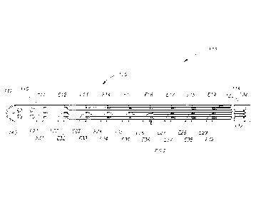
Figure 1 illustrates an implantable electrode array assembly 100.
While the embodiment of the assembly 100 illustrated is configured for
implantation in a rat 500 (see Figure 5A), embodiments may be constructed for
use in other subjects, such as other mammals, including humans, and such
embodiments are within the scope of the present teachings. The assembly 100
is for use with a subject that has a spinal cord 330 (see Figure 3) with at
least
one selected spinal circuit (not shown) and a neurologically derived paralysis
in
a portion of the subject's body. By way of a non-limiting example. the
assembly 100 may be implanted epidurally along the spinal cord 330. The
assembly 100 may be positioned at one or more of a lumbosacral region, a
cervical region, and a thoracic region of the spinal cord 330.
By way of non-limiting examples, when activated, the selected
spinal circuit may (a) enable voluntary movement of muscles involved in at
least
one of standing, stepping, reaching, grasping, voluntarily changing positions
of
one or both legs, voiding the subject's bladder, voiding the subject's bowel,
postural activity, and locomotor activity; (b) enable or improve autonomic
control of at least one of cardiovascular function, body temperature, and
metabolic processes; and/or (c) help facilitate recovery of at least one of an
autonomic function, sexual function, vasomotor function, and cognitive
function.
Without being limited by theory, it is believed that the selected spinal
circuit has
a first stimulation threshold representing a minimum amount of stimulation
required to activate the selected spinal circuit. and a second stimulation
7
CA 02824782 2013-07-12
WO 2012/100260
PCT/US2012/022257
threshold representing an amount of stimulation above which the selected
spinal circuit is fully activated and adding the induced neurological signals
has
no additional effect on the at least one selected spinal circuit.
The paralysis may be a motor complete paralysis or a motor
incomplete paralysis. The paralysis may have been caused by a spinal cord
injury classified as motor complete or motor incomplete. The paralysis may
have been caused by an ischemic or traumatic brain injury. The paralysis may
have been caused by an ischemic brain injury that resulted from a stroke or
acute trauma. By way of another example, the paralysis may have been
caused by a neurodegenerative brain injury. The neurodegenerative brain
injury may be associated with at least one of Parkinson's disease,
Huntington's
disease, Alzheimer's, ischemia, stroke, amyotrophic lateral sclerosis (ALS),
primary lateral sclerosis (PLS), and cerebral palsy.
If the paralysis was caused by a spinal cord injury at a first
location along the spinal cord 330, the assembly 100 may be implanted (e.g.,
epidurally) at a second location below the first location along the spinal
cord
relative to the subject's brain (not shown).
The assembly 100 is configured to apply electrical stimulation to a
portion of a spinal cord 330 of the subject. The electrical stimulation may
include at least one of tonic stimulation and intermittent stimulation. The
stimulation applied may be pulsed. The electrical stimulation may include
simultaneous or sequential stimulation of different regions of the spinal
cord.
The electrical stimulation applied by the assembly 100 may be below the
second stimulation threshold such that the at least one selected spinal
circuit is
at least partially activatable by the addition of signals generated by the
subject.
By way of a non-limiting example, such subject generated signals may be
induced by subjecting the subject to physical activity or training (such as
stepping on a treadmill). These signals may be induced in a paralyzed portion
of the subject. By way of another non-limiting example, the subject generated
signals may include supraspinal signals.
As mentioned above, the embodiment of the assembly 100
illustrated in Figures 1-3 is configured for implantation in the rat 500 (see
Figure
5A). Thus, the embodiment of the assembly 100 illustrated is sized (e.g.,
about
8
CA 02824782 2013-07-12
WO 2012/100260
PCT/US2012/022257
59 mm by about 3 mm) and shaped for implantation into the rat 500. However,
through application of ordinary skill in the art to the present teachings,
embodiments may be constructed for use with other subjects, such as other
mammals, including humans.
Figure 2 illustrates an enlarged portion 200 of the assembly 100
depicted in Figure 1. The assembly 100 may be characterized as being a
microelectromechanical systems ("MEMS") device. As mentioned above, the
assembly 100 is configured for implantation along the spinal cord 330 (see
Figure 3) and to provide electrical stimulation thereto. For example. the
assembly 100 may provide epidural stimulation to the spinal cord 330. The
assembly 100 allows for a high degree of freedom and specificity in selecting
the site of stimulation compared to prior art wire-based implants, and
triggers
varied biological responses that can lead to an increased understanding of the
spinal cord 330 and locomotion recovery for victims of spinal cord injury.
Turning to Figure 1, the assembly 100 includes a body portion
110, an electrode array 120, and a plurality of electrically conductive traces
130. The body portion 110 includes a distal end portion 112, a proximal end
portion 114 (opposite the distal end portion), a frame 140, and a grid
structure
210 (see Figure 2) for each electrode E11-E19, E21-E29, and E31-E39 of the
electrode array 120. Each of the grid structures 210 defines a plurality of
cells
212. By way of a non-limiting example, the grid structures 210 may each be
constructed from parylene (e.g., parylene-C). In the embodiment illustrated,
the
grid structure 210 includes 40 cells.
As mentioned above, the electrode array 120 includes the plurality
.. of electrodes El 1-E19, E21-E29, and E31-E39 (e.g., 9 x 3 electrodes). The
electrodes El 1-E19, E21-E29, and E31-E39 are arranged in a two-dimensional
array. Each of the electrodes E11-E19, E21-E29, and E31-E39 includes a
plurality of electrically conductive contacts 220. The contacts 220 are sites
at
which the electrode (e.g., the electrode E37 illustrated in Figure 2) will
contact
the spinal cord (e.g., the dura). The contacts 220 are in electrically
communication with one another. The embodiment of the electrode E37
illustrated includes 40 contacts 220. However, this is not a requirement. As
mentioned above, each of the electrodes El 1-E19. E21-E29, and E31-E39
9
CA 02824782 2013-07-12
WO 2012/100260
PCT/US2012/022257
corresponds to a unique one of the grid structures 210. In the embodiment
illustrated, for each of the electrodes El 1-E19, E21-E29, and E31-E39, each
of
the contacts 220 is positioned within a different one of the cells 212 of the
corresponding grid structure 210. The grid structure 210 may help prevent
.. delamination of the layers of the assembly 100 (see Figure 1). As is
apparent
to those of ordinary skill in the art and as will be explain below, the grid
structure 210 and contacts 220 may be formed by selectively etching a layer of
substantially electrically non-conductive material (e.g., parylene) adjacent a
pad
of electrically conductive material (e.g., metal) to define the grid structure
210
and expose portions of the electrically conductive material within the cells
212
of the grid structure to define the contacts 220.
While the electrode array 120 illustrated includes 27 electrodes, in
other embodiments, the number of electrodes may range from one electrode to
about 100,000 electrodes or more. In certain embodiments, the electrode array
120 includes at least 10, at least 15, at least 20, at least 25. at least 50,
at least
100, at least 250, at least 500, or at least 1000 electrodes. In various
embodiments, the interelectrode spacing of adjacent electrodes in the
electrode
array 120 varies from about 100 pm or about 500 pm, or about 1000 pm or
about 1500 pm to about 2000 pm, or about 3000 pm, or about 4000 pm, or
about 4500 pm, or about 5000 pm. In various embodiments, interelectrode
spacing ranges from about 100 pm. about 150 pm, about 200 pm, or about 250
pm up to about 1,000 pm, about 2000 pm, about 3000 pm, or about 4,000 pm.
In some embodiments, the diameter (or width) of each of the electrodes El 1-
E19, E21-E29, and E31-E39 ranges from about 50 pm, 100 pm. 150 pm, 200
.. pm, or 250 pm up to about 500 pm, about 1000 pm, about 1500 pm, or about
2000 pm.
The electrode array 120 can be formed in any geometric shape
such as a square shape, rectangular shape, or circular shape. Typically the
size of the electrode array 120 will be on the order of about 0.1 mm to about
2
.. cm, wide or in diameter, depending in part on the number of electrodes in
the
electrode array 120. In various embodiments, the length of the electrode array
120 ranges from about 0.01 mmm, or 0.1 mm up to about 10 cm or greater.
CA 02824782 2013-07-12
WO 2012/100260
PCT/US2012/022257
One or more of the traces 130 is connected to each of the
electrodes El 1-E19, E21-E29, and E31-E39. Referring to Figure 2, in the
embodiment illustrated, two traces "Ti" and "T2" are connected to each of the
electrodes E11-E19, E21-E29, and E31-E39. In alternate embodiments, more
than two traces 130 may be connected to each of the electrodes E11-E19. E21-
E29, and E31-E39. Connecting more than one of the traces 130 to each of the
electrodes El 1-E19, E21-E29. and E31-E39 helps ensure signals reach each
of the electrodes El 1-E19, E21-E29, and E31-E39. In other words,
redundancy may be used to improve reliability. For each of the electrodes Eli-
E19, E21-E29, and E31-E39, the traces 130 are connected to each of the
contacts 220 of the electrode and carry signals thereto. Openings 132 (see
Figure 3) formed (e.g., etched) in the body portion 110 expose portions of the
traces 130.
The traces 130 may be used to selectively deliver electrical
signals (e.g., pulsed signals) to the electrodes E11-E19. E21-E29, and E31-
E39. In this manner, only a selected one or more of the electrodes E 1 1-E19,
E21-E29, and E31-E39 may deliver stimulation to the spinal cord 330 (see
Figure 3). The electrodes E 1 1-E19, E21-E29, and E31-E39 are operably linked
by the traces 130 to control circuitry (not shown). The control circuitry (not
shown) is configured to select one or more of the electrodes El 1-E19, E21-
E29, and E31-E39 to activate/stimulate and/or to control the parameters (e.g.,
frequency, pulse width, amplitude, and the like) of the electrical
stimulation. In
various embodiments, the electrode selection, frequency, amplitude, and pulse
width are independently selectable. For example, at different times, different
electrodes can be selected. At any time, different electrodes can provide
stimulation having different parameter values (e.g., frequencies, amplitudes,
and the like). In various embodiments, at least a portion of the electrodes
may
be operated in a monopolar mode and/or a bipolar mode. In such
embodiments, constant current or constant voltage may be used to deliver the
stimulation.
In some embodiments, the traces 130 may receive signals from
implantable control circuitry (not shown) and/or an implantable power source
(not shown). The implantable control circuitry (not shown) may be programmed
11
CA 02824782 2013-07-12
WO 2012/100260
PCT/US2012/022257
and/or reprogrammed by an external device (e.g., using a handheld device that
communicates with the control circuitry through the skin). The programming
may be repeated as often as necessary.
Figure 3 illustrates a cable system 300 incorporating the
assembly 100. The cable system 300 is illustrated implanted along the spine
320 and spinal cord 330 of the rat 500 (see Figure 5A). Due to the difficulty
preventing infection at connectors that cross the skin (not shown), in chronic
experiments, it is often highly desirable to pass signals through a headplug
310
positioned on the head (not shown) of the rat 500, where the large bone
surface, lack of muscle tissue, and minimal movement of skin help minimize the
risk of infection. Because some preliminary experiments in living animals have
shown that mechanical strains imposed by the animals' movements might make
some embodiments of an all-MEMS device configured to extend from the
headplug 310 to the spinal cord 330 unreliable, the cable system 300 was
devised to confine strain imposed on the assembly 100 to acceptable limits.
Figure 3 illustrates how the cable system 300 (including the
assembly 100) is positioned along the spine 320 of the subject (e.g., the rat
500
illustrated in Figure 5A) after implantation. The cable system 300 is composed
of a spinal baseplate 340, a wire bundle 350, and the headplug 310. Another
set of wires (not shown) may be implanted in the leg(s) 520 (see Figure 5A) of
the subject to record electromyography ("EMG") signals. The baseplate 340
may be constructed from a standard FR-4 PCB substrate. The baseplate 340
is attached (e.g., by a suture 342) to a selected vertebrae (e.g., vertebrae
"L2").
In the embodiment illustrated, the baseplate 340 is attached to the "L2"
vertebrae. The assembly 100 is attached (e.g., by a suture 344) to the spinal
cord 300. In the embodiment illustrated, the distal end portion 112 of the
assembly 100 is attached to the spinal cord 300 at a location adjacent
vertebrae "T13." The proximal end portion 114 of the assembly 100 is attached
to the baseplate 340 using a conductive material (e.g., conductive epoxy) to
bridge electrical connections. By way of a non-limiting example, the proximal
end portion 114 of the assembly 100 may be secured to the baseplate 340
using Loctite M-121HP Medical device epoxy.
12
CA 02824782 2013-07-12
WO 2012/100260
PCT/US2012/022257
The wire bundle 350 includes a plurality of wires 352. By way of a
non-limiting example, the wires 352 may include a different wire for each of
the
electrodes El 1-E19, E21-E29, and E31-E39 (e.g., 27 wires total for a 9 x 3
array of electrodes). Each of the wires 352 may be constructed from gold and
include a Teflon coating. For example, 75 pm gold wires (e.g., Teflon coated
gold wire manufactured by AM Systems) may be used. The wires 352 may be
soldered to the baseplate 340 and connected by high density connectors 360 to
the headplug 310. The traces 130 are connected to the baseplate 340 via the
openings 132 formed in the body portion 110 of the assembly 100. By way of a
non-limiting example, silver epoxy (not shown) may be used to connect the
traces 130 to the baseplate 340.
The entire cable system 300 (except a portion 368 of the
assembly 100) may be coated with a coating 370 configured to insulate
electrical connections and provide mechanical strength while retaining the
.. flexibility wherever necessary. By way of a non-limiting example, the
coating
370 may include a biomedical grade epoxy and a silicone elastomer (e.g., MDX
4-4210 Biomedical grade silicone).
A silicone cap 380 (or overhanging portion) is formed on the end
of the baseplate 340 to protect the assembly 100 from external moving tissue.
The cap 380 may be formed from the same material as the coating 370. Along
portions of the assembly 100, the coating 370 may be implemented as a thin
layer of silicone (e.g., about 100 pm thick) to reduce stress concentration as
the
assembly 100 bends with the subjects spine 320 during movement. A thicker
layer of silicone applied to the assembly 100 may be detrimental to the health
of
the spinal cord 330 because of increased pressure that is applied by a more
rigid assembly to the spinal cord. in other words, flexibility may be an
important
feature of a successful chronic implantable electrode array assembly.
FABRICATION
The assembly 100 may be fabricated using a method somewhat
similar to that described in D.C. Rodger, et al., "Flexible microfabricated
parylene multielectrode arrays for retinal stimulation and spinal cord field
modulation," Proc. 4th International IEEE-EMBS Special Topic Conference on
13
CA 02824782 2013-07-12
WO 2012/100260
PCT/US2012/022257
Microtechnologies in Medicine and Biology, Okinawa, Japan, pp. 31-34 (2006),
which describes a method of forming a sandwich-like structure of parylene-
metal-parylene.
Turning to Figures 4A-40, the assembly 100 may be constructed
using a method 400. For ease of illustration, the method 400 will be described
with respect to using parylene-C, which is substantially electrically
nonconductive. Parylene-C is a United States Pharmacopeia! Convention
("USP") class VI biocompatible material, and its mechanical properties provide
the necessary flexibility to make good epidural contact with the spinal cord
330
(see Figure 3). However, those of ordinary skill in the art appreciate that
other
materials may be used instead of or in combination with parylene-C. Examples
of other materials include flexible materials such as parylene-A, parylene-AM,
parylene-F, parylene-N, parylene-D, and the like. Further, the electrode
arrays
120 will be described as including metal, which may be implemented using one
or more biocompatible metals (e.g., gold, platinum, chromium, titanium,
iridium,
tungsten, and/or oxides and/or alloys thereof). For ease of illustration, the
method 400 will be described with respect to using platinum (and titanium) to
construct the electrode arrays 120.
The method 400 begins at the top of Figure 4A. A first
subassembly "SAl" is constructed by applying (e.g., spinning) an optional
first
layer of sacrificial photoresist 410 on a substrate 412 (e.g., a silicon
wafer).
Then, a second subassembly "SA2" is constructed by depositing
(e.g., using conventional vapor-deposition) a first (frame) layer of parylene-
C
416 on the first layer of photoresist 410. By way of a non-limiting example.
the
first (frame) layer of parylene-C 416 may be about 10 pm thick.
A third subassembly "SA3" is constructed by applying (e.g.,
spinning) a second layer of photoresist 422 on the second subassembly "SA2."
Next, a fourth subassembly "SA4" is constructed by exposing and
developing the second layer of photoresist 422 to define the frame 140 (see
Figure 1) using conventional photoresist techniques.
Turning to Figure 4B, a fifth subassembly "SAS" is constructed by
removing (e.g., etching) at least a portion of the first (frame) layer of
parylene-C
416 to define the at least a portion of the frame 140 that surrounds the
14
CA 02824782 2013-07-12
WO 2012/100260
PCT/US2012/022257
electrode array 120. Then, the second layer of photoresist 422 is removed
(e.g., dissolved using acetone).
Next, a sixth subassembly "SA6- is constructed by depositing
(e.g., using conventional vapor-deposition) a second (base) layer of parylene-
C
420 on the fifth subassembly "SAS." By way of another non-limiting example,
the second (base) layer of parylene-C 420 may be about 5 tirll thick. The
second (base) layer of parylene-C 420 forms an underside for the body portion
110 (see Figure 1) of the assembly 100 (see Figure 1). The second (base)
layer of parylene-C 420 may also be characterized as defining at least a
portion
of the frame 140 because the first (frame) layer of parylene-C 416 is
underneath and helps shape the second (base) layer of parylene-C 420. In
other words, the frame 140 may be characterized as including both first
(frame)
and the second (base) layers 416 and 420. Alternatively, the frame 140 may be
characterized as being defined entirely by the first (frame) layer 416.
A seventh subassembly "SAT' is constructed by applying (e.g.,
spinning) a third layer of photoresist 424 onto the sixth subassembly "SA6."
An eighth subassembly "SA8" is constructed by exposing and
developing the third layer of photoresist 424 to define a pattern using
conventional photoresist techniques. The pattern defines the electrode array
120 and the traces 130.
Turning to Figure 4C, a ninth subassembly "SA9" is constructed
by depositing (e.g., using ebeam evaporation) an electrically conductive layer
428 on the eighth subassembly "SA8." The electrically conductive layer 428
may be constructed by first depositing an adhesion layer of a first material
(e.g.,
100A of titanium) and then depositing an electrode layer of a second different
electrically conductive material (e.g., 2000A of platinum) suitable for
conducting
electrical stimulation. Thus, the electrically conductive layer 428 may be
constructed using more than one layer of material.
A tenth subassembly "SA10" is constructed by removing (e.g.,
dissolving) the third layer of photoresist 424, which removes portions of the
electrically conductive layer 428 positioned thereupon to form the electrode
array 120 and the traces 130. In other words, a conventional liftoff process
is
CA 02824782 2013-07-12
WO 2012/100260
PCT/US2012/022257
used to pattern the electrically conductive layer 428 to form the electrode
array
120 and the traces 130.
Next, an eleventh subassembly "SA11" is constructed by
depositing (e.g., using conventional vapor-deposition) a third (top) layer of
parylene-C 430 on the tenth subassembly "SA10." By way of another non-
limiting example, the third (top) layer of parylene-C 430 may be about 5 liM
thick.
Turning to Figure 4D, a twelfth subassembly "SA12" is created by
applying (e.g., spinning) a fourth layer of photoresist 432 onto the eleventh
subassembly "SA11."
A thirteenth subassembly "SA13" is constructed by exposing and
developing the fourth layer of photoresist 432 to define a pattern using
conventional photoresist techniques. The pattern defines the openings 132,
which are formed in the third (top) layer of paryiene-C 430.
A fourteenth subassembly "SA14" is created by forming the
openings 132 in the third (top) layer of parylene-C 430 to expose portions of
the
electrically conductive layer 428. The openings 132 may be formed using
etching (e.g., oxygen plasma etching). For each of the electrodes E1 1-El 9,
E21-E29, and E31-E39, at least a portion of the openings 132 provide access
to the contacts 220 and define the grid structure 210. The contacts 220
contact
the spinal cord 330 (see Figure 3) through the openings 132. A different
portion
of the openings 132 provide access to the traces 130 so that the baseplate 340
may be electrically connected thereto. Etching may also be used to define the
shape of the assembly 100. Then, the fourth layer of photoresist 432 is
removed (e.g., dissolved using acetone or water).
A fifteenth subassembly "SA15" is formed by removing (e.g..
dissolving) the first layer of photoresist 410 to release the layers above the
first
layer of photoresist 410 from the substrate 412. By way of a non-limiting
example, the first layer of photoresist 410 may be dissolved using acetone or
water.
Finally, the assembly 100 (see Figure 1) may be created by
annealing the fifteenth subassembly "SA15" in a vacuum oven at 200 C for 48
hours.
16
CA 02824782 2013-07-12
WO 2012/100260
PCT/US2012/022257
RESULTS AND DISCUSSION
Implementations of the cable system 300 (see Figure 3) were
implanted in rats and functioned for up to eight weeks. This level of
reliability
makes the cable system 300 (and assembly 100) suitable for studying stepping
ability overtime. The cable system 300 (and assembly 100) also provides site
selectivity, afforded by the high density microfabricated electrode array 120.
Figure 5A is an illustration of the rat 500 suspended over a
treadmill 510 by a jacket 530. The rat 500 has a completely transected spinal
cord and thus hindlimb paralysis. Stepping by the hind limbs was achieved in
the rat 500 by stimulating the rat's spinal cord 330 (see Figure 3) while with
the
rat was suspended over the treadmill 510. Figure 5A also illustrates portions
of
a motion capture system (e.g., dots D1-D5) used to record stepping ability.
Figure 5B is a stick diagram 550 representing hind limb motion when the rat's
spinal cord 330 was not stimulated. As expected, the rat 500 dragged its feet
when it's spinal cord 330 was not stimulated due to the hindlimb paralysis.
Figures 6A and 6B depict a pair of stick diagrams 610 and 620,
respectively, that illustrate hind limb motion when bipolar stimulation is
applied
to the rat's spinal cord 330 by two different electrode pairs. The diagrams
610
and 620 are believed to illustrate the first stepping achieved by a spinalized
rat
stimulated by a MEMS electrode array. Of note is that the stimulation site
pairs
for the two different stepping patterns illustrated in Figures 6A and 6B were
close together in the electrode array 120, suggesting that the high-density
electrode configuration of the assembly 100 is of great value in understanding
the biological mechanisms underlying locomotion and its application to
recovery
after spinal cord injury.
EMG recording may also be very valuable in obtaining biological
information. Figures 7A and 7B show two EMG recordings for two different
stimulation pairs at three different voltages. In other words, Figure 7A
depicts
an EMG recording recorded when stimulation was applied by one pair of
electrodes and Figure 7B depicts an EMG recording recorded when stimulation
was applied by a different pair of electrodes. Figure 7A illustrates a
monosynaptic response "Rl." Such monosynaptic responses generally occur
17
CA 02824782 2013-07-12
WO 2012/100260
PCT/US2012/022257
in the first six milliseconds of the recordings, while polysynaptic responses
(such as polysynaptic responses "Pl") generally occur later. Of note is that
the
recording depicted in Figure 7A includes both the monosynaptic response "Rl"
and the polysynaptic responses "P1," while the recording depicted in Figure 78
includes only polysynaptic responses "P2." This demonstrates that the high
density of electrode array 120 provides high-density stimulation sites (the
electrodes El 1-E19, E21-E29. and E31-E39) that are useful in eliciting
different
biological responses. The EMG signals of Figures 7A and 78 were obtained
during reflex tests (0.3 Hz stimulation pulses), and the stick diagrams of
Figures
6A and 6B were obtained during stepping testing (40 Hz).
The assembly 100 has been shown to survive in a living rat for up
to eight weeks and may survive much longer, because the impact of
mechanical damage observed on the functionality of the assembly 100 is
minimal. The cable system 300 provides a means for stimulating the spinal
cord 330 and recording evoked responses. Optionally, the electrodes E11-E19,
E21-E29, and E31-E39 of the assembly 100 may be used to detect neurological
signals in addition to delivering stimulation. The stimulation applied by the
assembly 100 may be used to induce stepping in a rat with a completely
transected spinal cord. The assembly 100 provides a means for controlling the
site of stimulation to produce different EMG responses and stepping patterns.
This level of control is useful for understanding neurobiological circuits
inside
the spinal cord 330 and developing possible treatments for locomotion recovery
in victims of spinal cord injury.
While the cable system 300 including the assembly 100 has been
.. described with respect to enabling stepping in a subject (e.g., the rat
500),
through application of ordinary skill in the art to the present teachings
embodiments can be constructed that enable other types of functionality, such
as to (a) enable voluntary movement of muscles involved in at least one of
standing, stepping, reaching, grasping, voluntarily changing positions of one
or
both legs, voiding the bladder, voiding the bowel, postural activity, and
locomotor activity; (b) enable or improve autonomic control of at least one of
cardiovascular function, body temperature, and metabolic processes; and/or (c)
18
, .
help facilitate recovery of at least one of an autonomic function, sexual
function,
vasomotor function, and cognitive function.
The foregoing described embodiments depict different
components contained within, or connected with, different other components. It
is to be understood that such depicted architectures are merely exemplary, and
that in fact many other architectures can be implemented which achieve the
same functionality. In a conceptual sense, any arrangement of components to
achieve the same functionality is effectively "associated" such that the
desired
functionality is achieved. Hence, any two components herein combined to
achieve a particular functionality can be seen as "associated with" each other
such that the desired functionality is achieved, irrespective of architectures
or
intermedial components. Likewise, any two components so associated can
also be viewed as being "operably connected," or "operably coupled," to each
other to achieve the desired functionality.
While particular embodiments of the present invention have been
shown and described, it will be obvious to those skilled in the art that,
based
upon the teachings herein, changes and modifications may be made without
departing from this invention and its broader aspects and, therefore, the
appended claims are to encompass within their scope all such changes and
modifications as are within the true spirit and scope of this invention.
Furthermore, it is to be understood that the invention is solely defined by
the
appended claims. It will be understood by those within the art that, in
general,
terms used herein, and especially in the appended claims (e.g., bodies of the
appended claims) are generally intended as "open" terms (e.g., the term
"including" should be interpreted as "including but not limited to," the term
"having" should be interpreted as "having at least," the term "includes"
should
be interpreted as "includes but is not limited to," etc.). It will be further
understood by those within the art that if a specific number of an introduced
claim recitation is intended, such an intent will be explicitly recited in the
claim,
and in the absence of such recitation no such intent is present. For example,
as an aid to understanding, the following appended claims may contain usage
19
CA 2824782 2018-05-28
CA 02824782 2013-07-12
WO 2012/100260
PCT/US2012/022257
of the introductory phrases "at least one" and "one or more" to introduce
claim
recitations. However, the use of such phrases should not be construed to imply
that the introduction of a claim recitation by the indefinite articles "a" or
"an"
limits any particular claim containing such introduced claim recitation to
inventions containing only one such recitation, even when the same claim
includes the introductory phrases "one or more" or "at least one" and
indefinite
articles such as "a" or "an" (e.g., -a" and/or "an" should typically be
interpreted
to mean "at least one" or "one or more"); the same holds true for the use of
definite articles used to introduce claim recitations. In addition, even if a
specific number of an introduced claim recitation is explicitly recited, those
skilled in the art will recognize that such recitation should typically be
interpreted to mean at least the recited number (e.g., the bare recitation of
"two
recitations," without other modifiers, typically means at least two
recitations, or
two or more recitations).
Accordingly, the invention is not limited except as by the
appended claims.