Note: Descriptions are shown in the official language in which they were submitted.
DEVICES, SYSTEMS, AND METHODS FOR EXTRACTING A MATERIAL
FROM A MATERIAL SAMPLE
:BACKGROUND OF THE INVENTION
Two commonly used techniques for dissecting specific areas from slide
mounted tissue sections are Manual Macrodissection and Laser Capture
Microdissection (LCM). Manual Macrodissection is predominately used in the
pathology field because it has negligible cost, is relatively quick, and
generally large
quantities of sample are obtained. However, the lower limit of precision is
about lntm,
which can limit accuracy, and the manual nature makes it error prone and
poorly
documented. LCM is spatially precise allowing capture resolution as small as
515m and
thus the ability to target single cells. However, equipment is very expensive,
it is slow
and requires full time interaction by a trained operator, and the spatial
precision comes
at the price of minute quantities of recovered sample, making downstream
biochemical
analysis challenging and often requiring extensive amplification that can bias
results.
A third dissection technique using needles and micromanipulators has not
gained wide
spread acceptance because it is difficult and labor intensive,
SUMMARY OF THE INVENTION
The present disclosure provides devices, systems, and associated methods for
selectively extracting a material from a sample. ln one aspect, for example, a
method
for selectively extracting a material, such as a biological material, from a
sample, such
as a biological sample can include identifying a region of material to be
extracted from
a sample, applying an extraction tool to the region of material to disrupt
material from
the sample, and dispensing a liquid at the region of material, The method can
also
include aspirating the liquid and the disrupted material from the sample,
- 1 -
CA 2825612 2018-03-28
CA C282S012 HI} 07 .24
WO 2012/102779 PCT/US2011/061075
The extraction tool can utilize a variety of motions to disrupt the material
from
the sample, and any such motion capable of disrupting material is considered
to be
within the present scope. In one aspect, for example, the extraction tool can
impart a
cutting motion to the region of material. Any cutting motion is contemplated,
non-limiting examples including rotating, vibrating, slicing, and the like,
including
combinations thereof In one specific aspect, the cutting motion is rotating.
Various methods of dispensing the liquid and aspirating the liquid and the
disrupted material are contemplated. In one aspect, for example, the liquid
can be
dispensed at an interface between the region of material and the extraction
tool. In this
manner disrupted material is readily mixed with the liquid as it is disrupted.
In another
aspect, the liquid is dispensed and aspirated simultaneously. Thus the
disrupted
material can be quickly removed by the aspirated liquid from the sample. In
yet another
aspect, the liquid is dispensed and aspirated by the extraction tool, or in
other words,
the liquid is dispensed and aspirated from ports coupled to, or otherwise
associated
with, or formed integrally with, the extraction tool.
Additionally, a variety of techniques for identifYing a region of material,
such
as biological material are contemplated. In one aspect, for example,
identifying a
region of material further includes obtaining a real time digital image of the
sample and
defining an area of interest on the digital image corresponding to the region
of material,
where movement of the sample is reflected by movement of the area of interest
and/or
the digital image to maintain position of the area of interest relative to the
material. In
another aspect, the sample is a series of sections, and the area of interest
is defined on
one section that corresponds to the region of material from a different
section.
The present disclosure additionally provides various material extraction
devices. In one aspect, for example, an extraction device for selectively
extracting
material, such as a biological material from a sample, such as a biological
sample, can
include a housing and at least one cutting tip rotatably coupled to the
housing and
configured to be rota tably driven by a motor. The cutting tip is operable to
disrupt
material from a region of a sample. The device can further include at least
one liquid
dispensing port coupled to the housing and located proximal to the cutting
tip, where
the liquid dispensing port is operable to dispense liquid at the cutting tip.
Additionally,
at least one liquid aspiration port is coupled to the housing and located
proximal to the
- 2 -
cutting tip, where the liquid aspiration port is operable to aspirate liquid
and disrupted
biological material from a region proximal to the cutting tip. In another
aspect, the at
least one liquid dispensing port and the at least one liquid aspiration port
rotate with the
cutting tip. In yet another aspect, the at least one liquid dispensing port
and the at least
one liquid aspiration port are operable to function simultaneously.
The cutting tip can be of any size, depending on the desired cutting task. In
one
aspect, for example, the cutting tip is sized to disrupt an area of biological
material of
from about 10 gm in size to about 1 mm in size. In another aspect, the cutting
tip is
sized to disrupt an area of material of from about 100 gm in size to about 250
pm in
size.
In accordance with one embodiment of the present invention there is
provided an extraction device for selectively extracting biological material
from a
biological sample. The extraction device comprises: a housing; a cutting tip
rotatably coupled to the housing and configured to be rotatably driven by a
motor,
the cutting tip being operable to disrupt material from a region of a sample;
a liquid
dispensing port coupled to the housing and located proximal to the cutting
tip, the
liquid dispensing port being operable to dispense liquid at the cutting tip;
and a
liquid aspiration port coupled to the housing and located proximal to the
cutting tip,
the liquid aspiration port being operable to aspirate liquid and disrupted
biological
material from a region proximal to the cutting tip. The liquid dispensing port
and
the liquid aspiration port are not the same port. The liquid dispensing port
and the
liquid aspiration port are operable to function simultaneously during rotation
of the
cutting tip. The extraction device is configured for inclusion in a system for
selectively extracting biological material from a biological sample.
- 3 -
CA 2825612 2020-01-30
The present disclosure additionally provides systems for selectively
extracting
a material from a sample. In one aspect, for example, a system for selectively
extracting material, such as biological material from a sample, such as a
biological
sample can include an extraction device as has been described herein
positioned to
operationally face a support substrate and to engage a sample disposed on the
support
substrate. A motor can be operationally coupled to the extraction device and
operable
to rotate the cutting tip. A fluidics system can be coupled to the extraction
device and
operable to deliver fluid to the liquid dispensing port and withdraw fluid
from the liquid
removal port. Furthermore, a positional movement system can be coupled to the
extraction device and operable to move either the cutting tip of the
extraction device
relative to the support substrate or the support substrate relative to the
cutting tip.
It can be beneficial to visualize the material extraction process during use.
As
such, in one aspect a visualization system is included and is positioned to
provide a
visual display of a sample, such as a biological sample, placed on the support
substrate.
The visualization system can include a variety of visualization devices,
including
without limitation, digital imagers, optical imagers, microscopes, inverted
microscopes, and the like, including combinations thereof. In one specific
aspect, the
support substrate is transparent. In another aspect, the visualization system
is an
inverted microscope positioned to provide the visual display from a side of
the
transparent support substrate opposite the cutting tip. In yet another aspect,
the
visualization system is operable to provide a real time visual display of the
cutting tip
during an extraction procedure.
- 3a -
CA 2825612 2020-01-30
In another aspect, the system for selectively extracting material, such as a
biological material from a sample, such as a biological sample, can further
include a
manual manipulation system. This manual system is functionally coupled to the
positional movement system and is operable to allow a user to move the cutting
tip
and/or the support substrate relative to one another.
In yet another aspect, the system for selectively extracting material, such as
a
biological material from a sample, such as a biological sample, can further
include an
automatic manipulation system. Such an automatic system is functionally
coupled to
the positional movement system and is operable to automatically move the
cutting tip
and/or the support substrate relative to one another. In another aspect, the
automatic
system further includes a processing system functionally coupled to the
automatic
manipulation system. The processing system is operable to identify and locate
a
predetermined region of material to be extracted from a sample and move the
cutting tip
and/or support substrate relative to one another to extract the biological
material via the
automatic manipulation system.
There has thus been outlined, rather broadly, the more important features of
the
invention so that the detailed description thereof that follows may be better
understood,
BRIEF DESCRIPTION OF THE DRAWINGS
FIG. 1 shows a side view of a material extraction device in accordance with
one
embodiment of the present invention.
FIG. 2A shows a view of a material extraction device in accordance with
another embodiment of the present invention.
FIG. 23 shows a view of a material extraction device in use in accordance with
another embodiment or the present invention.
FIG. 3 shows a view of a material extraction device in use in accordance with
another embodiment of the present invention.
-4 -
CA 2825612 2018-03-28
CA C282S012 HI} 07 .24
WO 2012/102779 PCT/US2011/061075
FIG. 4 shows a view of a cutting tip from a material extraction device in
accordance with another embodiment of the present invention.
FIG. 5 shows a schematic view of a material extraction system in accordance
with another embodiment of the present invention.
FIG. 6A-D show images of tissue being extracted by a material extraction
device in accordance with another embodiment of the present invention.
FIG. 7A-B show images of tissue having a defined area of interest in
accordance with another embodiment of the present invention.
FIG. 8 shows cross sectional views of a material extraction device having
various components separated out in accordance with one embodiment of the
present
invention.
FIG. 9 shows a side view of a material extraction system in accordance with
one
embodiment of the present invention.
The drawings will be described further in connection with the following
detailed description. Further, these drawings are not necessarily to scale and
are by
way of illustration only such that dimensions and geometries can vary from
those
illustrated.
DETAILED DESCRIPTION
Before the present invention is disclosed and described, it is to be
understood
that this invention is not limited to the particular structures, process
steps, or materials
disclosed herein, but is extended to equivalents thereof as would be
recognized by those
ordinarily skilled in the relevant arts. It should also be understood that
terminology
employed herein is used for the puipose of describing particular embodiments
only and
is not intended to be limiting.
It must be noted that, as used in this specification and the appended claims,
the
singular forms "a," "an," and "the" include plural referents unless the
context clearly
dictates otherwise. Thus, for example, reference to "the cutting tip" includes
one or
more of such tips, reference to "a liquid port" includes reference to one or
more of such
ports.
- 5 -
C. 028230122M-07.24
WO 2012/102779 PCT/US2011/061075
Definitions
In describing and claiming the present invention, the following terminology
will be used in accordance with the definitions set forth below.
As used herein, the term "substantially" refers to the complete or nearly
complete extent or degree of an action, characteristic, property, state,
structure, item, or
result. The exact allowable degree of deviation from absolute completeness may
in
some cases depend on the specific context. However, generally speaking the
nearness
of completion will be so as to have the same overall result as if absolute and
total
completion were obtained. The use of "substantially" is equally applicable
when used
in a negative connotation to refer to the complete or near complete lack of an
action,
characteristic, property, state, structure, item, or result. For example, a
composition
that is "substantially free of" particles would either completely lack
particles, or so
nearly completely lack particles that the effect would be the same as if it
completely
lacked particles. In other words, a composition that is "substantially free
of" an
ingredient or element may still actually contain such item as long as there is
no
measurable effect on the property of interest thereof.
As used herein, the term "about" is used to provide flexibility to a numerical
range endpoint by providing that a given value may be "a little above" or "a
little
below" the endpoint with a degree of flexibility as would be generally
recognized by
those skilled in the art. Further, the term about explicitly includes the
exact endpoint,
unless specifically stated otherwise.
As used herein, a plurality of items, structural elements, compositional
elements, and/or materials may be presented in a common list for convenience.
However, these lists should be construed as though each member of the list is
individually identified as a separate and unique member. Thus, no individual
member
of such list should be construed as a de facto equivalent of any other member
of the
same list solely based on their presentation in a common group without
indications to
the contrary.
Concentrations, amounts, and other numerical data may be expressed or
presented herein in a range format. It is to be understood that such a range
format is
used merely for convenience and brevity and thus should be interpreted
flexibly to
include not only the numerical values explicitly recited as the limits of the
range, but
- 6 -
CA C282S012 HI} 07 .24
WO 2012/102779 PCT/US2011/061075
also to include all the individual numerical values or sub-ranges encompassed
within
that range as if each numerical value and sub-range is explicitly recited. As
an
illustration, a numerical range of "about I to about 5" should be interpreted
to include
not only the explicitly recited values of about 1 to about 5, but also include
individual
values and sub-ranges within the indicated range. Thus, included in this
numerical
range are individual values such as 2, 3, and 4 and sub-ranges such as from. 1-
3, from
2-4, and from 3-5, etc., as well as I, 2, 3, 4, and 5, individually. This same
principle
applies to ranges reciting only one numerical value as a minimum or a maximum.
Furthermore, such an interpretation should apply regardless of the breadth of
the range
or the characteristics being described.
The Invention
The present disclosure relates to devices, systems, and methods for removing
material from a material sample. In some cases, the material that has been
extracted is
saved for further processing or analysis. Such may be the case for procedures
involved
in forensics, testing of material purity, histopathology, core sampling, and
the like. In
some cases, serial sections of a material sample can be generated that allows
a
destructive sampling of one section while retaining structural features from
adjacent
sections for further analysis.
One example of where such testing can be beneficial is in the area of
histopathology or other biological fields whereby biological material is
removed from
a biological sample. It should be noted, however, that although much of the
following
description is biological in nature, the present scope is not limited to such.
R.ather, the
present disclosure applies to any material and/or testing procedure relating
to the
current aspects.
In one aspect a method for selectively extracting biological material from a
biological sample is provided. In such a method, a region of biological
material to be
extracted from a biological sample is identified. In some cases, the
biological sample
is disposed on a surface, such as for example, a substantially planar or
planar surface.
In other cases, the biological sample can be in the form of a block or other
three
dimensional object. The biological material can be any type of biological
material, and
can be derived from a variety of biological organisms, including animals,
humans,
plants, fungus, and the like. The biological sample itself can include any
material
- 7 -
CA C282S012 HI} 07 .24
WO 2012/102779 PCT/US2011/061075
derived from a biological organism, including tissue, tissue sections, organs,
organ
sections, cells, cultured cells, cultured tissue, plant matter, secretions,
excretions, and
the like, including combinations thereof. The biological material can also be
embedded
in a matrix such as plastic, paraffin, a gel, or any other material or agent
useful to
present the material in a solid, semisolid, or suspended form, and can include
fresh or
frozen. biological sample or sample sections. Thus the region of biological
material is
an area from which biological material is to be extracted from. the biological
sample.
The method can further include applying an extraction tool to the region of
biological material to disrupt biological material from the biological sample.
In some
119 aspects, the extraction tool contacts the biological sample in the
identified region and
disrupts biological material therefrom. Any configuration of extraction tool
capable of
disrupting the biological material is considered to be within the present
scope.
Additionally, a variety of disruptive motions are contemplated. In one aspect,
for
example, the disruptive motion is a cutting motion. Non-limiting examples of
cutting
motions include rotating, vibrating, slicing, and the like, including
combinations
thereof in one specific aspect the cutting motion is rotation.
The method can also include dispensing a liquid at the region of biological
material. The liquid can be dispensed on a portion of the biological sample,
or it can be
dispensed over the entire or substantially the entire sample. In one aspect,
the liquid is
dispensed at an interface between the region of biological material and the
extraction
tool. The liquid can be any liquid that is beneficial for extracting
biological material
from a biological sample. The liquid can include any liquid medium capable of
mixing
with the disrupted biological material. In some cases, the liquid can be
designed to
merely mix with the biological material. in other cases, the liquid can be
formulated to
react with the biological material and/or the biological sample. For example,
the liquid
can contain enzymes or other chemical moieties to facilitate the disruption
and/or
breakdown of the biological material. As such, further processing steps can be
facilitated as the biological material is being extracted from the biological
sample.
Generally the liquid can contain one or more of various solvents, enzymes,
buffers, and
the like. In one aspect, the liquid can be water or purified water.
The method can also include removing the liquid and at least a portion of the
disrupted biological material from the biological sample. Thus, once the
disrupted
- 8 -
CA C282S012 HI} 07 .24
WO 2012/102779 PCT/US2011/061075
biological material is mixed with the liquid, both the liquid and the
biological material
can be removed for further processing or disposal. In addition to any
enzymatic
reactions, the liquid thus creates a slurry or suspension of the biological
material in
order to facilitate removal from the sample. Removal can occur via a variety
of
mechanisms, including without limitation, aspiration, wicking, gravity flow,
and the
like. In one specific aspect, the removal is by aspiration. The removal of the
liquid can
occur sequentially with the dispensing of the liquid or the removal can occur
simultaneously with the dispensing. In one specific aspect, the liquid is
dispensed and
aspirated simultaneously. Additionally, in some cases the dispensing and
removal of
the liquid occurs separately from the extraction tool. In one aspect, the
liquid is
dispensed and aspirated by the extraction tool.
While dispensing and removing liquid have been described with the disruption
of the material, it should be noted that such disruption can occur in the
absence of a
liquid, and that any other physical method of removing the disrupted material
is
considered to be within the present scope. For example, the disrupted material
can be
removed from the surface using a vacuum and recovered on an air filter.
The present disclosure additionally provides tools for the extraction of
material
from a sample. In one aspect, as is shown in FIG. 1 for example, an extraction
device
for selectively extracting biological material from a biological sample is
provided.
Such a device can include a housing 12 for containing the various components
of the
device and at least one cutting tip 14. As has been described, the cutting tip
14 can
disrupt biological material from the biological sample using a variety of
cutting
motions, such as for example, rotating, slicing, vibrating, punching, and the
like. In one
specific aspect, the cutting motion is rotational. In such cases, the cutting
tip 14 is
rotatably coupled to the housing 12 and configured to be coupled 16 to and
rotatably
driven by a motor (not shown). Thus as the cutting tip contacts the biological
sample,
the rotational motion disrupts biological material.
The extraction device can additionally include at least one liquid dispensing
port 18 coupled to the housing 12 and located in a position that is proximal
to the
cutting tip 14. As such, the liquid dispensing port 18 dispenses liquid at the
cutting tip
14, and in doing so may reduce the volume of liquid required to perform a
cutting
procedure. Furthermore, the extraction device can include at least one liquid
aspiration
- 9 -
CA C282S012 HI} 07 .24
WO 2012/102779 PCT/US2011/061075
port 19 coupled to the housing 12 and located in a position that is proximal
to the
cutting tip 14. As such, the liquid aspiration port 19 aspirates liquid and
disrupted
biological material from a region proximal to the cutting tip 14, thus
minimizing the
contact of liquid and biological material at other regions of the biological
sample.
In one aspect, the liquid dispensing port and the liquid aspiration port
rotate
with the cutting tip. One aspect of such a configuration is shown in FiGs. 2A.
& B.
FIG. 2.A shows one aspect having an extraction device 20 with a cutting tip
22, a liquid
dispensing port 24, and a liquid aspiration port 26 associated with the
cutting tip 22. it
should be noted that both the liquid dispensing ports 24 and the liquid
aspiration port
26 are associated with the cutting tip 22 in such a way that they rotate with
the cutting
tip. Liquid thus dispensed during a procedure will be located at an interface
between
the cutting tip and the biological sample. The arrow in FIG. 2A represents the
path of
the flow of liquid from the liquid dispensing port 24 to the liquid aspiration
port 26
during use.
FIG. 2B shows a cross section of the excision device of FIG. 2A while in use.
In this case a biological sample 27 is disposed on a substantially planar
surface 28 and
a rotating 25 cutting tip 22 is brought into contact with the biological
sample. A liquid
is dispensed from the liquid dispensing ports 24 associated with the cutting
tip 22 to
provide liquid at the interface between the cutting tip 22 and the biological
sample 27.
Biological material is disrupted from the biological sample and is mixed with
the
liquid at the interface. The liquid and biological material mixture is
aspirated from the
interface via the liquid aspiration port 26. Arrows 29 show the liquid and the
biological
material being aspirated through the liquid aspiration port 26 and through the
extraction
device.
In another aspect, the liquid dispensing port and the liquid aspiration port
are
operable to function simultaneously. It is noted that numerous designs can be
utilized
to achieve such functionality, and any such design is considered to be within
the present
scope. For example, in one aspect separate pumps can be utilized to
simultaneously
pump fluid out of the liquid dispensing port and aspirate liquid in through
the liquid
aspiration port. In other aspects, a single pump can be utilized having
sufficient
fluidics to allow simultaneous functionality, in one exemplary aspect shown in
FIG. 3,
the internal configuration of the extraction tool can allow such simultaneous
-10-
CA C282S012 HI} 07 .24
WO 2012/102779
PCT/US2011/061075
functionality. In the left panel of FIG. 3, an extraction device 30 is
positioned into a
liquid holding vessel 31 to contact a liquid 32. A plunger 33 creating a seal
within the
extraction device 30 is depressed in a direction toward the liquid dispensing
vessel 32.
This depression causes the liquid 32 to move through a liquid dispensing port
and an
associated dispensing channel 34 to fill a liquid dispensing reservoir 35
within the
extraction tool. The negative pressure created by the movement of the plunger
33 thus
fills the liquid dispensing reservoir 35 with liquid. As is shown in tb.e
center panel of
FIG. 3, the extraction tool 30 is then placed against a biological sample on a
substantially planar surface and rotated to disrupt biological material. While
the device
is rotating, the plunger 33 can be withdrawn in a direction away from the
substantially
planar surface 36 in order to create positive pressure in the liquid
dispensing reservoir
35. This positive pressure dispenses liquid through the dispensing channel 34
and out
of the liquid dispensing port at the interface 37 between the biological
sample and the
excision device. Simultaneously the withdrawal of the plunger 33 causes a
negative
pressure within a liquid aspiration reservoir 38 that causes liquid at the
interface 37 to
be aspirated through the liquid aspiration port and associated aspiration
channel 39 to
thus fill the liquid aspiration reservoir with liquid and disrupted biological
material.
The right panel of FIG. 3 shows the plunger 33 being depressed toward the
cutting tip
40, thus producing a positive pressure in the liquid aspiration reservoir 38
and expelling
the liquid and biological material into a liquid holding vessel 31. The liquid
holding
vessel can be the same or different from the liquid holding vessel from which
the
extraction device was filled.
The various components of the excision device can be made from a variety of
materials such as metals, polymers, rubbers, and the like. In general, the
seals can be
made from a compliant material such as soft plastic or rubber, the syringe
tubes and
cutting tip can be made of rigid materials such as, for example, hard plastic
or metal,
and the plunger can be made from a moderately compliant material. It can be
useful for
materials that will be in contact with liquid to have some degree of non-
reactivity
toward the liquid being used.
A variety of cutting tip designs are contemplated, and such designs can vary
depending on the type and/or configuration of material being processed, as
well as the
overall design of the system being used. Non-limiting examples of types of
cutting tips
-11-
CA C282S012 HI} 07 .24
WO 2012/102779 PCT/US2011/061075
include blades, scrapers, planers, rough surfaces, hooks, serrations, and the
like,
including combinations thereof. For example, a roughed surface such as a
grinding
wheel can be used to disrupt material from the sample. In one aspect, a useful
cutting
bit design is shown in FIG. 4. FIG. 4 shows an extraction device housing 42
into which
a rotatable cutting tip 44 is coupled. The cutting tip has at least one side-
oriented
opening 46 having an associated cutting bit 48. The cutting bit protrudes
slightly from
the underside surface 49 of the cutting tip 44. In this aspect, the broken
circular cutting
tip 44 functions effectively as a retaining "darn".
Liquid is dispensed out of a liquid dispensing port 45 positioned in the
housing
42. The liquid enters the "darn" through the opening 46, as well as between
the
underside surface 49 and the support substrate such as a slide. The liquid is
then
aspirated through the center of the cutting tip in proximity to the cutting
bit 48
(aspiration holes not shown). Thus, the material, such as biological material,
is
disrupted by the cutting bit 48 as the cutting tip 44 rotates, and the
disrupted material is
aspirated along with the liquid by the extraction tool. In another aspect, the
cutting tip
can lack an opening, and the liquid will primarily be drawn into the interior
of the
cutting tip 44 between the underside surface 49 and the support substrate.
Such a
design may minimize the loss of disrupted material on the support substrate
surface.
The size of the cutting tip can also vary widely depending on the desired use
of
.. the device. As such, any size of cutting tip is considered to be within the
present scope.
In one aspect, however, the cutting tip is sized to disrupt an area of
biological material
of from. about 10 gm in size to about 1 mm in size. In another aspect, the
cutting tip is
sized to disrupt an area of biological material of from about 100 gm in size
to about 250
gm in size.
A variety of uses of material extraction devices and systems are contemplated,
and any beneficial use is considered to be within the present scope. In one
aspect, for
example, the present disclosure includes systems, devices, and methods for
dissecting
specific areas of interest from slide mounted biological material, such as
tissue
sections, and recovering tissue fragments for downstream biochemical analysis.
Specifically, an extraction device can be utilized as has been described
herein to
facilitate such dissections. In one aspect, a system including the extraction
device can
further include a platform to hold a substantially planar substrate such as a
slide and
- 12-
CA C282S012 HI} 07 .24
WO 2012/102779 PCT/US2011/061075
move it in both .X and Y axis directions. The system can further include a
head piece
positioned above the slide, which is capable of Z-axis movement to which the
extraction device is coupled. Thus, the extraction device can displace very
specific
regions of biological material from the slide surface. In some aspects, a
microscope can
be positioned below the slide in an orientation to allow viewing of the
cutting process.
In other aspects, specialized software can be incorporated to designate an
area of
interest to be displaced.
In addition to the cutting tip discussions above, a specialized cutting bit
can be
similar to a mill bit in that rotational movement of the bit displaces
material from a
sample or from a surface. In those aspects whereby the cutting bit includes a
liquid
dispensing port and a liquid aspirating port, the cutting bit is capable of
simultaneously
dispensing and aspirating liquid directly on the cutting surface in order to
recover
displaced fragments of biological material in the aspirated liquid. In
addition to the
cutting tip designs described and contemplated above, the cutting bit can be a
modified
syringe where the seal of the syringe plunger divides the syringe body into
two
chambers, one on either side of the plunger seal. As the plunger is withdrawn,
liquid
from the plunger side chamber is displaced and routed through channels on the
outside
of the syringe body and dispensed on the slide in the immediate vicinity of
the cutting
tip, which is located on the opposite end of the syringe body from the
plunger. The
action of withdrawing the plunger also aspirates the dispensed liquid from the
slide into
the syringe chamber in the syringe body. While the syringe plunger is being
withdrawn,
the cutting bit is rotated as well as moved in X and Y directions on the slide
surface,
displacing tissue fragments. Thus, as the tissue is cut from the slide surface
it is picked
up by the flow of liquid and captured by the cutting bit. Following cutting,
the plunger
can be depressed to expel the cutting fluid into a tube and thus allow
recovery of the cut
and aspirated tissue fragments. (See for example, FIG. 3). Multiple sizes of
cutting bits
can allow either more precise or more rapid cutting. Of course such a syringe-
type
embodiment is merely exemplary, and should not be seen as limiting.
The present disclosure additionally provides systems for extracting material
from a material sample. In one aspect, for example as is shown in FIG. 5, a
system for
selectively extracting biological material from a biological sample can
include an
extraction device 52 positioned to operationally face a support substrate 54
and to
- 13 -
CA C282S012 HI} 07 .24
WO 2012/102779 PCT/US2011/061075
engage a biological sample disposed on the support substrate 54. The support
substrate
54 can be any substrate capable of supporting the biological sample and
functioning as
outlined herein. Non-limiting examples can include microscope slides, clamps,
Petri
dishes, solid support surfaces, and the like. In some aspects the support
substrate can
be at least substantially planar. In other aspects, the support substrate can
be
transparent or translucent. Such a transparent substrate allows viewing of the
cutting
procedure from beneath the substrate.
The system can also include a motor 56 operationally coupled to the extraction
device 52. The motor can be configured to rotate a cutting tip 57. Any motor
capable
of such rotation is contemplated, and any such is considered to be within the
present
scope. Such motors can include single speed, variable speed, reversible, and
the like,
including combinations thereof. Furthermore, the motor 56 can be operationally
coupled to the extraction device 52 via any functional type of connection,
including
belts, direct drive, gears, and the like.
The system can also include a fluidics system 55 coupled to the extraction
device 52 that is operable to deliver fluid to the liquid dispensing port and
withdraw
fluid from the liquid removal port (not shown). In some cases, the fluidics
system 55
can be incorporated into the extraction device 52 as is, for example,
described herein.
In other aspects, the fluidics system 55 can be separate from the extraction
device and
be fluidically coupled thereto.
In another aspect, the system can include a positional movement system 53
coupled to the extraction device 52 and operable to move either the cutting
tip 57 of the
extraction device 52 relative to the support substrate 54 or the support
substrate 54
relative to the cutting tip 57. 53a shows a positional movement system coupled
to the
extraction device 52, and 53b shows a positional movement system coupled to
the
support substrate 54. A given system can have either or both of these
positional
movements systems. Thus the positional movement system can move the extraction
device, the support substrate, or both the extraction device and the support
substrate
relative to one another. The positional movement system can be under manual
control
or automatic control. In one aspect, for example, the positional movement
system can
be under manual control. In such cases the user can have control of the axial
movement
(e.g. the .X and Y axis) of the support substrate, as well as vertical
movement to control
- 14 -
CA C282S012 HI} 07 .24
WO 2012/102779 PCT/US2011/061075
contact of 57 to 54 (Z-axis). In other aspects, the user can similarly control
the axial
and vertical movement of the extraction device. In one aspect, such control
can be
achieved via a joy stick or other manual manipulation instrument. Thus the
user can
extract regions of biological material from a slide surface using the real
time image
from a microscope to guide the process. FIG. 5 shows an inverted microscope 58
or
other imaging device positioned to observe the extraction procedure from.
beneath the
support substrate 54.
In other aspects, the user can also have control over Z-axis positioning of
the
cutting bit such that the bit can be lowered onto a specific region of the
biological
sample utilizing a positional movement system such as shown at 53a. Following
cutting of a region, the cutting bit can be raised and moved to a second
region, then a
third region, etc. Bit pressure on the support substrate can be controlled by
a variety of
mechanisms. In one aspect, such control can be imparted by the weight of the
instrument head, which rides on and thus is regulated by tension such as, for
example,
spring tension.
As has been described, the rotation of the cutting bit can be controlled by a
motor coupled to the cutting bit. For those aspects whereby a plunger is
utilized to
control the fluid flow within the extraction device, withdraw and depression
of the
plunger can be controlled by a Z-axis actuator. In one aspect, the rate of
plunger
withdraw is timed to the rate of X and Y axis movement; the faster the rate of
travel in
the .X and Y axis, the faster the rate of plunger withdraw. It is also
possible to cut and
recover tissue without X and Y movement simply by lowering the bit on a
region. In
this case, the plunger will be withdrawn slightly as the bit makes contact
with the slide,
but further plunger withdraw can be dependent on X and Y movement.
In another aspect, the positional movement system can be moved automatically.
For example, an automatic manipulation system 55 can be functionally coupled
to the
positional movement system 53a, b. Such an automatic manipulation system can
automatically move the extraction device and/or the support substrate relative
to one
another. While any form of automatic control is considered to be within the
present
scope, in one aspect the automatic manipulation system can be a computer
control
source or other processing system. For example, in one aspect a processing
system can
be functionally coupled to the automatic manipulation system. The processing
system
- 15-
CA C282S012 HI} 07 .24
WO 2012/102779 PCT/US2011/061075
can thus be operable to identify and locate a predetermined region of
biological
material to be extracted from a biological sample and to move the cutting tip
andlor
support substrate relative to one another to extract the biological material
via the
automatic manipulation system. It is also contemplated that a highly automated
multiple slide capacity version of the system in which movement in all three
axis will
be computer controlled can be implemented, as will the loading of the cutting
fluid
liquid and the recovery of fragments from the cutting bit.
It may also be beneficial for the system to include a visualization system to
allow an extraction process to be viewed both in manual and automatic modes.
In one
.. aspect, for example, a visualization system 58 can be positioned to provide
a visual
display of a biological sample placed on the support substrate. Any
visualization
system known is considered to be within the present scope, non-limiting
examples of
which include digital imagers, optical imagers, microscopes, inverted
microscopes, and
the like, including combinations thereof. In one aspect, for example, the
visualization
system is an inverted microscope positioned to provide the visual display from
a side
of the support substrate opposite the cutting tip. In other words, the
inverted
microscope allows the viewing of the cutting procedure from beneath a
transparent
support substrate. In another aspect, the visualization system is operable to
provide a
real time visual display of the cutting tip during an extraction procedure.
The visual system also allows the ability to indicate digitally a region or
area of
interest to be processed or excised on the live image of a biological sample.
This area
of interest can then be optionally locked in position relative to the
biological sample
section and moved with the live image as the slide is moved under the cutting
bit. In
addition, the area of interest can be generated for a different biological
sample section
from a series of sections cut from the same sample (e.g. a tissue block).
Because the
sections are cut very thin, neighboring tissue sections have a very similar in
overall
morphology, although they may not be identical. The advantage of generating
the area
of interest from a neighboring section is that one section can be stained with
a first type
of stain and cover slipped for optimal viewing, while the neighboring section
is stained
with a second type of stain but not cover slipped for optimal recovery and
downstream
biochemical testing.
- 16-
CA C282S012 HI} 07 .24
WO 2012/102779 PCT/US2011/061075
In one aspect, the system can be used to dissect and recover specific areas of
tissue from slide mounted tissue sections for further biochemical analysis.
However, in
other aspects additional uses for the system are contemplated. In some cases,
it can be
desirable to remove specific regions of tissue sections so these regions do
not interfere
with analysis of tissue sections that remain on the slide. For example, in the
case of
FISH (Fluorescent In Situ Hybridization) analysis on heterogeneous tissue
containing
both tumor and non tumor regions, it can be beneficial to first remove some or
all of the
non tumor tissue from the slide surface in order to improve processing and
analysis of
the remaining tumor tissue. In another aspect, the system can be used to
dissect thin
layers of biological material other than tissue sections immobilized on
standard
laboratory slides. For example, layers derived from biological material either
randomly
spread or cultured on the slide surface can be processed. Alternatively, the
biological
material can be immobilized on a transparent surface other than a slide, for
example a
tissue culture dish. It is also possible that the layers are non-biological
material, for
example thin geological or semiconductor layers. It is to be understood that
the
instrument and the accompanying software described here, either in combination
or
separately, could potentially be used in a wide variety of applications and
such uses are
within the present scope.
Aspects of the present disclosure can be utilized in various microdissection
procedures. In one aspect, for example, such microdissection procedures can be
carried
out on sequentially sliced sections of tissue. Tissue sections on slides are
typically very
thin (for example 3 microns) and are cut sequentially from the same block of
tissue. In
some cases, the block of tissue is chemically fixed, dehydrated, and embedded
in
paraffin wax. Sequentially cut tissue sections are termed neighboring tissue
sections,
and they are very similar, but not identical in overall morphology.
One specific example can include microscopic examination of formalin fixed,
paraffin embedded (FFPE) tissue sections mounted on glass slides. This method
relies
upon a pathologist's subjective interpretation of histologic features seen at
20x-1000x
magnification under brightfield microscopy. Ancillary testing is often
required to fully
classify human pathologic entities such as cancer, and FITE tissue is usually
used for
these studies for two main reasons: 1) fresh tissue is not often available,
and 2)
histologic examination allows for selection of an appropriate area of the
tissue for
- 17-
CA C282S012 HI} 07 .24
WO 2012/102779 PCT/US2011/061075
ancillary testing. Direct analysis of DNA or RNA. recovered from paraffin
embedded
tumor specimens is currently employed for diagnosis, risk stratification, and
treatment
planning for a number of solid tumors.
Tumors are generally heterogeneous in composition, requiring dissection of
neoplastic tissue from the surrounding non-neoplastic tissue in order to
obtain a
sufficiently high percentage of tumor cells for optimal analytic sensitivity
of
downstream testing. As has been described, dissection can be accomplished
using a
laser cutting tool or a variety of mechanical cutting tools under direct
microscopic
visualization (collectively termed "microdissection"), or by gross
visualization of an
.. area previously identified and marked under a microscope
("macrodissection"). Laser
directed methods, collectively termed laser capture microdissection (LCM),
include
laser cutting and either thermoplastic film or "catapulting" to capture areas
of tissue
selected by real-time microscopic visualization. LCM is spatially very precise
allowing capture of areas down to a few microns in size, but the technique has
several
drawbacks: the equipment is very expensive, and the procedure is very time
consuming
because it requires real-time histologic interpretation by the pathologist.
The latter
drawback may in fact be the main reason why LCM has not been adopted by most
laboratories.
Mechanical microdissection is done under a microscope using needles, sonic
chisels, or other scraping tools. The precision can approach that of LCM, but
the
equipment can be fairly expensive and like LCM the technique requires
significant
operator time and expertise particularly if the area has not been pre-selected
by a
pathologist. Macrodissection is done with the unaided eye using devices such
as
scalpels; the process is relatively easy and equipment expenses are often
negligible, but
precision is typically a few millimeters or more. Macrodissection is currently
a
popular method in many laboratories with a high test volume, because the
procedure
can be performed by a laboratory technologist without any training in
histopathology.
The pathologist simply circles the area to be tested on a slide and the
laboratory
technologist performs the actual macrodissection as well as downstream testing
on a
companion slide from the same FFPE tissue block.
The present devices and techniques overcome many of these problems and
provide a system whereby such processes can be automated. The present device
is
- 1=-
CA C282S012 HI} 07 .24
WO 2012/102779 PCT/US2011/061075
relatively inexpensive to produce and operate, and can semi-automate or fully
automate
slide based tissue macrodissection and provide spatial resolution (smallest
region
recoverable) of 1 mm or less and positional accuracy of 0.1 mm or less (closer
to
microdissection than to manual macrodissection).
As such, the various devices and systems described herein can be incorporated
with a software system that allows a user to indicate an. area of interest on
a digital
image of a tissue section immobilized on a particular slide of a series of
slides. The
software system then can transfer that area of interest to the analogous
location of a
digital image of a tissue section immobilized on an adjacent slide (directly
adjacent or
further along in the slide series), and generate area of interest location
information to a
system for disrupting and extracting the tissue from the slide.
In one aspect, for example, a slide based process and software system can
function as follows: A user can specify an area of interest on a tissue
section
immobilized on a first slide, possibly by generating a digital annotation. on
a digital
image of the tissue section. The area of interest can be digitally transferred
to an
analogous region of a neighboring tissue section on another slide, or in some
cases the
area of interest can be transferred to a separate section on the same slide.
The software
specifies the X and Y coordinates of the area of interest relative to the
slide and
generates location information. The software can then direct the extraction
device to
disrupt and recover the tissue located at the area of interest on the second
slide, while
the morphology of the tissue is maintained on the first slide.
In a more specific aspect, two slides, each supporting a sequential tissue
section
from the same tissue sample are treated with different stains. One stain is
used for
visualization of the tissue section and the second stain is more compatible
with tissue
recovery and downstream biochemical analysis. For example, H&E stain could be
used for visualization slide and Analine Blue stain could be used for tissue
recovery
slide. A high resolution digital image can be generated from the tissue
visualization
slide using a digital or other microscope. Using software drawing algorithms,
a user
such as a pathologist outlines an area of interest on the microscopic digital
image from
the tissue section visualization slide. The software also generates a digital
image
silhouette of the tissue section and positions the area of interest generated
by the
pathologist relative to the tissue section silhouette. For inventory purposes,
digital
- 19-
CA C282S012 HI} 07 .24
WO 2012/102779 PCT/US2011/061075
images, in some cases lower resolution digital images, of the entire slide
including the
tissue sections and slide edges can be generated from both the visualization
and tissue
recovery slides, in one aspect by a standard digital camera. The software can
generate
digital image silhouettes of the tissue sections and position them relative to
the edges
of the slide. It is also possible to incorporate bar code reading software
algorithms for
database interactions.
The tissue section silhouettes from the low and high resolution visualization
slide images are aligned by the operator or using image recognition algorithms
and the
location of the area of interest is transferred to the recovery tissue section
image. The
software then generates location information that is sent to the material
extraction
system, which allows it to recover tissue corresponding to the area of
interest. A digital
camera or barcode reader mounted on the extraction device checks bar codes on
the
slides and tubes to verify correct placement. After extraction is complete,
the digital
camera takes a picture of the tissue section to document the tissue region
that was
recovered.
Accordingly, such a software implementation can include a variety of software
modules, such as command modules, image recognition modules, mechanical
movement modules, barcode reading modules, graphical user interface modules,
and
the like. Generally such software and software modules would be resident in
hardware
within the extraction system or in an associated computer system or network.
To help guide the user in the microdissection process, the software has been
developed to indicate digitally an area of interest, which is superimposed on
the live
digital image of the tissue section. FIG. 6a shows an example of a tissue
section image
91 captured by a digital microscope and displayed on a computer screen. In the
upper
right corner is an example of a composite image 93 of the tissue section
stitched
together from a series of individual images generated by the digital
microscope. The
area currently being viewed live is indicated 92 on the composite image. FIG.
6b shows
a digitally indicated area of interest superimposed on the live image. The
areas of
interest can be of any size and shape, larger or smaller than the field of
view, and
multiple areas of interest can be created on a particular tissue section. Once
properly
positioned, the area of interest is "locked" in position relative to the
tissue section such
that when the tissue section is moved in the X and Y axis directions, the area
of interest
- 20 -
CA C282S012 HI} 07 .24
WO 2012/102779 PCT/US2011/061075
moves with the live image (FIG. 6c). In this way, the area of interest can
guide the user
to microdissect the proper region of tissue 95 using the cutting tip 96. Once
complete,
the area of interest is now devoid of tissue 95, which has been recovered by
the cutting
tip (FIG. 6D).
As has been described, in one aspect the software can generate an area of
interest from a neighboring tissue section. The advantage of generating the
area of
interest from a neighboring section is the preparation conditions of the
neighboring
section can be chosen for optimal viewing. For example, the use of a glued on
coverslip, and the use of multiple tissue stains, which provide significantly
more
biological information, but are inhibitory to the downstream biochemistries
typically
performed on microdissected tissue. For example, FIG. 7a shows an area of
interest 94
positioned on an image from a cover slipped H&E stained tissue section 99.
FIG. 7b
shows an image of a neighboring tissue section 91 optimized for tissue
microdissection
(for example stained with a non-inhibitory stain such as An.aline Blue and not
cover
slipped). A copy of the area of interest 94 has been positioned on the
corresponding
region of tissue, as determined by tissue morphology shared by the neighboring
tissue
sections.
Examples
Example 1: Material extraction device
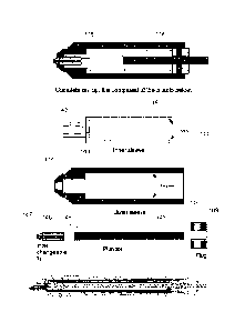
A material extraction device is shown in FIG. 8. Various parts are made using
an injection molding or sinter molding process and therefore are made of
plastic or
fused metal powder. The components are listed below.
- Two concentric syringe tubes, and inner tube 141 and an outer tube 143.
- A portion of the inner tube 142 is shaped to receive the cutting tip 147.
- A portion of the outer tube 144 is shaped to divert the dispensed liquid
onto the
cutting tip.
- A plunger 145 with a compression seal 149 against the inner wall of the
inner
syringe tube 141, which creates the plunger end chamber 116 and the non-
plunger end
chamber 115.
- An annular seal 146 between the inner and outer chambers located at the
plunger end.
- 21 -
CA C282S012 HI} 07 .24
WO 2012/102779 PCT/US2011/061075
- A slideable annular seal 109 between the plunger and the inner syringe tube.
- The cutting tip 147 that contacts the slide surface and displaces tissue.
- The cutting tip tube 148 that provides fluid communication between the slide
surface and the non-plunger end chamber 115 in order to aspirate the displaced
tissue
fragments.
- Two holes 111 providing fluid communication between the plunger end
chamber and the channels between the inner and outer syringe tubes.
Example 2: Material extraction system.
A material extraction system is shown in FIG. 9. The instrument head assembly
161 is mounted on a set of rails 162, which are mounted perpendicularly to the
plane of
the slide. Z-axis movement of the instrument head on the rails is controlled
by a linear
actuator 163, which controls contact of the cutting bit with the slide. The
pressure of the
cutting bit on the slide surface is created by the weight of the instrument
head assembly
riding on an adjustable spring 164. The instrument head assembly contains a
rotational
assembly with the axis of rotation oriented vertically and passing through the
center of
focus of the digital camera. The rotational assembly is comprised of an outer
cylinder
165 with a Morris taper 166 on the axis of rotation that matches the taper of
the cutting
bit. The outer cylinder is supported by bearings 167, which are held mounted
in the
instrument head assembly. The rotational assembly is also comprised of an
inner
cylinder 168, which is movable along the axis of rotation by a linear actuator
169. The
linear actuator is mounted to the instrument head assembly and is rotationally
decoupled from the rotational assembly by a bearing 170. The inner cylinder
contains
a grasping cassette 171, which allows reversible grasping of the cutting bit
plunger 101.
Control of the grasping cassette is via a rod 172, depression of which
releases the grip
of the grasping cassette on the plunger of the cutting bit and ejects the
cutting bit from
the Morris taper. Rotational force of the rotational assembly is generated by
a motor
173, which is mounted on the instrument head assembly.
Of course, it is to be understood that the above-described arrangements are
only
illustrative of the application of the principles of the present invention.
Numerous
modifications and alternative arrangements may be devised by those skilled in
the art
- 22 -
CA C282S012 HI} 07 .24
WO 2012/102779
PCT/US2011/061075
without departing from the spirit and scope of the present invention and the
appended
claims are intended to cover such modifications and arrangements. Thus, while
the
present invention has been described above with particularity and detail in
connection
with what is presently deemed to be the most practical and preferred
embodiments of
the invention, it will be apparent to those of ordinary skill in the art that
numerous
modifications, including, but not limited to, variations in size, materials,
shape, form,
fimction and manner of operation, assembly and use may be made without
departing
from the principles and concepts set forth herein.
- 23 -