Note: Descriptions are shown in the official language in which they were submitted.
CA 02826064 2015-08-26
FLASHBACK BLOOD COLLECTION NEEDLE
BACKGROUND OF THE INVENTION
1. Field of the Invention
[0001] The present invention relates to a device for collecting blood samples
by performing
venipuncture on a patient. More particularly, the present invention relates to
a needle assembly
for multiple sample blood collection that allows a phlebotomist to determine
whether vein entry
has occurred when collecting a blood sample from a patient into an evacuated
blood collection
tube.
2. Description of Related Art
[0002] Venipuncture is the primary method used for acquiring blood samples for
laboratory
testing. In performing venipuncture procedures, a phlebotomist must follow
several steps
simultaneously. Such steps include assessing the patient's overall physical
and psychological
condition so as to properly select a venipuncture site and technique. The
phlebotomist must also
select the proper corresponding equipment, perform the technique so as to
control bleeding, and
properly collect and identify fluid specimens for testing. The phlebotomist
must ascertain all of
these coinciding factors, as such factors may adversely affect the distension
of the vein and the
length of the venipuncture procedure.
[0003] Various venipuncture devices have been developed to address the above-
described
problems. These devices include products intended to assist the phlebotomist
in confirming that
vein entry has been made see e.g., United States. Patent Nos. 5,222,502 and
5,303,713. Such a
device contains a needle assembly with a housing that defines a chamber
therein. A single
cannula pointed at both ends is affixed to the housing. The intravenous (IV)
end of the cannula
is adapted for penetration of a patient's vein. The non-patient end of the
cannula has a sealable
sleeve and is adapted for penetration of a penetrable stopper positioned
within an evacuated
container.
1
CA 02826064 2015-08-26
[0004] Upon vein entry with the intravenous end of the cannula, blood will
flow through the
cannula, into the sealable sleeve and into the housing chamber, which is clear
or translucent for
visualization ("flashback"). Once air is vented from the flashback chamber,
the blood therein is
pressurized each time the sealable sleeve is pushed toward the housing chamber
upon activation
of an evacuated container.
[0005] Due to the length of time between vein entry and flashback, the
phlebotomist may
erroneously believe that satisfactory vein entry has not been achieved since
there is no immediate
indication of vein entry in the see-through chamber. The phlebotomist may
unnecessarily repeat
the venipuncture procedure, requiring replacement of the evacuated container
and/or the needle
assembly itself. Such a repetitive process prolongs the physical and emotional
discomfort
endured by the patient. In such cases, a phlebotomist may use a blood
collection set to provide
some entry indication, and will then incur the cost of the blood collection
set, as well as the cost
of a discard tube.
[0006] It would therefore be desirable to provide an improved blood collection
device that
permits blood flow through a relatively short flow path directly into a
flashback chamber,
thereby providing immediate indication of successful vein entry.
SUMMARY OF THE INVENTION
[0007] The invention provides a needle assembly for the extraction of at least
one fluid sample
into an evacuated container for laboratory testing. The needle assembly
provides a clear or
translucent housing with sufficient dead space for blood to flow into a
flashback chamber for
visualization by the user to confirm successful vein entry, with an internal
vent mechanism.
100081 In one embodiment, the invention relates to a needle assembly
comprising a housing
defining a housing interior, a cannula having a patient puncture tip extending
from a first end of
the housing, and a non-patient puncture tip extending from a second end of the
housing. The
non-patient puncture tip and the patient puncture tip are in fluid
communication with each other
through the cannula, such that the sole communication path between the housing
interior and the
external environment is via the patient puncture tip. A porous vent is
positioned within the
housing interior to separate the housing interior into a first chamber and a
second chamber, with
2
CA 02826064 2015-08-26
the cannula being in fluid communication with the first chamber. The porous
vent includes pores
for passage of blood therethrough from the first chamber to the second
chamber. The first
chamber and the second chamber are configured such that upon insertion of the
patient needle tip
into a patient, blood flows through the cannula and into the first chamber
without sealing the
porous vent. At this point in the process, the blood "flashback" can be
visualized in the first
chamber. Upon application of an evacuated container to the non-patient
puncture tip, blood is
drawn from the first chamber and air is drawn from the second chamber, thereby
establishing a
negative pressure within the second chamber with respect to an external
environment of the
needle assembly. Blood can thereafter be drawn into the first chamber and
through the porous
vent, with a negative pressure maintained in the second chamber.
[0009] In one embodiment, the cannula includes a first end comprising the
patient puncture tip
and a second end comprising the non-patient puncture tip, with an opening
between the first end
and the second end providing fluid communication between the cannula and the
first chamber of
the housing. In an alternate embodiment, the cannula comprises a first cannula
having a patient
puncture tip, with the needle assembly further comprising a second cannula
including the non-
patient puncture tip, with the first cannula and the second cannula
substantially axially aligned
and separated by a gap in fluid communication with the first chamber of the
housing. A sleeve
may also extend about the non-patient puncture tip.
[00010] In one embodiment, the second chamber may include multiple interior
regions in fluid
communication, such as a first interior region and a second interior region.
The first and second
interior regions of the second chamber are in fluid communication with each
other through the
porous vent.
[00011] In a particular embodiment, the first end of the housing comprises an
elongated
longitudinal first portion having a first diameter and the second end of the
housing comprises a
second portion having a second diameter larger than the first diameter of the
first portion. In
such an embodiment, the porous vent may be positioned within the housing
interior between the
first portion having a first diameter and the second portion having a second
diameter.
Alternatively, the porous vent may be positioned within the housing interior
at a location
spanning the transition between the first diameter of the first portion and
the second diameter of
3
CA 02826064 2015-08-26
the second portion. In embodiments where the second chamber includes multiple
interior
regions, such as a first interior region and a second interior region, the
first chamber may extend
along a portion of the longitudinal first portion, with at least one of the
interior regions, such as
the second interior region of the second chamber extending longitudinally
concentrically about
the first chamber. In this manner, the external diameter, and thus the
external profile of the
needle assembly, can be decreased.
[00012] In yet a further embodiment, a method of preventing leakage, such as,
for example,
blood droplets, at the patient puncture tip in a needle assembly is provided.
The method involves
receiving blood through a patient puncture tip and into a first chamber of a
needle assembly, with
the needle assembly including a needle housing defining a housing interior; a
cannula having the
patient puncture tip extending from a first end of the needle housing; a non-
patient puncture tip
extending from a second end of the needle housing, the non-patient puncture
tip and the patient
puncture tip being in fluid communication with each other through the cannula;
and a porous
vent positioned within the housing interior and separating the housing
interior into a first
chamber and a second chamber. The cannula is in fluid communication with the
first chamber
such that the sole communication path between the housing interior and the
external environment
is via the patient puncture tip, and the porous vent includes pores for
passage of blood
therethrough from the first chamber into the second chamber. Fluid
communication is
established between the non-patient puncture tip and an evacuated collection
container, such that
blood contained within the first chamber is drawn into the evacuated
collection container and air
is drawn out of the second chamber through the porous vent. As such, a
negative pressure is
established within the second chamber relative to the external environment of
the needle
assembly, such that blood flows through the cannula into the first chamber and
contacts the
porous vent. Blood is then drawn through the pores of the porous vent toward
the second
chamber such that after removing the patient puncture tip from the vasculature
of the patient any
blood contained within the cannula is displaced away from the patient puncture
tip toward the
second chamber based upon the negative pressure established within the second
chamber.
[00013] Additionally, a further step may include establishing fluid
communication between the
non-patient puncture tip and a second evacuated collection container prior to
drawing blood
through the patient puncture tip and through the cannula into the second
evacuated collection
4
CA 02826064 2015-08-26
container, followed by releasing the fluid communication between the non-
patient puncture tip
and the second evacuated collection container.
[00014] In yet a further embodiment, the invention is directed to a method of
collecting a
sample of blood from a patient into an evacuated blood collection tube using a
blood collection
assembly having a patient needle tip and a non-patient needle tip and a
housing having a
flashback visualization chamber. The method involves using a needle assembly
comprising a
housing having a porous vent positioned therein to separate an interior of the
housing into a first
chamber forming the flashback visualization chamber and a second chamber, the
first chamber
and second chamber being configured such that air is drawn out of the second
chamber through
the porous vent and into the evacuated blood collection tube along with the
blood sample,
thereby establishing a negative pressure within the second chamber. The
negative pressure
causes blood to be drawn into the first chamber and contact the porous vent,
such that after the
patient needle tip is removed from the patient, the negative pressure within
the second chamber
draws blood from the patient needle tip toward the second chamber, thereby
preventing leakage
of blood from the patient needle tip after removal from the patient.
[00015] In another embodiment, the invention is related to a needle assembly
having a housing
defining a housing interior wherein the housing comprises at least one cannula
having a patient
puncture tip extending from a first end of the housing and a non-patient
puncture tip extending
from a second end of the housing. The non-patient puncture tip and the patient
puncture tip are
in fluid communication with each other within the housing interior. The
assembly also includes
a porous vent positioned within the housing interior separating the housing
interior into a first
chamber and a second chamber within the housing interior. The porous vent
includes pores for
passage of fluid therethrough from the first chamber to the second chamber.
The porous vent
can comprise a tubular member including an axial bore which surrounds at least
a portion of the
at least one cannula. A blocking member is located adjacent to or within the
axial bore of the
porous vent and controls flow of the fluid through the vent such that the
fluid flows along the
longest path through the porous vent. This longest path depends upon the shape
of the porous
vent. In one embodiment wherein the porous vent is cylindrically or tubular
shaped having a
longer length than circumference, the flow can be in an axial direction along
a longitudinal path
through the porous vent. In another embodiment where the porous plug is washer
shaped having
CA 02826064 2015-08-26
a circumference which is greater than its length, the flow having the longest
path can be in the
radial direction. The porous vent with the blocking member reduces or
eliminates the amount of
uncontrolled flow of fluid through the porous vent along the shortest path or
the path of least
resistance. The sole communication path between the housing interior and the
external
environment is via the patient puncture tip. The porous vent has a first end
face, a second end
face, and a central portion extending between the first end face and the
second end face.
According to one embodiment, the porous vent is configured to cause the fluid
to flow along a
controlled longitudinal path from the first end face to either the central
portion or the second end
face of the porous vent and subsequently through a central opening between the
first and the
second chamber. This central opening can be located adjacent to the central
portion of the
porous vent. According to another embodiment, the porous vent can be
configured to cause the
fluid to flow along a controlled longitudinal path from the first end face to
the second end face
and subsequently into the second chamber through either the first end face
and/or the second end
face. According to yet another embodiment, the porous vent can be washer
shaped having the
first end face and the second end face blocked to cause the fluid to flow
along a controlled radial
path from an inner portion of the porous vent to the outer circumferential end
face of the porous
vent and into the second chamber.
1000161 The blocking member is configured to block at least a portion of the
porous vent to
render this portion of the vent non-porous to control the flow of fluid
therethrough. The
blocking member can block at least a portion of an inside surface of the axial
hole surrounding at
least a portion of the cannula. According to one embodiment, the blocking
member comprises a
non-porous bushing press-fitted into the inside surface of the porous vent.
This bushing can
comprise steel or any other type of metal cannula, an extruded plastic tube, a
tubular molded
part, and the like. The bushing can have a length that is substantially equal
to a length of the
porous vent. According to another embodiment, the blocking member can comprise
an adhesive
or sealant located in a space between an inside surface of the porous vent and
an outer diameter
of the cannula. According to another embodiment, the back end surface of the
porous member
can be blocked with an adhesive material to control the flow of fluid through
the porous vent.
According to yet another embodiment, the blocking member can be formed by
melting or fusing
the inner diameter surface portion of the porous vent. According to another
embodiment, the
6
CA 02826064 2015-08-26
blocking mem. ber can be a separate member, such as a plastic tubular or
cylindrical member that
is placed in abutting relationship with respect to the inside surface of the
porous vent. This
cylindrical member can extend from a portion of the housing.
[00017] According to one design, the at least one cannula can comprise a
single cannula
extending through the housing. The single cannula can include a lumen
extending therethrough,
a first end comprising the patient puncture tip, a second end comprising the
non-patient puncture
tip, and an opening through the cannula into the lumen at a location between
the first end and the
second end providing fluid communication between the lumen of the cannula and
the first
chamber of the housing. According to another embodiment, the at least one
cannula can
comprise a first cannula extending from the housing and comprising the patient
puncture tip, and
a second cannula extending from the housing and comprising the non-patient
puncture tip. The
first cannula and the second cannula are substantially axially aligned within
the housing interior
and separated from each other by a gap in fluid communication with the first
chamber of the
housing. The first chamber and the second chamber are configured such that
upon insertion of
the patient puncture tip into a patient causes blood to flow into the first
chamber without sealing
the porous vent, and upon application of a negative pressure source to the non-
patient puncture
tip, blood and air are drawn from the first chamber and air is drawn from the
second chamber,
thereby establishing a negative pressure within the second chamber with
respect to an external
environment of the needle assembly. Upon removal of the patient puncture tip
from the patient,
the negative pressure within the second chamber draws blood from the patient
needle tip toward
the second chamber to prevent blood droplets from being present at the patient
puncture tip.
[00018] In yet another embodiment, the invention relates to a needle assembly
comprising a
housing defining a housing interior. The housing comprises at least one
cannula having a patient
puncture tip extending from a first end of the housing and a non-patient
puncture tip extending
from a second end of the housing. The non-patient puncture tip and the patient
puncture tip are
in fluid communication with each other within the housing interior. A porous
vent is positioned
within the housing interior to separate the housing interior into a first
chamber and a second
chamber. The porous vent includes pores for passage of fluid therethrough from
the first
chamber to the second chamber. The porous vent is configured to control flow
of the fluid such
that the fluid flows along the longest path therethrough. The needle assembly
is designed such
7
CA 02826064 2015-08-26
that the sole communication path between the housing interior and the external
environment is
via the patient puncture tip and the first end of the housing comprises an
elongate longitudinal
first portion having a first diameter and the second end of the housing
comprises a second
portion having a second diameter larger than the first diameter of the first
portion. The porous
vent is positioned within the housing interior between the first portion
having a first diameter and
the second portion having a second diameter at a location spanning a
transition point between the
first diameter of the first portion and the second diameter of the second
portion. The porous vent
can comprise a tubular member having a first end face, a second end face, and
a central portion
located between the first end face and the second end face. The tubular member
further includes
an axial hole configured for surrounding at least a portion of the cannula.
The axial hole defines
an inside surface of the porous vent and the assembly further includes a
blocking member at the
inside surface of the axial hole for blocking at least a portion of the porous
vent to render this
portion of the vent non-porous to cause the fluid to flow along a controlled
longitudinal path
from the first end face to either the central location or the second end face
and subsequently
through a central aperture opening between the first chamber and the second
chamber. The
blocking member can be a non-porous bushing press-fitted into the inside
surface of the porous
vent or an adhesive located between an inside surface of the porous vent and
an outer diameter of
the cannula. Alternatively, the inside portion of the porous vent can be
rendered non-porous by
fusing this inner surface portion of the porous vent. According to another
embodiment, the
blocking member can be a separate member, such as a plastic tubular or
cylindrical member that
is placed in abutting relationship with respect to the inside surface of the
porous vent. This
cylindrical member can extend from a portion of the housing.
[00019] In another embodiment, the invention relates to a needle assembly
comprising a
housing defining a housing interior. The housing comprises at least one
cannula having a patient
puncture tip extending from a first end of the housing and a non-patient
puncture tip extending
from a second end of the housing. The non-patient puncture tip and the patient
puncture tip are
in fluid communication with each other within the housing interior. A porous
vent is positioned
within the housing interior separating the housing interior into a first
chamber and a second
chamber. The porous vent includes pores for passage of fluid therethrough from
the first
chamber to the second chamber. The porous vent is configured to control flow
of the fluid such
8
CA 02826064 2015-08-26
that the fluid flows in an axial direction therethrough. The housing includes
a rear hub which
can block a back end face of the porous vent. Alternatively, the rear hub can
leave a portion of
the second end face exposed. The rear hub includes a cylindrical portion
extending therefrom.
This cylindrical portion extends into the first chamber toward the first end
of the housing to
define a portion of the first chamber. The needle assembly is designed such
that the sole
communication path between the housing interior and the external environment
is via the patient
puncture tip. The porous vent comprises a tubular member having a first end
face, a second end
face, and a central portion extending between the first end face and the
second end face. The
tubular member further includes an axial hole configured for surrounding at
least a portion of the
cylindrical portion extending from the rear hub. The at least one cannula is
located within at
least a portion of the cylindrical portion. The cylindrical portion extending
from the rear hub
into the axial hole of the porous vent abuts against the inside surface of the
porous vent to act as
a blocking member to render a portion of the vent non-porous and to cause the
fluid to flow
along a controlled longitudinal path and consequently through either a central
aperture opening
between the first and the second chamber or through either the first end face
or the second end
face of the porous vent. An adhesive can be located between an inside surface
of the porous vent
and an outer diameter of the cannula and/or the cylindrical portion or the
inner surface portion of
the porous vent can be fused to render this portion non-porous in order to
assist in controlling the
flow of fluid through the porous vent.
[00020] According to still another embodiment, the invention relates to a
method of preventing
leakage of a blood droplet from a patient puncture tip of a needle assembly.
The method
includes: a) receiving blood through a patient puncture tip and into a first
chamber of a needle
assembly, the needle assembly comprising: i) a needle housing defining a
housing interior, the
housing comprising at least one cannula having a patient puncture tip
extending from a first end
of the housing and a non-patient puncture tip extending from a second end of
the housing; and ii)
a porous vent positioned within the housing interior and separating the
housing interior into a
first chamber and a second chamber, with the non-patient puncture tip and the
patient puncture
tip being in fluid communication with each other within the first chamber such
that the sole
communication path between the housing interior and the external environment
is via the patient
puncture tip. The porous vent includes pores for passage of blood and air
therethrough from the
9
CA 02826064 2015-08-26
first chamber into the second chamber and the porous vent is configured to
control flow of the
blood and air such that the blood and air flows along the longest path
therethrough. The method
further includes: b) establishing fluid communication between the non-patient
puncture tip and a
negative pressure source such that blood contained within the first chamber is
drawn out of the
non-patient puncture tip and air is drawn out of the second chamber through
the porous vent,
thereby establishing a negative pressure within the second chamber relative to
the external
environment of the needle assembly such that blood flows through the cannula
into the first
chamber and contacts the porous vent; and c) drawing blood and air through the
pores of the
porous vent toward the second chamber based upon the negative pressure
established within the
second chamber such that blood contained within a lumen of the patient
puncture tip is displaced
away from the patient puncture tip and toward the second chamber. The method
is such that the
receiving step a) comprises receiving blood through the lumen of the patient
puncture tip from a
patient's bloodstream, and the drawing step c) displaces blood away from the
patient puncture tip
after removing the patient puncture tip from the source of blood, such as, for
example, from the
vein. The method further includes the step that after step b) and prior to
step c), releasing the
fluid communication between the non-patient puncture tip and the negative
pressure source. The
porous vent can comprise a tubular member having a first end face and a second
end face and
wherein the tubular member further includes an axial hole configured for
surrounding at least a
portion of the cannula. The method includes rendering the inside surface of
the axial hole non-
porous to cause the fluid to flow along the longest path through the porous
vent and subsequently
into the second chamber. Depending upon the shape of the porous vent, this
longest path can be
a longitudinal path or a radial path. A blocking member can be provided to
render the inside
surface of the axial hole non-porous. This blocking member can be a bushing
formed from a
non-porous metal or plastic material press-fitted into the inside surface of
the porous vent, an
adhesive located between an inside surface of the porous vent and an outer
diameter of the
cannula, a fused inner surface portion of the porous vent, and/or a separate
member that is placed
in an abutting relationship with respect to the inside surface of the porous
vent.
DESCRIPTION OF THE DRAWINGS
[00021] FIG. 1 is a cross-sectional view of a typical embodiment of the needle
assembly of the
present invention.
CA 02826064 2015-08-26
[00022] FIG. 2 is a cross-sectional view of a second embodiment.
[00023] FIG. 3 is a cross-sectional view of a third embodiment.
[00024] FIG. 4 is a cross-sectional view of a fourth embodiment.
[00025] FIG. 5 is a schematic view of the needle assembly of FIG. 1 prior to
use.
[00026] FIG. 6 is a schematic view similar to FIG. 5, but showing the first
sign of venous
entry.
[00027] FIG. 7 is a schematic view of a fifth embodiment.
[00028] FIG. 8 is a perspective view of a needle assembly having a flash
chamber in a further
embodiment.
[00029] FIG. 9 is a rear perspective view of the needle assembly having a
flash chamber of
FIG. 8.
[00030] FIG. 10 is an exploded view of the needle assembly having a flash
chamber of FIG. 8.
[00031] FIG. 11A is a cross-sectional view of the needle assembly having a
flash chamber of
FIG. 8.
[00032] FIG. 11B is an enlarged cross-sectional view of a portion of the
needle assembly of
FIG. 11A.
[00033] FIG. 12A is a cross-sectional view of a needle assembly having a flash
chamber used
in connection with a blood collection assembly in yet a further embodiment.
[00034] FIG. 12B is an enlarged sectional view of a portion of the needle
assembly of FIG.
12A.
[00035] FIG. 13A is a cross-sectional view of a needle assembly having a flash
chamber used
in connection with a blood collection assembly in yet a further embodiment.
11
CA 02826064 2015-08-26
[00036] FIG. 13B is an enlarged sectional view of a portion of the needle
assembly of FIG.
13A.
[00037] FIG. 13C is an enlarged sectional view of a portion of the needle
assembly of FIG.
13B.
[00038] FIG. 14 is a perspective view of the needle assembly of FIG. 13A shown
in
combination with a blood collection holder, with a needle shield in a
shielding position.
[00039] FIG. 15 is a side view of the needle assembly of FIG. 14.
[00040] FIG. 16 is an enlarged side sectional view of the needle assembly of
FIG. 15 without
the cannula.
[00041] FIG. 17 shows a cross-sectional view of the needle assembly of the
invention, without
the cannula, according to one design including a blocking member located
within the porous
vent.
[00042] FIG. 18 shows a perspective view of the porous vent shown in FIG. 17.
[00043] FIG. 19 shows a cross-section view of the porous vent taken along line
XIX-XIX of
FIG. 18.
[00044] FIG. 20 is a cross-sectional view of the needle assembly using a
porous vent without a
blocking member showing the uncontrolled radial flow of fluid along the
shortest path through
the porous member.
[00045] FIG. 21 is a cross-sectional view of the needle assembly of the
invention including the
blocking member of the invention and showing one embodiment of the controlled
flow of fluid
along the longest path through the porous vent wherein the longest path is in
the longitudinal
direction.
[00046] FIG. 22A shows a cross-sectional view of the needle assembly according
to an
alternative design of the invention.
12
CA 02826064 2015-08-26
[00047] FIG. 22B shows a cross-sectional view of the needle assembly according
to another
design of the invention.
[00048] FIG. 22C shows a cross-sectional view of the needle assembly according
to yet
another design of the invention.
[00049] FIG. 22D shows a cross-sectional view of the needle assembly according
to still
another design of the invention.
[00050] FIG. 23A shows a cross-sectional perspective view of the needle
assembly of the
invention according to another design.
[00051] FIG. 23B shows a close-up cross-sectional perspective view of the
porous vent and
chamber arrangements of FIG. 23A.
[00052] FIG. 24 shows a cross-sectional view of the needle assembly of the
invention
according to another design.
[00053] FIG. 25 is a cross-sectional view of the needle assembly of the
invention including the
blocking member of the invention and showing another embodiment of the
controlled flow of
fluid along the longest path through the porous vent wherein the longest path
is in a radial
direction.
DETAILED DESCRIPTION
[00054] An embodiment of the invention provides a needle assembly for blood
collection that
provides a visual indication of vein entry ("flashback") upon collection of a
blood or other fluid
sample from a patient into one or more evacuated blood collection tubes and
inhibits leakage of
the blood or fluid sample from the IV cannula on removal from the patient.
[00055] Various embodiments of the present invention are shown in the FIGS.
With reference
to FIGS. 1-6, this embodiment is directed to a needle assembly 210 with a
housing 212 having a
fluid inlet end 214, a fluid outlet end 216 and a frustum-shaped exterior wall
218 extending
between the ends. Exterior wall 218 defines the housing interior 220. Housing
212 further
13
CA 02826064 2015-08-26
includes a cylindrical interior wall 224 that extends in the housing interior
220 from fluid inlet
end 214 substantially concentrically with cylindrical exterior wall 218 to a
vent plug 900.
Cylindrical interior wall 224 and vent plug 900 define a flashback chamber
226.
[00056] Needle assembly 210 also includes a fluid inlet cannula 236 having an
exterior end that
defines a sharpened bevel and an interior end 244 that is mounted fixedly in
fluid inlet end 214
of housing 212. Fluid inlet cannula 236 is characterized further by a
substantially cylindrical
lumen extending between the ends and communicating with the interior of
housing 212.
[00057] Needle assembly 210 further includes a fluid outlet cannula 252. With
reference to
FIGS. 5-6, outlet cannula 252 concludes a blunt interior end 254, an exterior
end defining a
sharpened bevel and a substantially cylindrical lumen extending between the
ends. Portions of
outlet cannula 252 between the ends are securely affixed in outlet end 216 of
housing 212.
Outlet cannula 252 is mounted so that interior end 254 passes substantially
coaxially into interior
wall 224 and so that interior end 254 of outlet cannula 252 substantially
aligns axially with
interior end 244 of inlet cannula 236. Additionally, interior end 254 of
outlet cannula 252 is
spaced only a small distance from interior end 244 of inlet cannula 236. An
axial gap between
interior end 254 of outlet cannula 252 and interior end 244 of inlet cannula
236 that is less than
0.5mm may result in a flashback that is inconsistent.
[00058] Cylindrical interior wall 224 is dimensioned relative to outlet
cannula 252 to achieve
both desirable flow of blood through assembly 210 and to achieve effective
flashback indication.
In particular, cylindrical interior wall 224 preferably is dimensioned to
provide a radial gap
around outlet cannula 252 of about 0.2mm, as indicated by dimension "c" in
FIG. 1. This gap
achieves a substantially laminar blood flow within flashback chamber 226 and
prevents blood
hemolysis. Additionally, the small radial gap between cylindrical inner wall
224 and outlet
cannula 252 enables a drop of blood to be spread thinly across the radial gap
in flashback
chamber 226 to provide a magnified flashback indication with a very small
volume of blood.
Thus, an easily visualized flashback indication is achieved quickly at the
first appearance of
blood from interior end 244 of inlet cannula 236.
[00059] Needle assembly 210 further includes a sealable sleeve 261 mounted to
fluid outlet end
216 of housing 212 and covering exterior end 258 of outlet cannula 252 when
sealable sleeve
14
CA 02826064 2015-08-26
261 is in an unbiased condition. However, sealable sleeve 261 can be collapsed
in response to
pressure exerted by the stopper of an evacuated tube for urging exterior end
260 of outlet cannula
252 through both sealable sleeve 261 and stopper of an evacuated tube, as
known in the art.
[00060] The above embodiment is described in terms of a vent plug. However,
any vent
mechanism is suitable. The vent mechanism may be, for example, a porous vent
plug formed
from a matrix or carrier material, typically hydrophobic, that is coated with,
impregnated with, or
otherwise, contains a hydrophilic material that swells on contact with aqueous
or water
containing substances. The hydrophobic carrier material can be but is not
limited too, high-
density polyethylene, polytetrafluoroethylene, ultra-high molecular weight
polyethylene, Nylon
6, polypropylene, polyvinylidine fluoride and polyethersulfone. The swellable
nature of the
hydrophilic material thereby provides the sealing function in the vent upon
contact with blood. It
is also possible to use a porous vent plug that becomes sealed upon contact
with blood using
biological phenomena, e.g., by clotting and/or cell agglutination that blocks
the vent; a
superabsorbant material to seal the vent by swelling on contact with an
aqueous fluid; a porous
vent configured to form a tortuous path for fluid movement therethrough; or a
one-way valve,
(e.g., a thin flap such as plastic film covering a vent, a deformable seal
such as a rubber or plastic
duckbill valve, or a deformable wrap over a vent). It should be noted that any
combination of
these various mechanisms is also possible.
[00061] FIGS 2-4 show embodiments with varying vent plugs. FIG. 2 shows a vent
plug
900a, which is located at the end of the cylindrical inner wall 224a and
fitted into a recess 301 in
the housing interior non-patient wall 300. FIG. 3 shows a vent plug in a
similar location to that
of FIG. 2, however, vent plug 900b has a shoulder 901b. FIG. 4 shows a vent
plug 900c that is
located both within the cylindrical inner wall 224c and the recess 301 in the
housing interior non-
patient wall 300, and has a shoulder 901c. The vent plug location in each of
these embodiments
is such that no air can flow out of the flashback chamber 226 into the housing
interior 220
without passing through the vent mechanism (900 a, b, c).
[00062] FIGS. 5 and 6 provide schematic representations of the needle assembly
210 of FIG. 1
before and after a conventional venipuncture, in which, the needle assembly
210 is connected to
a holder (not shown) and punctures the patient's skin to make a vein entry.
Upon vein entry,
CA 02826064 2015-08-26
blood enters the IV cannula 236 and flows toward the flashback chamber 226.
The blood flows
from inlet cannula 236 into the space between inlet and outlet cannula, such
that blood flows
both into the outlet cannula 252 and into flashback chamber 226. At this point
in time, flashback
chamber 226, indicates successful vein entry and reduces the volume of air
present in housing
212 shown in FIG. 6. Air that was at atmospheric pressure within the lumen of
the IV cannula
248, flashback chamber 226, housing interior 220, and the lumen of the non-
patient cannula 262
prior to vein entry, thus experiences compression due to the influence of
venous pressure and this
air is therefore forced through the IV cannula 236 shown in FIG. 6 into the
flashback chamber
226 and through the vent plug 900 into chamber 220. Blood flow into housing
interior 220 is
prevented by the vent plug 900, which allows the pressurized air to flow
through it, but seals, and
sometimes completely seals, on contact with blood, thereby trapping the
compressed air (at
venous pressure) in housing interior 220. Blood flow in the entire needle
assembly ceases once
the pressure within chamber 226 and the venous pressure are equal.
[00063] Once the steps set forth in the previous paragraph occur, and venous
entry is visually
confirmed by the phlebotomist, an evacuated container (not shown), is then
inserted into the
holder such that exterior end 260 of second cannula 252 penetrates the stopper
of the container,
as known in the art. Upon penetration of the stopper by second cannula 252, a
negative pressure
gradient is transmitted to chamber 226, causing blood to flow from chamber 226
into the
container.
[00064] The needle assemblies described above desirably should be small for
convenient use,
but should be constructed to ensure reliable and rapid flashback. The
occurrence of flashback in
the needle assemblies described and illustrated above operate pursuant to the
ideal gas law. In
particular, at very low densities all gases and vapors approach ideal gas
behavior and closely
follow the Boyle's and Charles' laws given by:
Pi V1 = P2 V2
where:
Pi denotes the pressure of air within the needle assembly before needle
insertion;
P2 denotes the pressure of air within the needle assembly after vein entry;
16
CA 02826064 2015-08-26
V1 denotes the volume of air within the needle assembly before vein entry; and
V2 denotes the volume of air within the needle assembly after vein entry.
[00065] Design parameters should keep the needle device as small as possible
for easy use,
while ensuring an appropriate volume as specified by the preceding equation.
FIGS. 5 and 6
provide schematic representations of the needle assembly 210 of FIG. 1 for
purposes of
depicting the application of the ideal gas law. In this regard, A identifies
the volume of lumen
248 through inlet cannula 236. B denotes the total volume of the housing
interior 220, flashback
chamber 226, lumen 242 through outlet cannula 252 and sealable sleeve 261.
Referring again to
the preceding equation, P1 is the pressure within needle assembly 210 before
use, and hence
substantially equals atmospheric pressure. Atmospheric pressure will vary
slightly from time to
time and from location to location. However, for purposes of this analysis,
atmospheric pressure
P1 will be assumed to be 760mm Hg. V2 in the preceding equation is the volume
of the dead
space in needle assembly 210 after vein entry. More particularly, after vein
entry, blood will fill
lumen 248 of inlet cannula 236, thereby reducing the volume to be occupied by
gas in remaining
portions of needle assembly 210 and hence increasing the pressure of air in
the remaining portion
of needle assembly 210. A needle assembly with dimensions approximately as
shown in FIG. 1
will have a pressure P2 of about 790mm Hg at venous pressure (with
tourniquet). V1 in the
preceding equation defines the volume of the total dead spaced in needle
assembly 210 before
use, and hence will equal A + B as shown in FIG. 5. V2 defines the dead space
in the device
after vein entry, and with lumen 248 of inlet cannula 236 filled with blood.
Hence, V2 in the
preceding equation will equal B. These input parameters can be employed to
define a minimum
desired size for the respective components of needle assembly 200 as shown in
the following
application of the ideal gas law equation.
Pt V1 ¨ P2 V2
Pi/P2 = V2/ VI
760/790 = B/(A+B)
0.962 = B/(A+B)
0.962(A+B) = B
0.038B = 0.962A
17
CA 02826064 2015-08-26
B=25 .3A
[00066] Therefore, dead space in housing 212, outlet cannula 252 and sleeve
261
advantageously is at least 25.3 times the volume defined by lumen 248 through
inlet cannula 236
and most advantageously is about 26 times the volume of lumen 248. However,
other
configurations are possible and will function as described herein.
100067] The immediate response when an evacuated tube is placed in
communication with
outlet cannula 252 is to draw blood from the vein into tube (not shown). The
highest-pressure
gradient is always maintained between the vein and the evacuated tube. An
axially aligned inlet
cannula 236 and outlet cannula 252, therefore provide an unobstructed path for
blood flow from
the vein into evacuated tube.
[00068] When the requisite tubes are filled with blood, the needle assembly is
removed from
the vein. The sealed nature of the vent plug 900 inhibits the pressurized air
within housing
interior 220 from then moving into the flashback chamber 226 and into the
inlet cannula 236,
which could promote dripping of blood from the IV cannula tip.
[00069] The preceding embodiments show structurally separate inlet and outlet
cannulas that
are axially aligned with one other and placed in close end-to-end relationship
with one another.
However, the principles of the invention described above also can be achieved
with a single
cannula formed with a transverse slot or aperture within the flashback
chamber. For example,
FIG. 7 schematically shows a needle assembly 310 with a housing 312 and
housing interior 320
that is substantially identical to housing 212 described and illustrated
above. Needle assembly
310 differs from needle assembly 210 in that a single double end needle
cannula 336 is provided
and passes entirely through housing 312. More particularly, needle cannula 336
includes a
venous entry end 338, a non-patient end 340 and a lumen 342 extending
therebetween. Portions
of cannula 336 within inner wall 324 include a slot or aperture 344 to provide
communication
between lumen 342 and flashback chamber 326 within inner wall 324. Needle
assembly 310
functions substantially in the same manner as needle assembly 210 described
and illustrated
above.
18
CA 02826064 2015-08-26
[00070] FIGS. 8-10, 11A, and 11B depict a needle assembly in yet a further
embodiment of
the invention. In certain embodiments of the needle assembly described with
respect to FIGS. 1-
7, the housing interior includes a vent plug 900, which seals the flashback
chamber 226/326 from
the housing interior 220/320. In such previously described embodiments, the
vent plug is
described as sealing upon flow of blood into the flashback chamber, thereby
inhibiting any
pressurized air that may build up within the housing chamber 220/320 (such as
upon
displacement of air from the flashback chamber 226/326 into the housing
chamber 220/320
during the initial flash procedure) from moving in a reverse direction toward
the inlet cannula.
In the embodiment of FIGS. 8-10, 11A and 11B, a porous vent is positioned
within the housing
at a location such that the vent divides the housing into two chambers having
sizes and
dimensions to establish predetermined volumes thereto. Moreover, the porous
vent remains
porous to blood and does not seal upon contact with blood. Desirably the blood
does not contact
the porous vent at the initial flash indication, but such contact occurs at a
later point during use
of the assembly, as will be described in more detail herein.
[00071] For example, FIGS. 8-10, 11A, and 11B show a needle assembly 410
similar to that
described in connection with FIG. 1-6 above. As shown in FIGS. 8-10, 11A, and
11B, needle
assembly 410 includes a housing 412 having a fluid inlet end or first end 414
and a fluid outlet
end or second end 416. Needle assembly 410 includes exterior wall 418 defining
the housing
interior. Exterior wall 418 extends generally longitudinally at the first end
414 forming an
elongate longitudinal first portion 419 having a first diameter. At second end
416, exterior wall
418 forms a second portion 421 that has a second diameter that is generally
larger than the first
diameter of the first portion 419. Accordingly, housing 412 may form a
structure having a
generally T-shaped cross-section. The exterior wall 418 at second end 416 may
be a separate
element 428 that is attachable to main body portion 430 forming housing 412,
thereby assisting
in manufacture and assembly of needle assembly 410. First portion 419 and
second portion 421
may be arranged relative to each other in a variety of arrangements, so long
as they are capable
of functioning for transport of air therebetween as discussed herein.
[00072] Needle assembly 410 further includes a fluid inlet cannula 436
extending from first end
414 of housing 412. Fluid inlet cannula 436 includes an exterior end that
defines a first puncture
tip such as a sharpened bevel at patient puncture tip 438, and extends within
first end 414 of
19
CA 02826064 2015-08-26
housing 412 at open end 439, and may be fixedly mounted therein. Fluid inlet
cannula 436 is
characterized further by a substantially cylindrical lumen extending between
the ends and
communicating with the interior of housing 412.
[00073] Needle assembly 410 also includes a second puncture tip such as non-
patient puncture
tip 462 extending from second end 416 of housing 412. As seen in FIG. 10, this
may be
accomplished by providing needle assembly 410 with a second cannula in the
form of fluid
outlet cannula 452. In particular, the end of fluid outlet cannula 452 may
define a sharpened
bevel forming non-patient puncture tip 462. Fluid outlet cannula 452 extends
within second end
416 of housing 412, and may be fixedly mounted therein. Fluid outlet cannula
452 is
characterized further by a substantially cylindrical lumen communicating with
the interior of
housing 412. Outlet cannula 452 is mounted within housing 412 so that an
interior end 464
passes substantially coaxially therein such that outlet cannula 452
substantially aligns axially
with the interior end of inlet cannula 436. Desirably, this is achieved by
mounting outlet cannula
452 at a location adjacent second end 416 of housing 412, such that the
interior end 464 of outlet
cannula 452 extends within housing 412 to a location adjacent the interior end
439 of inlet
cannula 436. As seen in FIG. 11B, the interior end 464 of outlet cannula 452
is spaced only a
small distance from the interior end 439 of inlet cannula 436, thereby forming
an axial gap
therebetween for flow of blood into flashback chamber 426 about outlet cannula
452. The
distance between the interior end 464 of outlet cannula 452 and the interior
end 439 of inlet
cannula 436 forming the axial gap is sufficient to provide for flow of blood
into flashback
chamber 426, based upon the patient's blood pressure after venipuncture.
In certain
embodiments, an axial gap that is less than 0.5mm may result in a flashback
that is inconsistent.
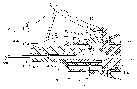
[00074] As seen in FIG. 11B, fluid inlet cannula 436 and fluid outlet cannula
452 are
positioned and dimensioned within housing 412 so as to achieve both desirable
flow of blood
through assembly 410 and to achieve effective flashback indication. In
particular, wall 418 of
housing 412 is dimensioned to provide a radial gap around outlet cannula 452
of about 0.2 mm at
an area surrounding the internal end 464 thereof. This gap achieves a
substantially laminar blood
flow within flashback chamber 426 and prevents blood hemolysis. Additionally,
the small radial
gap between the inner surface of wall 418 and outlet cannula 452 at the area
surrounding the
internal end 464 enables a drop of blood to be spread thinly across the radial
gap in flashback
CA 02826064 2015-08-26
chamber 426 to provide a magnified flashback indication with a very small
volume of blood.
Thus, an easily visualized flashback indication is achieved quickly at the
first appearance of
blood within flashback chamber 426. It is contemplated that internal end 464
of outlet cannula
452 may be partially supported within housing 412, so long as blood flow into
flashback
chamber 426 is achieved about the internal end 464.
[00075] In an alternate arrangement, a single cannula is provided, similar to
that embodiment
discussed in connection with FIG. 7. Such an arrangement is depicted in the
embodiment of
FIG. 12A and 12B (shown in connection with a blood collection assembly as will
be described
in more detail herein). In such an arrangement, the fluid inlet cannula and
the fluid outlet
cannula represent one single cannula 470, having a patient puncture tip 438 a
non-patient
puncture tip 462, and a lumen 442 extending therethrough, and with the body of
the cannula 470
being fixedly attached to a portion of the housing 412 and passing entirely
through housing 412.
A portion of cannula 470 extending through housing 412 includes one or more
openings such as
a slot or aperture 444 to provide communication between lumen 442 and
flashback chamber 436
within housing 412. In the embodiment seen in FIGS. 12A and 12B, two semi-
circular cuts
forming the aperture are shown on opposing sides of cannula 470, although it
is contemplated
that any number of such openings can be included to provide for blood flow
into flashback
chamber 426.
[00076] Returning to the embodiment of FIGS. 8-10, 11A, and 11B, needle
assembly 410
further includes a sealable sleeve 461 mounted to fluid outlet end 416 of
housing 412. This may
be accomplished by providing a mounting protrusion 429 at second end 416 of
housing 412, such
as on element 428, with sealable sleeve 461 representing an elastomeric
element that can be
frictionally fit or otherwise affixed over protrusion 429. Sealable sleeve 461
covers non-patient
puncture tip 462 at the exterior end of outlet cannula 452 when sealable
sleeve 461 is in an
unbiased condition. However, sealable sleeve 461 can be collapsed in response
to pressure
exerted by the stopper of an evacuated tube for urging exterior end 462 of
outlet cannula 452
through both sealable sleeve 461 and the stopper of an evacuated tube, as
known in the art.
[00077] The embodiment of FIGS. 8-10, 11A, and 11B further includes a porous
vent 910
positioned within the interior of housing 412. Porous vent 910 is positioned
within housing 412
21
CA 02826064 2015-08-26
to divide housing 412 into two distinct chambers, namely, a first chamber
represented by
flashback chamber 426 and a second chamber represented by secondary chamber
427. Porous
vent 910 may be constructed of a suitable material as described above with
respect to vent plug
900, albeit without the hydrophilic material that swells on contact. In this
manner, porous vent
910 is adapted to vent air therethough, and represents a porous structure
including a plurality of
pores that allow for passage of blood therethrough without sealing from fluid
flow therethrough
upon contact with blood, as is known in the art with vent plugs including a
hydrophilic material.
As discussed in more detail herein, during use of needle assembly 410, the
internal pores within
porous vent 910 at least partially fill with blood due to the negative
pressure established within
secondary chamber 427. Such filled pores in combination with the negative
pressure within
secondary chamber 427 prevent air flow between the secondary chamber 427 and
the flashback
chamber 426, and provide for fluid resistance of the blood flow through porous
vent 910, as will
be described in further detail.
[00078] Desirably, porous vent 910 is positioned within the interior of
housing 412 between
first portion 419 and second portion 421. In this manner, first portion 419 of
housing 412
essentially defines the flashback chamber 426, and second portion 421 of
housing 412 essentially
defines the secondary chamber 427. Alternatively, porous vent 910 may be
positioned within the
interior of housing 412 at a location spanning the transition between the
first diameter of first
portion 419 and the second diameter of second portion 421, as shown in the
embodiment of
FIGS. 12A and 12B. In any event, porous vent 910 is generally a cylindrically-
shaped member
with a central opening therein axially encircling a portion of the cannula,
particularly fluid outlet
cannula 452.
[00079] The interior volume of housing 412 is defined by the sum of the
volumes of flashback
chamber 426 and secondary chamber 427 as well as the volume represented by the
pores of
porous vent 910. Such interior volume is configured so as to provide for
certain attributes to the
needle assembly 410, in particular with respect to the ability of the
secondary chamber 427 to be
at least partially evacuated of a portion of the air therein to establish a
negative pressure therein
upon application of an evacuated tube to needle assembly 410 during use
thereof Such negative
pressure within secondary chamber 427 draws blood through the pores of porous
vent 910 based
on when blood contacts porous vent 910 and partially fills the pores thereof
In a particular
22
CA 02826064 2015-08-26
embodiment of the invention, the overall interior volume of housing 412 may be
from about 300
mm3 to about 400 mm3. Such a volume is particularly useful for the intended
use of needle
assembly 410 for conventional venipuncture for drawing a blood sample from a
patient using a
needle cannula having a conventional gauge for venipuncture as is known in the
art. Such a
volume also enables the needle assembly to be particularly useful with
patients having relatively
low blood pressure, in that the interior volume of the housing 412 is
sufficient so as to allow
adequate displacement of air so that blood will travel the complete length of
fluid inlet cannula
436 and into flashback chamber 426.
1000801 Porous vent 910 is desirably positioned within housing interior so as
to define
flashback chamber 426 as having a volume that represents from about 5 percent
to about 20
percent of the total overall volume of housing 412, desirably from about 7
percent to about 12
percent of the total overall volume of housing 412, including the volume of
secondary chamber
427 and the volume of the pores within porous vent 910. In this manner, the
remaining internal
volume of housing 412, defined by the internal volume positioned downstream
from the interface
between porous vent 910 and flashback chamber 426 including the internal pores
of porous vent
910 and the volume of secondary chamber 427, represents a significant portion
of the internal
volume of housing 412. Such a ratio of the flashback chamber 426 to the total
overall volume of
the housing 412 assures that flashback chamber 426 has sufficient volume to
properly visualize
the initial flash, desirably while preventing blood from fully contacting the
porous vent 910 at
initial venipuncture, based on the initial build-up of pressure within
secondary chamber 427
caused by venous pressure forcing the blood into flashback chamber 426. Such
volume ratios
are effective for the intended use as described in further detail herein,
wherein blood flowing into
flashback chamber 426 upon initial venipuncture does not fully contact porous
vent 910, and
desirably does not contact porous vent 910, and wherein at least a portion of
the air is drawn out
from secondary chamber 427 based upon application of an evacuated blood
collection tube to the
needle assembly 410. In this manner, secondary chamber 427 can effectively
draw blood from
within flashback chamber 426 and from within fluid inlet cannula 426 toward
secondary
chamber 427, such as into and through the pores of porous vent 910, so that
when the patient
puncture tip 438 is removed from the patient and is exposed to the external
environment, blood is
drawn away from the pucture tip 438, preventing the leakage of blood droplets
from the puncture
23
CA 02826064 2015-08-26
tip 438. In one particular embodiment, the total interior volume of the
housing 412 is about 380
mm3, with the flashback chamber 426 having a volume of about 30 mm3, the
secondary chamber
427 having a volume of about 300 mm3, and the pores of the porous vent 910
representing a
volume of about 50 mm3.
[00081] Needle assembly 410 may be assembled as follows. Fluid inlet cannula
436 is
positioned through first end 414 of housing 412 such that the open interior
end 439 is positioned
within an interior portion of housing 412 at first portion 419 and patient
puncture tip 438 extends
externally of first end 414. Fluid outlet cannula 452 is positioned within
housing 412 through
the opposite end, such that open internal end 464 is positioned within an
interior portion of
housing 412 at first portion 419 adjacent interior end 439 of fluid inlet
cannula 436, with a slight
gap therebetween, and with non-patient puncture tip extending externally of
second end 416.
Fluid inlet cannula 436 and fluid outlet cannula 452 may be affixed therein in
any known
manner, desirably through a medical grade adhesive.
[00082] In alternate embodiments including only a single cannula 470, such
cannula 470 is
affixed within housing 412 such that opening 444 is positioned within the
interior of housing 412
at first portion 419, with patient puncture tip 438 extending externally of
first end 414 and non-
patient puncture tip 462 extending externally of second end 416.
[00083] Porous vent 910 is then inserted within housing 412 and positioned
over fluid outlet
cannula 454 (or over the single cannula 470), and element 428 is thereafter
affixed to the second
end 416, enclosing the interior of housing 412. Sealable sleeve 461 is then
affixed over
protrusion 429. As such, the interior of housing 412 is closed from the
external environment,
with the sole path for fluid communication between the interior of housing 412
and the external
environment being provided through the patient puncture tip 438.
[00084] Needle assembly 410 assembled as such can be used in connection with a
blood
collection tube holder 800, as depicted in the embodiment shown in FIGS. 12A
and 12B. Such
assembly may be accomplished through the rear open end of blood collection
tube holder 800, so
that the entire needle assembly 410 is inserted to a portion where at least
patient puncture tip 438
and at least a portion of inlet cannula 436 extend out through the front end
of blood collection
tube holder 800. In embodiments where second portion 421 of needle assembly
410 is radially
24
CA 02826064 2015-08-26
larger than first portion 419, such an insertion and arrangement enables the
secondary chamber
427 to be fully contained within the internal space within collection tube
holder 800, and with
flashback chamber 426 extending out from a front end thereof.
[00085] In use, needle assembly 410 may be provided with collection tube
holder 800 attached
thereto. Patient puncture tip 438 is inserted through the skin of a patient
and into the patient's
vasculature, desirably into a vein. Upon venipucture, a closed environment is
achieved within
housing 412, since housing 412 is an entirely closed structure, and since
sealable sleeve 461
closes off the only outlet of housing 412 (i.e., fluid outlet cannula 452).
The patient's blood
pressure causes blood to flow through patient puncture tip 438, into fluid
inlet cannula 436, and
out interior end 439 (or through opening 444 in the embodiment of FIGS. 12A
and 12B), into
flashback chamber 426 surrounding interior end 464 of outlet cannula 452. The
transparent or
translucent nature of housing 412 permits visualization of the blood within
flashback chamber
426, providing an indication that venipuncture is achieved.
[00086] Since the interior of housing 412 is a closed environment, the flow of
blood into
flashback chamber 426 causes air to be trapped within the housing interior,
including within
flashback chamber 426, porous vent 910 and secondary chamber 427, as well as
within fluid
outlet cannula 452, causing such trapped air to be slightly pressurized
therein. Flashback
chamber 426 and secondary chamber 427 are configured through their size and
dimensions such
that the volumes thereof permit blood to flow into flashback chamber 426 at
this initial
venipucture, but the build up of air pressure within the pores of porous vent
910 and within
secondary chamber 427 prevents blood from fully contacting porous vent 910,
and desirably
prevents blood from even partially contacting porous vent 910 at the initial
venipuncture.
[00087] After such initial venipuncture and flash visualization, a sample
collection container
having a negative pressure therein, such as an evacuated blood collection tube
(not shown) as is
commonly known in the art, is inserted within the tube holder 800. The stopper
(not shown) of
such evacuated container contacts and displaces sealable sleeve 461, causing
non-patient
puncture tip 462 to puncture through sealable sleeve 461 and through the
stopper of the
evacuated container. At this point, fluid communication is established between
the non-patient
puncture tip 462 and the interior of the evacuated collection container. The
negative pressure
CA 02826064 2015-08-26
within the evacuated collection container draws the blood that has collected
within flashback
chamber 426 into fluid outlet cannula 452 and into the evacuated collection
container. Along
with the blood within flashback chamber 426, the negative pressure within the
evacuated
collection container will also draw at least a portion of the air out of the
flashback chamber 426
and out of the secondary chamber 427 through the pores of porous vent 910,
toward and into the
evacuated collection container. In addition, the close proximity and alignment
of fluid outlet
cannula 452 and fluid inlet cannula 436 causes blood to be drawn from fluid
inlet cannula 436
and from the patient, simultaneously with such air being drawn from the
flashback chamber 426
and secondary chamber 427.
[00088] Such drawing of air reduces the pressure within the flashback chamber
426 and the
secondary chamber 427, establishing a negative pressure therein with respect
to the patient's
bloodstream and with respect to the external environment. This negative
pressure that has been
established within the interior of housing 412, and specifically within
flashback chamber 426 and
secondary chamber 427, draws additional blood from within fluid inlet cannula
436 and from the
patient into flashback chamber 426, with the blood contacting porous vent 910.
With such blood
filling flashback chamber 426, the blood fully contacts the surface of porous
vent 910 that
extends within flashback chamber 426, and begins to fill the pores of porous
vent 910. Such
filling of the pores of porous vent 910 that are directly at the interface of
porous vent 910 and
flashback chamber 426 closes off the porous vent from airflow therethrough,
but does not fully
act as a seal, in that the blood does not cause the material of the porous
vent to swell or close off
to air flow, but instead merely physically fills the voids within the porous
vent. Moreover, since
a portion of the air within secondary chamber 427 has been drawn out from
secondary chamber
427, secondary chamber 427 represents a closed chamber with a negative
pressure therein
relative to the external environment. Since the volume of secondary chamber
427 represents a
substantial portion of the overall interior volume of housing 412, a
significant portion of interior
volume of housing 412 downstream of the filled pores at the interface of
porous vent 910 and
flashback chamber 426 remains at a negative pressure with respect to the
remainder of the
interior volume. Secondary chamber 427 will therefore continue to have a
drawing effect on the
blood within the pores of porous vent 910 and within flashback chamber 426
through the pores
of porous vent 910 toward secondary chamber 427, without releasing any air
from the secondary
26
CA 02826064 2015-08-26
chamber 427 in the opposite direction due to the pores of porous vent 910 at
the interface of the
flashback chamber 426 being filled with blood, thereby effectively preventing
air flow through
porous vent 910 due to the filled pores. The draw created by the negative
pressure within
secondary chamber 427 has a fluid resistance based on the blood filling the
pores of porous vent
910 and based on the tortuous path created by the pores of porous vent 910,
and therefore is a
gradual draw with reduced fluid movement.
[00089] At this point, the evacuated collection container and the secondary
chamber 427 are
both at a negative pressure with respect to the external environment (and with
respect to the
patient's bloodstream), and therefore both effect a draw from the fluid inlet
cannula 436. This
mutual drawing effect may essentially establish an equilibrium within the
flashback chamber
426, such that the blood contained within the flashback chamber 426 is not
drawn toward or into
either the secondary chamber 427 through the pores of porous vent 910 or into
the evacuated
collection container through the fluid inlet cannula 436, but instead
essentially remains within
flashback chamber 426 in a steady state. The negative pressure of the
evacuated collection
container draws blood directly from the patient through fluid inlet cannula
436, due to the close
proximity and alignment of fluid outlet cannula 452 and fluid inlet cannula
436, as well as due to
the equilibrium established within flashback chamber 426 (based on the
opposite draw forces
between the evacuated collection container and the evacuated secondary chamber
427). The
continual draw of blood into the evacuated collection container gradually
causes the pressure
within the collection container to increase.
[00090] Once the evacuated collection container is filled with the desired
amount of blood, the
container is removed from the non-patient puncture tip 462, thereby releasing
the fluid
communication between the non-patient puncture tip 462 and the evacuated
collection container,
with sealable sleeve 461 then covering and closing off non-patient puncture
tip 462. Absent such
draw from the negative pressure of the evacuated collection tube, the negative
pressure within
the secondary chamber 427 effects a slight draw on the blood within flashback
chamber 426
through the pores of porous vent 910. Such draw, however, is slow and gradual,
due to the
tortuous path of blood flow through the pores of porous vent 910.
27
CA 02826064 2015-08-26
[00091] Additional evacuated collection containers can thereafter be inserted
into tube holder
800 and used for sample collection through non-patient puncture tip 462 as
described above, by
placing a second evacuated collection container within the holder 800 and
establishing fluid
communication between the non-patient puncture tip 462 and the interior of the
evacuated
collection container by puncturing the stopper, as discussed. In such further
sampling, the
evacuated collection container and the secondary chamber 427 are both at a
negative pressure,
and therefore both effect a draw from the fluid inlet cannula. As above, this
effect essentially
establishes an equilibrium within the flashback chamber 426, thereby
preventing the blood
contained within the flashback chamber 426 from being drawn toward or into
either the
secondary chamber 427 (through the porous vent 910). The negative pressure of
the evacuated
collection container draws blood directly from the patient through fluid inlet
cannula 436 as
discussed above, due to the close proximity and alignment of fluid outlet
cannula 452 and fluid
inlet cannula 436. Once any such additional evacuated collection containers
are filled with the
desired amount of blood, the container is removed from the non-patient
puncture tip 462, thereby
releasing the fluid communication between the non-patient puncture tip 462 and
the evacuated
collection container, with sealable sleeve 461 then covering and closing off
non-patient puncture
tip 462.
[00092] Once all of the desired blood samples have been drawn in this manner,
patient puncture
tip 438 is removed from the vasculature of the patient (i.e., from the
bloodstream), thereby
exposing the opening of patient puncture tip 438 to the external environment.
Since the sole
communication path between the housing interior and the external environment
is through
patient puncture tip 438, the negative pressure established within secondary
chamber 427 relative
to the external environment will affect a gradual draw on the blood contained
within flashback
chamber 426 and within fluid inlet cannula 436 toward and through porous vent
910. Such
drawing effect will displace and move any blood contained within fluid inlet
cannula 436 away
from patient puncture tip 438, toward secondary chamber 427, thereby
preventing any blood
from leaking from patient puncture tip 438 out of fluid inlet cannula 436.
Such negative pressure
within secondary chamber 427 may continue to have a gradual drawing effect
through the porous
vent 910 for a prolonged period of time after removal of patient puncture tip
438 from the
patient, and may draw all of the remaining blood contained within fluid inlet
cannula 436 and
28
CA 02826064 2015-08-26
flashback chamber 426 through porous vent 910 and/or into secondary chamber
427. Needle
assembly 410 can then be properly disposed of in known manner.
[00093] FIGS. 13A, 13B, and 13C depict yet a further embodiment of a needle
assembly. The
needle assembly shown in FIGS. 13A-13C is similar to the embodiment described
above in
connection with FIGS. 8-10, 11A, and 11B, albeit with the secondary chamber
further
comprising a plurality of interior regions that are in fluid communication
with each other, and
desirably gas venting fluid communication, to define the interior volume of
the secondary
chamber.
[00094] In particular, as depicted in FIG. 13A, needle assembly 510 includes a
housing 512
having a fluid inlet end or first end 514 and a fluid outlet end or second end
516. Needle
assembly 510 further includes a fluid inlet cannula 536 extending from first
end 514 of housing
512. Fluid inlet cannula 536 extends between an exterior end that defines a
first puncture tip
such as a sharpened bevel at patient puncture tip 538, and an interior open
end 539 extending
within first end 514 of housing 512, and may be fixedly mounted therein. Fluid
inlet cannula
536 is characterized further by a substantially cylindrical lumen extending
between the ends and
communicating with the interior of housing 512.
[00095] Needle assembly 510 also includes a second puncture tip such as non-
patient puncture
tip extending from second end 516 of housing 512, such as through a second
cannula in the form
of fluid outlet cannula 552. In particular, the end of fluid outlet cannula
552 may define a
sharpened bevel forming non-patient puncture tip 562. Fluid outlet cannula 552
extends within
second end 516 of housing 512, and may be fixedly mounted therein. Fluid
outlet cannula 552 is
characterized further by a substantially cylindrical lumen communicating with
the interior of
housing 512. Outlet cannula 552 is mounted within housing 512 so that an
interior end 564
passes substantially coaxially therein such that outlet cannula 552
substantially aligns axially
with the interior end of inlet cannula 536, in a similar manner as discussed
in connection with the
embodiment depicted in FIGS. 8-10, 11A, and 11B described above. For example,
the interior
end 564 of outlet cannula 552 may be spaced only a small distance from the
interior end 539 of
inlet cannula 536, thereby forming an axial gap therebetween for flow of blood
into flashback
29
CA 02826064 2015-08-26
chamber 526 about outlet cannula 552 as shown in FIG. 13C, or may be a single
cannula having
an opening therein, as described in connection with the embodiment of FIGS.
12A-12B.
[00096] As shown in FIGS. 13A-13C, needle assembly 510 includes a generally
elongate
longitudinal portion at first end 514, which generally includes an interior
wall 515 and an
exterior wall 517. Interior wall 515 extends generally longitudinally within
housing 512, with a
first diameter defining an interior chamber in the form of flashback chamber
526. Second end
516 defines a second portion having a second diameter that is generally larger
than the first
diameter of interior wall 515. Interior wall 515 is dimensioned to provide a
radial gap around
outlet cannula 552 of about 0.2 mm at an area surrounding the internal end 564
thereof, thereby
achieving a substantially laminar blood flow within flashback chamber 526, as
discussed above.
Internal end 564 of outlet cannula 552 may be supported within housing 512, as
in the
embodiment discussed above. Needle assembly 510 may further include a sealable
sleeve 561
mounted to fluid outlet end 516 of housing 512, such as through a mounting
protrusion 529, as
discussed above.
[00097] As with the embodiment of FIGS. 8-10, 11A, and 11B, needle assembly
510 further
includes a porous vent 910a positioned within the interior of housing 512.
Porous vent 910a is
generally a cylindrically-shaped member with a central opening therein axially
spaced from and
encircling a portion of the cannula, particularly fluid outlet cannula 552.
Porous vent 910a may
be constructed of any suitable material as described above in connection with
the embodiment of
FIGS. 8-10, 11A, and 11B. Porous vent 910a is positioned within housing 512 in
a manner
such that housing 512 is divided into at least two distinct chambers, namely,
a first chamber
represented by flashback chamber 526 and a second chamber, representing the
total internal
volume of housing 512 that is positioned downstream of porous vent 910a.
The term
downstream is used herein to represent location with respect to the intended
flow of blood
through the housing 512 of needle assembly 510, i.e., blood flows through
housing 512 from
patient puncture tip 538 at fluid inlet cannula 536, through open end 539,
into flashback chamber
526, into porous vent 910a, and toward the secondary chamber.
[00098] Porous vent 910a may be positioned within the interior of housing 512
at a location
spanning the transition between the first end 514 and the second end 516. The
interior volume of
CA 02826064 2015-08-26
housing 512 is defined by the sum of the volumes of the flashback chamber and
the secondary
chamber as well as the volume represented by the pores of porous vent 910a.
Such interior
volume is configured so as to provide for certain attributes to the needle
assembly 510, in
particular with respect to the ability of the secondary chamber to be at least
partially evacuated of
a portion of the air therein to establish a negative pressure therein upon
application of an
evacuated tube to needle assembly 510 during use thereof, as described in
connection with the
embodiments set forth above. Such negative pressure within the secondary
chamber draws blood
into the pores of porous vent 910a based on when blood contacts porous vent
910a at the
interface between the porous vent 910a and flashback chamber 526 and partially
fills the pores
thereof.
[00099] In the embodiment of FIGS. 13A-13C, the secondary chamber comprises a
plurality of
distinct interior regions, such as a first interior region 527a and a second
interior region 527b. In
particular, in the embodiment of FIGS. 8-10, 11A, and 11B, the secondary
chamber 427
represents a radially enlarged portion at the second end 416 of housing 412,
which enlarged
portion accommodates the proper size of porous vent 910 and the proper
internal volume
required for secondary chamber 427 to function in the intended manner (i.e.,
to represent a
substantial volume of the total interior volume of housing 512 so as to be
able to establish a
negative pressure therein during use, as described above). When used in
connection with
traditional blood collection needle assemblies, it is desirable to maintain a
low profile for the
assembly. This may be accomplished by providing for a reduced overall profile,
and in
particular an overall reduced diameter, of the secondary chamber.
[000100]
In order to maintain the appropriate volume of the secondary chamber for the
intended use, the secondary chamber may extend longitudinally along the
housing 510. It is
important, however, to ensure that sufficient volume exists between the
secondary chamber and
the pores of porous vent 910a in order to ensure a sufficient drawing effect
once the secondary
chamber is evacuated in its intended use. Accordingly, the secondary chamber
may be divided
into a plurality of regions, such as in the embodiment of FIGS. 13A-13C, in
which the
secondary chamber includes first interior region 527a and second interior
region 527b, with first
and second interior regions 527a, 527b in fluid communication with each other
through porous
vent 910a, and also in fluid communication with respect to flashback chamber
526 downstream
31
CA 02826064 2015-08-26
of flashback chamber 526. In this manner, the total volume of the secondary
chamber
downstream of the flashback chamber, which is made up of a plurality of
interior regions
separated by the porous vent, is sufficient to achieve the intended use of the
device as described
herein, by maintaining the secondary chamber as a significant amount of the
total volume of the
needle housing.
[000101] While the present embodiment depicts two interior regions 527a and
527b, it is
contemplated that the number of interior regions can be any number, so long as
the total interior
volume of the secondary chamber (represented by the total volume of the
combined interior
regions positioned downstream of porous vent 910a), define a downstream
secondary chamber
volume corresponding to the volume and ratios described above with respect to
the embodiment
of FIGS. 8-10, 11A, and 11B.
[000102] First interior region 527a of the secondary chamber may generally
be located
adjacent the second end 516 of housing 512, while second interior region 527b
of the secondary
chamber may be positioned generally concentric about a portion of the
flashback chamber 526.
This may be accomplished by providing housing 512 as a two-part housing, with
first end 514
representing a main body portion 530 of the housing, and second end 516
representing a separate
body portion 528 of the housing that is attachable to the main body portion
530, forming housing
512. For example, main body portion 530 of the housing may include interior
wall 515 defining
flashback chamber 526 and exterior wall 517 defining second interior region
527b. Main body
portion 530 extends generally along the axis defining needle assembly 510 to
define an elongate
longitudinal portion, with interior wall 515 defining a first diameter for
flashback chamber 526,
and exterior wall 517 defining a second diameter for second interior region
527b. The exterior
wall of separate body portion 528 at second end 516 of housing 512 generally
defines the first
interior region 527a, and exterior wall 517 of main body portion 530 of
housing 512 generally
defines second interior region 527b. In this manner, second interior region
527b extends distally
from the porous vent 910a longitudinally and annularly surrounding a portion
of flashback
chamber 526. Desirably, both interior wall 515 and exterior wall 517 are
transparent or
translucent, such that the contents of flashback chamber 526 (such as blood
flow therein) can be
viewable through the second interior region 527b and/or through the first
interior region 527a.
32
CA 02826064 2015-08-26
[000103] Exterior wall 517 of housing 512 may generally taper from a larger
diameter to a
smaller diameter toward first end 514. A portion of exterior wall 517 shown in
FIG. 13B at
portion 517p may include a substantially constant diameter for accommodating
porous vent 910a
therein in a tightly sealed arrangement. Alternatively, porous vent 910a may
include dimensions
that taper to coincide with the interior wall surface along tapering exterior
wall 517.
[000104] FIGS. 14-16 depict a further embodiment, in which needle assembly
510 is
shown in use in connection with a safety blood collection needle assembly,
including tube holder
810 for accommodating an evacuated blood collection tube (not shown) during a
standard blood
collection procedure in known manner, and a pivoting safety shield 812 for
protecting the needle
after use of the blood collection needle assembly.
[000105] In use, needle assembly 510 works in substantially the same manner
as needle
assembly 410 described above in connection with FIGS. 8-10, 11A, 11B, 12A, and
12B, with
first and second interior regions 527a, 527b acting in the same manner as
secondary chamber
427 described in the prior embodiment. In particular, needle assembly 510 is
provided in
combination with a tube holder, such as tube holder 810. Upon venipuncture of
fluid inlet
cannula 536 with a patient, blood flows into fluid inlet cannula 536 based on
blood pressure of
the patient and out the open end 539 thereof, into flashback chamber 526, such
as shown in FIG.
13A, for visualization of blood flow, but does not fully contact the pores of
porous vent 910a.
After flash visualization, an evacuated blood collection container is inserted
into tube holder 810
for piercing by the non-patient puncture tip 562 of fluid outlet cannula 552,
which draws blood
out from flashback chamber 526 and draws air out from first and second
interior regions 527a,
527b, thereby reducing the pressure within flashback chamber 526 and first and
second interior
regions 527a, 527b, in a manner as described above. Thereafter, the negative
pressure within
flashback chamber 526 and first and second interior regions 527a, 527b draws
blood from the
patient through fluid inlet cannula 536, fully contacting the surface of
porous vent 910a at the
interface between porous vent 910a and flashback chamber 526 to fill the pores
thereof. Since
the interior volume within first and second interior regions 527a, 527b has
been evacuated, first
and second interior regions 527a, 527b represent a closed environment with a
negative pressure
therein, and therefore continue to have a drawing effect on the blood within
the filled pores of
porous vent 910a and within flashback chamber 526, as discussed above. Once
all tubes are
33
CA 02826064 2015-08-26
filled and removed, the negative pressure is maintained within first and
second interior regions
527a, 527b due to the filled pores of porous vent 910a sealing off first and
second interior
regions 527a, 527b from the external environment, and such negative pressure
within first and
second interior regions 527a, 527b continues to affect a gradual draw on the
blood contained
within the pores of porous vent 910a and flashback chamber 526 and within
fluid inlet cannula
536 away from patient puncture tip 538, thereby preventing any blood from
leaking from patient
puncture tip 538. Such continual draw may cause blood to flow completely
through the pores of
porous vent 910a and into one or both of first and second interior regions
527a, 527b.
[000106]
Reference is now made to FIGS. 17 and 21 which show a cross-sectional view
of the needle assembly of the invention according to another design, generally
indicated as 610,
and having a porous vent 920 including a blocking member 925 located within
the porous vent
920. According to this needle design, the needle assembly 610 includes a
housing 612 defining a
housing interior 620. The housing includes a first fluid inlet end 614, a
second fluid outlet end
616, and an exterior wall 618 extending between the ends 614 and 616. The
housing 612
includes a cylindrical interior wall 624 that extends in the housing interior
620. The exterior
wall 618 includes a frusto-conical shaped portion 618a that extends toward the
first fluid inlet
end 614. This frusto-conical shaped portion 618a, the porous vent 920, and the
cylindrical
interior wall 624 define a flashback chamber or first chamber 626. The
cylindrical interior wall
624, the exterior wall 618 of the housing, and a portion of the second fluid
outlet end 616 define
a second chamber 627. The first chamber 626 and the second chamber 627 are
separated by a
central aperture opening 628. The housing comprises at least one cannula 632a,
632b, as shown
in FIG. 21 having a patient puncture tip 638 extending from a first end 614 of
the housing 612
and a non-patient puncture tip 662 extending from a second end 616 of the
housing 612. The
non-patient puncture tip 662 and the patient puncture tip 638 are in fluid
communication with
each other within the housing interior 620.
[000107]
Needle assembly 610 may be assembled according to one design as follows.
Fluid inlet cannula 632 is positioned through first end 614 of housing 612
such that the open
interior end 639 is positioned within an interior portion of housing 612 and
patient puncture tip
638 extends externally of first end 614. Fluid outlet cannula 652 is
positioned within housing
612 through the opposite end, such that open internal end 664 is positioned
within an interior
3 4
CA 02826064 2015-08-26
portion of housing 612 adjacent interior end 639 of fluid inlet cannula 632,
with a slight gap
therebetween, and with non-patient puncture tip extending externally of second
end 616. Fluid
inlet cannula 632 and fluid outlet cannula 652 may be affixed therein in any
known manner,
desirably through a medical grade adhesive.
[000108] This type of needle design assembly is also shown in FIGS. 13A-13C
which
shows a first fluid inlet cannula 536 extending from the housing 512,
comprising the patient
puncture tip 538 and a second fluid outlet cannula 552 extending from the
housing 512,
comprising the non-patient puncture tip 562. The first cannula 536 and the
second cannula 552
are substantially axially aligned within the housing interior 520 and
separated from each other by
a gap between an interior end 539 of the first inlet cannula 536 and an
interior end 564 of the
second outlet cannula 552, the gap being in fluid communication with the first
chamber 526 of
the housing.
[000109] It can be appreciated that the alternative design, as discussed in
relation to FIGS.
12A-12B can be used for the needle assembly shown in FIGS. 17 and 21 wherein
only a single
cannula is affixed within housing 612, such that an opening is positioned
within the interior of
housing 612, with patient puncture tip 638 extending externally of first end
614 and non-patient
puncture tip 662 extending externally of second end 616.
[000110] The porous vent 920 is positioned within the housing interior 620
to separate the
housing interior 620 into the first chamber 626 and the second chamber 627.
The porous vent
920 includes pores for passage of fluid therethrough from the first chamber
626 to the second
chamber 627 and the sole communication path between the housing interior 620
and the external
environment is via the patient puncture tip 638.
[000111] The porous vent 920 of the present invention is configured to
control flow of the
fluid such that the fluid flows in an axial direction therethrough, as
specifically shown in FIG.
21. The porous vent 920, as shown in FIGS. 18-19, comprises a tubular member
having a first
end face 930, a second end face 932, and a central portion 934 extending
between the first end
face 930 and the second end face 932. The tubular member includes an axial
hole 936
configured for surrounding at least a portion 632b of the at least one cannula
632a, 632b and the
blocking member 925 causes the fluid (air and blood) to flow along a
controlled longitudinal
CA 02826064 2015-08-26
path from the first end face 930 to the central portion 934 along a length L,
as shown in FIG. 21,
and/or to the second end face 932 of the porous vent 920 and, according to the
design shown in
FIGS. 17 and 21, through the central aperture opening 628 between the first
chamber 626 and
the second chamber 627 wherein the central aperture opening 628 is located
adjacent to the
central portion 934 of the porous vent 920.
[000112] As illustrated in FIG. 20, when using a porous vent 920a' without
the blocking
member 925 of the present invention, the fluid (air and blood) would enter
into the porous vent
920a' through either or both of the first end face 930a' and/or a second end
face 932a' of the
porous vent 920a' and through the surface of the plug hole and flow outward
through the porous
media toward the second chamber 627a' along a plurality of radial paths in a
random manner
along the path of least resistance and subsequently through central aperture
opening 628a'
separating the first chamber 626a' from the second chamber 627a'.
Alternatively, the fluid
would flow at the seal interface with the plastic hub housings 612a'. The low
resistance of the
porous vent media 920a' can result in pooling of blood in the second chamber
627a' which
depletes the differential pressure between the housing interior 620a' and the
atmosphere.
Consequently, this depleted differential pressure can result in a blood
droplet being expelled
from the patient puncture tip (not shown) when an evacuated tube is removed
from the non-
patient puncture tip (not shown) and the needle cannula 632a' is removed from
the patient and
exposed to atmospheric pressure due to the higher pressure being present in
the hub.
[000113] The addition of the blocking member 925 to the inside diameter
and/or along the
axial bore 936 of the porous vent 920, according to the present invention and
shown in FIGS. 18
and 19, prevents fluid from flowing radially along the shortest path from the
inside diameter to
the outside diameter of the porous vent 920. In particular, the present
invention causes the fluid
(blood and air) to flow axially or along the longest path through the porous
vent 920, as shown in
FIG. 21, through a controlled length of the porous media of the vent 920 from
the end faces 930,
932 toward the central aperture opening 628 into the second chamber 627. The
added blocking
member 925 creates a longer and more tortuous path through the media which
improves the fluid
resistance and helps to retain differential vacuum in the second chamber 627.
36
CA 02826064 2015-08-26
[000114]
Referring back to FIGS. 17-19, the blocking member 925 can comprise a
bushing press-fitted into the inside surface 938 of the porous vent 920.
According to one design,
the bushing can be a stainless steel cannula cut to the length of the porous
vent 920. One
example can include a cut 17 gauge 302 stainless steel cannula press-fit as a
bushing into the
internal diameter of a sintered polyethylene cylindrical porous vent having a
7-12 micron pore
size range. This particular arrangement has been found to have a slower decay
of vacuum within
the second chamber even after multiple tube draws, reduced pooling of blood
into the second
chamber, reduced bubbling in the flash chamber, and reduced blood droplet
occurrences. Other
examples of bushings include extruded plastic tubing and tubular molded parts.
Other blocking
members for 925 can include potting an annular space between the porous vent
920 and the at
least one cannula 632 with an adhesive or sealant or blocking an end face 930,
932 of the porous
vent 920 or any other location of the porous vent 920 to limit the flow path
of fluid from the first
chamber 626 to the second chamber 627 and maximize the tortuous flow length
and vent
resistance. According to yet another design, the blocking member 925 can be
formed by melting
or fusing a portion of the porous vent 920 on the inside surface portion 938
to render this portion
non-porous. The melting or fusing of the vent 920 can be achieved by either
heat or ultrasonic
friction to the inside diameter portion 938 of the vent 920. According to
still another design, the
blocking member 925 can be a separate member, such as a plastic tubular
member, that is placed
in abutting relationship with respect to the inside surface 938 of the porous
vent 920.
[000115]
Referring back to FIGS. 12A-12B and previously discussed in relation to FIG.
21, the first and second cannulas 632, 652 can be replaced with a single
cannula design. As
shown in FIGS. 12A-12B, a single cannula 470 extends through the housing 412
wherein the
single cannula 470 includes a lumen extending therethrough, a first end
comprising the patient
puncture tip, a second end comprising the non-patient puncture tip, and an
opening 444 through
the cannula 470 into the lumen at a location between the first end and the
second end providing
fluid communication between the lumen of the cannula 470 and the first chamber
426 of the
housing.
[000116]
With continuing reference to FIGS. 12A-12B, the porous vent 910 can be
replaced with the porous vent 920 of the invention, including the blocking
member 925, and can
be used in this needle assembly 410. This needle assembly 410 has a housing
design 412
37
CA 02826064 2015-08-26
wherein the first end 414 of the housing comprises an elongate longitudinal
first portion 419
having a first diameter and the second end of the housing 416 comprises a
second portion 421
having a second diameter larger than the first diameter of the first portion
419. In this manner,
the first portion 419 of the housing 412 essentially defines the flashback or
first chamber 426,
and the second portion 421 of the housing 412 essentially defines the
secondary chamber 427.
The porous vent 920 of the invention may be positioned within the interior of
housing 412 at a
location spanning the transition between the first diameter of first portion
419 and the second
diameter of second portion 421. The porous vent 920 including the blocking
member 925 of the
invention causes the fluid to flow along a controlled longitudinal path from
the first end face 930
to one of the central location 934 and the second end face 932 and
subsequently through a central
aperture 428 opening between the first chamber 426 and the second chamber 427.
[000117] Referring now to FIGS. 22A-22D, the porous vent 920a, 920b, 920c,
and 920d,
may be used in needle assemblies to separate the first chamber 626a, 626b,
626c, and 626d from
the second chamber 627a, 627b, 627c, and 627d wherein the fluid flows through
the first or
second end face. In particular, as shown in FIG. 22A, the second end face 932a
of the porous
vent 920a is blocked such that fluid flows from the first chamber 626a to the
back end of interior
cylindrical wall 624a and from this second end face 932a, through the porous
vent 920a and
exits through the first end face 930a into the second chamber 627a. In FIG.
22A, the housing
612a includes a cylindrical interior wall portion 628a which extends into the
axial hole of the
porous vent 920a in an abutting relationship with respect to the inside
surface 938a of the porous
vent 920a. This cylindrical portion 628a functions as the blocking member to
control the fluid
flow within the porous vent 920a. In FIGS. 22B-22D, the fluid flows from the
first end face
930b, 930c, 930d along a longitudinal path to the second end face 932b, 932c,
932d and exits
through this second end face into the second chamber 627b, 627c, 627c1.
[000118] In the designs shown in FIGS. 22B-22D, the housing 612b, 612c,
612d includes a
rear hub 680b, 680c, 680d having a cylindrical portion 682b, 682c, 682d in an
abutting
relationship with respect to the inside surface 938b, 938c, 938d of the porous
vent 920b, 920c,
920d. This cylindrical portion 682b, 682c, 682d functions as the blocking
member to control the
fluid flow within the porous vent 920b, 920c, 920d.
38
CA 02826064 2015-08-26
[000119]
Referring now to FIGS. 23A-23B there is shown a needle assembly, generally
indicated as 710 comprising a housing 712 defining a housing interior 720. The
housing
comprises at least one cannula 732 having a patient puncture tip 738 extending
from a first fluid
inlet end 714 of the housing 712 and a non-patient puncture tip 762 extending
from a second
fluid outlet end 716 of the housing 712. The non-patient puncture tip 762 and
the patient
puncture tip 738 are in fluid communication with each other within the housing
interior 720.
The porous vent 920 is positioned within the housing interior 720 separating
the housing interior
into a first chamber 726 and a second chamber 727. The porous vent includes
pores for passage
of fluid therethrough from the first chamber 726 to the second chamber 727 and
the porous vent
920 is configured to control flow of the fluid such that the fluid flows in an
axial direction
through the vent. In this design, the housing 712 includes a rear hub 780
having a cylindrical
portion 782 extending therefrom and into the first chamber 726 toward the
first end 714 of the
housing 712 to define a portion of the first chamber 726. The sole
communication path between
the housing interior 720 and the external environment is via the patient
puncture tip 738. As
discussed in detail above, the porous vent 920 comprises a tubular member
having a first end
face 930, a second end face 932, and a central portion 934 extending between
the first end face
930 and the second end face 932. The tubular member includes an axial hole
936, as shown in
FIGS. 18, 19, and 23B, configured for surrounding at least a portion of the
cylindrical portion
782 extending from the rear hub 780. The at least one cannula 732 is located
within at least a
portion of the cylindrical portion 782. The cylindrical portion 782 extends
into the porous vent
920 and abuts against the inside surface 938, as shown in FIGS. 18, 19, and
23B, of the porous
vent 920. This cylindrical portion 782 functions as the blocking member 925 to
control the flow
of fluid such that it flows along a controlled longitudinal path and
subsequently through a central
aperture opening 628 between the first chamber 726 and the second chamber 727.
In the design
shown in FIGS. 23A-23B, the rear hub 780 and the cylindrical portion 782
extending therefrom
also block the second end face 932 of the porous vent 920 to prevent fluid
flow through the
second end face 932.
[0001201
According to an alternative design, as shown in FIG. 24, the rear hub 780a can
include a tapered cylindrical member 782a which extends into the first chamber
726 toward the
first end 714 of the housing 712 to define a portion of the first chamber 726.
Fluid, in the form
39
CA 02826064 2015-08-26
of liquid and air enters into the porous vent 920 from the first end face 930,
flows along a
longitudinal path therethrough due to the presence of blocking member 925, and
exits into the
second chamber 727 through the second end face 932.
[000121] It can be appreciated that the needle assemblies shown in FIGS.
22A-22D, 23A-
23B, and 24 can be used with the single cannula design, as described above in
relation to FIGS.
12A-12B, or with a pair of cannulas including a gap between the interior blunt
ends as described
above in relation to FIGS. 13A-13C.
[000122] The blocking member 925 can be formed from a variety of methods
and/or
devices as discussed in detail above, such as from a bushing press-fitted into
the inside surface of
the porous vent, an adhesive located between an inside surface of the porous
vent and an outer
diameter surface of the cylindrical member 782, 782a, a fused inner surface
portion of the porous
vent 920, a separate member, such as a plastic member or a wall portion of the
housing in
abutting relationship with respect to the inside surface of the porous vent,
or any other previously
disclosed techniques for rendering a portion of the porous vent non-porous to
control the axial
flow of the fluid therethrough and to reduce random radial movement of the
fluid.
[000123] The porous vent and blocking member of the present invention
causing the fluid
to flow along a controlled path through the porous vent along the longest
path, depending upon
the shape of the porous vent, results in slower decay of vacuum within the
second chamber of the
needle assembly even after multiple tube draws, reduced pooling of blood into
the second
chamber, and reduced blood droplet occurrences.
[000124] FIG. 25 shows a cross-sectional view of the needle assembly of the
invention
including the blocking member of the invention and showing another embodiment
of the
controlled flow of fluid along the longest path through the porous vent 920
wherein the longest
path is in a radial direction. The needle assembly, generally indicated as
1010, includes a
housing 1012 having a fluid inlet end or first end 1014 and a fluid outlet end
or second end 1016.
Needle assembly 1010 includes exterior wall 1018 defining the housing
interior. Exterior wall
1018 extends generally longitudinally at the first end 1014 forming an
elongate longitudinal first
portion 1019 having a first diameter. At second end 1016, exterior wall 1018
forms a second
portion 1021 that has a second diameter that is generally larger than the
first diameter of the first
CA 02826064 2015-08-26
portion 1019. First portion 1019 and second portion 1021 may be arranged
relative to each other
in a variety of arrangements, so long as they are capable of functioning for
transport of air
therebetween as discussed herein. Needle assembly 1010 further includes a
fluid inlet cannula
1036 extending from first end 1014 of housing 1012. Fluid inlet cannula 1036
includes an
exterior end 1042 that defines a first puncture tip such as a sharpened bevel
at patient puncture
tip 1038, and extends within first end 1014 of housing 1012 and may be fixedly
mounted therein.
Fluid inlet cannula 1036 is characterized further by a substantially
cylindrical lumen extending
between the ends and communicating with the interior of housing 1012.
[000125] Needle assembly 1010 also includes a second puncture tip such as
non-patient
puncture tip 1062 extending from second end 1016 of housing 1012. Fluid outlet
cannula 1052
extends within second end 1016 of housing 1012, and may be fixedly mounted
therein. Fluid
outlet cannula 1052 is characterized further by a substantially cylindrical
lumen communicating
with the interior of housing 1012. Outlet cannula 1052 is mounted within
housing 1012 so that
an interior end 1064 passes substantially coaxially therein such that outlet
cannula 1052
substantially aligns axially with the interior end of inlet cannula 1036. The
interior end 1064 of
outlet cannula 1052 is spaced only a small distance from the interior end 1039
of inlet cannula
1036, thereby forming an axial gap therebetween for flow of blood into
flashback chamber 1026
about outlet cannula 1052.
[000126] The porous vent 920 of FIG. 25 is washer-shaped, such that the
longest path
extends in the radial direction. The first face 930 and second face 932 of
porous vent 920 are
arranged to abut inside surfaces of first portion 1019 and second portion
1021. The first portion
1019 and second portion 1021 abutting the porous vent function as the blocking
member to
control the flow of the fluid (blood and air) such that it moves along the
longest path, which is in
a controlled radial direction from an inner portion of the porous vent to the
outer circumferential
end surface 933 of the porous vent 920, and out into secondary chamber 1027.
[000127] The relative dimensional calculations, volumes and pressures apply
both the
preferred embodiments described in detail, and to other embodiments falling
within the full
scope of the described invention. Accordingly, the scope of the invention as
defined by the
claims is not limited to the specific embodiments that were described in
detail. Various other
41
CA 02826064 2015-08-26
changes and modifications may be effected by one skilled in the art without
departing from the
broad of the invention as described herein, and it is intended to claim all
such changes and
modifications as fall within the scope of the invention.
42