Note: Descriptions are shown in the official language in which they were submitted.
CA 02827653 2013-08-16
WO 2012/119056 PCT/US2012/027440
PROTEASOME INHIBITOR DELANZOMIB FOR USE IN
THE TREATMENT OF LUPUS
TECHNICAL FIELD
Lupus therapy.
BACKGROUND
Lupus (systemic lupus erythematosus, SLE) is a chronic autoimmune disease
characterized by the presence of activated T and B cells, autoantibodies and
chronic
inflammation that attacks various parts of the body including the joints,
skin, kidneys,
CNS, cardiac tissue and blood vessels. In severe cases, antibodies are
deposited in the
cells (glomeruli) of the kidneys, leading to inflammation and possibly kidney
failure, a
condition known as lupus nephritis.
Although the cause of lupus remains unknown, manifestations of the disease
have
been linked to genetic polymorphisms, environmental toxins and pathogens
(Morel 2010;
Fairhurst, Wandstrat et al. 2006). In addition, gender, hormonal influences
and cytokine
dysregulation have been tightly linked to the development of lupus (Aringer
and Smolen
2004; Smith-Bouvier, Divekar et al. 2008). Lupus affects nine times as many
women as
men. It may occur at any age, but appears most often in people between the
ages of 10
and 50 years. African Americans and Asians are affected more often than people
from
other races.
There is no cure for lupus. Current treatments for lupus are aimed at
controlling
symptoms and are limited to toxic and immunosuppressive agents with severe
side-effects
such as high dose glucocorticoids and/or hydroxchloroquine. Severe disease
(e.g., patients
that have signs of renal involvement) require more aggressive drugs including
mycophenolate mofetil (MMF), azathioprine (AZA) and/or cyclophosphamide (CTX)
(Bertsias and Boumpas 2008). CTX, AZA and MMF are very toxic and
immunosuppressive, and only 50% of treated patients enter complete remission,
with
relapse rates up to 30% over a 2-year period.
Proteasome inhibitors have shown some potential as treatments for lupus. In
recent studies, bortezomib ¨ the only FDA approved proteasome inhibitor
(marketed by
Millennium Pharmaceuticals under the trade name Velcade0 for multiple myeloma)
¨
- 1 -
CA 02827653 2013-08-16
WO 2012/119056
PCT/US2012/027440
markedly prolonged survival of lupus prone NZB/W Fl mice as compared to
vehicle, and
also significantly reduced proteinuria and improved renal pathology (Neubert,
Kirsten et
al., 2008; Lee, S.W. and Kim, B.S., 2010). In a more limited study (6 control
animals, 5
on drug), bortezomib also significantly improved survival of MRL/lpr mice (p =
0.03)
(Neubert, Kirsten et al., 2008). In a case study of a woman with both multiple
myeloma
and lupus, bortezomib combined with prednisolone improved the patient's lupus
symptoms (Frohlich, Karen et al., 2010). An impediment to using bortezomib as
a
treatment for lupus is that bortezomib is associated with serious side effects
such as
polyneuropathy, thrombocytopenia and gastrointestinal complications (Lee, S.W.
and
Kim, B.S., 2010; Frohlich, Karen et al., 2010).
A need exists for new treatments for lupus, including lupus nephritis.
SUMMARY
Provided are methods for treating lupus in a subject comprising the step of
administering to the subject COMPOUND A.
0 r--
I H
N N N BOH
H 1
0 OH
HO
COMPOUND A.
In one embodiment, the subject is a human. In one embodiment, the COMPOUND A
is
administered as a prodrug. In one embodiment, the prodrug is a boronic ester
of
COMPOUND A. In one embodiment, the prodrug is COMPOUND B
, 0
N
,
HO
COMPOUND B.
In one embodiment, the COMPOUND A is administered once per week. In one
embodiment, the COMPOUND A is administered at a dose of about 0.5 mg/m2 to
about 5
mg/m2. In one embodiment, the COMPOUND A is administered at a dose of about 1
- 2 -
CA 02827653 2013-08-16
WO 2012/119056 PCT/US2012/027440
mg/m2 to about 3 mg/m2. In one embodiment, the COMPOUND A is administered at a
dose of about 2 mg/m2.
In one embodiment, the subject experiences a decrease in one or more serum
cytokines during treatment. In one embodiment, the subject experiences a
decrease in IL-
12 during treatment. In one embodiment, the subject experiences a decrease in
one or
more serum antinuclear antibodies during treatment. In one embodiment, the
subject
experiences a decrease in serum anti-chromatin IgG during treatment. In one
embodiment, the subject experiences a decrease in serum anti-Smith Ag IgG
during
treatment. In one embodiment, the subject experiences a decrease in serum anti-
dsDNA
IgG during treatment. In one embodiment, the subject experiences a decrease in
proteinuria during treatment. In one embodiment, the subject experiences an
increase in
serum C3 during treatment.
BRIEF DESCRIPTION OF THE DRAWINGS
Fig. 1 depicts an overview of the experimental design for testing COMPOUND A
and bortezomib in the acute lupus MRL/lpr mouse model.
Fig. 2 depicts the body weight progression of MRL/lpr mice across treatment
groups for the study duration. MRL/lpr mice were treated as outlined in the
legend.
Graph shows Mean SEM body mass for each group the duration of the study.
Fig. 3 depicts the survival of MRL/lpr mice across treatment groups for the
study
duration. MRL/lpr mice were treated as outlined in the legend. Graph shows
percent of
live mice for each week of the experiment.
Fig. 4 depicts lymphomegaly for MRL/lpr mice across treatment groups for the
study duration. MRL/lpr mice were treated as outlined in the legend.
Lymphomegaly or
presence of enlarged lymph nodes were observed weekly and noted. Graph shows
percent
of mice with non-enlarged lymph nodes (LNs).
Fig. 5 depicts splenomegaly for MRL/lpr mice across treatment groups for the
study duration. MRL/lpr mice were treated as outlined in the legend. The
spleen masses
for all mice that survived until the end of the experiment are graphed. Each
symbol
represents the spleen weight for one mouse at 25 weeks of age.
Fig. 6 depicts serum IL-12p40/p70 concentration over course of disease
treatment
in MRL/lpr mice. MRL/lpr mice were treated as outlined in the legend. Graph
shows
- 3 -
CA 02827653 2013-08-16
WO 2012/119056 PCT/US2012/027440
Mean SEM for the concentration of mouse serum IL-12p40/p70 from treated
MRL/lpr
mice. Cytokines were analyzed using Luminex bead kits.
Fig. 7 depicts serum IL-10 concentration over course of disease treatment in
MRL/lpr mice. MRL/lpr mice were treated as outlined in the legend. Graph shows
Mean SEM for the concentration of mouse serum IL-lb from treated MRL/lpr mice.
Cytokines were analyzed using Luminex bead kits.
Fig. 8 depicts serum TNFa concentration over course of disease treatment in
MRL/lpr mice. MRL/lpr mice were treated as outlined in the legend. Graph shows
Mean SEM for the concentration of mouse serum TNFa from treated MRL/lpr mice.
Cytokines were analyzed using Luminex bead kits.
Fig. 9 depicts frequency of anti-Smith antigen and anti-dsDNA antibody
secreting
cells in the spleens of MRL/lpr mice. MRL/lpr treated mice spleens were
processed for
splenocytes for ex vivo Elispot assays. Elispot wells were coated with 10
ilg/mL of smith
antigen, dsDNA or ovalbumin protein. Fresh, whole splenocytes were added to
each well
at 500,000 cells per well in cell culture medium. Cells were incubated
overnight at 37 C.
Developed wells provided spots that were counted as frequency of ASCs per
million
splenocytes. Graph shows Mean SEM.
Fig. 10 depicts the frequency of anti-chromatin antibody secreting cells in
the
spleens of MRL/lpr mice. MRL/lpr treated mice spleens were processed for
splenocytes
for ex vivo Elispot assays. Elispot wells were coated with 10 ilg/mL of boiled
chicken
chromatin or ovalbumin protein. Fresh, whole splenocytes were added to each
well at
50,000 cells per well in cell culture medium. Cells were incubated overnight
at 37 C.
Developed wells provided spots that were counted as frequency of ASCs per
million
splenocytes. Graph shows Mean SEM.
Fig. 11 depicts anti-chromatin anti-nuclear antibody concentrations in MRL/lpr
mice over time. Treated MRL/lpr mouse serum samples were analyzed for the
presence of
anti-chromatin IgG circulating ANAs via ELISA assay (see Materials and
Methods).
Graph shows Mean SEM of anti-chromatin ANA concentration in ng/ml, 2000-fold
dilution from original stock.
Fig. 12 depicts anti-Smith antigen antinuclear antibody concentrations in
MRL/lpr
mice over time. Treated MRL/lpr mouse serum samples were analyzed for the
presence of
anti-smith antigen IgG circulating ANAs via ELISA assay (see Materials and
Methods).
- 4 -
CA 02827653 2013-08-16
WO 2012/119056 PCT/US2012/027440
Graph shows Mean SEM of anti-smith antigen ANA concentration in ng/mL, 100-
fold
dilution from original stock.
Fig. 13 depicts anti-dsDNA antinuclear antibody concentrations in MRL/lpr mice
over time. Treated MRL/lpr mouse serum samples were analyzed for the presence
of anti-
dsDNA IgG circulating ANAs via ELISA assay (see Materials and Methods). Graph
shows Mean SEM of anti-dsDNA ANA concentration in ng/mL, 100-fold dilution
from
original stock.
Fig. 14 depicts the proportion of CD138hi spleen plasma cells from MRL/lpr
mice.
MRL/lpr spleens were removed and processed for leukocytes (RBC lysis). Total
spleen
leukocytes were stained with anti-CD19-FITC, anti-intracellular IgL kappa
light chain-PE
and anti-CD138-APC for plasma cell immunophenotype. Plasma cell stains were
paired
with appropriate isotype controls. Cells were ran on an Accuri C6 sampler flow
cytometer
and 200,000 events collected and analyzed for SSC'd, FSC'd (live size gate),
CD19-
negative cells that expressed CD138 and were positive for intracellular IgL
kappa light
chain. Plasma cell frequencies are shown as a percent of live size gated
lymphocytes,
Mean SEM shown in graph.
Fig. 15 depicts total urine protein (proteinuria) over time in MRL/lpr mice.
Urine
collected from treated MRL/lpr mice was analyzed for total protein content
using a rat
urinalysis kit. Graph shows Mean SEM of protein concentration in mg/mL.
Fig. 16 depicts the presence of urine leukocytes (leukoria) in MRL/lpr mice.
Urine
from treated MRL/lpr mice was tested using Uristix assays for the presence of
leukocytes
or leukoria. Graph shows score given to each strip according to the
manufacturer's
instructions for the last 4 time points for the study. Graphed result is Mean
SEM.
Fig. 17 depicts renal histopathology results from MRL/lpr mice (H&E stained
paraffin wax embedded kidney tissue sections from 25 week old mice). Images
show
most severely affected area of tissue sections selected blindly by the
pathologist.
Fig. 18 depicts the activity of the 20S proteasome in spleen of MRL/lpr mice.
Spleens from treated MRL/lpr mice were lysed and analyzed using a functional
ex vivo
test for the 20S proteasome.
Fig. 19 depicts phospho-IKBa cellular accumulation 3 hours post drug treatment
in
kidney of MRL/lpr mice. Kidneys were lysed and analyzed using a commercial
ELISA kit
that measures the accumulation of cellular IKBa as a function of proteasome
activity.
- 5 -
CA 02827653 2013-08-16
WO 2012/119056 PCT/US2012/027440
Fig. 20 depicts an overview of the experimental design for testing COMPOUND A
and bortezomib treatment of progressive lupus in the NZM lupus nephritis mouse
model.
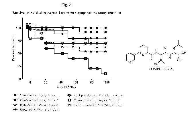
Fig. 21 depicts survival of NZM mice across treatment groups for the study
duration. NZM mice were treated as outlined in the legend. Graph shows percent
of live
mice for each week of the study.
Fig. 22 depicts the body weight progression for NZM mice across treatment
groups
for the study duration. NZM mice were treated as outlined in the legend. Graph
shows
Mean SEM body mass for each group across the duration of the study.
Fig. 23 depicts splenomegaly for NZM mice across treatment groups for the
study
duration. NZM mice were treated as outlined in the legend. The spleen weights
for all
mice that survived until the end of the study are graphed. Values represent
the
mean SEM spleen weight at 25 weeks of age.
Fig. 24 depicts total urine protein (proteinuria) in NZM mice. Urine collected
from NZM mice was analyzed for total protein content using a rat urinalysis
kit. Graph
shows Mean SEM of protein concentration in mg/mL.
Fig. 25 depicts anti-chromatin antinuclear antibody concentrations in serum of
NZM mice. NZM mouse serum samples were analyzed for the presence of anti-
chromatin
IgG circulating ANAs via ELISA assay. Graph shows Mean SEM of anti-chromatin
ANA concentration in ng/mL, 2000-fold dilution from original stock.
Fig. 26 depicts anti-Smith antigen antinuclear antibody concentrations in NZM
mice. NZM mouse serum samples were analyzed for the presence of anti-smith
antigen
IgG circulating ANAs via ELISA assay. Mean SEM values of anti-Smith Ag ANA
concentration in ng/mL, 100-fold dilution from original stock.
Fig. 27 depicts anti-dsDNA antinuclear antibody concentrations in NZM mice.
NZM mouse serum samples were analyzed for the presence of anti-dsDNA IgG
circulating ANAs via ELISA assay. Mean SEM values of anti-dsDNA ANA
concentration in ng/mL, 100-fold dilution from original stock.
Fig. 28 depicts serum IL-12p40/p70 concentration over course of disease
treatment
in NZM mice. NZM mice were treated as outlined in the legend. Mean SEM values
for
the concentration of mouse serum IL-12p40/p70 from treated NZM mice. Cytokines
were
analyzed using Luminex bead kits.
- 6 -
CA 02827653 2013-08-16
WO 2012/119056 PCT/US2012/027440
Fig. 29 depicts serum MIG concentration in NZM mice. NZM mice were treated
as outlined in the legend. Graph shows Mean SEM values for the concentration
of mouse
serum monokine, MIG, from treated NZM mice. Cytokines were analyzed using
Luminex
bead kits.
Fig. 30 depicts serum IP-10 concentration in NZM mice. NZM mice were treated
as outlined in the legend. Graph shows Mean SEM values for the concentration
of mouse
serum IFN-y inducible protein, IP-10, from treated NZM mice. Cytokines were
analyzed
using Luminex bead kits.
Fig. 31 depicts serum IL-13 concentration in NZM mice. NZM mice were treated
as outlined in the legend. Graph shows Mean SEM values for the concentration
of mouse
serum Th2 cytokine, IL-13, from treated NZM mice. Cytokines were analyzed
using
Luminex bead kits.
Fig. 32 depicts serum TNFa concentration in NZM mice. NZM mice were treated
as outlined in the legend. Graph shows Mean SEM values for the concentration
of mouse
serum proinflammatory cytokine, TNFa, from treated NZM mice. Cytokines were
analyzed using Luminex bead kits.
Fig. 33 depicts serum IL-17A concentration in NZM mice. NZM mice were
treated as outlined in the legend. Graph shows Mean SEM values for the
concentration of
mouse serum Th17 cytokine, IL-17A, from treated NZM mice. Cytokines were
analyzed
using Luminex bead kits.
Fig. 34 depicts the frequency of anti-chromatin antibody secreting cells in
the
spleens of NZM mice. NZM treated mice spleens were processed for splenocytes
for ex
vivo Elispot assays. Elispot wells were coated with 10 ilg/mL of boiled
chicken
chromatin or ovalbumin protein. Fresh, whole splenocytes were added to each
well at
50,000 cells per well in cell culture medium. Cells were incubated overnight
at 37 C.
Developed wells provided spots that were counted as frequency of ASCs per
million
splenocytes. Graph shows Mean SEM values.
Fig. 35 depicts the frequency of total IgG antibody secreting cells in the
spleens of
NZM mice. NZM treated mice spleens were processed for splenocytes for ex vivo
Elispot
assays. Elispot wells were coated with 10 ilg/mL of anti-mouse IgG, IgH, and
IgL chains.
Fresh, whole splenocytes were added to each well at 50,000 cells per well in
cell culture
medium. Cells were incubated overnight at 37 C. Developed wells provided spots
that
- 7 -
CA 02827653 2013-08-16
WO 2012/119056 PCT/US2012/027440
were counted as frequency of ASCs per million splenocytes. Graph shows Mean
SEM
values.
Fig. 36 depicts serum C3 complement levels in NZM mice. NZM serum samples
were processed as described for ANA analysis. Serum was tested for total
complement
factor 3 (C3) concentration using a commercial kit. Graph shows serum sample
diluted 1
to 50,000 in saline as mean SEM values.
Fig. 37 depicts serum concentration of collagen type I cross-linker
telopeptide
(CTx) in NZM mice. NZM serum samples were processed as described for ANA
analysis.
Serum was tested for total collagen type I cross-linker (CTx) concentration, a
biomarker
for systemic bone resorption, using a commercial kit. Graph shows serum sample
diluted
1 to 6 in saline as mean SEM values.
Fig. 38 depicts renal histopathology scores from NZM mice.
Fig. 39 depicts renal and pulmonary histopathology results from 40 week old
NZM
mice (H&E stained paraffin wax embedded kidney tissues sections from 25 week
old
mice). Images show worst affected area of section selected blindly by the
pathologist.
Fig. 40 depicts inhibition of the 20S proteasome in spleens of NZM mice.
Spleens
from treated NZM mice were lysed and analyzed using a functional ex vivo test
for the
20S proteasome. Graph represents mean SEM percent control for treatment
groups.
Fig. 41 depicts kidney IKBa accumulation 3 hours post dosing of NZM mice.
Kidneys were lysed and analyzed using a commercial ELISA kit that measures the
accumulation of cellular IKBa as a function of proteasome activity. Graph
represents
mean SEM percent control for treatment groups.
Fig. 42 depicts an overview of the experimental design for testing
subcutaneous
administration of COMPOUND A and bortezomib in the NZM lupus nephritis mouse
model.
Fig. 43 depicts the body weight progression for NZM mice across treatment
groups
for the study duration. Graph shows Mean SEM body mass for each group at the
end of
the study.
Fig. 44 depicts survival of NZM mice across treatment groups for the study
duration. NZM mice were treated as outlined in the legend. Graph shows percent
of live
mice for each week of the study. The overall percentage of surviving mice at
the 91 day
endpoint is listed in parentheses within the legend. * indicates p < 0.05.
- 8 -
CA 02827653 2013-08-16
WO 2012/119056 PCT/US2012/027440
Fig. 45 depicts total urine protein (proteinuria) in NZM mice. Urine collected
from NZM mice was analyzed for total protein content using a rat urinalysis
kit. Graph
shows Mean SEM of protein concentration in mg/mt. * indicates p < 0.05.
Fig. 46 depicts anti-Smith antigen antinuclear antibody concentrations in NZM
mice. NZM mouse serum samples were analyzed for the presence of anti-smith
antigen
IgG circulating ANAs via ELISA assay. Mean SEM values of anti-Smith Ag ANA
concentration in ng/mL is listed as 100-fold dilution from original stock. *
indicates p <
0.05 as compared to vehicle control G10.
Fig. 47 depicts anti-dsDNA antinuclear antibody concentrations in NZM mice.
NZM mouse serum samples were analyzed for the presence of anti-dsDNA IgG
circulating ANAs via ELISA assay. Mean SEM values of anti-dsDNA ANA
concentration in ng/mL is listed as 100-fold dilution from original stock. *
indicates p <
0.05 as compared to the vehicle control G10.
Fig. 48 depicts serum IL-12 concentration in NZM mice. Mean SEM values for
the concentration of mouse serum IL-12 from treated NZM mice at the end of the
study.
Cytokines were analyzed using Luminex bead kits. * indicates p < 0.05 as
compared to
the vehicle control G10.
Fig. 49 depicts the frequency of anti-Smith antibody secreting cells in the
spleens
of NZM mice. NZM treated mice spleens were processed for splenocytes for ex
vivo
Elispot assays. Elispot wells were coated with 10 ilg/mL of purified smith
antigen or
ovalbumin protein as a third party background control antigen. Fresh, whole
splenocytes
were added to each well at 500,000 cells per well in cell culture medium.
Cells were
incubated overnight at 37 C. Developed wells provided spots that were counted
as
frequency of ASCs per million splenocytes. Graph shows Mean SEM values. *
indicates
p < 0.05 compared to vehicle control G10.
Fig. 50 depicts the frequency of anti-dsDNA antibody secreting cells in the
spleens
of NZM mice. NZM treated mice spleens were processed for splenocytes for ex
vivo
Elispot assays. Elispot wells were coated with 10 ilg/mL of bovine dsDNA or
ovalbumin
protein as a third party background control antigen. Fresh, whole splenocytes
were added
to each well at 500,000 cells per well in cell culture medium. Cells were
incubated
overnight at 37 C. Developed wells provided spots that were counted as
frequency of
- 9 -
CA 02827653 2013-08-16
WO 2012/119056 PCT/US2012/027440
ASCs per million splenocytes. Graph shows Mean SEM values. * indicates p <
0.05
compared to vehicle control G10.
Fig. 51 depicts frequency of spleen CD38/CD138+ plasma cells in NZM mice. A
total of 100,000 to 200,000 events were collected per tube of splenocytes
stained with
anti-CD19-FITC, anti-CD38 ¨ PE and anti-CD138-APC, acquired and analyzed on an
Accuri flow cytometer. Live size gating was performed based on lymphocyte Fsc
and Ssc
scatter. CD19 negative histogram gated events were plotted as shown, by CD 138
and
CD38. Representative data shown from a total of three mice analyzed randomly.
T and B
cell subset analysis not shown. Plots show proportion of spleen plasma cells
in treated
NZM mice. Graph shows Mean SEM. * indicates p < 0.05 compared to vehicle
control
G10.
Fig. 52 depicts renal histopathology scores from NZM mice (H&E stained
paraffin
wax embedded kidney tissues sections from 25 week old mice). Graph shows
Mean SEM. * indicates p < 0.05 compared to vehicle control G10.
Fig. 53 depicts spleen and kidney IKBa accumulation 3 hours post dosing of NZM
mice. Spleen and kidneys were lysed and analyzed using a commercial ELISA kit
that
measures the accumulation of cellular IKBa as a function of proteasome
activity. Graph
represents mean SEM percent control for treatment groups.
DETAILED DESCRIPTION OF ILLUSTRATIVE EMBODIMENTS
The term "about" as used herein when referring to a measurable value such as
an
amount, a temporal duration, and the like, is meant to encompass reasonable
variations of
the value, such as, for example, 10% from the specified value. For example,
the phrase
"about 50%" encompasses reasonable variations of 50%, such as 10% of the
numerical
value 50, or from 45% to 55%.
As used herein, the term "subject" includes warm blooded animals, preferably
mammals, including humans. In a preferred embodiment, the subject is a
primate. In an
even more preferred embodiment, the subject is a human.
Provided are methods for treating lupus in a subject by administering to the
subject
COMPOUND A. COMPOUND A is a proteasome inhibitor with the chemical name
[(1R)-1-[[(25,3R)-3-hydroxy-2-[6-phenyl-pyridine-2-carbonyl)amino]-1-
oxobutyl]amino]-
- 10 -
CA 02827653 2013-08-16
WO 2012/119056
PCT/US2012/027440
3-methylbutylboronic acid (see Bernardini, et al., U.S. Patent No. 7,576,206).
COMPOUND A has the following structure:
/ 1 0
I H
40 B OH
N .,.,õ..,-----,N
N
E H I
We have found that COMPOUND A is superior to bortezomib in the treatment of
lupus. This is surprising because COMPOUND A and bortezomib are both
reversible
boronic acid proteasome inhibitors that induce cell death through activation
of the
extrinsic and intrinsic apoptotic signaling pathways (Chauhan D, Anderson K.C.
2003;
Piva R, Ruggeri B, et al. 2008). Furthermore, both agents primarily target the
proteasome's chymotrypsin-like catalytic activity, with minor inhibition of
the caspase-
like and little inhibition of the trypsin-like activities (Piva R, Ruggeri B
et al. 2008; Demo
SD, Kirk CJ et al. 2007). Thus, COMPOUND A and bortezomib appear to have
similar
mechanisms of action. In addition, the compounds have very similar chemical
structures.
Thus, the mechanism by which COMPOUND A provides enhanced efficacy against
lupus
as compared to bortezomib is unknown.
The COMPOUND A used in the present invention may be administered in any
suitable chemical form, including as a prodrug. Suitable prodrugs include
pharmaceutically acceptable ester forms of the parent compound. Preferably,
the prodrug
converts to the parent compound (i.e., COMPOUND A) after administration. As
used
herein, "pharmaceutically acceptable ester" refers to a derivative of the
parent compound
in which the boronic acid residue is modified by making an ester thereof
Preferably, the
prodrug is a boronic ester. More preferably, the prodrug is a cyclic boronic
ester.
Examples of cyclic boronic esters include, but are not limited to,
diethanolamine boronic
ester, diisopropanolamine boronic ester, aminodiacetic acid boronic ester,
pinanediol
boronic ester, pinacol boronic ester, 1,2-ethanediol boronic ester, 1,3-
propanediol boronic
ester, 1,2-propanediol boronic ester, 2,3-butanediol boronic ester, 1,1,2,2-
tetramethylethanediol boronic ester, 1,2-diisopropylethanediol boronic ester,
5,6-
decanediol boronic ester, 1,2-dicyclohexylethanediol boronic ester,
bicyclohexyl- 1,1 '-
diol, and 1,2-diphenyl- 1,2-ethanediol boronic ester. Preferably, the prodrug
is a
- 11 -
CA 02827653 2013-08-16
WO 2012/119056
PCT/US2012/027440
diethanolamine boronic ester, diisopropanolamine boronic ester, or
aminodiacetic acid
boronic ester. More preferably, the prodrug is a diethanolamine boronic ester
or
diisopropanolamine boronic ester. More preferably, the prodrug is a
diethanolamine
boronic ester ¨ i.e., COMPOUND B
, 0
I ENLA HO' 10 N 0
/
COMPOUND B.
Therefore, in certain embodiments the COMPOUND A is administered as a
prodrug. In one embodiment, the COMPOUND A is administered as a boronic ester
derivative of COMPOUND A. In one embodiment, the COMPOUND A is administered
as a cyclic boronic ester derivative of COMPOUND A. In one embodiment, the
COMPOUND A is administered as the cyclic boronic ester COMPOUND B
, 0
,
01
HO
COMPOUND B.
Any suitable method of administration may be used. Examples include injection
(subcutaneous, intravenous, parenteral, intraperitoneal, intrathecal, etc.),
oral, rectal,
transmucosal, inhalation, and transdermal. When administered by injection, the
injection
can be bolus or continuous infusion. COMPOUND A is preferably administered by
intravenous (IV) injection, subcutaneous (SQ) injection, or orally, such as in
a tablet or
capsule. More preferably, COMPOUND A is administered by intravenous (IV)
injection
or subcutaneous (SQ) injection. For example, the COMPOUND A may be provided as
a
sterile lyophilized powder, which may be reconstituted with, e.g., sterile
Water for
Injection, aqueous saline (NaC1), or aqueous mannitol before injection.
Therefore, in one
embodiment the COMPOUND A is administered by injection. In another embodiment,
the COMPOUND A is administered by IV injection. In another embodiment, the
- 12 -
CA 02827653 2013-08-16
WO 2012/119056 PCT/US2012/027440
COMPOUND A is administered by SQ injection. In another embodiment, the
COMPOUND A is administered orally. In one embodiment, the COMPOUND A is
administered orally in a tablet. In another embodiment, the COMPOUND A is
administered orally in a capsule.
The COMPOUND A used in the present invention is typically administered to the
subject as a pharmaceutical composition. Pharmaceutical compositions of
COMPOUND
A typically contain, in addition to COMPOUND A and/or a prodrug thereof, at
least one
pharmaceutically acceptable excipient. Such excipients enable the preparation
of
solutions, tablets, pills, dragees, powders, capsules, liquids, gels, syrups,
slurries,
suspensions, emulsions, and the like.
Pharmaceutical preparations for oral use can be obtained by combining
COMPOUND A and/or a prodrug thereof with a solid excipient, optionally
grinding the
resulting mixture, and processing the mixture of granules, after adding
suitable auxiliaries,
if desired, to obtain tablets or dragee cores. Suitable excipients include
fillers or diluents,
binders, disintegrants, lubricants, antiadherents, glidants, wetting and
surface active
agents, colors and pigments, flavoring agents, sweeteners, adsorbents, and
taste-maskers.
Diluents are typically added to a small amount of the active drug to increase
the
size of a tablet. The most common diluent is lactose, which exists in two
isomeric forms,
alpha-lactose or beta-lactose, and can be either crystalline or amorphous.
Various types of
TM
lactose include spray dried lactose monohydrate (such as Super-Tab ), alpha-
lactose
monohydrate (such as Fast Floc)), anhydrous alpha-lactose, anhydrous beta-
lactose, and
agglomerated lactose. Other diluents include sugars, such as compressible
sugar NF,
dextrose excipient NF, and dextrates NF. A preferred diluent is lactose
monohydrate
(such as Fast Flo ). Other preferred diluents include microcrystalline
cellulose (such as
0 TM 0
Avicel PH, and Ceolus ), and microfine cellulose (such as Elcema ). Diluents
may
include starch and starch derivatives. Starches include native starches
obtained from
wheat, corn, rice and potatoes. Other starches include pregelatinized starch
NF, and
sodium starch glycolate NF. Starches and starch derivatives also function as
disintegrants.
Other diluents include inorganic salts, such as dibasic calcium phosphate USP
(such as Di-
Tab
and Emcompress0 ), tribasic calcium phosphate NF (such as Tri-Tab and Tri-
Cafoso ), and calcium sulfate NF (such as Compactrolo ). Such polyols as
mannitol USP,
sorbitol NF, and xylitol NF may also serve as diluents. Many diluents also
function as
- 13 -
CA 02827653 2013-08-16
WO 2012/119056
PCT/US2012/027440
disintegrants and binders, and these additional properties must be taken into
account when
developing a formulation.
Disintegrants are included in tablet formulations to break the tablets into
particles
of the active pharmaceutical ingredient and excipients which will facilitate
dissolution of
the active ingredient and enhance bio availability of the active ingredient.
Starch and
starch derivatives, including cross-linked sodium salt of a carboxymethyl
ether of starch
(such as sodium starch glycolate NF, Explotab , and Primoge1 ) are useful
disintegrants.
A preferred disintegrant is pregelatinized starch, such as Starch 1500 .
Another preferred
disintegrant is cross-linked sodium carboxymethyl cellulose (such as
Croscarmellose
Sodium NF, Ac-Di-Sol ). Other disintegrants include cross-linked
polyvinylpyrrolidone
(such as Crospovidone NF), microcrystalline cellulose (such as Avicel PH).
Binders are used as wet granulation excipients to agglomerate the active
pharmaceutical ingredient and the other excipients. A binder is selected to
improve
powder flow and to improve compactibility. Binders include cellulose
derivatives such as
microcrystalline cellulose NF, methylcellulose USP, carboxymethycellulose
sodium USP,
hydroxypropyl methylcellulose USP, hydroxyethyl cellulose NF, and
hydroxypropyl
cellulose NF. Other binders include polyvidone, polyvinyl pyrrolidone, gelatin
NF,
natural gums (such as acacia, tragacanth, guar, and pectin), starch paste,
pregelatinized
starch NF, sucrose NF, corn syrup, polyethylene glycols, and sodium alginate,
ammonium
calcium alginate, magnesium aluminum silicate, polyethylene glycols. A
preferred binder
is polyvinyl pyrrolidone, in particular, Povidone USP, and preferably,
povidone K-29/32.
Lubricants are used in tablet formulations to prevent sticking of the tablet
to the
punch faces and to reduce friction during the compression stages. Lubricants
typically
include vegetable oils (such as corn oil), mineral oils, polyethylene glycols
(such as PEG-
4000 and PEG-6000), salts of stearic acid (such as calcium stearate and sodium
stearyl
fumarate), mineral salts (such as talc), inorganic salts (such as sodium
chloride), organic
salts (such as sodium benzoate, sodium acetate, and sodium oleate) and
polyvinyl
alcohols. A preferred lubricant is magnesium stearate.
Dragee cores may be provided with suitable coatings. For this purpose,
concentrated sugar solutions may be used, which may optionally contain gum
arabic, talc,
polyvinyl pyrrolidone, carbopol gel, polyethylene glycol, and/or titanium
dioxide, lacquer
solutions, and suitable organic solvents or solvent mixtures. Dyestuffs or
pigments may
- 14 -
CA 02827653 2013-08-16
WO 2012/119056
PCT/US2012/027440
be added to the tablets or dragee coatings for identification or to
characterize different
combinations of active compound doses.
Pharmaceutical preparations that can be used orally include push-fit capsules
made
of gelatin, as well as soft, sealed capsules made of gelatin and a
plasticizer, such as
glycerol or sorbitol. The push-fit capsules can contain the COMPOUND A and/or
a
prodrug thereof in admixture with filler such as lactose, binders such as
starches, and/or
lubricants such as talc or magnesium stearate and, optionally, stabilizers. In
soft capsules,
the COMPOUND A and/or a prodrug thereof may be dissolved or suspended in
suitable
liquids, such as fatty oils, liquid paraffin, or liquid polyethylene glycols.
In addition,
stabilizers may be added. All formulations for oral administration should be
in dosages
suitable for such administration.
The COMPOUND A and/or a prodrug thereof may be formulated for parenteral
administration by injection, e.g., by bolus injection or continuous infusion.
Formulations
for injection may be presented in unit dosage form, e.g., in ampoules or in
multi-dose
containers, optionally with an added preservative. The compositions may take
such forms
as suspensions, solutions or emulsions in oily or aqueous vehicles, and may
contain
excipients such as suspending, stabilizing and/or dispersing agents.
Pharmaceutical formulations for parenteral administration include aqueous
solutions of the COMPOUND A and/or prodrug thereof in water-soluble form.
Additionally, suspensions of the COMPOUND A and/or prodrug thereof may be
prepared
as appropriate oily injection suspensions. Suitable lipophilic solvents or
vehicles include
fatty oils such as sesame oil, or synthetic fatty acid esters, such as ethyl
oleate or
triglycerides, or liposomes. Aqueous injection suspensions may contain
substances which
increase the viscosity of the suspension, such as sodium carboxymethyl
cellulose, sorbitol,
or dextran. Optionally, the suspension may also contain suitable stabilizers
or agents
which increase the solubility of the COMPOUND A to allow for the preparation
of highly
concentrated solutions.
The COMPOUND A and/or prodrug thereof may be in powder form for
constitution with a suitable vehicle, e.g., sterile Water for Injection,
before use. For
example, the pharmaceutical composition may be a lyophilized powder.
Preferably, the
lyophilized powder is reconstituted, for example using 0.9% NaC1, and
administered by
- 15 -
CA 02827653 2013-08-16
WO 2012/119056
PCT/US2012/027440
injection. Lyophilized powders suitable for use in the present invention are
disclosed in
WO 2010/114982.
Excipients for lyophilized powders include bulking agents that have "generally
regarded as safe" (GRAS) status from the United States Food and Drug
Administration
(FDA). Such bulking agents are well known in the art of pharmaceutical
lyophilization,
tend to strengthen the structure of the resulting lyophilized cake, and may be
used in the
present invention. Preferred bulking agents include saccharides, preferably
monosaccharides or oligosaccharides, amino acids, sugar alcohols, and mixtures
thereof.
More preferred bulking agents include saccharides, preferably monosaccharides
or
oligosaccharides, sugar alcohols, and mixtures thereof. More preferably,
bulking agents
used in the present invention include sucrose, dextrose, maltose, lactose,
sorbitol, glycine,
and dextran. A most preferred bulking agent is mannitol.
Cyclodextrins may also be used in lyophilized powder pharmaceutical
compositions. Preferred cyclodextrins include the naturally occurring
cyclodextrins,
methyl-f3-cyclodextrin, dimethy1-13-cyclodextrin, trimethy1-13-cyclodextrin, 2-
hydroxymethy1-13-cyclodextrin, hydroxyethy1-13-cyclodextrin, 2-hydroxypropyl-3-
cyclodextrin, 3-hydroxypropy1-13-cyclodextrin,13-cyclodextrin sulfate, 13-
cyclodextrin
sulfonate, or 13-cyclodextrin sulfobutyl ether. Most of these are commercially
available
from such suppliers as Aldrich Chemical Company, Milwaukee Wisconsin and
Wacker
Chemicals, New Canaan, Connecticut. Preferred cyclodextrins include f3-
cyclodextrin,
hydroxypropyl-f3-cyclodextrin and f3-cyclodextrin sulfobutyl ether.
Preferably, the
cyclodextrin is hydroxypropyl 13 cyclodextrin, hydroxypropyl y cyclodextrin,
sulfobutyl
ether 13-cyclodextrin, or a mixture thereof Preferred cyclodextrins include
hydroxypropyl-f3-cyclodextrin and f3-cyclodextrin sulfobutyl ether. In one
embodiment,
the cyclodextrin is 13-cyclodextrin sulfobutyl ether. In another embodiment,
the
cyclodextrin is hydroxypropyl-13-cyclodextrin. A particularly preferred
cyclodextrin is
KLEPTOSEO HPB, available from Roquette Freres, France.
The pharmaceutical composition preferably contains from 1% to 95% (w/w) of the
active compound (i.e., compound of the present invention). More preferably,
the
pharmaceutical composition contains from 5% to 70% (w/w) of the active
compound.
Preferably, the pharmaceutical composition contains at least one unit dose of
the
active compound. In general, the unit dose of COMPOUND A and/or prodrug
thereof is
- 16 -
CA 02827653 2013-08-16
WO 2012/119056
PCT/US2012/027440
from about 0.1 mg/m2 to about 10 mg/m2 for a typical subject. More preferably,
the unit
dose of COMPOUND A and/or prodrug thereof is from about 0.5 mg/m2 to about 5
mg/m2. More preferably, the unit dose is from about 1 mg/m2 to about 5 mg/m2.
More
preferably, the unit dose is from about 2 mg/m2 to about 4 mg/m2. More
preferably, the
unit dose is from about 1 mg/m2 to about 3 mg/m2. More preferably, the unit
dose is from
about 2 mg/m2 to about 3 mg/m2. More preferably, the unit dose is from about 2
mg/m2 to
about 2.5 mg/m2. More preferably, the unit dose is about 2 mg/m2.
The COMPOUND A is administered in an amount effective to treat lupus, i.e., an
amount effective to prevent, alleviate, or ameliorate symptoms of the disease,
prolong
survival of the subject being treated, and/or favorably impact lupus-related
biomarkers in
the subject. Determination of the effective amount of COMPOUND A is well
within the
capability of those skilled in the art in light of the detailed disclosure and
examples
provided herein. The effective amount can vary depending on such factors as
the size of
the subject, the severity of the lupus disease, the frequency of
administration, the
bioavailability of the compound, the health and co-morbid conditions of the
subject, and
the quantity and nature of any concurrent treatment (e.g., glucocorticoids).
For example,
the effective amount of COMPOUND A for monotherapy may be a higher dose than
the
amount of COMPOUND A that is effective when COMPOUND A is used together in
combination with other lupus therapies. Effective doses can be extrapolated
from dose-
response curves derived from in vitro or animal model test systems, and may be
based on
the surface area or weight of the subject.
Treatment can be initiated with smaller dosages which are less than the
optimum
dose of the compound. Thereafter, the dosage can be increased by small
increments until
the optimum effect under the circumstances is reached. The total daily dosage
may be
divided and administered in portions during the day if desired. To optimize
the dosing
regimen, the effectiveness of COMPOUND A can be monitored by monitoring the
effect
of treatment on various biomarkers in a subject undergoing treatment. Useful
biomarkers
include those listed in the Examples section herein (e.g., antinuclear
antibodies, cytokines
such as IL-12, proteinuria, serum complement, etc.). Two especially convenient
biomarkers for monitoring the effectiveness of lupus treatment are proteinuria
and
antinuclear antibodies. An effective dose of COMPOUND A preferably alters the
biomarker(s) in the desired way as compared to the biomarker level prior to
treatment
- 17 -
CA 02827653 2013-08-16
WO 2012/119056 PCT/US2012/027440
(e.g., decrease proteinuria, decrease antinuclear antibodies, increase serum
C3, etc.).
Thus, an effective dose of COMPOUND A can be optimized by starting at a low
dose, and
then titrating up whilst monitoring one or more of these biomarkers. In
general, it is
preferable to obtain the initial assessment of the biomarker(s) (e.g.,
proteinuria or
antinuclear antibodies) from the patient prior to beginning therapy and one or
more
additional assessments at different time points during treatment. In such a
use, a baseline
determination prior to therapy is determined and then changes in biomarker(s)
(e.g.,
proteinuria or antinuclear antibodies) are determined during the course of
therapy and the
dose adjusted as needed. Alternatively, two or more successive determinations
can be
made during treatment without the need of a pre-treatment baseline
measurement. In such
a use, the first assessment of biomarker(s) (e.g., proteinuria or antinuclear
antibodies)
should be made from the subject as a baseline level for determining whether
the level is
increasing or decreasing and the dose adjusted or maintained accordingly.
In preferred embodiments, the subject undergoing treatment with COMPOUND A
experiences a desirable change in one or more biomarkers associated with lupus
disease.
Suitable biomarkers associated with lupus include lymphomegaly, splenomegaly,
serum
IL-12, serum C3, serum antinuclear antibodies, anti-chromatin IgG, anti-Smith
Ag IgG,
serum anti-dsDNA antinuclear antibodies, serum IFNa, proteinuria, serum IL-
17A, serum
IL-6, serum CCL3/MIP-la, serum CXCL10/IP-10, serum CXCL9/MIG, serum IL-4,
serum IL-13, serum IL-113, serum TNFa, serum KC/IL-8, and serum CTx.
Therefore, in
one embodiment the subject experiences a decrease in lymphomegaly during
treatment
with COMPOUND A. In another embodiment, the subject experiences a decrease in
splenomegaly during treatment with COMPOUND A. In another embodiment, the
subject
experiences a decrease in serum IL-12 during treatment with COMPOUND A. In
another
embodiment, the subject experiences an increase in serum C3 during treatment
with
COMPOUND A. In another embodiment, the subject experiences a decrease in serum
antinuclear antibodies during treatment with COMPOUND A. In another
embodiment,
the subject experiences a decrease in anti-chromatin IgG during treatment with
COMPOUND A. In another embodiment, the subject experiences a decrease in anti-
Smith Ag IgG during treatment with COMPOUND A. In another embodiment, the
subject
experiences a decrease in serum anti-dsDNA antinuclear antibodies during
treatment with
COMPOUND A. In another embodiment, the subject experiences a decrease in serum
- 18 -
CA 02827653 2013-08-16
WO 2012/119056
PCT/US2012/027440
IFNa during treatment with COMPOUND A. In another embodiment, the subject
experiences a decrease in proteinuria during treatment with COMPOUND A. In
another
embodiment, the subject experiences a decrease in serum IL-17A during
treatment with
COMPOUND A. In another embodiment, the subject experiences a decrease in serum
IL-
6 during treatment with COMPOUND A. In another embodiment, the subject
experiences
a decrease in serum CCL3/MIP-la during treatment with COMPOUND A. In another
embodiment, the subject experiences a decrease in serum CXCL10/IP-10 during
treatment
with COMPOUND A. In another embodiment, the subject experiences a decrease in
serum CXCL9/MIG during treatment with COMPOUND A. In another embodiment, the
subject experiences a decrease in serum IL-4 during treatment with COMPOUND A.
In
another embodiment, the subject experiences a decrease in serum IL-13 during
treatment
with COMPOUND A. In another embodiment, the subject experiences a decrease in
serum IL-113 during treatment with COMPOUND A. In another embodiment, the
subject
experiences a decrease in serum TNFa during treatment with COMPOUND A. In
another
embodiment, the subject experiences a decrease in serum KC/IL-8 during
treatment with
COMPOUND A. In another embodiment, the subject experiences a decrease in serum
CTx during treatment with COMPOUND A.
The COMPOUND A may be administered to the subject at any suitable dose. In
one embodiment, the COMPOUND A dose is in the range of about 0.1 mg/m2 to
about 10
mg/m2. In another embodiment, the COMPOUND A dose is about 0.5 mg/m2 to about
5
mg/m2. In another embodiment, the COMPOUND A dose is about 1 mg/m2 to about 5
mg/mg2. In another embodiment, the COMPOUND A dose is about 0.5 mg/m2 to about
3
mg/m2. In another embodiment, the COMPOUND A dose is about 1 mg/m2 to about 4
mg/mg2. In another embodiment, the COMPOUND A dose is about 2 mg/m2 to about 4
mg/m2. In another embodiment, the COMPOUND A dose is about 1 mg/m2 to about 3
mg/mg2. In another embodiment, the COMPOUND A dose is about 1.5 mg/m2 to about
3
mg/m2. In another embodiment, the COMPOUND A dose is about 2 mg/m2 to about 3
mg/m2. In another embodiment, the COMPOUND A dose is about 2 mg/m2 to about
2.5
mg/m2. In another embodiment, the COMPOUND A dose is about 2 mg/m2. Preferred
COMPOUND A doses include, but are not limited to, 1.1 mg/m2, 1.5 mg/m2, 1.8
mg/m2,
2.1 mg/m2, 2.4 mg/m2, 2.7 mg/m2, and 3.0 mg/m2. More preferably, the COMPOUND
A
dose is 1.8 mg/m2, 2.1 mg/m2, 2.4 mg/m2, or 2.7 mg/m2. More preferably, the
- 19 -
CA 02827653 2013-08-16
WO 2012/119056 PCT/US2012/027440
COMPOUND A dose is 2.1 mg/m2 or 2.4 mg/m2. The preceding doses are suitable
for
any method of COMPOUND A administration, and are especially suitable for
subcutaneous or intravenous dosing. Oral doses of COMPOUND A will typically be
at
the high end of the preceding ranges, such as about 1 mg/m2 to about 7 mg/m2.
In one
embodiment, the oral dose of COMPOUND A is about 2 mg/m2 to about 6 mg/m2,
such as
about 3 mg/m2 to about 5 mg/m2. Exemplary oral COMPOUND A doses include, but
are
not limited to, 2 mg/m2, 3 mg/m2, 4 mg/m2, 5 mg/m2 and 6 mg/m2.
The regimen of administration of each COMPOUND A dose can vary depending
on such factors as the pharmacokinetics of the dosage form, the type of lupus
symptoms
being treated or inhibited, the size of the subject, and the severity of the
lupus disease.
The timing of administration of the COMPOUND A can be readily varied by the
treating
physician to optimize efficacy and minimize side effects in light of the above
considerations and the present detailed disclosure. There is wide flexibility
in the dosing
schedules for COMPOUND A according to present invention.
The COMPOUND A may be administered at the above-described doses according
to any suitable schedule. The COMPOUND A dose amounts may be constant or
varied
within the dosing schedule. Preferably, the COMPOUND A dose is maintained at a
constant level during the schedule unless significant drug-related toxicity is
observed, in
which case subsequent doses can be reduced, for example by about 20-30%. A
suitable
COMPOUND A schedule will typically range from once-daily dosing to once-weekly
dosing or even once-monthly dosing. Preferably, the COMPOUND A is administered
less
frequently than once-daily, such as one dose every 2-14 days. Preferably, the
COMPOUND A is administered every 3 to 28 days, such as every 3 to 14 days. For
example, the COMPOUND A may be administered twice per week. In another
example,
COMPOUND A may be administered once per week. In another example, COMPOUND
A may be administered once every two weeks. The schedule may include, after
treatment
with COMPOUND A for one or more weeks, such as 2, 3, or 4 weeks, a period of
at least
5 days during which COMPOUND A is not administered, such as a period of about
7 to
21 days. In one embodiment, the rest period is about 10 to 17 days, such as
about 10 days
or about 17 days. For example, the COMPOUND A can be administered on days 1,
4, 8
and 11 of a 21 day cycle, wherein days 12-21 are a rest period. In another
embodiment,
the COMPOUND A can be administered on days 1, 4, 8, and 11 of a 28 day cycle,
- 20 -
CA 02827653 2013-08-16
WO 2012/119056
PCT/US2012/027440
wherein days 12-28 are a rest period. In another embodiment, the COMPOUND A
can be
administered on days 1, 8 and 15 of a 28 day cycle, wherein days 16-28 are a
rest period.
In another embodiment, the COMPOUND A can be administered on days 1 and 8 of a
21
day cycle, wherein days 12-21 are a rest period. In another embodiment, the
COMPOUND A can be administered on days 1 and 8 of a 28 day cycle, wherein days
12-
28 are a rest period. In another embodiment, the COMPOUND A can be
administered on
days 1 and 15 of a 21 day cycle. In another embodiment, the COMPOUND A can be
administered on days 1 and 15 of a 28 day cycle. The scheduled dosing cycles
can be
repeated one or more times. For example, the scheduled cycle may be repeated
until
maximum response is observed, plus one or two additional cycles. As another
example,
the scheduled cycle may be repeated for 6 to 12 cycles. Optionally, after the
initial cycles
are completed, a "maintenance schedule" may be used in which the COMPOUND A is
administered less frequently and/or at a lower dose than in the initial
schedule, such as
once per week, once every two weeks, once every three weeks, or once every
four weeks.
The maintenance schedule may be continued either for a fixed period of time,
generally 1-
2 years, or indefinitely as long as the patient is continuing to show no signs
of progressive
disease and is tolerating the treatment without significant toxicity. In
certain
embodiments, the dosing schedules can be adapted from COMPOUND A dosing
schedules suitable for the treatment of other diseases, such as multiple
myeloma. For
example, COMPOUND A is currently being investigated for the treatment of
multiple
myeloma by administering COMPOUND A (about 2 mg/m2) on days 1, 8, and 15 of a
repeating 28 day cycle.
One or more additional lupus treatments can be used in combination with the
administration of the COMPOUND A. Such treatments include, but are not limited
to,
glucocorticoids, hydroxchloroquine, mycophenolate mofetil (MMF), azathioprine
(AZA),
and cyclophosphamide (CTX). Appropriate doses of these agents are well known
in the
art.
MATERIALS AND METHODS
Compounds
COMPOUND A may be obtained as a solid off-white powder by a procedure
analogous to that reported herein. Bortezomib may be obtained by a procedure
analogous
-21 -
CA 02827653 2013-08-16
WO 2012/119056 PCT/US2012/027440
to that reported herein. Dexamethasone (DEX, 10 mg/mL, liquid, Lot#089016) may
be
purchased from Hanna's Pharmaceuticals (Wilmington, DE). Cyclophosphamide
(CTX;
Hanna's Pharmaceuticals, Wilmington, DE) is used at 50 mg/kg, once weekly
injection,
ip. Vehicle used for the suspension of COMPOUND A and bortezomib is 87% PBS,
3%
DMSO, 10% Solutol (Mutchler Inc., Solutol HS 15).
COMPOUND A and bortezomib are stored in 75 1AL of DMSO at -80 C in single
use aliquots. These aliquots are diluted to final concentrations via the
addition of 87%
PBS plus 10% Solutol to equal a final concentration of 3% DMSO per
formulation.
Syntheses
Preparation 1. (1R)-1-[(3a5, 4S, 6S, 7aR)-Hexahydro-3a,5,5-trimethyl-4,6-
methano-1,3,2-
benzodioxaborol-2-yll-3-methylbutylamine hydrochloride salt.
A 20 liter Chemglass0 jacketed reactor equipped with overhead stirring,
nitrogen
sweep, thermocouple with temperature readout, a 1 liter addition funnel, sub-
surface gas
dispersion tube and auxiliary heater/chiller is charged with 8.0 liters of
anhydrous methyl
tert-butyl ether. The chiller is set to -40 C. The solvent is cooled to -31.3
C with
agitation. Next, 714.4g (19.71 mol, 5.0eq) of HC1(g) is added subsurface over
1.75 hours
while maintaining the temperature between -25.7 and -10.0 C. Next, 1.6235 kg
(3.964mo1) of N,N-Bis(trimethylsily1)-(1R)- 1- [ (3 aS,45,65,7aR)-hexahydro-
3a,5,5-
trimethy1-4,6-methano-1,3,2-benzodioxaborol-2-y1]-3-methylbutylamine (obtained
by a
method similar to that disclosed in U.S. Patent Publication No. 2005/0240047
(Pickersgill
et al.), is dissolved in 2.1 liters of methyl tert-butyl ether. Next, the
solution is added to
the HC1 solution over 40 minutes while maintaining the reaction temperature
between -25
and -10 C. After addition is complete the reaction is warmed to ambient
temperature and
the chiller is turned off. The reaction is allowed to warm to ambient
temperature and is
stirred overnight. Next, the reaction is concentrated on the rotary evaporator
to a volume
of 1-2 liters. 3 liters of heptanes are added to the mixture and the
distillation continued to
remove 3 more liters of distillate. Next, 6 more liters of heptanes are added
portion wise
while removing 1 more liter of distillate. The product mixture is transferred
to the 20 liter
Chemglass0 jacketed reactor equipped as previously described and allowed to
slowly stir
overnight at ambient temperature. The next morning the mixture is cooled to
about -15 C
to -10 C and allowed to agitate for 1 hour. The product is filtered through a
medium glass
- 22 -
CA 02827653 2013-08-16
WO 2012/119056 PCT/US2012/027440
sintered filter funnel equipped with a #1 Whatman0 filter paper. The product
cake is
washed with 2 liters of cold (0 C) heptane and dried in an oven under vacuum
(29mmHg)
at 35 C and purged with nitrogen. The yield is 996.0g (84%) with a purity of
93.9A%,
and a diastereomer ratio of 98.75:1.25 (d.e. = 97.5%).
Preparation 2. 6-(25,3R)-N-[(1R)-1-(1,3,6,2-Dioxazaborocan-2-yl)-3-
methylbutyl}-3-
hydroxy-2-{(6-phenylpyridin-2-yl)formamido] butanamide (i.e., COMPOUND B)
Step A. Preparation of 6-Phenyl-pyridine-2-carbonyl chloride. A 2-L three neck
round bottomed flask equipped with an overhead stirrer, thermocouple, heating
mantle
with digital temperature controller, condenser and nitrogen inlet/outlet is
charged with
100.0g (0.502 mol) of 6-phenyl-2-pyridinecarboxylic acid and 1500 mL of
toluene (Kf <
0.02wt%) then warmed to 40 C. Thionyl chloride (110 mL; 1.51 mol, 3 eq) is
then added
to the thin slurry via addition funnel over 20 minutes. The thin slurry is
heated to 75 C
and stirred overnight (about 10-16 hr), until it becomes a clear solution.
After cooling the
reaction mixture to room temperature the solvent and excess thionyl chloride
are removed
in vacuo as follows: Reaction mixture is stripped under full vacuum at 40 C
(bath
temperature) to approximately 1/3 its original volume (-500 mL) and then (1000
mL) of
fresh toluene is added. Concentration is continued, again stripping to 1/3
original volume
(-500m1) followed by re-dilution with 1000 mL of fresh toluene. The total
amount of
toluene removed is ¨2000 mL.
Step B. Preparation of (2S,3R)-3-Hydroxy-21oxo-2-(6-phenyl-pyridin-2-yl)-
ethyl}-butyric acid. A 3-L three neck round bottomed flask is equipped with an
overhead
stirrer, thermocouple, pressure equalizing dropping funnel, nitrogen
inlet/outlet and
ice/water cooling bath. L-threonine, 62.8g (0.53 mol) is added, followed by
117g (1.1
mol) of sodium carbonate and 1500 mL of deionized water. The aqueous solution
is
cooled to 10.0 C. During this time the addition funnel is charged with the
acid
chloride/toluene solution prepared in Step A. This toluene solution is added
dropwise to
the aqueous reaction over approximately 10 minutes at ¨10 C. Once the addition
is
complete, the reaction is warmed to room temperature (-22-25 C) and vigorously
stirred
until it is shown to be complete by HPLC analysis (typically ¨ 3 hr). The
reaction mixture
is then transferred to a separatory funnel and the two layers are separated.
The lower
aqueous phase is then recharged to the reaction flask. Methanol (800 mL) is
then added to
- 23 -
CA 02827653 2013-08-16
WO 2012/119056 PCT/US2012/027440
the mixture followed by pH adjustment (target pH=1-2) with 2.5M HC1 (-850 mL),
keeping the temperature at 15-20 C. Some off-gassing typically occurs at ¨
pH=5,
followed by precipitation of the product at pH=3. The slurry is allowed to
stir at room
temperature for 30 minutes post pH adjustment. The white solid is collected by
vacuum
filtration, (mother liquor losses <2 mg/mL), washed with deionized water
(2X500 ml) then
dried in a vacuum oven at 40 C with a nitrogen sweep to a constant weight to
provide 141
g (0.471 mol, 94%) of the title compound with an HPLC purity of 99A% (95 wt%).
1H
NMR (d6-DMSO, 400MHz) 6 12.9 (s, 1H, b), 8.71 (d, 1H, J=9.16 Hz), 8.23 (d, 1H,
J=7.24 Hz), 8.1 (m, 3H), 8.03 (d, 1H, J=7.0 Hz), 7.55 (m, 3H), 5.34 (s, 1H,
b), 4.46 (dd,
1H, J=2.52, 9.16 Hz), 4.34 (dd, 1H, J=1.92, 6.24 Hz), 1.15 (d, 3H, J=6.4 Hz).
Step C. Preparation ofN-[(15,2R)-1[[[(1R)-1-1[(3a5,4S,6S,7aR)-hexahydro-
3a,5,5-trimethy1-4,6-methano-1,3,2-benzodioxaborol-2-y1]-3-
methylbutyliaminoicarbony412-hydroxypropyli-6-phenyl-2-pyridinecarboxamide. A
10
liter jacketed reaction vessel equipped with a thermocouple, stirring shaft
with impeller,
addition funnel, and low temperature recirculating bath is charged with 156.1g
(0.52 mol,
1.0 eq) of (2S,3R)-3-hydroxy-2-[oxo-2-(6-phenyl-pyridin-2-y1)-ethy1]-butyric
acid, 218.8g
(0.575 mol, 1.1 eq) of 0-(7-azabenzotriazol-1-y1)-N,N,N'N'-tetramethyluronium
hexafluorophosphate (HATU), 157.7g (0.522 mol, 1.0 eq) of (1R)-1-[(3a5, 4S,
6S, 7aR)-
hexahydro-3a,5,5-trimethy1-4,6-methano-1,3,2-benzodioxaborol-2-y1]-3-
methylbutylamine hydrochloride salt (98.8:1.2 mixture of isobutyl
diastereomers (R:S)),
and 2355 mL of N,N-dimethylformamide (DMF). Agitation is begun to dissolve the
solids before cooling the reaction mixture to <-25.0 C. Diisopropylethylamine
(218.6 mL,
162.2g, 1.25 mol, 2.4 eq) is charged to the addition funnel and then added
dropwise to the
reaction mixture over ¨30 minutes at -25 C to -30 C. Once addition is complete
the
reaction is stirred at -30 C for six hours. In a separate twenty-two liter
four-neck reaction
flask equipped with an overhead stirrer and thermocouple is charged 3925 mL of
DI water
and 3925 mL of ethyl acetate. The reaction mixture is transferred to this
flask over five
minutes at RT. The lower aqueous layer is separated and discarded. A solution
of 393g of
sodium phosphate monobasic, monohydrate in 3925 mL of DI water is prepared and
the
organic phase is washed with this solution. The lower aqueous phase is again
removed
and discarded. A solution of 376.9g of sodium bicarbonate in 4710 mL of DI
water is
prepared and the organic phase is washed with this solution after splitting
into two
- 24 -
CA 02827653 2013-08-16
WO 2012/119056 PCT/US2012/027440
portions. Once again the lower aqueous phase is separated and discarded. A
saturated
sodium chloride solution is prepared using 481.4g of sodium chloride in 3140
mL of DI
water and the organic phase is washed with this solution, the layers are
separated and the
lower aqueous phase discarded. Norit GAC 1240+ carbon (157g) is added to the
organic
phase and the suspension is stirred at RT overnight (13.8 hours). The carbon
is removed
by vacuum filtration through Whatman GF/C glass fiber filter paper, then
washed with
350 mL of ethyl acetate. The filtrate is concentrated to a foam on a rotary
evaporator
under vacuum with a 33-44 C bath temperature to provide 231.5 g (0.422 mol,
80.9%) of
the title compound as a foam with a chemical purity of 96.4%. The level of
threonine
isomer is 1.16A%. %. 1FINMR (d6-DMSO, 400 MHz) 6 8.98 (d, b, 1H, J=2.99 Hz),
8.76
(d, 1H, J=8.55 Hz), 8.2 (m, 3H), 8.11 (t, 1H, J=7.71 Hz), 8.02 (d, 1H, J=7.54
Hz), 7.54 (m,
3H), 5.26 (d, 1H, J=4.95 Hz), 4.49 (dd, 1H, J=4.22, 8.52 Hz), 4.13 (m, 2H),
2.6 (m, b, 1H),
2.19 (m, b, 1H), 2.02 (m, b, 1H), 1.83 (t, 1H, J=5.38 Hz), 1.75 (s, b, 1H),
1.68 (m, b, 1H),
1.62 (d, 1H, J=13.9 Hz), 1.36 (d, 1H, J=10.05 Hz), 1.3 (m, b, 3H), 1.22 (d,
6H, J=11.65
Hz), 1.12 (d, 3H, J=6.26 Hz), 0.84 (d, 6H, J=6.57 Hz), 0.79 (s, 3H).
Step D. Preparation of 6-(25,3R)-N-[(1R)-1-(1,3,6,2-dioxazaborocan-2-y1)-3-
methylbuty1}-3-hydroxy-2-{(6-phenylpyridin-2-y1)formamidoibutanamide (i.e.,
COMPOUND B).
Option 1 ¨ Two Step Procedure: A twelve liter four neck round bottom flask is
equipped with an overhead stirrer, thermocouple and nitrogen outlet before
being charged
with a solution of 229.8g (0.42 mol, 1 eq) of N-[(1S,2R)-1[[[(1R)-1-
1[(3aS,4S,6S,7aR)-
hexahydro-3a,5,5-trimethy1-4,6-methano-1,3,2-benzodioxaborol-2-y1]-3-
methylbutyl]amino]carbonyl]2-hydroxypropy1]-6-pheny1-2-pyridinecarboxamide in
2310
mL of methanol. To this is added 3465 mL of n-heptane, 108g (1.06 mol, 2.5 eq)
of (2-
methylpropyl)boronic acid and a solution of 70 mL (84g, 0.85 mol, 2.0 eq) of
37%
hydrochloric acid in 353 mL of DI water. Agitation is begun and the two phase
mixture is
stirred at RT for 16 hours. The reaction mixture is transferred in portions to
a four liter
separatory funnel and the lower methanolic phase is separated and returned to
the reaction
flask. The upper heptane layer is discarded. A fresh charge of 3465 mL of n-
heptane is
added to the reaction and the reaction is agitated at RT for an additional two
hours.
Agitation is stopped and the phases are separated and the lower methanolic
layer is
extracted with n-heptane (2 X 4600 mL). The heptane phases are discarded and
the
- 25 -
CA 02827653 2013-08-16
WO 2012/119056 PCT/US2012/027440
methanolic phase is concentrated in vacuo with a bath temperature of 40 C.
Ethyl acetate
(4620 mL) is charged to the evaporation flask and the sticky yellow residue is
dissolved
before transferring to a twelve-liter reaction flask. A solution of 665.4 g of
sodium
bicarbonate in 7650 mL of DI water is prepared and used to wash the ethyl
acetate layer in
two portions (1 X 4000 mL and 1 X 3850 mL). A solution of 1059.7g of sodium
chloride
in 2700 mL of DI water is prepared and then used to wash the ethyl acetate
phase.
After separation of layers the ethyl acetate layer is treated with 47.3g (0.45
mol,
1.1 eq) of diethanolamine. The mixture is allowed to stir at RT overnight.
Precipitated
solids are collected by vacuum filtration using a closed filtration flask and
the wet cake is
washed with 500 mL of ethyl acetate. The sealed filter funnel is transferred
to a glove box
where it is opened and the 481.8 g of wet cake is transferred to two pyrex
drying trays
which are then placed into a vacuum oven. The product is dried to a constant
weight at
23.5 in of Hg and 50 C over 27 hours to provide 179.7g (0.372 mol, 88.8%) of
the title
compound with a chemical purity of 98.6% and a chiral purity of 98.8% de. 1H
NMR (d6-
DMSO, 400 MHz) 6 8.8 (d, 1H, J=8.52 Hz), 8.2 (m, 3 H), 8.1 (t, 1H, J=7.68Hz),
8.0 (dd,
1H, J=6.7, 0.9 Hz), 7.5 (m, 3H), 7.2 (d, 1H), 6.5 (t,b, 1H), 5.1 (d, 1H,
J=4.92 Hz), 4.5 (dd,
1H), 4.2 (m, 1H), 3.6 (m, 2H), 3.5 (m, 2H), 3.1 (m, 1H), 3.0 (m, 2H), 2.7 (m,
2H), 1.6 (m,
1 H), 1.3 (m, 1H), 1.2 (m, 1H), 1.1 (d, 3H, J=6.32 Hz), 0.8 (2d, 6H, J=6.68,
6.52 Hz).
Option 2 - One Step Procedure: A 50 mL three neck round bottom flask is
equipped with a thermocouple, stir bar, nitrogen inlet/outlet, heating mantle
and
temperature controller. The flask is charged with 2.0g (3.65 mmol, 1.0 eq) of
N-[(1S,2R)-
1[[[(1R)-1-1[(3aS,4S,6S,7aR)-hexahydro-3a,5,5-trimethy1-4,6-methano-1,3,2-
benzodioxaborol-2-y1]-3-methylbutyl]amino]carbonyl]2-hydroxypropy1]-6-pheny1-2-
pyridinecarboxamide and 20 mL of MTBE. The reaction mixture is stirred for
approximately 10 minutes until all the solids dissolved. Diethanolamine (0.44
mL, 0.48g,
4.57 mmol, 1.25 eq) is charged via syringe, along with 2 drops of
methanesulfonic acid, to
the light yellow solution and the mixture is heated to 50 C. After
approximately 30
minutes a white precipitate begins to form. Stirring is continued overnight
before cooling
to room temperature. The solids are collected by vacuum filtration, washed
with MTBE
(1 X 20 mL) then dried under vacuum at 60 C overnight to give 0.92g (1.9 mmol,
52%) of
the title compound as a white solid with a chemical purity of 91.9% and a
chiral purity of
>99.5% de.
- 26 -
CA 02827653 2013-08-16
WO 2012/119056 PCT/US2012/027440
Step E (optional). Purification of 6-(25,3R)-N-[(1R)-1-(1,3,6,2-dioxazaborocan-
2-
yl)-3-methylbutyl}-3-hydroxy-2-{(6-phenylpyridin-2-yl)formamido] butanamide
(COMPOUND B). A two liter four neck round bottom flask is equipped with an
overhead
stirrer, thermocouple, condenser, heating mantle, temperature controller and
nitrogen
outlet before being charged with 175g (0.363 mol) of 6-(2S,3R)-N-R1R)-1-
(1,3,6,2-
dioxazaborocan-2-y1)-3-methylbutyl} -3-hydroxy-2- {(6-phenylpyridin-2-
yl)formamidoThutanamide and 1400 mL (8 volumes ) of 95% ethanol. Agitation is
begun
and the resultant suspension is heated to 75.7 C over 21 minutes. Once at
temperature the
solution is stirred for 80 minutes at 74.9-75.8 C before cooling to 2.7 C over
80 minutes.
The reaction slurry is then stirred at 2.2-6.0 C overnight (17 hours) to fully
crystallize the
product. Precipitated solids are collected by vacuum filtration using a closed
filtration
flask and the wet cake is washed with 350 mL of 95% ethanol. The sealed filter
funnel is
transferred to a glove box where it is opened and the 203.8g of wet cake is
transferred to a
pyrex drying tray which is then placed into a vacuum oven. The product is
dried to a
constant weight at 23.5 in of Hg and 50 C over 19 hours to provide 147.3g
(0.306, mol,
84.2%) of the title compound with a chemical purity of 99.76% and an optical
purity of
>99.8%de.
Preparation 3. [(1R)-1-[[(25,3R)-3-Hydroxy-21[(6-phenylpyridin-2-yl)carbony]
aminor
1-oxobutyl 1 amino]-3-methylbutyllboronic acid (i.e., COMPOUND A). A 50 mL
three
neck round bottom flask equipped with a thermocouple, stir bar and nitrogen
outlet is
charged with 1.65g (3.4 mmol) of 6-(2S,3R)-N-[(1R)-1-(1,3,6,2-dioxazaborocan-2-
y1)-3-
methylbuty1}-3-hydroxy-2- {(6-phenylpyridin-2-yl)formamido]butanamide
(chemical
purity = 99.5%, chiral purity >99.5% de), 17 mL of methyl isobutyl ketone and
1.7 mL of
2N hydrochloric acid. The mixture is stirred overnight. The layers of the
reaction are
separated and the organic layer is dried over magnesium sulfate, filtered and
evaporated to
dryness in vacuo. The residue is triturated in pentane and the resultant white
solid is
collected by vacuum filtration before drying in a vacuum oven overnight at 60
C to give
1.26g (3.1 mmol, 90%) of the title compound. HPLC indicates a purity of
99.6A%. Chiral
purity > 99.5% de. 1H NMR (d4-Me0D, 400 MHz) 6 8.17 (m, 2H), 8.13 (m, 1H),
8.05
(m, 2H), 7.5 (m, 3H), 4.75 (d, 1H, J=3.04 Hz), 4.42 (dq, 1H, J=2.92, 6.4), 2.7
(t, b, 1H),
1.61 (m, 1H), 1.35 (t, 2H, J=7.48 Hz), 1.29 (d, 3H, J=6.36 Hz), 0.89 (d, 6H,
J=6.52 Hz).
-27 -
CA 02827653 2013-08-16
WO 2012/119056
PCT/US2012/027440
Preparation 4. (2S)-N-[(1R)-1-(1,3,6,2-dioxazaborocan-2-yl)-3-methylbutyll-3-
phenyl-2-
(pyrazin-2-ylformamido)propanamide (i.e., diethanolamine ester of bortezomib):
Step A. Preparation of pyrazine-2-carbonyl chloride. A 500 ml three neck round
bottomed flask equipped with a stir bar, thermocouple, heating mantle with
digital
temperature controller, condenser and nitrogen inlet/outlet is charged with
15g (0.12 mol)
of pyrazine carboxylic acid, 225 mL of toluene (Kf < 0.02wt%) and 26.4 ml (43
g, 0.36
mol) of thionyl chloride. The thin slurry is heated to 75 C and stirred
overnight (10-16
hr). After cooling the reaction mixture to room temperature the solvent and
excess thionyl
chloride are removed in vacuo as follows: Reaction mixture is stripped under
full vacuum
at 60 C (bath temperature) to approximately 1/3 its original volume and then
(175 ml) of
fresh toluene is added. Concentration is continued, again stripping to 1/3
original volume
followed by re-dilution with 225 ml of fresh toluene to provide the pyrazine
acid chloride
in a toluene solution.
Step B. Preparation of (S)-3-phenyl-21(pyrazine-2-carbonyl)-aminorpropionic
acid. A second 500 ml three neck round bottomed flask is equipped with a stir
bar,
thermocouple, pressure equalizing dropping funnel, nitrogen inlet/outlet and
ice/water
cooling bath. L-Phenylalanine, 20.2g (0.122 mol) is added, followed by 28.2g
(0.266 mol)
of sodium carbonate and 225 mL of deionized water. The aqueous solution is
cooled to
10.0 C. During this time the addition funnel is charged with the acid
chloride/toluene
solution prepared in Step A (-125 mL). This toluene solution is added dropwise
to the
aqueous reaction over approximately 10 minutes at ¨10 C. Once the addition is
complete,
the reaction is warmed to room temperature (-22-25 C) and vigorously stirred
for 3 h.
The reaction mixture is then transferred to a separatory funnel and the two
layers are
separated. The lower aqueous phase is then recharged to the reaction flask.
Methanol
(125 mL) is then added to the red solution followed by pH adjustment (target
pH=1-2)
with 3.0 M HC1 (-175 mL), keeping the temperature at 15-20 C. Some off-gassing
occurs
at ¨ pH=5, followed by precipitation of the product at pH=3. The slurry is
allowed to stir
at room temperature for 30 minutes at ambient temperature post pH adjustment.
The
resulting pink solid precipitate is collected by vacuum filtration, (mother
liquor losses <2
mg/mL), washed with deionized water (1X50 ml) then dried in a vacuum oven at
40 C
with a nitrogen sweep to a constant weight to provide 11.92g (0.43.9 mmol,
36%) of the
- 28 -
CA 02827653 2013-08-16
WO 2012/119056 PCT/US2012/027440
title compound with an HPLC purity of 99A%. 1H NMR (d6-DMSO, 400MHz) 6 13.04
(s, 1H), 9.14 (d, 1H, J=1.44 Hz), 8.88 (dd, 2H, J=2.48, 6.16 Hz), 8.75 (dd,
1H, J=1.52, 2.4
Hz), 7.25 (m, 4H), 7.18 (m, 1H), 4.75 (dt, 1H, J=5.48, 8.08 Hz), 3.2 (dd, 2H,
J=1.79, 5.32
Hz).
Step C. Preparation ofN-[(1S)-1 [[[(1R)-113a5,4S,6S,7aR)-hexahydro-3a,5,5-
trimethy1-4,6-methano-1,3,2-benzodioxaborol-2-y1:1-3-methylbutyl _1 amino]
carbony1]-2-
benzyl _12-pyrazine carboxamide. A 500 ml three neck round bottomed flask
equipped with
a stir bar, addition funnel, thermocouple, nitrogen inlet/outlet and cooling
bath is charged
with llg (99.9 mmol) of (S)-3-pheny1-2-[(pyrazine-2-carbony1)-amino]-propionic
acid,
15.5.0g (40.6 mmol) of 0-(7-azabenzotriazol-1-y1)-N,N,N'N'-tetramethyluronium
hexafluorophosphate (HATU), 12.2g (40.6 mmol) of (1R)-1-[(3aS, 4S, 6S, 7aR)-
hexahydro-3a,5,5-trimethy1-4,6-methano-1,3,2-benzodioxaborol-2-y1]-3-
methylbutylamine hydrochloride salt (87:13 mixture of isobutyl diastereomers
(R :5)) and
165 mL of N,N-dimethylformamide (DMF). The pale yellow reaction solution is
cooled
to -35 C where 12.6g (17 mL, 97.3 mmol) of N,N-di-isopropyl ethyl amine is
added
dropwise over six minutes at -34 C to -35 C. The resulting solution is then
stirred
overnight at -40 to -11 C. The reaction mixture is quenched onto 600 ml of a
1:1 cold
water/ethyl acetate mixture. After transferring into a separatory funnel the
layers are
separated. The organic phase is then washed successively with 10% aqueous
sodium
hydrogen phosphate (1 X 200 mL), 8% aqueous sodium bicarbonate (2 X 200 mL)
and
saturated sodium chloride (1 X 200 mL). The product solution is dried over
magnesium
sulfate then filtered. The filtrate is evaporated to dryness in vacuo to give
19.57 g (37.7
mmol, 93%) of the title compound as a light brown foam with an HPLC purity of
92A%.
1H NMR (d6-DMSO, 400 MHz) 6 9.15 (d, 1H, J=1.44 Hz), 8.87 (d, 1H, J=2.48Hz),
8.7
(m, 3H), 7.25 (m, 4H), 7.18 (m, 1H), 4.89 (q, 1H, J=6.88, 15.4 Hz), 4.13 (dd,
1H, J=1.8,
8.56 Hz), 3.15 (d, 2H, J=6.88 Hz), 2.7 (m, b, 1H), 2.22 (m, b, 1H), 2.05 (m,
b, 1H), 1.87 (t,
1H, J=5.40 Hz), 1.81 (s, b, 1H), 1.67 (d, b, 1H), 1.52 (m, b, 1H), 1.13-1.33
(m, 9H), 0.83
(dd, 6H, J=2.48, 6.56 Hz), 0.80 (s, 3H).
Step D. Preparation of (25)-N-[(1R)-1-(1,3,6,2-dioxazaborocan-2-y1)-3-
methylbuty1:1-3-phenyl-2-(pyrazin-2-ylformamido)propanamide (i.e.,
diethanolamine ester
of bortezomib). A one liter four neck round bottomed flask is equipped with an
overhead
stirrer, thermocouple and nitrogen inlet/outlet then charged with 19.0g (36.6
mmol) of N-
- 29 -
CA 02827653 2013-08-16
WO 2012/119056 PCT/US2012/027440
R1S)-1[[[(1R)-1-[(3aS,4S,6S,7aR)-hexahydro-3a,5,5-trimethy1-4,6-methano-1,3,2-
benzodioxaborol-2-y1]-3-methylbutyl]amino]carbonyl]-2-benzyl]2-pyrazine
carboxamide
9.32g (91.5 mmol) of isobutylboronic acid, 190 mL of methanol, 34.7 mL (69.4
mmol) of
2M aqueous hydrochloric acid and 285 mL of heptane. The two phase reaction is
stirred
at room temperature overnight until an IPC showed <2% starting material
remaining by
area. The reaction mixture is transferred to a separatory funnel and the
layers are
separated. The lower methanol layer is washed with heptanes (2 X 250 mL)
before being
removed to a one-liter round bottomed flask and evaporating to dryness in
vacuo. The
resulting residue is dissolved in 300 mL of ethyl acetate which is washed with
8% aqueous
sodium bicarbonate (2 X 200 mL) and brine (1 X 300 mL), before transferring to
a clean
one liter three neck round bottom flask equipped as above.
To the ethyl acetate solution is added 4.1g (38.4 mmol) of diethanolamine and
the
mixture is stirred at room temperature for about 10 to 100 hours. The
resulting solids are
collected by vacuum filtration, washed with ethyl acetate (1 X 30 mL) then
dried in a
vacuum oven at 50 C overnight to provide the title compound as a white solid
(15.8g, 34.9
mmol, 95.2%), which is shown by HPLC to be a 91:9 mixture of diastereomers
(i.e., 82%
de).
Step E (optional). Purification of (2S)-N-[(IR)-1-(1,3,6,2-dioxazaborocan-2-
yl)-3-
methylbutyll-3-phenyl-2-(pyrazin-2-ylformamido)propanamide (i.e.,
diethanolamine ester
of bortezomib). (2S)-N-[(1R)-1-(1,3,6,2-dioxazaborocan-2-y1)-3-methylbuty1]-3-
pheny1-
2-(pyrazin-2-ylformamido)propanamide is charged to a 250 ml three round bottom
flask
equipped with a stir bar, thermocouple, heating mantle, controller, condenser
and nitrogen
inlet/outlet. Ethanol (absolute, 128 mL) is then charged to the flask and
heated to reflux.
Undissolved solids (which are enriched about (2:8) in the undesired isomer)
are removed
by vacuum filtration. The filtrate is returned to the round bottom flask and
cooled to room
temperature to crystallize the product which is isolated by vacuum filtration,
washed with
cold absolute ethanol (1X 50 ml), and dried in a vacuum oven at 50 C overnight
to provide
11.6g (25.6 mmol, 70%) of the title compound as a 94:6 mixture of
diastereomers (i.e.,
88% de). The chemical purity is >99.9A%. 1H NMR (d6-DMSO, 400 MHz) 6 9.10 (d,
1H, J=1.4 Hz), 8.88 (d, 1H, J=2.48 Hz), 8.83 (d, 1H, J=8.84 Hz), 8.75 (dd, 1H,
J=1.52,
2.32 Hz), 7.3 (m, 5H), 6.55 (s, b, 1H), 4.75 (m, 1H), 3.65 (m, 2H), 3.55 (m,
1H), 3.45 (m,
- 30 -
CA 02827653 2013-08-16
WO 2012/119056 PCT/US2012/027440
1H), 2.9-3.2 (m, 4H), 2.8 (m, 1H), 2.7 (m, 2H), 1.56 (m, 1H), 1.33 (dt, 1H,
J=4.04, 13.80
Hz), 1.18 (dt, 1H, J=3.48, 9.88 Hz), 0.8 (dd, 6H, J=6.64, 12.56 Hz).
Preparation 5. Bortezomib. A 100 ml three neck round bottom flask is equipped
with a
stir bar, thermocouple and nitrogen inlet/outlet then charged with 5.0g (10.4
mmol) of
(2S)-N-[(1R)-1-(1,3,6,2-dioxazaborocan-2-y1)-3-methylbuty1]-3-pheny1-2-
(pyrazin-2-
ylformamido)propanamide (i.e., diethanolamine ester of bortezomib), 50 ml of
methanol
and 10.4 ml of 2N aqueous hydrochloric acid. The reaction is stirred at room
temperature
overnight before removing the solvent in vacuo at 40 C. The resulting residue
is dissolved
in 50 ml of ethyl acetate and washed with saturated sodium bicarbonate (1X 50
mL)
before once again concentrating the organic layer to dryness in vacuo. The
residue is then
triturated overnight at room temperature with 50 mL of pentane under nitrogen.
The
resulting free flowing solids are collected by vacuum filtration, washed with
pentane (1 X
ml) then dried in a vacuum oven at 30 C overnight to provide 3.29g (8.56 mmol,
15 82.3%) of the title compound as a white solid with chemical purity
>99.8A% and a
93.5:6.5 ratio of diastereomers (i.e., 87% de). 1H NMR (d4-Me0H, 400 MHz) 6
9.15 (d,
1H, J=1.36 Hz), 8.77 (d, 1H, J=2.48 Hz), 8.68 (dd, 1H, J=1.52, 2.44 Hz), 7.27
(m, 4H),
7.21 (m, 1H), 5.05 (t, 1H, J=7.68 Hz), 3.2 (m, 2H), 2.66 (t, 1H, J=7.56 Hz),
1.39 (m, 1H),
1.17 (t, 2H, J=7.12 Hz), 0.83 (dd, 6H, J=5.32, 6.40 Hz).
Preparation 6. 6-Phenyl-pyridine-2-carboxylic acid {(1S,2R)-1-[(R)-1-(4,8-
dimethyl-
[1,3,6,2-dioxaborocan-2-y1)-3-methylbutylcarbamoy1}-2-2-hydroxypropyl}amide
(i.e.,
diisopropanolamine ester of COMPOUND A).
A 50 mL four neck round bottom flask is equipped with a stir bar,
thermocouple,
heating mantle with temperature controller, condenser and nitrogen inlet then
charged with
2.0 g (3.65 mmol) of N-[(1S,2R)-1[[[(1R)-1-1[(3aS,4S,6S,7aR)-hexahydro-3a,5,5-
trimethy1-4,6-methano-1,3,2-benzodioxaborol-2-y1]-3-
methylbutyl]amino]carbonyl]2-
hydroxypropy1]-6-pheny1-2-pyridinecarboxamide (chemical purity = 95.7%, chiral
purity
about 97.5% de (based on the fact that the (1R)-1-[(3aS, 4S, 6S, 7aR)-
hexahydro-3a,5,5-
trimethy1-4,6-methano-1,3,2-benzodioxaborol-2-y1]-3-methylbutylamine used to
make the
N-[(1S,2R)-1[[[(1R)-1-1[(3aS,4S,6S,7aR)-hexahydro-3a,5,5-trimethy1-4,6-methano-
1,3,2-
benzodioxaborol-2-y1]-3-methylbutyl]amino]carbonyl]2-hydroxypropy1]-6-pheny1-2-
- 31 -
CA 02827653 2013-08-16
WO 2012/119056 PCT/US2012/027440
pyridinecarboxamide has a 97.5% de)), 30 mL of t-butyl methyl ether (MTBE) and
0.61 g
(94.56 mmol, 1.25 eq) of diisopropanolamine. The resultant yellow solution is
stirred at
20-25 C for 16 hours. An additional 1.2 g (9 mmol, 2.5 eq) of
diisopropanolamine is
charged and the mixture heated at 40 C for 16 hours before cooling to room
temperature.
The white solid is collected by vacuum filtration, washed with 15 mL of MTBE
then dried
under vacuum overnight at 33 C to yield 1.31 g (2.55 mmol, 70%) of the desired
product
based on 1H NMR. The chemical purity is 96.8A% and no diastereomer is detected
by
HPLC (>99.8% de).
Acute Systemic Lupus Erythematosus (SLE) Model
Animals
Six week old lupus-prone, female, MRL/lpr (Jackson Labs, #000485) and non-
lupus prone control MRL/MpJ (Jackson Labs, #000486) mice are obtained from
Jackson
Laboratories (Bar Harbor, ME) at 6-weeks of age. All mice are maintained on a
24 hour
light/dark cycle, with food and water available ad libitum. All experimental
animal
procedures are approved by and in accordance to the regulations of the
Institutional
Animal Care and Use Committee (IACUC) of Cephalon, Inc; approved IACUC
protocol
#03-040 and #03-03-041.
MRL/lpr mice develop a rapid lymphoproliferative disease due to an inactive
Fas
molecule preventing the proper apoptosis of self-reactive T and B cells in
primary and
secondary lymphoid tissues (deficiency in both central and peripheral
tolerance
mechanism). Due to this impaired tolerance mechanism, a percentage of all T
and B cells
that enter the periphery have a high likelihood of responding to self
protein/tissues and
thus initiate autoimmunity early in life. The MRL/lpr model mice develop
several chronic
inflammatory disease-like symptoms that are characteristic of early and late
lupus
including the generation of anti-nuclear antibodies, arthritis, dermatological
manifestations, immunocomplex-mediated glomerulonephritis leading to
proteinuria and
eventual death. Lesser characterized phenomenon are CNS and cardiac
manifestations,
both of which are more common in humans. Previous optimization and validation
studies
clearly showed that disease only manifests in diseased MRL/lpr animals and not
the
control mice, MRL/Mp, and that antinuclear antibodies (ANAs) rapidly form
between
weeks 4-12 leading to the onset of lupus nephritis around weeks 18-25 with the
presence
- 32 -
CA 02827653 2013-08-16
WO 2012/119056 PCT/US2012/027440
of proteinuria. Mortality is higher in the females and occurs around 25 weeks
of age, renal
disease includes glomerulonephritis, glomerular infiltrates, sclerosis and
vasculitis. Both
splenomegaly and lymphomegaly associated with dermatitis can also be observed.
Antinuclear Antibody (ANA) ELISA Assays
The measurement of serum anti-dsDNA and anti-Smith antigen antibodies is done
by an in-house generated custom ELISA assay. Chromatin coated plates are
purchased
from Inova Diagnostics, Inc. Purified bovine thymus dsDNA (Sigma, St. Louis,
MI) or
purified bovine Smith antigen (GenWay, San Diego, CA) are used as coating
antigen for
the detection of anti-dsDNA and anti-Smith Ag antibodies respectively. Coated
plates are
washed with Borate Sulfate Saline (BSS) and blocked with BSS containing 1%
Bovine
Serum Albumin (BSA) and 0.1% Tween-20 detergent. Standard curves are generated
using mouse anti-chromatin antibody (Sigma, 2B1) or 25 week old MRL/lpr serum.
Mouse anti-dsDNA antibody (Abcam, Cambridge, MA), or mouse anti-Smith antigen
antibody (Abcam) are used as standards for each assay. Secondary antibody is
purchased
from Abcam (goat anti-mouse pAb-HRP), the substrate is purchased from Rockland
(Gilbertsville, PA) (TMB), and stop reaction buffer is generated using 1 mL of
concentrated sulfuric acid into 20 mL of dH20. Developed plates are read using
a Victor-
X4 spectrophotometer reading at 450 nM with a reference wavelength of 570 nM.
For all
ANA ELISA assays, values below the limit of detection set by the lowest point
along the
standard or set by the manufacturer are considered out of range and are not
estimated, but
assumed the lowest point along the standard curve for that particular assay.
Antibody Secreting B-cell Elispot Assays
B-cell Elispot components are ordered from MabTech (Nacka Strand, Sweden) and
nitrocellulose IP filter plates are ordered from Millipore (Billerica, MA).
Elispot wells are
coated with either purified bovine thymus dsDNA (Sigma), purified bovine Smith
antigen
(GenWay) or boiled filtered purified chicken chromatin from lysed chicken red
blood cells
(Rockland, Gilbertsville, PA) at 10 lg/mL. Spleens are processed using glass
homogenization, filtered through a 60 ilm sterile cell strainer and red blood
cells (RBCs)
lysed using BioLegend (San Diego, CA) lysis buffer. Processed splenocytes are
added to
each well in culture medium. To avoid skewing of true ex vivo frequencies of
antibody
- 33 -
CA 02827653 2013-08-16
WO 2012/119056 PCT/US2012/027440
secreting cell types (ASCs), cells are not stimulated with a polyclonal
mitogen like LPS,
but rather incubated in media alone to allow monitoring of genuine ex vivo
antibody
release. Anti-mouse pan-IgG is used as a positive control for total IgG
producing ASCs
and is used to normalize results. Frequencies for each antigen are identified
in an initial
test phase for each model. For chromatin and total IgG only, 30,000
splenocytes are
added to each well; for Smith antigen and dsDNA, 500,000 cells are added to
each well.
Different numbers of splenocytes are added for different antigens due to
saturation limits
of spot frequencies per well. These limits are previously established. B-cell
Elispots are
incubated overnight at 37 C. To develop each assay, secondary antibody is
added to each
well, incubated, washed and alkaline phosphatase strepavidin is used as the
conjugate;
substrate used is BCIP-NBT. Plates are developed until spots are visible. All
Elispot
analyses are performed using an Immunospot C.T.L. scanner and Biospot software
(Cellular Technology Ltd., Shaker Heights, OH). Results are shown as values
from which
media and cells only wells are subtracted. Some values are negative due to
high
background.
Flow Cytometry
Washed, RBC-lysed splenocytes are stained for plasma cell markers as
previously
used in published reports (Neubert, Meister et al. 2008). As used herein the
term "plasma
cell" will refer to the following immunophenotype definition: Plasma cells are
defined as
live CD19-negative, CD138-positive, intracellular kappa light chain-positive
events. A
total of 200,000-500,000 events are collected per tube/sample. Flow staining
protocol is
as follows, briefly, cells are suspended in complete medium (defined below)
and 2.5 [tg of
anti-CD16/CD32 (FcBlock) with anti-CD19-FITC and anti-CD138-APC antibodies.
After
20 minutes of staining on ice, samples are washed then fixed and permeabilized
using a
BD intracellular staining kit. Samples are stained with anti-kappa Ig light
chain-PE in
permeabilization buffer for at least 1 hour, washed twice in permeabilization
buffer then
finally washed in medium and prepared for flow analysis on ice. All samples
are
replicated with appropriate, matched isotypes as described below.
BD Trucount Tubes (Beckman Dickson, San Diego, CA, cat#340334, Lot#63050,
Bead count = 50,979) are used for all flow cytometry based counting.
Antibodies used for
flow cytometry consist of anti-mouse CD138-APC (eBioscience, San Diego, CA),
- 34 -
CA 02827653 2013-08-16
WO 2012/119056 PCT/US2012/027440
anti-mouse CD19-FITC (eBioscience, San Diego, CA), anti-mouse kappa light
chain-PE
(eBioscience, San Diego, CA), Rat IgGi-APC isotype control (eBioscience, San
Diego,
CA), Rat IgG2a-PE isotype control (eBioscience, San Diego, CA). All samples
are
analyzed using an Accuri C6 Flow Cytometer (Accuri Cytometers, Ann Arbor, MI).
Complete media (R10) is used for all experiments involving the ex vivo culture
of
splenocytes for all Elispot experiments. Complete media consists of RPMI1640
(Cellgro,
Manassas, VA), plus 1% Pen-Strep (Cellgro, Manassas, VA), 1% L-Gln (Cellgro,
Manassas, VA), 1% NEAA (Cellgro, Manassas, VA), 13-ME (Cellgro, Manassas, VA),
plus 10% fetal bovine serum (FBS) (Cellgro, Manassas, VA).
Luminex Analysis of Serum Cytokine Samples
For the processing of serum samples for cytokine analysis, frozen plasma at -
80 C
is thawed on ice, vortexed, and centrifuged for 10 minutes to remove debris
and
aggregates. A total of 25-50 iut of serum is used for Luminex assays
following the
manufacturer's instructions. Ten different mouse cytokines are measured using
the mouse
cytokine 10-plex bead kit (Invitrogen, Carlsbad, CA, no. LMC0001). Briefly,
filter plates
(Millipore, Billerica, MA, no. MAIPSW1J10), are pre-wet with 200 iut of wash
solution
(kit component) and 25 iut of beads are added per well. Serum samples are
diluted and a
total volume of 50 iut is added per well (ie, 25 iut of sample serum plus 25
iut of assay
diluent as provided by the manufacturer). Plates with beads are incubated for
2 hours at
room temperature (RT) on an orbital shaker in the dark. At the end of the
incubation,
plate(s) are washed twice in kit buffer, secondary biotinylated antibody is
added at a 1:10
dilution (100 L) in biotin diluent provided with the kit. Plates are
incubated at RT for 1
hour in the dark then washed twice in kit buffer. Strepavidin in assay diluent
is added at
100 iut per well, then incubated for 30 minutes at RT in the dark. The plates
are washed 3
times then 100 iut of kit wash solution is added and agitated for 2-3 minutes
at RT in the
dark. Plates are run immediately following this incubation period on a Luminex
xMAP
200 unit with data acquisition and analysis software (Invitrogen, San Diego,
CA, no.
MAP0200). All bead washing is performed using a vacuum manifold unit (Pall,
Ann
Arbor, MI no. 5017). For all cytokine Luminex assays, values below the limit
of detection
set by the lowest point along the standard or set by the manufacturer are
considered out of
- 35 -
CA 02827653 2013-08-16
WO 2012/119056 PCT/US2012/027440
range and are not estimated, but assumed the lowest point along the standard
curve for that
particular assay.
Urinalysis
Urine samples are acid precipitated for total protein recovery and analysis.
Samples for which enough urine remained are used for Uristix leukouria
analysis.
Briefly, a standard protein solution is prepared from normal mouse sera and is
used as a
standard for mouse urinary protein assay by turbidity. Standard preparation is
as follows:
0, 5, 10, 15, 20, 30, 40, and 50 L of a 4 mg/mL mouse sera standard protein
solution as
provided in the kit are added in duplicate into two columns. PBS is added to
individual
wells to adjust the final volume to 50 L. For urine sample preparation, urine
samples are
centrifuged at 9880 x g for 3 minutes using a tabletop micro-centrifuge. Urine
supernatant
(1-50 L) is added in duplicate. PBS is added to adjust the volume to 50 L
total. For the
turbidity assay: 25 L of 0.1 N HC1 is added into blank columns and 250 L of
3%
sulfosalicyclic acid into the test columns. The microplate is incubated for 10
minutes at
RT and plates are read using an ELISA reader with single beam at 450 nm. For
the
Uristix strip assay, strips are laid out and labeled. Twenty microliters of
urine are placed
onto each test strip square and incubated for at least 30 seconds before the
result is
recorded. For all total urine analysis plate-based assays, values below the
limit of
detection set by the lowest point along the standard or set by the
manufacturer are
considered out of range and are not estimated, but assumed the lowest point
along the
standard curve for that particular assay.
Histology
For histological analyses, the left kidney from each animal is removed and
fixed in
10% buffered neutral 10% formalin (EMD) for 48 hours on an orbital rocker at
25 C, then
washed overnight with running dH20 and stored in 70% ethanol at 4 C until
ready to be
processed. Kidney stains used for histological analyses included hematoxylin
and eosin
(H&E), Periodic Acid Schiff (PAS) and Trichrome stains (Wistar Institute,
Philadelphia,
PA). All 3 stained sections are used by the pathologist for scoring purposes.
All
histological scoring is performed blinded and by an independent board
certified veterinary
medical pathologist (Julie Engiles, VMD, DACVP). Multiple stains are used to
determine
- 36 -
CA 02827653 2013-08-16
WO 2012/119056 PCT/US2012/027440
score values. Images in Fig. 17 show worst affected area of section selected
blindly by the
pathologist.
Kidney IC-GN Scoring Method (Smith, Dong et al. 2007):
1. Glomerular cellularity - Score 1-5: Null, low, moderate, high, severe
2. Glomerular necrosis - Score 1-5: Null, low, moderate, high, severe
3. Glomerulosclerosis - Score 1-5: Null, low, moderate, high, severe
4. Interstitial infiltration - Score 1-5: Null, low, moderate, high, severe
5. Tubular atrophy - Score 1-5: Null, low, moderate, high, severe
6. Interstitial fibrosis - Score 1-5: Null, low, moderate, high, severe
7. Vasculitis - Score 1-5: Null, low, moderate, high, severe
Specific Pathologist Definitions:
= Necrosis is defined by the presence of nuclear debris/pyknosis within the
glomerular tuft indicative of "necrosis." In many samples, this is a very
mild, rare finding within only a very few segments of the glomerular tuft.
Also consider effacement of the glomerular tuft by PAS positive,
Trichrome negative material necrosis.
= Glomerular sclerosis is defined by increased fibroplasia or fibrosis
within
the glomerular tuft or capsule. Sometimes glomerular sclerosis is
considered an "end-stage" glomerular change, but here it is classified as
those demonstrating "active" progressive fibrosis as glomerular "sclerosis."
= Tubular atrophy is classified by increased luminal diameter and/or
flattening of renal tubular epithelium, or drop-out of tubules.
= Interstitial infiltration is considered as inflammatory infiltrates.
= Vasculitis ¨ A more loose definition of vasculitis to take into account
non-
inflammatory (degenerative/proliferative) vascular pathology. Most of the
vascular changes are mild and could be considered "arteriosclerosis"
characterized by proliferation of the smooth muscle cells, and hyalinization
of the extracellular matrix. This change may reflect hypertension
secondary to glomerular pathology. In very few animals is there overt
"vasculitis." Most inflammatory changes are within the walls of the blood
-37 -
CA 02827653 2013-08-16
WO 2012/119056
PCT/US2012/027440
vessels, with little to no damage to the endothelium, and no overt
hemorrhage/fibrin leakage. Many also have perivascular rims of
fibroblasts.
Pharmacodynamic (PD) Analysis
Spleen and kidney are collected on day 118 three hours post final dose
administration and snap frozen on dry ice with cooled isopentene and stored at
-80 C.
Lysed spleen and kidney are processed using the following protocol. Seven
hundred
microliters "tissue extraction buffer" generated by combining protease
inhibitor cocktail
(Calbiochem, no. 539136) and Halt phosphatase inhibitor cocktail (Thermo
scientific, no.
78420) or Roche phosphatase inhibitor cocktail (Roche, no. 04906837001) in
tissue
extraction reagent I (Invitrogen, no. FNN0071) are added to each sample.
Samples are
homogenized frozen using a PT 10-35 Polytron homogenizer (VWR, no. 97036-082).
After homogenization the sample is centrifuged at 4 C for 2000 x g for 10
minutes,
supernatant is re-centrifuged at 4 C at maximum speed, 14,000 x g, for 15
minutes.
Supernatants are carefully removed to avoid picking up the top layer of
lipids/adipose
debris. Protein concentration is adjusted to 3 mg/ml using a BCA protein assay
(Pierce,
no. 83228). Spleen proteasome activity is analyzed using 20S fluorogenic assay
(Cayman
Chemical Company, Cat#10008041, Lot#0414698-1) and modulation of proteasome
activity in the kidney is analyzed using the IKBa accumulation ELISA assay
(Cell
Signaling, Cat#7355, Lot#17). Both assays are performed per manufacture's
instructions.
Spontaneous Progressive Lupus Nephritis Model
Animals
Spontaneous SLE-prone NZBWF1/J (NZM) (catalog no. 100008) female mice
and non-lupus prone (within time from of experiment) NZW/LacJ (catalog no.
001058)
female mice are obtained from Jackson Laboratories (Bar Harbor, ME) at 6 weeks
of age.
All mice are maintained on a 24 hour light/dark cycle, with food and water
available ad
libitum. All experimental animal procedures are approved by and in accordance
to the
regulations of the Institutional Animal Care and Use Committee (IACUC) of
Cephalon,
Inc; approved IACUC protocol #03-040 and #03-03-041.
- 38 -
CA 02827653 2013-08-16
WO 2012/119056 PCT/US2012/027440
The NZM strain acts as a mouse model of genetically-driven, progressive,
systemic
lupus erythematosus (SLE). The evolution of the disease in NZM mice is
characterized by
an abnormal polyclonal B cell activation with a high production of various
autoantibodies,
including those directed against DNA and other nuclear antigens, and against
cytoskeleton
proteins. Elevated circulating immune complexes can lead to fatal
glomerulonephritis in
older mice. For experiments, animals are age-matched and studies are initiated
at 28
weeks of age and mice are treated up to 40 weeks of age.
Flow Cytometry and Antibodies
Antibodies used for flow cytometry consist of anti-mouse CD138-APC
(eBioscience, San Diego, CA), anti-mouse CD19-FITC (eBioscience, San Diego,
CA),
anti-mouse CD38-PE (eBioscience, San Diego, CA) and anti-mouse CD45R/B220-Cyc
(eBioscience, San Diego, CA), Rat IgGi-APC isotype control (eBioscience, San
Diego,
CA), Rat IgG2a-PE isotype control (eBioscience, San Diego, CA). All samples
are
analyzed using an Accuri C6 Flow Cytometer (Accuri Cytometers, Ann Arbor, MI).
Complete media is used for all experiments involving the ex vivo culture of
splenocytes
for all Elispot experiments. Complete media consists of RPMI1640 (Cellgro,
Manassas,
VA), plus 1% Pen-Strep (Cellgro, Manassas, VA), 1% L-Gln (Cellgro, Manassas,
VA),
1% NEAA (Cellgro, Manassas, VA), 13-ME (Cellgro, Manassas, VA), plus 10% fetal
bovine serum (FBS) (Cellgro, Manassas, VA).
Antinuclear Antibody (ANA) ELISA Assays
The measurement of serum anti-dsDNA and anti-Smith antigen antibodies is done
by an in-house generated custom ELISA assay. Chromatin coated plates are
purchased
from Inova Diagnostics, Inc. Purified bovine thymus dsDNA (Sigma, St. Louis,
MI) or
purified bovine Smith antigen (GenWay, San Diego, CA) are used as coating
antigen for
the detection of anti-dsDNA and anti-Smith Ag antibodies respectively. Coated
plates are
washed with Borate Sulfate Saline (BSS) and blocked with BSS containing 1%
Bovine
Serum Albumin (BSA) and 0.1% Tween-20 detergent. Standard curves are generated
using mouse anti-chromatin antibody (Sigma, 2B1) or 25 week old MRL/lpr serum.
Mouse anti-dsDNA antibody (Abcam, Cambridge, MA), or mouse anti-Smith antigen
antibody (Abcam) are used as standards for each assay. Secondary antibody is
purchased
- 39 -
CA 02827653 2013-08-16
WO 2012/119056 PCT/US2012/027440
from Abeam (goat anti-mouse pAb-HRP), the substrate is purchased from Rockland
(Gilbertsville, PA) (TMB), and stop reaction buffer is generated using 1 mL of
concentrated sulfuric acid into 20 mL of dH20. Developed plates are read using
a Victor-
X4 spectrophotometer reading at 450 nM with a reference wavelength of 570 nM.
Antibody Secreting B-cell Elispot Assays
B-cell Elispot components are ordered from MabTech (Nacka Strand, Sweden) and
nitrocellulose IP filter plates are ordered from Millipore (Billerica, MA).
Elispot wells are
coated with either purified bovine thymus dsDNA (Sigma), purified bovine Smith
antigen
(GenWay) or boiled filtered purified chicken chromatin from lysed chicken red
blood cells
(Rockland, Gilbertsville, PA) at 10 lg/mL. Spleens are processed using glass
homogenization (frosted slides). Dispersed cell contents are washed in sterile
HBSS and
RBCs lysed using lx RBC lysis buffer (BioLegend, Cat# 420301). Processed
splenocytes
are added to each well and are not stimulated with lipopolysaccharide (LPS) to
avoid
skewing of true ex vivo frequencies of antibody-secreting cell types (ASCs).
Anti-mouse
pan-IgG is used as a positive control for total IgG producing ASCs and is used
to
normalize results. Frequencies for each antigen are identified in an initial
test phase for
each model. For chromatin and total IgG only, 30,000 splenocytes are added to
each well;
for Smith antigen and dsDNA, 500,000 cells are added to each well. Different
numbers of
splenocytes are added for different antigens due to saturation limits of spot
frequencies per
well. B-cell Elispots are incubated overnight at 37 C. To develop each assay,
secondary
antibody is added to each well, incubated, washed and alkaline phosphatase
strepavidin
(Jackson ImmunoResearch, West Grove, PA) is used as the conjugate; substrate
used is
BCIP-NBT (MabTech, Cincinnati, OH). Plates are developed until spots are
visible. All
Elispot analyses are performed using an Immunospot C.T.L. scanner and Biospot
software
(Cellular Technology Ltd., Shaker Heights, OH). Results are shown as values
from which
media and cells only wells are subtracted. Some values are negative due to
high
background.
Luminex Analysis of Serum Cytokine Samples
For the processing of serum samples for cytokine analysis, frozen plasma at -
80 C
is thawed on ice, vortexed, and centrifuged for 10 minutes (10,000 x g) to
remove debris
- 40 -
CA 02827653 2013-08-16
WO 2012/119056 PCT/US2012/027440
and aggregates. A total of 25-50 iut of serum is used for Luminex assays
following the
manufacturer's instructions. Ten different mouse cytokines are measured using
the mouse
cytokine 20-plex bead kit (Invitrogen, Carlsbad, CA, no. LMC0006). Briefly,
filter plates
(Millipore, Billerica, MA, no. MAIPSW1J10), are pre-wet with 200 iut of wash
solution
(kit component) and 25 iut of beads are added per well. Serum samples are
diluted and a
total volume of 50 iut is added per well (ie, 25 iut of sample serum plus 25
iut of assay
diluent as provided by the manufacturer). Plates with beads are incubated for
2 hours at
room temperature (RT) on an orbital shaker in the dark. At the end of the
incubation,
plate(s) are washed twice in kit buffer, secondary biotinylated antibody is
added at a 1:10
dilution (100 L) in biotin diluent provided with the kit. Plates are
incubated at RT for 1
hour in the dark then washed twice in kit buffer. Strepavidin in assay diluent
is added at
100 iut per well, then incubated for 30 minutes at RT in the dark. The plates
are washed 3
times then 100 iut of kit wash solution is added and agitated for 2-3 minutes
at RT in the
dark. Plates are run immediately following this incubation period on a Luminex
xMAP
200 unit with data acquisition and analysis software (Invitrogen, San Diego,
CA, no.
MAP0200). All bead washing is performed using a vacuum manifold unit (Pall,
Ann
Arbor, MI no. 5017). For all cytokine Luminex assays, values below the limit
of detection
set by the lowest point along the standard or set by the manufacturer are
considered out of
range and are not estimated, but assumed the lowest point along the standard
curve for that
particular assay.
Urinalysis
Urine samples are acid precipitated for total protein analysis. For urine
sample
preparation, urine samples are centrifuged at 10,000 rpm for 3 minutes using a
tabletop
micro-centrifuge. 1-50 iut of urine supernatant is added in duplicate. PBS is
added to
adjust the volume to 50 iut total. For the turbidity assay: 25 iut of 0.1 N
HC1 is added into
blank columns and 250 iut of 3% sulfosalicyclic acid into the test columns.
The
microplate is incubated for 10 minutes at RT and plates are read using an
ELISA reader
with single beam at 450 nm. A standard protein solution is prepared from
normal mouse
sera and is used as a standard for mouse urinary protein assay by turbidity.
Standard
preparation is as follows: 0, 5, 10, 15, 20, 30, 40, and 50 iut of a 4 mg/mL
mouse sera
standard protein solution as provided in the kit are added in duplicate into
two columns.
-41 -
CA 02827653 2013-08-16
WO 2012/119056 PCT/US2012/027440
PBS is added to individual wells to adjust the final volume to 50 L. Samples
for which
enough urine remains are used for Uristix leukouria analysis. For the Uristix
strip
assay, strips are laid out and labeled. Twenty microliters of urine are placed
onto each test
strip square and incubated for at least 30 seconds before the result is
recorded. For all
total urine analysis plate-based assays, values below the limit of detection
set by the
lowest point along the standard or set by the manufacturer are considered out
of range and
are not estimated, but assumed the lowest point along the standard curve for
that particular
assay.
Histology
For histological analyses, the left kidney from each animal is removed and
fixed in
10% buffered neutral formalin (EMD) for 48 hours on an orbital rocker at 25 C,
then
washed overnight with running dH20 and stored in 70% ethanol at 4 C until
ready to be
processed. Kidney stains used for histological analyses included hematoxylin
and eosin
(H&E), Periodic Acid Schiff (PAS) and Trichrome stains (Wistar Institute,
Philadelphia,
PA). All 3 stained sections are used by the pathologist for scoring purposes.
All
histological scoring is performed blinded and by an independent board
certified veterinary
medical pathologist (Julie Engiles, VMD, DACVP). Multiple stains are used to
determine
score values. Images in Fig. 39 show worst affected area of section selected
blindly by the
pathologist.
Kidney IC-GN Scoring Method (Smith, Dong et al. 2007):
1. Glomerular cellularity - Score 1-5: Null, low, moderate, high, severe
2. Glomerular necrosis - Score 1-5: Null, low, moderate, high, severe
3. Glomerulosclerosis - Score 1-5: Null, low, moderate, high, severe
4. Interstitial infiltration - Score 1-5: Null, low, moderate, high, severe
5. Tubular atrophy - Score 1-5: Null, low, moderate, high, severe
6. Interstitial fibrosis - Score 1-5: Null, low, moderate, high, severe
7. Vasculitis - Score 1-5: Null, low, moderate, high, severe
Specific Pathologist Definitions:
= Necrosis is defined by the presence of nuclear debris/pyknosis within the
glomerular tuft indicative of "necrosis." In many samples, this is a very
- 42 -
CA 02827653 2013-08-16
WO 2012/119056
PCT/US2012/027440
mild, rare finding within only a very few segments of the glomerular tuft.
Also consider effacement of the glomerular tuft by PAS positive,
Trichrome negative material necrosis.
= Glomerular sclerosis is defined by increased fibroplasia or fibrosis
within
the glomerular tuft or capsule. Sometimes glomerular sclerosis is
considered an "end-stage" glomerular change, but here it is classified as
those demonstrating "active" progressive fibrosis as glomerular "sclerosis."
= Tubular atrophy is classified by increased luminal diameter and/or
flattening of renal tubular epithelium, or drop-out of tubules.
= Interstitial infiltration is considered as inflammatory infiltrates.
= Vasculitis ¨ A more loose definition of vasculitis to take into account
non-
inflammatory (degenerative/proliferative) vascular pathology. Most of the
vascular changes are mild and could be considered "arteriosclerosis"
characterized by proliferation of the smooth muscle cells, and hyalinization
of the extracellular matrix. This change may reflect hypertension
secondary to glomerular pathology. In very few animals is there overt
"vasculitis." Most inflammatory changes are within the walls of the blood
vessels, with little to no damage to the endothelium, and no overt
hemorrhage/fibrin leakage. Many also have perivascular rims of
fibroblasts.
Collagen Type-I Cross-linker (CTx) Bone Resorption Biomarker ELISA
Serum samples are analyzed using a commercial kit (RatLaps EIA, Cat#AC-06F1,
Lot#4538 IDS, Scottsdale, AZ). Assay is performed as instructed by the
manufacturer.
Briefly, biotinylated ratlaps antigen is first added in each well of a 96-well
ELISA plate.
This plate is covered and incubated for 30 minutes at RT. Plate is washed five
times with
washing solution and incubated with standards or unknown samples in
appropriate wells
followed by primary Ab. Incubate overnight for 18 hours at 4 C followed by
washing
with buffer. Secondary incubation includes a peroxidase conjugated goat anti-
rabbit IgG
added to each well and incubation at RT for 1 hour. Incubation with
chromogenic
substrate solution for 15 minutes at RT in the dark develops the plate for
analysis.
Stopping solution halts the reaction, absorbance is measured within 2 hours at
450 nm.
- 43 -
CA 02827653 2013-08-16
WO 2012/119056 PCT/US2012/027440
Serum C3 Complement ELISA
Serum samples are analyzed using a commercial kit (GenWay, Cat#40-374-
130047, Lot#10E). Assay is performed as instructed by manufacturer. Serum
samples are
diluted 1 to 50,000 times in dilution buffer. Standard or unknown serum
samples are
added to each well and the plate is incubated at RT for 20 minutes. Plate is
washed four
times with wash solution buffer. Diluted enzyme-antibody conjugate is added to
each well
and incubated at RT for 20 minutes. Plate is washed 4 times with wash
solution. TMB
substrate solution is added to each well and incubated in the dark at RT for
10 minutes.
Stop solution halts the reaction. Plate is read at absorbance 450 nm.
Serum IFNa ELISA
Serum samples are analyzed using a commercial kit (Interferon Source,
Cat#42100-1, Lot#LF1318051 PBL, Piscataway, NJ). Assay is performed as
instructed by
the manufacturer. Standards or unknown samples are added to each well and
incubated
for 1 hour at RT. Plate is washed once and antibody solution is added to each
well and
incubated for 24 hours at RT. Wells are washed 3 times and blotted to semi-
dry. HRP-
conjugate solution is added to each well and incubated for 1 hour at RT. Plate
is then
washed and incubated with TMB substrate solution for 15 minutes at RT in the
dark.
Optical density is determined at 450 nm 5 minutes after addition of the stop
solution.
Data Analysis
Data is calculated using Microsoft Excel or Prism GraphPad 5.0 which is also
used
for graphing. Statistical analysis is performed using 1-way, 2-way ANOVA test
or Mann-
Whitney two-tailed paired student t-test.
Pharmacodynamic (PD) Analysis
Spleen and kidney are collected and snap frozen on dry ice with cooled
isopentene
and stored at -80 C. Lysis of spleen and kidney is conducted using the
following protocol:
Seven hundred microliters "tissue extraction buffer" generated by combining
protease
inhibitor cocktail (Calbiochem, no. 539136) and Halt phosphatase inhibitor
cocktail
(Thermo scientific, no. 78420) or Roche phosphatase inhibitor cocktail (Roche,
no.
- 44 -
CA 02827653 2013-08-16
WO 2012/119056 PCT/US2012/027440
04906837001) in tissue extraction reagent I (Invitrogen, no. FNN0071) are
added to each
sample. Samples are homogenized frozen using a PT 10-35 Polytron homogenizer
(VWR,
no. 97036-082). After homogenization the sample is centrifuged at 4 C for 2000
x g for
minutes, supernatant is re-centrifuged at 4 C at maximum speed, 14,000 x g,
for
5 15 minutes. Supernatants are carefully removed to avoid picking up the
top layer of
lipids/adipose debris. Protein concentration is adjusted to 3 mg/ml using a
BCA protein
assay (Pierce, no. 83228). Spleen proteasome activity is analyzed using 20S
fluorogenic
assay (Cayman Chemical Company, Cat#10008041, Lot#0414698-1) and modulation of
proteasome activity in the kidney is analyzed using the IKBa accumulation
ELISA assay
10 (Cell Signaling, Cat#7355, Lot#17). Both assays are performed per
manufacture's
instructions.
EXAMPLES
Example 1. COMPOUND A effectively treats lupus in MRL/lpr mice
Protocol
MRL/lpr mice are first randomized, initial bleeds collected, and body weights
recorded for baseline measurements (Fig. 1). All mice are individually ear
tagged and are
monitored throughout the entire experiment (e.g., "Group#-Cage letter-Mouse#",
so "3B5"
= Group 3 (G3), cage B, mouse#5). Mice are age-matched and treatment started
at 6-8
weeks of age. Cheek bleeds and urine collection continues weekly throughout
the
experiment. Mice are individually tracked for several parameters including
cytokine and
ANA levels, proteinuria, body mass, lymphomegaly, general health, and
mortality. All
parameters are evaluated bi-monthly except mortality which is monitored daily.
All
groups consist of a minimum of at least 10-12 mice per group. Ex vivo
experiments
include flow cytometry analyses for plasma cells, serum complement C3 and ANA
levels;
urine protein and leukocytes, serum cytokine profiling, renal histo pathology,
and
determination of compound levels (pharmacokinetics (PK)) in the spleen, kidney
and
plasma. Mice are tracked on an individual basis. For all data, mice are
grouped into
populations and data graphed as mean SEM. End-stage readouts are analyzed as
described in the MATERIALS AND METHODS section (above) and in Fig. 1.
As shown in Fig. 1, mice are treated either iv or ip with COMPOUND A or
bortezomib, or orally with COMPOUND A. Standard of care agent used is
- 45 -
CA 02827653 2013-08-16
WO 2012/119056 PCT/US2012/027440
dexamethasone in saline. Vehicle alone is provided orally twice weekly (p.o.).
Both
proteasome inhibitors are suspended in the vehicle solution. Bi-monthly bleeds
and urine
are collected and frozen at 80 C until assayed. End analyses include kidney
histology,
splenocyte B-cell Elispots, flow cytometry for B and T cell populations, PD
markers,
serum cytokines, antibody and complement levels. In addition, urine
proteinuria is
determined for all animals.
Initially the group one (G1) dose of COMPOUND A was 4 mg/kg, iv once weekly.
Due to animal mortality (4 out of 15 mice total at day 71) in this group, the
dose was
modified to 3 mg/kg iv once per week. Animals that died at COMPOUND A 4 mg/kg
dose were not included in the analyses. In group two (G2), COMPOUND A was
administered at a dose of 3 mg/kg iv twice weekly. In group three (G3),
COMPOUND A
was administered at a dose of 10 mg/kg p.o. twice weekly. In group four (G4),
bortezomib was initially administered at a dose of 0.5 mg/kg iv twice weekly.
However,
due to animal mortality in this group, the group was split into two groups, G4
and G5, and
in the new G4 group bortezomib was administered at a dose of 0.5 mg/kg iv once
weekly.
In the new group five (G5), bortezomib was administered at a dose of 0.3 mg/kg
iv twice
weekly. Dexamethasone was administered at a dose of 1.5 mg/kg/2d (M,W,F) ip.
Vehicle
was 3%DMS0/10%Soluto1/87%PBS. In those instances in which mice could not be
injected via the iv route, agents were administered ip as noted in Table 1.
Table 1. Days and Route of Injections
CI G2 G3 G4 G5 G6 C7
1 IV (4 mg/kg) IV PO IV IP PO
4 IP (4 mg/kg) IP PO IP IP PO
7 IP
8 IV (4 mg/kg) IV PO IV IP PO
11 IV PO IP PO
14 IV (4 mg/kg) IP PO IP IP IP PO
16 IP
17 IP PO IP PO
18 IP
21 IP (4 mg/kg) IP PO IP IP IP PO
- 46 -
CA 02827653 2013-08-16
WO 2012/119056
PCT/US2012/027440
=
23 I IP
24 IP PO IP PO
25 IP
28 IV (4 mg/kg) IV PO IV IV IP PO
29 IP
31 IP PO IP IP PO
35 IP (4 mg/kg) IP PO IP IP IP PO
36 IP
38 IP PO IP IP PO
42 IV (4 mg/kg) IV PO IV IV IP PO
44 IP
45 IP PO IP PO
46 IP
49 IV (4 mg/kg) IV PO IV IV IP PO
51 IP
52 IP PO IP PO
53 IP
56 IV (4 mg/kg) IV PO IV IV IP PO
58 IP
59 IP PO IP PO
60 IP
63 IP
64 IV (4 mg/kg) IV PO IV IV PO
65 IP
67 IP PO IP IP PO
70 IP
71 IV (4 mg/kg) IV PO IV IV PO
72 IP
74 IP PO IP IP PO
77 IP (3 mg/kg) IP PO IP IP IP PO
-47 -
CA 02827653 2013-08-16
WO 2012/119056 PCT/US2012/027440
81 IP PO IP IP PO
83 IP
85 IV (3 mg/kg) IV PO IV IV IP PO
86 IP
88 IP PO IP IP PO
91 IV (3 mg/kg) IV PO IV IV IP PO
93 IP
94 IP PO IP IP PO
98 IV (3 mg/kg) IV PO IV IV IP PO
100 IP
101 IP PO IP PO
102 IP
105 IP
106 IV (3 mg/kg) IV PO IV IV PO
107 IP
109 IP PO IP IP PO
113 IV (3 mg/kg) IV PO IV IV IP PO
114 IP
116 IP PO IP IP PO
119 IP (3 mg/kg) IP PO IP IP IP
PO
Body Weight and Survival
The body weight of MRL/lpr mice in the treatment groups decreased as compared
to vehicle (see Fig. 2 and Tables 2 and 20). Body weight loss was equivalent
in all treated
animals except for those in the bortezomib (0.5 mg/kg iv once weekly) and
COMPOUND
A (3 mg/kg iv once weekly) groups which compared to the vehicle group had
reduced
body mass (17% and 15% decrease respectively; p<0.05).
- 48 -
CA 02827653 2013-08-16
WO 2012/119056
PCT/US2012/027440
Table 2. Statistics for Group Comparisons: Body Weight Progression for MRL/lpr
Mice across Treatment Groups for the Study Duration
G4 G5 G6 G7
Group Bortezomib, 0.5 Bortezomib 0.3
Dexamethasone 1.5 Vehicle,
mg/kg lx wk iv mg/kg, 2x wk iv mg/kg, 3x wk
ip 2x wk po
Gl:COMPOUND A,
NS NS NS p<0.05
3 mg/kg lx wk iv
G2: COMPOUND A,
NS NS NS NS
3 mg/kg 2x wk iv
G3: COMPOUND A,
NS NS NS NS
mg/kg 2x wk po
G6: Dexamethasone, 1.5
NS NS NA NS
mg/kg 3x wk ip
G7: Vehicle, 2x wk po p<0.05 NS NS
NA
Statistics used for comparisons was 1-way ANOVA; NS = not significant
(p>0.05); NA = not applicable
5
COMPOUND A treatment resulted in a 5% increase in survival for 0.3 mg/kg once
weekly regimen and 8.4% increase in survival for 0.3 mg/kg iv and 10 mg/kg
oral, twice
weekly regimen over that of vehicle (see Fig. 3 and Tables 3 and 20).
Bortezomib
treatment was associated with unexpected toxicities based upon initial dose
range finding
studies and resulted in a 2% decrease (0.5 mg/kg iv once weekly) and 19%
decrease (0.3
10 mg/kg
iv twice weekly) in survival over that of vehicle at EOS. Increased survival
was
observed for COMPOUND A-treatment groups relative to both of the bortezomib
treatment groups (p<0.05) (see Fig. 3 and Tables 3 and 20). Greater mortality
in the
lower dose of bortezomib combined with the extended survival provided by
COMPOUND
A treatment suggests that bortezomib is both less effective at preventing
disease related
mortality and less well tolerated than COMPOUND A.
Table 3. Statistics for Group Comparisons: Survival for MRL/lpr Mice across
Treatment Groups for the Study Duration
G4 G5 G6 G7
Group Bortezomib, 0.5 Bortezomib 0.3
Dexamethasone 1.5 Vehicle, 2x
mg/kg lx wk iv mg/kg, 2x wk iv mg/kg, 3x wk ip
wk po
Gl:COMPOUND A,
NS p<0.05 NS NS
3 mg/kg lx wk iv
G2: COMPOUND A,
P<0.05 p<0.05 NS NS
3 mg/kg 2x wk iv
G3: COMPOUND A,
P<0.05 p<0.05 NS NS
10 mg/kg 2x wk po
G6:Dexamethasone,
NS p<0.05 NA NS
1.5 mg/kg 3x wk ip
G7: Vehicle, 2x wk po NS p<0.05 NS NA
Statistics used for comparisons was a Mann-Whitney two-tailed paired t-test;
NS = not significant (p>0.05);
NA = not applicable
- 49 -
CA 02827653 2013-08-16
WO 2012/119056
PCT/US2012/027440
Lymphomegaly and Splenomegaly
MRL/lpr mice treated iv with COMPOUND A exhibited reduced lymphomegaly
(ie presence of swollen cervical lymph nodes) as compared to vehicle (71% and
34%
decrease; p<0.05) (see Fig. 4 and Tables 4 and 20). Bortezomib treatment did
not
significantly reduce lymphomegaly. A reduction in splenomegaly (spleen
swelling) was
observed for all treatment groups as compared to the vehicle treatment group
(568 mg
down to 150-300 mg by EOS) (p<0.01) (see Fig. 5 and Table 5). The greatest
decrease
was observed for the COMPOUND A 3 mg/kg iv twice weekly treatment groups as
compared to the vehicle treatment groups at the end of the study (74%
decrease, p<0.001)
(see Table 20).
Table 4. Statistics for Group Comparisons: Lymphomegaly for MRL/lpr Mice
across Treatment Groups for the Study Duration
G4 G5 G6 G7
Group Bortezomib, 0.5
Bortezomib 0.3 Dexamethasone 1.5 Vehicle, 2x
mg/kg lx wk i.v. mg/kg, 2x wk i.v. mg/kg, 3x wk i.p. wk p.o.
Gl: COMPOUND A,
p<0.05 p<0.05 p<0.05 p<0.05
3 mg/kg lx wk iv
G2: COMPOUND A,
NS p<0.05 p<0.05
p<0.05
3 mg/kg 2x wk iv
G3: COMPOUND A,
NS NS NS NS
10 mg/kg 2x wk po
G6:Dexamethasone, 1.5
NS NS NA NS
mg/kg 3x wk ip
G7:Vehicle, 2x wk po NS NS NS NA
Statistics used for comparisons was a Mann Whitney two-tailed paired t-test;
NS = not significant (p>0.05);
NA = not applicable
Table S. Statistics for Group Comparisons: Splenomegaly for MRL/lpr Mice
across
Treatment Groups for the Study Duration
Group G4 G5 G6 G7
Bortezomib, 0.5 Bortezomib 0.3 Dexamethasone 1.5 Vehicle,
mg/kg lx wk iv mg/kg, 2x wk iv mg/kg, 3x wk ip 2x wk po
Gl:COMPOUND A,
NS p<0.05 NS
p<0.01
3 mg/kg lx wk iv
G2:COMPOUND A,
NS p<0.001 NS p<0.001
3 mg/kg 2x wk iv
G3:COMPOUND A,
NS NS p<0.05 p<0.001
10 mg/kg 2x wk po
G6:Dexamethasone,
NS p<0.05 NA p<0.001
1.5 mg/kg 3x wk ip
G7:Vehicle, 2x wk po p<0.01 p<0.01 p<0.001 NA
Statistics used for comparisons was a two-tailed Mann-Whitney t-test; NS = not
significant (p>0.05); NA =
not applicable.
- 50 -
CA 02827653 2013-08-16
WO 2012/119056
PCT/US2012/027440
These results are important because splenomegaly and lymphomegaly are
indicative of lupus disease in MRL/lpr mice. Therefore, the reduction in
spleen and lymph
node size indicates that COMPOUND A partially controlled systemic autoimmune
responses during treatment. Significantly, COMPOUND A was superior to
bortezomib in
reducing lymphomegaly, and was superior to the twice weekly dose of bortezomib
in
reducing splenomegaly.
Serum Cytokines
MRL/lpr mice treated with COMPOUND A exhibited reduced levels of several
serum cytokines (IL-12p40/p70, TNFa and IL-10) compared to the vehicle-treated
mice
(see Table 20). All three COMPOUND A treatment regimens significantly impacted
serum IL-12 levels, whereas only the highest dose of bortezomib at 0.5 mg/kg
lx week iv
significantly reduced IL-12 levels below vehicle treated animals (p<0.05, see
Fig. 6 and
Table 6). All treatment groups reduced serum IL-10 as compared to vehicle, but
only the
COMPOUND A 3 mg/kg twice weekly dose provided a significant reduction (16.8
pg/mL
down to 5.7 pg/mL by EOS) (p<0.05) (see Fig. 7 and Table 7). All treatment
groups
experienced a decrease in serum TNFa as compared to vehicle (see Fig. 8 and
Tables 8
and 20).
Table 6. Statistics for Group Comparisons: Serum IL-12p40/p70 Concentration
Over Course of Disease Treatment in MRL/lpr Mice
Group G4 G5 G6 G7
Bortezomib, 0.5 Bortezomib 0.3
Dexamethasone 1.5 Vehicle, 2x
mg/kg lx wk iv mg/kg, 2x wk iv mg/kg, 3x wk ip
wk po
Gl: COMPOUND A, NS NS NS
p<0.05
3 mg/kg lx wk iv
G2: COMPOUND A, NS NS NS
p<0.0 0 1
3 mg/kg 2x wk iv
G3: COMPOUND A, NS NS NS
p<0.0 0 1
10 mg/kg 2x wk po
G6:Dexamethasone, 1.5 NS NS NA
p<0.0 0 1
mg/kg 3x wk ip
G7: Vehicle, 2x wk po p<0.00 1 NS p<0.0 0 1 NA
Statistics used for comparisons was 1-way ANOVA; NS = not significant
(p>0.05); NA = not applicable
-51 -
CA 02827653 2013-08-16
WO 2012/119056
PCT/US2012/027440
Table 7. Statistics for Group Comparisons: Serum IL-1I3 Concentration Over
Course of Disease Treatment in MRL/lpr Mice
G4 G5 G6 G7
Group Bortezomib, 0.5 Bortezomib 0.3
Dexamethasone 1.5 Vehicle, 2x
mg/kg lx wk iv mg/kg, 2x wk iv mg/kg, 3x wk ip
wk po
G1: COMPOUND A,
3 mg/kg lx wk iv NS NS NS NS
G2: COMPOUND A,
3 mg/kg 2x wk iv NS NS NS
p<0.05
G3: COMPOUND A,
mg/kg 2x wk po NS NS NS NS
G6:Dexamethasone, 1.5
mg/kg 3x wk ip NS NS NA NS
G7: Vehicle, 2x wk po NS NS NS NA
Statistics used for comparisons was 1-way ANOVA; NS = not significant
(p>0.05); NA = not applicable
5 Table 8. Statistics for Group Comparisons: Serum TNFa Concentration Over
Course of Disease Treatment in MRL/lpr Mice
G4 G5 G6 G7
Group Bortezomib, 0.5 Bortezomib 0.3
Dexamethasone 1.5 Vehicle, 2x
mg/kg lx wk iv mg/kg, 2x wk iv mg/kg, 3x wk ip
wk po
G1: COMPOUND A,
3 mg/kg lx wk iv NS NS NS
p<0.05
G2: COMPOUND A,
3 mg/kg 2x wk iv NS NS NS
p<0.0 1
G3: COMPOUND A,
10 mg/kg 2x wk po NS NS NS
p<0.0 1
G6:Dexamethasone, 1.5
mg/kg 3x wk ip NS NS NA
p<0.001
G7: Vehicle, 2x wk po P<0.05 p<0.0 1 p<0.0 0 1 NA
Statistics used for comparisons was 1-way ANOVA; NS = not significant
(p>0.05); NA = not applicable
This data is important because IL-12 and IL-10 are elevated in lupus patients
and
10 can be used as prognostic markers of disease progression (Chun et al
2007, Tucci et al
2008). Several of the common pro-inflammatory cytokines are also elevated
during the
course of lupus disease such as IL-10 and TNFa, (Aringer et al 2004). COMPOUND
A
was superior to bortezomib in that treatment with COMPOUND A significantly
reduced
IL-12, but treatment with bortezomib twice weekly was ineffective. In
addition, the twice
weekly COMPOUND A dose was the only treatment that significantly reduced IL-
10.
C3
MRL/lpr mice treated orally with COMPOUND A exhibited significantly
increased serum C3 concentration relative to vehicle (128% increase over
vehicle at day
99; p<0.001). COMPOUND A was superior to bortezomib in this respect because
the
- 52 -
CA 02827653 2013-08-16
WO 2012/119056 PCT/US2012/027440
bortezomib treatment groups did not exhibit a significant increase in serum C3
relative to
vehicle.
This data is important because the level of C3 in the serum is indirectly
correlated
to the magnitude or extent of inflammation. SLE patients show reduced levels
of C3 and
C4 over time, which is indicative of increased systemic inflammation, which
results in
tissue organ damage. But with treatment these factors rebound, indicating that
the
treatment is reducing systemic inflammation and thus effectively treating the
disease.
Therefore, an increase in serum C3 is indicative of lupus disease resolution
and treatment
(Boumpas, Furie et al. 2003).
Antibody-secreting Cells
MRL/lpr mice treated with COMPOUND A exhibited a reduced frequency of anti-
Smith Ag and anti-dsDNA IgG producing antibody-secreting cells (ASC) as
compared to
vehicle (97%, 100% and 86% decrease for Smith Ag; 80%, 84%, 82% decrease for
dsDNA ASCs for Groups 1-3, respectively) (see Fig. 9 and Tables 9 and 20;
p<0.01-
p<0.05). MRL/lpr mice treated iv with COMPOUND A exhibited a reduced frequency
of
anti-Smith Ag ASC as compared to bortezomib (see Fig. 9 and Table 9; p<0.01-
p<0.05).
MRL/lpr mice treated with COMPOUND A exhibited a reduced frequency of anti-
Smith
Ag and anti-dsDNA IgG producing ASC as compared to both bortezomib 0.3 mg/kg
iv
twice weekly and dexamethasone (see Fig. 9 and Table 9; p<0.01-p<0.05).
MRL/lpr mice
treated with COMPOUND A exhibited a reduced frequency of anti-chromatin IgG-
producing spleen ASC below that of bortezomib 0.3 mg/kg iv twice weekly,
dexamethasone, and vehicle (see Fig. 10 and Table 10; p<0.01-p<0.05).
30
- 53 -
CA 02827653 2013-08-16
WO 2012/119056
PCT/US2012/027440
Table 9. Statistics for Group Comparisons: Frequency of Anti-smith Antigen and
Anti-dsDNA Antibody Secreting Cells in the Spleens of Treated MRL/lpr Mice
G4 G5 G6 G7
Bortezomib, Bortezomib 0.3 Dexamethasone 1.5
Vehicle,
Group Readout
0.5 mg/kg lx mg/kg, 2x wk iv mg/kg, 3x wk ip
2x wk po
wk iv
Gl: COMPOUND A, SMITH Ag p<0.05 p<0.01 p<0.05
p<0.01
3 mg/kg lx wk iv - dsDNA NS p<0.01 p<0.05
p<0.05
G2: COMPOUND A, SMITH Ag p<0.01 p<0.01 p<0.05
p<0.01
3 mg/kg 2x wk iv - dsDNA NS p<0.01 p<0.05
p<0.05
G3:COMPOUND A, SMITH Ag NS p<0.01 p<0.05
p<0.01
mg/kg 2x wk po - dsDNA NS p<0.01 p<0.05
p<0.05
G6: Dexamethasone, SMITH Ag p<0.05 NS NA NS
1.5 mg/kg 3x wk ip - dsDNA NS NS NA NS
G7:Vehicle, 2x wk po - SMITH Ag p<0.05 NS NS NA
dsDNA NS NS NS NA
Statistics for comparisons included a two-tailed Mann-Whitney t-test. Single
outlier value greater than 2.5
times the standard deviation in Group-7, vehicle, was excluded from the
analysis for both dsDNA and Smith
5 antigen.
Table 10. Statistics for Group Comparisons: Frequency of Anti-Chromatin
Antibody Secreting Cells in the Spleens of Treated MRL/lpr Mice
G4 G5 G6 G7
Group Bortezomib, 0.5 Bortezomib 0.3
Dexamethasone 1.5 Vehicle, 2x
mg/kg lx wk iv mg/kg, 2x wk iv mg/kg,
3x wk ip wk po
Gl: COMPOUND A,
3 mg/kg lx wk iv NS P<0 .0 1 P<0.05
P<0 .0 1
G2: COMPOUND A,
3 mg/kg 2x wk iv NS P<0 .0 1 P<0.01
P<0 .0 1
G3: COMPOUND A,
10 mg/kg 2x wk po NS P<0.05 P<0.05
P<0 .0 1
G6:Dexamethasone, 1.5
mg/kg 3x wk ip NS NS NA NS
G7: Vehicle, 2x wk po P<0.05 NS NS NA
Statistics for comparisons included two-tailed Mann-Whitney t-test.
This data is important because circulating autoantibody secreting cell types
are
directly correlative to a poor SLE prognosis (Neubert et al 2008, Sanz et al
2010, Muller et
al 2008). Significantly, COMPOUND A was superior to bortezomib in reducing
anti-
Smith Ag ASC, and the weekly and twice weekly iv doses of COMPOUND A dose were
both superior to the twice weekly dose of bortezomib in reducing both anti-
dsDNA IgG
producing ASC and anti-chromatin IgG-producing spleen ASC.
- 54 -
CA 02827653 2013-08-16
WO 2012/119056
PCT/US2012/027440
Antinuclear Antibodies (ANAs)
MRL/lpr mice treated with COMPOUND A exhibited reduced levels of anti-
chromatin IgG as compared to vehicle (89%, 98%, and 79% for Groups 1-3,
respectively)
(see Fig. 11 and Tables 11 and 20; p<0.001). Only the weekly bortezomib dose
reduced
anti-chromatin IgG levels as compared to vehicle (64% decrease) (see Fig. 11
and Table
11; p<0.001 and Table 20). Similar responses were observed for anti-Smith Ag
IgG ANA
levels (97%, 100%, 86%, and 74% decrease for Groups 1-4 compared to vehicle-
treated
animal at EOS) (see Fig. 12 and Tables 12 and 20). The iv doses of both
COMPOUND A
and bortezomib reduced anti-dsDNA IgG ANA as compared to vehicle (80%, 84%,
82%,
and 58% decreases for Groups 1, 2, 4 and 5, respectively) (see Fig. 13 and
Tables 13 and
20).
Table 11. Statistics for Group Comparisons: Anti-chromatin Anti-nuclear
Antibody
Concentrations in Treated MRL/lpr Mice Over Time
G4 G5 G6 G7
Group Bortezomib, 0.5
Bortezomib 0.3 Dexamethasone 1.5 Vehicle,
mg/kg lx wk iv mg/kg, 2x wk iv mg/kg, 3x wk ip
2x wk po
G1: COMPOUND A,
3 mg/kg lx wk iv NS NS NS
p<0.001
G2: COMPOUND A,
3 mg/kg 2x wk iv NS NS NS
p<0.001
G3: COMPOUND A,
10 mg/kg 2x wk po NS NS NS
p<0.001
G6:Dexamethasone, 1.5
mg/kg 3x wk ip NS NS NA NS
G7: Vehicle, 2x wk po p<0.001 NS NS NA
Statistics performed was 1-way ANOVA.
Table 12. Statistics for Group Comparisons: Anti-Smith Antigen Antinuclear
Antibody Concentrations in Treated MRL/lpr Mice Over Time
G4 G5 G6 G7
Group Bortezomib, 0.5
Bortezomib 0.3 Dexamethasone 1.5 Vehicle,
mg/kg lx wk iv mg/kg, 2x wk iv mg/kg, 3x wk ip
2x wk po
G1: COMPOUND A,
3 mg/kg lx wk iv NS NS NS
p<0.001
G2: COMPOUND A,
3 mg/kg 2x wk iv NS NS NS
p<0.001
G3: COMPOUND A,
10 mg/kg 2x wk po NS NS NS
p<0.001
G6:Dexamethasone, 1.5
mg/kg 3x wk ip NS NS NA NS
G7: Vehicle, 2x wk po p<0.0 0 1 NS NS NS
Statistics performed was 1-way ANOVA.
- 55 -
CA 02827653 2013-08-16
WO 2012/119056
PCT/US2012/027440
Table 13. Statistics for Group Comparisons: Anti-dsDNA Antinuclear Antibody
Concentrations in Treated MRL/lpr Mice Over Time
G4 G5 G6 G7
Group Bortezomib, 0.5 Bortezomib 0.3
Dexamethasone 1.5 Vehicle, 2x
mg/kg lx wk iv mg/kg, 2x wk iv mg/kg, 3x wk ip
wk po
Gl: COMPOUND A,
3 mg/kg lx wk iv NS NS NS p<0
.0 0 1
G2: COMPOUND A,
3 mg/kg 2x wk iv NS NS NS p<0
.0 0 1
G3: COMPOUND A,
mg/kg 2x wk po NS NS NS NS
G6:Dexamethasone, 1.5
mg/kg 3x wk ip NS NS NA NS
G7: Vehicle, 2x wk po p<0 .0 0 1 p<0 .0 0 1 NS NA
Statistics performed was 1-way ANOVA.
5 This data is important because the presence of anti-dsDNA antibodies is
associated
with a poor prognosis of lupus and is strongly associated with the development
of
nephritis, which may be fatal (Egner 2000; Kiss, Lakos et al. 2009). COMPOUND
A was
superior to bortezomib in that treatment with COMPOUND A significantly reduced
both
anti-chromatin IgG and anti-Smith Ag IgG ANA levels, but twice weekly
treatment with
10 bortezomib failed to significantly reduce serum levels of either ANA.
Plasma Cells
MRL/lpr mice treated with COMPOUND A exhibited decreased spleen plasma
cells as compared to vehicle (defined by CD19-CD138+ intracellular IgL kappa
light
chain+ cells) (67% decreases for Groups 1-3; p<0.05) (see Fig. 14 and Tables
14 and 20).
Similar results were obtained for bortezomib (66% decrease for Group 4 and 33%
decrease for Group 5). Treatment with COMPOUND A provided a significant
improvement in total spleen plasma cells as compared to dexamethasone, similar
to
weekly treatment with bortezomib (p<0.05).
25
- 56 -
CA 02827653 2013-08-16
WO 2012/119056
PCT/US2012/027440
Table 14. Statistics for Group Comparisons: Proportion of CD138hi Spleen
Plasma
Cells from Treated MRL/lpr Mice
G4 G5 G6 G7
Group Bortezomib, 0.5 Bortezomib 0.3
Dexamethasone 1.5 Vehicle, 2x
mg/kg lx wk iv mg/kg, 2x wk iv mg/kg, 3x wk ip
wk po
Gl: COMPOUND A,
3 mg/kg lx wk iv NS NS p<0.05 NS
G2: COMPOUND A,
3 mg/kg 2x wk iv NS NS p<0.05 NS
G3: COMPOUND A,
mg/kg 2x wk po NS NS p<0.05 NS
G6:Dexamethasone, 1.5
mg/kg 3x wk ip p<0.05 NS NA NS
G7: Vehicle, 2x wk po NS NS NS NA
A one-tailed Mann-Whitney t-test was used as the statistical test.
5 This
data is important because plasma cells are responsible for the generation of
autoantibodies, and long-lived plasma cells are thought to be one of the root
propagators
of continued lupus pathogenesis in humans (Espeli, Bokers et al. 2011;
Neubert, Meister et
al. 2008). Long-lived plasma cells (LL-PCs) primarily populate the bone marrow
(BM),
but can also be found in the spleen at sites of inflammation, and are known to
be resistant
10 to cyclophosphamide, one of the few accepted therapies for lupus
nephritis (Chevrier,
Genton et al. 2009). COMPOUND A was superior to bortezomib in that treatment
with
COMPOUND A significantly reduced spleen plasma cells as compared to
dexamethasone,
but treatment with twice weekly bortezomib did not.
Protein uria and Leukouria
MRL/lpr mice treated with COMPOUND A exhibited significantly reduced total
urine protein over the course of the study for all three treatment groups
relative to vehicle
(60%, 70%, and 71% for Groups 1-3, respectively; p<0.01) (Fig. 15 and Tables
15 and
20). However, only the MRL/lpr mice treated weekly with bortezomib exhibited
significantly reduced proteinuria (55% decrease; p<0.05) (Fig. 15 and Tables
15 and 20).
Both COMPOUND A and bortezomib treatment reduced leukouria levels below that
of
vehicle (69-96% decrease for Groups 1-5; p<0.001) (Fig. 16 and Tables 16 and
20).
- 57 -
CA 02827653 2013-08-16
WO 2012/119056 PCT/US2012/027440
Table 15. Statistics for Group Comparisons: Total Urine Protein (Proteinuria)
Over
Time in Treated MRL/lpr Mice
G4 G5 G6 G7
Group Bortezomib, 0.5 Bortezomib 0.3
Dexamethasone 1.5 Vehicle,
mg/kg lx wk iv mg/kg, 2x wk iv
mg/kg, 3x wk ip 2x wk po
G1: COMPOUND A,
3 mg/kg lx wk iv NS NS NS p<0.0
1
G2: COMPOUND A,
3 mg/kg 2x wk iv NS NS NS p<0.0
1
G3: COMPOUND A,
mg/kg 2x wk po NS NS NS p<0.0 1
G6:Dexamethasone,
1.5 mg/kg 3x wk ip NS NS NA NS
G7: Vehicle, 2x wk
po p<0.05 NS NS NA
Statistical test used was 1 way ANOVA.
5 Table 16. Statistics for Group Comparisons: Presence of Urine Leukocytes
(Leukoria) in MRL/lpr Mice
G4 G5 G6 G7
Group Bortezomib,
0.5 Bortezomib 0.3 Dexamethasone 1.5 Vehicle,
mg/kg lx wk iv mg/kg, 2x wk iv mg/kg, 3x wk ip 2x wk po
G1: COMPOUND A,
3 mg/kg lx wk iv NS NS NS
p<0.001
G2: COMPOUND A,
3 mg/kg 2x wk iv NS NS NS
p<0.001
G3: COMPOUND A,
10 mg/kg 2x wk po NS NS NS
p<0.001
G6:Dexamethasone, 1.5
mg/kg 3x wk ip NS NS NA
p<0.001
G7: Vehicle, 2x wk po p<0.00 1 p<0.0 0 1 p<0.0 0 1 NA
Statistical test used was 1 way ANOVA.
These results are important because increases in urine protein (proteinuria)
and
10 urine leukocytes (leukouria) are the direct result of renal damage
associated with lupus
nephritis. COMPOUND A was superior to bortezomib in that treatment with
COMPOUND A significantly reduced proteinuria, but twice weekly treatment with
bortezomib failed to provide a significant reduction in urine protein as
compared to
vehicle.
Histopathological Analyses
End-stage lupus nephritis was evaluated by histopathology and scored by a
board
certified pathologist for the assessment of total renal damage in diseased
animals.
MRL/lpr mice treated with COMPOUND A exhibited significantly reduced severity
of
- 58 -
CA 02827653 2013-08-16
WO 2012/119056
PCT/US2012/027440
various renal pathologies as compared to vehicle (1.55-1.86-fold decrease in
average
score) (see Fig. 17 and Tables 17 and 20). However, only the MRL/lpr mice
treated
weekly with bortezomib exhibited significantly reduced renal pathology
severity (1.68-
fold decrease in average score as compared to vehicle) (Fig. 17 and Tables 17
and 20).
Importantly, all 3 COMPOUND A treatment groups exhibited reduced renal
interstitial
infiltration as compared to vehicle (up to 61% reduction), whereas the maximum
reduction
achieved by bortezomib was 47% (see Table 20).
These results are important because they show that treatment of MRL/lpr mice
with COMPOUND A can slow and/or prevent the development of lupus nephritis in
lupus-prone mice. COMPOUND A was superior to bortezomib in that treatment with
COMPOUND A significantly reduced renal damage in MRL/lpr mice, but twice
weekly
treatment with bortezomib failed to provide a significant reduction.
Table 17. Statistics for Group Comparisons: Renal Histopathology Results from
25
Week Old, Treated MRL/lpr Mice
Pathology G4 ¨ G5 ¨ G6 ¨ G7
¨
Borteiontib Bortemmib Dexamethasone Vehicle 2x.
0.5 mg/kg 0.3 mg/kg 1.5 mg/kg 3x wk
wk po
ip
GI ¨ Glomerular
COMPOUND A, cellularity NS NS NS
p<0.001
3 mg/kg lx wk iv Glomerular necrosis NS NS NS
p<0.01
Glomerulo- sclerosis NS NS NS p<0.01
Interstitial
infiltration NS NS NS
p<0.001
Tubular atrophy NS NS NS NS
Interstitial fibrosis NS NS NS NS
Vasculitis NS NS NS
p<0.01
G2 ¨ Glomerular
COMPOUND A, cellularity NS NS NS
p<0.001
3 mg/kg 2x wk iv Glomerular necrosis NS NS NS
p<0.001
Glomerulo- sclerosis NS p<0.05 NS p<0.001
Interstitial
infiltration NS NS NS
p<0.001
Tubular atrophy NS NS NS
p<0.001
Interstitial fibrosis NS NS NS
p<0.01
Vasculitis NS NS NS
p<0.001
G3 ¨ Glomerular
COMPOUND A, cellularity NS NS NS
p<0.001
10 mg/kg 2x wk Glomerular necrosis NS NS NS p<0.01
po
Glomerulo- sclerosis NS NS NS p<0.01
Interstitial
infiltration NS NS NS
p<0.001
- 59 -
CA 02827653 2013-08-16
WO 2012/119056
PCT/US2012/027440
Pathology G4 ¨ G5 ¨ G6 ¨ G7
¨
Bortezomib Bortezomib Dexamethasone Vehicle 27i.
0.5 mg/kg 0.3 mg/kg 1.5 mg/kg 3x wk
wk po
Ix wk iv 2x wk iv ip
Tubular atrophy NS NS NS
p<0.01
Interstitial fibrosis NS NS NS NS
Vasculitis NS NS NS
p<0.0 1
G6 ¨ Glomerular
Dexamethasone, cellularity NS NS NS NS
1.5 mg/kg 3x wk Glomerular necrosis NS NS NS NS
ip Glomerulo- sclerosis NS NS
NA NS
Interstitial
infiltration NS NS NS NS
Tubular atrophy NS NS NS NS
Interstitial fibrosis NS NS NS NS
Vasculitis NS NS NS NS
G7 ¨ Vehicle, 2x Glomerular
wk po cellularity p<0.0 1 NS NS NS
Glomerular necrosis P<0.05 NS NS NS
Glomerulo- sclerosis p<0.00 1 NS NS NA
Interstitial
infiltration p<0.00 1 p<0.00 1 p<0.00 1 NS
Tubular atrophy P<0.0 1 NS NS NS
Interstitial fibrosis P<0.0 1 NS NS NS
Vasculitis P<0.00 1 NS NS NS
Pharmacodynamics
Both the 20S proteasome assay and an IKBa accumulation ELISA were used to
measure the pharmacodynamic activity of COMPOUND A in the spleen and kidneys
of
treated mice. MRL/lpr mice treated with COMPOUND A exhibited decreased
function of
the 20S proteasome in the spleen as compared to vehicle (40%, 45% and 41%
decreases
for Groups 1-3, respectively; p<0.01) (see Fig. 18 and Tables 18 and 20).
Similar
findings were observed for bortezomib treatment groups (40% and 41% decreases
relative
to vehicle treatment, respectively, for Groups 4 and 5). For kidney, where
active and often
fatal disease is precipitated, the proteasome inhibition was of greater
magnitude as
measured by an IKBa accumulation assay: both COMPOUND A and bortezomib
treatment
led to accumulation of cytoplasmic IKBa 3-6 fold over that of the vehicle
treatment group
(266-537% increases over vehicle; p<0.001) (see Fig. 19 and Tables 19 and 20).
- 60 -
CA 02827653 2013-08-16
WO 2012/119056
PCT/US2012/027440
Table 18. Statistics for Group Comparisons: Activity of the 20S Proteasome in
Spleen of MRL/lpr Mice
G4 G5 G7
Group Bortezomib, 0.5 mg/kg lx Bortezomib
0.3 mg/kg, Vehicle, 2x wk
wk iv 2x wk iv po
G1: COMPOUND A,
3 mg/kg lx wk iv NS NS
p<0.01
G2: COMPOUND A,
3 mg/kg 2x wk iv NS NS
p<0.01
G3: COMPOUND A,
mg/kg 2x wk po NS NS p<0.01
G7: Vehicle, 2x wk po p<0.05 p<0.01 NA
Statistical test used was a two-tailed Mann-Whitney t-test.
5 Table 19. Phospho-Itil3a Cellular Accumulation 3 Hours Post Drug
Treatment in
Kidney of MRL/lpr Mice
G4 G5 G7
Group Bortezomib, 0.5 mg/kg Bortezomib 0.3 mg/kg,
Vehicle, 2x wk po
lx wk iv 2x wk iv
G1: COMPOUND A,
3 mg/kg lx wk iv NS p<0.01 P<0.01
G2: COMPOUND A,
3 mg/kg 2x wk iv NS p<0.01 P<0.01
G3: COMPOUND A,
10 mg/kg 2x wk po NS NS P<0.01
G7: Vehicle, 2x wk po p<0.01 p<0.05 NA
Statistical test used was a two-tailed Mann-Whitney t-test.
Table 20. MRL/lpr Summary Percent Change vs Vehicle for Multiple Parameters
Parameter
........................................
1
AUC body mass -15 -13 -4 -17 -11 -3 n/a
EOS survival' 87 100 100 90 73 83 92
AUC lymphomegaly 71 34 9 13 21 22 n/a
EOS spleen mass -67 -74 -56 -68 -44 -71 n/a
Cytokine AUC serum IL-12 -48 -72 -62 -54 -54 -74 n/a
Cytokine AUC serum IL-1I3 -56 -61 -58 -46 -51 -44 n/a
Cytokine AUC serum TNFa -62 -79 -71 -64 -71 -87 n/a
ASC EOS Smith Ag -97 -110 -86 -74 +73 +40 n/a
ASC EOS dsDNA -80 -84 -82 -58 +219 +129 n/a
ASC EOS chromatin -89 -98 -79 -64 +5 -22 n/a
ANA AUC chromatin -94 -94 -91 -82 -58 -47 n/a
ANA AUC Smith Ag -96 -96 -94 -84 -58 -45 n/a
ANA AUC dsDNA -91 -95 -37 -88 -76 -59 n/a
EOS plasma cells -67 -67 -67 -67 -33 +25 n/a
-61 -
CA 02827653 2013-08-16
WO 2012/119056
PCT/US2012/027440
Parameter CT C2 C Ct C6 C7
.:.; ................................ =
..:.:.:
Urine AUC proteinuria -60 -70 -71 -55 -50 -46 n/a
Urine AUC leukocytes -96 -94 -94 -78 -70 -100 n/a
EOS glomerular cellularity -41 -36 -33 -31 -15 -15 n/a
EOS glomerular necrosis -38 -47 -31 -28 -25 -25 n/a
EOS glomerulosclerosis -39 -45 -33 -42 -18 -27 n/a
EOS interstitial infiltration -42 -61 -50 -47 -39 -50
n/a
Kidney EOS tubular atrophy -35 -46 -42 -50 -27 -23 n/a
Kidney EOS interstitial fibrosis -31 -42 -27 -46 -27 -27
n/a
EOS vasculitis -33 -45 -43 -42 -21 -18 n/a
PD EOS 20S proteasome -40 -45 -41 -40 -41 n/a n/a
PD EOS IKBa accumulation +537 +227 +166 +340 +99 n/a n/a
Notes: 'Survival data shows true EOS mouse percentages from starting
population for each group; values in
bold are significant (p<0.05) relative to vehicle treatment. ANA=antinuclear
antibodies; AUC=area under
the curve; EOS=end of study; Lymphomegaly values represent the percent of non
enlarged lymph nodes
relative to the vehicle-treatment group. G1=COMPOUND A 3 mg/kg iv, 1 x wk;
G2=COMPOUND A 3
mg/kg iv, 2 x wk; G3=COMPOUND A 10 mg/kg po, 2 x wk; G4=bortezomib 0.5 mg/kg
iv, 1 x wk;
G5=bortezomib 0.3 mg/kg iv, 2 x wk; G6=dexamethasone 1.5 mg/kg ip, 3 x wk;
G7=vehicle po, 2 x wk
Summary
Treatment of lupus-prone MRL/lpr mice with COMPOUND A resulted in a
reduction of several lupus-associated immune-parameters compared to both
dexamethasone and bortezomib. COMPOUND A proved superior to bortezomib in many
respects. For example, all three dosing regimens of COMPOUND A resulted in a
significant reduction in the incidence and severity of renal pathologies as
compared to the
bortezomib 0.3 mg/kg 2x week group, with these reductions directly correlated
with
reduced proteinuria and SLE-related mortality in MRL/lpr mice.
Example 2. COMPOUND A effectively treats lupus in NZM mice
Protocol
Age mated female NZM or NZW/LacJ mice were matured for the study of lupus
nephritis for a total of 7 months or 210 days at which time urine was
collected for
proteinuria detection. Mice with 0.5-1.0 mg/ml of urine protein as determined
by an
optical density-based total protein precipitation assay or 30-300 mg/di of
protein as
determined by a stick assay were considered proteinuria positive and selected
for study
entry. Groups were normalized to contain a Gaussian distribution of
proteinuria positive
animals (i.e., 1/3 low proteinuria, 1/3 medium, 1/3 high) then randomized
between groups
- 62 -
CA 02827653 2013-08-16
WO 2012/119056
PCT/US2012/027440
before ear marking and taking baseline measures including body weight, urine
and serum
collections. A total of five mice from the total population were randomly
selected for
baseline kidney histology evaluation.
Treatments and tests for each group are as described in Fig. 20. Briefly, in
Group
one (G1) COMPOUND A was administered at a dose of 3mg/kg ip once per week. In
group two (G2), COMPOUND A was administered at a dose of 3 mg/kg ip twice
weekly.
In group three (G3), bortezomib was administered at a dose of 0.3 mg/kg ip
once per
week. In group four (G4), bortezomib was administered at a dose of 0.3 mg/kg
ip twice
weekly. Groups five and six (G5 and G6) were standard of care agents
cyclophosphamide
(CTX) and dexamethasone. In G5, CTX was administered in saline at 50 mg/kg,
once
weekly, ip. Along with MMF and Azathioprine, Cyclophosphamide is a current
treatment
option for patients suffering from lupus nephritis as it acts as a potent
immunosuppressive
agent, however, due to its severe side-effects it is used only in the most
severe cases. In
G6, dexamethasone was administered in saline at 1.5 mg/kg, three times per
week, ip. In
Group seven (G7), vehicle was provided ip with Solutol as the preservative
suspended in
saline and 1% DMSO. COMPOUND A and bortezomib were suspended in the vehicle
solution. Treatments started on day of age, 212, some mice died shortly after
treatment
but all animals regardless of health status were counted against the total
group size from
time of dosing. Bi-monthly bleeds and urine were collected and serum and urine
were
frozen at -80 C until assayed. End analyses included kidney histology,
splenocyte B-cell
Elispots, flow cytometry for B and T cell populations, pharmacodynamic
markers, serum
cytokines, antibody and complement levels. All graphs shown in the relevant
Figures
show day-0 as being the "start of treatment" and thus represents 212 days of
age. Day-98
or "end of treatment/study" represents 310 days of age for the NZM animals.
All graphs
show time as "day on study" with day on study starting at 212 days of age and
ending at
310 days of age.
Body Weight and Survival
NZM mice treated with COMPOUND A exhibited significantly increased body
weight as compared to vehicle (9% and 10% respectively; p<0.001), similar to
bortezomib
and standards of care (see Fig. 22 and Tables 22 and 41).
- 63 -
CA 02827653 2013-08-16
WO 2012/119056
PCT/US2012/027440
NZM mice treated with COMPOUND A exhibited significantly increased survival
relative to vehicle (55% and 46%, respectively; p<0.001), and significantly
extended
survival over that of both standard of care agents (CTX and dexamethasone) and
both
bortezomib groups (p<0.01) (see Fig. 21 and Tables 21 and 41). At day 56, 100%
of mice
were still alive for both COMPOUND A treatment groups and only 75% of mice
were still
alive for both of the bortezomib-treatment groups. At the end of the study
(day 310), only
45% of mice survived in the vehicle treatment group, whereas in the COMPOUND A
treatment groups 100% of animals remained.
Table 21. Statistics for Group Comparisons: Survival for NZM Mice Across
Treatment Groups for the Study Duration
G5 ¨ Bortezomib, G6 ¨ G7 - CTX G8 ¨ Dex
G9 ¨
Groups 0.3 mg/kg lx wk ip Bortezomib 0.3 50mg/kg
1.5mg/kg, 3x Vehicle, lx
mg/kg, 2x wk ip lx wk ip wk ip wk ip
G1 ¨ COMPOUND A,
p <0.01 p <0.001 p<0.001 p<0.001 p<0.001
3 mg/kg lx wk ip
G2 ¨ COMPOUND A,
p<0.01 p<0.001 p<0.001 p<0.001 p<0.001
3 mg/kg 2x wk ip
G7 ¨ CTX 50 mg/kg lx
NS NS NA p<0.01
p<0.001
wk ip
G8 ¨Dex, 1.5 mg/kg 3x
p<0.001 p<0.001 p<0.01 NA p<0.05
wk ip
G9 ¨ Vehicle, lx wk ip p<0.01 p<0.01 p<0.001 p<0.05 NA
Statistics used for comparisons was a two-tailed paired student t-test; NS =
not significant (p>0.05); NA =
not applicable
Table 22. Statistics for Group Comparisons: Body Weight Progression for NZM
Mice Across Treatment Groups for the Study Duration
G5 ¨ Bortezomib, G6 ¨ Bortezomib G7 - CTX G8 ¨ Dex G9
¨
Groups 0.3 mg/kg lx wk 0.3 mg/kg, 2x wk
50mg/kg 1.5mg/kg, 3x Vehicle, lx
ip ip lx wk ip wk ip
wk ip
G1 ¨ COMPOUND A, NS NS<0 05
001
P = <0
P = p<0.001
3 mg/kg lx wk ip
G2 ¨ COMPOUND A, NS NS<0 01
001
P = <0
P = p<0.001
3 mg/kg 2x wk ip
G7 ¨ CTX 50 mg/kg lx p<0.001 p<0.001 NA NS NS
wk ip
G8 ¨ Dex, 1.5 mg/kg 3x p<0.001 p<0.001 NS NA NS
wk ip
G9 ¨ Vehicle, lx wk ip p<0.001 p<0.001 NS NS NS
Statistics used for comparisons was 1-way ANOVA; NS = not significant
(p>0.05); NA = not applicable
This data is important because it shows that COMPOUND A provided a significant
survival benefit over bortezomib in this mouse model of lupus nephritis.
- 64 -
CA 02827653 2013-08-16
WO 2012/119056
PCT/US2012/027440
Splenomegaly
NZM mice treated with COMPOUND A exhibited decreased splenomegaly as
compared to vehicle control (37% and 34% decrease at EOS) (see Fig. 23 and
Tables 23
and 41). Treatment with COMPOUND A or CTX also reduced spleen mass
significantly
compared to once-a-week bortezomib (p<0.05) (see Fig. 23 and Tables 23 and
41).
Changes in spleen mass were compared against the vehicle treatment group
spleen mass
and not normalized for body mass at EOS, day-98, as body mass for all groups
were not
significantly different at EOS, day-98, as analyzed using a 2-way ANOVA test
(p>0.05).
Table 23. Statistics for Group Comparisons: Splenomegaly for NZM Mice Across
Treatment Groups for the Study Duration
G3 ¨ G4 ¨ G5 - CTX G6 ¨ Dex G7 ¨ Vehicle,
Groups Bortezomib, 0.3 Bortezomib 0.3 50mg/kg 1.5mg/kg,
3x wk lx wk ip
mg/kg lx wk ip mg/kg, 2x wk ip lx wk ip ip
G1 ¨ COMPOUND
p<0.01 NS p<0.05 NS NS
A, 3 mg/kg lx wk ip
G2 ¨ COMPOUND
p<0.05 NS p<0.01 NS NS
A, 3 mg/kg 2x wk ip
G5 ¨ CTX 50 mg/kg
p<0.01 p<0.01 NA NS p<0.01
lx wk ip
G6 ¨ Dex, 1.5 mg/kg
p<0.05 NS NS NA NS
3x wk ip
G7 ¨ Vehicle, lx wk
NS NS p<0.01 NS NA
ip
Statistics used for comparisons was a two-tailed Mann-Whitney test; NS = not
significant (p>0.05); NA =
not applicable.
This data is important because splenomegaly (spleen swelling) is indicative of
lupus disease in NZM mice. COMPOUND A was superior to bortezomib in that
splenomegaly was significantly reduced in animals treated with COMPOUND A as
compared to weekly bortezomib.
Protein uria
NZM mice treated with COMPOUND A exhibited reduced proteinuria as
compared to once weekly bortezomib, DEX, or vehicle (61% and 72% for Groups
1&2 as
compared to vehicle) (p<0.01) (see Fig. 24 and Tables 24 and 41).
- 65 -
CA 02827653 2013-08-16
WO 2012/119056
PCT/US2012/027440
Table 24. Statistics for Group Comparisons: Total Urine Protein (proteinuria)
in
Treated NZM Mice
G5 ¨ Bortezomib, G6 ¨ G7 - CTX G8 ¨ Dex 1.5
G9 ¨
Groups 0.3 mg/kg lx wk Bortezomib 0.3 50
mg/kg mg/kg, 3x wk ip Vehicle,
ip mg/kg, 2x wk ip lx
wk ip lx wk ip
G1 ¨ COMPOUND A,
p<0.001 NS NS p<0.001 p<0.01
3 mg/kg lx wk ip
G2 ¨ COMPOUND A,
p<0.001 NS NS p<0.001 p<0.001
3 mg/kg 2x wk ip
G7 ¨ CTX 50 mg/kg lx
p<0.001 NS NA p<0.001 p<0.01
wk ip
G8 ¨ Dex, 1.5 mg/kg 3x
NS p<0.001 p<0.001 NA NS
wk ip
G9 ¨ Vehicle, lx wk ip NS p<0.05 p<0.001 NS NA
Statistical test used was 1-way ANOVA. NS=not significant; NA=not applicable;
ND=not determined
These results are important because an increase in urine protein (proteinuria)
is the
direct result of renal damage associated with lupus nephritis. COMPOUND A was
superior to bortezomib in that proteinuria was significantly reduced in
animals treated with
COMPOUND A as compared to weekly bortezomib.
Antinuclear Antibodies (ANAs)
NZM mice treated weekly or twice weekly with COMPOUND A exhibited
significantly decreased serum anti-chromatin Ab as compared to vehicle (63%
and 79%
decreases, respectively; p<0.05) (see Tables 25 and 41 and Fig. 25). These
reductions in
anti-chromatin Ab by COMPOUND A were greater than reductions observed for the
bortezomib, Dex or CTX treatment groups, none of which was significant
relative to the
vehicle treatment group. NZM mice treated with COMPOUND A twice weekly
exhibited
decreased serum anti-chromatin Ab (79% decrease compared to vehicle) compared
to
twice weekly bortezomib (22% decrease compared to vehicle) (see Fig. 25 and
Tables 25
and 41). NZM mice treated with COMPOUND A once or twice weekly exhibited
decreased serum anti-chromatin Ab as compared to once weekly bortezomib
(p<0.001)
(see Fig. 25 and Tables 25 and 41). Once weekly administration of COMPOUND A
decreased serum anti-Smith Ag Ab as compared to once weekly bortezomib (88%
versus
22% decrease compared to vehicle) (see Fig. 26 and Tables 26 and 41). Both
once and
twice weekly administration of COMPOUND A decreased serum anti-dsDNA Ab levels
below that of once and twice weekly bortezomib (21% and 68% decrease versus
95%
- 66 -
CA 02827653 2013-08-16
WO 2012/119056
PCT/US2012/027440
increase and 2% decrease as compared to vehicle controls) (see Fig. 27 and
Tables 27 and
41).
Table 25. Statistics for Group Comparisons: Anti-chromatin Antinuclear
Antibody
Concentrations in Serum of NZM mice
Bortezomib, 0.3 0.3 mg/kg, 2x wk 50 mg/kg 3x wk ip
Vehicle,
Groups
mg/kg lx wk ip ip lx wk ip lx wk
ip
G1 ¨ COMPOUND A,
p<0.001 NS NS NS p<0.05
3 mg/kg lx wk ip
G2 ¨ COMPOUND A,
p<0.001 NS NS NS p<0.05
3 mg/kg 2x wk ip
G7 ¨ CTX 50 mg/kg lx wk
p<0.001 NS NA NS NS
ip
G8 ¨ Dex, 1.5 mg/kg 3x wk
p<0.001 NS NS NA NS
ip
G9 ¨ Vehicle, lx wk ip p<0.001 NS NS NS NA
Statistical analysis was performed by 1-way ANOVA; NS=not significant; NA=not
applicable
Table 26. Statistics for Group Comparisons: Anti-smith Antigen Antinuclear
Antibody Concentrations in NZM Mice
G5 ¨ Bortezomib, G6 ¨ Bortezomib G7 ¨ CTX 50 G8 ¨ Dex 1.5
G9 ¨
Groups 0.3 mg/kg lx wk 0.3 mg/kg, 2x wk ip
mg/kg lx wk mg/kg, 3x wk Vehicle, lx
ip ip ip wk
ip
G1 ¨
COMPOUND A, NS NS NS NS
p<0.01
3 mg/kg lx wk ip
G2 ¨
COMPOUND A, NS p<0.01 NS p<0.01 NS
3 mg/kg 2x wk ip
G7 ¨ CTX 50
NS NS NA NS
p<0.01
mg/kg lx wk ip
G8 ¨Dex, 1.5
NS NS NS NA NS
mg/kg 3x wk ip
G9 ¨ Vehicle, lx
NS NS p<0.01 p<0.05 NA
wk ip
Statistics were performed by 1-way ANOVA analysis. NS=not significant; ND=not
determined; NA=not
applicable
20
- 67 -
CA 02827653 2013-08-16
WO 2012/119056
PCT/US2012/027440
Table 27. Statistics for Group Comparisons: Anti-dsDNA Antinuclear Antibody
Concentrations in NZM Mice
G5 ¨ G6 ¨ G7 - CTX G8 ¨ Dex 1.5 G9
¨ Vehicle,
Grou Bortezomib, 0.3 Bortezomib 0.3 50 mg/kg mg/kg, 3x
wk ip lx wk ip
ps
mg/kg lx wk ip mg/kg, 2x wk lx wk ip
ip
G1 ¨
COMPOUND A,<0 05
P = NS NS NS NS
3 mg/kg lx wk ip
G2 ¨
COMPOUND A,001
<0
P = NS NS NS NS
3 mg/kg 2x wk ip
G7 ¨ CTX 50
p<0.001 NS NA NS NS
mg/kg lx wk ip
G8 ¨ Dex, 1.5
p<0.05 NS NS NA NS
mg/kg 3x wk ip
G9 ¨ Vehicle, lx
NS NS NS NS NA
wk ip
Statistics were performed using 1-way ANOVA analysis. NS=not significant;
ND=not determined; NA=not
applicable.
This data is important because the presence of anti-dsDNA antibodies is
associated
with a poor prognosis of lupus and is strongly associated with developing, and
often fatal,
lupus nephritis (Egner 2000; Kiss, Lakos et al. 2009). COMPOUND A was superior
to
bortezomib in that only COMPOUND A provided significantly decreased serum anti-
chromatin Ab as compared to vehicle, and only once weekly COMPOUND A provided
significantly decreased serum anti-Smith Ag Ab as compared to vehicle. In
addition, both
once and twice weekly administration of COMPOUND A decreased serum anti-dsDNA
Ab levels below that of once and twice weekly bortezomib.
Serum Cytokines
NZM mice treated with COMPOUND A exhibited significantly decreased serum
IL-12p40/p70 as compared to vehicle (83% and 67% decrease respectively;
p<0.01) (see
Fig. 28 and Tables 28 and 41). COMPOUND A provided a greater reduction in
serum
IL-12 levels than bortezomib as compared to the vehicle (67-83% decrease for
COMPOUND A versus 32-55% decrease for bortezomib) (see Fig. 28 and Tables 28
and
41). Serum monocyte chemokine (ie, monokine), CXCL9/MIG decreased upon
treatment
with COMPOUND A or bortezomib as compared to CTX (p<0.001) but not to DEX (see
Fig. 29 and Tables 29 and 41). Serum IFN-y-inducible chemokine, CXCL10/IP-10,
was
decreased following treatment with COMPOUND A for the twice weekly dose as
compared to vehicle (30% decrease), but significant changes were only seen as
compared
- 68 -
CA 02827653 2013-08-16
WO 2012/119056 PCT/US2012/027440
to CTX (see Fig. 30 and Tables 30 and 41). No significant change in IL-13
serum levels
were observed for the COMPOUND A or bortezomib treatment groups as compared to
vehicle (see Fig. 31 and Tables 31 and 41). Both once and twice weekly doses
of
COMPOUND A decreased serum TNFa over that of Dex (p<0.001) (see Fig. 32 and
Tables 32 and 41). No significant change in IL-17A serum levels were observed
for the
COMPOUND A or bortezomib treatment groups as compared to vehicle (see Fig. 33
and
Tables 33 and 41).
Table 28. Statistics for Group Comparisons: Serum IL-12p40/p70 Concentration
in
Treated NZM Mice
G5 ¨ G6 ¨ Bortezomib 0.3 G7 ¨ CTX 50 G8 ¨ Dex
G9 ¨
Bortezomib, 0.3 mg/kg, 2x wk ip mg/kg lx wk 1.5 mg/kg, Vehicle, lx
Groups mg/kg lx wk ip ip 3x wk ip wk
ip
G1 ¨ COMPOUND
NS NS NS NS
p<0.001
A, 3 mg/kg lx wk ip
G2 ¨ COMPOUND
NS NS NS NS
p<0.01
A, 3 mg/kg 2x wk ip
G7 ¨ CTX 50 mg/kg
NS NS NA NS
p<0.001
lx wk ip
G8 ¨ Dex, 1.5 mg/kg
NS NS NS NA NS
3x wk ip
G9 ¨ Vehicle, lx wk
NS p<0.05 p<0.001 NS NA
ip
Statistics were performed using 1-way ANOVA analysis; NS = not significant
(p>0.05); NA = not
applicable
Table 29. Statistics for Group Comparisons: Serum MIG Concentration in NZM
Mice
G5 ¨ Bortezomib, G6 ¨ Bortezomib G7 - CTX G8 ¨ Dex 1.5
G9 ¨
0.3 mg/kg lx wk 0.3 mg/kg, 2x wk 50 mg/kg mg/kg, 3x wk
Vehicle,
Groups ip ip lx wk ip ip
lx wk
ip
G1 ¨ COMPOUND A, NS NS p<0.001 NS NS
3 mg/kg lx wk ip
G2 ¨ COMPOUND A, NS NS p<0.001 NS NS
3 mg/kg 2x wk ip
G7 ¨ CTX 50 mg/kg p<0.001 p<0.001 NA p<0.001 P
lx wk ip
<0.001
G8 ¨ Dex, 1.5 mg/kg NS NS p<0.001 NA NS
3x wk ip
G9 ¨ Vehicle, lx wk ip NS NS p<0.001 NS NA
Statistics used for comparisons was 1-way ANOVA; NS = not significant
(p>0.05); ND=not determined;
NA = not applicable
- 69 -
CA 02827653 2013-08-16
WO 2012/119056
PCT/US2012/027440
Table 30. Statistics for Group Comparisons: Serum IP-10 Concentration in NZM
Mice
G5 ¨ Bortezomib, G6 ¨ Bortezomib G7 ¨ G8 ¨ Dex 1.5 mg/kg, G9 ¨
0.3 mg/kg lx wk 0.3 mg/kg, 2x wk CTX 50 3x wk ip Vehicle, lx
Groups ip ip mg/kg wk
ip
lx wk ip
G1 ¨ COMPOUND A, NS NS p<0.001 NS NS
3 mg/kg lx wk ip
G2 ¨ COMPOUND A, NS NS p<0.001 NS NS
3 mg/kg 2x wk ip
G7 ¨ CTX 50 mg/kg P<0.001 p<0.001 NA P<0.001 p
<0.001
lx wk ip
G8 ¨ Dex, 1.5 mg/kg NS NS p<0.001 NA p
<0.001
3x wk ip
G9 ¨ Vehicle, lx wk p <0.001 p <0.001 p<0.001 p <0.001 NA
ip
Statistics used for comparisons was 1-way ANOVA; NS = not significant
(p>0.05); NA = not applicable
Table 31. Statistics for Group Comparisons: Serum IL-13 Concentration in NZM
Mice
G5 ¨ G6 ¨ Bortezomib G7 ¨ CTX 50 G8
¨ Dex 1.5 G9 ¨
Bortezomib, 0.3 0.3 mg/kg, 2x wk ip mg/kg lx wk mg/kg, 3x wk
Vehicle,
Groups mg/kg lx wk ip ip ip
lx wk ip
G1 ¨ COMPOUND A, NS NS p <0.001 NS NS
3 mg/kg lx wk ip
G2 ¨ COMPOUND A, NS NS P<0.001 NS NS
3 mg/kg 2x wk ip
G7 ¨ CTX 50 mg/kg p<0.001 p<0.001 NA p<0.001
p <0.001
lx wk ip
G8 ¨ Dex, 1.5 mg/kg NS NS p<0.001 NA NS
3x wk ip
G9 ¨ Vehicle, lx wk ip NS NS P<0.001 NS NA
Statistics used for comparisons was 1-way ANOVA; NS = not significant
(p>0.05); ND=not complicated;
NA = not applicable
Table 32. Statistics for Group Comparisons: Serum TNFa Concentration in NZM
Mice
Groups G5 ¨ G6 ¨ Bortezomib G7
¨ G8 ¨ Dex 1.5 G9 ¨
Bortezomib, 0.3 0.3 mg/kg, 2x wk CTX mg/kg, 3x wk ip Vehicle, lx
mg/kg lx wk ip ip 50 mg/kg wk
ip
lx wk ip
G1 ¨ COMPOUND A, NS NS NS p <0.001 NS
3 mg/kg lx wk ip
G2 ¨ COMPOUND A, NS NS NS p <0.001 NS
3 mg/kg 2x wk ip
G7 ¨ CTX 50 mg/kg lx NS NS NA p<0.05 NS
wk ip
G8 ¨ Dex, 1.5 mg/kg 3x NS p<0.01 p<0.05 NA NS
wk ip
G9 ¨ Vehicle, lx wk ip NS NS NS NS NA
Statistics used for comparisons was 1-way ANOVA; NS = not significant
(p>0.05); ND=not determined;
NA = not applicable
- 70 -
CA 02827653 2013-08-16
WO 2012/119056
PCT/US2012/027440
Table 33. Statistics for Group Comparisons: Serum IL-17A Concentration in NZM
Mice
Groups G5 ¨ Bortezomib, G6 ¨ Bortezomib G7 - CTX G8
¨ Dex G9 ¨
0.3 mg/kg lx wk ip 0.3 mg/kg, 2x wk 50 mg/kg 1.5 mg/kg,
Vehicle,
ip lx wk ip 3x wk ip
lx wk ip
G1 ¨ COMPOUND A, NS p<0.01 p <0.001 NS NS
3 mg/kg lx wk ip
G2 ¨ COMPOUND A, NS NS p<0.001 NS NS
3 mg/kg 2x wk ip
G7 ¨ CTX 50 mg/kg lx wk ip NS p<0.001 NA p<0.001
p <0.001
G8 ¨ Dex, 1.5 mg/kg 3x wk ip NS NS p<0.001 NA NS
G9 ¨ Vehicle, lx wk ip NS NS NS NS NA
Statistics used for comparisons was 1-way ANOVA; NS = not significant
(p>0.05); NA = not applicable
These results are important because these cytokines are elevated in lupus
patients,
and are involved in the augmentation of lupus flares and ongoing immune
responses to
self antigens perpetuating the disease (Chun, Chung et al. 2007; Tucci,
Lombardi et al.
2008). Reduction or modulation in these cytokine profiles can provide a
favorable benefit
for patients and provides an indirect indicator of disease resolution.
(Morimoto, Tokano et
al. 2001; Aringer and Smolen 2004; Chun, Chung et al. 2007; Niewold, Hua et
al. 2007;
Fu, Chen et al. 2008; Tucci, Lombardi et al. 2008). COMPOUND A was superior to
bortezomib in that COMPOUND A provided a greater reduction in serum IL-12
levels
than bortezomib as compared to vehicle.
Antibody-secreting Cells
NZM mice treated with COMPOUND A exhibited decreased anti-chromatin ASC
frequencies below that of bortezomib, DEX, and vehicle (96-100% decrease in
anti-
chromatin ASC for COMPOUND A as compared to vehicle; p<0.01) (see Fig. 34 and
Tables 34 and 41). Similar results were observed for total IgG producing ASC
in the
spleen of treated mice. COMPOUND A reduced total IgG producing ASC below that
of
weekly bortezomib, DEX, CTX, and vehicle (62-67%% decrease for CEP-1870
compared
to vehicle) (see Fig. 35 and Tables 35 and 41).
- 71 -
CA 02827653 2013-08-16
WO 2012/119056 PCT/US2012/027440
Table 34. Statistics for Group Comparisons: Frequency of Anti-chromatin
Antibody
Secreting Cells in the Spleens of NZM Mice
Groups G5 - G6 - Bortezomib G7 - CTX
G8 - Dex 1.5 G9 -
Bortezomib, 0.3 0.3 mg/kg, 2x wk 50 mg/kg mg/kg, 3x wk
Vehicle, lx
mg/kg lx wk ip ip lx wk ip ip
wk ip
G1 - COMPOUND A,
p<0.001 p<0.01 NS p<0.01 p<0.01
3 mg/kg lx wk ip
G2 - COMPOUND A,
P<0.01 p<0.05 NS NS p<0.01
3 mg/kg 2x wk ip
G7 - CTX 50 mg/kg lx wk ip P<0.05 p<0.05 NA NS
p<0.01
G8 - Dex, 1.5 mg/kg 3x wk ip NS NS NS NA NS
G9 - Vehicle, lx wk ip NS NS p<0.01 NS
NA
Statistics for comparisons included a two-tailed Mann-Whitney t- test or if
required a Wilcox on matched
pairs test when Mann-Whitney not possible. NS=not significant; ND=not
determined; NA=not applicable
Table 35. Statistics for Group Comparisons: Frequency of Total IgG Antibody
Secreting Cells in the Spleens of NZM Mice
Groups G5 - G6 - Bortezomib G7 - CTX 50 G8 - Dex
1.5 G9 -
Bortezomib, 0.3 0.3 mg/kg, 2x wk mg/kg lx wk
mg/kg, 3x Vehicle, lx
mg/kg lx wk ip ip ip wk ip
wk ip
G1 - COMPOUND A,
p<0.01 NS p<0.01 p<0.01
p<0.05
3 mg/kg lx wk ip
G2 - COMPOUND A,
p<0.001 NS p<0.01 p<0.01
p<0.01
3 mg/kg 2x wk ip
G7 - CTX 50 mg/kg lx wk ip p<0.001 p<0.001 NA P<0.01
p<0.01
G8 - Dex, 1.5 mg/kg 3x wk ip NS NS p<0.01 NA
NS
G9 - Vehicle, lx wk ip NS NS p<0.01 NS
NA
Statistics for comparisons included a two-tailed Mann-Whitney t- test or if
required a Wilcox on matched
pairs test when Mann-Whitney not possible. NS=not significant; ND=not
determined; NA=not applicable
This data is important because circulating autoantibody secreting cell types
are
directly correlative to a poor SLE prognosis (Neubert et al 2008, Sanz et al
2010, Muller et
al 2008). COMPOUND A was superior to bortezomib in that COMPOUND A reduced
anti-chromatin ASC significantly more than bortezomib, and only COMPOUND A
(but
not bortezomib) significantly reduced total IgG producing ASC as compared to
vehicle.
Serum Complement
NZM mice treated with COMPOUND A exhibited increased complement C3
serum concentrations above that of CTX (p<0.001 for once weekly COMPOUND A;
p<0.05 for twice weekly COMPOUND A) (see Fig. 36 and Tables 36 and 41). Only
COMPOUND A once weekly increased C3 above vehicle (45% increase; p<0.05) (see
Fig. 36 and Tables 36 and 41). The once weekly COMPOUND A group (G1) also
significantly increased C3 levels above bortezomib twice weekly (p<0.01) (see
Fig. 36
and Tables 36 and 41).
- 72 -
CA 02827653 2013-08-16
WO 2012/119056 PCT/US2012/027440
Table 36. Statistics for Group Comparisons: Serum C3 Complement Levels in NZM
Mice
Groups G5 ¨ G6 ¨ G7 ¨ CTX 50 G8 ¨ Dex 1.5
G9 ¨
Bortezomib, Bortezomib 0.3 mg/kg lx wk mg/kg, 3x wk ip
Vehicle, 2x
0.3 mg/kg lx mg/kg, 2x wk ip ip wk
po
wk ip
G1 ¨ COMPOUND A, NS p<0.01 p <0.001 NS p
<0.05
3 mg/kg lx wk ip
G2 ¨ COMPOUND A, NS NS p <0.05 NS NS
3 mg/kg 2x wk ip
G7 ¨ CTX 50 mg/kg lx wk ip NS NS NS p<0.01 NS
G8 ¨ Dex, 1.5 mg/kg 3x wk ip NS p <0.05 p<0.01 NA NS
G9 ¨ Vehicle, 2x wk po NS NS NS NS NA
Statistics were performed using 1-way-ANOVA. NS=not significant; ND=not
determined; NA=not
applicable
This data is important because the level of C3 in the serum is indirectly
correlated
to the magnitude or extent of inflammation. SLE patients show reduced levels
of C3 and
C4 over time, which is indicative of increased systemic inflammation, which
results in
tissue organ damage. But with treatment these factors rebound, indicating that
the
treatment is reducing systemic inflammation and thus effectively treating the
disease.
Therefore, an increase in serum C3 is indicative of lupus disease resolution
and treatment
(Boumpas, Furie et al. 2003). COMPOUND A was superior to bortezomib in that
only
COMPOUND A (but not bortezomib) increased C3 as compared to vehicle.
Serum Collagen Type I Cross-linker (CTx) Bone Resorption Biomarker
NZM mice treated with either COMPOUND A or bortezomib exhibited no change
in CTx levels as compared to vehicle (see Fig. 37 and Tables 37 and 41).
Table 37. Statistics for Group Comparisons: Serum Concentration of Collagen
Type
I Cross-linker in NZM Mice
Groups G5 ¨ Bortezomib, G6 ¨ G7 ¨ CTX G8 ¨ Dex 1.5 G9 ¨
Vehicle,
0.3 mg/kg lx wk Bortezomib 0.3 50 mg/kg
mg/kg, 3x wk ip lx wk ip
ip mg/kg, 2x wk ip lx wk ip
G1 ¨ COMPOUND A,
NS NS NS P<0.01 NS
3 mg/kg lx wk ip
G2 ¨ COMPOUND A,
NS NS NS P<0.01 NS
3 mg/kg 2x wk ip
G7 ¨ CTX 50 mg/kg lx
NS NS NA ND NS
wk ip
G8 ¨ Dex, 1.5 mg/kg 3x
p<0.05 p<0.01 ND NA NS
wk ip
G9 ¨ Vehicle, lx wk ip NS NS NS NS NA
Statistics were performed using 1-way-ANOVA. NS=not significant; ND=not
determined; NA=not
applicable
-73 -
CA 02827653 2013-08-16
WO 2012/119056 PCT/US2012/027440
Histopathological Analyses
End-stage lupus nephritis was evaluated by histopathology and scored by a
board
certified pathologist for the assessment of total renal damage in diseased
animals. NZM
mice treated with COMPOUND A exhibited reduced incidence and severity of
several
renal histopathologies as compared to vehicle, including glomerular
cellularity (-50%
reduction), glomerulosclerosis (-53% reduction), interstitial infiltration (43-
46%
reduction), tubular atrophy (55-59% reduction), interstitial fibrosis (-55%
reduction) and
vasculitis (-51-55% reduction) (see Fig. 38 and Tables 38 and 41). Similar
magnitudes in
the reductions of the various renal histopathologies resulted from both once
and twice
weekly COMPOUND A treatment. Significant reductions in these renal
histopathologies
did not occur in this model in the bortezomib treatment group, excepting for a
reduction in
interstitial infiltration (44%) (see Fig. 38 and Tables 38 and 41). Both once
and twice
weekly administration of COMPOUND A decreased several renal pathologies below
that
of bortezomib, DEX- and vehicle-treated groups (glomerular cellularity,
glomerular
necrosis, glomerulosclerosis, interstitial infiltration, tubular atrophy,
interstitial fibrosis,
vaculitis). In general, COMPOUND A treatment positively impacted renal tissue
damage
and inflammation greater than that of bortezomib (e.g., ¨45-55% decrease in
score for
COMPOUND A over several parameters vs. ¨6-14% decrease in score for bortezomib
as
compared to vehicle) (see Fig. 38 and Tables 38 and 41). Both renal and lung
infiltrates
were observed in the vehicle-treated mice (see Fig. 39).
These results are important because they show that treatment of NZM mice with
COMPOUND A can slow and/or prevent the development of lupus nephritis in lupus-
prone mice. COMPOUND A was superior to bortezomib in that treatment with
COMPOUND A significantly reduced renal damage in NZM mice as compared to
vehicle
(six of seven histopathologies were significantly reduced), but treatment with
bortezomib
failed to provide a significant reduction (of the seven histopathologies only
interstitial
infiltration was significantly reduced, and only for the twice weekly dose)
(see Fig. 38 and
Tables 38 and 41). COMPOUND A provided a significant decrease in five of the
seven
renal pathologies associated with lupus nephritis as compared to once weekly
bortezomib
(see Fig. 38 and Tables 38 and 41).
- 74 -
CA 02827653 2013-08-16
WO 2012/119056
PCT/US2012/027440
Table 38. Statistics for NZM Group Comparisons: Renal Histopathology Results
G3 - Bortezomib 0.3 mg/kg lx wk G4 - Bortezomib 0.3 mg/kg
2x wk
GC - Glomeruluar cellularity GN - Glomerular necrosis GS -
Glomerulosclerosis
II - Interstitial infiltration TA - Tubular atrophy IF
- Interstitial fibrosis
VA -Vasculitis
............ ........ .......... .. .......... ..........
.......... .......... .............
Pathology G3 G4 .. G5 G6 G7 G8 ..... G9
..
GC
p<0.01 NS NS p<0.01 p<0.001 NS NS
COMPOUND GN p<0.05 NS NS p<0.01 NS NS NS
A GS
p<0.001 NS NS p<0.001 p<0.01 NS NS
3 mg/kg H NS NS NS p<0.01 p<0.05 NS
NS
lx wk
TA p<0.001 NS NS
p<0.001 p<0.001 NS NS
IF
p<0.01 NS NS p<0.001 p<0.01 NS NS
VA NS
NS NS p<0.01 p<0.01 NS NS
GC
p<0.01 NS NS p<0.01 p<0.001 NS NS
COMPOUND GN p<0.05 NS NS p<0.01 NS NS NS
A GS
p<0.001 NS NS p<0.001 p<0.01 NS NS
3 mg/kg H NS NS NS p<0.01 p<0.05 NS
NS
2x wk
TA p<0.01 NS NS
p<0.001 p<0.001 NS NS
IF
p<0.01 NS NS p<0.001 p<0.01 NS NS
VA NS NS NS NS p<0.05 NS
NS
GC NS NS NA NS p<0.05 NS
NS
CTX GN NS NS NA NS NS NS NS
50mg/kg
GS NS NS NA NS NS NS NS
lx wk
(G5) H NS NS NA NS NS
NS NS
TA NS NS NA NS NS NS NS
IF NS NS NA NS NS NS NS
VA NS NS NA NS NS NS NS
GC NS NS NS NA NS p<0.01 NS
DEX GN NS NS p<0.05 NA NS p<0.01 p<0.05
1.5mg/kg lx GS NS p<0.001 p<0.05 NA NS
p<0.001 p<0.05
wk H NS
p<0.01 p<0.05 NA NS p<0.05 NS
(G6)
TA NS
p<0.01 p<0.05 NA NS p<0.01 NS
IF NS NS p<0.05 NA NS p<0.05
NS
VA NS NS NS NA NS NS NS
GC NS
NS p<0.05 NS NA p<0.001 NS
Vehicle GN NS NS NS NS NA NS NS
(G7) GS NS NS NS NS NA
p<0.05 NS
H NS p<0.05 NS NS NA NS NS
TA NS NS NS NS NA p<0.05 NS
IF NS NS NS NS NA NS NS
VA NS NS NS NS NA NS NS
GC
p<0.01 NS NS p<0.01 p<0.001 NA NS
NZW/LacJ GN p<0.05 NS NS p<0.01 NS NA NS
non-diseased GS
p<0.01 NS NS p<0.001 p<0.05 NA NS
(G8)
H NS NS NS p<0.05 NS NA NS
TA NS
NS NS p<0.01 p<0.05 NA NS
IF NS NS NS p<0.05 NS NA NS
VA NS NS NS NS NS NA NS
- 75 -
CA 02827653 2013-08-16
WO 2012/119056
PCT/US2012/027440
Pathology G3 G4 G5 G6 G7 G8 G9
............................................................................
.......... .......... .......... .......... ..........
.......... ...........
GC NS NS NS NS NS NS NA
NZM GN
NS NS NS p<0.05 NS NS NA
diseased GS NS NS NS p<0.05 NS NS NA
baseline
H NS NS NS NS NS NS NA
(G9)
TA NS NS NS NS NS NS NA
IF NS NS NS NS NS NS NA
VA NS NS NS NS NS NS NA
Pharmacodynamics
Both 20S proteasome activity and IKBa accumulation were used as
pharmacodynamic indicators of COMPOUND A-mediated proteasome inhibition in the
spleen and kidneys of treated mice. NZM mice treated with COMPOUND A exhibited
decreased function of the spleen 20S proteasome as compared to vehicle
(p<0.05) (-40%
inhibition relative to vehicle) (see Fig. 40 and Tables 39 and 41). Twice
weekly
administration of COMPOUND A increased the accumulation of kidney IKBa levels
above that of the vehicle-treatment (-40% increase, p<0.05) (see Fig. 41 and
Tables 40
and 41).
Table 39. Statistics for Group Comparisons: Inhibition of 20S Proteasome in
Spleens of Treated NZM Mice
Groups G5 ¨ Bortezomib, 0.3 mg/kg G6 ¨ Bortezomib 0.3 mg/kg, 2x
G9 ¨ Vehicle, lx
lx wk ip wk ip wk ip
G1 ¨ COMPOUND A,
p<0.01 NS p<0.05
3 mg/kg lx wk ip
G2 ¨ COMPOUND A,
p<0.01 NS p<0.05
3 mg/kg 2x wk ip
G9 ¨ Vehicle, lx wk ip NS NS NA
Statistics were performed using a two-tailed Mann-Whitney t-test. NS=not
significant; ND=not determined;
NA=not applicable
Table 40. Statistics for Group Comparisons: Kidney Itil3a Accumulation 3 Hours
Post Dosing of NZM Mice
Groups G5 ¨ Bortezomib, 0.3 mg/kg G6 ¨ Bortezomib 0.3 mg/kg, 2x
G9 ¨ Vehicle, lx
lx wk ip wk ip wk ip
G1 ¨ COMPOUND A,
NS NS NS
3 mg/kg lx wk ip
G2 ¨ COMPOUND A,
NS NS
p<0.05
3 mg/kg 2x wk ip
G9 ¨ Vehicle, lx wk ip NS NS NA
Statistics were performed using a one-tailed Mann-Whitney t-test. NS=not
significant; ND=not determined;
NA=not applicable
- 76 -
CA 02827653 2013-08-16
WO 2012/119056 PCT/US2012/027440
Table 41. NZM Summary Percent Change vs Vehicle for Multiple Parameters
CT G2 C3t G1' C G6 C7
Body mass AUC +9 +10 +13 +11 -1 +4
n/a n/a n/a
EOS survival 100 91 58 75 10 70
45 ND n/a
Spleen mass EOS -37 -34 +14 -19 -59 -42
n/a n/a n/a
Cytokine MIG AUC -55 -66 -67 -72 -68 359
n/a n/a n/a
Cytokine IL-12 AUC -83 -67 -32 -55 -48 -78
n/a n/a n/a
Cytokine IP-10 AUC +17 -30 -50 -59 -37 338
n/a n/a n/a
Cytokine IL-13 AUC -27 -33 -66 -65 -56 486
n/a n/a n/a
Cytokine IL-17A AUC -41 -89 -73 -85 -55 508
n/a n/a n/a
Cytokine TNFa AUC -61 -57 12 -29 79 -25 n/a
n/a n/a
ASC total IgG EOS -62 -67 -1 -31 +19 -93
n/a n/a n/a
ASC chromatin EOS -96 -100 -46 -74 -42 -100
n/a n/a n/a
Serum C3 complement AUC 45 32 8 -11 45 -18 n/a
n/a n/a
Serum CTx linker AUC -41 -40 -27 -35 21 -26 n/a
n/a n/a
Urine proteinuria AUC -61 -72 30 -57 27 -68 n/a
n/a n/a
ANA chromatin AUC -63 -79 113 -22 -8 -51 n/a
n/a n/a
ANA Smith Ag AUC -88 8 -22 -53 -56 -
86 n/a n/a n/a
ANA dsDNA AUC -21 -68 95 -2 -19 -
66 n/a n/a n/a
Kidney glomerular cellularity -48 -50 -12 -21 -38 -7
n/a -57 -19
Kidney glomerular necrosis -29 -29 18 3 -16 42 n/a -
43 -24
Kidney glomerulosclerosis -54 -53 8 -29 -17 34 n/a -
53 -14
Kidney interstitial infiltration -46 -43 -15 -44 -36 11
n/a -51 -26
Kidney tubular atrophy -59 -55 -6 -32 -25 18 n/a
-48 -10
Kidney interstitial fibrosis -56 -55 -5 -22 -36 11 n/a
-51 -2
Kidney vasculitis -55 -51 -21 -14 -40 9 n/a
-46 -19
PD 20S proteasome activity -42 -41 -14 -27 ND ND n/a
n/a n/a
PD IKE3a accumulation +33 +60 +61 +29 ND ND
n/a n/a n/a
Notes: End of study values of percent live mice from original number for each
group; values in bold are
significant (p<0.05) relative to vehicle treatment. Percent differences were
calculated based on AUC or EOS
values as listed. Lymphomegaly values represent the percent of mice with non
enlarged lymph nodes.
ANA=antinuclear antibodies; ASC=antibody secreting cells; AUC=area under the
curve; EOS=end of study;
PD=pharmacodynamic; CTx= carboxyterminal telopeptides of type I collagen.
G1=COMPOUND A 3
mg/kg ip, 1 x wk; G2=COMPOUND A 3 mg/kg ip, 2 x wk; G3=bortezomib 0.3 mg/kg
ip, 1 x wk;
G4=bortezomib 0.3 mg/kg ip, 2 x wk; G5=Dex 1.5 mg/kg ip, 3 x wk; G6=CTX 50
mg/kg ip, 1 x wk;
G7=vehicle PBS 3% DMS0+10% Solutol, ip, lx wk; G8=NZW/LacJ Non-Disease Mouse;
G9=7 month
old NZM Baseline, Od
- 77 -
CA 02827653 2013-08-16
WO 2012/119056 PCT/US2012/027440
Summary
Treatment of lupus-prone NZM mice with COMPOUND A reduced disease
symptoms of lupus nephritis and promoted survival. COMPOUND A resulted in
greater
survival, tolerability and reduction of several lupus-associated immune-
parameters
compared to dexamethasone, cyclophosphamide and bortezomib. For example,
COMPOUND A reduced anti-chromatin autoantibody secreting cells (ASC)
significantly
more than bortezomib, only COMPOUND A (but not bortezomib) increased serum
complement C3 as compared to vehicle, COMPOUND A (but not bortezomib) resulted
in
a significant reduction in the incidence and severity of multiple renal
pathologies as
compared to vehicle.
Example 3. COMPOUND A administered subcutaneously effectively treats lupus
in NZM mice
Protocol
Treatments are summarized in Fig. 42 and Table 42. The study duration was 91
days, commencing at 212 days of age and ending at 303 days of age.
Table 42. Treatment Groups
Group Mouse/Strain Drug Dose Route
G1 .
24
NZM COMPOUND A ip
mg/kg#
G2 NZM COMPOUND A 1 mg/kg* ip
G3 .
03
NZM COMPOUND A ip
mg/kg**
G4 NZM COMPOUND A 3 mg/kg Sc
G5 NZM COMPOUND A 1 mg/kg* Sc
G6 .
03
NZM COMPOUND A Sc
mg/kg**
G7 NZM Bortezomib 0.3 mg/kg ip
G8 NZM Bortezomib 0.3 mg/kg sc
G9 NZM cyclophosphamide 50 mg/kg ip
G10 NZM Saline n/a ip
G11 NZW/LacJ n/a n/a n/a
#initial dosing was 3.0 mg/kg, but on Day 15 it was reduced to 2.4 mg/kg due
to observed toxicity
*initial dosing was 0.945 mg/kg, but on Day 36 it was increased to 1.0 mg/kg
**initial dosing was 0.23 mg/kg, but on Day 36 it was increased to 0.3 mg/kg
ip=intraperitoneal, sc=subcutaneous, n/a = not applicable.
- 78 -
CA 02827653 2013-08-16
WO 2012/119056 PCT/US2012/027440
Body Weight and Survival
Body weight was relatively comparable across all treatment groups at end of
study
Day 91 (see Fig. 43). NZM mice treated with COMPOUND A in Groups 1, 2, and 4
exhibited significantly increased survival relative to vehicle (see Fig. 44).
Protein uria
NZM mice treated with COMPOUND A in Groups 1, 2, 4, and 5 exhibited reduced
proteinuria as compared to vehicle (Group 10) and bortezomib administered ip
(Group 7)
(see Fig. 45).
Antinuclear Antibodies (ANAs)
NZM mice treated with COMPOUND A exhibited significantly decreased serum
anti-smith ANAs as compared to vehicle (93.8%, 85.0%, 94.5%, and 92.4% for
Groups 1,
2, 4, and 5, respectively) (see Fig. 46). These reductions in anti-smith ANAs
by
COMPOUND A were greater than the reduction observed for the bortezomib ip
treatment
group (Group 7), which exhibited an insignificant reduction of only 66.7%. The
bortezomib sc group (Group 8) exhibited a 85.7% decrease relative to the
vehicle
treatment group. NZM mice treated with COMPOUND A exhibited decreased serum
anti-dsDNA ANAs compared to vehicle (79.6%, 69.2%, 84.1%, and 81.0% for Groups
1,
2, 4, and 5, respectively) (see Fig. 47). These reductions in anti-dsDNA ANAs
by
COMPOUND A were greater than the reduction observed for the bortezomib ip
treatment
group (Group 7), which exhibited an insignificant reduction of only 25.8%. The
bortezomib sc group (Group 8) exhibited a 55.4% decrease relative to the
vehicle
treatment group.
Serum Cytokines
NZM mice treated with COMPOUND A exhibited decreased serum IL-12
cytokine levels compared to vehicle (92.8%, 76.6%, 75.7%, and 70.8% for Groups
1, 2, 4,
and 5, respectively) (see Fig. 48). These reductions in serum IL-12 cytokine
levels by
COMPOUND A were greater than the reduction observed for the bortezomib ip
treatment
group (Group 7), which exhibited an insignificant reduction of only 65.8%. The
- 79 -
CA 02827653 2013-08-16
WO 2012/119056 PCT/US2012/027440
bortezomib sc group (Group 8) exhibited a 75.6% decrease relative to the
vehicle
treatment group.
Antibody-secreting Cells
NZM mice treated with COMPOUND A exhibited decreased anti-smith ASCs
compared to vehicle (98.7%, 96.6%, 98.6%, and 97.6% for Groups 1, 2, 4, and 5,
respectively) (see Fig. 49). These reductions in anti-smith ASCs by COMPOUND A
were
greater than the reduction observed for the bortezomib treatment group, which
exhibited
reductions of 46.3% and 82.4% for Groups 7 and 8, respectively. NZM mice
treated with
COMPOUND A exhibited decreased anti-sdDNA ASCs compared to vehicle (97.9%,
96.4%, 100.7%, and 95.9% for Groups 1, 2, 4, and 5, respectively) (see Fig.
50). These
reductions in anti-dsDNA ASCs by COMPOUND A were greater than the reduction
observed for the bortezomib treatment group, which exhibited reductions of
64.7% and
88.9% for Groups 7 and 8, respectively.
Spleen CD38/CD138+ Plasma Cells
NZM mice treated with COMPOUND A exhibited decreased proportions of spleen
CD19/CD45R double negative CD138/CD38 double positive plasma B cells compared
to
vehicle (56.6%, 38.1%, 49.8%, 24.3%, and 47.8% for Groups 1, 2, 4, 5, and 6,
respectively) (see Fig. 51). The bortezomib treatment groups exhibited
reductions of
35.4% and 57.8% for Groups 7 and 8, respectively.
Histopathological Analyses
NZM mice treated with COMPOUND A exhibited decreased combined average
renal histopathology blinded scores compared to vehicle (53%, 43.6%, 18.9%,
48.7%, and
40.5% for Groups 1-5, respectively) (see Fig. 52). These reductions in
combined average
renal histopathology blinded scores by COMPOUND A were greater than the
reduction
observed for the bortezomib ip treatment group (Group 7), which exhibited an
insignificant reduction of only 19.4%. The bortezomib sc group (Group 8)
exhibited a
43.6% decrease relative to the vehicle treatment group.
- 80 -
CA 02827653 2013-08-16
WO 2012/119056
PCT/US2012/027440
Pharmacodynamics
The IxBa accumulation ELISA was used as the standard pharmacodynamic (PD)
assay to measure proteasome inhibitory activity in the spleen and kidneys of
treated mice
(see Fig. 53).
Summary
Treatment of lupus-prone NZM mice with COMPOUND A ip or sc reduced
disease symptoms of lupus nephritis and promoted survival. The results
observed were
generally superior to bortezomib, particularly as compared to the ip treatment
group.
- 81 -
CA 02827653 2013-08-16
WO 2012/119056
PCT/US2012/027440
PREFERRED EMBODIMENTS
Preferred embodiments of the present invention include:
Embodiment 1: A method for treating lupus in a subject, comprising
the step of
administering to the subject COMPOUND A
/ 1 0
I H
40 N H
N - N B
H I
0
i\ OH
HO
COMPOUND A.
Embodiment 2: A method for treating lupus in a subject, comprising
the step of
administering to the subject an effective amount of COMPOUND A.
Embodiment 3: Use of COMPOUND A in the manufacture of a medicament
for
treating lupus in a subject.
Embodiment 4: COMPOUND A for use in the treatment of lupus in a
subject.
Embodiment 5: The method, use, or compound of any of Embodiments 1 to
4,
wherein the COMPOUND A is administered intravenously.
Embodiment 6: The method, use, or compound of any of Embodiments 1 to
4,
wherein the COMPOUND A is administered subcutaneously.
Embodiment 7: The method, use, or compound of any of Embodiments 1 to
4,
wherein the COMPOUND A is administered orally.
Embodiment 8: The method, use, or compound of any of Embodiments 1 to
7,
wherein the COMPOUND A is administered at a dose of about 0.5 mg/m2 to about 5
mg/m2.
Embodiment 9: The method, use, or compound of any of Embodiments 1 to
7,
wherein the COMPOUND A is administered at a dose of about 1 mg/m2 to about 5
mg/mg2.
- 82 -
CA 02827653 2013-08-16
WO 2012/119056 PCT/US2012/027440
Embodiment 10: The method, use, or compound of any of Embodiments 1 to
7,
wherein the COMPOUND A is administered at a dose of about 0.5 mg/m2 to about 3
mg/m2.
Embodiment 11: The method, use, or compound of any of Embodiments 1 to
7,
wherein the COMPOUND A is administered at a dose of about 1 mg/m2 to about 4
mg/mg2.
Embodiment 12: The method, use, or compound of any of Embodiments 1 to
7,
wherein the COMPOUND A is administered at a dose of about 1 mg/m2 to about 3
mg/mg2.
Embodiment 13: The method, use, or compound of any of Embodiments 1 to 7,
wherein the COMPOUND A is administered at a dose of about 1.5 mg/m2 to about 3
mg/m2.
Embodiment 14: The method, use, or compound of any of Embodiments 1 to
7,
wherein the COMPOUND A is administered at a dose of about 2 mg/m2.
Embodiment 15: The method, use, or compound of any of Embodiments 1 to 7,
wherein the COMPOUND A is administered at a dose of about 2 mg/m2 to about 2.5
mg/m2.
Embodiment 16: The method, use, or compound of any of Embodiments 1 to
7,
wherein the COMPOUND A is administered at a dose of about 1.5 mg/m2.
Embodiment 17: The method, use, or compound of any of Embodiments 1 to 7,
wherein the COMPOUND A is administered at a dose of about 2.5 mg/m2.
Embodiment 18: The method, use, or compound of any of Embodiments 1 to
7,
wherein the COMPOUND A is administered at a dose of about 0.1 mg/m2 to about
10
mg/m2.
Embodiment 19: The method, use, or compound of any of Embodiments 1 to 7,
wherein the COMPOUND A is administered at a dose of about 1 mg/m2 to about 7
mg/m2.
- 83 -
CA 02827653 2013-08-16
WO 2012/119056 PCT/US2012/027440
Embodiment 20: The method, use, or compound of any of Embodiments 1 to
7,
wherein the COMPOUND A is administered at a dose of about 2 mg/m2 to about 6
mg/m2.
Embodiment 21: The method, use, or compound of any of Embodiments 1 to
7,
wherein the COMPOUND A is administered at a dose of about 3 mg/m2 to about 5
mg/m2.
Embodiment 22: The method, use, or compound of any of Embodiments 1 to 7,
wherein the COMPOUND A is administered at a dose of about 3 mg/m2.
Embodiment 23: The method, use, or compound of any of Embodiments 1 to
7,
wherein the COMPOUND A is administered at a dose of about 4 mg/m2.
Embodiment 24: The method, use, or compound of any of Embodiments 1 to
7,
wherein the COMPOUND A is administered at a dose of about 5 mg/m2.
Embodiment 25: The method, use, or compound of any of Embodiments 1 to
7,
wherein the COMPOUND A is administered at a dose of about 6 mg/m2.
Embodiment 26: The method, use, or compound of any of Embodiments 1 to
25,
wherein the COMPOUND A is administered twice weekly.
Embodiment 27: The method, use, or compound of any of Embodiments 1 to 25,
wherein the COMPOUND A is administered once weekly.
Embodiment 28: The method, use, or compound of any of Embodiments 1 to
25,
wherein the COMPOUND A is administered once every two weeks.
Embodiment 29: The method, use, or compound of any of Embodiments 1 to
25,
wherein the COMPOUND A is administered on days 1, 4, 8 and 11 of a 21 day
cycle.
Embodiment 30: The method, use, or compound of any of Embodiments 1 to
25,
wherein the COMPOUND A is administered on days 1, 4, 8, and 11 of a 28 day
cycle.
Embodiment 31: The method, use, or compound of any of Embodiments 1 to
25,
wherein the COMPOUND A is administered on days 1, 8 and 15 of a 28 day cycle.
- 84 -
CA 02827653 2013-08-16
WO 2012/119056
PCT/US2012/027440
Embodiment 32: The method, use, or compound of any of Embodiments 1 to
25,
wherein the COMPOUND A is administered on days 1 and 8 of a 21 day cycle.
Embodiment 33: The method, use, or compound of any of Embodiments 1 to
25,
wherein the COMPOUND A is administered on days 1 and 8 of a 28 day cycle.
Embodiment 34: The method, use, or compound of any of Embodiments 1 to 25,
wherein the COMPOUND A is administered on days 1 and 15 of a 21 day cycle.
Embodiment 35: The method, use, or compound of any of Embodiments 1 to
25,
wherein the COMPOUND A is administered on days 1 and 15 of a 28 day cycle.
Embodiment 36: The method, use, or compound of any of Embodiments 1 to
25,
wherein the COMPOUND A is administered on days 1, 6, 11, and 17 of a 28 day
cycle.
Embodiment 37: The method, use, or compound of any of Embodiments 1 to
25,
wherein the COMPOUND A is administered 1, 6 and 11 of a 21 day cycle.
Embodiment 38: The method, use, or compound of any of Embodiments 29
to 37,
wherein the cycle is repeated at least once.
Embodiment 39: The method, use, or compound of any of Embodiments 1 to 38,
wherein the subject experiences a decrease in lymphomegaly during treatment.
Embodiment 40: The method, use, or compound of any of Embodiments 1 to
39,
wherein the subject experiences a decrease in splenomegaly during treatment.
Embodiment 41: The method, use, or compound of any of Embodiments 1 to
40,
wherein the subject experiences a decrease in one or more serum antinuclear
antibodies
during treatment.
Embodiment 42: The method, use, or compound of any of Embodiments 1 to
41,
wherein the subject experiences a decrease in one or more serum cytokines
during
treatment.
Embodiment 43: The method, use, or compound of any of Embodiments 1 to 42,
wherein the subject experiences a decrease in serum IFNa during treatment.
- 85 -
CA 02827653 2013-08-16
WO 2012/119056
PCT/US2012/027440
Embodiment 44: The method, use, or compound of any of Embodiments 1 to
43,
wherein the subject experiences a decrease in proteinuria during treatment.
Embodiment 45: The method, use, or compound of any of Embodiments 1 to
44,
wherein the subject experiences a decrease in serum IL-12 during treatment.
Embodiment 46: The method, use, or compound of any of Embodiments 1 to 45,
wherein the subject experiences a decrease in serum IL-17A during treatment.
Embodiment 47: The method, use, or compound of any of Embodiments 1 to
46,
wherein the subject experiences a decrease in serum IL-6 during treatment.
Embodiment 48: The method, use, or compound of any of Embodiments 1 to
47,
wherein the subject experiences a decrease in serum CCL3/MIP-la during
treatment.
Embodiment 49: The method, use, or compound of any of Embodiments 1 to
48,
wherein the subject experiences a decrease in serum CXCL10/IP-10 during
treatment.
Embodiment 50: The method, use, or compound of any of Embodiments 1 to
49,
wherein the subject experiences a decrease in serum CXCL9/MIG during
treatment.
Embodiment Si: The method, use, or compound of any of Embodiments 1 to 50,
wherein the subject experiences a decrease in serum IL-4 during treatment.
Embodiment 52: The method, use, or compound of any of Embodiments 1 to
51,
wherein the subject experiences a decrease in serum IL-13 during treatment.
Embodiment 53: The method, use, or compound of any of Embodiments 1 to
52,
wherein the subject experiences a decrease in serum TNFa during treatment.
Embodiment 54: The method, use, or compound of any of Embodiments 1 to
53,
wherein the subject experiences a decrease in serum KC/IL-8 during treatment.
Embodiment 55: The method, use, or compound of any of Embodiments 1 to
54,
wherein the subject experiences a decrease in serum CTx during treatment.
- 86 -
CA 02827653 2013-08-16
WO 2012/119056
PCT/US2012/027440
Embodiment 56: The method, use, or compound of any of Embodiments 1 to
55,
wherein the subject experiences an increase in serum C3 during treatment.
Embodiment 57: The method, use, or compound of any of Embodiments 1 to
56,
wherein the subject experiences a decrease in serum anti-chromatin IgG during
treatment.
Embodiment 58: The method, use, or compound of any of Embodiments 1 to 57,
wherein the subject experiences a decrease in serum anti-Smith Ag IgG during
treatment.
Embodiment 59: The method, use, or compound of any of Embodiments 1 to
58,
wherein the subject experiences a decrease in serum IL-10 during treatment.
Embodiment 60: The method, use, or compound of any of Embodiments 1 to
59,
wherein the subject experiences a decrease in serum anti-dsDNA antinuclear
antibodies
during treatment.
Embodiment 61: The method, use, or compound of any of Embodiments 1 to
60,
wherein the COMPOUND A is administered as a prodrug.
Embodiment 62: The method, use, or compound of Embodiment 61, wherein
the
prodrug is a boronic ester derivative of COMPOUND A.
Embodiment 63: The method, use, or compound of Embodiment 62, wherein
the
prodrug is a cyclic boronic ester derivative of COMPOUND A.
Embodiment 64: The method, use, or compound of Embodiment 63, wherein
the
prodrug is COMPOUND B
40 I
, 0
,
HO
COMPOUND B.
Embodiment 65: The method, use, or compound of any of Embodiments 1 to
64,
wherein the subject is a human.
- 87 -
CA 02827653 2013-08-16
WO 2012/119056 PCT/US2012/027440
Embodiment 66: The method, use, or compound of any of Embodiments 1-
56,
wherein the subject experiences a decrease in serum anti-chromatin ANA during
treatment.
Embodiment 67: The method, use, or compound of any of Embodiments 1-56
or 66,
wherein the subject experiences a decrease in serum anti-Smith Ag ANA during
treatment.
Embodiment 68: The method, use, or compound of any of Embodiments 1-
56, 66, or
67, wherein the subject experiences a decrease in serum IL-10 during
treatment.
Embodiment 69: The method, use, or compound of any of Embodiments 1-56
or 66-
68, wherein the subject experiences a decrease in serum anti-dsDNA ANA during
treatment.
Embodiment 70: The method, use, or compound of any of Embodiments 1-56
or 66-
69, wherein the COMPOUND A is administered as a prodrug.
Embodiment 71: The method, use, or compound of Embodiment 70, wherein
the
prodrug is a boronic ester derivative of COMPOUND A.
Embodiment 72: The method, use, or compound of Embodiment 71, wherein the
prodrug is a cyclic boronic ester derivative of COMPOUND A.
Embodiment 73: The method, use, or compound of Embodiment 72, wherein
the
prodrug is COMPOUND B
/
0 I , 0
HO
COMPOUND B.
Embodiment 74: The method, use, or compound of any of Embodiments 1-56
or 66-
73, wherein the subject is a human.
Additional Preferred Embodiments include:
- 88 -
CA 02827653 2013-08-16
WO 2012/119056
PCT/US2012/027440
1. A method for treating lupus in a subject, comprising the step of
administering to
the subject COMPOUND A
0
N
- N B
0 H OH
COMPOUND A.
2. The method of preferred embodiment 1, wherein the subject is a human.
3. The method of preferred embodiment 2, wherein the COMPOUND A is
administered as a prodrug.
4. The method of preferred embodiment 3, wherein the prodrug is a boronic
ester of
COMPOUND A.
5. The method of preferred embodiment 4, wherein the prodrug is COMPOUND B
0
ri it
H 0 -NH
0
HO
COMPOUND B.
6. The method of preferred embodiment 2, wherein the COMPOUND A is
administered once per week.
7. The method of preferred embodiment 2, wherein the COMPOUND A is
administered at a dose of about 0.5 mg/m2 to about 5 mg/m2.
8. The method of preferred embodiment 2, wherein the COMPOUND A is
administered at a dose of about 1 mg/m2 to about 3 mg/m2.
9. The method of preferred embodiment 2, wherein the COMPOUND A is
administered at a dose of about 2 mg/m2.
- 89 -
CA 02827653 2013-08-16
WO 2012/119056
PCT/US2012/027440
10. The method of preferred embodiment 5, wherein the COMPOUND A is
administered at a dose of about 0.5 mg/m2 to about 5 mg/m2.
11. The method of preferred embodiment 5, wherein the COMPOUND A is
administered at a dose of about 1 mg/m2 to about 3 mg/m2.
12. The method of preferred embodiment 5, wherein the COMPOUND A is
administered at a dose of about 2 mg/m2.
13. The method of any of preferred embodiments 1 to 12, wherein the subject
experiences a decrease in one or more serum cytokines during treatment.
14. The method of any of preferred embodiments 1 to 12, wherein the subject
experiences a decrease in IL-12 during treatment.
15. The method of any of preferred embodiments 1 to 12, wherein the subject
experiences a decrease in one or more serum antinuclear antibodies during
treatment.
16. The method of any of preferred embodiments 1 to 12, wherein the subject
experiences a decrease in serum anti-chromatin IgG during treatment.
17. The method of any of preferred embodiments 1 to 12, wherein the subject
experiences a decrease in serum anti-Smith Ag IgG during treatment.
18. The method of any of preferred embodiments 1 to 12, wherein the subject
experiences a decrease in serum anti-dsDNA IgG during treatment.
19. The method of any of preferred embodiments 1 to 12, wherein the subject
experiences a decrease in proteinuria during treatment.
20. The method of any of preferred embodiments 1 to 12, wherein the subject
experiences an increase in serum C3 during treatment.
As those skilled in the art will appreciate, numerous modifications and
variations
of the present invention are possible in light of the above teachings. It is
therefore
- 90 -
CA 02827653 2013-08-16
WO 2012/119056
PCT/US2012/027440
understood that within the scope of the appended claims, the invention may be
practiced
otherwise than as specifically described herein, and the scope of the
invention is intended
to encompass all such variations.
All publications referenced herein are incorporated by reference in their
entireties
for all purposes.
- 91 -
CA 02827653 2013-08-16
WO 2012/119056 PCT/US2012/027440
REFERENCES
Aringer, M. and J. S. Smolen (2004). "Tumour necrosis factor and other
proinflammatory
cytokines in systemic lupus erythematosus: a rationale for therapeutic
intervention." Lupus 13(5): 344-7.
Bertsias, G. and D. T. Boumpas (2008). "Update on the management of lupus
nephritis: let
the treatment fit the patient." Nat Clin Pract Rheumatol 4(9): 464-72.
Boumpas, D. T., R. Furie, et al. (2003). "A short course of BG9588 (anti-CD40
ligand
antibody) improves serologic activity and decreases hematuria in patients with
proliferative lupus glomerulonephritis." Arthritis Rheum 48(3): 719-27.
Chevrier, S., C. Genton, et al. (2009). "CD93 is required for maintenance of
antibody
secretion and persistence of plasma cells in the bone marrow niche." Proc Natl
Acad Sci U S A 106(10):3895-900.
Chauhan D, Anderson KC. (2003). "Mechanisms of cell death and survival in
multiple
myeloma (MM): Therapeutic implications." Apoptosis 8:337-43;
Chun, H. Y., J. W. Chung, et al. (2007). "Cytokine IL-6 and IL-10 as
biomarkers in
systemic lupus erythematosus." J Clin Immunol 27(5): 461-6.
Demo SD, Kirk CJ, Aujay MA, et al. (2007). "Antitumor activity of PR-171, a
novel
irreversible inhibitor of the proteasome." Cancer Res. 67:6383-91.
Egner, W. (2000). "The use of laboratory tests in the diagnosis of SLE." J
Clin Pathol
53(6): 424-32.
Espeli, M., S. Bokers, et al. (2011) "Local Renal Autoantibody Production in
Lupus
Nephritis." J Am Soc Nephrol. 22(2):296-305.
Fairhurst, A. M., A. E. Wandstrat, et al. (2006). "Systemic lupus
erythematosus: multiple
immunological phenotypes in a complex genetic disease." Adv Immunol 92: 1-69.
Frohlich, K., Holle, J.U., et al. (2010). "Successful use of bortezomib in a
patient with
systemic lupus erythematosus and multiple myeloma." Ann. Rheum. Dis.
doi:10.1136/ard.2010.133256 (published online ahead of print article).
Fu, Q., X. Chen, et al. (2008). "Association of elevated transcript levels of
interferon-
inducible chemokines with disease activity and organ damage in systemic lupus
erythematosus patients." Arthritis Res Ther 10(5): R112.
Kiss, E., G. Lakos, et al. (2009). "Anti-nuscleosome antibody, a reliable
indicator for
lupus nephritis." Autoimmunity 42(5): 393-8.
- 92 -
CA 02827653 2013-08-16
WO 2012/119056 PCT/US2012/027440
Lee, S.W. and Kim, B.S. (2010). "Comparison of therapeutic efficacy between
bortezomib
and combination treatment of prednisolone and mycophenolate mofetil on
nephritis
in NZB/WF1 mice." Clin. Exp. Rheumatol. 28(3):393-396.
Morel, L. (2010). "Genetics of SLE: evidence from mouse models." Nat Rev
Rheumatol
6(6): 348-57.
Morimoto, S., Y. Tokano, et al. (2001). "The increased interleukin-13 in
patients with
systemic lupus erythematosus: relations to other Thl-, Th2-related cytokines
and
clinical findings." Autoimmunity 34(1): 19-25.
Muller, S., J. Dieker, et al. (2008). "Pathogenic anti-nucleosome antibodies."
Lupus 17(5):
431-6.
Neubert, K., S. Meister, et al. (2008). "The proteasome inhibitor bortezomib
depletes
plasma cells and protects mice with lupus-like disease from nephritis." Nat
Med
14(7): 748-55.
Niewold, T. B., J. Hua, et al. (2007). "High serum IFN-alpha activity is a
heritable risk
factor for systemic lupus erythematosus." Genes Immun 8(6): 492-502.
Piva R, Ruggeri B, Williams M, et al. (2008). "CEP-18770: a novel orally-
active
proteasome inhibitor with a tumor-selective pharmacological profile
competitive
with bortezomib." Blood 111:2765-75.
Sanz, I. and F. E. Lee (2010). "B cells as therapeutic targets in SLE." Nat
Rev Rheumatol
6(6): 326-37.
Smith, D. L., X. Dong, et al. (2007). "A female preponderance for chemically
induced
lupus in SJL/J mice." Clin Immunol 122(1): 101-7.
Smith-Bouvier, D. L., A. A. Divekar, et al. (2008). "A role for sex chromosome
complement in the female bias in autoimmune disease." J Exp Med 205(5): 1099-
108.
Tucci, M., L. Lombardi, et al. (2008). "Overexpression of interleukin-12 and T
helper 1
predominance in lupus nephritis." Clin Exp Immunol 154(2): 247-54.
- 93 -