Note: Descriptions are shown in the official language in which they were submitted.
CA 02828158 2013-08-23
WO 2012/113906
PCT/EP2012/053163
Solid Support and Method of Enhancing the Recovery of Biological Material
Therefrom
Field of the Invention
The present invention relates to solid supports and is particularly concerned
with
solid supports which can be used in the storage, recovery and further
processing
of biological materials such as biopharmaceutical drugs.
Background to the Invention
The use of solid supports such as filter paper for the collection and analysis
of
human blood dates back to the early 1960s, when Dr. Robert Guthrie used dried
blood spot (DBS) specimens to measure phenylalanine in newborns for the
detection of phenylketonuria (Mei, J., et al., 2001; Journal of Nutrition,
131:1631S-1636S). This novel application for collecting blood led to the
population screening of newborns for the detection of treatable, inherited
metabolic diseases. DBS have now been used for over 40 years to screen for a
large range of neonatal metabolic disorders.
DBS specimens are collected by spotting whole blood onto a solid support, such
as a membrane, glass fiber or paper, either from venous blood or directly from
a
finger or heel prick, making this method particularly suitable for the
shipment of
specimens from peripheral clinics to central laboratories. Furthermore, DBS
packed in zip-lock plastic bags with desiccant can be stored and shipped at
ambient temperature, thus avoiding the need for i) cold chain storage and ii)
fast
specialized transportation. DBS collected by applying a drop of blood onto an
absorbent material such as Whatman 903 Neonatal STD paper are not subject to
the IATA Dangerous Goods Regulations (Addendum II, Mar 2005).
1
CA 02828158 2013-08-23
WO 2012/113906
PCT/EP2012/053163
Additional solid paper supports that are used for collecting, transportation
and
storing DBS and other bodily fluids for newborn and neonatal screening
purposes
include ¨
1. Ahlstrom 226
2. Munktell TFN (CE marked)
3. Toyo Roshi grade 545 Advantec Toyo, Tokyo (see Elvers L et al 2007; J.
Inherit Medtab Dis 30, 4, 609).
All of these papers like the Whatman 903 Neonatal STD paper consist of cotton
.. linters. The VVhatman 903 Neonatal STD and Ahlstrom 226 papers are
classified
as Class II Medical devices. Solid paper supports that have the potential to
be
developed into devices for newborn and neonatal screening purposes include
those manufactured by Macherey Nagel (e.g. MN818), Reeve Angel (e.g. Double
ring) and Hahnemuhle Grade 2292.
The consumable costs for DBS are less than US$1 per test, and transport costs
are markedly reduced compared with plasma, which requires a liquid format and
specialized transportation conditions (Johannessen, A., et al., 2009; J
Antimicrobial Chemotherapy, 64, 1126-1129). Although the actual assay costs
remain unchanged, and the extraction of analytes from DBS involves some extra
hands-on time at a centralised laboratory, the use of DBS and specifically
solid
paper supports is increasingly used in the storage and /or analysis of
biological
materials such as nudeic acids, proteins etc. In addition, DBS have also been
utilised during the drug discovery process in which candidate low molecular
weight drug compounds have been introduced into test animals and
concentration levels in the blood monitored.
In recent years, biotechnologically-derived recombinant proteins, peptides and
antibody-based drugs, as well as antisense oligonucleotides and DNA for gene
therapy, have developed into mainstream therapeutic agents and now constitute
a substantial portion of the compounds under clinical development. These
agents
2
CA 02828158 2013-08-23
WO 2012/113906
PCT/EP2012/053163
are commonly termed "biotech-drugs" or "biopharmaceutical drugs" to
differentiate them from low molecular weight drug compounds.
Drug Metabolism and Pharmacokinetic (DMPK) analysis of Biotech-drugs and
low molecular weight drug compounds is important as DMPK analysis is vital to
drug discovery as it provides insight into how drug candidates may be
absorbed,
metabolised and excreted by the body. Analyses are routinely performed at the
drug discovery stage and involve dosing animals with the compound of interest,
and measuring the drug (or metabolite) concentration in biological fluids as a
function of time. This generates valuable information such as drug clearance,
bioavailability etc, but demands a significant amount of time and resource
(Beaudette, P., et al., 2004; J. of Chromatography B 809, 153-158).
Major problems associated with the DMPK analysis, typically conducted in drug
screening programmes, are the apparent lack of a suitable storage media for
maintaining stability and integrity in blood samples prior to analysis.
Current
methodologies use plasma or whole blood collected from the dosed animals at
designated times. However, this method has a number of drawbacks including
the involvement of time-consuming procedures which create a bottleneck in the
analysis process. In addition, the multiple bleeding of individual animals for
time-
course experiments is restrictive. This puts a limitation on throughput and
increases the use of animals, which has the result that fewer lead compounds
can be advanced.
The small blood volume needed for DBS enables serial blood sampling from one
animal rather than composite bleeds from several animals which significantly
improves the quality of DMPK and toxicokinetic data and assessments. The
ethical benefits of the reduced blood volume (typically 15 -20 pl per spot)
needed
for DBS with regard to the "3Rs" (reduction, refinement, and replacement) are
obvious in preclinical drug development. The numbers of test animals can be
significantly reduced. In addition, non-terminal blood sampling is possible in
3
CA 02828158 2013-08-23
WO 2012/113906
PCT/EP2012/053163
juvenile toxicity studies which are increasingly required by authorities as
part of
the safety evaluation of drugs for paediatric use. Another advantage for
regulatory animal toxicology studies is the increase in data quality.
Therefore due to the growing need for rapid analysis of large quantities of
blood
samples in pharmacokinetic research, DBS have become an attractive option.
For paper to perform as a solid support for DBS it is desirable that the paper
combines satisfactory mechanical properties with an ability to hold the
biological
material of interest in a stable condition in such a way that it can be
subjected to
further processing and/or analysis post-storage. Examples of such papers used
for DMPK analyses are those known as 903 Neonatal specimen collection
papers and also papers known as FTA and FTA Elute described, for example, in
US Patent Numbers 5,75,126 and 5,939,259.
Additional solid paper supports used for DMPK analyses include the following ¨
1. Ahlstrom grade 226 paper:
Use of Dried Plasma Spots in the Determination of Pharmacokinetics in Clinical
Studies: Validation of a Quantitative Bioanalytical Method.
Barfield, M., et al., (2011), Anal., Chem., 83, 118-124.
2. Standardized Filter paper:
Drug monitoring of lamotrigine and oxcarbazepine combination during pregnancy
Wegner, I., et al., (2010), Epilepsia, 51, 2500-2502.
3. Whatman 903, FTA (DMPK-A) and FTA Elute (DMPK-B) substrates:
Effect of storage conditions on the weight and appearance of dried blood spot
samples on various cellulose-based substrates.
Denniff, P., et al., (2010), Bioanalysis, 2, 11, 1817-22.
4. Whatman DMPK-A, -B, -C:
4
CA 02828158 2013-08-23
WO 2012/113906
PCT/EP2012/053163
Application of DBS for quantitative assessment of the peptide Exendin-4;
comparison of plasma and DBS method by UHPLC¨MS/MS.
Kehler, R., et al., (2010), Bioanalysis, 2,8, 1461-1468.
5. Ahlstrom grade 237 paper:
Application of a Liquid Extraction Based Sealing Surface Sampling Probe for
Mass Spectrometric Analysis of DBS & Mouse Whole-Body Thin Tissue Sections
Van Berke!, G., et al., (2009), Anal., Chem., 2009, 81, 21, 9146-9152.
6. Whatman ETA blood spot cards:
Dried blood spots as a sample collection technique for the determination of
pharmacokinetics in clinical studies: considerations for the validation of a
quantitative bioanalytical method.
Spooner, N., et al., (2009), Anal Chem. 81, 1557-63.
7. Whatman FTA Elute Micro card:
Study of dried blood spots technique for the determination of dextromethorphan
and its metabolite dextrorphan in human whole blood by LC-MS/MS.
Liang, X., et al., (2009), J. Chrom B, Anal. Tech Biomed & Life Sci, 877, 799-
806.
8. Whatman filter paper cards:
A liquid chromatography/Tandem mass spectrometry method for determination of
25-hydroxy vitamin D2 and 25-hydroxy vitamin D3 in dried blood spots: a
potential adjunct to diabetes and cardiometabolic risk screening.
Newman, M., et al., (2009), J Diabetes Sci and Tech. 3, 156-162.
9. Toyo Roshi No. 545 filter paper (Advantec Toyo, Tokyo):
Simultaneous determination of 17a-hydroxypregnenolone and 17a-
hydroxyprogesterone in DBS from low birth weight infants using LC-MS/MS.
Higashi, T., et al., (2008), J. Pharm and Biomedical Analysis, 48, 1, 177-182.
5
CA 02828158 2013-08-23
WO 2012/113906
PCT/EP2012/053163
10. Whatman specimen collection paper BFC 180:
Determination of morphine & 6-acetylmorphine in blood with use of dried blood
spots.
Garcia-Boy, R., et al., (2008), Therapeutic Drug Monitoring, 30, 6, 733-739.
11. Whatman filter paper (catalog no. 10535097):
Quantification of cationic anti-malaria agent methylene blue in different
human
biological matrices using cation exchange chromatography coupled to tandem
mass spectrometry.
Burhenne, J., et al., (2008), J. Chrom B, Anal. Tech Biomed & Life Sci, 863,
273-
282.
12. Whatman 3MM:
Use of filter paper for sample collection and transport in steroid
pharmacology.
Howe, C., et al., (1997), Olin Chem. 43, 1408-15.
13. Whatman FTA, FTA Elute, DMPK-A, B, C, Ahlstrom 226 -
Determination of Tamiflue and active metabolite in dried blood spots using the
SCAPTM DBS system and column-switching LC-MS/MS.
Heinig, K., et al., F. Hoffmann-La Roche, Basel, Switzerland.
(see:
http://www.presearch. co.uk/pag es/products/applications/1
725/Determination%20e/020T
amiflu%C2%AE%20and%20active%20metabolite%20in%20dried%2Oblood%20spots%
20using%20the%20SCAPTM%20DBS%20system.ndf)
Solid paper supports that have the potential to be developed into devices for
DMPK purposes include Munktell TFN grade, Toyo Roshi grade 545, Macherey
Nagel (e.g. MN818), Reeve Angel (e.g. Double ring) and Hahnemuhle Grade
2292).
6
CA 02828158 2013-08-23
WO 2012/113906
PCT/EP2012/053163
For effective downstream processing and analysis, the analyte of interest
(such
as endogenous proteins or Biotech drugs) must be easy to extract from the
solid
paper support using relatively simple techniques that are amenable to high
throughput.
The combination of DBS and the detection of endogenous protein has been
described in the scientific literature. For example, the biomarker for cystic
fibrosis
(CF) immunoreactive trypsin (IT), the first reported use of endogenous IT from
DBS for CF screening was published by Ryley et al., in 1981 (J. Clin. Pathol.
34,
906-910). Since then, IT has been routinely used as an indicator of CF using
DBS from neonates. A number of commercial organisations supply FDA
approved immunoassay kits for this application. Many simply use a "paper-in"
approach, in which a paper punch containing the DBS is applied directly in to
the
immunoassay and the analyte of interest is extracted in situ. Recently (Lindau-
Shepard & Pass, 2010, Clinical Chem. 56, 445-450) demonstrated that IT exists
in two different isoforms. These authors reported the development of a
suspension (or paper-in) array-based immunoassay for the diagnosis of CF using
the two different isoforms of IT. All these protein-based studies were carried
out
on uncoated Guthrie cards (Whatman 903 paper).
Since the inception of anonymous human immuno-deficiency (HIV) screening,
over 1.2 million DBS tests have been carried out for the serological detection
of
endogenous anti-HIV antibodies in the blood from expectant mothers.
These studies have proved that i) concerns about long-term storage of blood
and
any associated proteins of interest have proved unfounded and ii) the presence
of haem in the DBS does not interfere with assay performance.
It is therefore desirable to produce solid supports which provide a simple,
stable
storage medium for biological materials, including i) endogenous moieties and
ii)
biopharmaceutical or biotech drugs, which give a high yield or recovery of the
7
CA 02828158 2013-08-23
WO 2012/113906
PCT/EP2012/053163
biological material on further processing. The present invention addresses
these
needs and provides methods that enhance the recovery levels of biological
materials such as biopharmaceutical drugs from biological samples stored as
DBS on solid supports, particularly solid paper supports.
Definitions
The term "biological material" as used herein shall mean any "biomolecule",
"synthetically-derived biomolecule", "biopharmaceutical drug" or "cellular
component" as defined below:
i) A biomolecule is any organic molecule that is produced by a living
organism,
including large polymeric molecules such as proteins, polysaccharides, and
nucleic acids as well as small low molecular weight molecules such as primary
metabolites, secondary metabolites, and natural products.
ii) A synthetically-derived biomolecule, is a "biomolecule" as defined in i)
above
that is generated using recombinant DNA technologies or chemically synthesised
by other non-living in-vitro methods.
iii) A biopharmaceutical drug (or "biotech drug") is a biotechnologically-
derived
recombinant protein, peptide or antibody-based drug, or an antisense
oligonucleotide, protein nucleic acid (PNA) or deoxy ribonucleic acid (DNA)
for
gene therapy.
iv) A cellular component is a unique, highly organized substance or substances
of which cells, and thus living organisms, are composed. Examples include
membranes, organelles, proteins, and nucleic acids. Whilst the majority of
cellular components are located within the cell itself, some may exist in
extracellular areas of an organism.
8
CA 02828158 2013-08-23
WO 2012/113906
PCT/EP2012/053163
Summary of the Invention
According to a first aspect of the present invention, there is provided a
solid
support having at least one surface coated with a chemical that enhances the
recovery of a biological material from said surface, wherein the chemical is
selected from the group consisting of vinyl polymer, non-ionic synthetic
polymer
and protein.
In one aspect, the solid support is selected from the group consisting of
paper,
glass microfiber and membrane.
In another aspect, the paper is a cellulose paper. Preferably the paper is a
903
Neonatal STD or a DMPK-C card.
In a further aspect, the membrane is selected from the group consisting of
polyester, polyether sulfone (PES), polyamide (Nylon), polypropylene,
polytetrafluoroethylene (RIFE), polycarbonate, cellulose nitrate, cellulose
acetate
and aluminium oxide.
In another aspect, the vinyl polymer is polyvinyl pyrrolidone (PVP).
In a further aspect, the non-ionic synthetic polymer is poly-2-ethyl-2-
oxazoline
(PEOX).
In one aspect, the protein is selected from the group consisting of albumin
and
casein.
According to a second aspect of the present invention, there is provided a
method of recovering a biological material from a solid support comprising the
steps of
9
CA 02828158 2013-08-23
WO 2012/113906
PCT/EP2012/053163
i) contacting a surface of a solid support as hereinbefore
described with a sample containing a biological material;
ii) drying the sample on the surface of the support;
iii) storing the support; and
iv) extracting the biological material from the surface.
In one aspect, step iii) comprises storing the paper support at a temperature
in
the range of 15 to 40 C. Preferably, the temperature is in the range of 20 to
30
C. In another aspect, the paper support is stored at a lower temperature
depending on the thermal stability of the biological material.
The nature of the sample will depend upon the source of the biological
material.
For example, the source may be from a range of biological organisms including,
but not limited to, virus, bacterium, plant and animal. Preferably, the source
will
be a mammalian or a human subject. For mammalian and human sources, the
sample may be selected from the group consisting of tissue, cell, blood,
plasma,
saliva and urine.
In another aspect, the biological material is selected from the group
consisting of
biomolecule, synthetically- derived biomolecule, cellular component and
biopharmaceutical drug.
In a further aspect, the biological material is a biopharmaceutical drug.
In one aspect, the support is a paper. Preferably the paper is a cellulose
paper.
More preferably, the paper is a 903 Neonatal STD or a DMPK-C card.
According to a third aspect of the present invention, there is provided a
method
of making a solid support as hereinbefore described, comprising coating at
least
one surface of a solid support with a solution of a chemical that enhances the
recovery of a biological material from said surface, wherein the chemical is
81773335
selected from the group consisting of vinyl polymer, non-ionic synthetic
polymer and
protein.
In one aspect, the chemical is selected from group consisting of polyvinyl
pyrrolidone (PVP), poly-2-ethyl-2-oxazoline (PEOX), albumin and casein.
In another aspect, the solid support is a paper. Preferably the paper is a
cellulose
paper. More preferably, the cellulose paper is a 903 Neonatal STD or a DMPK-C
card.
According to a fourth aspect of the present invention, there is provided a use
of a
solid support as hereinbefore described for enhancing the recovery of a
biological
material from a surface thereof.
In one aspect, the biological material is a biopharmaceutical drug.
In one aspect, there is provided a method of recovering a biological material
from a
solid support having at least one surface coated with a chemical selected from
the
group consisting of polyvinyl pyrrolidone (PVP), poly-2-ethyl-2-oxazoline
(PEOX),
albumin and casein, comprising the steps of i) contacting the surface of the
solid
support with a sample containing the biological material; ii) drying said
sample on
said surface of said support; iii) storing the support; and iv) extracting
said biological
material from the surface.
In one aspect, there is provided use of a solid support having at least one
surface
coated with a chemical selected from the group consisting of polyvinyl
pyrrolidone
(PVP), poly-2-ethyl-2-oxazoline (PEOX), albumin and casein, for enhancing the
recovery of a biological material therefrom.
In one aspect, there is provided use of a solid support having at least one
surface
coated with a chemical selected from the group consisting of polyvinyl
pyrrolidone
(PVP), poly-2-ethyl-2-oxazoline (PEOX), albumin and casein, for enhancing the
recovery of a biopharmaceutical drug therefrom.
11
Date recue/Date Received 2021-01-20
81773335
In one aspect, there is provided a method of recovering a biological material
from a
solid support having at least one surface coated with a chemical selected from
the
group consisting of polyvinyl pyrrolidone (PVP), poly-2-ethyl-2-oxazoline
(PEOX),
albumin and casein, comprising the steps of i) contacting a surface of a solid
support
with a sample containing a biological material; ii) drying said sample on said
surface
of said support; iii) storing the support; and iv) extracting said biological
material from
the surface.
In one aspect, there is provided use of a solid support having at least one
surface
coated with a chemical selected from the group consisting of polyvinyl
pyrrolidone
(PVP), poly-2-ethyl-2-oxazoline (PEOX), albumin and casein to facilitate the
recovery
of a biological material therefrom.
In one aspect, there is provided use of a solid support having at least one
surface
coated with a chemical selected from the group consisting of polyvinyl
pyrrolidone
(PVP), poly-2-ethyl-2-oxazoline (PEOX), albumin and casein to facilitate the
recovery
of a biopharmaceutical drug therefrom.
Brief Description of the Figures
Figure 1 presents the recovery of exogenously-added IL-2 from dried blood
spots
applied to various paper matrices.
Figure 2 presents the recovery of exogenously-added IL-2 from dried blood
spots
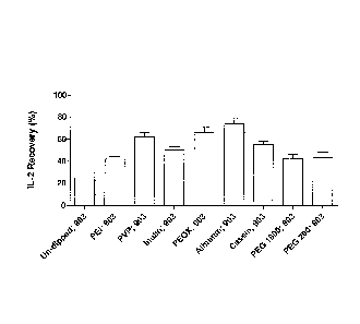
applied to 903 Neonatal STD papers coated with various chemicals.
Figure 3 presents the recovery of exogenously-added IL-2 from dried blood
spots
applied to DMPK-C papers coated with various chemicals.
11a
Date recue/Date Received 2021-01-20
CA 02828158 2013-08-23
WO 2012/113906
PCT/EP2012/053163
Detailed Description of the Invention
Recombinant IL-2 carrier (R & D Systems; Cat. 202-IL-CF-10pg; lot
AE4309112 and Cat. 202-IL-I0pg, lot AE4309081 respectively) was dissolved in
either Dulbecco's PBS without calcium and magnesium (FAA; Cat. H15-002, lot
H00208-0673), EDTA-anti-coagulated human, rabbit or horse blood (TCS
Biosciences) at 50 pg or 100 pg/pl.
Aliquots (1 pl containing 0, 50 or 100 pg of IL-2) were applied to the
following GE
Healthcare filter papers; 903 Neonatal STD card, Cat. 10538069, lot 6833909
W082; DMPK-A card, Cat. WB129241, lot F16847509; DMPK-B card, Cat.
WB129242, Lot FE6847609 and DMPK-C card, Cat. WB129243, Lot FE6847009.
Samples were allowed to dry overnight at ambient temperature and humidity.
Punches (3 mm diameter) were extracted from each paper type using the
appropriately sized Harris Uni-core punch (Sigma, Cat.Z708860-25ea, lot 3110).
Single punches were placed into individual wells of the IL-2 microplate
derived
from the Human IL-2 Quantikine ELISA (R & D Systems, Cat. D0250, lot 273275).
These plates are coated with a mouse monoclonal antibody against IL-2. The IL-
2 protein was eluted from the paper punch using the assay buffer (100 pl)
supplied with the Quantikine kit. All subsequent steps were performed
according
to the instructions supplied with the Quantikine kit using a "paper in" method
(paper punches are placed directly into the assay buffer and the analyte
eluted
directly in situ). On completion of the assay the optical density of the
microplate
was monitored at 450 nm using a Thermo Electron Corporation, Multiskan
Ascent. The recovery of IL-2 was determined by comparing values to a standard
curve of known IL-2 concentrations. A fresh IL-2 standard curve was prepared
for
each individual experiment.
Additional experiments involved the addition of IL-2-spiked blood to the 903
Neonatal STD and DMPK-C cards after the cards had been saturation dipped in
several chemical solutions (as described below).
12
CA 02828158 2013-08-23
WO 2012/113906
PCT/EP2012/053163
Chemicals Used
A list of the chemicals and their sources is given below.
Poly-ethyl-enemine, 50% in water (Fluka; Cat. P3143, lot 29k1492).
Poly-vinyl-pyrolodine, 1% in water (Sigma; Cat.PVP40-100 mg, lot 11pk0097).
lnulin, 1% in water (Sigma; Cat. 12255-100 g, lot 079F7110).
Poly-2-ethyl-2-oxazoline, 1 % in water (Aldrich Cat. 372846, lot 30498PJ).
Albumin, 1% in water (Sigma, Cat A2153-10 g, lot 049k1586).
Caesin from bovine milk, 1% in water (Sigma, Cat. C5890-500 g, lot 089k0179).
Poly-ethylene glycol 1000, 1% in water (Biochemika, Cat. 81189, lot 1198969).
Poly-ethylene glycol 200, 1% in water (Fluka, Cat. 81150, lot 1384550).
13
CA 02828158 2013-08-23
WO 2012/113906 PCT/EP2012/053163
Experimental Results
When IL-2 was dissolved in EDTA-anti-coagulated blood, the 903 and DMPK-C
cards facilitated the recovery of 45 - 55% of the cytokine, while only 2 -3 %
was
recovered from the DMPK-A and B cards (see Table 1 and Figure 1). The 903
and DMPK-C cards are the basic base papers and have not been dipped or
coated with any chemical, whilst the DMPK-A and B cards are coated with a
proprietary mixture of chemicals that facilitate the denaturation and
inactivation of
proteins, micro-organisms and cells respectively. These cards have been
designed to facilitate the transportation and prolonged storage of nucleic
acids.
Therefore the low IL-2 recovery levels observed when using the DMPK-A and B
cards may actually be a reflection of the presence of these denaturing
reagents
and the ELISA-based antibody detection system used. The ELISA detection
system requires the eluted IL-2 to exhibit an intact native structure.
Paper type IL-2 recovery ( /0) p-value
903; minus carrier 46.9 13.3 > 0.05
903; plus carrier 50.7 5.8
DMPK A; minus carrier 2.0 0.0 > 0.05
DMPK A; plus carrier 2.0 0.0
DMPK B; minus carrier 2.0 0.0 > 0.05
DMPK B; plus carrier 2.0 0.0
DMPK C; minus carrier 53.9 4.8 > 0.05
DMPK C; plus carrier 45.2 5.4
Table 1 - The Recovery of exogenously-added IL-2 from dried blood spots
applied to various paper types. The p-value compares carrier for each paper
type. The presence of the carrier had no significant effect on the recovery of
IL-2
(p-value > 0.05).
No IL-2 recovery was observed when the cytokine was dissolved in PBS
irrespective of the paper type used (data not shown). The IL-2 recovery levels
observed in the absence of added IL-2 were essentially equivalent to
background
14
CA 02828158 2013-08-23
WO 2012/113906
PCT/EP2012/053163
levels indicating that the EDTA-anti-coagulated blood contain negligible
amounts
of endogenous IL-2 (data not shown).
Several chemicals were used to saturation dip the 903 Neonatal STD and
DMPK-C cards, some of which appeared to facilitate the recovery of elevated IL-
2 levels compared to non-dipped papers (p-value <0.05). For both the 903
Neonatal STD and DMPK-C cards (Tables 2 and 3; Figures 2 and 3), chemicals
such as poly-vinyl-pyrolodine, poly-2-ethyl-2-oxazoline, albumin and casein
facilitated a significant increase in IL-2 recovery levels (mean > 55 %
compared
to ¨45% observed for the corresponding un-dipped paper).
Chemical IL-2 recovery (%) p-value
Un-dipped 44.9 6.5 nia
Poly-ethyl-anent-le (PEI) 41.8 6.0 > 0.05
Poly-vinyl-pyrolodine (PVP) 62.0 10.7 <0.05
Inulin 50.4 7.6 > 0.05
Poly-2-ethyl-2-oxazoline (Pe0X) 66.1 12.6 <0.05
Albumin 73.8 13.6 <0.05
Caesin 55.0 7.8 <0.05
Poly-ethylene glycol 1000 (PEG 1000) 42.5 9.1 > 0.05
Poly-ethylene glycol 200 (PEG 200) 43.3 11.0 > 0.05
Table 2 ¨ The Recovery of exogenously-added IL-2 from dried blood spots
applied to 903 Neonatal STD papers coated with various chemicals. The table is
derived from 2 independent experiments (n = 6). The p-value compares the
values derived from the dipped papers to those derived from the Un-dipped 903
paper.
CA 02828158 2013-08-23
WO 2012/113906
PCT/EP2012/053163
Chemical IL-2 recovery (%) p-value
Un-dipped 49.0 2.1 .. nia
Poly-ethyl-enemine (PEI) 55.8 12.2 .. > 0.05
Poly-vinyl-pyrolodine (PVP) 74.7 7.8 <0.05
Inulin 33.6 15.4 .. > 0.05
Poly-2-ethyl-2-oxazoline (Pe0X) 62.2 2.0 <0.05
Albumin* 63.7 increase
Caesin 57.7 1.5 .. <0.05
Poly-ethylene glycol 1000 (PEG 1000) 31.0 2.8 >0.05
Poly-ethylene glycol 200 (PEG 200) 33.5 15.7 .. > 0.05
Table 3 ¨ The Recovery of exogenously-added IL-2 from dried blood spots
applied to DMPK-C coated with various chemicals (n = 3). The p-value
compares the values derived from the dipped papers to those derived from the
Un-dipped DMPK-C paper. Albumin* n = 1.
While preferred illustrative embodiments of the present invention are
described,
one skilled in the art will appreciate that the present invention can be
practised by
other than the described embodiments, which are presented for the purposes of
illustration only and not by way of limitation. The present invention is
limited only
by the claims that follow.
16