Note: Descriptions are shown in the official language in which they were submitted.
CA 02829543 2013-10-15
SPEC05-4CA
1
Spatial Filter to Combine Excitation Light and Emission Light in an Episcopic
Multiplexed Confocal Scanning Microscope
TECHNICAL FIELD
[0001] The technology relates generally to episcopic optical microscopes, and
more
particularly to fluorescence imaging microscopes and to scanning confocal
optical
microscopes.
BACKGROUND
[0002] Episcopic microscopy is defined as the use of a microscope system
whereby the
illumination light illuminates the sample through the objective lens of the
microscope for the
observation of scattered light. The scattered light may be elastically
scattered at the same
wavelength as the illumination wavelength or inelastically scattered at a
different wavelength.
Scattered light from the sample may be collected by the same objective lens
and directed
towards an eyepiece of the microscope or towards an imaging device, such as a
camera.
[0003] In episcopic imaging, the illumination light path and the scattered
light path are
typically combined by a beam splitting optic in the form of a half-silvered
mirror or a cube
beam splitter, for example. In one example, the half-silvered mirror reflects
the illumination
light and transmits the scattered light. This is illustrated in the example
wide-field microscope
system of Figure 1. Illumination light having a wavelength X,1 is transmitted
from a light
source 102 and reflected by a beam splitter 104 through an objective lens 106
of a microscope
and onto a sample 108 in a sample plane 110. Scattered light having a
wavelength X2 is
collected by the objective lens 106 of the microscope. Where the wavelength X2
of the
scattered light differs from the wavelength Xi of the illumination light, that
is where X2 ,
the scattered light may be transmitted, via the beam splitter 104, to an
imaging device 112
with an optional tube lens (not shown).
[0004] The word "combine", as used herein, may refer to the joining or
coupling of the
illumination light path with the scattered light path in one direction, and
the splitting or
separating of the illumination light path from the scattered light path in
another direction. In
the example of Figure 1, the path of the illumination light, as it is being
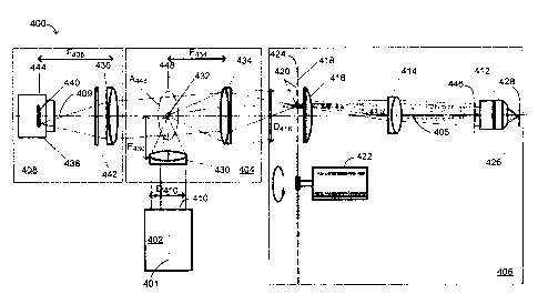
transmitted toward the
CA 02829543 2013-10-15
SPEC05-4CA
2
microscope objective lens 106, is being combined with the scattered light path
via the beam
splitter 104. Conversely, the scattered light, as it is coming from the
microscope objective lens
106, is being split from the illumination light by the beam splitter 104.
[0005] It is noted that the illumination light path and the scattered light
path are mutually
interchangeable. For example, the positions of the light source 102 and the
imaging device 112
may be switched relative to one another, and the beam splitter 104 may be used
to transmit the
illumination light, and to reflect the scattered light.
[0006] Use of the beam splitter 104 may result in high losses of illumination
or excitation
light, as well as scattered light. In one example, at least half of the
illumination or excitation
light may be lost when it is reflected by the beam splitter 104, and at least
half of the scattered
light may be lost when it is being transmitted through the beam splitter 104.
Therefore, use of
the half-silvered beam splitter 104 in an episcopic microscope may result in a
significant
portion being lost, for example 50% of the excitation light and 50% of the
scattered light.
[0007] The optical setup of Figure 1 with the half-silvered beam splitter 104
may be used for
fluorescence imaging with the addition of a narrowband filter (not shown)
between the beam
splitter 104 and the imaging device 112. A narrowband filter may be used to
suppress
elastically scattered light having a wavelength X2 = Ai and to transmit
inelastically scattered
light having a wavelength X2 X1. Given that fluorescence microscopy is often
done with
weak or very sensitive samples that result in minimal fluorescence light being
emitted, the
inefficiency of the half-silvered beam splitter 104 may be unacceptable for
such applications.
[0008] In fluorescence imaging, the illumination or excitation light path and
the scattered or
emission light path are typically combined by a dichroic mirror. In the
example illustrated in
Figure 1, in the event that the beam splitter 104 is a dichroic mirror, the
dichroic mirror 104
may reflect the illumination light and transmit the scattered light.
Illumination light having a
wavelength k1 is transmitted from the light source 102 and reflected by the
dichroic mirror
104 through the objective lens 106 of the microscope and onto the sample 108
in the sample
plane 110. Fluorescence emission light having a wavelength X2 that differs
from the
wavelength X1, that is X2 Xi, is collected by the objective lens 106 of the
microscope and
transmitted, via the dichroic mirror 104, to the imaging device 112.
[0009] The wavelength structure of the dichroic mirror 104 may be designed to
work with
one or more wavelength bands of illumination light and one or more wavelength
bands of
CA 02829543 2013-10-15
SPEC05-4CA
3
scattered light. However, such dichroic mirrors rarely work for all the
wavelengths of interest.
Thus, it is common for multiple dichroic minors to be mounted in a mirror
turret or minor
wheel so that different dichroic mirrors can be selected for different
wavelength requirements.
[0010] As an alternative to the use of half-silvered mirrors or dichroic
mirrors, some systems
may combine scattered and illumination light bands using a polarization beam
splitter. This
may be done with linearly polarized light or circularly polarized light.
Figure 2 illustrates the
combining of illumination and scattered light using a polarizing beam splitter
204 and a
quarter wave plate 206. In this implementation, only light that retains the
circular polarization
is passed back through the polarizer 204.
[0011] While such techniques may be used effectively for elastically scattered
light,
fluorescence scattering is generally unpolarized. Therefore, using a
polarizing beam splitter on
fluorescence scattering may result in a significant portion ¨ at least half ¨
of the scattered light
being lost. The inefficiency of a polarizing beam splitter may be unacceptable
for such
applications, because of the reasons mentioned above.
[0012] Figure 3 illustrates another technique, as described in U.S. Patent No.
6,888,148, for
spatial coupling of illumination and scattered light paths. This technique,
which is also
described in German Patent No. 679547, U.S. Patent No. 3,229,564, and U.S.
Patent No.
4,314,763, involves the use of a spatial filter in the form of a small mirror
or prism, also
known as a "pick-off' mirror or prism, placed in the back focal plane of a
microscope lens.
[0013] A collimated illumination light beam may be transmitted from a light
source 302 and
focused by a lens 308 onto the back focal plane 306 of an objective lens 310
of a microscope.
The illumination light beam may be reflected by a pickoff minor 304 toward the
objective lens
310. The illumination light is focused by the objective lens 310 onto a sample
312 to achieve a
dark-field illumination of the sample 312. The light reflected by the sample
312 may be
collected by the objective lens 310. An aperture of the objective lens 310 is
located at the back
focal plane 306. The majority of the light collected by the objective lens 310
may be
transmitted, via an optional tube lens 314, to an imaging device 316, thus
providing a dark-
field image of the same 312 at an image plane 318 coinciding with an image
sensor of the
imaging device 316.
[0014] As a result of the pick-off minor 304, a portion of the light collected
by the objective
lens 310 is blocked from being transmitted to the imaging device 316. The size
of the pick-off
CA 02829543 2013-10-15
SPEC05-4CA
4
mirror 304 may be substantially smaller than the size of the aperture of the
objective lens 310
at the back focal plane 306.
[0015] In all of the microscope systems described herein, the illumination
light path and the
scattered light path may be mutually interchangeable. That is, the half-
silvered mirror or the
dichroic mirror and the polarizing beam splitter may be used to transmit the
illumination light,
and to reflect the scattered light. The same is true for the spatial beam
coupler, in the case that
the illumination light beam is focused onto the back focal plane of the
objective lens through
an opening in a high reflection mirror, which reflects the scattered light to
the imaging device.
[0016] High-resolution confocal laser microscopy is an established field in
modern imaging
and bioimaging technologies. This technique provides sharp, high-
magnification, three-
dimensional imaging with submicron resolution by non-invasive optical
sectioning and
rejection of out-of-focus information (see, T. Code and G. Kino, "Confocal
Scanning Optical
Microscopy and Related Imaging Systems," Academic Press, San Diego, 1996).
[0017] A point scanning confocal system images a single point at a time. Light
is projected
onto the sample through a single illumination aperture, such as a pinhole.
Light from the focal
plane of the sample then passes through the same pinhole or a conjugate
pinhole to reject out-
of-focus light. The rejection of out-of-focus light is a significant advantage
of a confocal
imaging system as it provides an uncluttered image of the focal plane.
[0018] In episcopic confocal imaging, the illumination light path and the
elastically scattered
light path may be combined by a half-silvered mirror or dichroic beam
splitter, or by use of a
polarization beam splitter. A system with a spatial coupling/separation of the
illumination light
path and the scattered light path provides a nearly collimated beam that
illuminates a total field
of view of the objective lens, as illustrated in Figure 3. This system cannot
be used to provide
a focused light spot on the sample, and therefore is not applicable to single
point confocal
scanning imaging.
[0019] Multi-point confocal systems employ a similar principle, but image
multiple points in
the sample plane simultaneously. These multi-point confocal systems typically
use an array of
pinholes or other illumination apertures that simultaneously capture multiple
points of the
image. Some models of confocal systems use a spinning disk with pinholes
arranged in a
pattern, such as a Nipkow pattern, to provide uniform illumination of the
sample. Examples of
a confocal scanner employing the conventional Nipkow disk method have been
described in
U.S. Patent No. 3,926,500 and U.S. Patent No. 4,927,254. Another example of a
confocal
CA 02829543 2013-10-15
SPEC05-4CA
scanner employing a two-dimensional array of pinholes that translate in a
plane of the array is
described in U.S. Patent No. 4,806,004. Multi-point confocal systems have
become important
microscopy tools, for example, in life science research. They are seen as
simple, cost effective
instruments that acquire images quickly and with less damaging effects on
samples than point
scanning systems.
100201 In a conventional apparatus for confocal scanning microscopy such as
that described
above, the pinhole disk of the confocal optical scanner unit thereof may be
tilted to reduce the
amount of surface reflection from the pinhole disk surface into an imaging
device.
100211 The use of optical fibers as flexible laser delivery subsystems has
proven particularly
useful in confocal microscopy, and is described, for example, in JP0420912,
U.S. Patent No.
5,557,452, U.S. Patent No. 8,275,226, and U.S. Patent Application Publication
No.
20110134519.
SUMMARY
[0022] Previous implementations of spinning disk systems do not use collimated
light for
illumination. (Rather, spinning disk systems that use lenslet arrays typically
focus light
through the pinholes, where the numerical aperture of the lenslets is chosen
to fill the back
aperture of the objective. In that case, the focused light passing through the
pinholes is
necessarily uncollimated.) Single disk confocal instruments have traditionally
been designed
for use with large area light sources such as light pipes or arc lamps. Large
area light sources
are not conducive to creating collimated light. As known in the art, the back
aperture of the
objective must be filled in order to achieve resolution at the diffraction
limit. In the absence of
a pinhole disk, collimated light at an image plane will focus to a small spot
at the back
aperture of the objective. To persons of ordinary skill in the art, this is a
perceived barrier to
using collimated light with spinning disk systems.
100231 It is also known in the art that placing a small pick-off member, for
example, a small
pick-off mirror, in an illumination path will necessarily produce collimated
light. Thus it is not
known and it is not obvious to place a small pick-off member, for example, a
small pick-off
mirror, at a conjugate plane to the back aperture of the objective of a
spinning disk system,
because the small pick-off member will necessarily illuminate the pinholes of
the spinning
disk with collimated light.
CA 02829543 2013-10-15
SPEC05-4CA
6
[0024] Notwithstanding the foregoing, this document describes methods and
arrangements in
which a spatial filter (for example, a small pick-off member, or a perforated
mirror comprising
a window) is placed at a conjugate plane to the back aperture (back focal
plane) of the
objective of a multiplexed confocal scanning microscope system. In these
methods and
arrangements, illumination apertures of a scanning disk are necessarily
illuminated with
collimated light. Through experimentation, the inventors discovered that it is
in fact not only
possible to use collimated light for illumination but it can be advantageous.
This was a
surprising and unexpected result. The inventors discovered that diffraction of
light by the
pinholes filled an area of the back aperture of the objective that was
sufficient to project a
resolved pinhole to the sample plane, within the resolving power of the
objective. Thus the
filled area of the back aperture of the microscope objective is determined by
the size of the
pinholes, and the use of collimated light will work for all sizes of pinholes.
This makes it
possible to match the pinhole size to the objective resolving power and
optimally fill the
objective under all conditions.
[0025] The area of the spatial filter may be substantially smaller than the
area of the image of
the back aperture at the conjugate plane. For example, the area of the spatial
filter may be
approximately 5% of the area of the back aperture image. In another example,
the area of the
spatial filter may be approximately 10% of the area of the back aperture
image. In yet another
example, the area of the spatial filter may be approximately 15% of the area
of the back
aperture image. In a further example, the area of the spatial filter may be
approximately 20%
of the area of the back aperture image.
[0026] In the methods and arrangements described in this document, the
presence of the
spatial filter enables illumination light in a multiplexed confocal scanning
microscope system
to be efficiently separated from scattered light, such as fluorescence light
or luminescence
light excited and/or backscattered in a specimen. In contrast to the above-
described prior art
techniques, the separation is not wavelength-dependent and is therefore
particularly suitable
for use in multi-wavelength fluorescence microscopy, for example, for the
simultaneous
excitation of different dyes. Accordingly, fast switching between several
excitation
wavelengths or spectral detection wavelength ranges ¨ so-called multitracking
¨ may be
achieved in a particularly simple manner.
[0027] In accordance with the technology described herein, there is provided a
multiplexed
confocal scanning microscope system which comprises a scanning disk with a
plurality of
CA 02829543 2013-10-15
SPEC05-4CA
7
pinholes arranged in a predetermined pattern, such as a spiral path. The
combining of the
illumination light and the scattered light may be achieved through the use of
a beam
combining module, which may comprise a spatial filter, for example, a small
pick-off member
or a perforated mirror comprising a window.
[0028] In one example, illumination light originating from a light source may
be focusing
onto a small pick-off member having an aperture conjugate to a back focal
plane of an
objective lens of the confocal microscope. The illumination light is reflected
by the pick-off
member and collimated by a lens providing a collimated or a nearly collimated
beam of light
illuminating a surface of the scanning pinhole disk. The surface of the
scanning pinhole disk is
arranged to coincide with a first image plane that is conjugate to a plane of
a specimen or
sample to be imaged.
[0029] The pinholes of the scanning pinhole disk diffract the impinging light
and the incident
beam is split by the scanning disk into an array of individual narrow
diffracted beams which
travel through an optional field lens and a tube lens and converge on the
objective lens.
[0030] Each individual beam may be focused onto the sample plane, providing an
array of
diffraction limited focal spots. The pinhole array disk may be rotated at a
constant speed, and
since the pinholes are arranged along the spiral path and since the plane of
the pinhole array
disk is conjugate to the sample plane, the rotation of the disk forms a raster
scan across the
sample. The light focused on the multiple points of the sample may be
scattered by the sample,
captured by the objective lens at its operating numerical aperture NAms and
focused back at
the same pinholes. Most of the light passing through the pinholes and used to
create an image
of the back aperture of the objective lens may continue unimpeded to a second
image plane,
where it may be captured by a high sensitivity imaging device, such as a
camera. However, a
small portion of the light will be obstructed by the pick-off member. The
sample plane, the
first image plane, and the second image plane are conjugate.
[0031] The light that continues unimpeded by the pick-off member is focused
with a relay
system onto the second image plane, which coincides with an image sensor of
the camera,
providing an array of diffraction limited focal spots. Rotation of the pinhole
array disk forms a
raster image, thus providing a magnified confocal image of the sample for
detection by the
image sensor.
[0032] In another example, the illumination light path and the imaging light
path may be
mutually interchanged. In this case, illumination light originating from a
light source may be
CA 02829543 2013-10-15
SPEC05-4CA
8
focusing onto a window in a high reflection perforated mirror, where the
window is located at
a plane conjugate to a back focal plane of the objective lens. The
illumination light transmitted
through the window is ultimately used by the multiplexed confocal microscope
to illuminate a
sample. The scattered light from the sample is ultimately reflected to the
imaging device by
the high reflection mirror.
[0033] In another example, illumination light from a light source may be
delivered to the
beam combining module by a flexible light delivery subsystem, for example, in
the form of an
optical fiber. Different configurations may be used to provide Kohler
illumination of a sample,
or critical illumination of the sample.
[0034] The efficiency of the illumination may be improved, for example, by
using a light
source module and a beam combining module having optics that are configured to
provide a
magnification such that an area of substantially uniform illumination on the
plane of the
pinhole array disk is not substantially bigger than a region of interest (ROI)
corresponding to
an area of the sample plane that is imaged by the imaging device. In the case
where
illumination light is delivered to the beam combining module by an optical
fiber, the
efficiency of the illumination may be improved, for example, by selecting the
fiber such that
the dimensions of the cross section of the core of the fiber and the numerical
aperture NAF of
the fiber satisfy a particular relationship with respect to the numerical
aperture NAms of the
microscope and properties of the pinhole array disk.
100351 An "illumination aperture", as used herein, refers to any illumination
aperture that is
suitable for use in confocal microscopy, as would be apparent to someone
skilled in the art.
For example, an "illumination aperture array" may refer to a plurality of
pinholes, a plurality
of slits, a Nipkow array, or any other suitable plurality of illumination
apertures. It may also be
part of a confocal scanner employing a rotating disk with the plurality of the
illumination
apertures, or two-dimensional array of pinholes translatorily movable in a
plane of the array.
Any suitable plurality of illumination apertures can be substituted for the
pinhole array of
pinholes employed in the examples described herein.
[0036] The terms "light" and "radiation" may be used interchangeably and refer
to light in
the UV-visible-NIR (ultraviolet-visible-near infrared) spectral range. The
terms "light source"
and "radiation source" may refer to any source able to generate and emit light
or radiation,
including but not limited to, lasers, light emitting diodes (LEDs), solid
state devices, super
CA 02829543 2013-10-15
SPEC05-4CA
9
luminescent diodes (SLDs), arc lamps, or any other suitable light sources as
would be apparent
to someone skilled in the art.
[0037] "Illumination light" or "excitation light", as used herein, refers to
any light provided
by a light source to be used for illumination of a sample. "Scattered light"
or "returned light"
refers to the light returning from the sample, and used for obtaining images
of the sample. The
returned light is often produced by fluorescence but can result from multi-
photon excitation
emission, reflection, Raman scattering or any other elastic or inelastic light
scattering effect as
would be known to someone skilled in the art.
[0038] The term "nearly collimated beam", as used herein, may refer to a light
beam having
a divergence that is greater than a diffraction-limited light beam, but low
enough so as not to
change the beam geometry on the scale of the optical system under
consideration. Real beam
divergence will be specified in every particular application.
[0039] The term "combining", as used herein, may refer to the joining or
coupling of the
illumination light path with the scattered light path in one direction, and
the splitting or
separating of the illumination light path from the scattered light path in
another direction.
[0040] As used herein, a "microscope" comprises at least an objective lens, as
illustrated, for
example, by the objective lens 106 in Figures 1 and 2, and by the objective
lens 310 in Figure
3. In other examples, microscopes may be considered to have the more
conventional form of
an infinity corrected micro-objective and a tube lens. Both cases are
interchangeable. A
"microscope system" is a system that may be used to probe a sample by
projecting light or
radiation onto the sample, thus producing reflected light or scattered light
or fluorescence light
or any combination thereof from the sample. As used herein, "multiplexed
confocal
microscopy" or "multipoint confocal microscopy" refers to the use of a
plurality of
illumination apertures to apply the confocal technique in parallel to a
microscope.
CA 02829543 2013-10-15
SPEC05-4CA
BRIEF DESCRIPTION OF THE DRAWINGS
[0041] Figure 1 illustrates a prior art episcopic imaging optical microscope
system
employing a half-silvered mirror beam splitter and/or dichroic beam splitter.
[0042] Figure 2 illustrates a prior art episcopic imaging optical microscope
system
employing a polarizing beam splitter and quarter wave plate.
[0043] Figure 3 illustrates a prior art episcopic imaging optical microscope
system
employing spatial combining of the illumination light path and the scattered
light path.
[0044] Figure 4-1 illustrates a first example multiplexed confocal microscope
system
employing a pick-off member for spatial combining of the illumination light
path and the
scattered light path.
[0045] Figure 4-2 illustrates a second example multiplexed confocal microscope
system
employing a pick-off member for spatial combining of the illumination light
path and the
scattered light path.
100461 Figure 5 illustrates a third example multiplexed confocal microscope
system
employing a pick-off member for spatial combining of the illumination light
path and the
scattered light path.
[0047] Figure 6 illustrates an example multiplexed confocal microscope system
employing a
perforated minor for spatial combining of the illumination light path and the
scattered light
path.
[0048] Figure 7 illustrates an example multiplexed confocal microscope system
employing a
pick-off member for spatial combining of the illumination light path and the
scattered light
path, comprising an optical fiber as a flexible light delivery subsystem and
configured to
provide Kohler illumination of a sample
[0049] Figure 8 illustrates an example multiplexed confocal microscope system
employing a
pick-off member for spatial combining of the illumination light path and the
scattered light
path, comprising an optical fiber as a flexible light delivery subsystem and
configured to
provide critical illumination of a sample.
[0050] Figure 9 illustrates an example multiplexed confocal microscope system
employing a
perforated minor for spatial combining of the illumination light path and the
scattered light
path, comprising an optical fiber as a flexible light delivery subsystem and
configured to
provide Kohler illumination of a sample.
CA 02829543 2013-10-15
SPEC05-4CA
11
100511 Figure 10 illustrates an example multiplexed confocal microscope system
employing
a perforated mirror for spatial combining of the illumination light path and
the scattered light
path, comprising an optical fiber as a flexible light delivery subsystem and
configured to
provide critical illumination of a sample.
[0052] It will be appreciated that for simplicity and clarity of illustration,
elements illustrated
in the figures have not necessarily been drawn to scale. For example, the
dimensions of some
of the elements may be exaggerated relative to other elements for clarity.
CA 02829543 2013-10-15
SPEC05-4CA
12
DETAILED DESCRIPTION
[0053] Figure 4-1 illustrates a first example multiplexed confocal microscope
system
employing a pick-off member for spatial combining of the illumination light
path and the
scattered light path.
[0054] The system 400 of Figure 4-1 comprises a light source module 402, a
beam
combining module 404, a multiplexed confocal microscope module 406 and an
imaging
module 408.
[0055] The light source module 402 may be configured to generate and to
optionally
condition light to be emitted from an exit aperture 410. The light source
module 402 may
comprise one or more light sources, including, for example, lasers, light
emitting diodes
(LEDs), solid state devices, super luminescent diodes (SLDs), arc lamps, or
any other suitable
light sources as would be apparent to someone skilled in the art. In one
example, the light
sources may comprise one or more lasers, each laser generating light at a
different wavelength.
The light source module 402 may comprise additional optical elements,
including, for
example, one or more lenses, one or more mirrors, one or more optical fibers,
and/or one or
more light guides. The optical fibers may include single-mode and/or multi-
mode fibers, and
the light guides may include fiber bundles, and liquid-filled light guides.
[0056] One or more optical components may be used to form a collimated or
nearly
collimated beam of illumination light at the exit aperture 410 of the light
source module 402,
where the exit aperture 410 has a diameter D410. The one or more optical
components may
include, for example, lenses, mirrors, prisms, and the like.
[0057] The light source module 402 may also comprise a beam-shaping means (not
shown)
for increasing evenness of the transverse intensity distribution of the
illumination beam. The
beam shaping means may be implemented, for example, by a diffractive beam
shaper element,
refracting optical components, mirror optics, or a filter means. The beam-
shaping means may
be selected so as to produce a collimated illumination beam having a
transverse intensity
distribution that is as uniform as possible. In one example, the beam profile
may have a "top
hat" shape with a flat transverse energy distribution. In another example, the
collimated
illumination beam may have a non-uniform profile, such as Gaussian profile, a
cosine profile
or any other profile as would be apparent to someone skilled in the art.
CA 02829543 2013-10-15
SPEC05-4CA
13
[0058] The multiplexed confocal microscope module 406 comprises an objective
lens 412, as
well as an optional tube lens 414 and an optional field lens 416. The
multiplexed confocal
microscope module 406 further comprises a pinhole array disk 418, which
comprises a
plurality of pinholes 420. The pinhole array disk 418 may be rotatable about
its axis, for
example, using a motor 422. Instead of the plurality of pinholes 420, the
pinhole array disk
418 may comprise a plurality of slits, or any other suitable plurality of
illumination apertures.
The pinhole array disk 418 may be built in the form of a two-dimensional array
of pinholes
420 translatorily movable in a plane of the pinhole array disk 418. For the
sake of simplicity,
the pinhole array disk 418 is herein assumed to have the form of a Nipkow
disk, such that the
pinholes 420 are disposed in a pattern of several interleaved spirals. The
pinholes 420 may be
spaced approximately two to ten pinhole diameters apart. The plane of the
pinhole array disk
418 may coincide with a first image plane 424 of the multiplexed confocal
microscope module
406.
[0059] The pinholes 420 of the pinhole array disk 418 diffract the impinging
light. The light
beam incident on the pinhole array disk 418 is split by the pinhole array disk
418 into an array
of individual narrow diffracted beams that travel through the optional field
lens 416 and the
optional tube lens 414 and converge on the objective lens 412. One of the
individual narrow
diffracted beams is schematically illustrated in Figure 4-1 (not to scale) by
a shadowed area
starting from the light source module 402 and terminating on a sample 426,
where the sample
426 is located at a sample plane 428. Each individual beam may be focused onto
the sample
plane 428, providing an array of diffraction limited focal spots. The pinhole
array disk 418
may be rotated by the motor 422 at a constant speed. Since the pinholes 420
are arranged
along a spiral path and since the first image plane 424 and the sample plane
428 are conjugate,
the rotation of the pinhole array disk 418 may form a raster scan across the
sample 426. Where
a transverse distribution of light exiting the light source module 402 is
substantially uniform, it
may be possible to achieve a substantially uniform intensity of light
transmitted through
corresponding the pinholes 420 of the pinhole array disk 418. Accordingly,
after a complete
scan of the pinholes 420, it may be possible to achieve a substantially
uniform illumination of
the sample 426.
[0060] The beam combining module 404 comprises a focusing lens 430 with a
focal length
F430, a pick-off member 432, and a collimating lens 434 with a focal length
F434. The pick-off
member 432 acts as a spatial filter and may be placed a distance F430 from the
lens 430 along
CA 02829543 2013-10-15
SPEC05-4CA
14
the optical axis 401 of the light source module 402, such that the focusing
lens 430 focuses the
collimated or nearly collimated beam onto the pick-off member 432. The
illumination light is
reflected by the pick-off member 432 towards the collimating lens 434, where
the collimating
lens 434 is placed a distance F434 from the pick-off member 432 along the
optical axis 405 of
the multiplexed confocal microscope module 406. The lenses 430 and 434 form a
telescopic
device providing a collimated or nearly collimated beam for illumination of
the first image
plane 424 of the multiplexed confocal microscope module 406 and the conjugate
sample plane
428. Using the paraxial ray approximation, a light ray originating from the
center of the exit
aperture 410 of the light source module 402 at an angle 0410 relative to the
optical axis 401
will be incident, in normal operation, on the first image plane 424 at an
angle 0434 relative to
the optical axis 405, where the angle 0434 is expressed in equation 1 as:
[0061] 0434 =0410*(F430/F434) (1)
[0062] A longest dimension D418 of an area on the pinhole array disk 418 to be
illuminated,
which is roughly equal to a working aperture D434 of the lens 434, is
proportional to a
diameter D410 of the illumination beam exiting the exit aperture 410. This is
expressed by
equation 2:
[0063] D4180410 1"--F4341F430 (2)
[0064] The lenses 430 and 434 are presented in the form of regular achromatic
doublets.
Alternatively, they may be built in the form of singlet spherical, aspheric,
gradient index,
triplet, or multi-component lenses, or any other focusing elements, including
reflective
focusing elements, Fresnel's optical elements, and diffractive focusing
elements, as would be
apparent to someone skilled in the art.
[0065] The pick-off member 432 is illustrated in Figure 4-1 in the form of a
small flat
reflecting minor. However, the pick-off member 432 may be, for example, any
mirrored
surface that is able to reflect the illumination light towards the microscope,
or a small right
angle prism. Alternatively, rather than using reflection, the pick-off member
432 may use
transmission, diffraction or refraction, as would be apparent to someone
skilled in the
art. Also, in some applications, it may be of interest to use a small
polarizing beam splitter or a
small dichroic mirror as the pick-off member 432. It should be noted that if a
dichroic mirror
CA 02829543 2013-10-15
SPEC05-4CA
is used as the pick-off member 432, little light will be obstructed by the
pick-off member 432,
but the flexibility to work at all wavelengths of interest may be compromised.
In the example
of Figure 4-1, the illumination light has an angle of incidence of 45 degrees
relative to the
surface of the pick off member 432, so that an angle between the axis 401 and
the axis 405 is
90 degrees. It will be apparent to someone skilled in the art that this angle
between the axes
401 and 405 may be varied in a wide range, for example, from 5 degrees to 150
degrees,
where the angle may be dependent on a type of the pick-off member 432 being
used and on
the incidence angle.
[0066] The beam combining module 404 is configured to image the exit aperture
410 of the
light source module 402 onto the sample plane 428 (or a plane conjugate to the
sample plane
428, such as the first image plane 424). Such a configuration of the beam
combining module
404 may result in substantially uniform illumination of the sample 426.
[0067] Typically, it is desired in a confocal microscope system for the
pinhole spot to be
imaged at or near the diffraction limit of the microscope. To produce the
minimum imaged
spot size, a divergence 0420 of the light transmitted through the pinhole 420
should be the
same or larger than the largest numerical aperture NAms of the microscope as
measured at the
first image plane 424 where the pinhole 420 is located. If the numerical
aperture of light
exiting the pinhole 420 is larger than numerical aperture NAms of the
microscope, then the
minimum imaged spot size can be achieved, but some of the light will be
rejected by the
microscope optics. Ideally, the numerical aperture of light exiting the
pinhole 420 should
closely match the numerical aperture NAms of the microscope so that the
optimum resolution
and light transmission to the sample 426 can be achieved.
[0068] An acceptance numerical aperture NAms of the objective 412 of the
microscope (a
half of its back aperture angle) is expressed by equation 3 as:
[0069] NAms =sinOms D446/2L =D446/2F414 (3)
[0070] where Oms is the divergence angle from the microscope, D446 is a
diameter of the
back aperture of the microscope, L is the distance between the tube lens 414
and the back
focal plane 446 of the objective 412, and F414 is the focal length of the tube
lens 414.
CA 02829543 2013-10-15
SPEC05-4CA
16
[0071] The diameter d420 of the pinholes 420 should be chosen so that the
light exits the
pinhole 420 at an angle 0420 given by the numerical aperture NAms of the
microscope as
determined by optical diffraction theory. The divergence angle 0420 of the
light exiting any
individual pinhole 420 depends on the divergence angle 0434 of the
illumination beam
collimated with the lens 434 and on the diffraction at the pinhole 420, which
is a function of
the wavelength X of the illumination light and the pinhole diameter d420. This
is expressed by
equation 4 as:
[0072] 0420 =scIrt(04342 +(0.61X/d420)2) ..5. Oms7,..,= NAms (4)
[0073] The pinhole array is typically designed in a multi-focal confocal
microscope such that
the light exiting the pinhole is at or near the designed acceptance numerical
aperture NAms of
the microscope. Using equation 4, the divergence angle 0434 of the light
illuminating the
pinhole array disk 418 may be expressed as follows:
[0074] 0434 sqrt(NAm52 - y2) (5)
[0075] where y =0.61X/ d420 is an angle of the diffraction.
[0076] Equations 1, 2 and 5 may be used with the paraxial approximation to
derive a
relationship between (i) the product of a desired dimension D418 of the area
on the pinhole
array disk 418 to be illuminated and the microscope acceptance numerical
aperture NAms and
(ii) the product of the illumination beam diameter D410 of the light source
module 402 and the
exit divergence angle 0410. This relationship is expressed in equation 6 as:
[0077] D4100410 =D4340434 =- D418'sqrt(NAms2
- Y2) <D418NAMS (6)
[0078] It is of interest to have a light beam of relatively low etendue. For
example, the
etendue of the light beam may be lower than the etendue of the microscope.
That is, the
product of the light beam diameter D410 and the exit divergence angle 0410 may
satisfy the
requirements given by equation 6.
[0079] For a pinhole diameter d420 =30 1.1M, a wavelength X =488 nm, a
dimension D418 =
mm, and a microscope acceptance numerical aperture NAms =0.015, the
diffraction angle
CA 02829543 2013-10-15
SPEC05-4CA
17
is y = 0.61X/d420 =0.01, and the optimal product of the divergence 0410 of the
illuminating
beam and its diameter 13410 is expressed as:
[0080] D4100410 0.12mm <0.15mm (7)
[0081] The light focused on the multiple points of the sample 426 may be
scattered from the
sample 426 and captured by the objective lens 412 at the operating numerical
aperture NAms
of the objective lens 412. The captured light may be focused at the same
pinholes 420 at the
first image plane 424 after traveling through the optional tube lens 414 and
the optional field
lens 416.
[0082] The imaging module 408 may be configured to provide an analog or
digital image of
the first image plane 424 and, consequently, a magnified region of interest
(ROI) of the sample
426 under investigation conjugated to the first image plane 424. The imaging
module 408
comprises an imaging lens 436 with a focal length F436 and an imaging device
438, such as a
high-sensitivity camera, with an image sensor 440. Optics of the imaging
module 408 may
comprise additional elements such as a blocking filter 442, essential for
fluorescence imaging,
and/or narrow band filters for multi-spectral imaging, a relay lens (not
shown), a diaphragm
(not shown) and other additional elements, as would be apparent to someone
skilled in the art.
[0083] The imaging lens 436 is placed on the optical axis 409 of the imaging
module 408
(which coincides with the optical axis 405 of the multiplexed confocal
microscope module
406) at a distance F436 from the image sensor 440. The imaging lens 436 is
schematically
illustrated in Figure 4-1 in the form of a regular achromatic doublet.
Alternatively, the imaging
lens 436 may be built in the form of a multi-component lens, aspheric singlet,
gradient index,
triplet lenses, or any other focusing elements, including reflective focusing
elements, Fresnel's
optical elements, and diffractive focusing elements, as would be apparent to
someone skilled
in the art.
[0084] A non-exhaustive list of examples of the imaging device 438 includes a
charge-
coupled device (CCD) camera, a complementary metal-oxide semiconductor (CMOS)
camera,
an intensified CCD (ICCD) camera, and any other suitable camera as would be
apparent to
someone skilled in the art. A 3CCD camera with additional narrowband filters
may be applied
for simultaneous multi-spectral imaging.
CA 02829543 2013-10-15
SPEC05-4CA
18
100851 In order provide an image of the sample 426, the imaging device 438,
the sample 426
and the relay optics (comprising, for example, the lenses 434 and 436) may be
arranged such
that the sample plane 428 and a plane of the image sensor 440 are optically
conjugate with
each other.
100861 The light passing through the pinholes 420 is focused by the lens 434
and the imaging
lens 436 onto the image sensor 440, providing an array of diffraction limited
focal spots.
Rotation of the pinhole array disk 418 forms a raster image, thus providing a
magnified
confocal image of the sample 426. The sample plane 428, a first image plane
424 (coinciding
with the surface of the pinhole array disk 418), and a second image plane 444
(coinciding with
the image sensor 440) are conjugate to one another.
100871 The light path of the individual narrow diffraction beam being
reflected by the sample
426, collected by the objective lens 412, and further focused onto the first
image plane 424,
and the second image plane 444 is schematically illustrated in Figure 4-1 by
means of
marginal rays (not in scale).
100881 The beam combining module 404 may be configured to provide a
magnification such
that the area of substantially uniform illumination on the sample 426 is not
substantially bigger
than the ROI of the sample 426 that is imaged by the image sensor 440 of the
imaging device
438. This may improve the efficiency of the illumination.
100891 Within the relay lens optics (comprising, for example, the lenses 434
and 436), an
image of the back focal plane 446 of the objective lens 412 may be formed. The
plane at
which the image of the back focal plane 446 is formed may be defined as the
conjugate back
focal aperture plane 448. The image of the back focal plane 446 in the
conjugate aperture
plane 448, also called the back aperture image, has an area A448. The field
lens 416, which
may be located at or near the first image plane 424, may be used to collimate
the scattered
light entering the relay lens 434. This means that the image of the back focal
plane 446 may be
located at the focal length F434 of the lens 434. It is substantially at the
conjugate aperture
plane 448 that the pick-off member 432 is located. The pick-off member 432 may
have an area
A432 that is substantially smaller than the area A448 of the back aperture
image. For example,
the diameter D432 of the pick-off member 432 may be substantially less than
the diameter
D448 of the back aperture image, that is D432 <<D448.
CA 02829543 2013-10-15
SPEC05-4CA
19
[0090] While an aperture of the pick-off member 432 may be as small as
possible, it should
be large enough to reflect all the focused illumination light toward the
multiplexed confocal
microscope module 406. A minimal diameter D432 of the aperture of the pick-off
member 432
may be found using the paraxial ray approximation, and is expressed in
equation 8 as:
[0091] D432 20410F430 (8)
[0092] In one example, the area A432 of the aperture of the pick-off member
432 is
approximately 5% of the area A448 of the back aperture image. Most of the
light used to create
the back aperture image may continue on to the second image plane 444, where
it may be
captured, for example, by the image sensor 440 of the imaging device 438.
However, a portion
of the light will be blocked, partially blocked, reflected or otherwise
obstructed by the pick-off
member 432. The portion of scattered light that is obstructed corresponds to
the area A432,
whereas the portion of the scattered light that is not obstructed corresponds
to an area A'432,
where the area A448 is the union of the area A432 and the area A'432, and
where the area A432
does not intersect with the area A'432. That is, A448 =A432 u A'432 and where
A432 n A'432
=0.
[0093] An efficiency E of the beam combining module 404 is related to the
ratio of the area
A432 to the area A448 as expressed in equation 9:
[0094] E =1 - (- --tA^32)/(- -+A A48) (9)
[0095] For example, one may consider the case that the back aperture image has
a diameter
D448 = 2.5 mm, and the pick-off member 432 is in the form of a right angle
prism having
aperture dimensions 0.5mm x 0.5mm. In this case, the area A432 obscured by the
pick-off
member 432 is approximately 5% of the area A448 of the back aperture image,
that is A432 =
0.05 =A448. Thus, approximately 5% of the scattered light may be blocked by
the pick-off
member 432. Since the remaining portion A'432 of the area A448 is
approximately 95% of the
area A448 of the back aperture image, that is A'432 =0.95 A448, it follows
that approximately
95% of the scattered light may be transmitted to the imaging device 438. This
may provide a
highly efficient and flexible means of combining the scattered and
illumination light paths.
CA 02829543 2013-10-15
SPEC05-4CA
While it is contemplated that all of the area A432 corresponding to the pick-
off member 432
may be located within the area A448 of the back aperture image, it is
alternatively possible that
some portion of the area A432 may be outside of the area A448 of the back
aperture image.
Only that portion of the area A432 that is within the area A448 of the back
aperture image may
block or obstruct the scattered light from reaching the second image plane
444.
100961 Collimated light beams are traditionally used in complex optical
systems, where it is
possible the collimated beams are less aberrated than non-collimated beams by
optical
components that they travel through. However, the illumination light beam
provided by the
light source module may be neither collimated nor nearly collimated, and may
have a
considerable divergence or convergence. An example of this is illustrated in
Figure 4-2.
100971 The multiplexed confocal microscope system 450 of Figure 4-2 comprises
a light
source module 452, a beam combining module 454, a multiplexed confocal
microscope
module 456 and an imaging module 458.
100981 The beam combining module 454 is similar to the beam combining module
404
illustrated in Figure 4-1. The multiplexed confocal microscope module 456 is
similar to the
multiplexed confocal microscope module 406, except that it does not include
the optional
lenses 414 and 416. The imaging module 458 is similar to the imaging module
408, except
that the blocking filter 442 is positioned on the opposite side of the imaging
lens 436.
100991 The light source module 452 is similar to the light source module 402,
in that it may
be configured to generate and optionally to condition light to be emitted from
an exit aperture
410. In this case, the light source module 452 comprises a point-like light
source illustrated in
the form of an arc lamp 460 and a collective lens 462.
[00100] The illumination beam exiting the exit aperture 410 of the light
source module 452
may have a rather high numerical aperture absolute value INIA410 I =sin a ,
where a is an angle
between a marginal ray of the light beam and the optical axis 401.
1001011 In the event that the light beam has a relatively high numerical
aperture absolute
value INIA4m I, such that sin a 1/20, for example, in order for the lens 430
to focus the light
beam onto the aperture of the pick-off member 432, the lens 430 should be
placed a distance
430 from the pick-off member 432, where the distance 430 is expressed in
equation 10 as:
1001021 Z43o430, /(1 -2a_F
43o/3410) (10)
CA 02829543 2013-10-15
SPEC05-4CA
21
[00103] The relationship in equation 10 uses the thin lens approximation and
paraxial
approximations.
[00104] To correctly image the pick-off mirror to the back aperture of the
objective, it is
common practice to use an optional field lens (not shown) located close to the
disk plane 424.
To avoid the use of a field lens, the light beam illuminating the pinhole
array disk 418 may be
somewhat convergent or divergent to match the microsope optics without a field
lens, as it is
illustrated in Figure 4-2.
[00105] It should be noted that the total area of the pinholes 420 illuminated
at any one time is
about five percent of A418 (the illuminated part of the pinhole array disk
418) or less. Thus,
only a few percent of the illuminating light reaching the plane of the pinhole
array disk 418
actually passes through the pinholes 420. A major part of the light is
reflected from the disk
418 itself toward the imaging module 408. For this reason, it may be
beneficial to eliminate
the reflected light, particularly when observing weakly-scattering samples.
One technique for
doing this is described in the U.S. Patent No. 4,927,254, in which the
rotating disk is tilted to
eliminate reflections from the disk. The disk may be tilted sufficiently far
so that the light
reflected from the disk is deviated far enough from the optical axis that it
exits the imaging
system and is directed towards a light stop in front of the imaging lens 436
or a light stop
located at the aperture plane 448 and is eliminated. In the examples of
Figures 4-1 and 4-2,
the collimated beam of the remaining reflected light from the pinhole array
disk 418 is focused
by the collimating lens 434 back onto the pick-off member 432, which is
located at the focal
point of the lens 434, thus removing remaining reflected light from the
imaging light beam
travelling to the imaging module 408. For luminescence imaging, it may be of
interest to place
an optional narrow band filter 442 in the emission path between the detection
device and the
pick-off member 432, to further reduce scattered excitation light, as
reflected from the pinhole
array disk 418 and from the optical components of the confocal microscope
system. Another
technique is to place an adjustable iris at the conjugate plane 448, where the
adjustable iris is
centered around the pick-off member 432 (or more generally, around the spatial
filter). This
technique may be used in combination with the optional narrow band filter 442
or in
combination with the tilting of the rotating disk or in combination with both.
[00106] Figure 5 illustrates a third example multiplexed confocal microscope
system
employing a pick-off member for spatial combining of the illumination light
path and the
scattered light path.
CA 02829543 2013-10-15
SPEC05-4CA
22
[00107] Similarly to the example of Figure 4-1, the multiplexed confocal
microscope system
500 of Figure 5 comprises the multiplexed confocal microscope module 406 and
the imaging
module 408. However, in place of the light source module 402 and the beam
combining
module 404, the system of Figure 5 comprises a light source module 502 and a
beam
combining module 504, respectively.
[00108] The light source module 502 comprises a light source 503, a beam
conditioning unit
505, an exit aperture 510, and an optional folding mirror 512.
[00109] The light source 503 may be built, for example, in the form of multi-
wavelength light
source assembly described in U.S. Patent No. 8,275,226, to generate multi-
wavelength light
that is suitable for illumination in confocal microscopy. Depending on the
application, the
light source module 502 may comprise one or more lasers, each laser generating
light at a
different wavelength, for example.
[00110] The beam conditioning unit 505 may comprise, for example, two sub-
units (not
shown) ¨ a beam-shaping means for increasing an evenness of intensity
distribution of the
illumination light beam, and a beam expander to provide the illumination beam
with a desired
diameter. The beam shaping means may be implemented by a diffractive beam
shaper
element, refracting optical components, mirror optics, or a filter means. The
beam expander
may be built in the form of telescopic setup of any kind, as would be apparent
to someone
skilled in the art, and may include one or more lenses, one or more mirrors,
and one or more
prisms.
[00111] The light source module 502 may be configured to provide a collimated
beam of
illumination having a desired diameter and substantially high evenness of the
transverse
intensity distribution of the illumination.
[00112] The beam combining module 504 comprises a collimating lens 434 and a
pick-off
member in the form of a convex mirror 514. The convex mirror 514 may be
spherical mirror
or an off-axis parabolic mirror, and it has a focal length -F514 and a
diameter D514 that is
substantially less than the diameter D448 of the back aperture image, that is
D514 <<D448.
The convex mirror 514 defocuses the collimated beam and reflects it towards
the collimating
lens 434, which is placed a distance 434F
=- 434 ¨ F514 from the convex mirror 514. The
convex mirror 514 and the lens 434 form a telescopic device providing a
collimated or nearly
collimated beam for illumination of the first image plane 424.
CA 02829543 2013-10-15
SPEC05-4CA
23
[00113] In another example, the mirror 514 for the narrow collimated
illumination beam
provided by the light source module 502 may be built in the form of a concave
spherical
mirror or an off-axis parabolic mirror having a focal length F514. In this
case, the collimating
lens 434 would be placed at a distance Z'434 =F434 +F514 from the pick-off
member 514 in
order to form a telescopic setup. Similarly to the pick-off member 432, the
mirror 514 acts as a
spatial filter.
[00114] Figure 6 illustrates an example multiplexed confocal microscope system
employing a
perforated mirror for spatial combining of the illumination light path and the
scattered light
path. In this example, the illumination light path and the imaging light path
are mutually
interchanged.
[00115] Similarly to the example of Figure 4-1, the multiplexed confocal
microscope system
600 of Figure 6 comprises the light source module 402, the multiplexed
confocal microscope
module 406, and the imaging module 408. The system includes a beam combining
module
604, which comprises the focusing and collimating lenses 430 and 434. The beam
combining
module 604 also comprises a highly reflecting folding perforated mirror 632
with a small
coupling window 633 for transmitting illumination light. The window 633 may
also be
described as a hole or an opening. The perforated mirror 632, which acts as a
spatial filter, is
placed so that the window 633 is fixed in the plane 448 conjugate to the back
focal plane of
the objective lens of the multiplexed confocal microscope module 406. The
window 633 in the
folding perforated mirror 632 functions as a beam combining (pick-off) member.
The
illumination light beam exiting the light source module 402 is focused by the
focusing lens
430 into the window 633 and is transmitted to the multiplexed confocal
microscope module
406. The scattered imaging light from the multiplexed confocal microscope
module 406 is
reflected by the folding perforated mirror 632 toward the imaging module 408.
[00116] Figure 7 illustrates an example multiplexed confocal microscope system
employing a
pick-off member for spatial combining of the illumination light path and the
scattered light
path, comprising an optical fiber as a flexible light delivery subsystem and
configured to
provide Kohler illumination of a sample.
[00117] Similarly to the example of Figure 4-1, the multiplexed confocal
microscope system
700 of Figure 7 comprises the beam combining module 404, the multiplexed
confocal
microscope module 406, and the imaging module 408. The system includes a light
source
module 702 configured to provide a collimated (or nearly collimated) beam of
illumination
CA 02829543 2013-10-15
SPEC05-4CA
24
light. The light source module 702 comprises a light source 703, which may be
built, for
example, in the form of multi-wavelength light source assembly described in
U.S. Patent No.
8,275,226, to generate and optionally to condition multi-wavelength light that
is suitable for
illumination in confocal microscopy. Depending on the application, the light
source 703 may
comprise one or more lasers, each laser generating light at a different
wavelength, for
example.
[00118] The light source module 702 may also comprise a beam-shaping means
(not shown)
for increasing an evenness of an angular distribution of the illumination
light beam. The beam
shaping means may be implemented, for example by a diffractive beam shaper
element,
refracting optical components, mirror optics, or a filter means.
[00119] The light source module 702 may comprise a fiber optic light delivery
subsystem
705. The fiber optic light delivery subsystem 705 is presented in the form of
an optical fiber.
The optical fiber 705 may be a single-mode or multi-mode optical fiber with a
core diameter
DF and numerical aperture NAF, a fiber bundle, or a light guide.
[00120] A multimode fiber may have a core of circular cross section, square
cross section,
rectangular cross section, or any other suitable cross section. A step-index
multimode fiber is
an example of a multimode fiber for which the transverse distribution of light
exiting the distal
end is substantially uniform. It is contemplated that other multimode fibers
also exhibit the
behavior that the angular distribution or the transverse distribution of light
exiting the distal
end is substantially uniform. In the expression "angular distribution of light
exiting the distal
end of the multimode fiber" means distribution of a radiated power as a
function of an
inclination angle relative to the optical axis of the fiber; "transverse
distribution of light
exiting the distal end of the multimode fiber", the term "transverse" means
transverse to an
optical axis of the multimode fiber.
[00121] It may be of interest to have an angular distribution of the light
beam exiting a distal
end 706 of the optical fiber 705 be as uniform as possible, thus making it
possible to achieve a
substantially uniform intensity of light transmitted through corresponding
pinholes 420 of a
pinhole array disk 418, which, after a complete scan of the pinholes 420, may
result in
substantially uniform illumination of the sample 426.
[00122] Collimating optics 707 having a focal length F707 may be placed a
distance F707
from the fiber tip 706. The collimating optics 707 is illustrated in the form
of an achromatic
doublet lens. Alternatively, the collimating optics 707 may be built in the
form of singlet
CA 02829543 2013-10-15
SPEC05-4CA
spherical, aspheric, gradient index, triplet, or multi-component lens, or any
other focusing
elements, including reflective focusing elements and diffractive focusing
elements, as would
be apparent to someone skilled in the art. The fiber optic light delivery
subsystem 705 may
comprise additional elements not illustrated in Figure 7. For example, the
fiber optic light
delivery subsystem 705 may comprise means for phase randomization or
additional optical
fibers or both.
[00123] It should be noted that the plane 710 of the fiber tip 706, the plane
448 in which the
pick-off member 432 is fixed, and the back focal plane 446 of the microscope
objective lens
412 are conjugate. That means that lenses 707 and 430 form a relay optical
device providing
an image of the fiber tip 706 onto the pick-off member 432, and further onto
the back focal
plane 446 of the objective lens 412. This optical setup provides relatively
uniform illumination
of the sample, which is known as "Kohler illumination."
[00124] The light exits the fiber 705 with a spread of angles given by the
numerical aperture
NAF of the fiber 705. In general, a numerical aperture NAF of a fiber is
expressed in equation
11 as:
[00125] NAF =n=sinOF (11)
[00126] where n is a refractive index of the surrounding medium to which the
light exits from
the distal end of the fiber, exit angle OF is the angle of divergence of light
with respect to an
optical axis of the fiber. In the case that the surrounding medium is air, the
refractive index n =
1.
[00127] A beam of the illumination light exiting the aperture 410 of the light
source module
702 is characterized by its diameter D410 and divergence angle 0410 near the
beam axis 401,
where both depend on the core diameter DF of the fiber 705, its numerical
aperture NAF, and
the focal length F707 of the collimating lens 707. They may be found using the
paraxial ray
approximation, and are expressed in equations 12 as:
[00128] 0410---DF42F707); D410 =20706 F707 (12)
[00129] where 0706 is the divergence angle at the fiber tip 706.
[00130] Equations 6 and 12 may be used with the paraxial approximation to
derive the
following desired product of the core diameter DF of the fiber 705 and the
exit angle 0706:
CA 02829543 2013-10-15
SPEC05-4CA
26
[00131] DF0706D A
=-410-410 D418 sqrt(NAms2
¨ 72) <D418NAMS (13)
[00132] For a pinhole diameter d420 =30 p.m, a wavelength X. =488 nm, a
dimension D418 =
mm, and a microscope acceptance numerical aperture NAms =0.015, the
diffraction angle
is y = 0.6 1XA1420 =0.01, and the optimal product of the divergence 0410 of
the illuminating
beam and its diameter D410 is given by equation 7. If the numerical aperture
NAF of the fiber
705 is NAF =0.22, the optimal fiber core diameter DF is expressed as DF .._
0.55mm.
[00133] An efficiency E of the beam combining module 404 is defined by the
numerical
aperture NAms and the dimension D418 of the ROI image of the multiplexed
confocal
microscope module 406, and by the numerical aperture NAF and the core diameter
DF of the
fiber 705. The efficiency E may be found using the paraxial approximation and
Lagrange
invariant, and is expressed in equation 14 as:
[00134] E 1 =((DFNAF)/(D4181=1Ams))2 (14)
[00135] By placing the distal end 706 of the fiber 705 a distance F707 from
the lens 707, light
passing through the lens 707 may be collimated. A diameter of the lens 707
should be large
enough to capture the light emitted from the distal end 706 of the fiber 705.
[00136] In order to provide an image of the distal end 706 of the fiber 705
onto the aperture of
the pick-off member 432, the lens 430 of the beam combining module 404 may be
placed a
distance F430 from the pick-off member 432.
1001371 A magnification M of the image provided by the relay optics formed by
the lenses
707 and 430 is defined by the respective focal lengths F707 and F430 of the
lenses, and is
expressed in equation 15 as:
[00138] m F /F
¨ =- 430¨ 707 (15)
[00139] The numerical aperture sin0432 of the illumination beam reflected by
the pick-off
member 432 is expressed in equation 16 as:
[00140] sin0432 -=NAF/M =sin0F(F707/F430) (16)
[00141] where 0432 is the divergence of the illumination beam reflected by the
pick-off
member 432. The aperture diameter D432 of the pick-off member 432 depends on
the core
CA 02829543 2013-10-15
SPEC05-4CA
27
diameter DF of the fiber 705 and on the magnification 5. In order to achieve
an effective
coupling of the illumination light path with the microscope imaging light path
and to avoid
undesired loss of the illumination light, the aperture diameter D432 may be
selected to satisfy
equation 17:
[00142] D432 ..>_. M=DF ---,--DF(F430/F707) (17)
[00143] Since the core diameter DF and the numerical aperture NAF =sin0706 of
the fiber 705
have been selected to approximately follow the relation of equation 11, the
etendue of light
emitted from the distal end 706 of the fiber 705 does not substantially exceed
the etendue of
light passing through the pinholes 420 of the pinhole array disk 418, thereby
achieving
efficient illumination of the sample 426.
[00144] In normal operation, illumination light from the light source module
702 will arrive at
the center of a central pinhole 420 at an angle 0434 relative to the optical
axis 405. The angle
0434 is the divergence of the illumination beam collimated by the collimating
lens 434, and is
expressed in equation 18 as:
[00145] 0434 -=(DFF430)/(F707F434) (18)
[00146] The radiant intensity of the light at the plane of the pinhole array
disk 418 may be
substantially uniform as long as the distal end 706 is imaged sufficiently
close to the plane of
the pinhole array disk 418, even if the distal end 706 is not imaged precisely
onto the plane of
the pinhole array disk 418. Substantially uniform illumination of the sample
after a complete
scan of the illumination apertures may therefore be achievable where (i) the
transverse
distribution of light exiting the distal end 706 of the fiber 705 is
substantially uniform and (ii)
light-coupling optics of the light source module 702 and the beam combining
module 404 are
configured to image the distal end 706 of the fiber 705 onto or sufficiently
close to the plane
of the pinhole array disk 418.
[00147] Persons of ordinary skill in the art will appreciate that
substantially uniform
illumination of a sample may also be achievable when, instead of or in
addition to imaging the
exit aperture of the fiber tip 706 onto or sufficiently close to the sample
plane 426, the light
source module 702 and the beam combining module 404 image the exit aperture of
the light
source module 702 onto or sufficiently close to a plane that is optically
conjugate to the
sample plane 426.
CA 02829543 2013-10-15
SPEC05-4CA
28
[00148] Figure 8 illustrates an example multiplexed confocal microscope system
employing a
pick-off member for spatial combining of the illumination light path and the
scattered light
path, comprising an optical fiber as a flexible light delivery subsystem and
configured to
provide critical illumination of a sample.
[00149] Similarly to the example of Figure 4-1, the multiplexed confocal
microscope system
800 of Figure 8 comprises the beam combining module 404, the multiplexed
confocal
microscope module 406, and the imaging module 408. The system includes a light
source
module 802 configured to provide a nearly collimated beam of illumination
light. The light
source module 802 comprises the light source 703, and the fiber optic light
delivery subsystem
705 with a fiber distal end tip 706. The light source module 802 further
comprises magnifying
relay optics in the form of a first lens 804 having a short focal length F804
and a second lens
806 having a long focal length F806, where the focal length F806 is
substantially greater than
the focal length F804, that is F806 >>F804. The first lens 804 may be placed a
distance F804
from the plane 810 of the fiber tip 706. A relay optics aperture 808 may be
placed close to the
first lens 804.
[00150] The light source module 802 may also comprise a beam-shaping means
(not shown)
for increasing an evenness of an angular distribution of the illumination
light beam. The beam
shaping means may be implemented, for example, by a diffractive beam shaper
element,
refracting optical components, mirror optics, or a filter means.
[00151] The relay optics is illustrated in the form of pair of achromatic
doublet lenses 804 and
806. Alternatively, it may be built in the form of single doublet, triplet,
aspheric, or gradient
index lens having low aberrations, in the form of a multi-component relay
lens, or in the form
of a finite microscope objective. The relay optics 804 and 806 is configured
to image the distal
end 706 of the fiber 705 laying in the plane 810 onto an intermediate image
plane 812 that is
optically conjugated to the first image plane 424 coinciding with the surface
of the scanning
pinhole disk 418, and to the plane 428 of the sample 426, both conjugated to
the second image
plane 444 coinciding with the image sensor 440. This optical setup provides
highly uniform
illumination of the sample 426, which is known as "critical illumination".
[00152] The lens 806 may be placed at a distance equal to or approximately
equal to its focal
length F806 from the lens 804 and/or from the aperture 808. In this case the
lens 806 together
with the focusing lens 430 of the beam combining module 404 forms a relay
optical device
CA 02829543 2013-10-15
SPEC05-4CA
29
providing an image of the lens 804 and/or the aperture 808 onto the pick-off
member 432, and
further onto the back focal plane 446 of the objective lens 412. It says that
plane 814 of the
lens 804 and/or aperture 808, the plane 448 in which the pick-off member 432
is fixed, and the
back focal plane 446 of the microscope objective lens 412 are conjugate. Such
arrangement of
the fiber tip 706, the lens 804, the lens 806, the lens 430, and the pick-off
member 432
provides a light spot with a minimal diameter in the plane 448 and,
consequently, a minimal
required aperture of the pick-off member 432. The pick-off member 432 should
be large
enough to direct toward the multiplexed confocal microscope module 406 all the
illumination
light coming through the aperture 808.
[00153] Alternatively, the light source module 802 and the beam combining
module 404 may
be configured to image the distal end 706 of the fiber 705 sufficiently close
to the plane 428 of
the sample 426 or sufficiently close to the first image plane 424 such that a
radiant intensity of
light at the sample plane 428 or the first image plane 424 is substantially
uniform. In one
example, the fiber tip 706 may be fixed at a small distance from the plane 810
conjugate to the
first image plane 424, the sample plane 428, and the second image plane 444.
Such a
configuration of the light source module 802 and the beam combining module 404
may result
in a substantially uniform illumination of the sample 426.
[00154] The beam combining module 404 may be configured to provide a
magnification such
that the area of substantially uniform illumination on the sample plane 428 is
not substantially
bigger than an imaged area of the sample plane 428 that is imaged by the image
sensor 440 of
the imaging device 438. This may improve the efficiency of the illumination.
[00155] The excitation light is emitted from the distal end 706 of the fiber
705 and is turned
into diverging rays with a maximum spread-angle dependent on the numerical
aperture NAF
of the fiber 705. The light then passes through the lens 804, the aperture
808, and the lens 806.
The lenses 804 and 806 and the fiber 705 may be configured to image the tip
706 of the fiber
705 onto, or sufficiently close to, the plane 812 conjugated to the first
image plane 424
coinciding with the surface of the pinhole array disk 418. The lenses 430 and
434 may be
configured to reimage the tip 706 of the fiber 705 from the plane 812 onto the
first image
plane 424 so that the radiant intensity of light at the first image plane 424
is substantially
uniform because the transverse distribution of light exiting the fiber 705 is
substantially
uniform. Specifically, since the lenses 804, 806, 430 and 434 have been
selected to provide a
magnification equal to or greater than the minimum magnification Mmin
according to equation
CA 02829543 2013-10-15
SPEC05-4CA
16, the size of the image of the tip 706 of the fiber 705 at the first image
plane 424 is sufficient
to illuminate with substantial uniformity all the pinholes 420 that are used
by the imaging
module 408 to construct the confocal image of the sample 426. The minimum
magnification
Mmin is expressed by equation 19 as:
[00156] Mmin =M802 M404 =(F806T804)' (F434/F430) ?-- D418/DF (19)
[00157] A beam of the illumination light exiting the aperture 810 of the light
source module
802 is characterized by its diameter D810 and divergence angle 0810 near the
beam axis 401.
These values in turn depend on the core diameter DF of the fiber 705, its
numerical aperture
NAF, and the focal length F804 of the collimating lens 804. They may be found
using the
paraxial ray approximation and are expressed in equation 20 as:
[00158]
0810=07061M802 =-0706F804/F806; D810'4'-= DFM802 =DFF806/F804 (20)
[00159] Equations 6 and 12 may be used with the paraxial approximation to
derive the
following desired product of the core diameter DF of the fiber 705 and the
exit angle 0706,
again resulting in the expression given in equation 13.
[00160] The light exiting the pinhole array disk 418 is passed through
optional field lens 416
and optional tube lens 414 and converged to the sample 426 by the objective
lens 412.
[00161] Since the core diameter DF and the numerical aperture NAF of the fiber
705 have
been selected to approximately follow the relation of equation 13, the etendue
of light emitted
from the distal end 706 of the multimode fiber 705 does not substantially
exceed the etendue
of light passing through the pinholes 420 of the pinhole array disk 418,
thereby achieving
efficient illumination of the sample 426.
[00162] A signal from the sample 426, for example a fluorescence signal,
passes back through
the objective lens 412, and is converged to the individual pinholes 420 of the
pinhole array
disk 418. The fluorescence signal passing through the individual pinholes 420
is passed
through the beam combining module 404, so as to be imaged onto the sensor 440
of the
imaging device 438 via the relay lenses 434 and 436.
[00163] It should be noted that the plane 814 of the aperture 808, the plane
448 in which the
pick-off member 432 is fixed, and the back focal plane 446 of the microscope
objective lens
412 are conjugate. It should also be noted that the plane 810 of the fiber tip
706, the first
image plane 424, and the sample plane 428 are conjugate. That means that the
lenses 806 and
CA 02829543 2013-10-15
SPEC05-4CA
31
430 form a relay optical device providing an image of the aperture 808 onto
the pick-off
member 432, and further onto the back focal plane 446 of the objective lens
412.
[00164] A minimal aperture diameter D432 of the pick-off member 432 depends on
a
magnification of the image provided by relay optics 806 and 430, as defined by
the respective
focal lengths F806 and F430 of the lenses, and by a diameter D808 of the
aperture 808 as
defined by the focal length F804 of the lens 804:
[00165] D432 _._ D808 (F430/F806) =NAF(F430.F804/F806) (21)
[00166] To achieve an effective coupling of the illumination light path with
the microscope
imaging light path and to avoid undesired loss of the illumination light, the
aperture diameter
D432 of the pick-off member 432 should satisfy the equation 18.
[00167] The divergence angle 0434 of the illumination beam collimated by the
collimating
lens 434 is expressed in equation 22 as:
[00168] 0434 r.:, NAF/(M802M404) N¨A (P430P804)MP80uF434) N
AF(DFM418) (22)
[00169] where M802 is the magnification of the source module 802, and M404 is
the
magnification of the beam combining module module 404.
[00170] As an alternative to the use of the beam combining module 404 for
Kohler
illumination and critical illumination, as illustrated in Figures 7 and 8, a
beam combining
module with a folding perforated mirror may be employed, as described with
respect to Figure
6. This alternative is illustrated in the examples of Figures 9 and 10.
[00171] Figure 9 illustrates an example multiplexed confocal microscope system
employing a
perforated mirror for spatial combining of the illumination light path and the
scattered light
path, comprising an optical fiber as a flexible light delivery subsystem and
configured to
provide Kohler illumination of a sample.
[00172] Similarly to the example of Figure 4-1, the multiplexed confocal
microscope system
of Figure 9 comprises the light the multiplexed confocal microscope module 406
and the
imaging module 408. The system of Figure 9 further comprises a combined light
source and
beam combining module 902. The combined module 902 comprises the light source
703 and
the fiber optic light delivery subsystem 705 with a fiber distal end tip 706,
as described
previously with respect to the example of Figure 7. The combined module 902
also comprises
the collimating lens 434 and the highly reflecting folding perforated mirror
632 with the small
CA 02829543 2013-10-15
SPEC05-4CA
32
coupling window 633 for transmitting illumination light, as described
previously with respect
to the example of Figure 6.
[00173] As previously described, the perforated mirror 632 is placed so that
the window 633
is fixed in the plane 448 conjugate to the back focal plane of the objective
lens of the
multiplexed confocal microscope module 406. The window 633 in the folding
perforated
minor 632 works as a beam combining (pick-off) member. The distal end tip 706
of the fiber
705 is placed on the optical axis 401 of the combined light source and beam
combining
module 902, close to the window 633 of the mirror 632, such that the
illumination light exiting
the distal end 706 is transmitted to the multiplexed confocal microscope
module 406.
[00174] After passing through the window 633 of minor 632, the light may be
collimated by
means of the collimating lens 434, thereby providing a collimated or nearly
collimated beam
for illumination of the first image plane 424 of a multiplexed confocal
microscope module
406, which coincides with the plane of the pinhole array disk 418 and the
conjugated plane of
the sample to be probed. The scattered imaging light from the multiplexed
confocal
microscope module 406 is reflected by the folding perforated mirror 632 toward
the imaging
module 408.
[00175] As described previously with respect to the example of Figure 7, the
plane of the fiber
tip 706 is conjugate to the back focal plane 446 of the microscope objective
lens 412. This
optical setup provides Kohler illumination of the sample.
[00176] Figure 10 illustrates an example multiplexed confocal microscope
system employing
a perforated mirror for spatial combining of the illumination light path and
the scattered light
path, comprising an optical fiber as a flexible light delivery subsystem and
configured to
provide critical illumination of a sample.
[00177] Similarly to the example of Figure 4-1, the multiplexed confocal
microscope system
of Figure 10 comprises the light the multiplexed confocal microscope module
406 and the
imaging module 408. The system of Figure 10 further comprises a combined light
source and
beam combining module 1002. The combined module 1002 comprises the light
source 703
and the fiber optic light delivery subsystem 705 with a fiber distal end tip
706, as described
previously with respect to the example of Figure 7. The combined module 1002
also
comprises the collimating lens 434 and the highly reflecting folding
perforated minor 632
with the small coupling window 633 for transmitting illumination light, as
described
previously with respect to the example of Figure 6.
CA 02829543 2013-10-15
SPEC05-4CA
33
[00178] As previously described, the perforated mirror 632 is placed so that
the window 633
is fixed in the plane 448 conjugate to the back focal plane of the objective
lens of the
multiplexed confocal microscope module 406. The window 633 in the folding
perforated
mirror 632 works as a beam combining (pick-off) member.
[00179] In contrast to the combined light source and beam combining module
902, the
module 1002 further comprises a lens 1004 having a short focal length F1004.
The distal end
tip 706 of the fiber 705 is placed on the optical axis 401 of the combined
module 1002. The
fiber tip 706 and the lens 1004 may be positioned such that the lens 1004 is a
distance F100,4
from the plane 448 (and the window 633 of the perforated mirror 632), and such
that the fiber
tip 706 is a distance F1004 from the lens 1004. In this manner, the plane 448
will coincide with
a Fourier plane of the lens 1004. The lenses 1004 and 434 form relay optics
providing a
magnified image of the distal end 706 of the fiber 705 laying in the plane 810
onto the first
image plane 424 coinciding with the surface of the pinhole array disk 418, and
to the sample
plane, both conjugated to the second image plane 444 coinciding with the
detection plane of
the image sensor 440. This optical setup provides critical illumination of the
sample 426.
[00180] The light emitted from the distal end 706 of the fiber 705 is imaged
by means of the
relay optics formed by the lenses 1004 and 434 to provide a nearly collimated
beam for
illumination of the first image plane 424 of a multiplexed confocal microscope
module 406.
The scattered imaging light from the multiplexed confocal microscope module
406 is reflected
by the folding perforated mirror 632 toward the imaging module 408.
[00181] In all of the luminescent confocal microscope systems mentioned above,
fluorescent
dyes are used for specific labeling of the preparations. The number of dye
signatures that may
be used simultaneously, i.e., the number of characteristics, for example, of
cells that can be
investigated simultaneously, can be increased by means of the technology
disclosed herein.
When the spectral signatures of the individual dyes overlap extensively or are
very close to
one another, the detected wavelength range or numerical aperture should be
limited, according
to the prior art, for separate detection of the fluorescence signals of
individual dyes. This
reduces the sensitivity of detection, i.e., increases the noise of the
detectors, because greater
amplification must be used. This may be avoided by the methods and
arrangements disclosed
herein.
CA 02829543 2013-10-15
SPEC05-4CA
34
1001821 The technology disclosed herein may be used in image-generating
confocal
microscope systems as well as in analytic confocal microscope systems. The
confocal
microscope systems are episcopic image-generating systems such as multipoint
confocal
scanning microscopes for three-dimensional examination of biological
preparations,
fluorescence correlation microscopes for quantitative determination of
molecular
concentrations and for measuring molecular diffusions. They may be applied for
fluorescence
recovery after photobleaching (FRAP), fluorescence lifetime imaging (FLIM),
structured
illumination (SIM), super resolution and localization microscopy. Also
included are methods
based on fluorescence detection for screening dyes and methods for flow
cytometry.