Note: Descriptions are shown in the official language in which they were submitted.
CA 02829629 2013-09-09
WO 2012/122544
PCT/US2012/028640
PATENT
Attorney Docket No.: 93331-000310PC-825868
Client Reference No.: OTC-6034-JOH
PROTEIN NANOPARTICLE DISPERSIONS
RELATED APPLICATIONS
[0001] This application claims priority to U.S. Provisional Patent Application
No. 61/587,648
filed, January 17, 2012, entitled "HIGH PROTEIN CONCENTRATION NANOPARTICLE
DISPERSIONS" and U.S. Provisional Patent Application No. 61/451,571 filed,
March 10, 2011,
entitled "LOW VISCOSITY HIGH CONCNETRATION NANOPARTICLE ANTIBODY
DISPERSIONS". The disclosure of each of the above-referenced applications is
incorporated by
reference herein in their entirety.
STATEMENT AS TO RIGHTS TO INVENTIONS MADE UNDER
FEDERALLY SPONSORED RESEARCH OR DEVELOPMENT
[0002] This invention was made with government support under grants NSFSTC-CHE-
9876674, CBET-0968038, CBET-1065357 awarded by the National Science
Foundation. The
Government has certain rights in the invention.
BACKGROUND OF THE INVENTION
[0003] The present invention relates in general to the field of high
concentration protein
dispersion, and methods of making dispersions of protein nanoparticles. There
is a need in the art
for highly concentrated protein dispersion for a variety of applications
including, for example,
pharmaceutical formulations of subcutaneous administration. The present
inventions addresses
these and other needs in the art.
BRIEF SUMMARY OF THE INVENTION
[0004] In a first aspect, a transparent, low viscosity, high protein
concentration dispersion is
provided. The dispersion includes a plurality of nanoclusters. Each of the
plurality of
nanoclusters includes a plurality of proteins and each of the plurality of
proteins shares amino
acid sequence identity.
[0005] In a second aspect a pharmaceutical composition is provided, including
any of the
dispersions as described herein (including embodiments), wherein the plurality
of proteins is a
plurality of pharmaceutically active proteins.
1
CA 02829629 2013-09-09
WO 2012/122544
PCT/US2012/028640
[0006] In a third aspect a kit is provided, wherein the kit includes a
dispersion or
pharmaceutical composition described herein (including embodiments).
[0007] In a fourth aspect, a method of making a transparent, low viscosity,
high protein
dispersion of protein nanoclusters is provided, including concentrating a
protein-crowder liquid
combination and thereby forming the dispersion. The dispersion includes a
plurality of
nanoclusters, each of the plurality of nanoclusters includes a plurality of
proteins, and each of the
plurality of proteins shares amino acid sequence identity. The dispersion is a
transparent, low
viscosity, dispersion; wherein the dispersion includes a concentration of the
protein of greater
than about 200 mg/mL (e.g. greater than 200 mg/mL), and wherein the dispersion
includes a
plurality of a crowder.
[0008] In a fifth aspect, a method of making a transparent, low viscosity,
high protein
dispersion of protein nanoclusters is provided, including the step of
combining a protein in
powder form with a crowder and a dispersion liquid thereby forming a
dispersion, the dispersion
including a plurality of nanoclusters, the nanoclusters including a plurality
of the protein. Each of
the plurality of proteins shares amino acid sequence identity. The dispersion
is a transparent, low
viscosity, dispersion; wherein the dispersion includes a concentration of the
protein of greater
than about 200 mg/mL (e.g. greater than 200 mg/mL).
[0009] In a sixth aspect, a method of making a transparent, low viscosity,
high protein
dispersion of protein nanoclusters is provided, including the step of
combining a protein in
powder form with a dispersion liquid thereby forming a dispersion,the
dispersion including a
plurality of nanoclusters, the nanoclusters including a plurality of the
protein. Each of the
plurality of proteins shares amino acid sequence identity. The dispersion is a
transparent, low
viscosity, dispersion; wherein the dispersion includes a concentration of the
protein of greater
than about 200 mg/mL (e.g. greater than 200 mg/mL).
[0010] In a seventh aspect, a method is provided for treating a disease in a
patient in need of
such treatment, the method including administering an effective amount of any
one of the
dispersions described herein (including embodiments) to the patient.
[0011] In an eigth aspect, a method is provided for modifying the average
protein nanocluster
diameter of a transparent, low viscosity, high protein dispersion of protein
nanoclusters including
increasing or decreasing the concentration of a crowder or the protein in the
dispersion. The
2
CA 02829629 2013-09-09
WO 2012/122544
PCT/US2012/028640
dispersion includes a plurality of nanoclusters and each of the plurality of
nanoclusters includes a
plurality of proteins. Each of the plurality of proteins shares amino acid
sequence identity. The
dispersion is a transparent, low viscosity, dispersion; and the dispersion
includes a concentration
of the protein of greater than about 200 mg/mL (e.g. greater than 200 mg/mL).
[0012] In a further aspect a kit is provided, wherein the kit includes protein
in powder form or
a protein-crowder mixture in powder form, and a dispersion liquid.
BRIEF DESCRIPTION OF THE DRAWINGS
[0013] Figure 1. Digital image of transparent dispersion of the present
invention: FIG. lA 157
mg/ml¨ 0.08 (pp/0.16 (pN, FIG. IB 275 mg/ml. All of the dispersions in Table 1
looked very
similar.
[0014] Figure 2. Dynamic light scattering (DLS) hydrodynamic diameters of
nanoclusters:
FIG. 2A: Trehalose is only extrinsic crowder and mass ratio of trehalose to
protein is 1:1 (At 142
mg/ml, (PT = 0.09), FIG. 2B: 157 mg/ml IgG dispersion with (pp = 0.16 or 0.24.
Additional
sample information can be found in Table 1.
[0015] Figure 3. DLS hydrodynamic diameters at constant extrinsic crowder
concentrations
versus protein to determine protein solubilities: FIG. 3A: Initial 250 mg/ml
IgG in pH 6.4 buffer
with 250 mg/ml trehalose ((PT = 0.15). The protein monomer solubility is
between ¨31 and 50
mg/ml, FIG. 3B: Initial 200 mg/ml IgG in pH 6.4 buffer with 0.16 (pN/0.08 (pp
and 200 mg/ml
trehalose ((PE = 0.34). The protein monomer solubility is between ¨1.5 and 2.5
mg/ml.
[0016] Figure 4. Representative cryo-SEMs (Fig 4a) and STEM (Fig 4b) images of
the 157
mg/ml - 0.08 (pp/0.16 (pN IgG dispersion in Table 1.
[0017] Figure 5. Figure 5A shows the hydrodynamic diameter of protein
nanoclusters at a
constant IgG concentration of 50 mg/ml. In path 1, trehalose concentration was
increased with
500 mg/ml trehalose in pH 6.4 phosphate buffer along with small amounts of
dispersion of 200
mg/ml IgG with IgG:trehaolose (1:1 w/w) to maintain constant IgG
concentration. For
decreasing sugar conc. set, pure buffer was added while maintaining const. IgG
conc. in the same
way. In path 2, solid sugar crystals were added to a 50 mg/ml IgG solution to
increase the sugar
concentration. In path 3, trehalose concentration was decreased in a way
similar to the decrease
in path 1 using pure pH 6.4 phosphate buffer and 200 mg/mL IgG dispersion with
1:1
IgG:trehalose by weight. The values for cluster diameters obtained from theory
are also
3
CA 02829629 2013-09-09
WO 2012/122544
PCT/US2012/028640
superimposed on the plot. FIG. 5B shows IgG and trehalose concentration both
constant at 30
mg/ml. Volume fractions of PEG300 and NMP were increased by adding a 1:2
volume solution
of PEG300:NMP along with lyophilized powder with 1:1 weight ratio of IgG and
trehalose to
maintain constant IgG and trehalose concentrations.
[0018] Figure 6. Plotted distribution of the hydrodynamic diameter from DLS
for selected
samples from FIG. 5A at different concentrations of trehalose for different
paths of preparing the
solution from FIG. 5A.
[0019] Figure 7. Universal scaling of hydrodynamic diameter measured by DLS
for data in
FIG. 5A with increasing trehalose concentrations, (plus signs) pure sugar
crowder. (diamonds)
const. (PT= 0.018 with increasing NMP/PEG300 at conditions in FIG. 5B.
[0020] Figure 8. Static light scattering (SLS) data on dilutions of the
protein/trehalose
nanocluster dispersions with constant 0.08 (pp/0.16 (pN.
[0021] Figure 9. Total potential (Vtot(r)), attractive potentials from van der
Waals, Vvd,(r),
specific short-range attraction, Vsr(r), and depletion-attraction, Vdep(r) for
a 0.5 nm radius
crowder and electrostatic repulsive potential, Velectrostatic, for: FIG. 9A
Electrostatically stabilized
protein monomer with added crowders, FIG. 9B Unstable protein monomer near the
pI with
added crowders, FIG. 9C an electrostatically stabilized protein nanocluster
near the pI (assuming
1 charge/protein molecule) with added crowders, and FIG. 9D An
electrostatically stabilized
protein nanocluster near the pI (assuming 2 charges/protein molecule) with
added crowders.
[0022] Figure 10. Phase diagram for a protein dispersion based on the theory
described herein.
The steep solid line is the gel line above which the solution forms a gel
phase. The lines indicate
clusters of the same size or aggregation number. The number in the legend is
the diameter of the
cluster in nanometers for that particular curve.
[0023] Figure 11. Figure 11A shows a digital image of transparent dispersion
of BSA at 200
mg/ml with 300 mg /ml of trehalose according to the present invention. FIG.
11B and 11C show
SEM images of a 1B7 nanocluster (11B) and a sheep IgG nanocluster (11C).
Spherical protein
monomer with a halo of trehalose molecules around them can be seen in the
figure. FIG. 11D
shows the distribution of hydrodynamic diameter by DLS of a concentrated
nanocluster
dispersion and protein dilution at a constant crowder (trehalose)
concentration of 270 mg/ml. The
size of the nanocluster is seen to be nearly constant until the concentration
drops to 50 mg/ml of
4
CA 02829629 2013-09-09
WO 2012/122544
PCT/US2012/028640
protein. FIG. 11E shows the distribution of hydrodynamic diameters from DLS
for high
concentration dispersions of Sheep IgG with a mass ratio of 1:0.5 of IgG to
trehalose which
demonstrates the concept at higher concentrations.
[0024] Figure 12. Figure 12A shows the distribution of hydrodynamic diameters
of 1B7
clusters from DLS for a range of sugar concentrations. The concentration of
the 1B7 is
maintained constant at 70 mg/ml for all these dispersions. FIG. 12B shows
average cluster size
versus crowder concentration from theoretical predictions based upon the
theory described
herein and the actual experimentally observed size. FIG. 12C is a plot similar
to the plot in
FIG. 10, it is a theoretical prediction for cluster sizes giving a phase
diagram for mAb 1B7. It
shows protein volume fraction against the volume fraction of extrinsic
crowder. The gel line
indicates the locus of points above which the dispersion is predicted to gel
up while the other
curves on the plot are curves indicating constant cluster size. FIG. 12D is a
plot showing the
potential between the protein nanoclusters. The electrostatic repulsion and
the attractive forces
namely the specific short ranged forces, the depletion forces and the Van der
Waals forces
together create a potential barrier of about 19 kT. This barrier serves to
prevent the protein
nanoclusters from aggregating together.
[0025] Figure 13. Pharmacokinetics for 1B7 administered to mice by different
administration
methods. The concentration of the antibody was monitored at different
timepoints by ELISA.
The dispersion was 235 mg/ml 1B7 with 235 mg/ml trehalose in the solution.
[0026] Figure 14. Fraction of protein folded as a function of the volume
fraction of the protein
based on calculations from the coarse grained model. At high volume fractions,
the protein gets
self-crowded causing the protein molecules to favor being in the folded form.
[0027] Figure 15. SEM micrographs (Figs. 15A-C) of dried IgG powders frozen at
20 mg/ml
with a 1:1 by weight ratio of protein to trehalose after lyophilization of the
slow frozen
lyophilized sheep IgG.
[0028] Figure 16. Calibration curve for small conical vials for viscosity
measurements using
various solution standards. DI water (rio = 1 cP), PEG200 (r10 = 50 cP),
PEG300 (r10 = 70 cP),
PEG400 (rio = 90 cP), and benzyl benzoate (rio = 8.8 cP). The time for the
liquid level to be
drawn from 0.4" to 0.1" in small conical vial was measured from a video of the
solution
converted to a stack of images with 30 images per second.
5
CA 02829629 2013-09-09
WO 2012/122544
PCT/US2012/028640
[0029] Figure 17. IEF analysis of sheep IgG solution, from left to right lanes
are IEF markers
(Bio-Rad), 2 ,g sheep IgG and 1 iug sheep IgG. B) Zeta potential measurements
on sheep IgG
solution.
[0030] Figure 18. Hydrodynamic diameter distribution. Figure 18A is the
hydrodynamic
diameter distribution from DLS on a concentrated (10% solids weight)
polystyrene standard of
298nm spheres while FIG. 18B is the correlation function for sample in A, raw
data (G2(Raw)),
and fit using CONTIN algorithm (G2(Rec)).
[0031] Figure 19. Plots showing the static light scattering measurement at
various angles to
determine the fractal dimension of the nanoclusters. FIG. 19A is for
nanoclusters at 50 mg/ml
with 250 mg/ml trehalose and FIG. 19B is for nanoclusters at 10 mg/ml with 8%
PEG300/16%NMP.
[0032] Figure 20. Plot of the maximum emission wavelength measured from an IgG
sample at
various concentrations of urea.
[0033] Figure 21. Plot of the hydrodynamic diameter distributions from DLS of
a Sheep IgG
dispersion with a 1:1 by weight ratio of IgG to trehalose as it is serially
diluted using a solution
of phosphate buffer at pH 6.4. The size can be seen to decrease as the
solution becomes dilute.
[0034] Figure 22. SEM images of 1B7 clusters showing the morphology of the
clusters.
[0035] Figure 23. Plot of the potential between two monomeric protein
molecules as a
function of the inter-monomer distance for a protein near its pI.
[0036] Figure 24. Distribution of hydrodynamic diameter from DLS for
nanoclusters of BSA
at high concentrations of 400 mg/ml and 350 mg/ml.
[0037] Figure 25. Schematic of the SWIFT freezing process. The unfrozen
protein solution in
a cylindrical vial is placed on its side and rolled while exposed to liquid
nitrogen. This causes a
thin film of the protein solution to freeze on the inside edge of the vial
followed by subsequent
films towards the center of the vial resulting in a frozen annulus of protein
solution which is
placed in the lyophilizer to remove water.
[0038] Figure 26. Image of an iso-electric focusing (IEF) gel to determine the
isoelectric point
(pI) of mAb 1B7. Lane 1: IEF standards, ranging from 4.45 to 9.6 (BioRad); 2:
1 mg/ml 1B7; 3:
2 mg/ml 1B7; 4: 5 mg/ml 1B7.
6
CA 02829629 2013-09-09
WO 2012/122544
PCT/US2012/028640
[0039] Figure 27. Figures 27A and 27B are calibration data for the anti-
pertussis toxin activity
ELISA: FIG. 27A shows sample spiked serum pertussis ELISA assay analyzed using
parallel
line fit to a 100 g/ml spiked serum standard to determine EC50 in SpectraMax
Pro software,
FIG. 27B shows a measurement of the correlation between standards: natural log
of the sample
EC50 divided by the EC50 of the 100 g/ml spiked serum standard versus the
spiked serum
concentration. For each sample, the natural log of the EC50/EC50 of the 100
g/ml standard and
used to determine the serum mAb 1B7 concentration.
[0040] Figure 28. Schematic of SWIFT freezing process and dry powder SEM. A)
The
unfrozen protein solution in a cylindrical vial is placed on its side and
rolled while exposed to
liquid nitrogen. This causes a thin film of the protein solution to freeze on
the inside edge of the
vial followed by subsequent films towards the center of the vial resulting in
a frozen annulus of
protein solution which is placed in the lyophilizer to remove water. B)
Morphology of SWIFT
powder after lyophilization by SEM.
[0041] Figure 29. Comparison of unprocessed, lyophilized and dispersed 1B7 by
DLS. All
samples were diluted to 5 mg/ml in PBS.
[0042] Figure 30. Comparison of unprocessed, lyophilized and dispersed 1B7 by
PTx ELISA
to monitor antibody activity.
[0043] Figure 31. SWIFT freezing temperature profiles of lysozyme solutions
(10 mg/ml)
inside vials. The solutions were frozen in different film thicknesses 2.6 mm
and 0.6 mm
corresponding to the total liquid volume of 4 ml and 2.6 ml in vials with 15
mm diameter. The
coolant temperature was 80 K and the vial rotation speed was 30 rpm.
[0044] Figure 32. Effect of antibody concentration on particle size in
dispersion buffer. At
high concentration (200 mg/ml) in dispersion buffer, dynamic light scattering
(DLS) detects only
large particles of ¨200 nm. Upon dilution to 5 mg/ml in dispersion buffer, the
nanoparticles
equilibrate between the large 200 nm and smaller 50 nm nanoclusters. Further
dilution with
dispersion buffer to below the solubility limit (2.5 and 5 mg/ml), detects
only particles of ¨10 nm
size, the expected size for monomeric IgG.
[0045] Figure 33. Visual appearance of dispersion: FIG. 33A is a digital image
of suspended
particles, FIG. 33B is a SEM image of the mAblB7 dispersion (200 mg/ml) when
diluted to 100
mg/ml in the dispersion buffer, rapidly frozen with the water removed by
lyophilization.
7
CA 02829629 2013-09-09
WO 2012/122544
PCT/US2012/028640
[0046] Figure 34. Characterization of antibody recovered from dispersion: FIG.
34A is an
image of a SDS-PAGE gel comparing unprocessed, purified mAblB7 (lane 1) and
dispersion
diluted from 200 to 1 mg/ml in PBS (lane 2) and FIG. 34B shows a comparison of
unprocessed,
lyophilized and dispersed 1B7 by PTx ELISA to monitor antibody activity.
[0047] Figure 35. Non-reducing western blot to detect biotinylated 1B7 in the
terminal serum
samples. 4 iug of 1B7 from serum samples were combined with non-reducing SDS-
PAGE
loading buffer, boiled and applied to a 4-20% SDS-PAGE gel. After separation
and transfer to a
PVDF membrane, the blot was blocked with 5% BSA and probed with SA-HRP to
detect intact
and fragments of mAb 1B7. Lanes contain the following mouse samples: 1: IV
solution, mouse
#2; 2: IV solution #5; 3: SQ solution #7; 4: SQ solution #10; 5: SQ low dose
dispersion #13; 6:
SQ low dose dispersion #17; 7: SQ high dose dispersion #20; 8: SQ high dose
dispersion #24; 9:
SQ dispersion buffer only #18. The amount of serum used for lane 9
corresponded to amount of
serum used in the most dilute sample (SQ low dose dispersion #13).
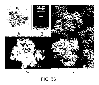
[0048] Figure 36. Nanocluster morphology for 1B7 antibody with trehalose as
extrinsic
crowder. A. Schematic of protein cluster where large circles represent
proteins, small dots,
counterions and medium circles, extrinsic crowders. Similar clusters are
observed for colloids in
organic solvent. B. Transparent dispersion at c = CE = 220 mg/ml. C. SEM image
of 36B
indicating closely-spaced, self-crowded protein. (The "halo" on the component
particles is an
artifact of trehalose deposition during sample preparation). D. Schematic of
dispersion of
nanoclusters drawn to scale.
[0049] Figure 37. Hydrodynamic diameter by DLS for 1B7 antibody and polyclonal
sheep
IgG with trehalose as extrinsic crowder. A. 1B7: serial dilutions in buffer
such that c/cE = 1. B.
1B7: dilution in pH 7.2 phosphate buffer with starting c = CE = 220 mg/ml as
in Fig. 36a
(squares) and decreasing CE with a constant c of 70 mg/ml with a starting CE
of 270 mg/ml
(diamonds). Error bars indicate s. d. in peak width. The predictions of Eq.
14 are in qualitative
agreement. C. 1B7: constant c of 70 mg/ml for decreasing CE of trehalose from
270 to 150 mg/ml
as shown in legend and then a final point where cE is raised back to 270
mg/ml, labeled as 270
mg/ml -2. D. polyclonal sheep IgG: constant c of 50 mg/ml for increasing
(diamonds) followed
by decreasing (squares) trehalose concentration. The reversibility suggests
equilibrium cluster
behavior. The theoretical predictions of Eq. 14 are in qualitative agreement
with the data.
8
CA 02829629 2013-09-09
WO 2012/122544
PCT/US2012/028640
[0050] Figure 38. BSA nanocluster size for high protein concentrations. A high
concentration
BSA dispersion formulated at c = 400 mg/ml and CE = 240 mg/ml forms
nanoclusters with
hydrodynamic diameter of 40 nm. Dispersions formulated with lower
concentrations of BSA
and/or trehalose yield progressively smaller nanoclusters. Also shown is BSA
monomer which is
3-4 nm diameter.
[0051] Figure 39. Antibody conformation and activity. A. Circular dichroism
spectra of
monoclonal antibody 1B7 control and 267 mg/ml dispersion. All samples were
diluted to 0.1
mg/ml in PBS and analyzed on a Jasco J-815 CD Spectrometer. B. Theoretical
prediction of the
fraction of folded protein suggesting that the native state would be favored
at high Ow= 0.6 found
in antibody nanocluster (Shen, Cheung et al. 2006).
[0052] Figure 40. Protein-protein, protein-cluster and cluster-cluster
hierarchical interactions
in nanocluster dispersions. (A) The potential of mean force includes specific
short-ranged (ssr),
depletion attraction (dep) and electrostatic (el) components: V(r) = Võ,(r) +
Vdep(r) + Vei(r). A.
(B) Components of V(r) for protein monomers at pI and 3 pH units away from pI.
B. Predicted
cluster diameter contours. The triangle denotes the conditions of the injected
dispersion into mice
at c= 235 mg/ml for 1B7 as given in Table 16. The diagonal pathway represents
dilution of the
dispersion (Fig. 37a). (C). V(r) for two 50 nm nanoclusters based on
experimental zeta potential
for polyclonal IgG. Inset, arc depicts range of long-ranged repulsion at the
edges of two clusters
and ring around circles indicates short-ranged inter-cluster attraction.
[0053] Figure 41. Pharmacokinetics of concentrated 1B7 dispersion and solution
controls.
Time course of serum antibody concentration normalized by dose after
administration of
intravenous solution, subcutaneous solution or subcutaneous dispersion. Serum
samples were
recovered from the tail vein and the 1B7 concentration determined by ELISA.
[0054] Figure 42. Schematic for the depletion attraction between two protein
particles (large
gray circles) induced by the presence of crowders (small circles) in solution.
The attractive force
reflects the entropic preference for configurations such as this where the
volume excluded to the
centers of the crowders is reduced by the size of the overlap region.
[0055] Figure 43. SEM images of antibody nanoclusters with trehalose as
extrinsic crowder.
A, Reproducibility of multiple SEM images of 1B7 antibody nanoclusters at c =
CE= 220mg/ ml
(identical conditions as in Fig. 36c). The SEM micrographs clearly show good
reproducibility in
9
CA 02829629 2013-09-09
WO 2012/122544
PCT/US2012/028640
the size of the ¨ 300 nm clusters in the dispersion for four clusters,
consistent with the DLS
results in Fig. 37a. The images were obtained from regular carbon film copper
TEM grids where
the nanoclusters were resting on the copper mesh. The individual protein
monomers, on the order
of 10 nm, appear to have a halo around them. This halo is a layer of trehalose
deposited during
freezing and lyophilization in sample preparation for SEM. B, Polyclonal IgG
nanocluster at c =
CE = 260 mg/ml.The imaging was done on a lacey carbon TEM grid and the
nanocluster is
resting on a strand of lacey carbon.
[0056] Figure 44. Static light scattering to determine fractal dimension.The
80 nm sheep IgG
nanoclusters were formed at c = 70 mg/ml IgG and CE= 270 mg/ml trehalose. The
intensity
which scales as the measured count rate was plotted versus the scattering
vector4n-sin (9/2)/2 at
various angles from 45 to 90 . The slope of the line fit through the data
multiplied by -1, i.e.,
2.6 is the fractal dimension.(Hiemenz and Rajagopalan 1997) In static light
scattering, we
assume that the structure factor is not a function of the scattering vector
and therefore, the
intensity is related to the scattering vector through the fractal dimension.
[0057] Figure 45. Hydrodynamic diameter by DLS of polyclonal IgG nanoclusters
upon
dilution in buffer (C/CE = 1). The protein concentrations are shown in the
legend. Sequential
dilution with phosphate buffer at constant c/cE yields progressively smaller
nanoclusters until
monomeric protein with a hydrodynamic diameter of ¨10 nm is observed at c = CE
= 47 mg/ml.
The behavior and mechanism for nanocluster dissociation is similar as observed
for monoclonal
antibody 1B7 in Fig. 37a and b.
[0058] Figure 46. Polyclonal IgG nanocluster size at high concentration.
Polyclonal sheep IgG
dispersions were formulated with 300 and 350 mg/ml protein with c/cE = 1:0.5
with trehalose
and the resulting nanocluster hydrodynamic diameter measured by DLS
[0059] Figure 47. HPLC SEC of monomer concentration after dilution of the
dispersion. All
samples were dilutedto 1 mg/ml in PBS and analyzed withWaters Breeze HPLC with
TOSOH
Biosciences TSKgel G2000SW and G3000SWxL columns. The mobile phase comprised
100 mM
sodium phosphate and 300 mM sodium chloride buffer (pH 7.0), and the eluate
was monitored
by absorbance at 214 nm. A. Chromatographs are shown for (1) solution control
1B7, (2)
lyophilized, reconstituted 1B7, and dispersion formulated with (3) 260 mg/ml
1B7 and 260
mg/ml trehalose. No increase was seen in aggregate concentration throughout
formation of the
dispersion, dilution of the clusters, and reformation of the clusters with
trehalose. B. The %
CA 02829629 2013-09-09
WO 2012/122544
PCT/US2012/028640
monomer values are given here for a wide range of indicated experiments, shown
in Fig. 37a and
37b.Error indicated is s. d.
[0060] Figure 48. SDS-PAGE gel. Absence of higher molecular weight aggregates
as assessed
by non-reducing SDS-PAGE. All dispersions were diluted to 1 mg/ml with PBS
prior to analysis.
5 iLig of each sample was combined with non-reducing loading buffer and loaded
on to a precast
4-20% SDS-PAGE gel (Bio Rad).Lane (1) molecular weight markers (Spectra BR);
(2) solution
control 1B7; (3) & (4) 1B7 post-lyophilization; (5) molecular weight markers
(Spectra BR); (6)
& (7) diluted 260 mg/ml 1B7 dispersion; (8) & (9) 260 mg/ml dispersion diluted
to 75 mg/ml
that was further diluted. None of the samples showed any change in molecular
weight, or
formation of any higher molecular weight aggregates.
[0061] Figure 49. Viscosity calibration curve for measurements with small
conical vials. The
calibration curve was created using the following solution standards: DI water
(rio = 1 cP),
benzyl benzoate (rio = 8.8 cP), PEG200 (rio = 50 cP), PEG300 (rio = 70 cP),
and PEG400 (rio = 90
cP). The time for the liquid level to be drawn from 0.4" to 0.1" in small
conical vial (0.1 mL V-
Vial, Wheaton) was measured from a video of the solution (taken with a Kodak
EasyShare Z812
IS), converted using ImageJ software to a stack of images with 30 images per
second. The time
was measured to within 0.05 seconds at least 3 times and averaged, while
maintaining the end of
the plunger at the 1 ml mark. A maximum volume of 10% of the cavity in the
syringe was filled
with dispersion to minimize variation in the pressure drop.
[0062] Figure 50. Dispersion characteristics before and after using a
centrifugal filtration-
concentration method ¨ pre and post-freezing. The dispersions were formulated
with 217 mg/mL
IgG and 70 mg/mL trehalose and frozen for 1 month.
[0063] Figure 51. Dispersion turbidity at varying wavelengths. Turbidity was
measured on a
Cary 3E UVNis spectrophotometer and is given for pre-filtrated dispersions.
[0064] Figure 52. SEM images of antibody nanoclusters with arginine as
extrinsic crowder.
The dispersion was diluted 4 fold at a constant crowder volume fraction of
0.077 using NMP as a
crowder before dropping on a copper TEM grid with lacey carbon film. Each
image contains a
single nanoparticle on top of a lacey carbon grid and is between 50-100 nm in
diameter.
[0065] Figure 53. Schematic for forming dispersions through centrifugal
filtration-
concentration. Protein is added to form a protein solution. To the protein
solution is added
11
CA 02829629 2013-09-09
WO 2012/122544 PCT/US2012/028640
crowder. The solution is transferred to a tube for centrifugal filtration-
concentration.
Concentration is achieved after centrifugation with some loss of the crowder
through the filter.
DETAILED DESCRIPTION OF THE INVENTION
[0066] While the making and using of various embodiments of the present
invention are
discussed in detail below, it should be appreciated that the present invention
provides many
applicable inventive concepts that can be embodied in a wide variety of
specific contexts. The
specific embodiments discussed herein are merely illustrative of specific ways
to make and use
the invention and do not limit the scope of the invention.
I. Definitions
[0067] To facilitate the understanding of this invention, a number of terms
are defined below.
[0068] As used herein, the term "nanocluster" refers to 10 or more proteins or
peptides that are
not irreversibly aggregated, having a diameter between 20 and 1,000
nanometers, which may
optionally be physically associated with additional compounds, components, or
compositions. In
some embodiments, the diameter is a hydrodynamic diameter. In some
embodiments, the
nanocluster may include subclusters of proteins or peptides that form a larger
cluster. In some
embodiments, the nanocluster may be self-crowding, wherein the crowding is
caused by the
proteins or peptides. In some embodiments, the nanoclusters may form in the
presence of an
extrinsic crowder. In some embodiments, the nanoclusters may be mostly self-
crowding. The
term nanocluster does not include protein or peptide crystals.
[0069] As used herein, the terms "syringable" and "syringeable" are used
interchangeably and
refer to a final composition for delivery to a subject that is sufficiently
fluid to be flowable
through a syringe (e.g. a syringe with a needle that is 21 to 27 gauge). For
example, a
composition that is "syringable" has a low enough viscosity to load the
syringe and inject a
subject from the syringe without undue force, wherein undue force is an amount
in excess of the
force exerted by a skilled practitioner in the medical field (e.g. doctor,
nurse) to deliver
compositions to a patient (e.g. through iv injection, SQ injection) through a
syringe (e.g. a
syringe with a needle that is 21 to 27 gauge) without adverse effects to the
patient solely due to
the force applied in the delivery.
[0070] As used herein, the term "non-settling" or "redispersible" refers to a
composition that
remains in solution phase (i.e., does not sediment) after an extended period
of time, e.g., 1 hour,
12
CA 02829629 2013-09-09
WO 2012/122544
PCT/US2012/028640
2 hours, 1 day, 3 days, 5 days, 1 week, 1 month, 3 months, 6 months, 1 year or
more). For
example, a composition is "re-dispersible" if upon re-dispersion it does not
flocculate so quickly
as to prevent reproducible dosing of a drug.
[0071] As used herein, the term "additive(s)" refers to salts, sugars,
organics, buffers, polymers
and other compositions that include: Disodium edetate, Sodium chloride, Sodium
citrate, Sodium
succinate, Sodium hydroxide, Sodium glucoheptonate, Sodium
acetyltryptophanate, Sodium
bicarbonate, Sodium caprylate, Sodium pertechnetate, sodium acetate, sodium
dodecyl sulfate,
aluminum hydroxide, aluminum phosphate, ammonium citrate, calcium chloride,
calcium,
potassium chloride, potassium sodium tartarate, zinc oxide, zinc, stannous
chloride, magnesium
sulfate, magnesium stearate, titanium dioxide, DL-lactic/glycolic acids,
asparagine, L-arginine,
arginine hydrochloride, adenine, histidine, glycine, glutamine, glutathione,
imidazole, protamine,
protamine sulfate, phosphoric acid, Tri-n-butyl phosphate, ascorbic acid,
cysteine hydrochloride,
hydrochloric acid, hydrogen citrate, trisodium citrate, guanidine
hydrochloride, mannitol,
lactose, sucrose, agarose, sorbitol, maltose, trehaloseõ surfactants,
polysorbate 80, polysorbate
20, poloxamer 188, sorbitan monooleate, triton n101, m-cresol, benyl alcohol,
ethanolamine,
glycerin, phosphorylethanolamine, tromethamine, 2-phenyloxyethanol,
chlorobutanol,
dimethylsulfoxide, N-methyl-2-pyrrolidone, propyleneglycol, Polyoxyl 35 castor
oil, methyl
hydroxybenzoate, tromethamine, corn oil-mono-di-triglycerides, poloxyl 40
hydrogenated castor
oil, tocopherol, n-acetyltryptophan, octa-fluoropropane, castor oil,
polyoxyethylated oleic
glycerides, polyoxytethylated castor oil, phenol (antiseptic), glyclyglycine,
thimerosal
(antiseptic, antifungal), Parabens (preservative), Gelatin, Formaldehyde,
Dulbecco's modified
eagles medium, Hydrocortisone, Neomycin, Von Willebrand factor,
Gluteraldehyde,
Benzethonium chloride, White petroleum, p-aminopheyl-p-anisate, monosodium
glutamate,
beta-propiolactone, Acetate, Citrate, Glutamate, Glycinate, Histidine,
Lactate, Maleate,
Phosphate, Succinate, Tartrate, Tris, Carbomer 1342 (copolymer of acrylic acid
and a long chain
alkyl methacrylate cross-linked with allyl ethers of pentaerythritol), Glucose
star polymer,
Silicone polymer, Polydimethylsiloxane, Polyethylene glycol,
carboxymethylcellulose,
Poly(glycolic acid), Poly(lactic-co-glycolic acid), Polylactic acid, Dextran
40, Poloxamers
(triblock copolymers of ethylene oxide and propylene oxide).
[0072] The terms "a" or "an," as used in herein means one or more. In
addition, the phrase
"substituted with a[n]," as used herein, means the specified group may be
substituted with one or
more of any or all of the named substituents. For example, where a group, such
as an alkyl or
13
CA 02829629 2013-09-09
WO 2012/122544
PCT/US2012/028640
heteroaryl group, is "substituted with an unsubstituted C1-C20 alkyl, or
unsubstituted 2 to 20
membered heteroalkyl," the group may contain one or more unsubstituted Ci-C20
alkyls, and/or
one or more unsubstituted 2 to 20 membered heteroalkyls. Moreover, where a
moiety is
substituted with an R substituent, the group may be referred to as "R-
substituted." Where a
moiety is R-substituted, the moiety is substituted with at least one R
substituent and each R
substituent is optionally different.
[0073] An "effective amount" is an amount sufficient to accomplish a stated
purpose (e.g.
achieve the effect for which it is administered, treat a disease, reduce
enzyme activity, or reduce
one or more symptoms of a disease or condition). An example of an "effective
amount" is an
amount sufficient to contribute to the treatment, prevention, or reduction of
a symptom or
symptoms of a disease, which could also be referred to as a "therapeutically
effective amount."
A "reduction" of a symptom or symptoms (and grammatical equivalents of this
phrase) means
decreasing of the severity or frequency of the symptom(s), or elimination of
the symptom(s). A
"prophylactically effective amount" of a drug is an amount of a drug that,
when administered to
a subject, will have the intended prophylactic effect, e.g., preventing or
delaying the onset (or
reoccurrence) of an injury, disease, pathology or condition, or reducing the
likelihood of the
onset (or reoccurrence) of an injury, disease, pathology, or condition, or
their symptoms. The full
prophylactic effect does not necessarily occur by administration of one dose,
and may occur only
after administration of a series of doses. Thus, a prophylactically effective
amount may be
administered in one or more administrations. An "activity decreasing amount,"
as used herein,
refers to an amount of a composition (e.g. antagonist, protein, low molecular
weight compound)
required to decrease the activity of an enzyme relative to the absence of the
composition (e.g.
antagonist). A "function disrupting amount," as used herein, refers to the
amount of antagonist
required to disrupt the function of an enzyme or protein relative to the
absence of the antagonist.
The exact amounts will depend on the purpose of the treatment, and will be
ascertainable by one
skilled in the art using known techniques (see, e.g., Lieberman,
Pharmaceutical Dosage Forms
(vols. 1-3, 1992); Lloyd, The Art, Science and Technology of Pharmaceutical
Compounding
(1999); Pickar, Dosage Calculations (1999); and Remington: The Science and
Practice of
Pharmacy, 20th Edition, 2003, Gennaro, Ed., Lippincott, Williams & Wilkins).
[0074] "Control" or "control experiment" is used in accordance with its plain
ordinary
meaning and refers to an experiment in which the subjects or reagents of the
experiment are
treated as in a parallel experiment except for omission of a procedure,
reagent, or variable of the
14
CA 02829629 2013-09-09
WO 2012/122544
PCT/US2012/028640
experiment. In some instances, the control is used as a standard of comparison
in evaluating
experimental effects.
[0075] "Contacting" is used in accordance with its plain ordinary meaning and
refers to the
process of allowing at least two distinct species (e.g. compounds including
biomolecules,
proteins, antibodies, or cells) to become sufficiently proximal to react,
interact or physically
touch. It should be appreciated, however, the resulting reaction product can
be produced directly
from a reaction between the added reagents or from an intermediate from one or
more of the
added reagents which can be produced in the reaction mixture.
[0076] As defined herein, the term "inhibition", "inhibit", "inhibiting" and
the like in reference
to a protein-inhibitor interaction means negatively affecting (e.g.
decreasing) the activity or
function of the protein relative to the activity or function of the protein in
the absence of the
inhibitor. In some embodiments inhibition refers to reduction of a disease or
symptoms of
disease. In some embodiments, inhibition refers to a reduction in the presence
of a disease-
related protein. Thus, inhibition includes, at least in part, partially or
totally blocking stimulation,
decreasing, preventing, or delaying activation, or inactivating,
desensitizing, or down-regulating
signal transduction or enzymatic activity or the amount of a protein.
Similarly an "inhibitor" is a
compound that inhibits the activity of a protein or production of a protein,
e.g., by binding,
partially or totally blocking stimulation (e.g. production), decreasing,
preventing, or delaying
activation, or inactivating, desensitizing, or down-regulating signal
transduction or enzymatic
activity. Inhibition may also reduce the amount of a protein by increasing
clearance or
degradation of the protein. In some embodiments, an inhibitor is an antibody.
[0077] The term "modulator" refers to a composition that increases or
decreases the level of a
target molecule or the function of a target molecule.
[0078] "Pharmaceutically acceptable excipient" and "pharmaceutically
acceptable carrier"
refer to a substance that aids the administration of an active agent to and
absorption by a subject
and can be included in the compositions of the present invention without
causing a significant
adverse toxicological effect on the patient. Non-limiting examples of
pharmaceutically
acceptable excipients include water, NaC1, normal saline solutions, lactated
Ringer's, normal
sucrose, normal glucose, binders, fillers, disintegrants, lubricants,
coatings, sweeteners, flavors,
salt solutions (such as Ringer's solution), alcohols, oils, gelatins,
carbohydrates such as lactose,
amylose or starch, fatty acid esters, hydroxymethycellulose, polyvinyl
pyrrolidine. and colors,
CA 02829629 2013-09-09
WO 2012/122544
PCT/US2012/028640
and the like. Such preparations can be sterilized and, if desired, mixed with
auxiliary agents such
as lubricants, preservatives, stabilizers, wetting agents, emulsifiers, salts
for influencing osmotic
pressure, buffers, coloring, and/or aromatic substances and the like that do
not deleteriously react
with the compositions (e.g. proteins, crowders, nanoclusters, dispersions) of
the invention. One
of skill in the art will recognize that other pharmaceutical excipients are
useful in the present
invention.
[0079] The term "preparation" is intended to include the formulation of the
active composition
(e.g. protein nanoclusters, protein-crowder nanoclusters, dispersions) with
encapsulating material
as a carrier providing a capsule in which the active component with or without
other carriers, is
surrounded by a carrier, which is thus in association with it. Similarly,
cachets and lozenges are
included. Tablets, powders, capsules, pills, cachets, and lozenges can be used
as solid dosage
forms suitable for oral administration.
[0080] As used herein, the term "pharmaceutically acceptable" is used
synonymously with
"physiologically acceptable" and "pharmacologically acceptable". A
pharmaceutical
composition will generally comprise agents for buffering and preservation in
storage, and can
include buffers and carriers for appropriate delivery, depending on the route
of administration.
[0081] The terms "isolated" "purified" or "biologically pure" refer to
material that is
substantially or essentially free from components which normally accompany it
as found in its
native state. Purity and homogeneity of biological molecules (e.g. nucleic
acids or proteins) are
typically determined using analytical chemistry techniques such as
polyacrylamide gel
electrophoresis or high performance liquid chromatography. A protein that is
the predominant
species present in a preparation is substantially purified. The term
"purified" may denote that a
nucleic acid or protein gives rise to essentially one band in an
electrophoretic gel. In some
embodiments, the nucleic acid or protein is at least 50% pure, optionally at
least 65% pure,
optionally at least 75% pure, optionally at least 85% pure, optionally at
least 95% pure, and
optionally at least 99% pure. As an example, an isolated cell or isolated
sample cells are a single
cell type that is substantially free of many of the components which normally
accompany the
cells when they are in their native state or when they are initially removed
from their native state.
In certain embodiments, an isolated cell sample retains those components from
its natural state
that are required to maintain the cell in a desired state. In some
embodiments, an isolated (e.g.
purified, separated) cell or isolated cells, are cells that are substantially
the only cell type in a
16
CA 02829629 2013-09-09
WO 2012/122544
PCT/US2012/028640
sample. A purified cell sample may contain at least 60%, 70%, 75%, 80%, 85%,
90%, 95%,
96%, 97%, 98%, 99%, or 100% of one type of cell. An isolated cell sample may
be obtained
through the use of a cell marker or a combination of cell markers, either of
which is unique to
one cell type in an unpurified cell sample. In some embodiments, the cells are
isolated through
the use of a cell sorter. In some embodiments, antibodies against cell
proteins are used to isolate
cells.
[0082] The term "hydrodynamic diameter" has its plain ordinary meaning within
Chemistry
and refers to the apparent diameter of a hypothetical hard sphere that
diffuses through a medium
at the same speed as the molecule under observation (e.g. as measured by
dynamic light
scattering).
[0083] As used herein, the term "transparent" refers to the physical property
of allowing light
to pass through a material. In some embodiments, transparent refers to the
property of allowing a
majority of the incident light, at a given wavelength(s), to pass through the
material. In some
embodiments, transparent refers to the property of allowing greater than about
75% of the
incident light at specified wavelengths (e.g. visible light, 600 nm, 400-700
nm) to pass through
the material. In some embodiments, transparent refers to the property of
allowing greater than
about 80% of the incident light at specified wavelengths to pass through the
material. In some
embodiments, transparent refers to the property of allowing greater than about
85% of the
incident light at specified wavelengths to pass through the material. In some
embodiments,
transparent refers to the property of allowing greater than about 90% of the
incident light at
specified wavelengths to pass through the material. In some embodiments,
transparent refers to
the property of allowing greater than about 95% of the incident light at
specified wavelengths to
pass through the material. In some embodiments, transparent refers to the
property of allowing
greater than about 96% of the incident light at specified wavelengths to pass
through the
material. In some embodiments, transparent refers to the property of allowing
greater than about
97% of the incident light at specified wavelengths to pass through the
material. In some
embodiments, transparent refers to the property of allowing greater than about
98% of the
incident light at specified wavelengths to pass through the material. In some
embodiments,
transparent refers to the property of allowing greater than about 99% of the
incident light at
specified wavelengths to pass through the material. In some embodiments,
transparent refers to
the property of allowing greater than about 99.5% of the incident light at
specified wavelengths
to pass through the material. In some embodiments, transparent refers to the
property of allowing
17
CA 02829629 2013-09-09
WO 2012/122544
PCT/US2012/028640
greater than about 99.6% of the incident light at specified wavelengths to
pass through the
material. In some embodiments, transparent refers to the property of allowing
greater than about
99.7% of the incident light at specified wavelengths to pass through the
material. In some
embodiments, transparent refers to the property of allowing greater than about
99.8% of the
incident light at specified wavelengths to pass through the material. In some
embodiments,
transparent refers to the property of allowing greater than about 99.9% of the
incident light at
specified wavelengths to pass through the material In some embodiments, when
referring to the
transparency of a dispersion of protein nanoclusters, the percentages above
(e.g. percentage
value of any one of 75, 80, 85, 90, 95, 96, 97, 98, 99, 99.5, 99.6, 99.7,
99.8, 99.9), is in
comparison to a control sample lacking the protein, which would be assigned
the value of 100%
incident light at specified wavelengths passing through the material. In some
embodiments,
transparency is measured by light extinction, wherein the term "light
extinction" as used herein
refers to the combined absorption and scattering of incident light at zero
degrees from the angle
of the incident light. In some embodiments, transparent means having a light
extinction of less
than about 0.05 cm-1. In some embodiments, transparent means having a light
extinction of less
than about 0.1 cm-1. In some embodiments, transparent means having a light
extinction of less
than about 0.25 cm-1. In some embodiments, transparent means having a light
extinction of less
than about 0.5 cm-1. In some embodiments, transparent means having a light
extinction of less
than about 1%. In some embodiments, transparent means having a light
extinction of less than
about 2%. In some embodiments, transparent means having a light extinction of
less than about
3%. In some embodiments, transparent means having a light extinction of less
than about 4%. In
some embodiments, transparent means having a light extinction of less than
about 5%. In some
embodiments, transparent means having a light extinction of less than about
10%. In some
embodiments, transparent means having a light extinction of less than about
15%. In some
embodiments, transparent means having a light extinction of less than about
20%. In some
embodiments, transparent means having a light extinction of less than about
25%. In some
embodiments, when referring to the transparency of a dispersion of protein
nanoclusters in terms
of light extinction, the percentages above (e.g. percentage value of any one
of 1, 2, 3, 4, 5, 10,
15, 20, or 25), is in comparison to a control sample lacking the protein,
which would be assigned
the value of 0% light extinction. A "light extinction measurement" refers to a
light extinction
value physically measured by a person of ordinary skill.
18
CA 02829629 2013-09-09
WO 2012/122544
PCT/US2012/028640
[0084] The term "viscosity" has its plain ordinary meaning within Chemistry,
as applied to
liquids and fluids.
[0085] As used herein, the term "low visocity" refers to a viscosity that is
less than about 100
centipoise. In some embodiments, "low viscosity" refers to a viscosity of less
than about 90
centipoise. In some embodiments, "low viscosity" refers to a viscosity of less
than about 80
centipoise. In some embodiments, "low viscosity" refers to a viscosity of less
than about 70
centipoise. In some embodiments, "low viscosity" refers to a viscosity of less
than about 60
centipoise. In some embodiments, "low viscosity" refers to a viscosity of less
than about 50
centipoise. In some embodiments, "low viscosity" refers to a viscosity of less
than about 40
centipoise. In some embodiments, "low viscosity" refers to a viscosity of less
than about 30
centipoise. In some embodiments, "low viscosity" refers to a viscosity of less
than about 20
centipoise. In some embodiments, "low viscosity" refers to a viscosity of less
than about 10
centipoise. In some embodiments, a low visocity is measured with a viscometer,
rheometer, or
syringe loading method as described herein. In some embodiments, a low
viscosity is measured
with a shear rate that is about 100, 200, 300, 400, 500, 600, 700, 800, 900,
1000, 2000, 3000,
4000, 5000, 6000, 7000, 8000, 9000, 10000, 20000, 30000, 40000, 50000, 60000,
70000, 80000,
90000, or 100000 second-1. In some embodiments, an average shear rate may be
determined
from the flow rate and geometric properties with a syringe loading method as
described herein.
In some embodiments, "low viscosity" refers to any one of the combinations of
viscosity and
shear rate shown in the table/matrix below having number 1 to 280, wherein
each cell
corresponds to the viscosity for that column and the shear rate for that row:
100 90 80 70 60 50 40 30 20 10
Viscosity less than
about(centipoise)/s
hear rate about
(second-1
100 1 2 3 4 5 6 7 8 9 10
200 11 12 13 14 15 16 17 18 19 20
300 21 22 23 24 25 26 27 28 29 30
400 31 32 33 34 35 36 37 38 39 40
19
CA 02829629 2013-09-09
WO 2012/122544
PCT/US2012/028640
500 41 42 43 44 45 46 47 48 49 50
600 51 52 53 54 55 56 57 58 59 60
700 61 62 63 64 65 66 67 68 69 70
800 71 72 73 74 75 76 77 78 79 80
900 81 82 83 84 85 86 87 88 89 90
1000 91 92 93 94 95 96 97 98 99 100
2000 101 102 103 104 105 106 107 108 109 110
3000 111 112 113 114 115 116 117 118 119
120
4000 121 122 123 124 125 126 127 128 129 130
5000 131 132 133 134 135 136 137 138 139 140
6000 141 142 143 144 145 146 147 148 149 150
7000 151 152 153 154 155 156 157 158 159 160
8000 161 162 163 164 165 166 167 168 169 170
9000 171 172 173 174 175 176 177 178 179 180
10000 181 182 183 184 185 186 187 188 189 190
20000 191 192 193 194 195 196 197 198 199 200
30000 201 202 203 204 205 206 207 208 209 210
40000 211 212 213 214 215 216 217 218 219 220
50000 221 222 223 224 225 226 227 228 229 230
60000 231 232 233 234 235 236 237 238 239 240
70000 241 242 243 244 245 246 247 248 249 250
80000 251 252 253 254 255 256 257 258 259 260
90000 261 262 263 264 265 266 267 268 269 270
CA 02829629 2013-09-09
WO 2012/122544
PCT/US2012/028640
100000 271 272 273 274 275 276 277 278 279 280
In some embodiments, the visocity is the value in the column heading and the
shear rate is the
value in the row heading that together correspond to any one of the cells
number 1 to 280 in the
table immediately above. In some embodiments, the visocity and shear rate
combinations in the
table above are measured by a syringe loading method as described herein.
[0086] As used herein, the term "high protein concentration" or "high protein"
refers to a
protein concentration of greater than about 200 mg/mL. In some embodiments,
the protein
concentration is greater than about 300 mg/mL. In some embodiments, the
protein concentration
is greater than about 400 mg/mL. In some embodiments, the protein
concentration is greater than
about 500 mg/mL. In some embodiments, the protein concentration is greater
than about 600
mg/mL. In some embodiments, the protein concentration is greater than about
700 mg/mL. In
some embodiments, the protein concentration is greater than about 800 mg/mL.
In some
embodiments, the protein concentration is greater than about 900 mg/mL. In
some embodiments,
the protein concentration is greater than about 1000 mg/mL. In some
embodiments, the protein
concentration is the concentration of one protein species (proteins
substantially identical). In
some embodiments, the protein concentration is the concentration of all
proteins in a mixture. In
some embodiments, "high protein concentration" or "high protein" refers to a
protein
concentration that is greater than about 200, 300, 400, 500, 600, 700, 800,
900, or 1000 mg/mL.
In some embodiments "high protein concentration" or "high protein" refers to a
protein
concentration range, wherein the range is entirely greater than about 200,
300, 400, 500, 600,
700, 800, 900, or 1000 mg/mL.
[0087] As used herein, the term "dispersion" has its plain ordinary meaning
within the field of
Chemistry and refers to a system containing particles dispersed in a
continuous phase of a
different composition (e.g. nanoparticles or nanoclusters dispersed in a
liquid phase). In some
embodiments, a dispersion may be a suspension, wherein a suspension has its
plain ordinary
meaning within Chemistry and refers to a dispersion of solid particles in a
continuous liquid
phase, wherein the solid particles are large enough for sedimentation. In some
embodiments, a
dispersion may be a colloid, wherein a colloid has its plain ordinary meaning
as used within
Chemistry. In some embodiments, a dispersion comprises nanoparticles dispersed
in a
continuous liquid phase. In some embodiments the dispersed particles are
protein nanoclusters.
In some embodiments, the continuous phase of a different composition comprises
protein in
21
CA 02829629 2013-09-09
WO 2012/122544
PCT/US2012/028640
solution. In some embodiments, the protein in solution is less than about 50%,
40%, 30%, 20%,
10%, 9%, 8%, 7%, 6%, 5%, 4%, 3%, 2%, 1%, 0.5%, 0.4%, 0.3%, 0.2%, 0.1%, of the
total
protein in the dispersion (i.e. particles and continuous phase combined).
[0088] The terms "polypeptide," "peptide" and "protein" are used
interchangeably herein to
refer to a polymer of amino acid residues, wherein the polymer may optionally
be conjugated to
a moiety that does not consist of amino acids (e.g. small molecular weight
compounds). The
terms apply to amino acid polymers in which one or more amino acid residue is
an artificial
chemical mimetic of a corresponding naturally occurring amino acid, as well as
to naturally
occurring amino acid polymers and non-naturally occurring amino acid polymer.
In some
embodiments, a protein comprises a non-protein composition (e.g. low molecular
weight
compound) conjugated (e.g. bonded) to the polymer of amino acid residues
(collectively a
"conjugate" or "conjugated protein"). In some embodiments, a protein consists
of a polymer of
amino acids (a "non-conjugated protein"). In some embodiments, a protein is a
polymer of about
10, 20, 30, 40, 50, 60, 70, 80, 90, 100, 200, 300, 400, 500, 600, 700, 800,
900, 1000, 2000, 3000,
4000, or 5000 amino acid residues. In some embodiments, a protein is a polymer
of 2, 3, 4, 5, 6,
7, 8, 9, 10, 11, 12, 13, 14, 15, 16, 17, 18, 19, or 20 amino acid residues.
[0089] The term "amino acid" refers to naturally occurring and synthetic amino
acids, as well
as amino acid analogs and amino acid mimetics that function in a manner
similar to the naturally
occurring amino acids. Naturally occurring amino acids are those encoded by
the genetic code,
as well as those amino acids that are later modified, e.g., hydroxyproline, y-
carboxyglutamate,
and 0-phosphoserine. Amino acid analogs refers to compounds that have the same
basic
chemical structure as a naturally occurring amino acid, i.e., an a carbon that
is bound to a
hydrogen, a carboxyl group, an amino group, and an R group, e.g., homoserine,
norleucine,
methionine sulfoxide, methionine methyl sulfonium. Such analogs have modified
R groups (e.g.,
norleucine) or modified peptide backbones, but retain the same basic chemical
structure as a
naturally occurring amino acid. Amino acid mimetics refers to chemical
compounds that have a
structure that is different from the general chemical structure of an amino
acid, but that functions
in a manner similar to a naturally occurring amino acid.
[0090] Amino acids may be referred to herein by either their commonly known
three letter
symbols or by the one-letter symbols recommended by the IUPAC-IUB Biochemical
22
CA 02829629 2013-09-09
WO 2012/122544
PCT/US2012/028640
Nomenclature Commission. Nucleotides, likewise, may be referred to by their
commonly
accepted single-letter codes.
[0091] "Conservatively modified variants" applies to both amino acid and
nucleic acid
sequences. With respect to particular nucleic acid sequences, conservatively
modified variants
refers to those nucleic acids which encode identical or essentially identical
amino acid
sequences, or where the nucleic acid does not encode an amino acid sequence,
to essentially
identical sequences. Because of the degeneracy of the genetic code, a large
number of
functionally identical nucleic acids encode any given protein. For instance,
the codons GCA,
GCC, GCG and GCU all encode the amino acid alanine. Thus, at every position
where an
alanine is specified by a codon, the codon can be altered to any of the
corresponding codons
described without altering the encoded polypeptide. Such nucleic acid
variations are "silent
variations," which are one species of conservatively modified variations.
Every nucleic acid
sequence herein which encodes a polypeptide also describes every possible
silent variation of the
nucleic acid. One of skill will recognize that each codon in a nucleic acid
(except AUG, which is
ordinarily the only codon for methionine, and TGG, which is ordinarily the
only codon for
tryptophan) can be modified to yield a functionally identical molecule.
Accordingly, each silent
variation of a nucleic acid which encodes a polypeptide is implicit in each
described sequence
with respect to the expression product, but not with respect to actual probe
sequences.
[0092] As to amino acid sequences, one of skill will recognize that individual
substitutions,
deletions or additions to a nucleic acid, peptide, polypeptide, or protein
sequence which alters,
adds or deletes a single amino acid or a small percentage of amino acids in
the encoded sequence
is a "conservatively modified variant" where the alteration results in the
substitution of an amino
acid with a chemically similar amino acid. Conservative substitution tables
providing
functionally similar amino acids are well known in the art. Such
conservatively modified variants
are in addition to and do not exclude polymorphic variants, interspecies
homologs, and alleles of
the invention.
[0093] The following eight groups each contain amino acids that are
conservative substitutions
for one another: 1) Alanine (A), Glycine (G); 2) Aspartic acid (D), Glutamic
acid (E); 3)
Asparagine (N), Glutamine (Q); 4) Arginine (R), Lysine (K); 5) Isoleucine (I),
Leucine (L),
Methionine (M), Valine (V); 6) Phenylalanine (F), Tyrosine (Y), Tryptophan
(W); 7) Serine (S),
Threonine (T); and 8) Cysteine (C), Methionine (M) (see, e.g., Creighton,
Proteins (1984)).
23
CA 02829629 2013-09-09
WO 2012/122544
PCT/US2012/028640
[0094] The terms "identical" or percent sequence "identity," or "shares amino
acid sequence
identity" in the context of two or more nucleic acids or polypeptide
sequences, refer to two or
more sequences or subsequences that are the same or have a percentage of amino
acide residues
or nucleotides that are the same over a specified region, or have a specified
percentage of amino
acid residues or nucleotides that are the same (i.e., about 60% identity,
preferably about 65%,
70%, 75%, 80%, 85%, 90%, 91%, 92%, 93%, 94%, 95%, 96%, 97%, 98%, 99%, 99.1%,
99.2%,
99.3%, 99.4%, 99.5%, 99.6%, 99.7%, 99.8%, 99.9%, or higher identity over a
specified region,
when compared and aligned for maximum correspondence over a comparison window
or
designated region) as measured using a BLAST or BLAST 2.0 sequence comparison
algorithms
with default parameters described below, or by manual alignment and visual
inspection (see, e.g.,
NCBI web site at ncbi.nlm.nih.gov/BLAST/ or the like). Such sequences are then
said to be
"substantially identical." This definition also refers to, or may be applied
to, the compliment of a
test sequence. The definition also includes sequences that have deletions
and/or additions, as
well as those that have substitutions. Employed algorithms can account for
gaps and the like.
[0095] For sequence comparisons, typically one sequence acts as a reference
sequence, to
which test sequences are compared. When using a sequence comparison algorithm,
test and
reference sequences are entered into a computer, subsequence coordinates are
designated, if
necessary, and sequence algorithm program parameters are designated.
Preferably, default
program parameters can be used, or alternative parameters can be designated.
The sequence
comparison algorithm then calculates the percent sequence identities for the
test sequences
relative to the reference sequence, based on the program parameters.
[0096] A "comparison window", as used herein, includes reference to a segment
of any one of
the number of contiguous positions selected from the group consisting of from
20 to 600, usually
about 50 to about 200, more usually about 100 to about 150 in which a sequence
may be
compared to a reference sequence of the same number of contiguous positions
after the two
sequences are optimally aligned. Methods of alignment of sequences for
comparison are well-
known in the art. Optimal alignment of sequences for comparison can be
conducted, e.g., by the
local homology algorithm of Smith & Waterman, Adv. Appl. Math. 2:482 (1981),
by the
homology alignment algorithm of Needleman & Wunsch, J. Mol. Biol. 48:443
(1970), by the
search for similarity method of Pearson & Lipman, Proc. Nat'l. Acad. Sci. USA
85:2444 (1988),
by computerized implementations of these algorithms (GAP, BESTFIT, FASTA, and
TFASTA
in the Wisconsin Genetics Software Package, Genetics Computer Group, 575
Science Dr.,
24
CA 02829629 2013-09-09
WO 2012/122544
PCT/US2012/028640
Madison, WI), or by manual alignment and visual inspection (see, e.g., Current
Protocols in
Molecular Biology (Ausubel et al., eds. 1995 supplement)).
[0097] A preferred example of algorithm that is suitable for determining
percent sequence
identity and sequence similarity are the BLAST and BLAST 2.0 algorithms, which
are described
in Altschul et al., Nuc. Acids Res. 25:3389-3402 (1977) and Altschul et al.,
J. Mol. Biol.
215:403-410 (1990), respectively.
[0098] Aptamers are nucleic acids that are designed to bind to a wide variety
of targets in a
non-Watson Crick manner. An aptamer can thus be used to detect or otherwise
target nearly any
molecule of interest, including an autoimmune, inflammatory autoimmune,
cancer, infectious
disease, or other disease associated protein. Methods of constructing and
determining the binding
characteristics of aptamers are well known in the art. For example, such
techniques are described
in U.S. Patent Nos. 5,582,981, 5,595,877 and 5,637,459. Aptamers are typically
at least 5
nucleotides, 10, 20, 30 or 40 nucleotides in length, and can be composed of
modified nucleic
acids to improve stability. Flanking sequences can be added for structural
stability, e.g., to form
3-dimensional structures in the aptamer.
[0099] As used herein, the term "crowder" refers to a compound that, when
present in a
solvent (e.g. a dispersion liquid) with concentrated proteins, aids formation
of a stable colloidal
dispersion containing nanoclusters of non-irreversibly aggregated proteins. In
some
embodiments, a crowder may be the protein itself (e.g. self-crowding protein).
In some
embodiments, the crowder may be an amino acid. In some embodiments, the
crowder may be a
second protein species (e.g. a dipeptide, tripeptide, oligopeptide, conjugated
protein, non-
conjugated protein). In some embodiments, the crowder may be a non-protein
crowder such as a
polysaccharide, polyelectrolyte, polyacid, dextran, polaxamer, surfactant, a
glycerol, an
erythritol, an arabinose, a xylose, a ribose, an inositol, a fructose, a
galactose, a maltose, a
glucose, a mannose, a trehalose, a sucrose, a poly(ethylene glycol), a
carbomer 1342, a glucose
polymers, a silicone polymer, a polydimethylsiloxane, a polyethylene glycol, a
carboxy methyl
cellulose, a poly(glycolic acid), a poly(lactic-co-glycolic acid), a
polylactic acid, a dextran, a
poloxamers, organic co-solvents selected from ethanol, N-methyl-2-pyrrolidone
(NMP), PEG
300, PEG 400, PEG 200, PEG 3350, Propylene Glycol, N,N Dimethylacetamide,
dimethyl
sulfoxide, solketal, tetahydrofurfuryl alcohol, diglyme, ethyl lactate, a
salt, a buffer or a
combination thereof
CA 02829629 2013-09-09
WO 2012/122544
PCT/US2012/028640
[0100] The terms "polysaccharide", "polyelectrolyte", "polyacid", "polaxamer",
"surfactant",
"buffer" have their plain ordinary meaning within the field of Chemistry.
[0101] As used herein, the term "dextran" refers to a branched polysaccharide
comprising
glucose molecules. In some embodiments, the dextran has a molecular weight
between about one
[0102] As used herein, the term "about", when modifying a number (e.g. an
amount,
26
CA 02829629 2013-09-09
WO 2012/122544
PCT/US2012/028640
number, using a common technique or apparatus for taking such measurement. In
some
embodiments, about includes 0.1, 1.0, or 10 times the number. In some
embodiments, about
includes plus and minus 0.1 times the number (e.g. about 200 mg/mL is 180 -220
mg/mL).
[0103] As used herein, the term "average diameter", when applied to
nanoclusters, refers to the
average diameter of the nanoclusters in a sample. In some embodiments, the
average diameter is
an average hydrodynamic diameter. In some embodiments, the average diameter is
the average
length of the longest axis of the nanocluster. In some embodiments the average
diameter is
measured by dynamic light scattering. In some embodiments the average diameter
is measured
by static light scattering. In some embodiments the average diameter is
measured by size
exclusion chromatography. In some embodiments the average diameter is measured
by
microscopy. In some embodiments the average diameter is measured by scanning
electron
microscopy. In some embodiments the average diameter is measured by
cryoelectron
microscopy. In some embodiments the average diameter is measured by
transmission electron
microscopy. In some embodiments the average diameter is measured by x-ray
scattering (e.g.
small angle x-ray scattering).
[0104] As used herein, the term "plurality" refers to more than one.
[0105] As used herein, the term "irreversibly aggregated" refers to proteins
physically
associated together in a mixture, comprising a liquid medium, wherein upon
dilution of the
concentration of the protein or concentration of crowder if the mixture
contains crowder, the
proteins do not dissociate from the aggregates to form functional protein
possessing the
secondary, tertiary, and quaternary structure appropriate for the medium and
concentration of
protein if the protein had not previously been aggregated. An irreversibly
aggregated protein may
also be termed an "unstable" protein. A "stable" protein (e.g. antibody) is a
protein that
dissociates from a protein aggregate or protein cluster upon dilution of
either the protein
concentration or crowder concentration, if a crowder is present and promoted
the formation of
the protein aggregate or protein cluster, to form functional (e.g. active,
enzymatically active)
proteins possessing the secondary, tertiary, and quaternary structure
appropriate for the medium
and concentration of protein if the protein had not previously been
aggregated.
[0106] As used herein, the term "low molecular weight compound" refers to a
composition
having a molecular weight less than 1 kilodalton. In some embodiments, the low
molecular
weight compound may be a diagnostic agent, pharmaceutical agent, contrast
agent, fluorophore,
27
CA 02829629 2013-09-09
WO 2012/122544
PCT/US2012/028640
paramagnetic agent, peptide, or toxin. In some embodiments, the low molecular
weight
compound may be conjugated (e.g. bonded) to another composition (e.g. protein,
antibody).
[0107] As used herein, the term "diagnostic agent" refers to a composition
that is useful for
detecting the presence of a disease state or a symptom of a disease state. In
some embodiments, a
diagnostic agent may be a label or detectable moiety.
[0108] A "label" or a "detectable moiety" is a composition detectable by
spectroscopic,
photochemical, biochemical, immunochemical, chemical, or other physical means.
For example,
useful labels include 32P, fluorescent dyes, electron-dense reagents, enzymes
(e.g., as commonly
used in an ELISA), biotin, digoxigenin, or haptens and proteins or other
entities which can be
made detectable, e.g., by incorporating a radiolabel into a peptide or
antibody specifically
reactive with a target peptide. Any method known in the art for conjugating an
antibody to the
label may be employed, e.g., using methods described in Hermanson,
Bioconjugate Techniques
1996, Academic Press, Inc., San Diego.
[0109] As used herein, a "pharmaceutical" refers to a composition that is
useful in the
treatment of a disease or a symptom of a disease.
[0110] As used herein, a "pharmaceutically active protein" refers to a protein
that is useful in
the treatment of a disease or a symptom of a disease.
[0111] The terms "treating" or "treatment" refers to any indicia of success in
the treatment or
amelioration of an injury, disease, pathology or condition, including any
objective or subjective
parameter such as abatement; remission; diminishing of symptoms or making the
injury,
pathology or condition more tolerable to the patient; slowing in the rate of
degeneration or
decline; making the final point of degeneration less debilitating; improving a
patient's physical
or mental well-being. The treatment or amelioration of symptoms can be based
on objective or
subjective parameters; including the results of a physical examination,
neuropsychiatric exams,
and/or a psychiatric evaluation. For example, the certain methods presented
herein could
successfully treat cancer by decreasing the incidence of cancer and or causing
remission of
cancer. The term "treating," and conjugations thereof, include prevention of
an injury, pathology,
condition, or disease.
[0112] "Disease" or "condition" refer to a state of being or health status of
a patient or subject
capable of being treated with the compositions, dispersions, or methods
provided herein.
28
CA 02829629 2013-09-09
WO 2012/122544
PCT/US2012/028640
[0113] As used herein, the term "contrast agent" refers to a composition that,
when
administered to a subject, improves the detection limit or detection
capability of a method,
technique, or apparatus for medical imaging (e.g. radiographic instrument, X-
ray, CT, PET,
MRI, ultrasound). A contrast agent may enhance the contrast of signals related
to different
structures or fluids within a subject.
[0114] "Patient" or "subject in need thereof" refers to a living organism
suffering from or
prone to a disease or condition that can be treated by administration of a
pharmaceutical
composition as provided herein. Non-limiting examples include humans, other
mammals,
bovines, rats, mice, dogs, monkeys, goat, sheep, cows, deer, and other non-
mammalian animals.
In some embodiments, a patient is human.
[0115] As used herein, the term "fluorophore" has its plain ordinary meaning
within Chemistry
and refers to a chromophore used in fluorescent imaging of spectroscopy. A
fluorophore absorbs
light within a first range of wavelengths and emits light within a second
range of wavelengths.
[0116] As used herein, the term "shear rate" has its plain ordinary meaning
within Chemistry
and fluid mechanics and refers to the rate (e.g. seconds-1) of application of
a shear, wherein shear
refers to a force or pressure applied to an object (e.g. deformable object,
liquid, solid object)
perpendicular to a given axis with greater value (i.e. greater force or
pressure) on one side of the
axis compared to the other. For flow through a cylinder, shear rate at the
wall is proportional to
the flow rate divided by the cube of the radius.
[0117] As used herein, the term "syringe loading method" refers to a method of
measuring the
viscosity of a liquid (e.g. dispersion, solution, suspension, mixture) by
using the same pressure
drop in a needle attached to a syringe wherein the piston of the syringe is
displaced by a set
amount, causing flow through the needle, wherein the needle has a known
diameter and length.
The unknown viscosity of liquid being measured is compared to a plurality of
measurements
conducted in exactly the same way as the current measurement, wherein the
plurality of
measurements is conducted on liquids with known viscosities. In some
embodiments, the needle
has a gauge between 21 and 27. In some embodiments, the needle is a 25 gauge
needle. In some
embodiments, the syringe is a 1 mL syringe. In some embodiments, the needle is
1.5 inches long.
In some embodiments, the time to draw the liquid (e.g. dispersion) from a
height in a conical
vial, wherein the distance from the liquid meniscus to the bottom of the cone
is at 0.4 inches, to a
height, wherein the distance from the liquid meniscus to the bottom of the
cone is at 0.1 inches,
29
CA 02829629 2013-09-09
WO 2012/122544
PCT/US2012/028640
corresponding to a volume of 48 microliters, is measured. In some embodiments,
a syringe
loading method is a method described herein above using the parameters
described in Example
VI of the present application.
[0118] As used herein, the term "packing fraction" refers to a ratio of the
volume occupied by
when combined with a composition as desribed herein (including embodiments)
releases the
composition at a controlled rate into a patient. Such compounds include high
molecular weight,
anionic mucomimetic polymers, gelling polysaccharides and finely-divided drug
carrier
substrates. These components are discussed in greater detail in U.S. Pat. Nos.
4,911,920;
[0120] As used herein, the term "paramagnetic agent" refers to a paramagnetic
compound
useful in diagnostic imaging methods (e.g. magnetic resonance imaging) as a
contrast agent. In
[0121] As used herein, the term "isotonic" refers to two liquids having the
same osmotic
pressure. A liquid is isotonic with another if it has the same effective
osmotic pressure as the
liquid inside the cell across the membrane of a given type of cell. In some
embodiments, a
CA 02829629 2013-09-09
WO 2012/122544
PCT/US2012/028640
administration of the dispersion in a patient. In some embodiments, a
dispersion is isotonic with
a subcutaneous site of administration of the dispersion.
[0122] As used herein, the term "antibody" refers to a polypeptide comprising
a framework
region from an immunoglobulin gene or fragments thereof that specifically
binds and recognizes
an antigen. The recognized immunoglobulin genes include the kappa, lambda,
alpha, gamma,
delta, epsilon, and mu constant region genes, as well as the myriad
immunoglobulin variable
region genes. Light chains are classified as either kappa or lambda. Heavy
chains are classified
as gamma, mu, alpha, delta, or epsilon, which in turn define the
immunoglobulin classes, IgG,
IgM, IgA, IgD and IgE, respectively. Typically, the antigen-binding region of
an antibody will
be most critical in specificity and affinity of binding. In some embodiments,
antibodies or
fragments of antibodies may be derived from different organisms, including
humans, mice, rats,
hamsters, camels, etc. Antibodies of the invention may include antibodies that
have been
modified or mutated at one or more amino acid positions to improve or modulate
a desired
function of the antibody (e.g. glycosylation, expression, antigen recognition,
effector functions,
antigen binding, specificity, etc.).
[0123] An exemplary immunoglobulin (antibody) structural unit comprises a
tetramer. Each
tetramer is composed of two identical pairs of polypeptide chains, each pair
having one "light"
(about 25 kD) and one "heavy" chain (about 50-70 kD). The N-terminus of each
chain defines a
variable region of about 100 to 110 or more amino acids primarily responsible
for antigen
recognition. The terms variable light chain (VI) and variable heavy chain (VH)
refer to these light
and heavy chains respectively.
[0124] Antibodies exist, e.g., as intact immunoglobulins or as a number of
well-characterized
fragments produced by digestion with various peptidases. Thus, for example,
pepsin digests an
antibody below the disulfide linkages in the hinge region to produce F(ab)'2,
a dimer of Fab
which itself is a light chain joined to VH-CH1 by a disulfide bond. The
F(ab)'2 may be reduced
under mild conditions to break the disulfide linkage in the hinge region,
thereby converting the
F(ab)'2 dimer into an Fab' monomer. The Fab' monomer is essentially Fab with
part of the hinge
region (see Fundamental Immunology (Paul ed., 3d ed. 1993). While various
antibody fragments
are defined in terms of the digestion of an intact antibody, one of skill will
appreciate that such
fragments may be synthesized de novo either chemically or by using recombinant
DNA
methodology. Thus, the term antibody, as used herein, also includes antibody
fragments either
31
CA 02829629 2013-09-09
WO 2012/122544
PCT/US2012/028640
produced by the modification of whole antibodies, or those synthesized de novo
using
recombinant DNA methodologies (e.g., single chain Fv) or those identified
using phage display
libraries (see, e.g., McCafferty et at., Nature 348:552-554 (1990)).
[0125] Methods for humanizing or primatizing non-human antibodies are well
known in the art
(e.g., U.S. Patent Nos. 4,816,567; 5,530,101; 5,859,205; 5,585,089; 5,693,761;
5,693,762;
5,777,085; 6,180,370; 6,210,671; and 6,329,511; WO 87/02671; EP Patent
Application 0173494;
Jones et at. (1986) Nature 321:522; and Verhoyen et at. (1988) Science
239:1534). Humanized
antibodies are further described in, e.g., Winter and Milstein (1991) Nature
349:293. Generally,
a humanized antibody has one or more amino acid residues introduced into it
from a source
which is non-human. These non-human amino acid residues are often referred to
as import
residues, which are typically taken from an import variable domain.
Humanization can be
essentially performed following the method of Winter and co-workers (see,
e.g., Morrison et at.,
PNAS USA, 81:6851-6855 (1984), Jones et al., Nature 321:522-525 (1986);
Riechmann et al.,
Nature 332:323-327 (1988); Morrison and 0i, Adv. Immunol., 44:65-92 (1988),
Verhoeyen et
at., Science 239:1534-1536 (1988) and Presta, Curr. Op. Struct. Biol. 2:593-
596 (1992), Padlan,
Molec. Immun., 28:489-498 (1991); Padlan, Molec. Immun., 31(3):169-217
(1994)), by
substituting rodent CDRs or CDR sequences for the corresponding sequences of a
human
antibody. Accordingly, such humanized antibodies are chimeric antibodies (U.S.
Patent No.
4,816,567), wherein substantially less than an intact human variable domain
has been substituted
by the corresponding sequence from a non-human species. In practice, humanized
antibodies are
typically human antibodies in which some CDR residues and possibly some FR
residues are
substituted by residues from analogous sites in rodent antibodies. For
example, polynucleotides
comprising a first sequence coding for humanized immunoglobulin framework
regions and a
second sequence set coding for the desired immunoglobulin complementarity
determining
regions can be produced synthetically or by combining appropriate cDNA and
genomic DNA
segments. Human constant region DNA sequences can be isolated in accordance
with well
known procedures from a variety of human cells.
[0126] A "chimeric antibody" is an antibody molecule in which (a) the constant
region, or a
portion thereof, is altered, replaced or exchanged so that the antigen binding
site (variable
region) is linked to a constant region of a different or altered class,
effector function and/or
species, or an entirely different molecule which confers new properties to the
chimeric antibody,
e.g., an enzyme, toxin, hormone, growth factor, drug, etc.; or (b) the
variable region, or a portion
32
CA 02829629 2013-09-09
WO 2012/122544
PCT/US2012/028640
thereof, is altered, replaced or exchanged with a variable region having a
different or altered
antigen specificity. The preferred antibodies of, and for use according to the
invention include
humanized and/or chimeric monoclonal antibodies.
[0127] In one embodiment, the antibody is conjugated to an "effector" moiety.
The effector
moiety can be any number of molecules, including labeling moieties such as
radioactive labels or
fluorescent labels, or can be a therapeutic moiety. In one aspect the antibody
modulates the
activity of a protein. Such effector moieties include, but are not limited to,
an anti-tumor drug, a
toxin, a radioactive agent, a cytokine, a second antibody or an enzyme. In
some embodiments,
the antibody of the invention is linked to an enzyme that converts a prodrug
into a cytotoxic
agent.
[0128] The immunoconjugate can be used for targeting the effector moiety to a
target molecule
or target molecule positive cell. Such differences can be readily apparent
when viewing the
bands of gels with approximately similarly loaded with test and controls
samples. Examples of
cytotoxic agents (e.g. toxins) include, but are not limited to ricin,
doxorubicin, daunorubicin,
taxol, ethidium bromide, mitomycin, etoposide, tenoposide, vincristine,
vinblastine, colchicine,
dihydroxy anthracin dione, actinomycin D, diphteria toxin, Pseudomonas
exotoxin (PE) A,
PE40, abrin, and glucocorticoid and other chemotherapeutic agents, as well as
radioisotopes.
Suitable detectable markers include, but are not limited to, a radioisotope, a
fluorescent
compound, a bioluminescent compound, chemiluminescent compound, a metal
chelator or an
enzyme.
[0129] Techniques for conjugating therapeutic agents to antibodies are well
known (see, e.g.,
Amon et al., "Monoclonal Antibodies For Immunotargeting Of Drugs In Cancer
Therapy", in
Monoclonal Antibodies And Cancer Therapy, Reisfeld et al. (eds.), pp. 243-56
(Alan R. Liss,
Inc. 1985); Hellstrom et al., "Antibodies For Drug Delivery"in Controlled Drug
Delivery (2nd
Ed.), Robinson et al. (eds.), pp. 623-53 (Marcel Dekker, Inc. 1987); Thorpe,
"Antibody Carriers
Of Cytotoxic Agents In Cancer Therapy: A Review" in Monoclonal Antibodies '84:
Biological
And Clinical Applications, Pinchera et al. (eds.), pp. 475-506 (1985); and
Thorpe et al., "The
Preparation And Cytotoxic Properties Of Antibody-Toxin Conjugates", Immunol.
Rev., 62:119-
58 (1982)).
[0130] The phrase "specifically (or selectively) binds" to an antibody or
"specifically (or
selectively) immunoreactive with," when referring to a protein or peptide,
refers to a binding
33
CA 02829629 2013-09-09
WO 2012/122544
PCT/US2012/028640
reaction that is determinative of the presence of the protein, often in a
heterogeneous population
of proteins and other biologics. Thus, under designated immunoassay
conditions, the specified
antibodies bind to a particular protein at least two times the background and
more typically more
than 10 to 100 times background. Specific binding to an antibody under such
conditions requires
an antibody that is selected for its specificity for a particular protein. For
example, polyclonal
antibodies can be selected to obtain only those polyclonal antibodies that are
specifically
immunoreactive with the selected antigen and not with other proteins. This
selection may be
achieved by subtracting out antibodies that cross-react with other molecules.
A variety of
immunoassay formats may be used to select antibodies specifically
immunoreactive with a
particular protein. For example, solid-phase ELISA immunoassays are routinely
used to select
antibodies specifically immunoreactive with a protein (see, e.g., Harlow &
Lane, Using
Antibodies, A Laboratory Manual (1998) for a description of immunoassay
formats and
conditions that can be used to determine specific immunoreactivity).
[0131] Protein levels can be detected using antibodies or antibody fragments
specific for that
protein, natural ligands, small molecules, aptamers, etc.
[0132] Antibody based techniques are known in the art, and described, e.g., in
Harlow & Lane
(1988) Antibodies: A Laboratory Manual and Harlow (1998) Using Antibodies: A
Laboratory
Manual; Wild, The Immunoassay Handbook, 3d edition (2005) and Law,
Immunoassay: A
Practical Guide (1996). The assay can be directed to detection of a molecular
target (e.g., protein
or antigen), or a cell, tissue, biological sample, liquid sample or surface
suspected of carrying an
antibody or antibody target.
[0133] A non-exhaustive list of immunoassays includes: competitive and non-
competitive
formats, enzyme linked immunosorption assays (ELISA), microspot assays,
Western blots, gel
filtration and chromatography, immunochromatography, immunohistochemistry,
flow cytometry
or fluorescence activated cell sorting (FACS), microarrays, and more. Such
techniques can also
be used in situ, ex vivo, in vitro, or in vivo, e.g., for diagnostic imaging.
[0134] As used herein, the term "protein-crowder liquid combination" refers to
a liquid
mixture including a plurality of a protein and a plurality of a crowder. In
some embodiments, the
protein-crowder liquid combination is a dispersion of protein nanoclusters. In
some
embodiments, the protein-crowder liquid combination is a dispersion of
nanoclusters comprising
a plurality of proteins and a plurality of crowder. In some embodiments, the
protein-crowder
34
CA 02829629 2013-09-09
WO 2012/122544
PCT/US2012/028640
liquid combination is a suspension of nanoclusters comprising a plurality of
protein. In some
embodiments, the protein-crowder liquid combination is a solution comprising a
plurality of
protein and a plurality of crowder.
[0135] As used herein, the term "protein-crowder mixture" refers to a mixture
of protein and
crowder, which may optionally include additional components or compounds. In
some
embodiments, a "protein-crowder mixture" is a "protein-crowder liquid
combination".
[0136] As used herein, the term "dispersion liquid" refers to the continuous
liquid mixture of a
dispersion. In some embodiments, a dispersion liquid is a liquid solution in
which protein
nanoclusters are dispersed. In some embodiments, a dispersion liquid is a non-
aqueous liquid in
which protein nanoclusters are dispersed. In some embodiments, a dispersion
liquid is an
aqueous liquid in which protein nanoclusters are dispersed.
[0137] As used herein, the term "cryogenic agent" refers to a composition
having a
temperature below -150 degrees Celsius. In some embodiments, a cryogenic agent
is liquid
nitrogen. In some embodiments, a cryogenic agent is liquid helium.
[0138] As used herein, the term "centrifugal filtration" refers to the process
of filtering or
separating components in a mixture by flowing one or more, but not all,
components of the
mixture through a filter, wherein the components are moved through the filter
by centrifugal
force. In some embodiments,the mixture and filter are spun in a centrifuge. In
some
embodiments the filtration is carried out in an Amicon, Microcon, or Centricon
device (available
from Millipore).
[0139] As used herein, the term "tangential flow filtration" refers to a
method of filtration
wherein the majority of movement of the liquid mixture, prior to passing
through the filter, is
tangential to the surface of the filter. The term "crossflow filtation" may be
used interchangeably
with "tangential flow filtration".
[0140] As used herein, the term "protein solution" refers to a mixture of a
plurality of protein
in a liquid medium (e.g. water, buffer), wherein the protein does not form
nanoclusters having an
average diameter of 20 to 1000 nm. In some embodiments, a protein solution
includes proteins
having a quaternary state appropriate for the dissociate constant of the
protein and concentration
of protein mixed in the liquid, without forming clusters of 10 or more
proteins. In some
CA 02829629 2013-09-09
WO 2012/122544
PCT/US2012/028640
embodiments, a protein dispersion may comprise a dispersion of protein
nanoclusters in a protein
solution.
[0141] As used herein, the term "thin film freezing" refers to a method
comprising freezing a
liquid on a cooled solid surface, wherein the liquid forms a thin film on the
surface of thickness
less than 500 micrometers and a surface area to volume ratio between 25 and
500 cm-1. In some
embodiments, the liquid and surface have temperatures differing by about 30
degrees Celsius or
more. The liquid may be delivered to the cooled solid surface as droplets. In
some embodiments,
the droplets freeze within 50, 75, 100, 125, 150, 175, 200, 250, 500, 1,000,
or 2,000 milliseconds
of contacting the cooled solid surface. In some embodiments, the droplets may
have an average
diameter of 0.1 mm to 5 mm at room temperature. In some embodiments, the
droplets will have a
cooling rate of between 50 and 250 K/second. The cooled solid surface may be
the interior
surface of a vial, a belt, platen, plate, roller, platter, or converyor
surface.
[0142] It should be noted that throughout the application that alternatives
are written in
Markush groups, for example, each amino acid position that contains more than
one possible
amino acid. It is specifically contemplated that each member of the Markush
group should be
considered separately, thereby comprising another embodiment, and the Markush
group is not to
be read as a single unit.
II. Compositions
[0143] In a first aspect, a transparent, low viscosity, high protein
concentration dispersion is
provided. The dispersion includes a plurality of nanoclusters. Each of the
plurality of
nanoclusters includes a plurality of proteins and each of the plurality of
proteins shares amino
acid sequence identity.
[0144] In some embodiments, the plurality of proteins shares complete amino
acid sequence
identity. In some embodiments, the plurality of proteins are substantially
identical. In some
embodiments, the plurality of proteins are about 75% identical. In some
embodiments, the
plurality of proteins are about 80% identical. In some embodiments, the
plurality of proteins are
about 85% identical. In some embodiments, the plurality of proteins are about
90% identical. In
some embodiments, the plurality of proteins are about 95% identical. In some
embodiments, the
plurality of proteins are about 96% identical. In some embodiments, the
plurality of proteins are
about 75% identical. In some embodiments, the plurality of proteins are about
97% identical. In
36
CA 02829629 2013-09-09
WO 2012/122544
PCT/US2012/028640
some embodiments, the plurality of proteins are about 98% identical. In some
embodiments, the
plurality of proteins are about 99% identical. In some embodiments, the
plurality of proteins are
about 99.5% identical. In some embodiments, the plurality of proteins are
about 99.6% identical.
In some embodiments, the plurality of proteins are about 99.7% identical. In
some embodiments,
the plurality of proteins are about 99.8% identical. In some embodiments, the
plurality of
proteins are about 99.9% identical. In some embodiments, the plurality of
proteins are the
identical except for drift in the sequence attributable to mistakes in
transcription or translation. In
some embodiments, the plurality of nanoclusters includes a mixture of proteins
with different
amino acid sequences.
[0145] In some embodiments of the dispersion, each of the plurality of
nanoclusters has an
average diameter between about 20 and about 1,000 nanometers. In some
embodiments of the
dispersion, the average diameter is an average hydrodynamic diameter. In some
embodiments of
the dispersion, the average diameter is an average of the longest dimension of
the plurality of
nanoclusters. In some embodiments of the dispersion, less than 5% of the
plurality of proteins in
the plurality of nanoclusters are irreversibly aggregated. In some embodiments
of the dispersion,
less than 2% of the plurality of proteins in the plurality of nanoclusters are
irreversibly
aggregated. In some embodiments of the dispersion, less than 1% of the
plurality of proteins in
the plurality of nanoclusters are irreversibly aggregated.
[0146] In some embodiments, the viscosity of the dispersion is between about 1
centipoise and
about 1000 centipoise (e.g. between 1 centipoise and 1000 centipoise). In some
embodiments,
the viscosity of the dispersion is between about 1 centipoise and about 500
centipoise (e.g.
between 1 centipoise and 500 centipoise). In some embodiments, the viscosity
of the dispersion
is between about 1 centipoise and about 250 centipoise (e.g. between 1
centipoise and 250
centipoise). In some embodiments, the viscosity of the dispersion is between
about 1 centipoise
and about 100 centipoise. In some embodiments, the viscosity of the dispersion
is between about
1 centipoise and about 90 centipoise (e.g. between 1 centipoise and 90
centipoise). In some
embodiments, the viscosity of the dispersion is between about 1 centipoise and
about 80
centipoise (e.g. between 1 centipoise and 80 centipoise). In some embodiments,
the viscosity of
the dispersion is between about 1 centipoise and about 70 centipoise (e.g.
between 1 centipoise
and 70 centipoise). In some embodiments, the viscosity of the dispersion is
between about 1
centipoise and about 60 centipoise (e.g. between 1 centipoise and 60
centipoise). In some
embodiments, the viscosity of the dispersion is between about 1 centipoise and
about 50
37
CA 02829629 2013-09-09
WO 2012/122544
PCT/US2012/028640
centipoise (e.g. between 1 centipoise and 50 centipoise). In some embodiments,
the viscosity of
the dispersion is between about 1 centipoise and about 40 centipoise (e.g.
between 1 centipoise
and 40 centipoise). In some embodiments, viscosity (e.g. of a dispersion) is
measured by a
syringe loading method. In some embodiments, viscosity (e.g. of a dispersion)
is measured with
a viscometer (e.g. Stormer viscometer, vibrating viscometer, rotating
viscometer, Marsh funnel
viscometer, U-tube viscometer, falling sphere viscometer, falling piston
viscometer, oscillating
piston viscometer, Stabinger viscometer, bubble viscometer, or Cannon-Fenske
viscometer). In
some embodiments, viscosity (e.g. of a dispersion) is measured with a
rheometer. In some
embodiments, viscosity (e.g. of a dispersion) is measured with a Zahn cup. In
some
embodiments, viscosity (e.g. of a dispersion) is measured with a Ford
viscosity cup. In some
embodiments, viscosity (e.g. of a dispersion) is measured with a syringe (e.g.
a syringe equipped
with a needle having a size between 21 gauge and 27 gauge, or a 25 gauge
needle, or a 1.5 inch
long needle, or a 25 gauge 1.5 inch long needle). In some embodiments,
viscosity (e.g. of a
dispersion) is measured with a plastometer. In some embodiments, the viscosity
of the dispersion
is about 50 centipoise and the shear rate of the dispersion is about 1000
second' (e.g. 50
centipoise and 1000 second-1). In some embodiments, the viscosity of the
dispersion is between
about 25 centipoise and about 75 centipoise and the shear rate of the
dispersion is about 1000
second' (e.g. between 25 centipoise and 75 centipoise and 1000 second-1). In
some
embodiments, the viscosity of the dispersion is between about 10 centipoise
and about 90
centipoise and the shear rate of the dispersion is about 1000 second' (e.g.
between 10 centipoise
and 90 centipoise and 1000 second-1). In some embodiments, the viscosity of
the dispersion is
about 50 centipoise and the shear rate of the dispersion is between about 100
second' and about
50000 second' (e.g. 50 centipoise and between 100 second' and 50000 second-1).
In some
embodiments, the viscosity of the dispersion is between about 25 centipoise
and 75 centipoise
and the shear rate of the dispersion is between about 100 second' and about
50000 second' (e.g.
between 25 centipoise and 75 centipoise and between 100 second' and 50000
second-1). In some
embodiments, the viscosity of the dispersion is between about 25 centipoise
and 75 centipoise
and the shear rate of the dispersion is between about 1000 second' and about
10000 second'
(e.g. between 25 centipoise and 75 centipoise and between 1000 second' and
10000 second-1). In
some embodiments, the dispersion is syringeable and wherein an aqueous
solution of the
plurality of proteins at an identical concentration is not syringeable. In
some embodiments, the
dispersion has a viscosity about two fold lower than the viscosity of an
aqueous solution of the
38
CA 02829629 2013-09-09
WO 2012/122544
PCT/US2012/028640
plurality of proteins at an identical concentration (e.g. 1.6 fold, 1.7 fold,
1.8 fold, 1.9 fold, 2 fold,
2.1 fold, 2.2 fold, 2.3 fold, 2.4 fold). In some embodiments, the dispersion
has a viscosity about
five fold lower than the viscosity of an aqueous solution of the plurality of
proteins at an
identical concentration (e.g. 4.6 fold, 4.7 fold, 4.8 fold, 4.9 fold, 5 fold,
5.1 fold, 5.2 fold, 5.3
fold, 5.4 fold). In some embodiments, the dispersion has a viscosity about ten
fold lower than the
viscosity of an aqueous solution of the plurality of proteins at an identical
concentration (e.g. 9.6
fold, 9.7 fold, 9.8 fold, 9.9 fold, 10 fold, 10.1 fold, 10.2 fold, 10.3 fold,
10.4 fold).
[0147] In some embodiments, the viscosity of the dispersion is between about
the two
viscosity values corresponding to any one of the cells in the table/matrix
immediately below
having number 1 to 240 (i.e. one viscosity for column and one viscosity for
row), wherein
between includes either of the two viscosity values):
90 80 70 60 50 40 30 20 10 1
Viscosity
(cP)/viscosity (cP)
100 1 2 3 4 5 6 7 8 9 10
95 11 12 13 14 15 16 17 18 19 20
90 21 22 23 24 25 26 27 28 29 30
85 31 32 33 34 35 36 37 38 39 40
80 41 42 43 44 45 46 47 48 49 50
75 51 52 53 54 55 56 57 58 59 60
70 61 62 63 64 65 66 67 68 69 70
65 71 72 73 74 75 76 77 78 79 80
60 81 82 83 84 85 86 87 88 89 90
55 91 92 93 94 95 96 97 98 99
100
50 101 102 103 104 105 106 107 108 109 110
45 111 112 113 114 115 116 117 118
119 120
39
CA 02829629 2013-09-09
WO 2012/122544
PCT/US2012/028640
40 121 122 123 124 125 126 127 128 129 130
35 131 132 133 134 135 136 137 138 139 140
30 141 142 143 144 145 146 147 148 149 150
25 151 152 153 154 155 156 157 158 159 160
20 161 162 163 164 165 166 167 168 169 170
15 171 172 173 174 175 176 177 178 179 180
181 182 183 184 185 186 187 188 189 190
9 191 192 193 194 195 196 197 198 199 200
8 201 202 203 204 205 206 207 208 209 210
7 211 212 213 214 215 216 217 218 219 220
6 221 222 223 224 225 226 227 228 229 230
5 231 232 233 234 235 236 237 238 239 240
[0148] In some embodiments, the dispersion includes between about 200 mg/mL
and about
600 mg/mL of the protein (e.g. between 200 mg/mL and 600 mg/mL). In some
embodiments, the
dispersion includes between about 200 mg/mL and about 400 mg/mL of the protein
(e.g.
CA 02829629 2013-09-09
WO 2012/122544
PCT/US2012/028640
[0149] In some embodiments, the dispersion includes a light extinction
measurement less than
about 0.05, about 0.1, about 0.25, or about 0.5 cm-1, wherein the light
extinction measurement
includes an average light extinction over wavelengths between 400 nm and 700
nm (e.g. less
than 0.05, 0.1, 0.25, or 0.5 cm-1). In some embodiments, the dispersion
includes a light extinction
measurement less than about 0.05, about 0.1, about 0.25, or about 0.5 cm-1,
wherein the light
extinction measurement is made at a wavelength of 600 nm (e.g. less than 0.05,
0.1, 0.25, or 0.5
-
cm'). In some embodiments, the dispersion includes a light extinction
measurement less than
about 0.05, about 0.1, about 0.25, or about 0.5 cm-1, wherein the light
extinction measurement is
made at a wavelength of between 400 nm and 700 nm (e.g. less than 0.05, 0.1,
0.25, or 0.5 cm-1,
and at a wavelength of 400, 450, 500, 550, 600, 650, or 700 nm or any other
intervening
wavelength).
[0150] In some embodiments of the dispersion, the plurality of nanoclusters
have an average
diameter between about 20 nanometers and about 800 nanometers (e.g. between 20
nanometers
and 800 nanometers). In some embodiments of the dispersion, the plurality of
nanoclusters have
an average diameter between about 20 nanometers and about 600 nanometers (e.g.
between 20
nanometers and 600 nanometers). In some embodiments of the dispersion, the
plurality of
nanoclusters have an average diameter between about 20 nanometers and about
400 nanometers
(e.g. between 20 nanometers and 400 nanometers). In some embodiments of the
dispersion, the
plurality of nanoclusters have an average diameter between about 20 nanometers
and about 200
nanometers (e.g. between 20 nanometers and 200 nanometers). In some
embodiments of the
dispersion, the plurality of nanoclusters have an average diameter between
about 20 nanometers
and about 100 nanometers (e.g. between 20 nanometers and 100 nanometers). In
some
embodiments of the dispersion, the plurality of nanoclusters have an average
diameter between
about 20 nanometers and about 75 nanometers (e.g. between 20 nanometers and 75
nanometers).
In some embodiments of the dispersion, the plurality of nanoclusters have an
average diameter
between about 20 nanometers and about 50 nanometers (e.g. between 20
nanometers and 50
nanometers).
[0151] In some embodiments of the dispersion, the plurality of nanoclusters
have an average
packing fraction between about 30% and about 80% (e.g. between 30% and 80%).
In some
embodiments of the dispersion, the plurality of nanoclusters have an average
packing fraction
between about 30% and about 70% (e.g. between 30% and 70%). In some
embodiments of the
dispersion, the plurality of nanoclusters have an average packing fraction
between about 30%
41
CA 02829629 2013-09-09
WO 2012/122544
PCT/US2012/028640
and about 60% (e.g. between 30% and 60%). In some embodiments of the
dispersion, the
plurality of nanoclusters have an average packing fraction between about 30%
and about 50%
(e.g. between 30% and 50%). In some embodiments of the dispersion, the
plurality of
nanoclusters have an average packing fraction between about 50% and about 60%
(e.g. between
50% and 60%). In some embodiments of the dispersion, the plurality of
nanoclusters have an
average packing fraction between about 60% and about 74% (e.g. between 60% and
74%).
[0152] In some embodiments, the dispersion includes a crowder. In some
embodiments, the
crowder is a monosaccharide. In some embodiments, the crowder is a
monosaccharide selected
from glucose, mannose, fructose, arabinose, xylose, ribose, and galactose. In
some embodiments,
the crowder is a disaccharide. In some embodiments, the crowder is a
disaccharide selected from
trehalose, lactulose, lactose, cellobiose, maltose, or sucrose. In some
embodiments, the crowder
is a polysaccharide. In some embodiments, the crowder is a polyelectrolyte. In
some
embodiments, the crowder is a polyacid. In some embodiments, the crowder is a
poly(ethylene
glycol). In some embodiments, the crowder is a poly(ethylene glycol) with a
molecular weight
between PEG 200 and PEG 5000. In some embodiments, the crowder is a salt. In
some
embodiments, the crowder is a dextran. In some embodiments, the crowder is a
polaxamer. In
some embodiments, the crowder is an alcohol. In some embodiments, the crowder
is an amino
acid or protein. In some embodiments, the crowder is a dipeptide, tripeptide,
four amino acid
peptide, five amino acid peptide, or oligopeptide. In some embodiments, the
crowder is a
conjugated protein. In some embodiments, the crowder is a non-conjugated
protein. In some
embodiments, the crowder is a non-protein crowder. In some embodiments, the
crowder is a
surfactant. In some embodiments, the dispersion includes a crowder selected
from the group
consisting of a trehalose, a poly(ethylene glycol), ethanol, N-methyl-2-
pyrrolidone (NMP), a
buffer, or a combination thereof. In some embodiments, the dispersion includes
about a 1:1
weight ratio of protein to a crowder (e.g. a 1:1 weight ratio). In some
embodiments, the
dispersion includes about a 2:1 weight ratio of protein to a crowder (e.g. a
2:1 weight ratio). In
some embodiments, the dispersion includes about a 3:1 weight ratio of protein
to a crowder (e.g.
a 3:1 weight ratio). In some embodiments, the dispersion includes about a 4:1
weight ratio of
protein to a crowder (e.g. a 4:1 weight ratio). In some embodiments, the
dispersion includes
about a 5:1 weight ratio of protein to a crowder (e.g. a 5:1 weight ratio). In
some embodiments,
the dispersion includes about a 6:1 weight ratio of protein to a crowder (e.g.
a 6:1 weight ratio).
In some embodiments, the dispersion includes about a 10:1 weight ratio of
protein to a crowder
42
CA 02829629 2013-09-09
WO 2012/122544
PCT/US2012/028640
(e.g. a 10:1 weight ratio). In some embodiments, the dispersion includes about
a 1:2 weight ratio
of protein to a crowder (e.g. a 1:2 weight ratio). In some embodiments, the
dispersion includes
about a 1:3 weight ratio of protein to a crowder (e.g. a 1:3 weight ratio). In
some embodiments,
the dispersion includes about a 1:4 weight ratio of protein to a crowder (e.g.
a 1:4 weight ratio).
In some embodiments, the dispersion includes about a 1:5 weight ratio of
protein to a crowder
(e.g. a 1:5 weight ratio). In some embodiments, the dispersion includes about
a 1:10 weight ratio
of protein to a crowder (e.g. a 1:10 weight ratio).
[0153] In some embodiments of the dispersion, the pH of the dispersion is at
about the
isoelectric point of the plurality of proteins (e.g. is at the isoelectric
point). In some embodiments
of the dispersion, the pH of the dispersion is less than about 2.5, 2.0, 1.5,
1.0, 0.8, 0.75, 0.5, 0.3,
0.2, 0.1, or 0.05 pH units different from the isoelectric point of the
plurality of proteins (e.g. less
than 2.5, 2.4, 2.3, 2.2, 2.1, 2.0, 1.9, 1.8, 1.7, 1.6, 1.5, 1.4, 1.3, 1.2,
1.1, 1.0, 0.95, 0.9, 0.85, 0.8,
0.75, 0.7, 0.65, 0.6, 0.55, 0.5, 0.45, 0.4, 0.35, 0.3, 0.25, 0.2, 0.15, 0.1,
0.09, 0.08, 0.07, 0.06, 0.05,
0.04, 0.03, 0.02, or 0.01 pH units). In some embodiments, the pH of the
dispersion is about 4,
4.5, 5, 5.5, 6, 6.5, 7, 7.5, 8, 8.5, 9, or 9.5. In some embodiments, the
dispersion is isotonic with
human blood. In some embodiments, the dispersion is hypotonic with human
blood. In some
embodiments, the dispersion has an osmolarity of about 300 mOsmo/L (e.g. 300
mOsmo/L). In
some embodiments, the dispersion has an osmolarity of between about 250
mOsmo/L and about
350 mOsmol/L (e.g. 250 mOsmo/L and 350 mOsmol/L). In some embodiments, the
dispersion
has an osmolarity of between about 150 mOsmo/L and about 450 mOsmol/L (e.g.
150 mOsmo/L
and 450 mOsmol/L). In some embodiments, the dispersion has an osmolarity of
between about
150 mOsmo/L and about 600 mOsmol/L (e.g. between 150 mOsmo/L and 600
mOsmol/L). In
some embodiments of the dispersion, each of the plurality of proteins is an
antibody, an antibody
fragment, a pegylated protein, a lipidated protein, a growth factor or growth
factor antagonist, a
cytokine or cytokine antagonist, a receptor or receptor antagonist, an
antigen, a vaccine, or an
anti-inflammatory agent. In some embodiments of the dispersion, the plurality
of proteins is a
plurality of conjugates, wherein each of the conjugates is a protein bonded to
low molecular
weight compound, wherein the low molecular weight compound is a diagnostic
agent, a
pharmaceutical agent, a contrast agent, a fluorophore, a radioisotope, a
toxin, a paramagnetic
agent, or an aptamer. In some embodiments of the dispersion, the plurality of
proteins is self-
crowding. In some embodiments of the dispersion, the plurality of proteins is
not a plurality of
conjugates and each of the proteins consists of amino acids (i.e. non-
conjugated protein).
43
CA 02829629 2013-09-09
WO 2012/122544
PCT/US2012/028640
[0154] In some embodiments, the plurality of nanoclusters include multiple
different protein
species. In some embodiments of the dispersion, the plurality of nanoclusters
is a first plurality
of nanoclusters and the plurality of proteins is a first plurality of
proteins, the dispersion further
includes a second plurality of nanoclusters wherein each of the second
plurality of nanoclusters
includes a second plurality of proteins and each of the second plurality of
proteins shares amino
acid sequence identity, and the second plurality of proteins is different from
the first plurality of
proteins. In some embodiments of the dispersion, the plurality of nanoclusters
further includes a
controlled release polymer. In some embodiments of the dispersion, the
plurality of nanoclusters
further includes a controlled release component. In some embodiments of the
dispersion, each of
the plurality of nanoclusters further includes a low molecular weight compound
and the low
molecular weight compound is a diagnostic agent, a pharmaceutical agent, a
contrast agent, a
fluorophore, a radioisotope, a toxin, a paramagnetic agent, a metal, a metal
oxide, or an aptamer.
In some embodiments of the dispersion, the dispersion further includes a
plurality of
nanoparticles. In some embodiments of the dispersion, the plurality of
nanoparticles include a
plurality of a compound selected from Au, a magnetic agent, an optical agent,
a diagnostic agent,
a pharmaceutical agent, a contrast agent, a fluorophore, a radioisotope, a
toxin, a paramagnetic
agent, a metal, a metal oxide, or an aptamer.
[0155] In a second aspect a pharmaceutical composition is provided, including
any of the
dispersions as described herein (including embodiments), wherein the plurality
of proteins is a
plurality of pharmaceutically active proteins. In some embodiments, the
pharmaceutical
composition is within a syringe attached to a 21 to 27 gauge needle. In some
embodiments,
pharmaceutical composition is within an osmotic pump. In some embodiments, the
pharmaceutical composition is within a controlled release component, liposome,
or microsphere.
[0156] In a third aspect a kit is provided, wherein the kit includes a
dispersion or
pharmaceutical composition described herein (including embodiments). In some
embodiments,
the kit includes instructions for using the included dispersion or
pharmaceutical composition. In
some embodiments, the kit includes a vessel containing a dispersion or
pharmaceutical
composition as described herein (including embodiments).
[0157] In a further aspect a kit is provided, wherein the kit includes protein
in powder form or
a protein-crowder mixture in powder form, and a dispersion liquid. In some
embodiments, the kit
may be used in a method of making a dispersion or pharmaceutical composition
described herein
44
CA 02829629 2013-09-09
WO 2012/122544
PCT/US2012/028640
(including embodiments). In some embodiments, the kit includes instructions
for making a
dispersion as described herein (including embodiments). In some embodiments,
the kit includes
protein in powder form and a dispersion liquid. In some embodiments, the kit
includes a protein-
crowder mixture in powder form and a dispersion liquid. In some embodiments,
the kit includes
a syringe and a needle. In some embodiments, the kit includes instructions for
mixing the protein
in powder form or protein-crowder mixture in powder form with the dispersion
liquid. In some
embodiments, the kit includes instructions for mixing the protein in powder
form or protein-
crowder mixture in powder form with the dispersion liquid and self-
administering the resulting
dispersion.
[0158] The present invention discloses a novel composition including a
dispersion of
submicron antibody particles and a method of making the same. A composition
described herein
is substantially transparent and allows for subcutaneous injection of highly
concentrated
antibody (¨ 200 mg/ml). A solution of monoclonal antibody (for example, 1B7)
was rapidly
frozen and lyophilized using a novel spiral-wound in situ freezing technology
technique
(SWIFT) to generate amorphous particles. Upon gentle stirring a transparent
dispersion of
protein formed rapidly in buffer containing one or more pharmaceutically
acceptable crowding
agents, trehalose, polyethylene glycol and n-methyl-2-pyrrolidone (NMP).
Formulation near an
antibody isoelectric point minimizes the charge per molecule, such that the
attractive forces were
sufficient to form large particles, specifically clusters composed of protein
molecules (-200 nm
diameter), with a low apparent viscosity (-24 cp).
[0159] In some embodiments, within each particle, there are no detectable
changes in antibody
tertiary structure, as the protein native state is stabilized by self-crowding
of the protein, limiting
unfolding and aggregation. In some embodiments, upon in vitro dilution of the
dispersion, the
particles revert to monomeric protein with full activity, as monitored by
dynamic light scattering
and ELISA. In some embodiments, when administered to mice as an intravenous
solution,
subcutaneous solution or subcutaneous dispersion at similar doses (4.6-7.3
mg/kg), the
distribution and elimination kinetics were similar. In some embodiments, a
dispersion
formulation makes ultra-high dosages possible (51.6 mg/kg); this also
exhibited a similar
pharmacokinetic profile. Moreover, analysis of the terminal serum samples by
in vitro binding
and cellular neutralization assays indicates antibody delivered as a sub-
cutaneous dispersion
retains full activity over the 14-day study period.
CA 02829629 2013-09-09
WO 2012/122544
PCT/US2012/028640
[0160] A method of generating high-concentration, low-viscosity dispersions of
submicron
antibody particles as described herein is readily generalizable and could lead
to improved
administration and patient compliance, providing new opportunities for the
biotechnology
industry.
[0161] Monoclonal antibodies continue to command a large market share with
numerous
entities in clinical trials for a variety of therapeutic indications. These
monoclonal antibodies
have generated considerable interest as therapeutics because they specifically
target distinct
antigens with favorable pharmacokinetic, production, and safety profiles.
Currently, 28
monoclonal antibodies have received FDA-approval for treatment of a wide
variety of diseases,
commanding an annual market size of over $20 billion dollars. In many cases,
the doses required
for therapeutic efficacy are large, limiting options for antibody delivery and
administration.
Despite advances in protein drug development which allow tailoring of key
biophysical
properties, such as solubility, stability, and binding affinity(Maynard,
Maassen et al.) via
recombinant DNA techniques, few options have been developed to deliver these
macromolecules
at desired dosages (>2 mg antibody/ kg body weight). Typically, large volumes
of dilute protein
solutions are delivered intravenously to avoid the chemical and physical
destabilization and
resulting loss in protein activity associated with high concentration
formulations. Self-
administered subcutaneous injections offer several major advantages over
intravenous infusion,
including increased accessibility and patient compliance, along with reduced
pain and cost.
However, the required therapeutic dosages would indicate protein
concentrations in excess of
100 mg/ml, given the maximum subcutaneous injection volume of 1.5 ml.
[0162] Formulation of therapeutic proteins at these high concentrations is
intrinsically difficult,
demanding solutions customized for each new product. Frequently, formulation
at high
concentrations is not possible due to low protein solubility, protein
instability and high solution
viscosity resulting from short-range attractive protein-protein interactions.
These interactions,
which include hydrophobic interactions, hydrogen bonds and fluctuating charge
dipoles, act over
distances up to ¨1 nm. At high protein concentrations (over 150 mg/ml), the
average separation
distance between individual antibody molecules is reduced to less than 10 nm.
(Miller, 2011)
Thus, the probability that two protein molecules will be less than 1 nm apart
is high and the
effect of the short-range attractive interactions between protein molecules
becomes significant.
This leads to the concentration-dependent formation of reversible and
irreversible aggregates
with potential adverse effects on protein activity, pharmacokinetics and
immunogenicity. Most
46
CA 02829629 2013-09-09
WO 2012/122544
PCT/US2012/028640
troubling are irreversible aggregates, high molecular weight aggregates
comprised of monomers
with altered native structure and reduced activity, which can result in a
turbid solution or
precipitation. The formation of these aggregates is highly protein specific
and can be formed
through physical mechanisms, via partially unfolded monomers with exposed
hydrophobic
residues or through chemical mechanisms, via formation of intermolecular bonds
mediated by
reactive thiols on cysteine or methionine residues.
[0163] Protein structure and activity in low viscosity formulations can be
preserved at high
protein concentrations by minimizing the effects of these short-range
interactions. For example,
concentrated suspensions of protein microparticles in water-insoluble organic
solvents and
aqueous suspensions of protein crystals with low viscosity have been reported.
These
formulations succeed by using micron-sized (5-20 gm) particles of proteins as
opposed to protein
monomers, thus increasing the average distance between protein particles for a
given protein
concentration. However, formulations of proteins in organic solvents may not
be patient-friendly
as they require large-bore needles and can result in additional side effects
such as redness and
swelling at the injection site. In addition, while highly concentrated aqueous
suspensions of
crystalline insulin have a history of clinical use, it is challenging to
routinely crystallize large
protein molecules such as immunoglobulins due to their high molecular weight,
surface
oligosaccharides, and high degree of segmental flexibility. Similarly,
controlled release
formulations in which proteins are encapsulated in polymeric matrices with non-
aqueous or
aqueous media have also been explored. In these cases, the low loadings of
protein within the
particle (-15-20 mass%) often result in a low deliverable dose even at high
particle volume
fractions. Moreover, most polymeric delivery systems suffer from challenges
with sterility,
protein stability, incomplete protein release, and increased immunogenicity.
[0164] The present inventors have previously reported a novel approach to
preserve protein
activity at high concentrations while achieving a low viscosity, in the form
of concentrated
dispersions of amorphous protein nanoclusters .(Miller 2011) The addition of
trehalose as a
"crowder" molecule occupies a large volume and increases the short-range
protein-protein
attractive interactions. (Miller, 2011) Consequently, most of the protein
molecules are
concentrated into densely packed equilibrium nanoclusters. (Miller, 2011) The
mechanisms of
nanocluster formation and stabilization were explained in terms of the
specific short-ranged
attraction, van der Waals and depletion attraction balanced against weak
electrostatic repulsion.
The weak electrostatic repulsion was accomplished by, formulation near the
protein isoelectric
47
CA 02829629 2013-09-09
WO 2012/122544
PCT/US2012/028640
point (pI) where the protein was only slightly charged. Simultaneously, the
nanoclusters do not
aggregate, since their large size reduces the impact of the short range
attractive interactions
between nanoclusters. Furthermore, the electrostatic repulsion increases from
the cumulative
effect of hundreds to thousands of slightly charged protein monomers. (Miller,
2011) This
hierarchy of attractive and repulsive interactions results in a colloidally
stable protein nanocluster
dispersion with low viscosity. In addition, the high volume fraction of the
protein within the
nanocluster, much higher than is possible with a protein solution, maintains
the protein native
structure due to a self-crowding, entropic stabilizing mechanism. (Shen,
Cheung et al. 2006) To
date, only a single extrinisic crowder, trehalose, has been reported for
formation of nanoclusters
of a therapeutic protein and the pharmacokinetics of that formulation.
(Miller, 2011)
[0165] The present invention includes description of a multicomponent mixture
of three
crowding agents that may be used to create stable dispersions of highly
concentrated, active
monoclonal antibody particles, which retain high activity and bioavailability
upon subcutaneous
administration in mice. Multicomponent crowding agent mixtures provide
flexibility in
formulation in response to specific biochemical characteristics of a
particular protein such as
high protein solubility.
[0166] The present invention discloses a novel composition comprising a
dispersion of
submicron antibody particles and a method of making the same. A composition
described herein
is substantially transparent and allows for subcutaneous injection of highly
concentrated
antibody (¨ 200 mg/ml). A solution of monoclonal antibody (for example, 1B7)
was rapidly
frozen and lyophilized using a novel spiral-wound in situ freezing technology
technique
(SWIFT) to generate amorphous particles. Upon gentle stirring, a transparent
dispersion of
protein formed rapidly in buffer containing one or more pharmaceutically
acceptable crowding
agents, trehalose, polyethylene glycol and n-methyl-2-pyrrolidone (NMP).
Formulation near the
antibody isoelectric point minimizes the charge per molecule, such that the
attractive forces were
sufficient to form large particles, specifically clusters composed of protein
molecules (-200 nm
diameter), with a low apparent viscosity (-24 cp).
[0167] Two submicron antibody particle formulations were prepared as examples
of the novel
low viscosity high concentration protein dispersions of the present invention:
(i) a polyclonal
sheep IgG dispersion comprising amorphous protein particles generated by
traditional tray
freezing lyophilization and (ii) a murine IgG2a monoclonal antibody 1B7
comprising amorphous
48
CA 02829629 2013-09-09
WO 2012/122544
PCT/US2012/028640
protein particles generated via a new freezing method, spiral-wound in situ
freezing technique
(SWIFT).
[0168] Combined diagnostics and therapy (theranostics)
In some embodiments, the compositions as described herein (including
embodiments) (e.g.
protein dispersions) may comprise a pharmaceutical and a diagnostic agent. The
term
theranostics is commonly used to describe a single composition comprising both
a therapeutic
and diagnostic agent. The synergy between treatment and monitoring or
diagnostics may be
useful for targeting the treatment more effectively and for selecting the
proper dosage. In some
embodiments, the composition may comprise a high dosage of a protein
therapeutic and a high
but non-toxic amount of a diagnostic agent (e.g. imaging agent, contrast
agent). The imaging
agent may be chemically attached to the protein (e.g. a conjugate) or it may
be dispersed with the
protein. In some embodiments, the imaging agent may itself be a nanoparticle,
for example Au
for optical imaging or iron oxide for magnetic imaging. In some embodiments,
the nanoparticle
may be chemically attached to the protein in the nanocluster, or the
nanocluster may comprise a
non-conjugated protein and a diagnostic agent or a nanoparticle comprising the
diagnostic agent.
In some embodiments of the compositions described herein, a dispersion
comprises a plurality of
protein nanoclusters and a plurality of diagnostic agent (e.g. Au, contrast
agent, paramagnetic
agent, magnetic, optical agents) nanoparticles.
[0169] With the use of magnetic nanoparticles, magnetic imaging methods like
MRI may be
used in conjugation with the therapeutic functionality of the protein. With
the use of
nanoparticles useful in optical techniques, methods such as photoacoustic
imaging, fluorescence
imaging, or optical coherence tomography, may be used in conjugation with the
therapeutic
functionality of the protein. In some embodiments of the compositions or
methods described
herein, multiple functionalities including optical and magnetic imaging
functionalities may be
combined to create not only a bi-functional but a multi-functional formulation
from the protein
dispersion.
[0170] In some embodiments of the compositions described herein, conjugate-
protein (i.e.
conjugated protein) dispersions may comprise an aptamer crosslinked with a
protein (e.g.
aptamer-gelonin treatment for prostate cancer (Chu et al (2006))). In some
embodiments, an
aptamer provides a targeting capability (e.g. binding to the prostate-specific
membrane antigen),
while the protein (e.g. gelonin) has significant toxicity. In some embodiments
of the
49
CA 02829629 2013-09-09
WO 2012/122544
PCT/US2012/028640
compositions described herein, a mAb (monoclonal antibody) conjugated to a
chemotherapeutic
drug (as described for example in both Hamblett et al (2004) and Krop et al
(2010)) can be used
for treatment of various cancers. In some embodiments of conjugates, the mAb
is a targeting
agent for proteins that are either only- or over-expressed on the surfaces of
tumor cells, for the
conjugated cytotoxic agent. Abraxane is a clinical cancer parenteral
nanoparticle therapy where
paclitaxel is complexed with serum albumin, whereby the albumin helps deliver
the abraxane.
[0171] In some embodiments of the transparent, low viscosity, high protein
concentration
dispersions as described herein (including embodiments), wherein the
dispersions have been
frozen, stored, and thawed, the average diameter of the plurality of
nanoclusters is about the
same (e.g. is the same) post-thawing as pre-freezing. In some embodiments of
the dispersions,
wherein the dispersions have been frozen, stored, and thawed, the post-thawing
average diameter
of the plurality of nanoclusters is within about 1% (e.g. within 1%) of the
pre-freezing average
diameter of the plurality of nanoclusters. In some embodiments of the
dispersions, wherein the
dispersions have been frozen, stored, and thawed, the post-thawing average
diameter of the
plurality of nanoclusters is within about 5% (e.g. within 5%) of the pre-
freezing average
diameter of the plurality of nanoclusters. In some embodiments of the
dispersions, wherein the
dispersions have been frozen, stored, and thawed, the post-thawing average
diameter of the
plurality of nanoclusters is within about 10% (e.g. within 10%) of the pre-
freezing average
diameter of the plurality of nanoclusters. In some embodiments of the
transparent, low viscosity,
high protein concentration dispersions as described herein (including
embodiments), wherein the
dispersions have been frozen and thawed, the average diameter of the plurality
of nanoclusters is
about the same (e.g. is the same) post-thawing as pre-freezing. In some
embodiments of the
dispersions, wherein the dispersions have been frozen and thawed, the post-
thawing average
diameter of the plurality of nanoclusters is within about 1% (e.g. within 1%)
of the pre-freezing
average diameter of the plurality of nanoclusters. In some embodiments of the
dispersions,
wherein the dispersions have been frozen and thawed, the post-thawing average
diameter of the
plurality of nanoclusters is within about 5% (e.g. within 5%) of the pre-
freezing average
diameter of the plurality of nanoclusters. In some embodiments of the
dispersions, wherein the
dispersions have been frozen and thawed, the post-thawing average diameter of
the plurality of
nanoclusters is within about 10% (e.g. within 10%) of the pre-freezing average
diameter of the
plurality of nanoclusters. As used herein, the term "store" or "storing", as
applied to a frozen
dispersion, refers to maintaining the dispersion in a frozen state. In some
embodiments, "store"
CA 02829629 2013-09-09
WO 2012/122544
PCT/US2012/028640
or "storing" refers to maintaining the dispersion at a temperature of about -
40 degrees Celsius. In
some embodiments, "store" or "storing" refers to maintaining the dispersion at
a temperature of
less than about -40 degrees Celsius. In some embodiments, "store" or "storing"
refers to
maintaining the dispersion at a temperature of about -80 degrees Celsius. In
some embodiments,
"store" or "storing" refers to maintaining the dispersion at a temperature of
about -20 degrees
Celsius.
[0172] In some embodiments of the transparent, low viscosity, high protein
concentration
dispersions as described herein (including embodiments), wherein the
dispersions have been
frozen, stored, and thawed, the viscosity of the dispersion is about the same
(e.g. is the same)
post-thawing as pre-freezing. In some embodiments of the dispersions, wherein
the dispersions
have been frozen, stored, and thawed, the post-thawing viscosity of the
dispersion is within about
1% (e.g. within 1%) of the pre-freezing viscosity of the dispersion. In some
embodiments of the
dispersions, wherein the dispersions have been frozen, stored, and thawed, the
post-thawing
viscosity of the dispersion is within about 5% (e.g. within 5%) of the pre-
freezing viscosity of
the dispersion. In some embodiments of the dispersions, wherein the
dispersions have been
frozen, stored, and thawed, the post-thawing viscosity of the dispersion is
within about 10% (e.g.
within 10%) of the pre-freezing viscosity of the dispersion. In some
embodiments of the
transparent, low viscosity, high protein concentration dispersions as
described herein (including
embodiments), wherein the dispersions have been frozen and thawed, the
viscosity of the
dispersion is about the same (e.g. is the same) post-thawing as pre-freezing.
In some
embodiments of the dispersions, wherein the dispersions have been frozen and
thawed, the post-
thawing viscosity of the dispersion is within about 1% (e.g. within 1%) of the
pre-freezing
viscosity of the dispersion. In some embodiments of the dispersions, wherein
the dispersions
have been frozen and thawed, the post-thawing viscosity of the dispersion is
within about 5%
(e.g. within 5%) of the pre-freezing viscosity of the dispersion. In some
embodiments of the
dispersions, wherein the dispersions have been frozen and thawed, the post-
thawing viscosity of
the dispersion is within about 10% (e.g. within 10%) of the pre-freezing
viscosity of the
dispersion.
[0173] In some embodiments of the transparent, low viscosity, high protein
concentration
dispersions as described herein (including embodiments), the dispersions are
frozen for about
one day (e.g. one day) and the average diameter of the plurality of
nanoclusters is about the same
(e.g. the same) post-thawing as pre-freezing. In some embodiments of the
transparent, low
51
CA 02829629 2013-09-09
WO 2012/122544
PCT/US2012/028640
viscosity, high protein concentration dispersions as described herein
(including embodiments),
the dispersions are frozen for about three days (e.g. three days) and the
average diameter of the
plurality of nanoclusters is about the same (e.g. the same) post-thawing as
pre-freezing. In some
embodiments of the transparent, low viscosity, high protein concentration
dispersions as
described herein (including embodiments), the dispersions are frozen for about
one week (e.g.
one week) and the average diameter of the plurality of nanoclusters is about
the same (e.g. the
same) post-thawing as pre-freezing. In some embodiments of the transparent,
low viscosity, high
protein concentration dispersions as described herein (including embodiments),
the dispersions
are frozen for about one month (e.g. one month) and the average diameter of
the plurality of
nanoclusters is about the same (e.g. the same) post-thawing as pre-freezing.
In some
embodiments of the transparent, low viscosity, high protein concentration
dispersions as
described herein (including embodiments), the dispersions are frozen for about
one year (e.g. one
year) and the average diameter of the plurality of nanoclusters is about the
same (e.g. the same)
post-thawing as pre-freezing.
[0174] In some embodiments of the transparent, low viscosity, high protein
concentration
dispersions as described herein (including embodiments), the dispersions are
maintained (e.g.
stored) as a frozen solid (e.g. at -40 degrees Celsius) for about one day
(e.g. one day) and the
average diameter of the plurality of nanoclusters is about the same (e.g. the
same) post-thawing
as pre-freezing. In some embodiments of the transparent, low viscosity, high
protein
concentration dispersions as described herein (including embodiments), the
dispersions are
maintained (e.g. stored) as a frozen solid (e.g. at -40 degrees Celsius) for
about three days (e.g.
three days) and the average diameter of the plurality of nanoclusters is about
the same (e.g. the
same) post-thawing as pre-freezing. In some embodiments of the transparent,
low viscosity, high
protein concentration dispersions as described herein (including embodiments),
the dispersions
are maintained (e.g. stored) as a frozen solid (e.g. at -40 degrees Celsius)
for about one week
(e.g. one week) and the average diameter of the plurality of nanoclusters is
about the same (e.g.
the same) post-thawing as pre-freezing. In some embodiments of the
transparent, low viscosity,
high protein concentration dispersions as described herein (including
embodiments), the
dispersions are maintained (e.g. stored) as a frozen solid (e.g. at -40
degrees Celsius) for about
one month (e.g. one month) and the average diameter of the plurality of
nanoclusters is about the
same (e.g. the same) post-thawing as pre-freezing. In some embodiments of the
transparent, low
viscosity, high protein concentration dispersions as described herein
(including embodiments),
52
CA 02829629 2013-09-09
WO 2012/122544
PCT/US2012/028640
the dispersions are maintained (e.g. stored) as a frozen solid (e.g. at -40
degrees Celsius) for
about one year (e.g. one year) and the average diameter of the plurality of
nanoclusters is about
the same (e.g. the same) post-thawing as pre-freezing.
III. Methods of making a dispersion
[0175] In a fourth aspect, a method of making a transparent, low viscosity,
high protein
dispersion of protein nanoclusters is provided, including concentrating a
protein-crowder liquid
combination and thereby forming the dispersion. The dispersion includes a
plurality of
nanoclusters, each of the plurality of nanoclusters includes a plurality of
proteins, and each of the
plurality of proteins shares amino acid sequence identity. The dispersion is a
transparent, low
viscosity, dispersion; wherein the dispersion includes a concentration of the
protein of greater
than about 200 mg/mL (e.g. greater than 200 mg/mL), and wherein the dispersion
includes a
plurality of a crowder. In some embodiments, the method includes, prior to the
concentrating,
combining a solution of the protein with a crowder in a vessel to form a
protein-crowder liquid
combination. In some embodiments of the method, the protein-crowder liquid
combination
includes a dispersion of protein nanoclusters with an average protein
nanocluster diameter
different from the average diameter of the plurality of protein nanoclusters
formed by the
concentrating. In some embodiments of the method of making a transparent, low
viscosity, high
protein dispersion of protein nanoclusters, the dispersion is selected from
the dispersions
described herein (including embodiments).
[0176] In a fifth aspect, a method of making a transparent, low viscosity,
high protein
dispersion of protein nanoclusters is provided, including the step of
combining a protein in
powder form with a crowder and a dispersion liquid thereby forming a
dispersion including a
plurality of nanoclusters including a plurality of the protein. Each of the
plurality of proteins
shares amino acid sequence identity. The dispersion is a transparent, low
viscosity, dispersion;
wherein the dispersion includes a concentration of the protein of greater than
about 200 mg/mL
(e.g. greater than 200 mg/mL). In some embodiments, the method includes, prior
to the
combining, removing a solvent from a protein mixture thereby forming the
protein in powder
form. In some embodiments of the method, the protein mixture is a protein
dispersion or a
protein solution. In some embodiments of the method, the removing includes
milling,
precipitating, dialyzing, sieving, spray drying, lyophilizing, or spray freeze
drying, spray
freezing the protein mixture; or the removing includes applying spiral wound
in situ freezing
53
CA 02829629 2013-09-09
WO 2012/122544
PCT/US2012/028640
technology (SWIFT) to the protein mixture. In some embodiments of the method,
the removing
includes thin film freezing. In some embodiments of the method, the solvent is
water. In some
embodiments of the method of making a transparent, low viscosity, high protein
dispersion of
protein nanoclusters, the dispersion is selected from the dispersions
described herein (including
embodiments).
[0177] In a sixth aspect, a method of making a transparent, low viscosity,
high protein
dispersion of protein nanoclusters is provided, including the step of
combining a protein in
powder form with a dispersion liquid thereby forming a dispersion including a
plurality of
nanoclusters including a plurality of the protein. Each of the plurality of
proteins shares amino
acid sequence identity. The dispersion is a transparent, low viscosity,
dispersion; wherein the
dispersion includes a concentration of the protein of greater than about 200
mg/mL (e.g. greater
than 200 mg/mL). In some embodiments, the method includes prior to the
combining, removing
a solvent from a protein-crowder mixture thereby forming the protein in powder
form, which
may optionally contain a crowder. In some embodiments of the method, the
protein-crowder
mixture is a protein dispersion or a protein solution. In some embodiments of
the method, the
removing includes milling, precipitating, dialyzing, sieving, spray drying,
lyophilizing, or spray
freeze drying, spray freezing the protein-crowder mixture; or the removing
includes applying
spiral wound in situ freezing technology (SWIFT) to the protein-crowder
mixture. In some
embodiments of the method, the solvent is water. In some embodiments of the
method of making
a transparent, low viscosity, high protein dispersion of protein nanoclusters,
the dispersion is
selected from the dispersions described herein (including embodiments).
[0178] In some embodiments of the methods of making a transparent, low
viscosity, high
protein dispersion of protein nanoclusters, as described herein (including
embodiments), the
dispersion liquid is water, an aqueous liquid, or a non-aqueous liquid. In
some embodiments of
the methods of making a transparent, low viscosity, high protein dispersion of
protein
nanoclusters, as described herein (including embodiments), the dispersion
liquid is benzyl
benzoate or benzyl benzoate plus one or more oils selected from safflower,
sesame, castor,
cottonseed, canola, saffron, olive, peanut, sunflower seed, a-tocopherol,
Miglyol 812, and ethyl
oleate.
[0179] In some embodiments of the methods of making a transparent, low
viscosity, high
protein dispersion of protein nanoclusters, as described herein (including
embodiments), the
54
CA 02829629 2013-09-09
WO 2012/122544
PCT/US2012/028640
removing includes applying spiral wound in situ freezing technology (SWIFT) to
the mixture. In
some embodiments of the methods of making a transparent, low viscosity, high
protein
dispersion of protein nanoclusters, as described herein (including
embodiments), applying
SWIFT includes the steps of: (1) rotating a vial, containing the mixture,
while contacting the vial
with a cryogenic agent; (2) freezing all of the mixture, wherein the freezing
results in a thin film
of the frozen mixture on the inner side of the vial and one or more subsequent
films in a spiral
orientation towards the center of the vial; and (3) lyophilizing the frozen
mixture. In some
embodiments, SWIFT may include contacting the vial with a cold substance (e.g.
dry ice) instead
of a cryogenic agent.
[0180] In some embodiments of the methods of making a transparent, low
viscosity, high
protein dispersion of protein nanoclusters, as described herein (including
embodiments), the
concentration of the protein in the dispersion is greater than about 300 mg/mL
(e.g. greater than
300 mg/mL). In some embodiments of the methods, the concentration of the
protein in the
dispersion is greater than about 400 mg/mL (e.g. greater than 400 mg/mL). In
some
dispersion of protein nanoclusters, as described herein (including
embodiments), the crowder is a
glycerol, an erythritol, an arabinose, a xylose, a ribose, an inositol, a
fructose, a galactose, a
maltose, a glucose, a mannose, a trehalose, a sucrose, a poly(ethylene
glycol), a carbomer 1342,
a glucose polymers, a silicone polymer, a polydimethylsiloxane, a polyethylene
glycol, a carboxy
CA 02829629 2013-09-09
WO 2012/122544
PCT/US2012/028640
dimethyl sulfoxide, solketal, tetahydrofurfuryl alcohol, diglyme, ethyl
lactate, a salt, a buffer,
protein, peptide, amino acid, or a combination thereof. In some embodiments of
the methods, the
crowder is a polysaccharide. In some embodiments of the methods, the crowder
is a poly
(ethylene glycol). In some embodiments of the methods, the crowder is NMP or
an alcohol. In
some embodiments of the methods, the crowder is an amino acid. In some
embodiments of the
methods, the crowder is a peptide. In some embodiments of the methods, the
crowder is a
peptide consisting of between two and 100 amino acids. In some embodiments of
the methods,
the crowder is a peptide consisting of between two and 75 amino acids. In some
embodiments of
the methods, the crowder is a peptide consisting of between two and 50 amino
acids. In some
embodiments of the methods, the crowder is a peptide consisting of between two
and 25 amino
acids. In some embodiments of the methods, the crowder is a peptide consisting
of between two
and 10 amino acids. In some embodiments of the methods, the crowder is a
peptide consisting of
between two and 5 amino acids. In some embodiments of the methods, the crowder
is a peptide
consisting of two amino acids. In some embodiments of the methods, the crowder
is a peptide
consisting of three amino acids. In some embodiments of the methods, the
crowder is a peptide
consisting of four amino acids.
[0182] In some embodiments of the methods of making a transparent, low
viscosity, high
protein dispersion of protein nanoclusters, as described herein (including
embodiments), the
concentrating is performed using filtration. In some embodiments of the
methods of making a
transparent, low viscosity, high protein dispersion of protein nanoclusters,
as described herein
(including embodiments), the concentrating is performed using centrifugal
filtration. In some
embodiments of the methods, the concentrating is performed using positive gas
pressure or
mechanical pressure. In some embodiments of the methods, the concentrating is
performed using
tangential flow filtration, dialysis, or absorption of buffer. In some
embodiments of the methods,
the concentrating is performed using a compound capable of absorbing liquid
(e.g. a molecular
sieve). In some embodiments of the methods, the concentrating includes adding
a compound
(e.g. a molecular sieve) to the protein-crowder mixture, wherein the added
compound absorbs
liquid. In some embodiments of the methods, the concentrating includes adding
a compound
(e.g. a molecular sieve) to the protein-crowder mixture, wherein the added
compound reduces the
water in the protein-crowder mixture by removing it from the bulk solution. In
some
embodiments of the methods, a crowder or the protein is added to the protein-
crowder liquid
combination during the concentrating.
56
CA 02829629 2013-09-09
WO 2012/122544
PCT/US2012/028640
[0183] In some embodiments, the methods of making a transparent, low
viscosity, high protein
dispersion of protein nanoclusters, as described herein (including
embodiments), further include
sterilizing the dispersion. In some embodiments, the methods of making a
transparent, low
viscosity, high protein dispersion of protein nanoclusters, as described
herein (including
embodiments), further include sterilizing the dispersion by filtration. In
some embodiments, the
methods of making a transparent, low viscosity, high protein dispersion of
protein nanoclusters,
as described herein (including embodiments), further include sterilizing the
dispersion by
filtration through a filter having pores of about 200 nm diameter (e.g. 200 nm
diameter).
[0184] In some embodiments, the methods of making a transparent, low
viscosity, high protein
dispersion of protein nanoclusters, as described herein (including
embodiments), further include
freezing, storing, and thawing the dispersion, and the average diameter of the
plurality of
nanoclusters is about the same (e.g. is the same) post-thawing as pre-
freezing. In some
embodiments of the methods, further including freezing, storing, and thawing
the dispersion, the
post-thawing average diameter of the plurality of nanoclusters is within about
1% (e.g. within
1%) of the pre-freezing average diameter of the plurality of nanoclusters. In
some embodiments
of the methods, further including freezing, storing, and thawing the
dispersion, the post-thawing
average diameter of the plurality of nanoclusters is within about 5% (e.g.
within 5%) of the pre-
freezing average diameter of the plurality of nanoclusters. In some
embodiments of the methods,
further including freezing, storing, and thawing the dispersion, the post-
thawing average
diameter of the plurality of nanoclusters is within about 10% (e.g. within
10%) of the pre-
freezing average diameter of the plurality of nanoclusters. In some
embodiments, the methods of
making a transparent, low viscosity, high protein dispersion of protein
nanoclusters, as described
herein (including embodiments), further include freezing and thawing the
dispersion, and the
average diameter of the plurality of nanoclusters is about the same (e.g. is
the same) post-
thawing as pre-freezing. In some embodiments of the methods, further including
freezing and
thawing the dispersion, the post-thawing average diameter of the plurality of
nanoclusters is
within about 1% (e.g. within 1%) of the pre-freezing average diameter of the
plurality of
nanoclusters. In some embodiments of the methods, further including freezing
and thawing the
dispersion, the post-thawing average diameter of the plurality of nanoclusters
is within about 5%
(e.g. within 5%) of the pre-freezing average diameter of the plurality of
nanoclusters. In some
embodiments of the methods, further including freezing and thawing the
dispersion, the post-
thawing average diameter of the plurality of nanoclusters is within about 10%
(e.g. within 10%)
57
CA 02829629 2013-09-09
WO 2012/122544
PCT/US2012/028640
of the pre-freezing average diameter of the plurality of nanoclusters. As used
herein, the term
"store" or "storing", as applied to a frozen dispersion, refers to maintaining
the dispersion in a
frozen state. In some embodiments, "store" or "storing" refers to maintaining
the dispersion at a
temperature of about -40 degrees Celsius. In some embodiments, "store" or
"storing" refers to
maintaining the dispersion at a temperature of less than about -40 degrees
Celsius. In some
embodiments, "store" or "storing" refers to maintaining the dispersion at a
temperature of about -
80 degrees Celsius. In some embodiments, "store" or "storing" refers to
maintaining the
dispersion at a temperature of about -20 degrees Celsius.
[0185] In some embodiments, the methods of making a transparent, low
viscosity, high protein
dispersion of protein nanoclusters, as described herein (including
embodiments), further include
freezing, storing, and thawing the dispersion, and the viscosity of the
dispersion is about the
same (e.g. is the same) post-thawing as pre-freezing. In some embodiments of
the methods,
further including freezing, storing, and thawing the dispersion, the post-
thawing viscosity of the
dispersion is within about 1% (e.g. within 1%) of the pre-freezing viscosity
of the dispersion. In
some embodiments of the methods, further including freezing, storing, and
thawing the
dispersion, the post-thawing viscosity of the dispersion is within about 5%
(e.g. within 5%) of
the pre-freezing viscosity of the dispersion. In some embodiments of the
methods, further
including freezing, storing, and thawing the dispersion, the post-thawing
viscosity of the
dispersion is within about 10% (e.g. within 10%) of the pre-freezing viscosity
of the dispersion.
In some embodiments, the methods of making a transparent, low viscosity, high
protein
dispersion of protein nanoclusters, as described herein (including
embodiments), further include
freezing and thawing the dispersion, and the viscosity of the dispersion is
about the same (e.g. is
the same) post-thawing as pre-freezing. In some embodiments of the methods,
further including
freezing and thawing the dispersion, the post-thawing viscosity of the
dispersion is within about
1% (e.g. within 1%) of the pre-freezing viscosity of the dispersion. In some
embodiments of the
methods, further including freezing and thawing the dispersion, the post-
thawing viscosity of the
dispersion is within about 5% (e.g. within 5%) of the pre-freezing viscosity
of the dispersion. In
some embodiments of the methods, further including freezing and thawing the
dispersion, the
post-thawing viscosity of the dispersion is within about 10% (e.g. within 10%)
of the pre-
freezing viscosity of the dispersion.
[0186] In some embodiments, the methods of making a transparent, low
viscosity, high protein
dispersion of protein nanoclusters, as described herein (including
embodiments), further includes
58
CA 02829629 2013-09-09
WO 2012/122544
PCT/US2012/028640
freezing the dispersion for about one day (e.g. one day) and thawing the
dispersion, wherein the
average diameter of the plurality of nanoclusters is about the same (e.g. the
same) post-thawing
as pre-freezing. In some embodiments, the methods of making a transparent, low
viscosity, high
protein dispersion of protein nanoclusters, as described herein (including
embodiments), further
includes freezing the dispersion for about three days (e.g. three days) and
thawing the dispersion,
wherein the average diameter of the plurality of nanoclusters is about the
same (e.g. the same)
post-thawing as pre-freezing. In some embodiments, the methods of making a
transparent, low
viscosity, high protein dispersion of protein nanoclusters, as described
herein (including
embodiments), further includes freezing the dispersion for about one week
(e.g. one week) and
thawing the dispersion, wherein the average diameter of the plurality of
nanoclusters is about the
same (e.g. the same) post-thawing as pre-freezing. In some embodiments, the
methods of making
a transparent, low viscosity, high protein dispersion of protein nanoclusters,
as described herein
(including embodiments), further include freezing the dispersion for about one
month (e.g. one
month) and thawing the dispersion, wherein the average diameter of the
plurality of nanoclusters
is about the same (e.g. the same) post-thawing as pre-freezing. In some
embodiments, the
methods of making a transparent, low viscosity, high protein dispersion of
protein nanoclusters,
as described herein (including embodiments), further includes freezing the
dispersion for about
one year (e.g. one year) and thawing the dispersion, wherein the average
diameter of the plurality
of nanoclusters is about the same (e.g. the same) post-thawing as pre-
freezing.
[0187] In some embodiments, the methods of making a transparent, low
viscosity, high protein
dispersion of protein nanoclusters, as described herein (including
embodiments), further includes
maintaining (e.g. storing) the dispersion as a frozen solid (e.g. at -40
degrees Celsius) for about
one day (e.g. one day) and then thawing the dispersion, wherein the average
diameter of the
plurality of nanoclusters is about the same (e.g. the same) post-thawing as
pre-freezing. In some
embodiments, the methods of making a transparent, low viscosity, high protein
dispersion of
protein nanoclusters, as described herein (including embodiments), further
includes maintaining
(e.g. storing) the dispersion as a frozen solid (e.g. at -40 degrees Celsius)
for about three days
(e.g. three days) and then thawing the dispersion, wherein the average
diameter of the plurality of
nanoclusters is about the same (e.g. the same) post-thawing as pre-freezing.
In some
embodiments, the methods of making a transparent, low viscosity, high protein
dispersion of
protein nanoclusters, as described herein (including embodiments), further
includes maintaining
(e.g. storing) the dispersion as a frozen solid (e.g. at -40 degrees Celsius)
for about one week
59
CA 02829629 2013-09-09
WO 2012/122544
PCT/US2012/028640
(e.g. one week) and then thawing the dispersion, wherein the average diameter
of the plurality of
nanoclusters is about the same (e.g. the same) post-thawing as pre-freezing.
In some
embodiments, the methods of making a transparent, low viscosity, high protein
dispersion of
protein nanoclusters, as described herein (including embodiments), further
include maintaining
(e.g. storing) the dispersion as a frozen solid (e.g. at -40 degrees Celsius)
for about one month
(e.g. one month) and then thawing the dispersion, wherein the average diameter
of the plurality
of nanoclusters is about the same (e.g. the same) post-thawing as pre-
freezing. In some
embodiments, the methods of making a transparent, low viscosity, high protein
dispersion of
protein nanoclusters, as described herein (including embodiments), further
includes maintaining
(e.g. storing) the dispersion as a frozen solid (e.g. at -40 degrees Celsius)
for about one year (e.g.
one year) and then thawing the dispersion, wherein the average diameter of the
plurality of
nanoclusters is about the same (e.g. the same) post-thawing as pre-freezing.
IV. Methods of Treating Diseases
[0188] In a seventh aspect, a method is provided for treating a disease in a
patient in need of
such treatment, the method including administering an effective amount of any
one of the
dispersions described herein (including embodiments) to the patient. In some
embodiments of
the method of treating a disease, the administered dispersion includes about
0.5, 1, 2, 4, 6, 8, 10
mg of protein for each kg of body weight of the patient (e.g. 0.5, 1, 2, 4, 6,
8, 10 mg of protein
for each kg of body weight).
[0189] The compositions (e.g. protein nanoclusters, protein-crowder
nanoclusters, dispersions)
of the invention can be administered alone or can be coadministered to the
patient.
Co administration is meant to include simultaneous or sequential
administration of the
compositions individually or in combination (more than one composition). Thus,
the
preparations can also be combined, when desired, with other active substances
(e.g. to reduce
metabolic degradation). The compositions described herein can be used in
combination with one
another, with other active agents known to be useful in treating a disease, or
with adjunctive
agents that may not be effective alone, but may contribute to the efficacy of
the active agent, or
with diagnostic agents.
[0190] In some embodiments, co-administration includes administering one
active agent
within 0.5, 1, 2, 4, 6, 8, 10, 12, 16, 20, or 24 hours of a second active
agent. Co-administration
includes administering two active agents simultaneously, approximately
simultaneously (e.g.,
CA 02829629 2013-09-09
WO 2012/122544
PCT/US2012/028640
within about 1, 5, 10, 15, 20, or 30 minutes of each other), or sequentially
in any order. In some
embodiments, co-administration can be accomplished by co-formulation, i.e.,
preparing a single
pharmaceutical composition including both active agents. In other embodiments,
the active
agents can be formulated separately. In another embodiment, the active and/or
adjunctive agents
may be linked or conjugated to one another.
[0191] The compositions (e.g. protein nanoclusters, protein-crowder
nanoclusters, dispersions)
of the present invention can be prepared and administered in a wide variety of
oral, parenteral
and topical dosage forms. Oral preparations include tablets, pills, powder,
dragees, capsules,
liquids, lozenges, cachets, gels, syrups, slurries, suspensions, etc.,
suitable for ingestion by the
patient. The compositions of the present invention can also be administered by
injection, that is,
intravenously, intramuscularly, intracutaneously, subcutaneously,
intraduodenally, or
intraperitoneally. Also, the compositions described herein can be administered
by inhalation, for
example, intranasally. Additionally, the compositions of the present invention
can be
administered transdermally. It is also envisioned that multiple routes of
administration (e.g.,
intramuscular, oral, transdermal) can be used to administer the compositions
described herein
(including embodiments). Accordingly, the present invention also provides
pharmaceutical
compositions including a pharmaceutically acceptable excipient and one or more
compositions
of the invention. The compositions disclosed herein can be administered by any
means known in
the art. For example, compositions may include administration to a subject
intravenously,
intradermally, intraarterially, intraperitoneally, intralesionally,
intracranially, intraarticularly,
intraprostaticaly, intrapleurally, intratracheally, intranasally,
intravitreally, intravaginally,
intrarectally, topically, intratumorally, intramuscularly, intrathecally,
subcutaneously,
subconjunctival, intravesicularlly, mucosally, intrapericardially,
intraumbilically, intraocularly,
orally, locally, by inhalation, by injection, by infusion, by continuous
infusion, by localized
perfusion, via a catheter, via a lavage, in a creme, or in a lipid
composition. Administration can
be local, e.g., to the site of disease (e.g. tumor in the case of cancer) or
systemic.
[0192] For preparing pharmaceutical compositions from the compositions as
described herein
(including embodiments), pharmaceutically acceptable carriers can be either
solid or liquid.
Solid form preparations include powders, tablets, pills, capsules, cachets,
suppositories, and
dispersible granules. A solid carrier can be one or more substance, that may
also act as diluents,
flavoring agents, binders, preservatives, tablet disintegrating agents, or an
encapsulating
material.
61
CA 02829629 2013-09-09
WO 2012/122544
PCT/US2012/028640
[0193] In powders, the carrier is a finely divided solid in a mixture with the
finely divided
active component (e.g. a compositions provided herein). In tablets, the active
composition is
mixed with the carrier having the necessary binding properties in suitable
proportions and
compacted in the shape and size desired. The powders and tablets may contain
from about 5% to
about 70% of the active compositions.
[0194] Suitable solid excipients include, but are not limited to, magnesium
carbonate;
magnesium stearate; talc; pectin; dextrin; starch; tragacanth; a low melting
wax; cocoa butter;
carbohydrates; sugars including, but not limited to, lactose, sucrose,
mannitol, or sorbitol, starch
from corn, wheat, rice, potato, or other plants; cellulose such as methyl
cellulose,
hydroxypropylmethyl-cellulose, or sodium carboxymethylcellulose; and gums
including arabic
and tragacanth; as well as proteins including, but not limited to, gelatin and
collagen. If desired,
disintegrating or solubilizing agents may be added, such as the cross-linked
polyvinyl
pyrrolidone, agar, alginic acid, or a salt thereof, such as sodium alginate.
[0195] Dragee cores are provided with suitable coatings such as concentrated
sugar solutions,
which may also contain gum arabic, talc, polyvinylpyrrolidone, carbopol gel,
polyethylene
glycol, and/or titanium dioxide, lacquer solutions, and suitable organic
solvents or solvent
mixtures. Dyestuffs or pigments may be added to the tablets or dragee coatings
for product
identification or to characterize the quantity of active compound (i.e.,
dosage). Pharmaceutical
preparations can also be used orally using, for example, push-fit capsules
made of gelatin, as
well as soft, sealed capsules made of gelatin and a coating such as glycerol
or sorbitol.
[0196] For preparing suppositories, a low melting wax, such as a mixture of
fatty acid
glycerides or cocoa butter, is first melted and the active composition is
dispersed homogeneously
therein, as by stirring. The molten homogeneous mixture is then poured into
convenient sized
molds, allowed to cool, and thereby to solidify.
[0197] When parenteral application is needed or desired, particularly suitable
admixtures for
the compositions are injectable, sterile solutions, preferably oily or aqueous
solutions, as well as
dispersions, suspensions, emulsions, or implants, including suppositories. In
particular, carriers
for parenteral administration include aqueous solutions of dextrose, saline,
pure water, buffers,
ethanol, glycerol, propylene glycol, peanut oil, sesame oil, polyoxyethylene-
block polymers, and
the like. Ampules are convenient unit dosages. The compositions can also be
incorporated into
liposomes or administered via transdermal pumps or patches. Pharmaceutical
admixtures suitable
62
CA 02829629 2013-09-09
WO 2012/122544
PCT/US2012/028640
for use are well-known to those of skill in the art and are described, for
example, in
Pharmaceutical Sciences (17th Ed., Mack Pub. Co., Easton, PA) and WO 96/05309,
the
teachings of both of which are hereby incorporated by reference.
[0198] Aqueous solutions suitable for oral use can be prepared by dissolving
the active
component in water and adding suitable colorants, flavors, stabilizers, and
thickening agents as
desired. Aqueous suspensions suitable for oral use can be made by dispersing
the finely divided
active component in water with viscous material, such as natural or synthetic
gums, resins,
methylcellulose, sodium carboxymethylcellulose, hydroxypropylmethylcellulose,
sodium
alginate, polyvinylpyrrolidone, gum tragacanth and gum acacia, and dispersing
or wetting agents
such as a naturally occurring phosphatide (e.g., lecithin), a condensation
product of an alkylene
oxide with a fatty acid (e.g., polyoxyethylene stearate), a condensation
product of ethylene oxide
with a long chain aliphatic alcohol (e.g., heptadecaethylene oxycetanol), a
condensation product
of ethylene oxide with a partial ester derived from a fatty acid and a hexitol
(e.g.,
polyoxyethylene sorbitol mono-oleate), or a condensation product of ethylene
oxide with a
partial ester derived from fatty acid and a hexitol anhydride (e.g.,
polyoxyethylene sorbitan
mono-oleate). The aqueous suspension can also contain one or more
preservatives such as ethyl
or n-propyl p-hydroxybenzoate, one or more coloring agents, one or more
flavoring agents and
one or more sweetening agents, such as sucrose, aspartame or saccharin.
Formulations can be
adjusted for osmolarity. The aqueous suspension or dispersion can be made in
water with a
crowder or with a non-aqueous solvent with or without a crowder.
[0199] Also included are solid form preparations that are intended to be
converted, shortly
before use, to liquid form preparations for oral administration (e.g. protein
in powder form or
protein-crowder mixtures in powder form, or another solid form). Such liquid
forms include
dispersions, suspensions, and emulsions. These preparations may contain, in
addition to the
active component, colorants, flavors, stabilizers, buffers, artificial and
natural sweeteners,
dispersants, thickeners, solubilizing agents, and the like.
[0200] Oil suspensions can contain a thickening agent, such as beeswax, hard
paraffin or cetyl
alcohol. Sweetening agents can be added to provide a palatable oral
preparation, such as
glycerol, sorbitol or sucrose. These formulations can be preserved by the
addition of an
antioxidant such as ascorbic acid. As an example of an injectable oil vehicle,
see Minto, J.
Pharmacol. Exp. Ther. 281:93-102, 1997. The pharmaceutical formulations can
also be in the
63
CA 02829629 2013-09-09
WO 2012/122544
PCT/US2012/028640
form of oil-in-water emulsions. The oily phase can be a vegetable oil or a
mineral oil, described
above, or a mixture of these. Suitable emulsifying agents include naturally-
occurring gums, such
as gum acacia and gum tragacanth, naturally occurring phosphatides, such as
soybean lecithin,
esters or partial esters derived from fatty acids and hexitol anhydrides, such
as sorbitan mono-
oleate, and condensation products of these partial esters with ethylene oxide,
such as
polyoxyethylene sorbitan mono-oleate. The emulsion can also contain sweetening
agents and
flavoring agents, as in the formulation of syrups and elixirs. Such
formulations can also contain a
demulcent, a preservative, or a coloring agent.
[0201] The pharmaceutical preparation is preferably in unit dosage form. In
such form the
preparation is subdivided into unit doses containing appropriate quantities of
the active
component. The unit dosage form can be a packaged preparation, the package
containing discrete
quantities of preparation, such as packeted tablets, capsules, and powders in
vials or ampoules.
Also, the unit dosage form can be a capsule, tablet, cachet, or lozenge
itself, or it can be the
appropriate number of any of these in packaged form. The unit dosage form can
be of a frozen
dispersion.
[0202] The compositions as described herein (including embodiments) may
additionally
include components to provide sustained release and/or comfort. Such
components include high
molecular weight, anionic mucomimetic polymers, gelling polysaccharides and
finely-divided
drug carrier substrates. These components may serve multiple functions as they
may also acts as
a crowder to aid nanocluster formation. These components are discussed in
greater detail in U.S.
Pat. Nos. 4,911,920; 5,403,841; 5,212,162; and 4,861,760. The entire contents
of these patents
are incorporated herein by reference in their entirety for all purposes. The
nanocluster
dispersions may be loaded into entities known to those in the field of drug
delivery to further
enable controlled (e.g. sustained) release including liposomes, microspheres,
capsules, osmotic
pumps, coating of polymer shells, matrices and implantable devices. In another
embodiment, the
nanocluster dispersions may be dried and then loaded into these entities.
[0203] The compositions as described herein (including embodiments) can be
delivered by
transdermally, by a topical route, formulated as applicator sticks,
dispersions, suspensions,
emulsions, gels, creams, ointments, pastes, jellies, paints, powders, and
aerosols.
[0204] The compositions as described herein (including embodiments) can also
be delivered as
microspheres for slow release in the body. For example, microspheres can be
administered via
64
CA 02829629 2013-09-09
WO 2012/122544
PCT/US2012/028640
intradermal injection of drug-containing microspheres, which slowly release
subcutaneously (see
Rao, J. Biomater Sci. Polym. Ed. 7:623-645, 1995; as biodegradable and
injectable gel
formulations (see, e.g., Gao Pharm. Res. 12:857-863, 1995); or, as
microspheres for oral
administration (see, e.g., Eyles, J. Pharm. Pharmacol. 49:669-674, 1997). Both
transdermal and
intradermal routes afford constant delivery for weeks or months.
[0205] The pharmaceutical compositions can be provided as a salt and can be
formed with
many acids, including but not limited to hydrochloric, sulfuric, acetic,
lactic, tartaric, malic,
succinic, etc. Salts tend to be more soluble in aqueous or other protonic
solvents that are the
corresponding free base forms.
[0206] In another embodiment, the compositions as described herein (including
embodiments)
are useful for parenteral administration, such as intravenous (IV)
administration or
administration into a body cavity or lumen of an organ. Among the acceptable
vehicles and
solvents that can be employed are water and Ringer's solution, an isotonic
sodium chloride. In
addition, sterile fixed oils can conventionally be employed as a solvent or
suspending medium.
For this purpose any bland fixed oil can be employed including synthetic mono-
or diglycerides.
In addition, fatty acids such as oleic acid can likewise be used in the
preparation of injectables.
These solutions are sterile and generally free of undesirable matter. These
formulations may be
sterilized by conventional, well known sterilization techniques (e.g.
filtration). For IV
administration, the formulation can be a sterile injectable preparation, such
as a sterile injectable
aqueous dispersion.
[0207] In another embodiment, the formulations of the compositions as
described herein
(including embodiments)can be delivered by the use of liposomes which fuse
with the cellular
membrane or are endocytosed, i.e., by employing ligands attached to the
liposome, or attached
directly to the oligonucleotide, that bind to surface membrane protein
receptors of the cell
resulting in endocytosis. By using liposomes, particularly where the liposome
surface carries
ligands specific for target cells, or are otherwise preferentially directed to
a specific organ, one
can focus the delivery of the compositions of the present invention into the
target cells in vivo.
(See, e.g., Al-Muhammed, J. Microencapsul. 13:293-306, 1996; Chonn, Curr.
Opin. Biotechnol.
6:698-708, 1995; Ostro, Am. J. Hosp. Pharm. 46:1576-1587, 1989).
[0208] Pharmaceutical compositions include compositions wherein the active
ingredient is
contained in a therapeutically effective amount, i.e., in an amount effective
to achieve its
CA 02829629 2013-09-09
WO 2012/122544
PCT/US2012/028640
intended purpose. The actual amount effective for a particular application
will depend, inter alia,
on the condition being treated. When administered in methods to treat a
disease, such
compositions will contain an amount of active ingredient effective to achieve
the desired result,
e.g., modulating the activity of a target molecule, and/or reducing,
eliminating, or slowing the
progression of disease symptoms. Determination of a therapeutically effective
amount of a
compound of the invention is well within the capabilities of those skilled in
the art, especially in
light of the detailed disclosure herein.
[0209] The dosage and frequency (single or multiple doses) administered to a
mammal can
vary depending upon a variety of factors, for example, whether the mammal
suffers from another
disease, and its route of administration; size, age, sex, health, body weight,
body mass index, and
diet of the recipient; nature and extent of symptoms of the disease being
treated, kind of
concurrent treatment, complications from the disease being treated or other
health-related
problems. Other therapeutic regimens or agents can be used in conjunction with
the methods and
compositions described herein (including embodiments). Adjustment and
manipulation of
established dosages (e.g., frequency and duration) are well within the ability
of those skilled in
the art.
[0210] For any composition described herein, the therapeutically effective
amount can be
initially determined from cell culture assays. Target concentrations will be
those concentrations
of active compound(s) that are capable of achieving the methods described
herein, as measured
using the methods described herein or known in the art.
[0211] As is well known in the art, therapeutically effective amounts for use
in humans can
also be determined from animal models. For example, a dose for humans can be
formulated to
achieve a concentration that has been found to be effective in animals. The
dosage in humans can
be adjusted by monitoring compounds effectiveness and adjusting the dosage
upwards or
downwards, as described above. Adjusting the dose to achieve maximal efficacy
in humans
based on the methods described above and other methods is well within the
capabilities of the
ordinarily skilled artisan.
[0212] Dosages may be varied depending upon the requirements of the patient
and the
compound being employed. The dose administered to a patient, should be
sufficient to effect a
beneficial therapeutic response in the patient over time. The size of the dose
also will be
determined by the existence, nature, and extent of any adverse side-effects.
Determination of the
66
CA 02829629 2013-09-09
WO 2012/122544
PCT/US2012/028640
proper dosage for a particular situation is within the skill of the
practitioner. Generally, treatment
is initiated with smaller dosages which are less than the optimum dose of the
compound.
Thereafter, the dosage is increased by small increments until the optimum
effect under
circumstances is reached.
[0213] Dosage amounts and intervals can be adjusted individually to provide
levels of the
administered compound effective for the particular clinical indication being
treated. This will
provide a therapeutic regimen that is commensurate with the severity of the
individual's disease
state.
[0214] Utilizing the teachings provided herein, an effective prophylactic or
therapeutic
treatment regimen can be planned that does not cause substantial toxicity and
yet is effective to
treat the clinical symptoms demonstrated by the particular patient. This
planning should involve
the careful choice of active compound by considering factors such as compound
potency, relative
bioavailability, patient body weight, presence and severity of adverse side
effects, preferred
mode of administration and the toxicity profile of the selected agent.
V. Methods of Modifying Nanocluster Size
[0215] In an eigth aspect, a method is provided for modifying the average
protein nanocluster
diameter of a transparent, low viscosity, high protein dispersion of protein
nanoclusters including
increasing or decreasing the concentration of a crowder or protein in the
dispersion. The
dispersion includes a plurality of nanoclusters and each of the plurality of
nanoclusters includes a
plurality of proteins. Each of the plurality of proteins shares amino acid
sequence identity. The
dispersion is a transparent, low viscosity, dispersion; and the dispersion
includes a concentration
of the protein of greater than about 200 mg/mL (e.g. greater than 200 mg/mL).
VI. Additional Compositions and Methods
[0216] In another aspect, low viscosity high concentration antibody
dispersions is provided as
well as methods of making the same. In another aspect is provided a
composition having
substantially transparent conformationally stabilized protein nanoclusters
that retain therapeutic
activity both in vivo and in vitro. In some embodiments, upon dilution, the
clusters reversibly
dissociate into native monomeric protein molecules with high biological
activity having low
viscosities. In some embodiments, the approach is broadly applicable to wide
classes of proteins,
without the need to modify the amino acid sequence.
67
CA 02829629 2013-09-09
WO 2012/122544
PCT/US2012/028640
[0217] In another aspect is provided a composition having a substantially
transparent, low
viscosity, high concentration dispersion of nanoclusters in a dispersion
medium, wherein the
nanoclusters include proteins or peptides and have an average diameter between
20 and 1,000
nanometers, wherein the proteins or peptides are stable and are clustered into
the nanoclusters.
[0218] In some embodiments, the composition as disclosed hereinabove includes
one or more
crowders selected from the group consisting of a glycerol, an erythritol, an
arabinose, a xylose, a
ribose, an inositol, a fructose, a galactose, a maltose, a glucose, a mannose,
a trehalose, a
sucrose, a poly(ethylene glycol), a carbomer 1342, a glucose polymers, a
silicone polymer, a
polydimethylsiloxane, a polyethylene glycol, a carboxy methyl cellulose, a
poly(glycolic acid), a
poly(lactic-co-glycolic acid), a polylactic acid, a dextran, a poloxamers,
organic co-solvents
selected from ethanol, N-methyl-2-pyrrolidone (NMP), PEG 300, PEG 400, PEG
200, PEG
3350, Propylene Glycol, N,N Dimethylacetamide, a dimethyl sulfoxide, a
solketal, a
tetahydrofurfuryl alcohol, a diglyme, an ethyl lactate, a salt, a buffer or a
combination thereof In
some embodiments, the nanocluster includes two or more different peptides or
proteins. In some
embodiments, the dispersion is a mixture of a first and a second dispersion of
nanoclusters,
wherein the first and second nanoclusters each having a different protein or
peptide. In some
embodiments, the dispersion includes nanoclusters that each have two or more
different peptides
or proteins. In some embodiments, the dispersion medium is at or near the
isoelectric point of the
proteins or peptides. In some embodiments, the dispersion medium is within
2.5, 2.0, 1.5, 1.0,
0.8, 0.75, 0.5, 0.3, 0.2, 0.1, 0.05 pH units of the isoelectric point of the
protein or peptides. In
some embodiments, the composition is sterilized by filtration. In some
embodiments, the
composition is an extended release composition.
[0219] In some embodiments, the proteins in the nanoclusters become a
biologically stable
monomer upon a decrease in protein concentration, the crowder or both. In some
embodiments,
the total concentration of peptides and proteins in the low viscosity high
concentration dispersion
is 25, 50, 100, 150, 200, 250, 300, 350, 400, 500 mg/mL or greater. In some
embodiments, the
low viscosity high concentration dispersion has a viscosity of less than 100,
90, 80, 70, 60, 50,
40, 30, 20, or 10 centipoise. In some embodiments, the nanocluster has a
diameter of
approximately 20, 30, 40, 50, 75, 100, 150, 250, 300, 400, 600, 800, or 1000
nm. In some
embodiments, the nanocluster diameter is a hydrodynamic diameter.
68
CA 02829629 2013-09-09
WO 2012/122544
PCT/US2012/028640
[0220] In some embodiments, the one or more proteins or peptides are selected
from an
antibody, an antibody fragment (e.g. Fab, Fc, Fv, Fab'), a pegylated protein,
a lipidated protein, a
growth factor or antagonist, a cytokine or antagonist, a receptor or receptor
antagonist, an
antigen, a vaccine, an anti-inflammatory agent, a therapeutic polypeptide or
peptide, or a
combination thereof In some embodiments, the viscosity of the dispersion of
the present
invention is less than that of an equivalent concentration of the protein or
peptide in solution. In
some embodiments, the protein or peptide is stable at a concentration where
the equivalent
protein or peptide concentration in solution is unstable. In some embodiments,
the nanocluster is
a reversible cluster having primary protein particles that dissociate into
stable monomeric
proteins upon parenteral administration. In some embodiments, the proteins are
self-crowded
within the cluster to maintain a stable conformation. In some embodiments, the
low viscosity
high concentration dispersion is syringeable through a 21 to 27-gauge needle.
[0221] In another aspect the one or more proteins or peptides are made into
micron or
submicron sized particles by one or more techniques selected from the group
consisting of
milling, precipitation, dialysis, sieving, spray drying, lyophilization,
spiral wound in situ freezing
technology (SWIFT), spray freeze drying, spray freezing into liquids, thin
film freezing, and
freezing directly in a dosage container. In yet another aspect the low
viscosity high concentration
dispersion is made by dispersing the micron or sub micron sized particles in
the dispersion
medium. In some embodiments, the dispersion medium includes a pharmaceutically
acceptable
solvent including a pharmaceutically acceptable aqueous solvent, a
pharmaceutically acceptable
non-aqueous solvent, or a combination. In some embodiments, the
pharmaceutically acceptable
solvents that may be used herein include benzyl benzoate or benzyl benzoate
plus one or more
oils selected from safflower, sesame, castor, cottonseed, canola, saffron,
olive, peanut, sunflower
seed, a-tocopherol, Miglyol 812, and ethyl oleate. In some embodiments, the
composition
described hereinabove may include one or more additives selected from the
group consisting of a
stabilizer, a surfactant, an emulsifier, a salt, a buffer, an amino acid, a
small peptide, a
polypeptide, a protein, a polymer, a cosolvent, and combinations thereof. In
some embodiments,
the proteins or peptides are self-crowding. In some embodiments, at least half
of the proteins or
peptides are not in solution. In some embodiments, following dilution from the
dispersion
medium, the proteins or peptides in the nanoclusters revert into a monomeric
form. In some
embodiments, the proteins or peptides retain at least 95%, 96%, 97%, 98%, 99%
and 100%
activity upon dilution from the dispersion medium.
69
CA 02829629 2013-09-09
WO 2012/122544
PCT/US2012/028640
[0222] Stability of the protein in the composition disclosed hereinabove may
be measured by
size exclusion chromatography, analytical ultracentrifugation, CD
spectroscopy, FTIR
spectroscopy, dynamic light scattering, static light scattering, ELISA, native
PAGE gel, or
biological activity assays. In some embodiments, the composition (e.g.
dispersion) exhibits
substantially similar pharmacokinetic properties on injection when compared to
an injectable
solution of the protein or the peptide, wherein the pharmacokinetic properties
include the
maximum serum concentration (Cmax), the time after injection that the maximum
concentration is
achieved tmax, the maximum available dose as represented by the area under the
curve (AUC),
tissue distribution (t112 alpha) and elimination times (t112 beta), or
combinations thereof In some
embodiments, the injected dosage is 0.1, 0.3, 0.5, 1, 2, 4, 6, 8, 10 mg/kg of
body weight of a
mammal. In some embodiments, the area under the curve (AUC)/dose in the blood
is 50%, 70%,
80%, 90%, 100%, 120%, 150%, 200%, 300% of the value observed for an
intravenous delivery
for an ending time between 2 and 30 days. In some embodiments, the AUC/dose in
the blood is
50%, 70%, 80%, 90%, 100%, 120%, 150%, 200%, 300% of the value observed for an
intravenous delivery for an ending time between 2 and 14 days. In some
embodiments, the total
AUC for an ending time of 2, 5, 7, 10, 14, 21, 28 and 30 days is 1, 2, 5, 6,
8, 10 times that of the
total AUC for a subcutaneous (SQ) solution.
[0223] In some embodiments, the total AUC for 20 days is 1, 2, 5, 6, 8, 10
times that of the
total AUC for an SQ solution, the total AUC for 14 days is 1, 2, 5, 6, 8, 10
times that of the total
AUC for an SQ solution, or the total AUC for 10 days is 1, 2, 5, 6, 8, 10
times that of the total
AUC for an SQ solution. In some embodiments, the C. of the composition reaches
0.5, 0.7,
0.9, 1.5, 2, 4, 6, 8 times the Cmax for a SQ solution injection. In some
embodiments, the tmax is
delayed by 1.2, 1.4, 1.6, 1.8, 2.0 times the tmax for an intravenous, oral,
parenteral, or SQ
solution. In some embodiments, the proteins or peptides retain at least 95%,
96%, 97%, 98%,
99%, and 100% activity upon dilution from the dispersion medium.
[0224] In some embodiments, half of the total AUC is observed in the blood
over 1, 2, 3, 5, 10,
20, or 30 days. In some embodiments, the therapeutic protein retains full
biological activity in
the serum over 1, 2, 3, 5, 10, 20, of 30 days. In some embodiments, the c/cmax
is >/= 0.5 at 2, 5,
10, 20, or 30 days, the c/cmax is >/= 0.3 at 2, 5, 10, 20, or 30 days, or the
c/cmax is >/= 0.1 at 2, 5,
10, 20, or 30 days. In some embodiments, the composition is adapted for
intravenous,
subcutaneous, parenteral, or oral administration.
CA 02829629 2013-09-09
WO 2012/122544
PCT/US2012/028640
[0225] In another aspect is provided a method for treating a mammal including
the step of
administering a therapeutically effective amount of the composition (e.g.
dispersion) as
described hereinabove to the mammal, wherein the mammal has a disorder
requiring treatment
with the protein in the formulation. In some embodiments, the mammal is a
human.
patient in need thereof a therapeutically effective amount of a formulation as
described herein.
[0227] In another aspect is provided a pertussis treatment method of
administration to a patient
in need thereof a therapeutically effective amount of a formulation as
described above with co-
administration of antibiotics.
including: forming a high concentration dispersion of nanoclusters in a
dispersion medium,
wherein the nanoclusters include proteins or peptides and have an average
diameter between 20
and 1,000 nanometers and the proteins or peptides are stable and the
composition is a
substantially transparent, high concentration, low viscosity protein or
peptide dispersion. In some
include a glycerol, an erythritol, an arabinose, a xylose, a ribose, an
inositol, a fructose, a
71
CA 02829629 2013-09-09
WO 2012/122544
PCT/US2012/028640
galactose, a maltose, a glucose, a mannose, a trehalose, a sucrose, a
poly(ethylene glycol), a
carbomer 1342, a glucose polymers, a silicone polymer, a polydimethylsiloxane,
a polyethylene
glycol, a carboxy methyl cellulose, a poly(glycolic acid), a poly(lactic-co-
glycolic acid), a
polylactic acid, a dextran, a poloxamers, organic co-solvents selected from
ethanol, N-methy1-2-
pyrrolidone (NMP), PEG 300, PEG 400, PEG 200, PEG 3350, Propylene Glycol, N,N
Dimethylacetamide, dimethyl sulfoxide, solketal, tetahydrofurfuryl alcohol,
diglyme, ethyl
lactate, a salt, a buffer, proteins, peptides, amino acids, conjugated
proteins, non-conjugated
proteins, or a combination thereof. In another embodiment, each nanocluster
includes two or
more different peptides or proteins. In another embodiment of the method the
dispersion includes
nanoclusters that each have two or more different peptides or proteins. In
another embodiment,
the method further includes the step of adjusting a pH of the dispersion
medium to at or near the
isoelectric point of the individual protein or peptide to assist in a
formation of the one or more
nanoclusters. In another embodiment, the dispersion medium is at or near the
isoelectric point of
the protein or peptides. In another embodiment, the dispersion medium is
within 2.5, 2.0, 1.5,
1.0, 0.8, 0.75, 0.5, 0.3, 0.2, 0.1, 0.05 pH units the isoelectric point of the
protein or peptides.
[0230] In another embodiment, the dispersion medium of the method of the
present invention
includes a pharmaceutically acceptable solvent including a pharmaceutically
acceptable aqueous
solvent, a pharmaceutically acceptable non-aqueous solvent, or a combination.
In some
embodiments, the pharmaceutically acceptable solvent includes benzyl benzoate
or benzyl
benzoate plus one or more oils selected from safflower, sesame, castor,
cottonseed, canola,
saffron, olive, peanut, sunflower seed, a-tocopherol, Miglyol 812, and ethyl
oleate. In some
embodiments, the composition may include one or more additives selected from
the group
consisting of a stabilizer, a surfactant, an emulsifier, a salt, an amino
acid, a small peptide, a
polypeptide, a protein, a polymer, a cosolvent, and combinations thereof. In
some embodiments,
the proteins in the nanoclusters become biologically stable monomers upon a
decrease in protein
concentration or the crowder. In some embodiments, the concentration of the
low viscosity high
concentration dispersion is 25, 50, 100, 150, 200, 250, 300, 350, 400, 500
mg/mL or greater. In
some embodiments, the viscosity of the low viscosity high concentration
dispersion is less than
100, 90, 80, 70, 60, 50, 40, 30, 20, or 10 centipoise. In some embodiments,
the nanocluster has a
hydrodynamic diameter of approximately 20, 30, 40, 50, 75, 100, 150, 250, 300,
400, 600, 800,
or 1000 nm. In some embodiments, the nanocluster has a diameter of
approximately 20, 30, 40,
50, 75, 100, 150, 250, 300, 400, 600, 800, or 1000 nm.
72
CA 02829629 2013-09-09
WO 2012/122544
PCT/US2012/028640
[0231] In some embodiments, the one or more proteins or peptides used in the
method of the
present invention are selected from an antibody, an antibody fragment (e.g.
Fab, Fc, Fv, Fab'), a
pegylated protein, a lipidated protein, a growth factor or antagonist, a
cytokine or antagonist, a
receptor or receptor antagonist, an antigen, a vaccine, an anti-inflammatory
agent, a therapeutic
polypeptide or peptide, or a combination thereof. In some embodiments, the
viscosity of the
dispersion is less than that of an equivalent concentration of the protein or
peptide in solution. In
some embodiments, the protein or peptide is stable at a concentration where
the equivalent
protein or peptide concentration in solution is unstable. In some embodiments,
the nanocluster is
a reversible cluster including primary proteins that dissociate into stable
monomeric proteins
upon parenteral administration. In some embodiments, the low viscosity high
concentration
dispersion is syringeable through a 21 to 27-gauge needle.
[0232] In some embodiments of the method disclosed herein the one or more
micron or
submicron sized particles of the protein or the peptide is formed by tray
lyophilization. In some
embodiments of the method disclosed herein the one or more micron or submicron
sized
particles of the protein or the peptide is formed by SWIFT. In some
embodiments, the step of
forming one or more micron or submicron sized particles of the protein or the
peptide by SWIFT
includes the steps of: (i) providing a concentrated and purified protein or
peptide solution in a
buffer, wherein the buffer is selected to maintain an integrity, a stability,
and activity of the
protein or the peptide during freezing, (ii) adding a cryoprotectant to the
purified protein or
peptide solution, (iii) sterilizing the protein or the peptide solution by a
membrane filtration, (iv)
transferring a fixed volume of the sterilized protein or peptide solution to a
sterile freezing vial,
(v) rotating the freezing vial on its side while contacting the vial base with
liquid nitrogen or any
other suitable cryogenic agent, (vi) freezing the entire volume of the protein
or the peptide
solution to form a powder, wherein the freezing results in a formation of an
initial thin film of
the frozen protein or the peptide solution on the inner side of the vial and
one or more subsequent
films in a spiral orientation towards the center of the vial, and (vii)
performing one or more
lyophilization cycles on the frozen powder.
[0233] In another embodiment, the method includes the step of assessing
protein or peptide
activity after reconstitution of the frozen powder in a buffer. In some
embodiments of the
method, the proteins or peptides are self-crowding. In some embodiments of the
method, the
proteins or peptides are not in solution. In some embodiments of the method,
the proteins or
peptides revert into a monomeric form upon dilution from the dispersion
medium. In some
73
CA 02829629 2013-09-09
WO 2012/122544
PCT/US2012/028640
embodiments of the method, the proteins or peptides retain at least 95%, 96%,
97%, 98%, 99%,
and 100% activity upon dilution from the dispersion medium. In some
embodiments of the
method, the composition exhibits substantially similar pharmacokinetic
properties on injection
when compared to an injectable solution of the protein or the peptide, wherein
the
pharmacokinetic properties include the maximum serum concentration (C.), the
time after
injection that the maximum concentration is achieved (t.), the maximum
available dose as
represented by the area under the curve (AUC), tissue distribution (t112
alpha) and elimination
times (t112 beta), or combinations thereof. In another embodiment, the
composition is formulated
to provide an injected dosage of 0.1, 0.3, 0.5, 1, 2, 4, 6, 8, 10 mg/kg of
body weight of a
mammal. In another embodiment, the composition provides an area under the
curve (AUC)/dose
in the blood of 50%, 70%, 80%, 90%, 100%, 120%, 150%, 200%, 300% of the value
observed
for an intravenous delivery with an ending time between 2 and 30 days.
[0234] In some embodiments of the method, the composition provides an AUC/dose
in the
blood of 50%, 70%, 80%, 90%, 100%, 120%, 150%, 200%, 300% of the value
observed for an
intravenous delivery with an ending time between 2 and 14 days. In some
embodiments of the
method, the composition provides a total AUC for 2, 5, 7, 10, 14, 21, 28, and
30 days is 1, 2, 5,
6, 8, 10 times that of the total AUC for a subcutaneous (SQ) solution. In some
embodiments of
the method, the composition provides a total AUC for: (i) 20 days of 1, 2, 5,
6, 8, 10 times that of
the total AUC for an SQ solution, (ii) 14 days of 1, 2, 5, 6, 8, 10 times that
of the total AUC for
an SQ solution, or (iii) 10 days of 1, 2, 5, 6, 8, 10 times that of the total
AUC 10 for an SQ
solution. In some embodiments of the method, the composition provides a C.
that reaches 0.5,
0.7, 0.9, 1.5, 2, 4, 6, 8 times the C. for a SQ solution injection. In some
embodiments of the
method, the composition provides a tmax that is delayed by 1.2, 1.4, 1.6, 1.8,
2.0 times the tmax for
an intravenous, oral, parenteral or subcutaneous solution.
[0235] In some embodiments of the method, upon dilution from the dispersion
medium the
proteins or peptides retain at least 95%, 96%, 97%, 98%, 99%, and 100%
activity. In some
embodiments of the method the composition provides one-half of the total AUC
observed in the
blood over 1, 2, 3, 5, 10, 20, or 30 days. In some embodiments of the method,
the protein or
peptide is a therapeutic protein or peptide that retains full biological
activity in the serum over 1,
2, 3, 5, 10, 20, or 30 days. In some embodiments of the method, the
composition provides a
c/cmax that is: (i) >/= 0.5 at 2, 5, 10, 20, or 30 days, (ii) >/= 0.3 at 2, 5,
10, 20, or 30 days, or (iii)
>/= 0.1 at 2, 5, 10, 20, or 30 days. In some embodiments of the method, the
composition is
74
CA 02829629 2013-09-09
WO 2012/122544
PCT/US2012/028640
adapted for intravenous, subcutaneous, parenteral or oral administration. In
some embodiments
of the method, the protein retains native conformation and activity within the
dispersion and after
dilution as measured by intrinsic tryptophan fluorescence, FTIR (fourier
transmission infra-red
spectroscopy), SEC, AUC, HPLC, light scattering, mass spectrometry, SEC, DLS,
gel
electrophoresis, antigen-specific or polyclonal ELISA, and specific in vitro
activity assay. In
some embodiments of the method, the composition may be made by the methods
described
hereinabove.
[0236] In another aspect is provided a method for administering a protein or
peptide
nanocluster composition for an application that requires one or more selected
pharmacokinetic
properties wherein the pharmacokinetic property is measured in a given medium
and
administered by a given route. The method includes the steps of: (i) providing
the protein or
peptide molecules that have an identifiable value for the one or more selected
pharmacokinetic
properties within a medium and in soluble form, (ii) forming the protein or
peptide nanocluster
composition having one or more protein or peptide nanoclusters and zero, one
or more crowders
in a dispersion medium, wherein the nanoclusters include proteins or peptides
and have an
average diameter between 20 and 1,000 nanometers, wherein the proteins or
peptides are stable
and are clustered into the nanoclusters, and (iii) administering the protein
or peptide nanoparticle
composition to a subject, wherein the nanoparticle composition has a value of
the selected
pharmacokinetic property that is substantially the same as the identifiable
value when measured
in the medium and when administered by the given route.
[0237] In another aspect is provided a method for administering protein or
peptide nanoparticle
compositions for an application that requires one or more selected
pharmacokinetic property
wherein the pharmacokinetic property is measured in a given medium and
administered by a
given route including the steps of: (i) providing the protein or peptide
molecules that have an
identifiable value for the one or more selected pharmacokinetic properties
within a medium and
in soluble form, (ii) forming the protein or peptide nanoparticle composition
including one or
more self-crowding protein or peptide nanoparticles including between 80 to
250 proteins or
peptides per nanoparticle in a dispersion medium, wherein the nanoparticles
are not in solution,
wherein the proteins or the peptides are stable, self-crowded and are
clustered when at or near
their individual isoelectric points, wherein the composition provides at least
200 mg/mL of the
protein or peptide on injection, and (iii) administering the protein or
peptide nanoparticle
composition to a subject, wherein the nanoparticle composition has a value of
the selected
CA 02829629 2013-09-09
WO 2012/122544
PCT/US2012/028640
pharmacokinetic property that is substantially the same as the identifiable
value when measured
in the medium and when administered by the given route.
[0238] In another aspect is provided a sterile nanocluster dispersion made by
the process of
forming one or more nanoparticles of the protein or the peptide in a
dispersion medium, which
optionally includes a crowder, under conditions that form protein or peptide
nanoclusters having
an average hydrodynamic diameter between 20 and 1,000 nanometers and the
proteins or
peptides are stable and the composition is a substantially transparent, high
concentration, low
viscosity protein or peptide dispersion.
EXAMPLE I
[0239] Polyclonal sheep IgG (Product No. 15131) was purchased from Sigma-
Aldrich, Inc.(St.
Louis, MO) and further purified by size-exclusion, fast protein liquid
chromatography (FPLC).
a-a trehalose, polyethylene glycol with an average molecular weight of 300
(PEG 300), n-methyl
2-pyrrolidone (NMP), and all other chemicals were purchased from Fisher
Chemicals (Fairlawn,
NJ).
[0240] Powder and dispersion formation: The pI of the protein was determined
to be 6.4 from
the zeta potential in 20 mM histidine buffer at a pH of 5.5, 6.4 and 7.4 and
confirmed by
isoelectric focusing gel electrophoresis (FIG. 17). The IgG solution, purified
by FPLC, at an
initial concentration of 20 mg/ml in histidine buffer, pH 5.5, with 1:1 wt
ratio of a-a trehalose,
was slowly frozen over 6 hours in 8 ml vials on a pre-cooled lyophilizer tray
at -40 C (VirTis
Advantage Plus Benchtop Freeze Dryer). The sample was then lyophilized to form
a dry powder
at 100 mTorr with 12 hours of primary drying at -40 C followed by a 6 hour
ramp to 25 C and
an additional 6 hours of secondary drying at 25 C. Scanning electron
microscopy images of the
powders formed upon lyophilization are shown in (FIGS. 15A-15C). Between 0.039
and 0.08 g
0.0005 g of powder were compacted with a spatula into a 0.1 ml conical vial
(Wheaton Science
Products No. 986211). 100 + 1 1 of an aqueous-based buffer were added to the
conical vial with
a 20-200 1 micropipette to yield a total dispersion volume of ¨0.1 ml. NaC1
was added to 50
mM pH 6.4 phosphate buffer (the pI of sheep IgG38) to yield a total ionic
strength of 154 mM.
The mixture of powder and buffer was stirred gently, at low shear, with the
tip of the 25 g needle
to remove air pockets and form a transparent dispersion without the appearance
of any visible
inhomogeneities (FIGS. lA and 1B) using the naked eye. The highly soluble
trehalose in the
powder dissolved and became an extrinsic crowding agent in the dispersion. In
certain studies,
76
CA 02829629 2013-09-09
WO 2012/122544
PCT/US2012/028640
the aqueous buffer contained a known volume of PEG300 as an additional
crowder, or mixture
of PEG300 and NMP. The total volumes of the various components in the
concentrated
dispersions are given in Table 1, based on known masses and densities (from
partial molar
volumes) of IgG and trehalose and known added volumes of the other (liquid)
components. The
volume fractions of the components ((p = 0, I for IgG, T for trehalose, P for
PEG300, and N for
NMP) are given in Table 1. This basic procedure was also used to determine
volume fractions
throughout the study.
[0241] Hydrodynamic diameters, Dh = Drõ of nanoclusters and/or protein monomer
in the
aqueous crowder solutions were measured by dynamic light scattering (DLS) at
various
concentrations on a custom-built (Brookhaven) apparatus with a 632.8 nm laser,
a fiber optic
detector and an avalanche photodiode at various scattering angles and a
temperature of ¨23 C,
unless otherwise specified. The measurements at high cp ranging from 0.12 to
0.21 were made at
160-165 scattering angle to minimize multiple scattering(Horn 2000) with a
specialized ¨60 1
sample cell (Beckman Coulter Part # A54094) to minimize the amount of protein
required. To
ensure that multiple scattering was minimized for the concentrated dispersion,
additional
measurements at a second scattering angle of 1350 were conducted and found to
give a Ph within
10% of the measurement at 160 . Data analysis was performed with CONTIN using
a digital
autocorrelator (Brookhaven BI-9000AT). DLS measurements in Table 1 were
performed in
triplicate. Reported average diameters corresponded to the DV50, or diameter
at which the
cumulative sample volume was under 50%. All samples contained one peak with a
narrow
distribution resulting in a relative standard deviation in peak width of less
than 20% (see Table 6
for detailed analysis). The particular samples containing PEG300 without NMP
(lines 3 and 4 in
Table 1) were measured in the same cell, but with a Delsa Nano Particle Size
Analyzer
(Beckman Coulter, Fullerton, CA) at a scattering angle of 165 . The technique
was validated
with a polystyrene standard ((p = 0.3) as shown in FIGS. 18A and 18B. A
variety of studies were
performed with much lower concentrations of protein (1 mg/ml) in 2 ml ampoules
(Wheaton
Scientific product #176776) at a scattering angle from 30 to 90 as
previously reported. In
addition, the average count rate for the larger volume, low concentration
dispersions was
recorded as the measured intensity for static light scattering (SLS) to
determine the porosity and
second osmotic virial coefficient.
77
CA 02829629 2013-09-09
WO 2012/122544
PCT/US2012/028640
[0242] To further characterize particle morphology by scanning electron
microscopy (SEM)
and scanning transmission electron microscopy (STEM) the aqueous dispersions
were diluted to
40 mg/ml and lyophilized. The degree of folding of the IgG within the
concentrated dispersed
particles was monitored from the kmax fluorescence of the tryptophan residues
in the fully
unfolded protein (350 nm) versus the folded protein (336 nm).25 A standard
curve of the sheep
IgG unfolding versus the concentration of a denaturant, urea, is shown in FIG.
20. The protein
activity was characterized by a polyclonal capture enzyme-linked immunosorbent
assay
(ELISA), after 10 1 of the dispersion was diluted to 1 mg/ml in a phosphate
buffer. These
samples were also measured by DLS at 30 to characterize the protein monomer
peak and to
identify the presence of any irreversible aggregates. The monomeric peak
obtained by DLS was
also verified by size exclusion chromatography (SEC), described herein and
Table 5.
[0243] Viscosity Measurement: The apparent viscosity of the IgG nanocluster
dispersions was
measured in triplicate with 10% relative standard deviation using a 25 gauge
(ID = 0.1mm) 1.5"
long needle attached to a lml tuberculin slip tip syringe, according to the
Hagen-Pouiselle
equation. The velocity through the needle was determined with a video camera
(Image J
software) on the basis of the time to draw the dispersion from a height 0.4"
from the bottom of
the cone to a height 0.1" (-50 1). The time was measured to within 0.05
seconds at least 3 times
and averaged, while maintaining a nearly constant suction force by holding the
end of the
plunger at the lml mark. A maximum volume of 10% of the cavity in the syringe
was filled with
dispersion to minimize variation in the pressure drop. A linear correlation
between the time to
draw 0.05 ml from the conical vial and the viscosity of various calibration
fluids is shown in
FIG. 16 .(Miller 2011) The mixed aqueous-based solvent mixture viscosity
(without protein) was
measured using a Cannon-Fenske calibrated viscometer tube (Fisherbrand Catalog
No. 13-617B)
at least 3 times and averaged.
[0244] Formation of Highly Concentrated Nanocluster Dispersions: A transparent
dispersion
was formed upon gentle stirring of high concentrations of the lyophilized
IgG:trehalose (1:1)
particles in aqueous pH 6.4 phosphate buffer (FIGS. lA and 1B). This
transparent appearance is
a consequence of the unusually low difference in refractive indices between
the protein (-1.42)
and the aqueous solvent (-1.33-1.37), despite the high protein concentration,
ci = c, of 150-275
mg/ml. The low turbidity enables visual observation that macroscopic particles
were not present
78
CA 02829629 2013-09-09
WO 2012/122544
PCT/US2012/028640
in all cases, which would be an important heuristic for the use of these
dispersions for parenteral
therapy.
[0245] The DLS results are first presented for the highly concentrated
dispersions in Table 1,
followed by more specialized studies to determine the "DLS solubility" of the
IgG and to vary
pathways to prove that the clusters reached equilibrium. At a scattering angle
of 160 , the Dh of
the protein nanoclusters was approximately an order of magnitude larger than
the value of lOnm
for an individual IgG molecule (Table 1). For the simplest cases in the first
two rows with
trehalose as the only extrinsic crowder, Dh -85-88 nm for ci of 214 and 275
mg/ml (FIG. 2A and
Table 1). The ability to accurately measure Dh at a scattering angle of 160
was determined by
additional measurements at angles of 145 and 135 confirming the particle
size within 10%. In
FIG. 2A for one representative DLS run, no larger aggregates are observed in
the size
distribution and the peak with had a relative standard deviation of less than
10%. All Dh values
reported in Table 1 are the average and standard deviation of 3 or more
individual runs. For a
lower concentration of trehalose as a crowder and a ci of 142 mg/ml ((PT =
0.09), smaller 58 nm
clusters were formed (FIG. 2A). When PEG300 was added ((pN = 0.16-0.24) to
raise the total
extrinsic crowder volume fraction, OE, to 0.25 and 0.34, the Dh increased
modestly to 111 nm
(FIG. 2B). Even larger 258 nm clusters were observed with a mixture of (pp =
0.08 and (pN = 0.16,
despite a similar total OE of 0.33 as for the case without NMP.
[0246] The IgG concentration, c, in the dispersion was diluted at constant
compositions of all
extrinsic crowders to define a "DLS solubility", as shown in FIGS. 3A and 3B.
The ci where the
Ph shifted from greater than ¨50nm, to the hydrodynamic radius of the IgG, 11
nm was defined
to be the solubility of the IgG in the extrinsic crowder solution. By this DLS
solubility technique,
the IgG solubility at the pI (pH 6.4) with 250 mg/ml trehalose (pr = 0.15) was
between a ci of 31
and 50 mg/ml as the large clusters were still visible at 50 mg/ml, however
only the soluble
monomer was visible at 31 mg/ml (FIG. 3A). When 0.16 (pN and 0.08 (pp with 200
mg/ml
trehalose are used in combination as crowders ((pE = 0.34), the IgG solubility
decreased by 1
order of magnitude, to between 1.25 and 2.5 mg/ml (FIG. 3B). The DLS
solubility at other
crowder conditions, including the extrinsic crowder combination of PEG300 and
trehalose, is
also investigated (Table 1). Using the DLS IgG dilution method, the solubility
was detected to be
less than 1 mg/ml for an added 0.24 (pp (Table 1). Solubilities of less than 1
mg/ml could not be
detected by the DLS as the intensity of the scattered laser light was too
weak.
79
CA 02829629 2013-09-09
WO 2012/122544
PCT/US2012/028640
[0247] Rapidly frozen and lyophilized SEM and STEM images of the concentrated
nanocluster
dispersions confirm the particle size and show the morphology of the clusters
formed with added
extrinsic crowders 0.16 (pN and 0.08 (pp ((PE = 0.33, FIGS. 4A and 4B). As
seen by both SEM
(FIG. 4A1) and STEM (FIG. 4B), the particles are clusters formed of ¨50 nm and
below primary
particles. From SLS measured concurrently with the DLS at various scattering
angles (30 , 45 ,
750 and 90 ), the fractal dimension of the nanoclusters can be determined as
the exponent from a
log-log plot of the scattering vector and the SLS intensity. The fractal
dimension of a cluster, 6f,
characterizes the structure of a flocculated particle by relating the volume
fraction of solid in the
particle, (pi to the primary particle diameter, D, and the cluster diameter,
D.
(1)
[0248] For a cluster composed of densely packed particles, 6f approaches 3.
For the
nanoclusters formed with 250 mg/ml trehalose (PT = 0.15), a 6f of 2.4 was
measured
experimentally (FIG. 19A) resulting in a (pi of individual proteins of 0.29.
For the clusters
formed with 0.16(pN and 0.08(pp ((PE = 0.34), a (pI of 0.29 was calculated
using the measured 6f of
2.6 (FIG. 19B).
[0249] Effect of Crowder Concentration on Equilibrium Nanocluster Diameter:
The effect of
the total extrinsic crowder volume fraction ((PE) on the size of the protein
nanoclusters was
determined by increasing and decreasing (PE by a variety of paths at a
constant ci (FIG. 5A). In
FIG. 5A path 1, (PT was increased from 50 mg/ml up to 300 mg/ml by adding
trehalose from a
concentrated solution of trehalose (500 mg/ml trehalose). At each point, the
ci was maintained at
50 mg/ml by simultaneously adding a small volume of a 200 mg/ml concentrated
protein
dispersion with 200 mg/ml trehalose prior to measuring the Dh. By DLS, the
protein at a
concentration of 50 mg/ml trehalose was present as a monomer, as seen from the
Dh of ¨ 10 nm,
up to a trehalose concentration of 150 mg/ml. Above 150 mg/ml of trehalose,
the protein formed
clusters as shown by the increasing Dhs. The protein cluster diameter
increased linearly with
trehalose concentration and reached ¨ 80 nm at a trehalose concentration of
300 mg/ml (FIG. 6).
In FIG. 7, the mass of the trehalose was converted to (PT by using the mass
density of trehalose
(1.64 g/m1). To verify the reproducibility of the cluster size by a separate
pathway, the trehalose
concentration starting from 300 mg/ml was decreased by adding a pure buffer
solution. Again,
CA 02829629 2013-09-09
WO 2012/122544
PCT/US2012/028640
the c, was maintained at 50 mg/ml by adding small amounts of the concentrated
protein
dispersion mentioned above. The experimentally measured cluster size decreased
at the same
rate, based on (PT, as it had increased while adding trehalose (FIG. 5A
decreasing sugar
concentration after path 1). In a separate study (FIG. 5A path 2), an
alternate method was used to
increase the trehalose concentration. Trehalose crystals were dissolved
directly in the protein
solution at 50 mg/ml IgG and 50 mg/ml trehalose to increase the trehalose
concentration. At each
trehalose concentration, the size of the protein clusters produced by both
methods, whether
increasing or decreasing trehalose concentration, closely agreed. In addition,
path 3 was tried in
which, the study was started with a trehalose concentration of 300 mg/ml and a
constant IgG
concentration of 50 mg/ml which was diluted to 100 mg/ml IgG by using pH 6.4
phosphate
buffer with the requisite small amounts of a 200 mg/ml IgG dispersion to
maintain the IgG
concentration at 50 mg/ml. All these different paths yield sizes that agree
well with each other at
the different trehalose concentrations that were tried and seem to fall on the
same straight line.
[0250] A second crowder composition, a 1:2 by volume solution of PEG300 and
NMP, was
also used to determine particle size at various total (PE. In this case, a
measured volume of the 1:2
volume solution of PEG300 and NMP was added to increase total (PE, while
constant protein and
trehalose concentrations of 30 mg/ml were maintained by the addition of a
small amount of the
1:1 wt ratio protein to trehalose lyophilized powder. Cluster growth was
observed as the (PE of
PEG300 and NMP was increased to 0.15 and higher. The largest particles of
¨180nm were seen
at a (PE of PEG300 and NMP of 0.3.
[0251] Actual hydrodynamic diameter distributions for some selected samples in
FIG. 5A
obtained by DLS are shown in FIG. 6. As can be seen the distributions are
fairly narrow with a
relative standard deviation of less than 10% over the mean. Also it can be
seen that not only do
the cluster sizes for different paths match up well as is shown in FIG. 5A but
the distributions
also match up well as can be seen in FIG. 6. The size of the protein clusters
formed for both the
trehalose crowder only and the 1:2 PEG 300: NMP crowder system was plotted
against the total
extrinsic crowder volume fraction in FIG. 7. Both types of crowder systems
give very
comparable linear growth of the protein cluster size as shown in FIG, 7. In
fact both the crowder
systems nearly fall on the same line.
[0252] Properties of Nanocluster Dispersions (Low Viscosity and High Molecular
Stability):
Syringable viscosities (e.g. <50 cP) were obtained for all of conditions in
Table 1, except the
81
CA 02829629 2013-09-09
WO 2012/122544
PCT/US2012/028640
final row with 204 mg/ml IgG (0.08 (pp/0.16 (pN). The viscosities for the
samples with (pp between
0.16-0.24 were modestly higher than those for the NMP-PEG mixtures. Even
higher volume
fractions of PEG300, 0.50, increased the viscosity to the point where it was
not syringeable at
150 mg/ml IgG. When only trehalose was used as an extrinsic crowding agent,
the viscosity of
the protein dispersion at 214 mg/ml, was 37 cP. Viscosities this low have
rarely been reported, if
reported at all, for therapeutic proteins at such a high concentrations.
Furthermore, solutions
often cannot be formed at 200-300 mg/ml as the protein solubilities are not
this high.
[0253] The apparent dispersion viscosity is commonly described as a function
of the intrinsic
viscosity, [i], maximum volume fraction of particles, (p., and the solvent
viscosity, lo, using
the Krieger-Dougherty equation (Eq. 2).
( --[771kax
0
¨ = 1¨ ¨1- (2)
770 Omax ,/ _
[0254] The 11 may be reduced by lowering flo or [i], which is a minimum of 2.5
for hard
sphere colloids, and increasing cp.. In Table 1, (pi is the volume fraction of
protein and (pmax =
0.55. Because we have an equal mass of trehalose as protein present in the
solvent, the solvent
viscosity is increased to account for the soluble sugar. For each of the PEG-
NMP formulations,
[1i] was fairly low, between 13 and 16. At a concentration of 275 mg/ml IgG
with only sugar as a
crowder, the [ri] for the protein dispersion is around the same value, 14. For
the three studies
with only PEG as an added crowder, the [ri] values are a little larger (19-20)
than for the NMP-
PEG samples but still smaller than for many reported proteins with intrinsic
viscosities as high as
100.
[0255] Given that the dispersions offer low viscosities at high
concentrations, the protein
stability within the dispersion and upon dilution is examined. At a ci of 100
mg/ml, diluted from
the concentrated dispersions with the crowders present, a fluorescence assay
was utilized to show
protein folding in the concentrated dispersion. Isolated protein amino acid
side chains,
tryptophan and to a lesser extent tyrosine, excited at 295 nm, will emit a
maximum signal at 350
nm. Due to the local environment within a fully folded protein, the maximum
emission
wavelength (k.) will shift to 336 nm for the sheep IgG (FIG. 20). Upon full
unfolding of the
protein, the local environment of the amino acid residues will change and kmax
will increase to
350 nm. Thus a scan of the emission at wavelengths between 336 and 350 where
the dispersed
82
CA 02829629 2013-09-09
WO 2012/122544
PCT/US2012/028640
particles are excited at 295nm and the maximum emission wavelength is recorded
will indicate
the folding of the protein within the nanoclusters. For both dispersions with
pure sugar crowder
and with NMP and PEG, the kmax of 336 nm indicates the fully folded state of
the protein (Table
2). High retention of monomeric protein and antibody activity for the protein
diluted and
dissolved to 1 mg/ml in a pH 7.0 phosphate buffer from the aqueous dispersion,
is shown in
Table 2. As the sheep IgG used in these initial studies does not bind a single
target, a polyclonal
anti-sheep IgG capture ELISA was used to monitor loss of conformational
epitopes due to
denaturation. The relative EC50 ¨ 1.1 indicates similar binding to the
standard IgG, within error
of the experiment (Table 2). The relative EC50 values close to 1 indicate a
negligible change in
activity. According to DLS measurements for the dissolved IgG from
nanoclusters at 1 mg/ml,
the protein dissolves to a Dh of ¨10nm, the Dh of the sheep IgG (Table 2).
Additional size
exclusion chromatography to quantify the % monomer of the protein upon
dilution into a pH 7.0
buffer is found herein (Table 5).
[0256] Protein Nanocluster Interactions: Interparticle interactions of the
nanoclusters in
dispersion were quantified by measuring the second virial coefficient, B2, by
static light
scattering (SLS). Since it was necessary to dilute the concentrated dispersion
to remove multiple
scattering, only the NMP-PEG system was utilized. A plot of KcP(0)/R0 versus c
was used to
determine B2 with the relationship
(
KC 1
1 + 2B2C (3)
Ro P(9)
[0257] where, K is an optical constant
ufl
47c2no2 ¨
d)
K= c (4)
NA /14
[0258] Here, no is the refractive index of the solvent and X, is the
wavelength of the incident
beam. The refractive index increment (dn/dc) for the nanoclusters was taken to
be the same as
that for protein aqueous solutions (0.185 ml/g), as additional crowders and
the formation of the
nanoclusters are not anticipated to affect the value of dn/dc. In the
nanoclusters, intraparticle
interference influences the measured intensity. To reduce this effect, an
additional factor, P(0),
83
CA 02829629 2013-09-09
WO 2012/122544 PCT/US2012/028640
was added to Eq. 3 to account for the change from pure Rayleigh scattering to
Debye scattering
(Eq. 5).
84
[0259] Table 1: Hydrodynamic diameter of clusters, protein monomer solubility
by DLS dilution, and viscosities in pH 6.4 50mM
phosphate buffer.
0
IgG (c1) or (pi 91, (Pp (PN (pp Hydrodynamic
DLS Dispersion Solvent Intrinsic n.)
o
Trehalose (Total Extrinsic
Diameter Solubility Viscosity Viscosity Viscosity
n.)
Conc. (mg/ml) Crowders)
(mg/ml) (cP) (cP)
n.)
t..)
214 0.16 0.13 0 0 0.13 85 25
31-50 37 1.5 17 u,
.6.
.6.
275 0.21 0.17 0 0 0.17 88 9
31-50 63 2.1 14
157 0.12 0.09 0.16 0 0.25 111 10
ND 51 7 3.4 20
162 0.12 0.10 0.24 0 0.34 111 10
<1 48 1 3.5 19
157 0.12 0.10 0.08 0.16 0.33 258 24
1.25-2.5 25 5 4.3 13
204 0.15 0.12 0.07 0.14 0.34 ND
1.25-2.5 102 5.6 16
0
0
iv
[0260] Table 2: Characterization of protein stability. Maximum emission
wavelength (Xmax) for concentrated protein dispersions from co
I.)
ko
0,
cio
tryptophan fluorescence. Xmax for fully
folded protein is 336nm and for fully unfolded protein is 349nm. For ELISA and
DLS, dispersions I.)
u,
ko
I.)
were diluted in pH 7.0 buffer to 1 mg/ml.
0
H
UJ
I
0
Dispersion name cp protein within
nanocluster (PE TRP ELISA DLS - ko
1
0
from SLS
(extrinsic) Xmax Relative Hydrodynamic ko
EC50*
Diameter
275 mg/ml 0.29
0.17 336 1 1.1 0.1 9 + 2
157 mg/ml - 0.08 (pp/0.16 (pN 0.29
0.33 336 1 1.1 0.2 10 1
* Relative EC50 was calculated as the difference between the EC50 of the
reconstituted dry powder to the orginal purified solution prior to Iv
n
processing.
1-3
cp
t..)
o
,-,
t..)
O-
t..)
cio
.6.
o
CA 02829629 2013-09-09
WO 2012/122544
PCT/US2012/028640
1 167r2R2 = 2le
p(e) = 1 + sin ¨ (5)
3/1., g
.2.,/
[0261] A low scattering angle, 30 , was chosen to reduce the second term of
the 1/P(0)
equation (Eq. 5). The slope in FIG. 7 indicates a positive B2 of 6.6 *10-5
mol*ml/g2 thus
signifying that the nanocluster interparticle interactions are slightly
repulsive. The repulsive
nature of the nanocluster interparticle interactions is supported by other
indirect characterization
techniques. If the nanocluster interparticle interactions were attractive, we
may not have seen
discrete individual particles by SEM or DLS.
[0262] Assembly of non-gelling dispersions of monodisperse protein
nanoclusters relies on
properly balancing hierarchical, multi-scale interactions. Protein molecules
should attract one
another (favoring cluster formation), individual proteins should interact
neutrally with the
clusters(Groenewold and Kegel 2001) (limiting cluster size), and nanoclusters
should repel one
another (avoiding gelation).
[0263] We begin by examining the potential of mean force between two proteins
at the
molecular level before discussing the nanoclusters. Fig. 40a shows estimates
for the
contributions to the potential of mean force V(r) for two 1B7 molecules (the
parameters used in
this case are given in Table 14). For pH 3 units away from pI, Vei(r) is
strongly repulsive. At
these conditions, as should be expected, only very small clusters have been
observed, as seen for
lysozyme.(Stradner, Sedgwick et al. 2004) Near the pI, Vei becomes very weak
and thus with a
strong Vdep for CE = 220 mg/ml, V(r) is attractive. This attraction may now be
shown to drive
formation of clusters, as described by the equilibrium free energy model.
[0264] To understand the cluster formation mechanism, consider an aqueous
solution of
protein and relatively concentrated crowder molecules at conditions near the
protein's pI. Two
protein molecules in this system will strongly attract one another because the
magnitude of the
electrostatic repulsion between the weakly charged monomers is vanishingly
small compared to
the short-range depletion attraction (Fig. 36A and 40A). However, the
interaction between a
protein monomer and a cluster of proteins is more complex because the monomer
feels, in
addition to the short-range depletion attraction, the net effect of many weak,
longer-ranged
repulsions from the charged protein within the cluster. This interaction can
be attractive or
repulsive depending on the size of the cluster.(Groenewold and Kegel 2001;
Groenewold and
86
CA 02829629 2013-09-09
WO 2012/122544
PCT/US2012/028640
Kegel 2004) If the cluster is sufficiently large, then these repulsions
balance the depletion
attraction, limiting further cluster growth (Fig. 36A). The equilibrium
cluster size increases with
increasing strength of depletion interactions between the protein monomers
(e.g., with increasing
crowder concentration) and decreases with the increasing strength of the
repulsive interactions
(e.g., with number pH units away from the pI). Because of their collective
electrostatic
repulsions, it will be shown that fully grown clusters in solution do not
attract one another.
[0265] The contours for protein cluster diameters, Dc, shown in Fig. 40B were
computed from
an extension of a simple equilibrium free energy model(Groenewold and Kegel
2001;
Groenewold and Kegel 2004) which has previously been applied to understand
clustering of
polymeric colloids in organic solvents.(Sedgwick, Egelhaaf et al. 2004) In
that model, Dc is
determined by a balance between short-range interparticle attractions and
weak, longer-range
electrostatic repulsions.
[0266] To understand the equilibrium model, consider ric proteins of radius R
that form a
cluster of radius Rc in solution, as shown in Fig. 36A. In our analysis, the
only attraction we
explicitly consider is the crowder-mediated depletion interactions, which (as
explained above) is
the dominant attractive interaction under strong clustering conditions. If the
depletion interaction
between two proteins is -e and each protein has C nearest neighbors in the
cluster interior, then
the effective depletion contribution to the free energy per protein molecule
in the cluster interior
will be -&C/2. The "missing" depletion interactions for proteins on the
cluster surface are
accounted for by adding an effective surface energy term (47cRc2y), where the
surface tension is
approximated as y = 8/47cR2 . In other words, the depletion attractions
contribute the following to
the cluster free energy,
eCn,
Fan, = ¨ ¨
(6)
2
[0267] Assuming that the charges are negligibly screened within the cluster
(as discussed
elsewhere herein), their repulsive self-energy can be approximated by that of
a uniform
distribution of point charges in a spherical volume with the cluster radius
Rc, i.e.,
3AksTniq2
Frgp ____________________________________ SR,
(7)
87
CA 02829629 2013-09-09
WO 2012/122544
PCT/US2012/028640
where 2 is the Bjerrum length (A. = e2/4n-CreokB7), Cr is the relative
permittivity of the medium,
and q is the charge per protein. The minimization of the F = Fall + Frei, with
respect to R, (or nc)
gives
lOrryR3
n, = 31c8T2q2
(8)
This simple result illustrates that the equilibrium ric increases with
attraction and decreases with
[0268] To further understand the cluster free energy in terms of the
translational and
combinatorial entropy of the counterions dissociating from the protein
molecules, it is instructive
to write(Groenewold and Kegel 2001)
The quantity qo represents the charge per protein q that minimizes the overall
cluster free energy
3/ -A
go = (4TEnd /3) z(R/b) 12./e I (2b-1)
(10)
where nd is the number of dissociable sites on a protein surface, b is the
distance of closest
approach between a counterion and a charge on the protein surface, and (1) is
the volume fraction
of proteins in solution. As discussed extensively elsewhere,(Groenewold and
Kegel 2001) higher
EC 47R 3/1/1,q2
¨= --+ irgT 2g {tri ¨ (11)
2 kuT kBT7t,
[0269] To take into account the porosity of the protein cluster, we modify the
original model
88
CA 02829629 2013-09-09
WO 2012/122544
PCT/US2012/028640
Rc frt,,,FfR
(12)
where 6f is the fractal dimension (2.6 from Fig. 44). The resulting modified
free energy equation
is
(14} õ
,6C 3;4,11 q -
¨= ¨ ____________________________________ .
(13)
kzT RaT -
KsT k = Ft õk
Minimizing fc with respect to n, at q = qd gives the following estimate for
the equilibrium
aggregation number (n*)
.=-= 5(6"f - 2)ER 28f-3
=k'6,t ,
_____________________________________________________________________ (14)
k,30f - 1.)kETAcit
Since we are interested here in cases where Võ, (hydrogen-bonding, hydrophobic
interactions,
etc.) is smaller in magnitude than the crowder-mediated depletion attraction
(Fig. 40A); we
approximate g as the contact value of the depletion potential in Eq. 16 [-
E(yoE,R/RE) =
Vdep(r=2R)]. In the limit of solid clusters with 6f= 3, Eq. 14 becomes Eq. 8
which is essentially
Table 15 summarizes our input variables for the model to determine the R,
contours in Fig. 40B.
The R, is determined from setting n* from Eq. 9 into Eq. 7. The total number
of dissociable sites
on the protein monomer at a given pH, nd, was chosen as 50 based on previous
estimates.(Chari,
[0270] The effects of and OE on R, are illustrated in Fig. 40B, from the
equilibrium model for
89
CA 02829629 2013-09-09
WO 2012/122544
PCT/US2012/028640
attractions (Eq. 16) at high values of OE as is evident at contact. This
attraction is balanced by
weak long-ranged repulsions with negligible electrostatic screening within the
dense clusters
(described elsewhere herein). On a horizontal pathway in Fig. 40B, increasing
OE at fixed 0
strengthens Vdep (crowding) and hence increases R. This pathway raises the
depletion attraction
between protein monomers (higher g) and therefore the numerator in Eq. 14 (and
likewise Eq. 8)
which increases R. The predictions of the model are in reasonable agreement
with experimental
data as shown in Fig. 37B and Fig. 37D where the cluster size increases with
an increase in the
CE. In addition, on a vertical pathway increasing cp at fixed OE lowers the
charge per protein in the
cluster, because fewer counterions per protein must dissociate to obtain the
same balance
between entropy and energy in the system,(Groenewold and Kegel 2001) which
also increases
R. For the combined change whereby 0 and OE decrease upon dilution along a
diagonal slant, Rc
decreases (Fig. 40B). Here both the decrease in depletion attraction and the
lower cp and its effect
on charge produce a decrease in R. Again this prediction is in agreement with
the experimental
data as shown in Figs. 37A-D. Our new model, as well as the one it is based
on,(Groenewold and
Kegel 2001; Groenewold and Kegel 2004) is only meant to provide qualitative
predictions. The
model does not consider intracluster charge screening, differences in er
inside and outside the
cluster, and variations in the attractive interaction with r. However, the
simple equilibrium model
substantiates the novel experimental discovery of reversible equilibrium
nanoclusters and
qualitatively predicts the experimental trends in D.
[0271] In contrast to the predominantly attractive interactions between
individual proteins near
their pI in Fig. 40A, the resulting nanocluster interactions are highly
repulsive (Fig. 40C). The
dominance of intercluster repulsions is due to the large number of weakly
charged proteins per
cluster (>1000 proteins/cluster and ¨1 elementary charge/protein) and the
longer range of Vei (Eq.
19) which scales as R. In contrast, the range of Vdep and Vss, (Eqs. 16 and
17) is < 1 nm, and thus
almost negligible versus the intercluster spacing (Fig. 40C inset).
[0272] Under conditions for which the electrostatic repulsion is insufficient
to balance the
attractive forces (i.e., very high crowder or protein concentrations), the
protein can also form a
gel.(Lu, Zaccarelli et al. 2008) The spinodal instability associated with this
transition in the
context of the clustering model(Groenewold and Kegel 2004) can be defined as
the locus of
points where calclq2 = 0 (see gray line in Fig. 40B). Note that equilibrium
clusters with various
CA 02829629 2013-09-09
WO 2012/122544
PCT/US2012/028640
sizes may be formed before the gel phase boundary, according to the
experimental data and the
theoretical cluster size contours.
Potential of mean force between two proteins in the presence of the
surrounding media
[0273] The potential of mean force V(r) between two protein particles, whether
protein
monomers or nanoclusters, in the presence of the other molecules in the media,
provides a basis
for understanding the relevant multiscale interactions. It can be modeled as a
sum of
components, which typically include depletion (dep) interactions, specific
short-ranged (ssr)
interactions, and van der Waals (vdw) interactions, as well as electrostatic
(el) interaction, i.e.,
V(r) = Vdep(r) +Vssr(r) + V(r)vd, + Vel(r)
(15)
Where r is the separation between particle centers.The depletion
attraction(Asakura and Oosawa
1958; Minton 2007; Zhou 2008; Zhou, Rivas et al. 2008) (commonly referred to
as "crowding")
is an effective (osmotic) interaction that particles experience due to the
presence of smaller
cosolutes or "extrinsic crowders" (here, trehalose molecules) in solution. It
arises because
entropy favors microstates where protein particles are close to one another;
i.e., configurations
which make more of the volume available to the smaller crowders (Fig. 42),In
Fig. 42, because
of the proximity of the proteins, the actual three dimensional volume
represented by the area
shaded dark gray between the two large gray circles (in the two dimensional
figure) becomes
available to the trehalose molecules.
[0274] The depletion attraction is often described by the Asakura-Oosawa
potential(S. Asakura
1954; Asakura and Oosawa 1958)
Vdep r ¨ 2R\2 3R r 2R'
¨ ¨ = 1 ¨ (2 + ¨RE +
(16)
BT ¨ 2 Z 2RE
whereR is the protein particle radius and OE and RE represent the volume
fraction and radius of
the extrinisic crowder, respectively.(Tuinier, Rieger et al. 2003) Since the
strength of the
depletion attraction is proportional to OE, it can be tuned experimentally by
modifying the
crowder concentration. The range of this attraction scales with RE(-0.5 nm for
trehalose), and so
it is considerably smaller than R (-5 .5 nm for the protein monomer).
[0275] What we term the specific short-ranged attraction between protein
particles represents a
combination of molecular-scale interactions including hydrogen bonding,
hydrophobic
91
CA 02829629 2013-09-09
WO 2012/122544
PCT/US2012/028640
interactions between exposed apolar protein patches, and fluctuating charge
dipoles.(ten Wolde
and Frenkel 1997; Curtis, Prausnitz et al. 1998; Rosenbaum, Kulkarni et al.
1999; Kulkarni,
Dixit et al. 2003)For simplicity, it is often modeled as asquare-well
potential ¨
r < ZR
vscr/k0T = {¨ VVkET 2R r 2E01 + A)
(17)
0 r> ZR(1 + A)
where Vo/kBT is the well depth (Vo/kBT ¨2.7 for a monoclonal antibody(Bajaj,
Sharma et al.
2007)) and the width (2R4) is ¨1nm.(ten Wolde and Frenkel ; Curtis, Prausnitz
et al.;
Rosenbaum, Kulkarni et al. ; Kulkarni, Dixit et al. ; Stradner, Sedgwick et
al. 2004)It is
reasonable to assume that the range of the specific short-ranged interactions
(2R4) is constant
and thus independent of particle size (R); i.e., A¨R-1.(Curtis, Prausnitz et
al. ; Kanai, Liu et al. ;
Yadav, Liu et al.) Thus, the range of influence of a 1 nm ssr interaction
becomes negligible for a
100 nm protein colloid relative to a 10 nm protein molecule, which will be
shown to play a key
role for the low viscosity of the nanocluster dispersions.
[0276] The van der Waals attraction between two particles can be expressed in
terms of a
Hamaker constant between two proteins through water Apõ,õ as(Hiemenz and
Rajagopalan 1997)¨
Vv4w. ZR2 2R 2 R)&+ ZR)
r
(18)
r4T 6ku Cr¨ ZRXr ZR)
It is relatively weak compared to the other interactions considered in this
study (4)õ,/,/kBT in
water is only ¨ 0.04), and hence it is not considered explicitly in our
analysis.
[0277] The electrostatic repulsion between particles is given by(Hiemenz and
Rajagopalan
1997)
VEL 647ERroz1,,
________________________________________ exp(¨/cfr ¨ ZR1)
(19)
kET IC 2
where ro is a function ofvo the particle surface potential(Hiemenz and
Rajagopalan 1997), qcois
the bulk ion concentration(50mM), K is the inverse Debye length (el = 0.7 nm
for the bulk buffer
solution). Note that the magnitude of the electrostatic repulsion depends on
both the charge and
the size of the particles.
92
CA 02829629 2013-09-09
WO 2012/122544
PCT/US2012/028640
Surface potential and zeta potential of IgG clusters
[0278] The zeta potential for 50 nm clusters of polyclonal sheep IgG, produced
from
dispersing the same powder as in Fig. 47 with c = 50 mg/ml and CE = 270 mg/ml
was measured
to be 3.9 0.75mV at a pH of 6.4 near the pI of 6.4. From this value and a
Debye length of 2 nm,
we estimated about 1-2 effective charges on the surface of each protein
molecule from the
relation
= 41TERs (1 KR's)
(20)
whereQ is the surface charge on a particle, Rs, the radius for the particle at
the shear plane, which
was approximated as equal to the radius of the particle (Re), and K is the
inverse Debye length.
Based on this Q, a surface potential of 64 mV was calculated for the IgG
nanoclusters
=
41re R
(21),
This surface potential gives a potential barrier of about 15 kBT in the
potential of mean force for
two protein clusters as shown in Fig.4c which stabilizes the clusters against
aggregation.Given
the large quantities of protein required to measure c, it was not feasible to
perform these
measurements for 1B7. However, given the similar molecular weights for the
proteins and
similar results for ric and the other properties, we believe the a similar
large surface potential
would stabilize the 1B7 clusters.
Low effective dielectric constant within the clusters
[0279] The concept of equilibrium cluster formation assumes long ranged
electrostatic forces
in the cluster, which is favored by a low dielectric constant.(Groenewold and
Kegel 2001) The
dielectric constant of water within the clusters will be influenced by
confinement between the
protein surfaces. Analogously, the heterogeneous environment within each dense
protein cluster
is very different from that of bulk water. As stated in the main text, we have
estimated that the
mint in the clusters is ¨0.60 based on the SEM images and SLS measurements on
IgG clusters
D )(6 1-3)
= (1- '
(22)
'11n)
93
CA 02829629 2013-09-09
WO 2012/122544
PCT/US2012/028640
where Dm is the diameter of a protein monomer. The dielectric constant in the
cluster Caccording
to effective medium theory is given as(Bottcher 1945; Reynolds and Hough 1957)
E Ez ¨ Ez
= 46, __________________________________________
(23)
3E Ei + ZE
whereCi is the protein dielectric constant (5), C2 is the dielectric constant
of water (80) and j is
the volume fraction of protein in the medium. The calculated Cis 20 for 01 of
0.6. This value is
similar to the choice of 25 in Table 17.
[0280] Assuming uniformly-spaced spherical proteins of R=5.5 nm at Oint= 0.6
implies from
simple geometry (if we assume each spherical protein to be contained in a cube
and the cubes
when put together side to side form the cluster of proteins where V -
,phere = 0.6 Vcubd that that
water inside the cluster is confined to channels on the order of mm or less
(see, (Rintoul and
Torquato 1998)) between protein surfaces. At this level of confinement,
(Paddison 2003; Biswas,
Rohman et al. 2008), (Senapati and Chandra 2001; Wang and Pan 2007; Ahmad, Gu
et al. 2011),
, the effective dielectric constant of water is reduced to ¨40.(Ahmad, Gu et
al. 2011) Also only
the ions that dissociate from the proteins and few of the extraneous ions tend
to be present in
these extremely confined spaces.(Kralchevsky, Danov et al. 2011) This low ion
concentration
and low C within the clusters will produce less Debye screening as compared to
bulk water
buffer solutions. This low screening level would further enhance the longer-
ranged electrostatic
repulsion that influences the cluster size.
Cluster Dissolution Time
[0281] The dissolution time for protein in the nanocluster is of interest for
understanding in
vitro dilution experiments, and more importantly, cluster dissociation upon in
vivo subcutaneous
injection. The dissolution time tF of a 300 nm cluster was calculated from a
shrinking sphere
model, assuming a solid sphere of protein(McCabe, Smith et al. 1985):
1
¨ ____________________________________________
4D.,õ (cs,t c
(24)b k ) 2
Where p is the density of the protein (1.34 g/m1),D, is the diffusion
coefficient of a single protein
in water (4.5 x 10⁻⁷ cm2/s, calculated using the Stokes-Einstein equation),
cõt is the concentration
of a saturated protein solution (assumed to be 50 mg/ml), Cbulk ¨ 0 mg/ml. The
dissolution time
94
CA 02829629 2013-09-09
WO 2012/122544 PCT/US2012/028640
was found to be 7 ms for a 300 nm diameter cluster. The rapid dissolution to
protein monomer is
favorable for rapid pharmacokinetics for high bioavailability. It may also be
beneficial for
minimizing time concentrated protein is exposed to fluids where protein
denaturation may
possibly take place.
[0282] FIG. 10 presents a set of contours for cluster sizes ranging from 20 to
230 nm, for a
given (pp, as a function of (pc. The calculated zo (= go), which depends upon
(pp, as shown in the
supplementary section, is on the order of one charge/protein molecule near the
pI.(Groenewold
and Kegel 2001; Chad, Jerath et al. 2009). Given the uncertainty in Er and zo
(= go), the value of
C was approximated as the contact value for the depletion potential in Eq. 7,
which depends
upon (pc, while the other attractive terms were neglected as they are not
influenced by crowding.
Horizontal and vertical pathways for varying either (pp or (pc, which were
also used in the
experimental studies, are shown explicitly in FIG. 10. For a given (pp, an
increase in (pc raises the
depletion attraction between protein monomer and hence increases the cluster
diameter, D, in
reasonable agreement with the data shown in FIG. 5A. As shown by the cluster
sizes in FIG. 5A,
the sizes predicted from theory shown in FIG. 10 are in reasonable agreement
with experiment
given the simplicity of the model. For instance, the model does not consider
charge screening,
differences in Er inside and outside the cluster, and variations in the
attractive interaction with H.
Similarly, at a given (pc when (pp increases, D increases as shown by the
vertical dashed black
line, corresponding to the data in FIGS. 3A and 3B. Here larger clusters are
the result of a
decrease in zo (= q0) with (pp, thus a decrease in the Coulombic repulsion. A
few specific cases
are also denoted. For Case 1 at 100 mg/ml sugar and a (pp of 0.037 (FIG. 10
and Table 3), the
predicted a is ¨35 nm while the experimentally value was 10 nm. For 275 mg/ml
sugar and a (pp
of 0.037, the predicted and measured values of a were ¨60 nm. For Case 3 (row
2 Table 1, 275
mg/ml sugar and protein) a ¨140 nm compared to the experimentally measured
size of 88 nm.
[0283] Table 3: Input and output variables for the proposed model.'
Quantity Case 1 FIG. 5A Case 2 FIG. 3A Kegel Case
6f 2.5 2.5 33
Dielectric constant (Cr) 15 15
10.72
Bjerrum Length (X) 3.733 3.733 5.22
No. of dissociable sites per 0.2 0.2 0.15
unit area of colloid surface
CA 02829629 2013-09-09
WO 2012/122544
PCT/US2012/028640
(a) (nm-2)
Distance between opposite
0.2 0.2 0.134
charges in an ionic bond (b)
Radius of primary particle 5 5 5 5 Disc ( 75nm
length,
..
(R) 15 nm diameter)
OE (if applicable) 0.061 0.167
Attractive energy (C/kT) 2.63 7.26 7.5
Surface Tension (y) 0.006919 0.01910
0.002122
0, 0.037 0.204 0.3
qo 0.435 0.185 0.0004
Q 9 116 80
Aggregation number (nc) 21 627
200000
96
[0284] Table 4: Material balance to determine volume fractions in the
dispersions
Name of sample Actual weight of 1:1 Actual volume
of total Composition of actual Vol. of Vol. of Vol. of
Volume of Volume Total Volume of 2
IgG: trehalose powder solvent added solvent added (excluding
IgG trehalose pure buffer PEG300 of NMP all components
=
1¨,
(mg) (mL) dissolved sugar) ( 1)
( 1) ( 1) ( 1) (m1) ( 1) n.)
1¨,
214 mg/ml 60.2 0.100 50mM phosphate buffer 23
18 100 0 0 141 n.)
n.)
275 mg/ml 79.3 0.090 50mM phosphate buffer 30 24
90 0 0 144 un
.6.
157 mg/ml ¨ 40.4 0.100 20%(v/v) PEG300 in 15 12
80 20 0 127 .6.
0.16 (pp 50mM phosphate buffer
162 mg/ml ¨ 41.4 0.100 30%(v/v) PEG300 in 15 13
70 30 0 128
0.24 (pp 50mM phosphate buffer
157 mg/ml ¨ 39.8 0.100 20%(v/v) NMP, 15 12
70 10 20 127
0.08 (pp/0.16 (pN 10%(v/v) PEG300 in
50mM phosphate buffer
204 mg/ml ¨ 56.5 0.100 20%(v/v) NMP, 21 17
70 10 20 138 n
0.08 10%(v/v) PEG 300 in
0
1.)
(pp/0.16 (pN 50mM phosphate buffer
co
1.)
ko
0,
1.)
1.)
0
H
CA
oI
l0
oI
l0
IV
n
,-i
cp
t..,
=
t..,
7:-:--,
t..,
oe
o
.6.
o
CA 02829629 2013-09-09
WO 2012/122544
PCT/US2012/028640
[0285] Table 5: Comparison of % monomer
Sample name % monomer after purification % monomer after
dilution
157 mg/ml ¨0.08 cp PEG, 0.16 96.2 0.6 97.0 0.1
(p NMP
[0286] Table 6: Summary of various individual DLS samples run for average
colloid sizes in
Table 1:
IgG or Trehalose Additional Run Mean diameter (nm) Percentage
Standard
Concentration Crowders Deviation over
mean
(mg/ml)
214 - Run 1 103 9
Run 2 68 21
275 - Run 1 92 8
Run 2 78 8
Run 3 94 9
157 0.16 (pp Run 1 95 9
Run 2 93 9
Run 3 140 23
162 0.24 (pp Run 1 118 10
Run 2 103 16
157 0.08 (pp Run 1 280 8
0.16 (pN Run 2 259 12
Run 3 256 8
Run 4 220 5
Run 5 275 6
[0287] The large clusters may be contrasted with small clusters of highly
charged lysozyme
monomer at a pH of about 8, far from the isoelectric point, with aggregation
numbers < 5 and
lifetimes of ¨25 ns.(Porcar, Falus et al. 2010) The small size and short
lifetime are consistent
with the dominance of the large repulsion for the highly charged particles
relative to the
attractive forces. In Table 3, results are shown for clusters of Boehmite rods
in ortho-
dichlorobenzene (8, = 29) where several of the parameters are similar to the
protein
clusters.(Groenewold and Kegel 2001) However, the low zo (= q0) results in
massive clusters
with an aggregation number of 200,000 as observed experimentally, as described
theoretically.(Groenewold and Kegel 2001) The nanoclusters in the current
study have a charge
98
CA 02829629 2013-09-09
WO 2012/122544
PCT/US2012/028640
intermediate between these two cases, which results in long lived equilibrium
clusters with n
¨102 to 103. In addition, the size could be manipulated by varying (PE at a
given (pp.
[0288] Intercluster interactions: Since the number of protein monomers is well
defined for an
equilibrium nanocluster well below the gel point, the nanocluster may be
viewed as an individual
colloidal particle. A sufficient repulsive Vtot between two nanoclusters is
required to prevent
aggregation of the clusters and to maintain a low viscosity. As shown in FIG.
9C, a large
repulsive barrier is present for an 85 nm protein nanocluster near the pI,
consistent with the
stable nanocluster dispersions for the case of (pc = 0.17 in Table 1. In
contrast, the protein
monomer dispersion was unstable in FIG. 9B, where the following parameters
were held
constant: 1 charge/protein monomer, A.a= mm, and (pc = 0.17. For ¨277 protein
molecules
within the cluster, the large charge of ¨ 277 produces a surface potential of
¨24 mV well above
the value for the monomer of only 0.71 mV. Thus, VEL is substantial for the
nanocluster, despite
the proximity to the pI, and simultaneously, negligible for the monomer.
Furthermore, VEL scales
as Rp such that the range of repulsion is much longer than for VDEp (<1 nm) in
and VssR. (Az
¨1nm). Since the range of these attractive forces is not influenced
significantly by Rp, it is
similar for the protein monomer and the nanoclusters. Thus, the reduced range,
r/a, of these
attractive interactions of < ¨1.01 for the nanocluster is far below that of
¨1.1 for the protein
monomer, In contrast, VEL versus r/a is relatively insensitive to R. Since VEL
is dominant for the
nanoclusters, they do not aggregate and remain colloidally stable with a W of
4.63* 107.
[0289] Of the various attractive interactions for the nanoclusters, the range
of the VDW
interaction is the longest as it scales with a. However, since the nanocluster
are porous, as shown
in the SEM and STEM images (FIGS. 4A and 4B), the Hamaker constant is reduced.
In
FIGS. 9C and 9D, the Hamaker constant is reduced two fold to 2.5 kT. Even when
including the
VDW attractions, the electrostatic interactions were much stronger and longer-
ranged than the
total attractive interactions.8 For a 250 nm protein nanocluster formed with a
higher crowder
volume fraction of 0.3 and greater charge (similar to the experimental
conditions in row 5 Table
1), electrostatic repulsion was even more dominant as shown in FIG. 9D. With
¨5870 protein
monomers, the larger charge corresponded to a higher surface potential of
¨32mV, and W
reached 6*1016. Repulsive interactions between the nanoclusters were measured
by SLS, with a
B2 of 6.6 x 10⁻⁵ mol*ml/g2 (FIG. 8).
99
CA 02829629 2013-09-09
WO 2012/122544
PCT/US2012/028640
[0290] In this case, the theoretical determination of B2 from Vtot is poorly
defined as the
contribution from VDEp diverges at very small r. A direct comparison of the
potentials in FIG. 9B
and 9C reveals a novel concept of hierarchical interactions where Vtot is
strongly attractive for
protein monomer and simultaneously, highly repulsive for the nanoclusters,
consistent with the
experimental data. This difference is due primarily to the increase in the
range and strength of
VEL with an increase in Q and Rp, relative to a negligible change in the range
of interaction for
VssR and VDEp. The strongly attractive intracluster interactions at r/a = 1.1
help generate the
nanoclusters, whereas the weak intercluster attraction at the same r/a
prevents aggregation. The
ability to control the hierarchical colloidal interactions may be expected to
be universal and
applicable to a wide variety of peptides and proteins.
[0291] Decreased Viscosity in nanocluster dispersions versus protein
solutions: Eventually, at
the point where the electrostatic repulsion becomes insufficient to balance
the attractive forces,
the protein forms a gel. The gel point may be defined by the spinodal curve,
where the second
derivative of f
-charge with respect to q is zero. FIG. 10 shows the calculated spinodal curve
and an
experimental condition which results in a gel of equilibrium nanoclusters (gel
point). The
location of the gel curve relative to the experimental point is quite
reasonable given the
simplicity of the model and complexity of the electrostatic interactions with
the cluster.
[0292] Protein stability section: For therapeutic proteins to retain activity
without inducing
adverse immunogenic reactions, it is important to maintain the native three-
dimensional
conformation during recovery and formulation.(Saluja and Kalonia) Currently,
antibodies are
challenging to formulate at high concentrations as solutions, since the high-
level of protein
mobility facilitates protein denaturation and exposure of internal hydrophobic
patches, leading to
reversible intermolecular association and, eventually, irreversible
aggregation. However,
excluded volume interactions from added crowding agents thermodynamically
increase the
stability of the native protein state. As the (pc increases, the protein
molecule will entropically
favor the reduced volume of the natively folded state over the unfolded
state.(Zhou, Rivas et al.
2008)For sugar as a crowding agent, the increase in the stability of a protein
solution has been
observed in terms of the negative preferential binding parameter between the
protein and sugar.
Thus, the extrinsically crowded solution environment of the protein
nanoclusters will prevent or
reduce unfolding of the protein molecules.
100
CA 02829629 2013-09-09
WO 2012/122544
PCT/US2012/028640
[0293] The low levels of protein denaturation observed are helped by
stabilization of the
protein native state during sample processing. Lyophilization is widely used
in
biopharmaceutical processing and has been shown to stabilize the protein
native state by
kinetically trapping protein molecules in an amorphous solid, thus reducing
protein mobility
which can lead to aggregation. Addition of the crowder, trehalose, during
lyophilization further
stabilizes the protein native state in solution by excluded volume and upon
dehydration by
forming hydrogen bonds with protein. Within the protein nanoclusters, the
solid state is
maintained, restricting protein mobility both in the particles and on the
particle surface, relative
to a solution.
[0294] Particle dissolution upon dilution occurs rapidly (-1 second), given
the high particle
surface area and solubility of the protein monomer in physiological buffers
(upon dilution of the
crowding agents). The presence of the diluted crowders such as trehalose and
PEG300 in the
dissolution buffer further prefers the native protein state by entropically
favoring it by excluded
volume.
EXAMPLE II
[0295] Murine IgG2a monoclonal antibody 1B7, which binds and neutralizes the
pertussis
toxin (PTx) associated with whooping cough infection(Sutherland and Maynard).
The
amorphous protein particles were generated via a new freezing method, spiral-
wound in situ
freezing technique (SWIFT). In contrast, previous protein nanocluster studies
used tray freezing
to produce the protein particles.(Miller, 2011) As opposed to traditional tray
freezing
lyophilization, the much more rapid SWIFT freezing may offer advantages for
achieving high
protein stability, as has been shown for spray freeze drying, spray freezing
into liquids and thin
film freezing. Unlike the other rapid freezing processes, in SWIFT, the
particles are produced in
the actual dosage vial to simplifying processing. The amorphous particles were
gently dispersed
in a dispersion buffer comprised of histidine buffer adjusted to the
approximate 1B7 pI
augmented with three pharmaceutically acceptable crowding agents, water-
soluble organic n-
methy1-2-pyrrolidone (NMP), polyethylene glycol (PEG), and trehalose to confer
low viscosity
and limit 1B7 solubility to prevent particle dissolution. Under these
conditions, the transparent
dispersion exhibits a low viscosity even at high antibody concentrations (<50
cP at 200 mg/ml),
with ¨200 nm 1B7 particles in equilibrium with 2.5-5 mg/ml dissolved 1B7, as
measured by
DLS. Importantly, the protein native structure is preserved, as seen by
comparing the activity of
the diluted dispersions and the untreated 1B7 by SDS-PAGE and ELISA analysis.
101
CA 02829629 2013-09-09
WO 2012/122544
PCT/US2012/028640
[0296] Furthermore, an in vivo murine pharmacokinetic study was performed to
compare the
bioavailability of three different subcutaneously administered dispersions
with traditional IV and
SQ administration of antibody solutions. To compare the dispersion and
solution controls at
similar dosages, we administered a standard dosage (-5 mg/kg) in a large 100
gl volume. The
1B7 distribution and elimination half-lives were very similar for these three
groups, while the
time to peak serum concentration (tmax) was delayed for the SQ injections,
consistent with the
expected slower diffusion kinetics from this injection site. The dispersion
was then prepared at
high concentration (200 mg/ml) to compare the pharmacokinetics resulting from
a small volume
(1 gl), standard dose injection and a large volume (100 gl), high-dose (52
mg/kg) injection.
Again, the pharmacokinetic profiles were remarkably similar for all SQ groups,
although the
small volume injection had a slightly shorter tmax, indicating that the more
rapid diffusion
kinetics of a small injection may impact the overall pharmacokinetics,
consistent with results for
the single crowder, trehalose, system (Miller, 2011). The analysis of terminal
serum samples for
total 1B7 protein and specific PTx binding activity by ELISA, as well as an in
vitro PTx
neutralization test, were unable to detect a loss in 1B7 activity or
development of anti-1B7
immune responses over a 14-day period. The high protein stability during
injection, residence in
the subcutaneous tissue and transport into the bloodstream is shown to be
consistent with in vitro
data previously reported (Miller, 2011), whereby folded protein molecules
rapidly diffuse away
from the surface of the equilibrium nanoclusters. The ability to achieve high
stabilities for the
severe test of extremely high dosages may offer new opportunities for more
modest increases in
dosages in subcutaneous injection. Dispersions are a promising approach to
highly concentrated,
low viscosity protein formulations that preserve activity and confer favorable
pharmacokinetics.
Moreover, dispersions can achieve dosages at least 10-fold higher than can be
attained via
solutions and can be formulated with a variety of pharmaceutically acceptable
agents.
[0297] Antibody expression, purification and biotinylation: Murine hybridoma
cells producing
the IgG2a antibody 1B7 were grown in T-flasks in Hybridoma-SFM serum-free
media at 37 C
with 5% CO2 until cell death, as reported previously (Sutherland and Maynard
2009; Miller
2011) Briefly purification of the antibody consisted of centrifugation at 3000
rpm for 20 minutes,
followed by filter sterilization using a 0.45 gm filter, dilution 1:1 with
binding buffer (20 mM
102
CA 02829629 2013-09-09
WO 2012/122544
PCT/US2012/028640
was measured with micro-bicinchonoinic acid (BCA) assay (Pierce, Rockford,
IL), while non-
reducing SDS-PAGE verified protein preparation homogeneity and purity.
Purified 1B7 was
labeled with biotin using EZ-link Sulfo-NHS-LC-Biotin (Pierce, Rockford, IL).
A 5 mM
solution of the biotin reagent was added at a 5:1 molar ratio to a 1 mg/ml
solution of the 1B7 in
PBS at room temperature and allowed to react for 30 minutes. Excess biotin was
removed by
buffer exchange using 50,000 MWCO Centricon concentrators with PBS.
[0298] Particle formation by spiral wound in-situ freezing technology (SWIFT):
Purified and
biotinylated 1B7 was buffer exchanged into 20mM pH 5.5 histidine buffer using
Centricon
filters, as above. The protein concentration was measured again, solid a-a
trehalose was added to
a 1:1 wt ratio as a cryoprotectant and gently mixed to dissolve. The resulting
solution was filter
sterilized (0.22 gm), diluted to 20 mg/ml protein and transferred to a sterile
8m1 (1.9 cm x 4.8
cm) glass vial for SWIFT freezing. During SWIFT, the base of the vial was
contacted with liquid
nitrogen while rotating the vial on its side (-1 revolution/second), resulting
in a thin film of
frozen solution on the inside edge of the vial, with subsequent thin films
freezing in a spiral
towards the center of the vial. After the entire volume was frozen (-10-40
seconds), the samples
were placed upright on a pre-cooled lyophilizer shelf at -40 C. The samples
were then
lyophilized for 12 hours at -40 C at 100mTorr, followed by a 6 hour ramp to 25
C at 50 mTorr,
and maintained for secondary drying at 25 C at 50 mTorr for at least an
additional 6 hours. To
assess protein activity after freezing, powder was reconstituted at 5 mg/ml in
PBS for analysis by
dynamic light scattering (DLS) and enzyme-linked immunosorbent assay (ELISA)
as described
below. Samples of the dry powders after lyophilization for scanning electron
microscopy (SEM)
analysis were placed on adhesive carbon tape to fix the sample to the SEM
stub. Each sample for
SEM was platinum-palladium sputter coated using a Cressington 208 bench top
sputter coater to
a thickness of lOnm. Micrographs were taken using a Zeiss Supra 40 VP scanning
electron
microscope with an accelerating voltage of 5 kV.
[0299] Dispersion formation: To form the dispersion, SWIFT frozen and
lyophilized 1B7
protein powder was compacted into 0.1m1 conical vials (Wheaton Science
Products No. 986211)
such that the total powder weight was 0.04 0.001g. An aqueous-based solvent
dispersion
buffer, containing 10% (v/v) PEG300 and 20% (v/v) n-methyl-2-pyrrolidone (NMP)
in a 50 mM
phosphate buffer with the pH adjusted to match the measured antibody pI (pH
7.2, see FIG. 26),
was added to the lyophilized protein. Gentle stirring with the tip of a needle
removed air pockets,
103
CA 02829629 2013-09-09
WO 2012/122544
PCT/US2012/028640
to yield a uniform, optically clear dispersion with a final 1B7 concentration
of 160-200 mg/ml.
Neither sonication nor violent mixing was necessary to form a uniform
transparent dispersion.
[0300] Viscosity measurement: The apparent viscosity of the 1B7 dispersion was
measured as
the time to draw 50 1 of the dispersion into a 25 gauge 1.5" long needle
attached to a lml
tuberculin slip tip syringe, as reported previously for sheep IgG dispersions.
Briefly, videos of
the conical vial containing the dispersion were taken and the time to draw
from a height 0.4"
from the bottom of the cone to a height 0.1" from the bottom of the cone was
measured using
Image J software. A standard curve using known solutions with various
viscosities provided a
linear correlation between the time to draw 0.05 ml from the conical vial to
the viscosity with an
r2 value greater than 0.99. These results are consistent with previous work
with suspensions of
model proteins and protein solutions which found that the time to draw up a
specified amount of
the sample in a syringe was correlated linearly to viscosity.
[0301] Colloidal size determination/ characterization: Dynamic light
scattering (DLS) was
used to measure the sizes of particles present in the purified 1B7
preparation, concentrated 1B7
dispersion and dilutions of the dispersion using a custom-built DLS apparatus
modified to
include backscattering angles up to 165 . (Miller, 2011) Particle sizes in the
concentrated
dispersion were measured with a small volume cell (60 1, Beckman Coulter
#A54094) at ¨23 C
and a 160 scattering angle, while all other measurements were made in a
standard 1 ml cell at
¨23 C and scattering angles optimized to detect the relevant particle size. To
estimate the
solubility of 1B7 in the dispersion buffer, the 200 mg/ml dispersion was
diluted 1:40, 1:80 and
1:160 in dispersion buffer and particle sizes measured at a 90 scattering
angle. The
concentration of 1B7 at which the protein monomer peak is observed by DLS is
defined as the
solubility.(Miller 2011) To mimic the effects of dilution on particle size and
detect formation of
aggregates, the dispersion was diluted 1:40 in PBS to give a final 5 mg/ml 1B7
concentration and
the resulting particle sizes measured at a scattering angle of 30 . The size
of purified 1B7
monomeric antibody in PBS was measured at 5 mg/ml and a scattering angle of 30
.
[0302] In vitro antibody activity and aggregation assays: The 1B7 tertiary
structure within the
dispersed particles was assessed by intrinsic tryptophan fluorescence, as the
emission maximum
shifts based on the local environment of the tryptophan side chain. A
SpectraMax M5
spectrophotometer (Molecular Devices) was used to fluoresce protein samples at
a wavelength of
295 nm, with the emission spectrum recorded at 1 nm increments between 310-
380nm. For 1B7,
104
CA 02829629 2013-09-09
WO 2012/122544
PCT/US2012/028640
a shift in the emission maximum is observed from 342 nm for folded protein to
350 nm for fully
unfolded protein. This approach was used to qualitatively look for evidence of
an altered tertiary
protein structure of the protein dispersed in 200nm particles at 200 mg/ml.
All subsequent assays
of protein activity and structural stability were conducted on 1B7 dispersion
diluted to 1 mg/ml
in PBS. Controls included lyophilized 1B7 (reconstituted in PBS at 1 mg/ml)
and purified 1B7
with no further processing (1 mg/ml in PBS). First, the formation of insoluble
and di-sulfide
linked aggregates was monitored by non-reducing SDS-PAGE. Briefly, 3 iLig 1B7
sample was
combined with loading buffer and separated on a 4-20% precast linear gradient
polyacrylamide
gel (Bio-Rad) and stained with Gel-Code Blue (Bio-Rad).
[0303] To monitor ligand-binding activity, an indirect PTx ELISA was employed
as reported
previously. (Sutherland and Maynard 2009; Miller 2011) High-binding ELISA
plates (Costar)
were coated with 50 p1 pertussis toxin (PTx, List Biological Laboratories) at
0.75 g/ml in PBS
and incubated at 4 C overnight. Wells were blocked with assay buffer (PBS-1%
milk) for lhr,
prior to addition of 1B7 samples in a -g10 serial dilution scheme from 50
g/ml in assay buffer.
After one hour incubation at room temperature and triplicate washes with PBS-
0.05% Tween 20,
50 1 goat anti-mouse IgG-horseradish peroxidase conjugate (1:2000 dilution in
assay buffer,
Sigma) was incubated for one hour at room temperature. Plates were washed in
triplicate and
signal developed with tetramethylbenzidine dihydrochloride (TMB) substrate
(Pierce), quenched
with 1N HC1 and the resulting absorbance at 450 nm recorded using a SpectraMax
M5
instrument. The EC50 value was calculated from the linear range of the dose-
response curve as
the antibody concentration corresponding to 50% of the maximum absorbance (Eq.
25).
EC50 =CO450,max C(A45011
(25)
2
[0304] For comparison between samples, the relative EC50 was calculated as the
ratio of the
sample EC50 to unprocessed control antibody EC50. All samples were run in
triplicate.
[0305] In vivo bioavailability in BALB/c mice: An in vivo pharmacokinetic
study of the 1B7
dispersion and control solution was performed over a 14 day period using four
to six healthy 24-
27 g, female BALB/c mice per group. Mice were administered a single 1 or 100
1 subcutaneous
(SQ) injection of 1B7 at low (4.6-7.3 mg 1B7/kg body weight) or high (51.6
mg/kg) doses. The
five sample groups compared in this study included two groups (1) IV and (2)
SQ injections of
105
CA 02829629 2013-09-09
WO 2012/122544
PCT/US2012/028640
100 gl of a 1B7 solution (1.4 mg/ml solution for a final 5.6 mg/kg dose)
reported previously
(Miller, 2011), as well as (3,4) SQ injections of 100 1 antibody dispersion
at low (4.6 mg/kg)
and high (51.6 mg/kg) doses; and (5) SQ injection of 1 1 at a low (7.3 mg/kg)
dose of the
antibody dispersion in the dispersion buffer (see Table 7). The previously
reported solution
samples (groups 1 and 2) were prepared from a 20 mg/ml 1B7 solution in PBS
diluted to 1.4
mg/ml in PBS(Miller, 2011) while the dispersion samples were diluted in the
dispersion buffer
from a 200 mg/ml 1B7 dispersion to a concentration of 1.2 mg/ml for group 3,
12.9 mg/ml for
group 4, and 182 mg/ml for group 5 immediately prior to injection.
[0306] Prior to the injection and at eight additional timepoints between 12
and 336 hours, mice
were weighed and a blood sample (-20 1) collected from the tail vein. After
collection, the
samples were allowed to clot, centrifuged at 5000 rpm for 10 minutes and serum
transferred to a
new tube. At the terminal timepoint (336 hours), mice were anaesthetized and
between 0.2 and 1
ml serum collected by cardiac puncture. These samples were used in ELISA
assays, to measure
the total and active concentrations of 1B7 in the serum and, for the terminal
time point, to
measure antibody activity via an in vitro neutralization assays and to provide
an initial estimate
of mouse anti-1B7 responses. This study was performed with approval by the
Institutional
Animal Care and Use Committee at the University of Texas at Austin (protocol
#AUP-2010-5
00070) in compliance of guidelines from the Office of Laboratory Animal
Welfare.
[0307] Table 7: Animal study data comparing various administration methods:
Type of N Dosage Cm/dose AUC0_14 thy/dose t1a. t112,ct
t112,R CHO neutralization
injection (mg/kg) ( g/m1)/ (mg/kg) (ug*hr/m1)/ (hrs.)
(hrs.) (hrs.) assay titer
(mg/kg) (m14tg)
IV solution 6 5.6 24.3 2073 12 + 0 45.7 + 227.1 + 500
injection 22.8 24.9
SQ solution 4 5.6 18.2 1630 21 + 6 43.4 210.0
400
injection 17.3* 17.4
SQ 1 Ill 6 9.4 13.8 2214 24 + 0 42.1 + 243.2 + TBD
injection 24.8* 35.5
dispersion
trehalose
[0308] Measurement of 1B7 in serum samples: To determine the concentration of
active 1B7
in serum samples, a standard ELISA approach was used with the following
modifications as
previously reported. (Miller, 2011) ELISA plates were coated with PTx at 1.5
g/ml in PBS. The
assay buffer used as diluent in all steps consists of 4% bovine serum albumin,
4% fetal bovine
serum (FBS), 0.05% Tween 20, in PBS, pH 7.4. After blocking with assay buffer,
2.3 1 serum
106
CA 02829629 2013-09-09
WO 2012/122544
PCT/US2012/028640
sample was serially 1:Ai 10 diluted in 50 1 per well assay buffer. Each plate
included mouse
serum (Sigma) as a negative control and a 1B7 standard curve diluted to an
initial concentration
of 100 g/ml in mouse serum. Additional samples were analyzed for total
protein detected using
a streptavidin coating on the ELISA plates to detect the biotinylated 1B7.
[0309] After measurement of the resulting absorbances, SoftMax Pro v5 was used
to calculate
EC50 values based on the serum dilution using a 4 parameter logistic (4PL)
model for each
individual curve. Concentrations of active 1B7 in each serum sample were
calculated from a
linear correlation between the log [(sample EC50)/ (standard EC50)] versus the
log of the known
1B7 concentration in the standard curve. A linear correlation with a fit >
0.95 from at least 5
independent standard curves was determined (FIG. 27).
[0310] Measurement of active antibody by CHO cell neutralization assay: As an
orthogonal
activity measurement to determine the concentration of serum 1B7 able to
neutralize PTx
activity in vitro, we employed a CHO cell neutralization assay.(Sutherland and
Maynard 2009)
The concentration of neutralizing antibody was measured as the sera dilution
that completely
inhibited PTx-induced CHO cell clustering relative to a standard curve of
purified 1B7 with
known concentration. Briefly, 50 1 of 1.5 ng/ml pertussis in Dulbecco's
Modified Eagle
Medium (DMEM) with 10% FBS was added directly to each well of a sterile 96
well tissue
culture plate. Terminal serum samples (2.3 1) were serially diluted using a
1:-q10 dilution
scheme to maintain a constant PTx concentration. After incubation for 30
minutes at 37 C and
5% carbon dioxide, 100 1/ well of freshly trypsinized CHO cells at 105cells/
ml were seeded in
each well. After 24 h of incubation at 37 C and 5% CO2, wells were scored for
CHO clustering
using 0-3 scale, with 0 as elongated (non-clustered) and 3 as completely
clustered.
[0311] Stable Protein Particles made by SWIFT freezing: As a first step in the
preparation of
concentrated aqueous dispersions, a dried powder of protein particles was
formed. The choice of
freezing method is critical to both protect antibody structure and activity
during freezing, as well
as to produce particles of the appropriate size and morphology to yield a
colloidally stable
dispersion. To address these concerns, a novel freezing technique, SWIFT, was
developed which
rapidly freezes an antibody solution directly in the final packaging vial
prior to lyophilization
(FIG. 25). The rationale in developing this technique is that two major
sources of protein
denaturation during freezing are exposure to liquid-gas interfaces during
spray-freeze drying and
the slow rate of freezing in larger volumes which can result in freeze
concentration and
107
CA 02829629 2013-09-09
WO 2012/122544
PCT/US2012/028640
subsequent concentration-dependent aggregation. By rotating the vial of
protein solution while in
contact with liquid nitrogen, each concentric layer freezes in less than a
second. The remaining
liquid is gently mixed due to rotation, normalizing any concentration
gradients.
[0312] SWIFT was used followed by lyophilization to form sub-micron particles
of the 1B7
antibody used in the dispersions. To prevent protein aggregation during
freezing, the protein
solution was adjusted to contain a 1:1 weight ratio of trehalose as a
cryoprotectant. The buffer
selected, 20 mM histidine pH 5.5, is commonly used during lyophilization
steps. An SEM
analysis of the frozen and lyophilized 1B7 clearly indicates the presence of
sub-micron particles,
similar to the size desired in the final dispersion (FIG. 28). Importantly,
antibody processed in
this manner retains native conformation and activity upon reconstitution with
PBS at 5 mg/ml.
At this concentration, DLS detected a single species with a ¨10nm hydrodynamic
diameter, as
expected for an antibody monomer (FIG. 29). The absence of larger particles
indicates that the
antibody did not form irreversible aggregates during SWIFT and lyophilization.
In addition, an
ELISA to monitor the specific PTx-binding activity of the reconstituted
antibody revealed no
significant change in activity due to these processing steps versus the
untreated control (FIG. 30).
[0313] The SWIFT process was designed to produce particles of the desired
morphology while
protecting protein structure and activity. This is achieved via rapid freezing
with minimal liquid-
air interface, goals inspired by related process, thin film freezing (TFF). In
SWIFT, each film
layer, corresponding to a single vial revolution, is ¨200nm thick. Indirect
contact with liquid
nitrogen as a heat sink confers cooling rates of ¨102 K/s. In TFF, a small
volume of protein
solution is deposited on a cryogenically cooled surface, where it spreads to
¨210 nm thickness,
freezing within a single second. Scaling-up to compare freezing times for
equal volumes, TFF
freezes at a rate of ¨5.1 seconds per ml of protein solution, while SWIFT
results in a similar rate,
¨7.5 seconds/ml (FIG. 31).
[0314] As a result of the similarities in freezing rates and film thicknesses,
TFF and SWIFT
processing of similar protein solutions yields dry particles with similar
morphologies (FIG. 28).
For TFF, and by extension, SWIFT, the rapid cooling and freezing rates
generate a large number
of ice nuclei, which exclude solute molecules due to freezing point depression
effects. The
remaining liquid present in thin channels between ice nuclei, becomes
supersaturated with
dissolved crowder molecules and protein. Rapid vitrification of these liquid
channels due to rapid
freezing decreases the collision rate between the protein and sugar molecules/
particles. As these
108
CA 02829629 2013-09-09
WO 2012/122544
PCT/US2012/028640
precipitate due to supersaturation, the coagulation of small particles
generates larger particles. In
addition, as the concentrations of dissolved solutes rise in the unfrozen
liquid, the associated
viscosity increase will further reduce the mobility of the growing particle
nuclei. In contrast,
traditional tray freezing lyophilization using a slower freezing rate contains
much larger liquid
channels and larger final particles after drying. Thus smaller submicron
protein particles, as
shown in FIG. 28 are formed during SWIFT freezing versus standard tray
freezing
lyophilization.
[0315] With both SWIFT and traditional tray lyophilization, low levels of
protein denaturation
and aggregation are achieved due to the kinetic and thermodynamic
stabilization of the native
protein structure during freezing and lyophilization. The native protein state
is stabilized during
lyophilization by kinetically trapping protein molecules in an amorphous
solid, thereby reducing
protein mobility which can lead to aggregation. Addition of the lyoprotectant
trehalose during
lyophilization further thermodynamically stabilizes the protein native state
during freezing by
entropically favoring the native folded state and during dehydration by
forming hydrogen bonds
with proteins. However, processes to form submicron protein particles such as
spray freeze
drying (SFD), have been shown to increase protein aggregation versus standard
tray freezing
lyophilization due to the large gas-liquid interface in the spraying step. The
large area/volume of
the gas-liquid interface of ¨6000 cm-1 in SFD for 10 gm sprayed droplets can
lead to protein
adsorption at the interface, denaturation and aggregation. In the case of
SWIFT, the gas-liquid
interface is minimized as the only exposure of the liquid protein solution to
the air is the liquid
interface inside the glass vial. As a result, the estimated gas-liquid
interface decreases 3 orders of
magnitude when compared to SFD to ¨4 cm-1. Thus the 1B7 was anticipated and
found to remain
stable upon reconstitution to monomer from the dry powder form after SWIFT
freezing and
lyophilization.
[0316] One practical advantage of SWIFT freezing is the ability to freeze
directly in the final
dosage vial when compared to other rapid freezing techniques such as TFF and
SFD. This
approach avoids the need for costly, solid transfer steps while maintaining
aseptic conditions. In
this case, if a dosage of 80 mg of the protein is required at a concentration
of 20 mg/ml, the 8 ml
vial used in the study can serve as both the freezing and reconstitution vial.
However, since the
cooling rate SWIFT freezing is governed by the liquid cryogen used and the
thickness of the
glass vial, as well as the heat transfer coefficients of the materials used,
the vial can be readily
scaled-up or down to meet dosage requirements. In addition, by removing the
transfer step to the
109
CA 02829629 2013-09-09
WO 2012/122544
PCT/US2012/028640
final vial, all of the protein can be recovered after lyophilization and
utilized in the formation of
the final dosage.
[0317] Colloidal Characterization of 1B7 particles in dispersion: To form the
colloidally
stable, transparent dispersion, the dry, sub-micron particles of antibody and
trehalose produced
via SWIFT were combined with a specially formulated dispersion buffer. To
reduce protein
solubility, this includes a 50 mM phosphate buffer adjusted to the antibody pI
(pH 7.2) and two
additional crowding agents: 20% n-methyl-2-pyrrolidone (NMP) and 10%
polyethylene glycol
300 (PEG300) by volume. After combining the SWIFT particles and dispersion
buffer, the
trehalose contained in the dry powder will dissolve. A fraction of the
trehalose will diffuse into
the solution, increasing the volume fraction of crowding agents as observed
previously for sheep
IgG.(Miller 2011) Sufficient dispersion buffer was added to the dry powder to
yield a final
antibody concentration of 160-200 mg/ml with a final volume fraction ((p) of
crowding agents of
0.34.
[0318] Under these conditions, DLS analysis of the dispersion using a low
volume (60 1) cell
identifies a single population of particles with a ¨200 +/- 14 nm diameter.
This colloid size was
reproduced in three separate studies, measured each time in triplicate, with a
representative curve
shown in FIG. 32. This particle size was further confirmed by SEM images of
the dispersion
after dilution to 100 mg/ml in the dispersion buffer, rapid freezing and
lyophilization onto an
SEM stage (FIG. 33). FIG. 33 shows nanoparticles of a size consistent with DLS
measurements,
but a different shape due to coating with crystallized trehalose. Previously,
SEM and STEM
images of dispersed sheep IgG and 1B7 particles at lower trehalose
concentrations, visualized the
dispersed particles as clusters of smaller particles (Miller 2011). Similar
images were obtained
for the current formulation after adding dispersion buffer to reduce the
trehalose concentration,
simultaneously reducing the 1B7 concentration to 40 mg/ml (FIG. 15C). To
confirm that these
results are not affected by dissolution of the 1B7 nanoparticles, the 1B7
solubility in dispersion
buffer was measured, using methods reported previously (Miller 2011). Starting
with the 200
mg/ml dispersion, added dispersion buffer was progressively added to reduce
the protein
concentration and measured the resulting particle sizes by DLS (FIG. 32). A
single peak at ¨200
nm was observable until the protein concentration was reduced to 2.5 mg/ml or
less. At this
concentration, only a single ¨10nm peak at is present, corresponding to the
hydrodynamic
diameter of a single monoclonal antibody molecule. From these data, it can be
concluded that the
110
CA 02829629 2013-09-09
WO 2012/122544
PCT/US2012/028640
solubility of 1B7 this dispersion buffer is ¨2.5 mg/ml and that 1B7
nanoparticles formed with
trehalose, PEG and NMP are fully reversible.
[0319] The dispersed particles were formed and exhibited colloidal stability
due to a balancing
of the intermolecular attractive and repulsive interactions at the protein
molecular and colloidal
levels, respectively (Miller, 2011). Briefly, individual protein molecules are
subject to highly
attractive depletion and specific short-ranged interactions such as
hydrophobic interactions,
hydrogen bonding and charge-dipole interactions resulting in low protein
solubility. (Miller
2011) Near the 1B7 pI, electrostatic repulsion is relatively weak and thus the
attraction force
dominates between individual protein molecules. However, once these molecules
assemble into
nanoclusters, the interactions between particles are slightly repulsive,
stabilizing the dominant
size.(Miller 2011) Each protein monomer on the cluster surface will have a
small number of
charges; summed over all the monomers on a particle surface, the repulsive
interactions become
significant. (Miller 2011). Attractive specific and short-range depletion-
attraction interactions
between clusters are minimized as the average distance between clusters
increases as the colloid
size increases, but the distance over which these forces act is fixed. To
exert the same force, an
interaction with a range of 1 nm on a lOnm protein monomer would need to act
over 20 nm on a
200 colloid. Between clusters, short-range attractive interactions are
negligible relative to
electrostatic repulsion resulting in a colloidally-stable dispersion of
protein nanoclusters, as has
been previously shown for sheep IgG.(Miller 2011) To formulate a stable
antibody dispersion
and balance repulsive forces, the depletion attraction forces need to be
adjusted by varying the
concentrations of the crowding agents .(Miller, 2011) As observed herein and
previously(Miller
2011), an increase in crowder concentrations dramatically reduces 1B7
solubility due to
depletion-attraction interactions. While 1B7 and the sheep IgG dispersions
could both be
formulated with a single crowding agent, trehalose, the ternary crowder system
as used herein
provides additional flexibility to tune solubility and formulate dispersions
with highly soluble
proteins or to further control the nanocluster size, protein stability,
dispersion viscosity and
nanocluster degradation during delivery.
[0320] The low apparent viscosity, 24 cP, of the ¨190 mg/ml 1B7 dispersion was
measured as
the viscosity through a 25 gauge 1.5 inch needle. This viscosity measurement
was previously
characterized for subcutaneous injections of highly concentrated solutions of
monoclonal
antibodies and non-aqueous suspensions of lyosyzme. The apparent dispersion
viscosity is
111
CA 02829629 2013-09-09
WO 2012/122544
PCT/US2012/028640
commonly described as a function of the intrinsic viscosity, [i], maximum
volume fraction of
particles, (p., and the solvent viscosity, flo, using the Krieger-Dougherty
equation (Eq. 26).
- / -kik.
11 = 1 _ 0
[0321] (26)
110Omax J
_ _
[0322] The 11 may be reduced by lowering flo, or [i], which has a minimum of
2.5 for hard
sphere colloids, and increasing (p.. For protein molecules in solution at high
concentrations, for
example (1) = 0.1 to 0.3, strong short-range specific attractive interactions,
often produce
viscosities 5 to 100 times the hard sphere value. For monoclonal antibody
solutions with
concentrations of 150 mg/mL, viscosities greater than 100cP have been
attributed to reversible
self-association of protein molecules, on the basis of measurements by
analytical
ultracentrifugation. In contrast, the low viscosities observed in the present
study for the
nanocluster dispersions may be consistent with the weak interactions between
the nanoclusters,
as reported previously (Miller 2011).
[0323] Table 8: Examples of protein dispersions made.
Protein Protein Conc. Trehalose Conc. 9 (PE
Dh
(mg/ml)
MAb 1B7 220 Trehalose 0.16 0.14 322
Sheep IgG 300 Trehalose 0.21 0.19 85
BSA 350 Trehalose 0.26 0.14 39
[0324] Sterile filtration of dispersions: 160-170 mg/mL dispersions of Bovine
Serum Albumin
(a 66 kDa protein) were formed using trehalose as the crowder. The dispersions
were found by
DLS to have a hydrodynamic radius of approximately 60 nm (see Table 9 for
details). These
dispersions were then filtered through a 0.22 micron Millex-GV syringe filter
(Durapore PVDF
membrane, 13 mm in diameter). Concentration of the filtered was less than 5%
different than the
original dispersion, and nanoclusters could be observed via DLS.
[0325] Table 9: Sterile Filtration Experiment.
Dispersion Pre Filtration Post Filtration Pre Filtration
Post Filtration
Concentration Concentration Cluster Size (nm) Cluster
Size (nm)
(mg/mL) (mg/ML)
200 mg/mL 1:1
BSA: Tre, 20% 160 161 57.7 31.9
112
CA 02829629 2013-09-09
WO 2012/122544
PCT/US2012/028640
NMP, 10% PEG
250 mg/mL 1:1
BSA: Tre 174 168 54.5 31.9
[0326] In vitro molecular stability of 1B7 in dispersion: The conformation of
antibody
contained within the dispersion and after dilution was assessed using multiple
techniques.
Tryptophan fluorescence assay, as reported previously for sheep IgG (Miller
2011), no change in
the maximum emission wavelength solvent exposed tryptophans was observed,
measured by a
tryptophan fluorescence assay, suggesting preservation of the active protein
structure within the
dispersed particles. After 10-fold dilution from the 200 mg/ml dispersion into
PBS, DLS
measured a single species with a ¨10 nm hydrodynamic diameter, as expected for
a single
antibody monomer (FIG. 32). As with the 1B7 reconstituted from the SWIFT
frozen and
lyophilized powder, a lack of larger particles suggests that the antibody does
not form
irreversible aggregates upon dispersion and can recover its individual monomer
size upon rapid
dilution. This is further confirmed by non-reducing SDS-PAGE (FIG. 34A), in
which a single
band is observed, with a molecular weight corresponding to that of an antibody
monomer, ¨150
kDa, indicating an absence of irreversible thiol-linked and SDS-resistant
aggregates. Finally, an
ELISA to monitor the specific PTx-binding activity of the antibody reveals no
significant change
in activity due to the formation or dilution of the dispersion versus
untreated control (FIG. 34B).
[0327] To maintain therapeutic efficacy without inducing an adverse
immunogenic response
upon in vivo injection (Saluja and Kalonia), the conformational stability of
the antibody must be
maintained through every processing and delivery step: from creation of the
dry, lyophilized
powder to dispersing of the powder in the dispersion buffer. While instability
of protein
molecules includes both chemical degradation as well as physical denaturation,
the higher order
dependence on protein concentration of physical denaturation is expected to be
a more severe
challenge for the successful development of stable high protein concentration
formulations and
thus is examined in further detail.(Saluja and Kalonia) As discussed
hereinabove, the protein
powder formed by SWIFT freezing and lyophilization did not lose activity or
developed
aggregates after reconstitution in buffer. Stability of the protein within the
dispersed particles is
maintained due to the high volume fraction of protein within each particle. A
high protein
volume fraction allows protein self-crowding effects to result in the
thermodynamic favoring of
the natively-folded lowest surface area conformation of the protein. (Shen,
Cheung et al. 2006;
Miller 2011) The concept of self-crowding to increase the fraction of natively
folded proteins is
113
CA 02829629 2013-09-09
WO 2012/122544
PCT/US2012/028640
similar to the idea that within cells proteins are stabilized by a high
concentration of molecular
crowding agents. In the case of self-crowding, the only difference is that the
protein acts as its
own molecular crowding agent.(Shen, Cheung et al. 2006) As a result when
compared to a solid
protein crystal, the difference between the amorphous protein particles formed
herein and
crystalline protein particles does not lead to a reduction in the stability of
the native
conformation of the protein. The entropic stabilization of the native protein
state from self-
crowding has been shown previously in theoretical arguments(Shen, Cheung et
al. 2006),
however, as protein solutions cannot achieve the high (>0.15) volume fractions
necessary, it has
only been realized recently for protein dispersions.(Miller 2011) In addition,
unfolding and
aggregation of the protein molecules in the dispersion are also reduced by
decreased protein
mobility of the solid state versus the solution state. Kinetically, the
protein molecules on the
outside of the particles in the dispersion are also stabilized by the
reduction in collisions which
could lead to the formation of aggregates.(Miller 2011)
[0328] The retention of active protein and lack of detectable aggregates of
the protein upon
dilution fiom the concentrated dispersion is an important indication of
potential in vivo protein
stability. The predicted dissolution time in PBS using the Noyes-Whitney
equation for high
surface/volume 200nm particles with a solubility of greater than 50 mg/ml is
less than 1 second.
In a previous study, a misfolded protein refolded during the slow dissolution
process. (Webb
2002) In the present study, the protein starts out in the folded state and has
little time to unfold
during the rapid dissolution. In addition, the molecular crowders present in
the dispersion
formulation will also be present simultaneously in the boundary layer
surrounding the protein
particles and help preserve the folded state.(Zhou, Rivas et al. 2008) For
1B7, high protein
stability was observed upon diluting the protein at a constant crowder
concentration by DLS and
ELISA measurements, consistent with this rapid dissolution/crowding
mechanism.(Miller 2011)
The fact that the clusters are natively-folded and reversible is highly
beneficial for maintaining
protein stability during dissolution of the clusters. With sheep IgG, at a
constant protein
concentration, a steady decrease in nanoparticle size was achieved upon
diluting a single low
molecular weight crowder, trehalose, to weaken the attractive forces.(Miller
2011)At each step
the protein in the cluster was found to be folded and full activity of the
protein upon dissolution
of the cluster was confirmed by ELISA.(Miller 2011) The protein molecules on
the cluster
surface are crowded by interior protein molecules and on the exterior by sugar
molecules. As the
114
CA 02829629 2013-09-09
WO 2012/122544
PCT/US2012/028640
folded molecules rapidly diffuse off the cluster surface into the PBS media,
they remain folded
as shown by the DLS and ELISA studies.
[0329] In vivo bioavailability of stable 1B7 from dispersions: No reliable in
vitro models exist
to mimic in vivo dissolution of the rapidly dissolving (<1 second predicted
dissolution time)
dispersion after subcutaneous injection. Thus, a mouse model was used to
measure the
pharmacokinetic parameters as well as the specific activity of in vivo
dissolved antibody
material. The five treatment groups included three control groups to allow
direct PK comparison,
a low volume, high concentration and large volume, high concentration
dispersion test groups.
The control groups received a standard antibody dose (4.6-5.6 mg/kg delivered
in 100 1) to
allow for a direct comparison of pharmacokinetics resulting from a
subcutaneous dispersion
injection and intravenous and sub-cutaneous delivery of an antibody solution.
The fourth group
was designed to assess the combined effects of dispersion concentration and
delivered volume on
in vivo dissolution rates and the resulting pharmacokinetics. These mice
received a standard dose
(7.3 mg/kg) administered as a high concentration dispersion (182 mg/ml) in a
small 1 1 volume.
The fifth group was designed to administer an ultra-high dose, which can only
be achieved with
high concentration, low viscosity formulations such as dispersions. These mice
received a ten-
fold higher dose than the other groups (51.6 mg/kg in 100 1). For all groups,
serum samples
were collected from the tail vein over 14 days, with the concentrations of
total and active 1B7
antibody in each sample measured by streptavidin and PTx capture ELISAs,
respectively. The
efficacy of antibody present at the terminal time point was also assessed
using an in vitro activity
assay, based on antibody-mediated inhibition of toxin activity.
[0330] Overall, the 1B7 pharmacokinetic profile is quite similar for all
groups, with nearly
identical distribution and elimination kinetics. The primary differences
result from the injection
site and injection volume, affecting the time to reach the maximum
concentration (t.) and the
value of the maximum concentration (C./dose). Looking first at the three
control groups,
delivery via subcutaneous dispersion resulted in a reduced burst phase (lower
C./dose and
delayed t.) as compared to IV and SQ delivery of solutions (Table 7; FIG. 13).
The IV solution
group reached a maximum serum concentration at the first measured time point
(12 hours),
followed by a rapid decrease as the antibody is distributed throughout the
tissues. (Miller 2011)
In comparison, the SQ solution group displayed a slightly reduced C./dose (24
versus 18
iLtg/m1/ mg/kg) and statistically significant delayed t. (12 versus 21 hrs;
p<0.05). While IV-
administered material is instantly diluted in the blood volume, material
administered SQ must
115
CA 02829629 2013-09-09
WO 2012/122544
PCT/US2012/028640
diffuse from the injection site through interstitial fluid to reach the
lymphatic and blood vessels
before distribution in the blood volume, delaying these PK parameters (FOG.
15A).(Miller 2011)
The SQ dispersion injections exhibit similar trends as the SQ solution but
with a lower C. and
delayed tmax when compared to the SQ solution. This may reflect the effects of
the dispersion
buffer on mixing and antibody diffusion, as the effect is minimized with SQ
dispersion 2, which
was injected as a 1 1 volume instead of a 100 1 volume. For this sample, the
tmax was identical
(within error) to that of the SQ solution.
[0331] Once the maximum serum concentration is attained, all groups show
similar 1B7
pharmacokinetics. As seen in FIG. 13, these data fit a biphasic exponential
profile, with a
distribution and 13 elimination time constants that are within experimental
error for all groups,
based on 1B7 concentrations measured by the PTx ELISA (Table 7). The 13
elimination half-life
was also within error for all groups when measured using a total protein ELISA
assay (results not
shown). The distribution phase represents passive antibody diffusion from the
well-mixed blood
volume into other tissues, driven by the 1B7 concentration gradient and the
elevated vascular
pressure, while antibody elimination rates are controlled by interactions with
specific receptors
such as the FcRn. Notably, both mechanisms require a monomeric, properly
folded antibody
molecule. A soluble aggregate will have a larger size and consequently larger
diffusion constant
and slower t1/2a, while a misfolded monomer or soluble aggregate will exhibit
different binding
kinetics for the FcRn and a different t1/2beta. The similar kinetics observed
for all groups
indicate that the antibody delivered as a SQ dispersion is able to dissociate
from the nanocluster
and diffuse away from the injection site while retaining an active, monomeric
form, similar to
our in vitro observation in which active 1B7 monomer is rapidly recovered upon
dispersion
dilution.
[0332] The studies described hereinabove were performed in mice, where the
large allowed
injection volume per body mass (100 1/25g) allows for direct comparisons
between solutions
and dispersions formulated at the same concentration. A similar comparison is
not possible in
humans, as SQ injections are restricted to ¨1.5 ml volume. To demonstrate that
dispersions can
achieve dosages relevant for humans, group 4 was prepared as a scaled-down
version of a human
dose. Here, a 1 1 volume of highly concentrated dispersion (182 mg/ml) was
administered
subcutaneously, for a final 7.6 mg/kg murine dosage. Scaling-up to calculate
the human dosage,
in which a 182 mg/ml dispersion could be administered in a 1.5 ml volume, this
is equivalent to a
116
CA 02829629 2013-09-09
WO 2012/122544
PCT/US2012/028640
4.3 mg/kg human dose, exceeding current dosing guidelines (2 mg/kg). To
evaluate the potential
for dispersions to result in less-frequent administration of ultra-high
antibody dosages, which are
not currently achievable with solutions, group 5 mice received a large, 100 1
injection volume
of highly concentrated dispersion (182 mg/ml), for a 51.6 mg/kg dose. This
group also exhibited
similar pharmacokinetics (similar I-
,max, t1/2,a5 t1/2,13) and 1B7 bioavailability indicating
concentration and dose-independent pharmacokinetics.
[0333] To provide an orthogonal measurement of antibody quality to complement
antigen
ELISA, we measured 1B7 biological activity with an in vitro CHO cell
neutralization assay
using sera from the terminal time point. Free PTx will bind cell-surface
receptors, undergo
receptor-mediated endocytosis and eventually ADP-ribosylation of Geo coupled
receptors;
phenotypically, this results in loss of contact inhibition and CHO cells grow
in a clustered
morphology. Antibody-mediated neutralization of PTx blocks toxin entry into
cells, protecting
normal growth phenotype. Sera were diluted in the presence of a fixed PTx
concentration, CHO
cells added and, after 24 hrs growth, scored for normal or clustered
morphology. The highest
sera dilution completely preventing CHO cell clustering was recorded and
compared versus
purified control 1B7 antibody. This assay resulted in no statistically
significant differences
between groups on titre per iug antibody basis. Based on this assay, there is
no evidence for a loss
in antibody efficacy as a result of injection site (SQ vs. IV) or formulation
(Table 7). Western
blot analysis was used to demonstrate the absence of gross physical changes in
serum antibody
due to formulation and administration route, such as formation of insoluble or
disulfide bonded
aggregates (FIG. 35).
[0334] As shown by both in vitro and in vivo data, the protein within the
dispersion shows no
detectable loss of native conformation during any processing step or after
dissolution and
systemic absorption. As described previously within the dispersion, the native
conformation is
maintained by protein self-crowding and the addition of crowding agents
entropically stabilizing
the native conformation. (Shen, Cheung et al. 2009) For aggregation to occur
upon dissolution,
the native protein must reversibly unfold to an aggregation-prone intermediate
and collide with
another aggregation-prone protein molecule, which leads to irreversible
inactivation of the
protein. As in the in vitro dilution experiments, initially in vivo the
crowders within the
dispersion (trehalose, PEG, and NMP) are still present as the particles
dissolve and thus
entropically prevent the protein from unfolding. Due to the fast dissolution
time, the nearby
117
CA 02829629 2013-09-09
WO 2012/122544
PCT/US2012/028640
crowders from the dispersion will help prevent unfolding of the protein. In
vivo, as the crowders
from the dispersion are diluted, additional crowders in the extracellular
environment around the
SQ injection or in the blood stream will help maintain the protein stability.
Thus during the in
vivo particle dissolution and distribution of 1B7 near the injection site,
stabilizing crowders,
either from the injection or naturally occurring within the body, reduce the
number of
aggregation-prone protein intermediates. Furthermore the fast dissolution
kinetics will dilute the
therapeutic protein and decrease the number of collisions that lead to the
formation of
aggregates. As the dissolved crowder is diluted upon injection, if individual
protein molecules
diffuse away from the nanocluster surface, as in the in vitro studies,
stabilization by self-
crowding will still be present in the remaining protein molecules in the
nanocluster. Preliminary
immunogenicity studies support this conclusion as no propensity for the
generation of anti-drug
antibodies is detected in any sample. Therefore, with the result from the CHO
assay, the protein
particles from dispersion likely retain the native antibody state even after
in vivo injection.
[0335] The nanocluster dispersions as described herein allow formulation of a
monoclonal
antibody at high concentration and low viscosity, with no detectable loss in
antibody structure or
activity in vitro or in vivo and similar pharmacokinetics when administered
subcutaneously to
mice. Highly concentrated ¨200 mg/ml aqueous-based dispersions of a
therapeutically relevant
antibody, 1B7 (Sutherland and Maynard 2009), were formed from stable,
submicron protein
particles (e.g. nanoclusters) containing a 1:1 weight ratio of trehalose in an
aqueous buffer with
multiple crowding agents, including trehalose, PEG and NMP. These particles
were produced by
rapid freezing in a dosage vial using spiral-wound in-situ film technology
(SWIFT) to minimize
protein denaturation and aggregation. The solubility of 1B7 was lowered in the
aqueous-based
solvent by adding pharmaceutically acceptable crowding agents, PEG300 and NMP,
along with
the trehalose from the dry powder to facilitate formation of the dispersion.
[0336] The protein particles described herein retained their native
conformation in the
dispersion as shown by fluorescence of the tryptophan residues on the protein.
Additional
analyses, ELISA, DLS and SDS-PAGE upon dilution of the dispersion into a pure
buffer,
indicate that the protein rapidly recovers monomeric form with full activity.
Similar in vivo
distribution and elimination half-lives were measured from the dispersion and
solution
formulations at similar doses, while the time to peak serum concentration
(tmax) was delayed for
the SQ injections, consistent with the expected slower diffusion kinetics from
this injection site.
Specific PTx binding activity by ELISA, as well as an in vitro PTx
neutralization test, were
118
CA 02829629 2013-09-09
WO 2012/122544
PCT/US2012/028640
unable to detect a loss in 1B7 activity or development of anti-1B7 immune
responses. The ability
to form stable, highly concentrated dispersions of a protein therapeutic with
low viscosities and
favorable bioavailability as described in the present invention will increase
the potential use of
subcutaneous injection, possibly for treatment of many chronic diseases.
EXAMPLE III
[0337] The crowded macromolecular environment within cells (-400 mg/ml) is
known to
favor the compact native state of proteins over unfolded conformations.(Hartl
and Hayer-Hartl
2002; Zhou, Rivas et al. 2008) As described with scaled particle theory,(Davis-
Searles, Saunders
et al. 2001; Zhou, Rivas et al. 2008) simulation,(Hall and Minton 2003; Cheung
and Truskett
2005; Shen, Cheung et al. 2006) and experiment,(Kendrick, Carpenter et al.
1998; Krishnan, Chi
et al. 2002; Cheung, Klimov et al. 2005; Zhou, Rivas et al. 2008; Dhar,
Samiotakis et al. 2010)
not only in cells, but also in vitro,(Hall and Minton 2003; Cheung and
Truskett 2005; Cheung,
Klimov et al. 2005; Oconnor, Debenedetti et al. 2007; Zhou, Rivas et al. 2008;
Dhar, Samiotakis
et al. 2010; Pielak and Miklos 2010) proteins are stabilized against unfolding
by the presence of
other macromolecules (volume fraction 0-0.3 to 0.4), which effectively "crowd
out" (i.e.,
entropically penalize) more expanded, non-native protein conformations.
Simulation and theory
with coarse-grain models (Cheung and Truskett 2005; Shen, Cheung et al. 2006)
also predict that
high concentrations (c> 400 mg/ml) of a single type of protein in solution
favor the compact
folded state via a mutual or self crowding mechanism. However, stable protein
solutions at these
ultrahigh concentrations have not been realized experimentally since proteins
are rarely soluble
and tend to gel at substantially lower concentrations in part due to specific
short-ranged attractive
interactions, especially hydrogen bonding and hydrophobic interactions.
(Rosenbaum, Zamora et
al. 1996; ten Wolde and Frenkel 1997; Shire, Shahrokh et al. 2004; Zaccarelli
2007; Zhou, Rivas
et al. 2008; Scherer, Liu et al. 2010) In fact, at concentrations of 100-200
mg/ml, proteins in
solution commonly undergo irreversible aggregation,(Fields, Alonso et al.
1992; Zhou, Rivas et
al. 2008; Young and Roberts 2009; Scherer, Liu et al. 2010) gelation and
precipitation.(Rosenbaum, Zamora et al. 1996; ten Wolde and Frenkel 1997;
Shire, Shahrokh et
al. 2004; Zaccarelli 2007) Therefore to avoid gelation, while simultaneously
attaining "local"
protein concentrations high enough to stabilize the native conformation via
self-crowding, novel
types of stable and reversible protein assemblies (e.g., nanoclusters) are
needed.
119
CA 02829629 2013-09-09
WO 2012/122544
PCT/US2012/028640
[0338] Insights into nanocluster formation and phase behavior of protein
solutions may be
obtained from considering model polymeric colloid suspensions.(Gast, Hall et
al. 1983;
Rosenbaum, Zamora et al. 1996; Sedgwick, Egelhaaf et al. 2004; Stradner,
Sedgwick et al. 2004;
Lu, Conrad et al. 2006; Zaccarelli 2007; Lu, Zaccarelli et al. 2008) In the
latter, tunable short-
range colloidal attractions (e.g., cosolute-induced depletion interactions)
are often
present.(Sedgwick, Egelhaaf et al. 2004; Lu, Zaccarelli et al. 2008)
Strengthening such
attractions (e.g., by increasing cosolute concentration) causes highly
polydisperse particle
assemblies to form, which percolate and then gel near the colloid phase
separation boundary.(Lu,
Zaccarelli et al. 2008)' (Sedgwick, Egelhaaf et al. 2004; Pan, Vekilov et al.
2010) Whereas phase
separation and gelation result from strong attractions between uncharged
colloids at high
concentrations,(Lu, Conrad et al. 2006; Zaccarelli 2007; Lu, Zaccarelli et al.
2008) the physics
change qualitatively when weak, longer-range electrostatic repulsion between
particles is also
present.(Sedgwick, Egelhaaf et al. 2004; Zaccarelli 2007) In such cases, as
predicted with an
equilibrium model,(Groenewold and Kegel 2001; Groenewold and Kegel 2004) long-
lived and
very large clusters of primary colloidal particles (i.e., cluster/ to particle
diameter ratio of 5-10
with low cluster-size polydispersity) have been observed in single-phase
organic solvents (Fig.
36a).(Groenewold and Kegel 2001; Groenewold and Kegel 2004; Sedgwick, Egelhaaf
et al.
2004; Stradner, Sedgwick et al. 2004; Zaccarelli 2007) These clusters form due
to the presence
of short and long-ranged interactions at the monomer scale which, in turn,
produce diverse multi-
scale (monomer-monomer, monomer-cluster, and cluster-cluster) interactions
that affect both
self-assembly and transport properties of the particle dispersions.
[0339] Clusters of proteins observed to date in water have been
small(Stradner, Sedgwick et
al. 2004; Porcar, Falus et al. 2010) (N-10, cluster/particle diameter ratio of
2.5), dilute,(Pan,
Vekilov et al. 2010) and short-lived.(Porcar, Falus et al. 2010) Recently,
reversible clusters of
Au particles in water have been assembled with diameters from 30 to 100 nm
(cluster/particle
diameter ratios from 6 to 20) by tuning the charge on the Au particles with a
weakly adsorbing
non-electrolyte.(Tam, Murthy et al. 2010; Tam, Tam et al. 2010) More recently,
nanoclusters
have been reported for CdSe.(Xia, Nguyen et al. 2011) It remains a challenge
to properly balance
the attractive and repulsive interactions to form large clusters of proteins.
[0340] In analogy with the model colloid systems discussed above, the strength
of effective
protein-protein attractions in solution can also be tuned through the presence
of cosolutes. For
example, even cosolutes that interact weakly with the proteins still produce
protein-protein
120
CA 02829629 2013-09-09
WO 2012/122544
PCT/US2012/028640
depletion attraction. (Davis-Searles, Saunders et al. 2001; Ping, Yang et al.
2006; Oconnor,
Debenedetti et al. 2007) These depletion attractions reflect the osmotic
pressure imbalance that
occurs when the surfaces of two protein molecules approach close enough to
exclude cosolutes
from the intervening gap (Fig. 42). They are known to strongly influence the
equilibrium
behavior(Minton 1999; Zhou, Rivas et al. 2008) and rates(del Alamo, Rivas et
al. 2005; Zhou,
Rivas et al. 2008) of association of proteins into dimers or small oligomers.
However, this
behavior has received far less attention than other related crowding (i.e.,
excluded volume)
effects that low(Lee and Timasheff 1981; Kendrick, Carpenter et al. 1998;
Davis-Searles,
Saunders et al. 2001; Krishnan, Chi et al. 2002; Chi, Krishnan et al. 2003;
Oconnor, Debenedetti
et al. 2007) and high(Hall and Minton 2003; Zhou, Rivas et al. 2008; Dhar,
Samiotakis et al.
2010; Pielak and Miklos 2010) molecular weight cosolutes (crowders) have on
protein folding
and/or site binding. The potential of mean force for depletion attraction
between proteins, Vdep(r)
is proportional to the volume fraction of the cosolute (extrinsic crowder) OE,
as described with
scaled particle theory (Davis-Searles, Saunders et al. 2001; Oconnor,
Debenedetti et al. 2007) or
by the Asakura-Oosawa model. (Asakura and Oosawa 1958; Vrij 1976; Gast, Hall
et al. 1983;
Sharma and Walz 1996; Sedgwick, Egelhaaf et al. 2004; Ping, Yang et al. 2006;
Mutch, van
Duijneveldt et al. 2007; Zaccarelli 2007). For model monomeric and oligomeric
cosolutes at a
fixed high concentration, Vdep can produce a strongly attractive osmotic
second virial coefficient
for a wide range in diameter (ratio of extrinsic crowder to that of protein
monomer) from 0.02 to
1.(Asakura and Oosawa 1958; Vrij 1976; Tuinier, Vliegenthart et al. 2000; Lu,
Conrad et al.
2006; Lu, Zaccarelli et al. 2008) An example of a diameter ratio of 0.1 would
be a 10 nm protein
molecule and a 1 nm disaccharide. Thus, similar to the behavior of model
colloids, depletion
attractions due to small crowders¨such as trehalose at high
concentrations¨could potentially
be utilized to provide sufficient attraction to balance weak electrostatic
interactions and form
large protein clusters.
[0341] Herein we assemble ¨100nm equilibrium clusters of proteins (mAb 1B7,
polyclonal
sheep IgG and BSA), which dissociate into stable protein monomer upon dilution
in buffer. The
nanoclusters are formed simply by gently mixing lyophilized protein powder
containing
trehalose, and buffer solution with protein concentrations up to 267 mg/ml for
mAb 1B7, 350
mg/ml for IgG and 400 mg/ml for BSA. To drive formation of large clusters in
water, we (1)
minimize the net protein charge with a buffer pH near the pI to weaken
electrostatic repulsion,
and (2) add high concentrations of a cosolute (extrinsic crowder), trehalose,
to provide strong
121
CA 02829629 2013-09-09
WO 2012/122544
PCT/US2012/028640
depletion attraction. The size of the clusters is either increased or
decreased reversibly over a
continuum by varying the concentration of cosolute (crowder), as shown by
dynamic light
scattering (DLS). The cluster size is predicted qualitatively by an extension
of an earlier free
energy model to account for the fractal dimension (6f) of the cluster. By
adjusting OE and the pH,
we balance hierarchical (protein-protein, protein-cluster, and cluster-
cluster) interactions in such
a way that promotes assembly of fluid dispersions of nearly monodisperse,
weakly-interacting
protein nanoclusters with ultra-high internal volume fractions (0 > 0.5 or c>
¨ 700 mg/ml). The
high internal c stabilizes proteins in their folded state via self-crowding,
as shown theoretically.
(Cheung and Truskett 2005; Shen, Cheung et al. 2006).
[0342] The stability of the protein after delivery from the clusters is of
interest in protein
therapeutics. After diluting the nanoclusters in buffer, the protein
nanoclusters are shown to
dissociate to protein monomers by dynamic light scattering (DLS),(Horn 2000)
size exclusion
chromatography (SEC), and sodium dodecyl sulfate polyacrylamide gel
electrophoresis (SDS-
PAGE). The protein is demonstrated to be folded by circular dichroism (CD),
thermodynamically stable by determination of the apparent melting temperature
(Tni),(Lavinder,
Hari et al. 2009) and biologically active by an enzyme-linked immunosorbent
assay
(ELISA).(Sutherland and Maynard 2009) Finally, the low viscosity of 40 cP,
resulting from
weak intercluster interactions, allows subcutaneous injection of the
concentrated clusters at
concentrations including up to 267 mg/ml. As an indication of the ability of
these dispersions to
dissociate and deliver active protein, an in vivo bioavailability study is
performed with mice. The
pharmacokinetic profile of the dispersed protein nanocluster dose is compared
to both
subcutaneous and intravenous doses of dilute antibody solution, with activity
of protein in the
bloodstream quantified by both ELISA and an in vitro antibody neutralization
assay.(Sutherland,
Chang et al. 2011)
Nanocluster morphology and tunability with trehalose and dilution in buffer
[0343] Fig. 36b shows a colloidally-stable, transparent dispersion of the
monoclonal antibody
1B7(Sutherland and Maynard 2009) that formed immediately upon gentle stirring
of lyophilized
protein powder (with a 1:1 mass ratio of trehalose to protein) in phosphate
buffer solution at the
pI (pH 7.2). The concentrations of protein, c, and extrinsic crowder,
trehalose, CE, were each 220
mg/ml. The low turbidity is a consequence of the small Dc and small difference
in refractive
indices of the porous cluster and solvent. The SEM images of the dispersions
after cryo-
122
CA 02829629 2013-09-09
WO 2012/122544
PCT/US2012/028640
preparation revealed ¨300 nm nanoclusters composed of primary particles about
the size of
protein monomer, ¨11 nm (Fig. 36c and Fig. 43), as shown with the help of a
graphic visualizing
these clusters in dispersion in Fig. 36d. The "halos" about the primary
particle the nanoclusters
are a result of trehalose deposition during SEM sample preparation, and thus
of minor interest.
For c = CE = 220 mg/ml, the average hydrodynamic diameter, Dc, of the clusters
from dynamic
light scattering (DLS) was 315 nm (std. dev. in peak width of 6% over the
mean) in agreement
with the SEM images (Fig. 37a). For the porous clusters, the volume fraction
of protein within a
cluster Oznt, was measured to be 0.6 with static light scattering (SLS, Fig.
44), as a function of the
fractal dimension (f) (Eq. 22). The 5f5 the slope in the log-log plot of the
intensity against the
scattering vector. The fractal dimension in the case of 80 nm IgG clusters was
found to be 2.6
versus 3, 2 and 1 for completely space filled spheres, disks and long thin
rods respectively,
which suggests that the protein has a high volume fraction inside the
nanoclusters.
[0344] Upon successive dilutions of the 220 mg/ml 1B7 dispersion in phosphate
buffer to
maintain a constant c/cE ratio, Dc decreased over a continuum as protein
molecules left the
cluster surface (Figs. 37a and 37b). Dc then reached a plateau at ¨12.3 nm for
c = CE= 75 mg/ml,
the expected size of an antibody monomer. Similarly, dilution of CE from 270
to 150 mg/ml with
C fixed at 70 mg/ml 1B7 was used to tune the cluster size until reaching a CE
below which only
¨10 nm species, presumably antibody monomers, were observed (Fig. 37b and
37c). The
trehalose concentration was decreased using pH 7.2 phosphate buffer along with
small amounts
of dispersion with c = CE = 100 mg/ml to maintain a constant C. Upon
subsequently increasing CE
back to 270 mg/ml, the original Dc values of ¨300 nm were recovered. Similar
experiments with
a polyclonal sheep IgG mixture (Fig. 37d and Figs. 43b and 45) resulted in the
same trends. Fig.
43b shows a nancocluster of sheep IgG from a dispersion at c = CE = 260 mg/ml,
which was
diluted down to 50 mg/ml followed by cryo-preparation. The IgG nanocluster
size decreased
from ¨80 nm at CE = 270 mg/ml to ¨10 nm (monomeric protein) for CE= 150 mg/ml
at a constant
C = 50 mg/ml (Fig. 37d). (When increasing CE a 500 mg/ml trehalose solution in
pH 6.4
phosphate buffer (pI of IgG) was used along with small amounts of dispersion
with c = CE= 200
mg/ml to maintain a constant c). Very similar values of Dc were observed upon
either increasing
or decreasing the trehalose concentration. This reversibility in the
nanocluster size suggests the
nanoclusters were in an equilibrium state, as further explained below with the
predictions from
the free energy model. The cluster size for the sheep IgG also decreases from
80 nm to 11 nm
(monomeric protein) when the dispersion was sequentially diluted in pH 6.4
phosphate buffer
123
CA 02829629 2013-09-09
WO 2012/122544
PCT/US2012/028640
from c = CE = 260 mg/ml to c = CE = 47 mg/ml as shown in Fig. 45. Taken
together, these data
demonstrate a novel type of long-lived (tested for several hours) well-defined
nanocluster in
aqueous media, with reversible equilibrium behavior, which was
unexpected.(Stradner,
Sedgwick et al. 2004; Pan, Vekilov et al. 2010; Porcar, Falus et al. 2010)
[0345] To demonstrate further the generality of the technique, clusters were
also formed with
macromolecular crowders including PEG (M.W. 300), N-methylpyrrolidone (NMP)
and dextran
(M.W. 10,000). With sheep IgG at a concentration of 162 mg/ml with 162 mg/ml
trehalose and
20% (v/v) PEG-300, the cluster diameter was 110 nm. For sheep IgG at a
concentration of 157
mg/ml with 157 mg/ml trehalose, 10% (v/v) PEG-300 and 20% by volume NMP, the
clusters
were ¨250 nm in diameter. Also 315 mg/ml BSA with 5% (v/v) PEG300 and 20%
(v/v) ethanol
yielded clusters of size 30 nm (BSA monomer is 4-5 nm). These examples with
macromolecular
crowders, illustrate the generality of the technique. Apart from that, in
order to demonstrate the
possibility of using this technique at higher concentrations of protein as a
proof of concept,
higher concentration dispersions of proteins were prepared. Fig. 38 shows
nanoclusters of BSA
at a very high c of 400 mg/ml and CE = 240 mg/ml which have a Dc= 40 nm. The
number of
protein monomers, about 1000, in the cluster is of the same order as the
clusters formed from
mAb 1B7 and sheep IgG. Highly concentrated dispersions are also shown for
sheep IgG in Fig.
46 where nanoclusters with Dc of ¨100 nm were observed for c = 300 and 350
mg/ml and c/cE =
1:0.5 where trehalose was the extrinsic crowder.
Protein stability after dilution of the nanoclusters
[0346] A major concern for protein formulations at high concentrations is the
potential for
individual protein monomers to misfold and form irreversible aggregates. These
events may
result from the dynamic nature of a protein molecule: at any given moment, a
system of identical
molecules will present an ensemble of related three-dimensional structures,
some of which
transiently expose normally buried hydrophobic patches. At low concentrations,
the protein will
frequently recover its native conformation, but at high concentrations the
probability of two
proteins with exposed hydrophobic patches colliding and associating
irreversibly is
high.(Kendrick, Carpenter et al. 1998) These misfolded and irreversibly
aggregated proteins do
not present the native structure and therefore exhibit reduced potency and,
due to their modified
apparent size and exposed surface charges, altered pharmacokinetics. Moreover,
the presentation
of these non-native surfaces to the immune system can induce a response
against the therapeutic
124
CA 02829629 2013-09-09
WO 2012/122544
PCT/US2012/028640
protein, which will in itself change biological activity and
pharmacokinetics.(Tabrizi, Tseng et
al. 2006)
[0347] As discussed below, simulation results of earlier studies(Cheung and
Truskett 2005;
Shen, Cheung et al. 2006; Zhou, Rivas et al. 2008) suggest that the folded
state is strongly
favored for model proteins at high concentrations (i.e., values comparable to
the local protein
concentration within the nanoclusters). To investigate this hypothesis,
experimental studies on
actual antibodies are needed to determine whether proteins in the nanoclusters
are in the folded
state upon dissociation of the nanoclusters to protein monomer. To determine
whether
irreversible protein aggregates are present in our 1B7 nanocluster dispersions
at 267 mg/ml, we
performed a battery of biophysical and biochemical tests. The dispersions were
diluted several
hours after formulation, as long term storage stability is outside the scope
of this work. (In
practical applications, the dispersions could be formed and then injected into
patients shortly
thereafter.) However, the protein within the dispersion was stressed through
viscosity testing
earlier, as it was drawn through a 25 gauge needle, subjecting it to
significant shear forces with a
shear rate estimated to be as high as 9500 s1 assuming a Newtonian fluid.
Remarkably, after
dilution to 1 mg/ml in PBS, we were unable to detect a change in protein
conformation or
activity relative to the control antibody in solution (Table 10). Prior to
dispersion, analysis of a
control 1B7 antibody solution in PBS exhibited a stability typical of
monoclonal
antibodies,(Garber and Demarest 2007) with an apparent thermal unfolding
transition
temperature (Tm) of 68 C (Table 10) and an unfolding midpoint at 6.2 M urea.
After dilution of
the dispersion, the Tm was again measured to be 68 C (Table 10). Since a Tm
change of two-to-
three degrees indicates a change in conformational stability, this data
demonstrates that the
average 1B7 thermal stability was not altered.(Kumar, Sharma et al. 2009)
Circular dichroism
(CD) was used to monitor the presence of secondary structure elements in the
protein as a
function of absorption of polarized light at particular wavelengths. Both the
control solution and
diluted dispersion retained the same strong negative signal at 217 nm,
indicative of the folded 13
sheet structure characteristic of antibodies (Fig. 39a and Table 11).(Chari,
Jerath et al. 2009)
Table 11 shows the secondary structure as estimated by Dichroweb, using the
CDSSTR fitting
algorithm. It is generally accepted that a normalized root mean square
deviation (NRMSD) of
<0.1 indicates a good fit.(Wallace, Janes et al. 2009) As shown in Table lithe
calculated percent
I3-sheet structure (the predominant secondary structure in antibodies) does
not differ between the
1B7 control solution and the diluted dispersion.
125
CA 02829629 2013-09-09
WO 2012/122544
PCT/US2012/028640
[0348] Finally, two additional sizing methods were used to directly assess
whether or not a
small population of misfolded and larger molecular weight aggregates was
present. As opposed
to analysis of high concentration antibody solutions,(Scherer, Liu et al.
2010) HPLC size
exclusion chromatography (HPLC-SEC) and SDS-PAGE analyses of the diluted
dispersions
show a negligible increase in higher molecular weight aggregates, when
compared with the
initial solution control (Table 10 and Figs. 47 and 48). The presence of
aggregates was also not
apparent by DLS in the sharp monomer peaks (Fig. 37a and 37c). HPLC size
exclusion
chromatography is able to discriminate antibody monomers from non-covalent and
covalent
aggregates, while non-reducing SDS-PAGE detects covalent multimers. Fig. 47
also shows the
HPLC-SEC data for the intermediate steps in the dilution experiment for the
1B7 dispersion that
are shown by DLS in Figs. 37a and 37c. In all cases, there was not an increase
in aggregates over
the initial solution control.
[0349] Table 10. 1B7 stability and activity in nanocluster dispersion samples
with c = CE =267
mg/ml diluted to 1 mg/ml in PBS prior to analysis. Error indicated is s. d
Sample Tm ( C)t % monomer (SEC)
EC50(ELISA)
Control solution 67.7 0.3 98.88 0.04 1.00 0.24
Diluted dispersion (from 267 mg/ml) 68.3 0.3 98.59 0.04 1.03 0.20
[0350] Table 11. Estimation of 1B7 Secondary Structure from Circular Dichroism
Sample % a-helix % 13-strand % Turn and
Unordered NRMSD*
Control solution 0 39 63
0.006
Diluted dispersion (from 267 mg/mL) 1 40 60
0.006
* NRMSD is the normalized root mean square deviations between the calculated
and
experimental CD spectra. The program CDSSTR was used for all secondary
structure estimates
via the Dichroweb online analysis.
[0351] Although these biophysical tests (SEC, DLS, CD and SDS-PAGE) did not
detect
protein structural perturbations or aggregation, it is possible that the
dispersed samples may have
folded monomeric protein that does not retain biological activity. Thus,
sensitive biological
assays were used for determining activity that may be applied for protein
concentrations <10
ng/ml. To monitor ligand binding activity, indirect ELISAs using pertussis
toxin as a capture
126
CA 02829629 2013-09-09
WO 2012/122544
PCT/US2012/028640
molecule measured the 1B7 activity in terms of the relative 50% effective
concentration
(EC50,clisp/EC50,control)= This ratio is the concentration of antibody
resulting in 50% of the
maximum ELISA response for the dispersion (after dilution to 1 mg/mL) versus
that for an
unmodified control solution. Here, the diluted dispersion yielded a relative
activity of 1.03
0.20, which is indistinguishable from measurements made with the solution
control (Table
10).(Crowther and Editor 1995) This result demonstrates that antigen binding
ability, a powerful
measure of protein activity, is identical for antibody recovered upon diluting
a dispersion and a
solution control.
[0352] The experimentally demonstrated stability of the native protein state
in the large self-
crowded nanoclusters may be anticipated from coarse-grain globular protein
models(Cheung and
Truskett 2005; Shen, Cheung et al. 2006) (Fig. 39b). Specifically, for
ultrahigh volume fractions
of proteins within the nanoclusters (0int ¨ 0.6), the fraction of folded
protein approaches unity.
This reflects the entropic self-crowding (inset in Figs. 39b, 36c and 36d)
penalty for unfolding to
more expanded non-native conformations, which overwhelms other factors (e.g.,
the increase in
both chain conformational entropy and favorable hydrophobic protein-protein
interactions upon
unfolding) that can otherwise destabilize the native state in less crowded
environments.
Importantly, the high 0, within the clusters (>400 mg/ml) strongly favors the
native state via
self-crowding, even for overall 0 values where proteins aggregate and unfold
when in solutions
without clusters.
[0353] Regarding protein stability and conformation, upon dilution the
proteins were clearly
active, stable, and monomeric. Thus irreversible aggregates appear to not have
been present
within the nanoclusters, despite the high protein concentrations. As discussed
above, within the
nanoclusters, the native conformation would be expected to be entropically
stabilized by protein
self-crowding. In addition, the relatively low mobility of the proteins in the
clusters, given the
high intracluster concentrations of 700 mg/ml, may kinetically frustrate
protein conformational
changes that could otherwise lead to contact between hydrophobic patches and
stabilize non-
native complexes and aggregated states.
[0354] During these in vitro dilution experiments, the rapid dissolution
(estimated at <1 msec)
also lowers the probability of protein collisions that may otherwise produce
irreversible
aggregates. Immediately upon dilution, concentration and solubility gradients
will result in
release of antibody molecules from the nanocluster surface, while molecules
buried within the
127
CA 02829629 2013-09-09
WO 2012/122544
PCT/US2012/028640
cluster remain self-crowded, thus favoring stable folded protein within the
cluster. This
hypothesis is supported by the lack of an increase in the aggregates based on
HPLC-SEC data
and SDS-PAGE data upon dilution of the clusters, which decreases Dc, as is
shown in Figs. 47
and 48. Finally, the trehalose within the dispersion is present as the
nanoclusters dissolve and
thus favors folded protein.
Viscosity of Nanocluster Dispersions
[0355] The very weak attraction between clusters led to a viscosity of the
dispersion of 1B7 at
c = 267 mg/ml of only 40 cP which is a syringeable value (Table 12).
Similarly, it was 63 cP for
polyclonal sheep IgG at c = 275 mg/ml. However, at c = 300 mg/ml for CE = 0
where the protein
did not form nanoclusters, the viscosity of the IgG solution was found to be
not measureable as
the solution was in the form of a gel that did not flow. This gelation was a
manifestation of the
attraction between the protein molecules with small spacings. In contrast, the
nanocluster
dispersions did flow at this c with c = CE' with a viscosity of 250 cP. In the
future, the viscosity
may be further lowered by optimizing the composition of the extrinsic crowder.
[0356] Table 12. Viscosity and hydrodynamic diameter for monoclonal 1B7
antibody and
polyclonal sheep IgG dispersions.
Protein concentration Trehalose concentration Viscosity Hydrodynamic Hydro.
Diam.
(c, mg/ml) (CE, mg/ml) (n, cP) diameter St. Dev.
267 (1B7) 270 40 315 17
275 (IgG) 275 63 88.0 9.0
In vivo study of protein stability and pharmacokinetics in mice
[0357] To test the potential for drug delivery of protein nanocluster
dispersions, we performed
an in vivo pharmacokinetics (PK) study in mice. Control groups received 100 ul
of dilute
antibody solution via intraveneous or subcutaneous injection to provide a
baseline defined as full
bioavailability. Using a highly concentrated 235 mg/ml nanocluster dispersion,
1 ul was injected
subcutaneously at pH 7.2 (Table 13). The viscosity of this dispersion was well
below 40 cP (see
Table 12), which is below the typical limit of 50 cP for subcutaneous
injection. Remarkably, the
resulting PK parameters, including normalized bioavailability (AUC/dose),
Cmax/dose, tmax and
elimination kinetics were statistically indistinguishable from those of the
two subcutaneous
128
CA 02829629 2013-09-09
WO 2012/122544
PCT/US2012/028640
groups (Fig. 41). The similar bioavailabilities suggest that the antibody
molecules in the
nanoclusters readily dissociated (the predicted time in buffer is 7 ms, Eq.
24), were transported
from the injection site and entered the blood stream, while identical alpha
and beta rates indicates
the presence of predominantly monomeric antibody in the blood. If the
antibodies were to
aggregate or misfold during dissolution, the molecular weight and surface
properties would
change, in turn affecting renal and hepatic clearance rates.(Tabrizi, Tseng et
al. 2006) Finally,
analysis of antibody activity in the terminal blood samples with an in vitro
toxin neutralization
test showed similar activities versus control antibody, indicating that, in
addition to antibody
conformation, activity was unaffected. It is likely this nanocluster drug
delivery concept could be
extended to even higher dosages, given that dispersion concentrations up to
400 mg/ml for BSA
and 350 mg/ml for polyclonal IgG, were attained (Figs. 38 and 46). The
dispersions could be
formed by mixing powder and buffer and then injected soon thereafter to avoid
the need for long
term storage stability. For proteins with an isoelectric point more than 2
units away from
physiological pH, this approach may require even greater concentrations of
crowder to overcome
electrostatic repulsion.
[0358] Table 13. Pharmacokinetic parameters for curves shown in Fig. 41. Error
is s.d.
Formulation Cm/ dose AUCo_oa/ dose (hrs) 11124 (hrs) t1/2,p
(Ins) Relative
( g/m1)/(mg/kg) (ug .hr/m1)/(mg/kg)
neutralization titer
IV solution 25.5 3.8 3582 990 15.1 45.7 227.1 2.3
1.7
0.7 22.8 24.9
SQ solution 18.8 4.4 2699 583 18.9 43.4 210.0 1.0
1.8
3.1 17.3 17.4
SQ 14.3 3.1 3269 291 21.4 42.1 243.2 1.3
0.5
nanocluster 2.9 24.8 35.5
dispersion
[0359] Low viscosity dispersions of concentrated protein in monodisperse
equilibrium
nanoclusters, with high conformational stability in vitro and high biological
activity in vivo upon
dilution, have been formed simply by mixing lyophilized protein, an extrinsic
crowder and
buffer. The high degree of self-crowding of the protein within the
nanoclusters at an unusually
high concentration of 700 mg/ml is shown theoretically to favor folding, as
confirmed
129
CA 02829629 2013-09-09
WO 2012/122544
PCT/US2012/028640
experimentally upon dilution of 1B7 nanoclusters. The size of the nanoclusters
is tunable by
adjusting the protein and extrinsic crowder concentrations near the pI, as
shown both
experimentally and with a free energy model. The ability to simultaneously
achieve self-crowded
clusters and low viscosities results from a general concept of tuning the
multi-scale interactions
with: attraction dominant at the protein monomer level, repulsion at the
intercluster level and a
neutral balance of the two for the monomer ¨ cluster interaction. The
intercluster repulsion
favors colloidal stability and low viscosity without gelation. Remarkably, an
analysis with a
variety of physical, chemical and biological assays indicated conformationally
stable protein
monomer without any loss of protein activity after dilution of the nanocluster
dispersions. In
vivo sub-cutaneous administration of dispersed antibody resulted in
indistinguishable
pharmacokinetics and activity compared to control antibody solutions. This
general approach for
formulating dispersions of protein nanoclusters with crowding agents and a pH
near the
isoelectric point, offers the potential of subcutaneous administration of a
variety of therapeutic
biologics, which would otherwise gel when formulated as solutions.
Formation of nanocluster dispersion
[0360] The murine IgG2a antibody 1B7 was expressed, purified and characterized
as
previously reported(Sutherland and Maynard 2009) and the pI determined via
silver stained
isoelectric focusing gel. Prior to lyophilization, the 1B7 solution was buffer
exchanged into a
20mM histidine buffer (pH 5.5) using a 50,000 molecular weight cutoff (MWCO)
Centricon
filter and solid a,a-trehalose added to a 1:1 protein: trehalose weight ratio
as a cryoprotectant.
The solution was filter-sterilized (0.22 gm), diluted to 20 mg/ml protein with
20mM histidine
buffer (pH 5.5), and transferred to a sterile 8 ml glass vial. It was frozen
over 6 hours on a pre-
cooled lyophilizer tray at -40 C (VirTis Advantage Plus Benchtop Freeze Dryer)
and then
lyophilized at 150 mTorr with 12 hours of primary drying at-40 C followed by a
6 hour ramp to
25 C and an additional 6 hours of secondary drying at 25 C. To create a
dispersion, typically 28
mg 0.02 mg of lyophilized protein was compacted into a tared 0.1 ml conical
vial (Wheaton
Science Products). After addition of 50 mM sodium phosphate buffer (pH 7.2)
the resulting
dispersion was stirred gently with the tip of a 25 gauge needle. The total
volume and volume
fractions of the components were calculated assuming ideal mixing based on
known masses, and
hypothetical pure liquid protein (1.35 g/cm3) and trehalose (1.64 g/cm3)
densities, from their
partial molar volumes at infinite dilution(Pilz, Puchwein et al. 1970; Miller,
dePablo et al. 1997)
and a known buffer volume. The final protein concentration was verified using
a BCA assay or
130
CA 02829629 2013-09-09
WO 2012/122544
PCT/US2012/028640
light absorbance at 280 nm with a mass extinction coefficient of 1.37 L/g=cm
(Nanodrop,
Thermo Scientific) to be within experimental error of the predicted value.
Characterization of the nanocluster dispersions
[0361] The hydrodynamic diameters of protein monomers and nanoclusters were
measured by
dynamic light scattering (DLS) with a 632.8 nm (red) laser and an avalanche
photodiode at
-23 C using CONTIN (Brookhaven BI-9000AT). The scattering angles ranged from
135 to
165 to minimize multiple scattering(Horn 2000) with the use of a 60 1 sample
cell (Beckman
Coulter). In order to verify the accuracy of this technique, the hydrodynamic
diameter of a 298
nm polystyrene standard was measured at 0-0.1 and found to be within 5% of the
actual size.
The scattering measurements for each sample of protein monomer or nanocluster
were done at
two separate angles consisting of 135 , 150 or 165 and the size was found to
be within 5-10%
for the two angles. According to a study of DLS and rheology of concentrated
colloids, the
calculation of the hydrodynamic diameter from the Stokes-Einstein equation
based on the solvent
viscosity is relatively accurate at our highest 0 of 0.25 .(Horn 2000) At
higher Os, interactions
between particles during the time scale of the measurement may produce much
larger deviations
from the Stokes-Einstein equation. To avoid these complexities, the particle
size may be
determined from small angle X Ray scattering.(Roosen-Runge, Hennig et al.
2011) For
determining the fractal dimension of the IgG nanoclusters (Fig. 44), the
scattered laser light
intensity was measured at scattering angles every 5 between 45 and 90 using
a cylindrical 2 ml
capacity ampoule.
[0362] To prepare samples for scanning electron microscopy (SEM, Hitachi S-
5500 at 30
KV), the dispersions were diluted to 40 mg/ml at a constant crowder volume
fraction of 0.18
(corresponding to original dispersion at 220 mg/ml) using PEG 300 as a
crowder, placed on a
copper TEM grid with a carbon film coated with formvar, blotted to remove the
excess liquid,
rapidly frozen by immersion in liquid nitrogen and lyophilized. The viscosity
of the nanocluster
dispersions were measured in triplicate using a 25 gauge (ID = 0.1 mm) 1.5"
long needle
attached to a 1 ml syringe, according to the Hagen-Pouiselle equation. The
time to draw the
dispersion from a height from the bottom of the cone from 0.4" to 0.1",
corresponding to a
volume of - 50 iut was determined from analysis of a digital video.(Miller,
Engstrom et al.
2010) A linear correlation between the time to draw 0.05 ml from the conical
vial and the
viscosity of various calibration fluids is shown in Fig. 49.(Liu, Nguyen et
al. 2005; Miller,
131
CA 02829629 2013-09-09
WO 2012/122544
PCT/US2012/028640
Engstrom et al. 2010) The shear rate varied decreased with the viscosity and
was 1000 s-1 at a
viscosity of 50 cp. The viscosities of trehalose solutions were calculated
from Uchida et
al.(Uchida, Nagayama et al. 2009)
Characterization of the protein structure and activity
[0363] To monitor antibody structure and ligand-binding activity, lyophilized
and dispersed
protein were diluted to 1 mg/ml in PBS, prior to analysis by a battery of
biophysical and
biochemical assays versus solution control antibody. Typically, the dilution
was performed
within ¨4-6 hours of the formation of the dispersion. Circular dichroism (CD)
measurements
were collected from 260 to 185 nm in 0.1 nm steps using a Jasco J-815 CD
Spectrometer. The
formation of insoluble and di-sulfide linked aggregates was monitored by
analysis of 5 [tg
samples of dilute protein on a 4-20% non-reducing SDS-PAGE gel. Formation of
non-covalent
aggregates were monitored by SEC, with 20 [tg of diluted dispersion analyzed
with a Waters
Breeze HPLC. To analyze ligand-binding activity, an indirect PTx ELISA was
performed as
previously described(Sutherland, Chang et al. 2011) and reported as the ratio
of 50% effective
concentration values (EC50) for the sample versus solution control. The
thermal melting
temperature (Tm) was quantified with using a 7900HT thermocycler from Applied
Biosystems
and SYPRO Orange Protein Gel Stain (Sigma-Aldrich).(Lavinder, Hari et al.
2009)
In vivo bioavailability in BALB/c mice
[0364] An in vivo pharmacokinetic study of the 1B7 dispersion and a control
solution was
performed over a 14 day period using 24-27g, female BALB/c mice. The three
sample groups
included (1) intravenous (IV) and (2) subcutaneous (SQ) control injections of
100 pl of a dilute
1B7 solution and (3) a test condition, SQ injection of an antibody dispersion
(235 mg/ml in a 1
1 volume to yield a 9.4 mg/kg dose). Prior to injection and at eight
additional time-points
between 12 and 336 hours, serum samples (-20 pl) were collected from the tail
vein. At the
terminal time-point, mice were anaesthetized and serum collected by cardiac
puncture. This
study was performed with approval by the Institutional Animal Care and Use
Committee at the
University of Texas at Austin (protocol #AUP-2010-00070) in compliance of
guidelines from the
Office of Laboratory Animal Welfare. To determine the concentration of active
1B7 in each
serum sample, an indirect PTx ELISA was performed as previously
described.(Sutherland and
Maynard 2009) Each plate included mouse serum (Sigma) as a negative control
and a 1B7
standard curve diluted in mouse serum. SoftMax Pro v5 was used to calculate
EC50values based
132
CA 02829629 2013-09-09
WO 2012/122544
PCT/US2012/028640
on the serum dilution using a 4 parameter logistic (4PL) model and total
concentrations of active
1B7 present in serum samples calculated from the standard curve. An orthogonal
antibody
activity assay, based on in vitro CHO cell neutralization of PTx, was
performed using serum
from the terminal time point.(Sutherland and Maynard 2009).
[0365] Table 14. Parameters used Figs. 40a and 40c to determine the potential
of mean force
Quantity Monomer at pI Monomer 3 pH units Cluster (Fig. 39c)
(Fig. 39a) from pI (Fig. 39a)
Charge per protein 1 25 0.6
Debye Length (K') 0.7 0.7 0.7
ro 0.036 0.72 0.76
4 0.17 0.17 0.17
[0366] Table 15. General parameters for calculating cluster diameter contours
in Fig. 40b
Value for 1B7 Value for
IgG
Quantity
(Figs. 37b and 39b) (Fig. 37c)
2.6
Fractal dimension (6f) 2.6
Dielectric constant (Cr) 25
No. of dissociable sites per unit area of particle surface 0.2
(a, nm-2) 0.2
Distance between opposite charges in an ionic bond 0.2
0.1
(b, nm)
5.5
Radius of primary particle (R, nm) 5.5
10 [0367] Table 16. Particular parameters for calculating cluster diameters
for specific case in
Fig. 40b
133
CA 02829629 2013-09-09
WO 2012/122544
PCT/US2012/028640
Quantity Case 1
Concentration of extrinsic crowder (CE, mg/ml) 220
Attractive energy (C/kB7) 6.52
Protein volume fraction (0) 0.16
Concentration of protein (c, mg/ml) 220
Charge on a protein monomer (q 0) 0.09
Aggregation number (nc) 4500
Predicted diameter of the cluster (Dr, nm) 280
Actual diameter of the cluster (Dr, nm) 320
EXAMPLE IV
[0368] A solution (typically 50 mg/mL) of polyclonal sheep IgG (abbreviated
IgG, Sigma
Aldrich or Rockland Immunochemicals) was prepared in the desired dispersion
buffer; the buffer
was formulated at 150 mM ionic strength and with pH in the range of the pI of
the protein,
typically within one or two pH units. The concentration was verified using
absorbance at 280 nm
with a mass extinction coefficient of 1.37 L/g=cm (Nanodrop, Thermo
Scientific). In some cases,
the protein was purified by FPLC as indicated below.
Formation of nanoclusters by removal of aqueous media
[0369] Tare weights were taken of the centrifugal filter assembly (Millipore
Microcon,
Ultracel YM-50 membrane, 50 kD nominal molecular weight limit, diameter of
filter, 0.25"
diameter). The required amount of crowder (e.g. trehalose, sucrose, small
molecular weight
polyethylene glycol, dextran or amino acid) was weighed into the retentate
chamber of the filter
assembly. The desired volume of protein solution was pipetted into the
retentate chamber, and
the solution was mixed with the pipet tip to ensure dissolution of the
crowder. The filter
assembly was then centrifuged (Eppendorf Centrifuge 5415D) at 10,000 rcf for a
measured
amount of time typically less than one hour. Post-centrifugation, the amount
of flow through the
filter was measured either by weighing the permeate tube or by computer-aided
image analysis
134
CA 02829629 2013-09-09
WO 2012/122544
PCT/US2012/028640
of the meniscus height in the permeate tube or in the filter capsule itself
The centrifugation was
repeated to obtain the desired level of concentration.
[0370] Once sufficient aqueous solution had been removed to reach the desired
concentration,
the dispersed protein in the retentate was recovered by inverting the filter
assembly into a
retentate recovery tube, and centrifuging it for 3-4 minutes at 1,000 ref. The
resulting dispersion
was transferred to a 0.1 mL conical vial (V-Vial, Wheaton), and the
concentration was verified
spectrophotometically using absorbance at 280 nm. The concentration of the
crowder will be
indicated as the starting concentration.
Viscosity measurement
[0371] The viscosity of the nanocluster dispersions were measured in
triplicate using a 25
gauge (ID = 0.1 mm) 1.5" long needle (Becton Dickinson & Co. Precision Glide
Needle)
attached to a 1 ml syringe (Becton Dickinson & Co. 1 mL syringe with LuerLokTM
tip),
according to the Hagen-Pouiselle equation. The time to draw the dispersion (in
a 0.1 mL conical
vial) from a height from the bottom of the cone from 0.4" to 0.1",
corresponding to a volume of
48 iut was determined from digital video. This time was correlated to
viscosity from a
calibration curve derived from a set of standards of known viscosities as
shown in Table 19.
Measurement of hydrodynamic diameter
[0372] The hydrodynamic diameters of protein monomers and nanoclusters were
measured by
dynamic light scattering (DLS) at an angle of 135 with a 632.8 nm laser and
an avalanche
photodiode at ¨23 C using the CONTIN algorithm (Brookhaven BI-9000AT). The
samples were
placed in a 60 1 sample cell (Beckman Coulter).
[0373] For analysis of non-covalent aggregates, the sample was diluted in
mobile phase (100
mM sodium phosphate, 300 mM sodium chloride, pH 7) to 1 mg/mL. A volume of
diluted
dispersion containing 20 [tg of protein was analyzed with a Waters Breeze
HPLC, using TOSOH
Biosciences TSKge13000SWXL and TSKge12000SW columns in series, with eluate
monitored
by absorbance at 214 nm.
Tonicity
[0374] One of the challenges in injectable administration is that the material
should be
isotonic. A media is isotonic with another if it has the same effective
osmotic pressure as the
135
CA 02829629 2013-09-09
WO 2012/122544
PCT/US2012/028640
liquid inside the cell across the membrane of a given type of cell. This
tonicity is a function of
the permeability of the cell membrane to the particular solute molecule and
therefore varies
depending on the type of cells involved and the identity of the solute
molecule. An isotonic
solution (compared to blood) is generally defined as a solution having the
same colligative
properties as a solution of sodium chloride containing 0.9 g NaC1 per 100 ml
of the solution. The
osmolality of a given formulation with its excipients was calculated relative
to the osmolality at
isotonic conditions based on equivalents of sodium chloride tabulated as a
function of the
relative permeability through biological membranes in the Merck Index (Twelfth
ed.) and Sinko
2006. The osmolality was assumed to be linearly additive for the individual
components.
[0375] Crowding agents such as polysaccharides which may be used to form
protein
nanoclusters dispersions for subcutaneous administration will influence the
tonicity of the
solution. For injection into the body, in some embodiments, formulation should
be as close to
isotonic in order to avoid pain due to the injection. For hypotonic fluids,
membrane transport will
influence the size of cells adjacent to the fluid. These changes could
potentially influence
immunogenicity. Therefore the crowding agents may be optimized for tonicity
and for
controlling the cluster size, without raising the viscosity above 50 cp or 100
cp or 150 cp.
Finally, in some embodiments, the formulation does not cause immunogenicity
upon
administration and the protein has the desired pharmacokinetics and biological
activity.
Formation of hypertonic protein dispersions by removing water with 100 mg/ml
or more
trehalose
[0376] The dispersions at the protein concentrations listed in Table 20
formulated with the
corresponding amount of trehalose were prepared by the centrifugation method
described earlier
and their viscosity was measured using a syringe. The viscosities were
observed to be lower and
the intrinsic viscosities given in Table 20 in the range of 5-6 in the first
three rows. The intrinsic
viscosities were higher in the case of row numbers 4 and 5 where sheep IgG
from a different
supplier (Rockland Immunochemicals) with a higher amount of aggregates in it
was used, which
might explain the higher viscosities. These low intrinsic and solution
viscosities were the result
of the new technique of generating the dispersion. Lower amounts of trehalose
in the solution
lead to a lower solvent viscosity. This method may provide advantages over
methods based on
dispersing protein powders made by lyophilization. The ability to avoid the
step of dissolution of
lyophilized powder may avoid potential complex colloidal gel states that may
result in the
136
CA 02829629 2013-09-09
WO 2012/122544
PCT/US2012/028640
viscosity being higher. This new technique also puts less stress on the
protein by avoiding the
lyophilization step and by increasing the protein concentration gradually. The
new technique also
allows flexibility in formulation as no cryoprotectant molecules are required.
[0377] The hydrodynamic diameter is observed (Table 20) to be around 30-35 nm
for these
nanoclusters which is much smaller than the ¨ 80 nm diameter observed for the
sheep IgG
nanoclusters formed as described herein. The lower size may be the result of
the lower levels of
trehalose present in the dispersion which leads to a lower magnitude of the
attractive depletion
attractions which drive cluster formation resulting in smaller cluster sizes.
Smaller clusters are
beneficial in terms of the clusters passing through a sterilizing filter more
easily leading to easier
sterilization of the dispersion. In addition, SEC data provides evidence that
there is little
irreversible aggregation among the protein molecules. Unmodified polyclonal
sheep IgG as
provided by Sigma Aldrich is 92.63 % monomer and as can be seen in Table 20,
the process of
cluster formation through centrifugation and the subsequent shearing through
the needle of the
syringe to measure viscosity do not lead to a significant decrease in the
amount of monomeric
protein in the solution. Therefore, the clustering process does not lead to
irreversible aggregation
of the protein and the protein dissociates back into monomer upon dilution in
vitro.
High or moderate concentration dispersions with hypotonic or isotonic low
concentrations of
crowding agents formed by lyophilization: comparision with solution and with
larger clusters.
[0378] The nanoclusters were made as described herein from lyophilized powders
of protein
and a cryoprotectant, trehalose for rows 1-3 in Table 21. The lyophilized
powder from Sigma
Aldrich sheep IgG was directly dispersed in the buffer containing dissolved
trehalose in order to
make a dispersion of protein nanoclusters at 350 mg/ml as shown in rows 1, 2
and 3 in Table 21.
The concentrations of these dispersions were determined by weight and volume.
The
nanoclusters formed stable colloidal dispersions in rows 2 and 3. In these
experiments we did not
measure the conformational stability of the protein or formation of protein
aggregates. For row 1,
no trehalose or other crowding agent was added, as a result of which, the
protein was in solution
and formed a highly viscous gel that was not syringeable. Nanoclusters were
not formed. Here
the attractive forces between protein molecules in solution led to gelation.
However with the
addition of trehalose as shown in Table 21, rows 2 and 3, nanoclusters formed
and the dispersion
was syringeable and had a much lower and measureable viscosity. The lower
viscosity for the
nanoclusters is expected on the basis of the colloidal forces. The trehalose
crowds the protein
137
CA 02829629 2013-09-09
WO 2012/122544
PCT/US2012/028640
into clusters as is evidenced by the cluster hydrodynamic diameter measured by
DLS as shown
in Table 21 rows 2 and 3. In these two cases the osmolality/osmolality at
isotonic conditions was
below unity.
[0379] Typically the ratio of cryoprotectant to protein is 1:1, weight by
weight for particles
produced by the lyophilization process. For a protein at 200 mg/ml, this ratio
corresponds to 200
mg/ml trehalose, well over the limit of ¨100 mg/ml for isotonic conditons.
(For disaccharides
like sucrose and lactose, the concentrations of an isotonic solution are 92.5
mg/ml and 97.5
mg/ml, respectively (Sinko 2006, Merck Index). The tonicity problem becomes
even more
severe for larger protein concentrations. One method to avoid this high
tonicity would be to use
less cryoprotectant (or lyoprotectant) during lyophilization. However, with
less cryoprotectant,
the protein may undergo denaturation during the lyophilization process. In
rows 1-3 in Table 21,
no cryoprotectant was used in the lyophilization process
High or moderate concentration hypotonic or isotonic dispersions with low
concentrations of
crowding agents formed by removal of water.
[0380] The high viscosities of these dispersions in rows 2-3 formed from
powders from
lyophilization in Table 21 indicate that alternative processes would be
beneficial to make the
nanoclusters. Another method to overcome the need for large amounts of
cryoprotectant to make
protein powders, which are then to form nanoclusters by mixing, would be to
avoid
lyophilization or other freeze drying processes. In this example, nanoclusters
as dispersions in
aqueous media were formed from monomeric protein solutions by removing water.
We
demonstrate this by using centrifugal filtration which allows the passage of
water and small
molecules including trehalose, thus increasing the protein concentration. As
the protein is
concentrated, nanoclusters were formed as a function of the amount of crowding
agent in the
solution and the protein concentration. In this type of process, which does
not require
lyoprotectants to make powder by lyophilization, the ratio of crowding agent
to protein may be
much smaller, that is, from 1:2 to even 1:5 weight by weight or less. Then if
the protein is stable
upon dilution of the nanoclusters, the need for lyoprotectants in powder
formation processes by
freezing or lyophilization may be circumvented.
[0381] Lower amount of cryoprotectant in the solution leads to a lower solvent
viscosity. A
lower solvent viscosity flo will produce a lower effective dispersion
viscosities based on equation
25. Also in this method it becomes possible to avoid the step of
lyophilization and dispersion of
138
CA 02829629 2013-09-09
WO 2012/122544
PCT/US2012/028640
lyophilized powder to form nanoclusters. During dispersion of powder, the
protein starts out in
highly concentrated powder form. As it mixes with the aqueous solvent, complex
colloidal gel
states may be formed which have the potential to raise the viscosity (see rows
1-3 of Table 21).
[0382] In the new process for removal of aqueous media to concentrate the
protein, the mass
transfer pathway is the opposite than in the case of mixing power with aqueous
media. Rather
than adding the aqueous solvent, it is removed, by centrifugation as described
above. Upon
removal of the aqueous solvent, the protein concentration never goes above the
protein
concentration in the final dispersion. This new technique also puts less
stress on the protein by
avoiding the lyophilization step and by increasing the protein concentration
gradually.
[0383] The formation of the nanoclusters depends upon the relevant colloidal
interactions as
shown experimentally and with a free energy model as described herein. Suppose
the final
protein concentration is 300 mg/ml. Then a crowder concentration of only 100
mg/ml or even
lower will often still lead to the formation of nanoclusters as shown by
actual experimental
examples in Table 21 in rows 4 to 11 and colloid theory presented earlier
herein In these
experiments the crowder concentration is lower than in many earlier examples
described herein.
[0384] In row 4, the osmolality/osmolality at isotonic conditions ratio was
not far above unity
while in row 5 it was essentially unity and in the subsequent rows 6 through
11, the ratio was
below unity. The dispersions from row 5 through 8 were found to have clusters
of hydrodynamic
diameters ranging from 28 to 45 nm by DLS which evidences that the protein was
present in the
form of nanoclusters in all these cases, as the protein monomer is only 11 nm
in diameter. Thus
the colloidally stable nanocluster dispersions in rows 5 to 11 were isotonic
or below isotonic
while still at an extremely high protein concentration. For the hypotonic
conditions, it would be
straightforward to add more buffer or salt to make the dispersion isotonic.
[0385] The dispersions in row 6 and 7 were made in a buffer with a lower total
salt
concentration 20 mM versus 50 mM in the earlier cases throughout these
examples of the sodium
monophosphate and sodium biphosphate buffer. This lower concentration of
buffer enables the
use of higher crowder concentrations while still maintaining the dispersion as
isotonic.
Furthermore, the use of lower crowder concentrations in Table 21 relative to
Table 20 lowers the
tonicity.
139
CA 02829629 2013-09-09
WO 2012/122544
PCT/US2012/028640
[0386] The concentrations of protein are above 290 mg/ml in rows 4-5, 7 and 9-
10. In rows 9
and 10 the relatively low crowder concentration produces a relatively low
solvent viscosity.
Furthermore, the intrinsic viscosity is below 8. These factors contribute to
the fact that the
solution viscosities are below 120 cp despite the very high protein
concentrations.
[0387] The concentration of crowder was decreased to 50 mg/ml (see row 12 of
table 21). This
dispersion was highly viscous and scattered light poorly, indicating that it
did not form uniform
nanoclusters. However, a dispersion at a similar concentration with a slightly
higher
concentration of crowder (row 4 of table 21) had approximately 50% lower
viscosity and formed
uniform clusters. Thus, a sufficient amount of crowder is required to form the
dispersion of
nanoclusters for a favorable viscosity. At lower crowder concentrations, the
morphology is of
proteins gelled from solution with uncontrolled morphologies as is well known
for colloidal gels
in the literature.
[0388] Morphology of clusters, colloidal properties and protein stability
after freezing and
thawing or freezing, storing, and thawing with implications for storage
stability. In order to be
acceptable for clinical use, it would be necessary for these high-
concentration formulations to be
stable from the time they are prepared until when they are injected. Here the
formulations are
stabilized by storage in a freezer. The dispersion (row 6 in table 21) was
frozen and stored at -
40 C for a week in a glass conical vial that was sealed for airtightness.
After passage of the
week, the dispersion was thawed gradually in a refrigerator at 4 C over the
course of a few hours
and then the thawed dispersion was characterized. The vials were not stirred
or shaken. The
nanoclusters were found in the dispersions without any need for agitation. The
viscosity,
hydrodynamic diameter and the concentration of the dispersion were quantified
(see table 22)
and were found to be the same as those measured before freezing the dispersion
for the first
example. For the second two examples, the size is only shown post freezing and
found to be well
below 100 nm. This demonstrates that the dispersion is stable upon freezing,
storage and
thawing. It is conceivable this approach may be used whereby the storage time
is months to even
a year given knowledge of the state of the art for storing proteins in the
frozen state.
Furthermore, the storage of the proteins as frozen nanoclusters has the
potential to provide even
greater stability than when storing proteins as solutions.
Effect of smaller Cluster size and higher packing fraction effect on viscosity
and other
properties.
140
CA 02829629 2013-09-09
WO 2012/122544
PCT/US2012/028640
[0389] The next experiments relate the dispersion viscosity to the morphology
of the
nanoclusters. In rows 8 and 9 in Table 21, the concentration of trehalose was
only 70 mg/ml
leading to an isotonic dispersion with a low viscosity in each case, despite
the high protein
concentrations. The hydrodynamic diameter is observed to be 40 nm in row 8 in
Table 21. This
size is much smaller than the ¨80 nm diameter observed for the sheep IgG
nanoclusters as
described herein, where the trehalose concentration was much higher. The
smaller size of the
nanocluster in Table 21 was expected given the lower amount of the extrinsic
crowding agent
trehalose, as described by the free energy model herein. The intrinsic
viscosity for the
nanoclusters shown in Table 20 and rows 8 to 10 in Table 21 and the dispersion
viscosity values
in these cases and in row 11 in Table 21 were lower than those achieved at
comparable protein
concentrations by the lyophilization/mixing process.
[0390] Physical arguments may be used to explain how the cluster size and
packing fraction of
protein within a cluster will influence the viscosity for a dispersion of
nanoclusters. The viscosity
depends upon the value of the nanocluster volume fraction (1) relative to Omax
as shown in Table
19 as well as other factors. For a given protein concentration in a
dispersion, where the protein is
in the form of nanoclusters, the effective volume fraction Oeff is now shown
to depend upon the
density or packing fraction of each nanocluster. The packing fraction increase
is a result of the
proteins not occupying the entire space in the cluster. Volume unoccupied by
protein is present
within the clusters. Water is present in the unoccupied volume. The packing
fraction depends
upon the cluster morphology, which depends upon the shape of the protein
molecules and the
composition of the dispersions. The internal volume fraction of protein within
a single
nanocluster mint is given by
( ¨0, ,(.81-3)
Cnt = !
(27)
where Dc is the cluster diameter, Dm is the protein monomer diameter and of is
the fractal
dimension of the cluster. An effective value the cluster volume fraction eff
may be defined as
/0,,,t. The viscosity in eq. 26 is a function of eff rather than since it
depends upon the volume
fraction occupied by the colloidal particles, which in this case are
nanoclusters.
[0391] An increase in (1),õt may be used to decrease (1) eff for a given
overall protein
concentration in a nanocluster dispersion. A decrease in eff would favor a
lower dispersion
141
CA 02829629 2013-09-09
WO 2012/122544
PCT/US2012/028640
viscosity according to eq 26. Furthermore, a decrease in (1) eff will
correspond to a greater spacing
between clusters. A larger spacing will favor weaker interactions and thus a
weaker intrinsic
viscosity, which would also lower the dispersion viscosity. In summary, the
ability to raise Oint
may be expected from a theoretical point of view to favor a lower dispersion
viscosity according
to eq. 26.
[0392] The value of effective cluster volume fraction Oeff was defined as
/0,õt, on the basis of
the mint from SLS, ¨0.6 which was obtained as described herein. It may also be
measured by
small angle x-ray scattering and by small-angle neutron scattering. For a
given protein and of, as
the cluster size, Dc increases, 'A*, For a fractal object, the packing
fraction decrease from the
center to the outside of the object. Thus 0,õt may be expected to be lower for
small clusters.
Therefore the use of smaller amounts of crowding agents that form small
nanocluster will favor
lower viscosities as long as short ranged attraction between clusters is weak
enough. Eventually
when the amount of crowding agent becomes too small the dispersion gels as
shown in row 1 of
table 21. Thus the concentration of crowding agent, as well as the composition
of the crowding
agent, may be optimized to achieve the lowest dispersion viscosity.For
example, for a cluster
size of around 30 to 40 nm as the intrinsic viscosity is lower (6-7, rows 1-3
in Table 20 and rows
8-11 in Table 21) when compared with the relatively higher values of ¨8 as
described herein for
certain clusters of size 80 nm
[0393] Balancing crowding for nanocluster size with tonicity by varying the
crowding agent
molecular weight. The formation of high concentration and low viscosity
dispersions is highly
dependent upon the crowding agent concentration and molecular weight based on
the free energy
model. The effect of crowder size on the depletion attraction is described by
equation 6. An
increase in crowder size increases the range of the depletion attraction,
which may further
increase the overall attraction between protein monomers. An increase in
depletion attraction
increases the cluster size. The molecular weight and composition of the
crowder also influences
the tonicity as well as the solvent viscosity.
[0394] A nanocluster dispersion was formed using 100 mg/ml lkD molecular
weight dextran
as the crowder as shown in row 10 in Table 21. The tonicity of the dispersion
was lower than
most of the other cases in Tables 20 and 21, indicating a benefit of the
higher molecular weight
of the crowder. The intrinsic viscosity is seen to be in the same range as
dispersions made with
Table 20. Therefore it is possible to use other crowding agents in solution
for forming clusters in
142
CA 02829629 2013-09-09
WO 2012/122544 PCT/US2012/028640
the solution. The polysaccharide dextran also acts as a crowding agent. The
dextran causes the
formation of clusters by causing depletion attraction between the protein
molecules as shown in
the free energy model. The solution viscosity for a dextran solution is higher
for the same mass
concentration as for trehalose solution. However, at a given mass
concentration, the dextran will
contribute to a lower extent to the osmolality of the solution due to its
higher molecular weight,
and thus smaller number of particles. Thus, the concentration of extrinsic
crowder may be
increased while maintaining isotonic conditions by raising the molecular
weight. Eventually, as
the molecular weight becomes too large, the solvent viscosity will be
prohibitive large. Thus, the
crowding agent molecular weight must be optimized to satisfy the constraints
of tonicity, cluster
size, solvent viscosity, and dispersion viscosity. It will also influence
protein stability.
[0395] Demonstration of lowering viscosity by raising crowder concentration to
form clusters
relative to solutions. Use of surfactant as an excipient to lower viscosity
and stabilize dispersion.
In row 11 of Table 21, the dispersion included trehalose as a crowder also had
1% polysorbate 80
(PS80) as an excipient. Protein molecules have patches on their surface that
are hydrophobic,
hydrophilic or charged. Interactions between the hydrophobic patches are short-
ranged and
attractive and cause increased attractions between the proteins or clusters in
solution leading to
higher intrinsic viscosity and hence increased effective viscosity (Equation
26). Surfactants like
PS 80 will coat the hydrophobic patches and convert them to hydrophilic
patches. The masking
of hydrophobicity may result in lowered attraction and hence lowered
dispersion viscosity. In
addition to this due to the lowered specific short ranged attractions,
surfactant usage will reduce
the tendency of aggregation for free protein monomers in the dispersion. The
lowered specific
short-ranged attractions between the protein monomers are also useful for
decreasing the solution
viscosity as the specific short-ranged forces will not bridge between clusters
through
hydrophobic patch interactions causing increased intrinsic viscosities. These
concepts agree with
the extremely low viscosity of ¨150 cP measured for a ¨350 mg/ml dispersion of
protein
nanoclusters as shown in row 11 in Table 21. In row 13, ethanol was added in
addition to
trehalose, with the thought of achieving a similar effect.
[0396] Table 17: Nanoclusters of Sheep IgG at pH 6 formed by mixing
lyophilized powders
with buffer with two different orders of mixing
No. Final conc IgG Trehalose Viscosity Intrinsic Method
mg/ml mg/ml cp viscosity
1 400 200 598 6.41 Buffer added to powder
143
CA 02829629 2013-09-09
WO 2012/122544
PCT/US2012/028640
2 350 175 269 7.36 Buffer added to
powder
3 350 175 248 7.24 Buffer added to
powder
4 300 300 304 9.17 Buffer added to
powder
275 275 63.0 7.42 Buffer added to powder
6 214 214 37.0 9.47 Buffer added to
powder
7 334 150 604 9.25 Powder added to
buffer
8 306 175 321 9.55 Powder added to
buffer
9 310 125 94 7.83 Powder added to
buffer
190 125 25 10.5 Powder added to buffer
[0397] The data in Table 17 are from dispersions manufactured in a manner
similar to
Example I. The IgG is lyophilized with the desired amount of trehalose (either
a mass ratio of 1:1
protein to trehalose or 1 to 0.5 protein to trehalose) as a cryoprotectant as
shown by the
5 concentrations in the table. Once lyophilized, the protein-trehalose
powder is weighed into a vial,
and a buffer solution (at the pH described above) is added to create the
dispersion. Rows 2 and 3
demonstrate that these dispersions are reproducible with regards to viscosity
measurement. In a
second method, powder is added to a buffer solution. In both methods, it was
possible to achieve
an intrinsic viscosity below 10.
10 EXAMPLE V
[0398] Number of iterations in the concentration process, which may be more
specifically a
filtration process, wherein the protein-crowder liquid combination is
concentrated. Concentration
processes include: centrifugal filtration, mechanical filtration, tangential
flow filtration, and
dialysis. The concentration (e.g. filtration) process may be performed in a
single iteration. Or it
may be performed in multiple iterations. A variety of strategies may be
performed during the
concentration (e.g. filtration) process to control the properties of the
dispersion produced by this
process. These variations will influence the mass transfer pathways during the
concentration (e.g.
filtration) process. Various agents may be added to the feed (e.g. to the
filter during the filtration
process). The agents may be added continuously or in increments. The
concentration (e.g.
filtration) may be performed in one iteration. Or it may be performed in
multiple interations. If it
is performed in multiple iterations, agent (e.g. crowder) may be added between
iterations or
during iterations, or both.
[0399] The agent added during concentration (e.g. filtration) may be a crowder
to influence
nanocluster morphology. Or it may be a concentration (e.g. filtration) aid
(e.g. to minimize
fouling of the filter by the protein for filtation). The tuning of the cluster
size with the addition of
144
CA 02829629 2013-09-09
WO 2012/122544
PCT/US2012/028640
crowder may be designed based on the concept of Figure 12 where (1) is plotted
versus E. An
increase in either of these quantities raises the nanocluster size. They will
also influence the
packing fraction of the cluster. The changes in cluster size and packing
fraction during the
concentration (e.g. filtration) process will influence the final cluster size
and packing fraction.
These morphological aspects will also influence the protein folding, which is
influenced by the
extrinsic crowder and self crowder.
[0400] The components in the dispersion may be designed to influence their
retention or
reduction during the concentration (e.g. filtration) process (e.g. permeation
through the filter for
filtration). The extrinsic crowding agent, including polysaccharides and amino
acids and peptides
and proteins may be designed such that they are retained or not retained
during the concentration
(e.g. filtration) process (e.g. permeate through the filter with the buffer
during filtration). The
agent may also be a nonsolvent for the protein such as an alcohol, nmp or
another organic
solvent or a salt. It may be an agent to change pH. The agent may also be
higher molecular
dextrans or polyethylene glycol or other polymeric crowders including peptides
and proteins and
natural polymers such as alginates and chitosan that do not get removed or
decreased during the
concentration (e.g. filtration) process (e.g. do not pass through the filter
in the case of filtration).
[0401] For the crowders that are reduced during the concentration (e.g.
filtration) process (e.g.
pass through the filter for filtration), the increase in the (1) raises the
nanocluster size. For these
the concentration of the crowder decreases during the concentration (e.g.
filtration) process as
the overall volume fraction of protein increases. For the crowders that are
retained during the
concentration (e.g. filtration) process (e.g. do not pass through the filter),
their concentration will
build up as the buffer permeates. Thus, the nanocluster size will increase
more in this non-
permeating crowder case as would be evident from the concepts in Fig. 12. It
would be possible
to make a formulation of a mixture of crowders, where one is reduced and one
is retained (e.g
one permeates and one does not, respectively, for filtration).
Starting and ending concentration of protein in the concentration (e.g.
filtration) process where
starting material is a protein solution of monomer
[0402] The starting and ending protein concentration (1) will influence the
nanocluster
properties. At a given OE, at a small 0, the protein may start as a monomer.
During a
concentration process, such as filtration, as (1) increases, clusters will be
formed as in Figure 12.
145
CA 02829629 2013-09-09
WO 2012/122544
PCT/US2012/028640
An increase in either 0 or OE or both will raise the cluster size. The
difference between the
starting 0 and the final 0 and the change in the pathway in 0 during
filtration will influence the
nanocluster morphology and protein stability.
Starting and ending concentration of protein in the filtration process where
the starting
material is already a nanocluster dispersion
[0403] The starting material may already be a nanocluster dispersion. In this
case an increase
in 0 or in OE during concentration, for example filtration, will influence the
nanocluster
morphology and raise the nanocluster size.
Reduction of nanocluster size by dilution
[0404] Buffer (e.g. aqueous buffer) may be added to lower protein and/or
extrinsic crowder
concentrations to lower the nanocluster size from a starting size.
EXAMPLE VI
[0405] Table 18. Theoretical shear rate as a function of viscosity for a
Newtonian fluid in a
0.1mm (25 gauge) syringe with a length of 1.5" where the viscosity and flow
rate was
determined from Table 19.
Viscosity of dispersion (cP) Flow rate (uL/s) Shear rate in the syringe
needle (s-1)
50 9.602 906
100 4.833 456
300 1.614 152
[0406] The principle of the syringe viscometer is to have a relatively small
variation in the
pressure drop in the needle by displacing the piston by a set amount cause
flow through the
needle of a known diameter. As the dispersion volume generally takes <5% of
the 1 mL syringe
volume upon flow, the pressure drop changes only a small amount. Since the
syringe plunger is
displaced the same amount each experiment, the pressure drop is constant. The
flow rate and the
viscosity are related through the calibration as described herein, the data
for which is given in
table 18. For the viscosity, listed in table 18 (with viscosity standards
listed in table 19), the flow
rate is determined through the calibration. The needle is a cylinder and so
assuming that the
equation for flow through a pipe with no slip at the walls holds, the shear
rate at the wall is
calculated based on the flow rate that was calculated at that viscosity. The
shear rate is a function
of the fluid's flow velocity and hence fluids which are more viscous have more
resistance to flow
146
CA 02829629 2013-09-09
WO 2012/122544 PCT/US2012/028640
as a result of which they flow slower and hence undergo a lower shear rate in
the needle. Even
for fluids with deviations from Newtonian behavior, an approximate shear rate
is given in Table
18.
[0407] Table 19. Standards for Syringe Viscometer Method
Sample Known Average Time to Draw Average Viscosity as
Calculated
Viscosity (cP) 48 iut (s, , rage by Equation (cP s. d.)
Deionized Water 1 0.20 0.04 1.8 0.4
Benzyl Benzoate 8.8 0.81 0.11 8.3 1.1
PEG 200 50 4.65 0.21 48.5 2.3
PEG 300 70 6.88 0.66 71.8 6.9
PEG 400 90 8.55 0.45 89.4 4.7
147
[0408] Table 20: Dispersion properties for nanoclusters. Dispersion
viscosities were determined by using the syringe viscosity method 0
t..)
o
described herein. Dynamic light scattering (DLS) was done to determine the
hydrodynamic diameter of the clusters and size exclusion .
t..)
chromatography (SEC) to check for the presence of irreversible aggregates.
Samples for SEC were diluted from dispersion down to 1 t..)
t..)
u,
.6.
mg/mL solution in 100 mM sodium phosphate buffer with 300 mM sodium chloride
prior to analysis. .6.
No. Initial cone Initial volume
Time of Final cone Cone of Osmolality/osmolality
Viscosity Intrinsic Hydrodynamic % monomer
of protein of protein centrifugation of protein trehalose
at isotonic cone (cP) viscosity diameter by DLS by SEC
(mg/ml) solution ( 1) (mM) (mg/ml) (mg/ml)
(nm)
1 33 498 80 220 100 1.19
12.70 6.63 33.50 91.0
2 51 499 80 260 125 1.41
13.16 5.25 30.07 92.3 n
3 67 250 40 268 125 1.41
20.73 6.00 32.27 92.6 0
I.)
4 42 353 60 310 125 1.41
94.28 7.83 co
I.)
l0
. 5 40 370 82 300 100 1.19
193.8 9.06 - - 0,
I.)
4=,
l0
00
I \ )
0
H
UJ
I
0
l0
I
0
l0
.0
n
,-i
cp
t..)
=
t..)
-c=-::.--,
t..)
oe
.6.
=
0
[0409] Table 21. Isotonic dispersions of nanoclusters of sheep IgG. Dispersion
viscosities were determined by using a syringe as t..)
o
described herein. Dynamic light scattering (DLS) was done to determine the
hydrodynamic diameter of the clusters and size exclusion t..)
t..)
t..)
chromatography (SEC) to check for the presence of irreversible aggregates. In
rows 1-3 dispersions were formed by lyophilization. In all the u,
.6.
.6.
other rows, dispersion were formed by centrifugation to remove water. For
comparison, all SEC samples except starred value were using
purified IgG with monomer content of 99.9%.
[0410] Table 21.
No Initial cone Initial volume Time of Total Cone Final cone
Cone of Osmolality/ Viscosity Intrinsic Hydrodynamic % monomer of
n
. of protein of protein centrifugation
of buffer of protein trehalose osmolality at (cP)
viscosity diameter (nm) diluted sample
(mg/ml) solution ( 1) (mm) (mM) (mg/ml) (mg/ml)
isotonic by SEC 0
1.)
conc
co
"
1 Lyophilized protein powder directly 50 350 0
0.30 Gel - - - ,0
0,
.
I.)
.6. 2 dispersed into solution of trehalose in a 50 350 35
0.61 591.13 8.92 21 - ,0
3 buffer. 50 350 70 0.92
873.27 9.24 24 - I.)
0
H
4 189 210 80 50 1.19
- L..,
1
0
331 100
368.44 8.75 ,0
1
0
5 67 517 202 50 1.01
45 - ,0
335 80
397.73 8.79
6 34 678 25 20 1
28.07 8.38 -
215 100
28
7 36 525 40 20 1
227.19 9.45 98.2
00
296 100
42 n
,-i
8 90 190 35 50 0.92
42.93 7.48 93.4*
cp
276 70
40 t..)
o
9 37 530 35 50 317 70 0.92
91.80 7.26 22** 99.9 t..)
'a
t..)
49 500 45 50 292 100 0.33
110.48 7.38 48** 98.0 oo
o,
.6.
(1kD
o
Dextran)
11 37 486 55 50 348 70 0.94
146.88 - - 99.5
(+1%
PS80)
0
12 36 - 50 50 333 50 0.74
552.50 9.51 - 99.7 ow
t..1
13 40 528 65 20 326 70 (2% 2.19
608.59 9.80 - 98.8
4
ethanol)
.6.
14 88 234 40 50 324 70 0.94
228.81 8.32 - _
P
0
cI\): ,
I\)
g
.
k ."
0"
H:
, 1 ,
01 )
l0
.0
n
1 - i
2
Z
' a
0 ow
c A
t
CA 02829629 2013-09-09
WO 2012/122544
PCT/US2012/028640
[0411] *Sigma Aldrich unpurified protein used ¨ 92.6% monomer prior to any
handling
**measured after freezing, storing, and thawing Rows 6-14 are purified by fplc
and from
Rockland. All others in this document are unpurified. Rows 4 and 5 are from
Rockland
unpurified. All of the others in Table 20 and 21 besides these are from sigma
without fplc.
[0412] Table 22: A description of the important dispersion characteristics
before and after a
dispersion was frozen for ¨1 week. % monomer can be compared with 99.9 %
monomer for
aliquot of purified IgG from same lot.
State of Dispersion Viscosity Intrinsic Hydrodynamic
% Monomer of Diluted
Viscosity Diameter Sample
215 mg/mL, 100 mg/mL 98.7
Trehalose ¨ pre-freezing
row 6 Table 21 28.07 8.38 27.57
215 mg/mL, 100 mg/mL 99.0
Trehalose ¨ post-freezing
row 6 table 21 25.37 8.84 25.8
Row 9 post freezing 24 -
Row 10 post freezing 48 -
EXAMPLE VII
[0413] Sheep IgG nanoclusters with Amino acid as crowding agent and sterile
filtration for
nanoclusters with trehalose as crowding agent
[0414] Use of Amino Acid as Crowding Agent: In row 1 of Table 23, a dispersion
of 298
mg/mL IgG was made using 100 mg/mL Arginine as the crowding agent. Amino acids
and
peptides are often used in protein formulations. In addition, arginine has
been shown to have a
stabilizing effect on protein solutions {Timasheff, 2006, Biophys Chem}, and
may have the
same effect on protein clusters. The viscosity was only 73 cp despite the very
high protein
concentration of 298 mg/ml. This concept may be utilized with a wide variety
of amino acids,
dipeptides, tripeptides, and oligopeptides as crowders. At a given amino acid
or peptides mass
concentration in unit of mg/ml, the tonicty will decreases with the molecular
weight of the
peptide.
[0415] Sterile Filtration of Nanocluster Dispersions: In rows 2a and 2b of
Table 23, a
dispersion of 223 mg/mL IgG was made using 70 mg/mL trehalose as the crowding
agent. The
size of the nanoclusters may be estimated to be smaller than 40 nm based on
similar nanocluster
formation conditions in Table. 21. The dispersion was then filtered through a
Millex PVDF 0.22
151
CA 02829629 2013-09-09
WO 2012/122544
PCT/US2012/028640
gm syringe filter (4 mm diameter) into a vessel, and the permeate
concentration was measured.
The receiving vessel contained a small amount of water, which accounted for
the very small
decrease in concentration. This experiment indicates that these dispersions
may be produced in a
non-sterile environment, then sterile-filtered (e.g. as part of the filling
process), which may
decrease the cost of manufacturing. The final concentration after going
through the filter was 199
mg/ml, indicating only a small amount of protein was lost in the filter.
[0416] Charged Crowding Molecule. In row 3 of Table 23, a dispersion of 238
mg/mL IgG
was made using 100 mg/mL sodium citrate as the crowding agent. Sodium citrate
is a charged
molecule similar in size to Trehalose that can act as a crowder, proving that
a dispersion of
nanoclusters can be formed with both charged and uncharged molecules.
152
0
[0417] Table 23. Sheep IgG Nanoclusters formed with arginine and nanocluster
filtered for sterile filtration. t..)
o
t..)
No. Initial cone of Initial volume of Time
of Total Cone of Final cone Cone of Osmolality/ Viscosity
Intrinsic
n.)
protein (mg/ml) protein solution ( 1) centrifugation
buffer (mM) of protein trehalose osmolality at (cP)
viscosity n.)
un
.6.
(min)
(mg/ml) (mg/ml) isotonic cone .6.
1 52 503 20 20 298
100 1.89 73.36 7.21
(arginine)
2a (pre 5 25.84 mL 40* 50 217
70 0.92 46.89 10.39
Filtration)
2b (post 5 25.84 mL 40* 50 200
70 0.92 26.27 9.91
Filtration)
n
3 44 609 66 20 238
100 (sodium 3.56 391 11.7 0
I.)
citrate)
co
1.)
*at 4500 rcf, due to larger sample volume required
q3.
0,
.
I.)
I.)
0
H
CA
I
0
l0
I
0
l0
IV
n
,-i
cp
t..)
o
t..)
w
oe
o
.6.
o
CA 02829629 2013-09-09
WO 2012/122544
PCT/US2012/028640
EXAMPLE VII
[0418] These experiments were performed similar to those in Example IV and VI
unless
indicated otherwise. All samples used Polyclonal Sheep IgG (manufactured by
Rockland
Immunochemicals), which was purified via FPLC prior to use (99.9 % monomer).
The
concentration after centrifugation was measured as follows: 2.00 ul of
dispersion was diluted
into 998 ul in either 20mM or 50 mM phosphate buffer (pH 6.4), corresponding
to the salt
concentration of the buffered solution the dispersion was formed in. The
sample was mixed well
by manual shaking and then transferred to a 1 mL Hellma QS 10 mm cell. The
absorbance at 280
nm was measured on a Cary 3E UVNis spectrophotometer, and then converted to
concentration
using Beer's law with an extinction coefficient (E =1%) of 1.43, as provided
by Rockland
Immunochemicals.
[0419] The Eft1% extinction coefficient is a commonly used molar extinction
coefficient with a
reference state of a 1 mg/mL protein solution. This normalization is
manifested as a change in
units from M-lcm-1 for the molar extinction coefficient, 8, to (mg/mL)-1cm-1
for the E(11%
coefficient. These two quantities are related by the expression 8 = (E *1%)MW.
Use of the E =1%
extinction coefficient in Beers' law gives protein concentration directly in
mg/mL, while the
molar extinction coefficient 8 yields molar concentrations. The E(11%
extinction coefficient is
more practical for direct mass concentration measurements, particularly when
molecular weight
is not known accurately.
[0420] Nanoclusters of sheep IgG were formed by centrifugal filtration-
concentration with
results shown in Table 24 and Table 25 with trehalose as the crowder.
Dispersion viscosities
were determined by using a syringe as previously described. Dynamic light
scattering (DLS) was
done to determine the hydrodynamic diameter of the clusters and size exclusion
chromatography
(SEC) to check for the presence of irreversible aggregates. The dispersions
were slightly
hypotonic in each case with the exception of row 3 in Table 25, which was
isotonic. As
indicated, longer centrifugation times coordinated accordingly to more
concentrated dispersions
and increased viscosities. The viscosities and hydrodynamic diameters for the
dispersions are
reported.
154
0
[0421] Table 24: Dispersion properties for nanoclusters of sheep IgG formed by
the centrifugal filtration concentration method.
o
,-,
t,..)
No. Initial cone of Initial volume of Time of Cone
of Osmolality/ Final cone of Solvent Viscosity Intrinsic
Hydrodynamic
n.)
protein (mg/ml) protein solution ( 1)
centrifugation trehalose osmolality at protein viscosity (cP)
viscosity diameter by DLS n.)
un
.6.
(min) (mg/ml) isotonic cone
(mg/ml) (cP) (nm) .6.
1 43 473 22 70 0.92 233
1.13 33 9 30
[0422] Table 25: Isotonic dispersions of nanoclusters of sheep IgG formed by
the centrifugal filtration concentration method.
n
No. Initial cone of Initial volume of Time of Cone
of Osmolality/ Final cone of Solvent Viscosity Intrinsic
Hydrodynamic
protein (mg/ml) protein solution ( 1) centrifugation
trehalose osmolality at protein viscosity (cP)
viscosity diameter (nm) 0
1.)
(min) (mg/ml) isotonic cone
(mg/ml) (cP) co
1.)
q3.
0,
1¨, 1 46 547 46 70 0.92 312
1.13 393 10 30 "
u,
q3.
u,
2 48 568 46 70 0.92 321
1.13 389 9 31 I.)
0
H
CA
1
3 48 598 37 100 1.0 254
1.22 128 11 40 0
q3.
1
0
q3.
1-d
n
c 4
=
oe
o
.6.
o
CA 02829629 2013-09-09
WO 2012/122544
PCT/US2012/028640
[0423] Using arginine as a crowder was also tested as arginine has been shown
to have a
stabilizing effect on protein solutions (Timasheff, 2006, Biophys Chem). As
shown in Table 26,
nanoclusters of sheep IgG from Rockland chemical were formed by centrifugal
filtration with
arginine as a crowder. The count rate in DLS was on the order of 1000 counts
per second (versus
tens of thousands of counts per second for the dispersions containing
trehalose), and thus too
small to produce accurate autocorrelation functions. For a final protein
concentration of 296
mg/ml, the viscosity was only 73 cp with an intrinsic viscosity of only 7.
Using arginine and
other amino acids as crowders has advantages because at a given amino acid or
peptides mass
concentration in unit of mg/ml, the tonicity will decreases with the molecular
weight of the
peptide.
[0424] A 296 mg/mL IgG dispersion consisting of 100 mg/mL arginine crowder in
pH 6.4
20mM sodium phosphate buffer (row 3 in Table 26) was analyzed by scanning
electron
microscopy (SEM). To prepare the arginine sample for SEM (using a Hitachi S-
5500 scanning
electron microscope at 30 KV), the dispersion was diluted to about 75 mg/ml (a
fourth of the
original protein concentration) at a constant crowder volume fraction of 0.077
(corresponding to
the volume of fraction of crowder in the original dispersion at 296 mg/ml)
using NMP as a
crowder, dropped on a copper TEM grid with a lacey carbon film, blotted to
remove the excess
liquid, rapidly frozen by immersion in liquid nitrogen and then lyophilized.
The images of
individual nanoclusters can be seen in Fig. 52. Each image contains a single
nanoparticle on top
of a lacey carbon gride. The nanoclusters are between 50-100 nm in diameter.
156
0
[0425] Table 26: Dispersions of nanoclusters of sheep IgG formed by the
centrifugal filtration concentration method with arginine as the t..)
o
crowder at a concentration of 100 mg/ml.
t..)
t..)
t..)
u,
No. Crowder Cone of Osmolality/ Initial cone of
Initial volume of Time of Final cone of Solvent
Viscosity Intrinsic .6.
.6.
used crowder osmolality at protein (mg/ml)
protein solution centrifugation protein (mg/ml) viscosity
(cP) viscosity
(mg/ml) isotonic cone ( 1) (mm)
1 Arginine 100 1.89 69 550 25
298 1.36 166 8
2 Arginine 100 1.89 69 550 25
323 1.36 207 8
n
3 Arginine 100 1.89 58 595 21
296 1.36 73 7 0
I.)
co
I.)
4 Arginine 100 1.89 47 555 17
218 1.36 24 9 ko
0,
.
I.)
u,
ko
-4
I.)
0
H
UJ
I
0
l0
I
0
l0
.0
n
,-i
cp
t..)
=
t..)
-c=-::.--,
t..)
oe
.6.
=
CA 02829629 2013-09-09
WO 2012/122544
PCT/US2012/028640
[0426] Storage stability. The storage stability for the dispersions prepared
using trehalose as
the extrinsic crowder was analyzed and is reported in Table 27 and Fig. 50. A
217 mg/mL IgG,
70 mg/mL trehalose dispersion formed by the centrifugal filtration
concentration method as
shown in Figure 53, in a 50 mM phosphate buffer (Osmolality/osmolality at
isotonic
concentration of 0.92) was frozen for ¨1 month. After one month of storage in
the frozen state at
-40 C, the sample was thawed in a fridge at 4 C and then analyzed. The
viscosity and
hydrodynamic diameter changed very little upon storage.
[0427] Table 27: Dispersion characteristics before and after freezing. Note:
All sizes were
obtained post thawing. In addition, the large initial volume is due to the use
of a Millipore
Centricon filter in order to gain the large volumes required for the sterile
filtration experiment
described in Table 28.
State of Initial cone of Initial volume of Time of
Viscosity Intrinsic Hydrodynamic
Dispersion protein (mg/ml) protein solution
centrifugation (cP) Viscosity Diameter (nm)
(ml) (mm)
Pre-freezing 5 25.84 35 36 9 9 30*
Post-freezing 5 25.84 35 35 9 26*
[0428] Forming sterile dispersions can be beneficial for industrial
applications to reduce
overall costs of manufacture. Therefore, use of sterile filtration was
analyzed. The results for
sterile filtration are given in Table 28 for samples also shown in Table 27.
The sterile filtration
experiment was done after forming the dispersions. A 217 mg/mL IgG, 70 mg/mL
trehalose
dispersion in a 50 mM phosphate buffer (Osmolality/osmolality at isotonic conc
of 0.92) was
initially prepared and passed through a 0.22 gm filter. The dispersions were
then frozen, stored
and thawed according to the conditions shown in Table 27. Subsequently, the
hydrodynamic
diameters were measured. The similar size, pre- and post-filtration, indicate
the nanoclusters
passed through the filter.
[0429] Turbidity of dispersions was measured on Cary 3E UVNis
spectrophotometer and is
reported in Table 296. Fig. 51 demonstrates that the turbidity for the pre-
filtration is very low at
varying wavelengths thus reinforcing the optical clarity of the dispersions.
158
0
[0430] Table 28. Sterile filtration. Dispersion characteristics before and
after freezing. Note: All sizes were obtained after samples were t..)
o
frozen and thawed. In addition, the large initial volume is due to the use of
a Millipore Centricon filter in order to gain the large volumes t..)
t..)
t..)
required for the sterile filtration experiment.
u,
.6.
.6.
State of Initial cone of Initial volume of Time of
Final cone of Viscosity Intrinsic Hydrodynamic diameter
dispersion protein (mg/ml) protein solution (ml) centrifugation
(mm) protein (mg/ml) (cP) viscosity (nm)
Pre- 5 25.84 35 217 36
9 9 30
filtration
Post- 5 25.84 35 200 26
10 26 0
filtration
0
I.)
co
I.)
ko
0,
.
I.)
u,
ko
,o
I.)
0
H
UJ
I
0
l0
I
0
l0
.0
n
,-i
cp
t..)
=
t..)
-c=-::.--,
t..)
oe
.6.
=
CA 02829629 2013-09-09
WO 2012/122544
PCT/US2012/028640
[0431] Table 29: Optical properties of dispersion of nanoclusters in Figure 2
Dispersion Absorbance at Turbidity at 600 Absorbance average
Turbidity average
600 nm nm (1/cm) 400-700 nm 400-700 nm
(1/cm)
217 mg/ml IgG + 0.113 0.261 0.145 0.335
70 mg/ml trehalose
[0432] It is contemplated that any embodiment discussed in this specification
can be
implemented with respect to any method, kit, reagent or composition of the
invention, and vice
versa. Furthermore, compositions of the invention can be used to achieve
methods of the
invention.
[0433] It will be understood that particular embodiments described herein are
shown by way of
illustration and not as limitations of the invention. The principal features
of this invention can be
employed in various embodiments without departing from the scope of the
invention. Those
skilled in the art will recognize or be able to ascertain using no more than
routine
experimentation, numerous equivalents to the specific procedures described
herein. Such
equivalents are considered to be within the scope of this invention and are
covered by the claims.
[0434] All publications and patent applications mentioned in the specification
are indicative of
the level of skill of those skilled in the art to which this invention
pertains. All publications and
patent applications are herein incorporated by reference to the same extent as
if each individual
publication or patent application was specifically and individually indicated
to be incorporated
by reference.
[0435] The term "or combinations thereof" as used herein refers to all
permutations and
combinations of the listed items preceding the term. For example, "A, B, C or
combinations
thereof" is intended to include at least one of: A, B, C, AB, AC, BC or ABC,
and if order is
important in a particular context, also BA, CA, CB, CBA, BCA, ACB, BAC or CAB.
Continuing
with this example, expressly included are combinations that contain repeats of
one or more item
or term, such as BB, AAA, MB, BBC, AAABCCCC, CBBAAA, CABABB, and so forth. The
skilled artisan will understand that typically there is no limit on the number
of items or terms in
any combination, unless otherwise apparent from the context.
160
CA 02829629 2013-09-09
WO 2012/122544
PCT/US2012/028640
[0436] All of the compositions and/or methods disclosed and claimed herein can
be made and
executed without undue experimentation in light of the present disclosure.
While the
compositions and methods of this invention have been described in terms of
preferred
embodiments, it will be apparent to those of skill in the art that variations
may be applied to the
[0437] It is understood that the examples and embodiments described herein are
for illustrative
15 VII. Embodiments
[0438] Embodiment 1. A transparent, low viscosity, high protein concentration
dispersion,
wherein the dispersion comprises a plurality of nanoclusters, wherein each of
the plurality of
nanoclusters comprises a plurality of proteins, wherein each of the plurality
of proteins shares
amino acid sequence identity.
nanoclusters has an average diameter between about 20 and about 1,000
nanometers.
[0440] Embodiment 3. The dispersion of embodiment 2, wherein the average
diameter is an
average hydrodynamic diameter.
[0441] Embodiment 4. The dispersion of embodiment 2, wherein the average
diameter is an
[0442] Embodiment 5. The dispersion of embodiment 1, wherein less than 5% of
the plurality
of proteins in the plurality of nanoclusters are irreversibly aggregated.
[0443] Embodiment 6. The dispersion of embodiment 1, wherein less than 2% of
the plurality
of proteins in the plurality of nanoclusters are irreversibly aggregated.
161
CA 02829629 2013-09-09
WO 2012/122544
PCT/US2012/028640
[0444] Embodiment 7. The dispersion of embodiment 1, wherein less than 1% of
the plurality
of proteins in the plurality of nanoclusters are irreversibly aggregated.
[0445] Embodiment 8. The dispersion of embodiment 1, wherein the viscosity of
the
dispersion is between about 1 centipoise and about 1000 centipoise.
[0446] Embodiment 9. The dispersion of embodiment 1, wherein the viscosity of
the
dispersion is between about 1 centipoise and about 500 centipoise.
[0447] Embodiment 10. The dispersion of embodiment 1, wherein the viscosity of
the
dispersion is between about 1 centipoise and about 250 centipoise.
[0448] Embodiment 11. The dispersion of embodiment 1, wherein the viscosity of
the
dispersion is between about 1 centipoise and about 100 centipoise.
[0449] Embodiment 12. The dispersion of embodiment 1, wherein the viscosity of
the
dispersion is between about 1 centipoise and about 90 centipoise.
[0450] Embodiment 13. The dispersion of embodiment 1, wherein the viscosity of
the
dispersion is between about 1 centipoise and about 80 centipoise.
[0451] Embodiment 14. The dispersion of embodiment 1, wherein the viscosity of
the
dispersion is between about 1 centipoise and about 70 centipoise.
[0452] Embodiment 15. The dispersion of embodiment 1, wherein the viscosity of
the
dispersion is between about 1 centipoise and about 60 centipoise.
[0453] Embodiment 16. The dispersion of embodiment 1, wherein the viscosity of
the
dispersion is between about 1 centipoise and about 50 centipoise.
[0454] Embodiment 17. The dispersion of embodiment 1, wherein the viscosity of
the
dispersion is between about 1 centipoise and about 40 centipoise.
[0455] Embodiment 18. The dispersion of embodiment 1, wherein the viscosity of
the
dispersion is about 50 centipoise and the shear rate of the dispersion is
about 1000 second-1.
[0456] Embodiment 19. The dispersion of claim 1, wherein the viscosity of the
dispersion is
between about 25 centipoise and about 75 centipoise and the shear rate of the
dispersion is about
1000 second-1.
162
CA 02829629 2013-09-09
WO 2012/122544
PCT/US2012/028640
[0457] Embodiment 20. The dispersion of embodiment 1, wherein the viscosity of
the
dispersion is between about 10 centipoise and about 90 centipoise and the
shear rate of the
dispersion is about 1000 second-1.
[0458] Embodiment 21. The dispersion of embodiment 1, wherein the viscosity of
the
dispersion is about 50 centipoise and the shear rate of the dispersion is
between about 100
second' and about 50000 second-1.
[0459] Embodiment 22. The dispersion of embodiment 1, wherein the viscosity of
the
dispersion is between about 25 centipoise and 75 centipoise and the shear rate
of the dispersion is
between about 100 second' and about 50000 second-1.
[0460] Embodiment 23. The dispersion of embodiment 1, wherein the viscosity of
the
dispersion is between about 25 centipoise and 75 centipoise and the shear rate
of the dispersion is
between about 1000 second' and about 10000 second-1.
[0461] Embodiment 24. The dispersion of any one of embodiments 8-23, wherein
the viscosity
is measured by a syringe loading method.
[0462] Embodiment 25. The dispersion of embodiment 1, wherein the dispersion
is syringeable
and wherein an aqueous solution of the plurality of proteins at an identical
concentration is not
syringeable.
[0463] Embodiment 26. The dispersion of embodiment 1, wherein the dispersion
has a
viscosity about two fold lower than the viscosity of an aqueous solution of
the plurality of
proteins at an identical concentration.
[0464] Embodiment 27. The dispersion of embodiment 1, wherein the dispersion
has a
viscosity about five fold lower than the viscosity of an aqueous solution of
the plurality of
proteins at an identical concentration.
[0465] Embodiment 28. The dispersion of embodiment 1, wherein the dispersion
has a
viscosity about ten fold lower than the viscosity of an aqueous solution of
the plurality of
proteins at an identical concentration.
[0466] Embodiment 29. The dispersion of embodiment 1, comprising between about
200
mg/mL and about 600 mg/mL of the protein.
163
CA 02829629 2013-09-09
WO 2012/122544
PCT/US2012/028640
[0467] Embodiment 30. The dispersion of embodiment 1, comprising between about
200
mg/mL and about 400 mg/mL of the protein.
[0468] Embodiment 31. The dispersion of embodiment 1, comprising between about
200
mg/mL and about 300 mg/mL of the protein.
[0469] Embodiment 32. The dispersion of embodiment 1, comprising between about
200
mg/mL and about 250 mg/mL of the protein.
[0470] Embodiment 33. The dispersion of embodiment 1, comprising greater than
about 200
mg/mL of the protein.
[0471] Embodiment 34. The dispersion of embodiment 1, comprising greater than
about 300
mg/mL of the protein.
[0472] Embodiment 35. The dispersion of embodiment 1, comprising greater than
about 400
mg/mL of the protein.
[0473] Embodiment 36. The dispersion of embodiment 1, comprising greater than
about 500
mg/mL of the protein.
[0474] Embodiment 37. The dispersion of embodiment 1, comprising greater than
about 600
mg/mL of the protein.
[0475] Embodiment 38. The dispersion of embodiment 1, comprising a light
extinction
measurement less than about 0.05, 0.1, 0.25, or 0.5 cm-1, wherein the light
extinction
measurement comprises an average light extinction over wavelengths between 400
nm and 700
nm.
[0476] Embodiment 39. The dispersion of embodiment 1, comprising a light
extinction
measurement less than about 0.05, 0.1, 0.25, or 0.5 cm-1, wherein the light
extinction
measurement is made at a wavelength of 600 nm.
[0477] Embodiment 40. The dispersion of embodiment 1, wherein the plurality of
nanoclusters
have an average diameter between about 20 nanometers and about 800 nanometers.
[0478] Embodiment 41. The dispersion of embodiment 1, wherein the plurality of
nanoclusters
have an average diameter between about 20 nanometers and about 600 nanometers.
164
CA 02829629 2013-09-09
WO 2012/122544
PCT/US2012/028640
[0479] Embodiment 42. The dispersion of embodiment 1, wherein the plurality of
nanoclusters
have an average diameter between about 20 nanometers and about 400 nanometers.
[0480] Embodiment 43. The dispersion of embodiment 1, wherein the plurality of
nanoclusters
have an average diameter between about 20 nanometers and about 200 nanometers.
[0481] Embodiment 44. The dispersion of embodiment 1, wherein the plurality of
nanoclusters
have an average diameter between about 20 nanometers and about 100 nanometers.
[0482] Embodiment 45. The dispersion of embodiment 1, wherein the plurality of
nanoclusters
have an average diameter between about 20 nanometers and about 75 nanometers.
[0483] Embodiment 46. The dispersion of embodiment 1, wherein the plurality of
nanoclusters
have an average diameter between about 20 nanometers and about 50 nanometers.
[0484] Embodiment 47. The dispersion of embodiment 1, wherein the plurality of
nanoclusters
have an average packing fraction between about 30% and about 80%.
[0485] Embodiment 48. The dispersion of embodiment 1, wherein the plurality of
nanoclusters
have an average packing fraction between about 30% and about 70%.
[0486] Embodiment 49. The dispersion of embodiment 1, wherein the plurality of
nanoclusters
have an average packing fraction between about 30% and about 60%.
[0487] Embodiment 50. The dispersion of embodiment 1, wherein the plurality of
nanoclusters
have an average packing fraction between about 30% and about 50%.
[0488] Embodiment 51. The dispersion of embodiment 1, wherein the plurality of
nanoclusters
have an average packing fraction between about 50% and about 60%.
[0489] Embodiment 52. The dispersion of embodiment 1, wherein the plurality of
nanoclusters
have an average packing fraction between about 60% and about 74%.
[0490] Embodiment 53. The dispersion of embodiment 1, comprising a crowder.
[0491] Embodiment 54. The dispersion of embodiment 1, comprising a crowder,
wherein the
crowder is a monosaccharide.
165
CA 02829629 2013-09-09
WO 2012/122544
PCT/US2012/028640
[0492] Embodiment 55. The dispersion of embodiment 1, comprising a crowder,
wherein the
crowder is a monosaccharide selected from glucose, mannose, fructose,
arabinose, xylose,
ribose, and galactose.
[0493] Embodiment 56. The dispersion of embodiment 1, comprising a crowder,
wherein the
crowder is a disaccharide.
[0494] Embodiment 57. The dispersion of embodiment 1, comprising a crowder,
wherein the
crowder is a disaccharide selected from trehalose, lactulose, lactose,
cellobiose, maltose, or
sucrose.
[0495] Embodiment 58. The dispersion of embodiment 1, comprising a crowder,
wherein the
crowder is a polysaccharide.
[0496] Embodiment 59. The dispersion of embodiment 1, comprising a crowder,
wherein the
crowder is a polyelectrolyte.
[0497] Embodiment 60. The dispersion of embodiment 1, comprising a crowder,
wherein the
crowder is a polyacid.
[0498] Embodiment 61. The dispersion of embodiment 1, comprising a crowder,
wherein the
crowder is a poly(ethylene glycol).
[0499] Embodiment 62. The dispersion of embodiment 1, comprising a crowder,
wherein the
crowder is a poly(ethylene glycol) with a molecular weight between PEG 200 and
PEG 5000.
[0500] Embodiment 63. The dispersion of embodiment 1, comprising a crowder,
wherein the
[0501] Embodiment 64. The dispersion of embodiment 1, comprising a crowder,
wherein the
crowder is a dextran.
[0502] Embodiment 65. The dispersion of embodiment 1, comprising a crowder,
wherein the
crowder is a polaxamer.
crowder is an alcohol.
[0504] Embodiment 67. The dispersion of embodiment 1, comprising a crowder,
wherein the
crowder is an amino acid or peptide or protein.
166
CA 02829629 2013-09-09
WO 2012/122544
PCT/US2012/028640
[0505] Embodiment 68. The dispersion of embodiment 67 wherein the protein
crowder is a
dipeptide, tripeptide, four amino acid peptide, or five amino acid peptide.
[0506] Embodiment 69. The dispersion of embodiment 1, comprising a crowder,
wherein the
crowder is a surfactant.
[0507] Embodiment 70. The dispersion of embodiment 1, comprising a crowder
selected from
the group consisting of a trehalose, a poly(ethylene glycol), ethanol, N-
methyl-2-pyrrolidone
(NMP), a buffer, or a combination thereof.
[0508] Embodiment 71. The dispersion of embodiment 1, comprising about a 1:1
weight ratio
of protein to a crowder.
[0509] Embodiment 72. The dispersion of embodiment 1, comprising about a 2:1
weight ratio
of protein to a crowder.
[0510] Embodiment 73. The dispersion of embodiment 1, comprising about a 3:1
weight ratio
of protein to a crowder.
[0511] Embodiment 74. The dispersion of embodiment 1, comprising about a 4:1
weight ratio
of protein to a crowder.
[0512] Embodiment 75. The dispersion of embodiment 1, comprising about a 1:2
weight ratio
of protein to a crowder.
[0513] Embodiment 76. The dispersion of embodiment 1, comprising about a 1:3,
1:4, or 1:10
weight ratio of protein to a crowder.
[0514] Embodiment 77. The dispersion of embodiment 1, comprising about a 10:1
weight ratio
of protein to a crowder.
[0515] Embodiment 78. The dispersion of embodiment 1, wherein the plurality of
nanoclusters
comprise multiple different protein species.
[0516] Embodiment 79. The dispersion of embodiment 1, wherein the plurality of
nanoclusters
is a first plurality of nanoclusters and the plurality of proteins is a first
plurality of proteins, the
dispersion further comprising a second plurality of nanoclusters wherein each
of the second
plurality of nanoclusters comprises a second plurality of proteins, wherein
each of the second
167
CA 02829629 2013-09-09
WO 2012/122544
PCT/US2012/028640
plurality of proteins shares amino acid sequence identity, wherein the second
plurality of proteins
is different from the first plurality of proteins.
[0517] Embodiment 80. The dispersion of embodiment 1, wherein the plurality of
nanoclusters
further comprises a controlled release component or a controlled release
polymer.
[0518] Embodiment 81. The dispersion of embodiment 1, wherein each of the
plurality of
nanoclusters further comprises a low molecular weight compound, wherein the
low molecular
weight compound is a diagnostic agent, a pharmaceutical agent, a contrast
agent, a fluorophore, a
radioisotope, a toxin, a paramagnetic agent, a metal, a metal oxide, or an
aptamer.
[0519] Embodiment 82. The dispersion of embodiment 1, wherein the dispersion
further
comprises a plurality of nanoparticles.
[0520] Embodiment 83. The dispersion of embodiment 82, wherein the plurality
of
nanoparticles comprise a plurality of a compound selected from Au, a magnetic
agent, an optical
agent, a diagnostic agent, a pharmaceutical agent, a contrast agent, a
fluorophore, a radioisotope,
a toxin, a paramagnetic agent, a metal, a metal oxide, or an aptamer.
[0521] Embodiment 84. The dispersion of embodiment 1, wherein the pH of the
dispersion is
at about the isoelectric point of the plurality of proteins.
[0522] Embodiment 85. The dispersion of embodiment 1, wherein the pH of the
dispersion is
less than about 2.5, 2.0, 1.5, 1.0, 0.8, 0.75, 0.5, 0.3, 0.2, 0.1, or 0.05 pH
units different from the
isoelectric point of the plurality of proteins.
[0523] Embodiment 86. The dispersion of embodiment 1, wherein the pH of the
dispersion is
about 4, 4.5, 5, 5.5, 6, 6.5, 7, 7.5, 8, 8.5, 9, or 9.5.
[0524] Embodiment 87. The dispersion of embodiment 1, wherein the dispersion
is isotonic
with human blood.
[0525] Embodiment 88. The dispersion of embodiment 1, wherein the dispersion
is hypotonic
with human blood.
[0526] Embodiment 89. The dispersion of embodiment 1, wherein the dispersion
has an
osmolarity of about 300 mOsmo/L.
168
CA 02829629 2013-09-09
WO 2012/122544
PCT/US2012/028640
[0527] Embodiment 90. The dispersion of embodiment 1, wherein the dispersion
has an
osmolarity of between about 250 mOsmo/L and 350 mOsmol/L.
[0528] Embodiment 91. The dispersion of embodiment 1, wherein the dispersion
has an
osmolarity of between about 150 mOsmo/L and 450 mOsmol/L.
[0529] Embodiment 92. The dispersion of embodiment 1, wherein the dispersion
has an
osmolarity of between about 150 mOsmo/L and 600 mOsmol/L.
[0530] Embodiment 93. The dispersion of embodiment 1, wherein each of the
plurality of
proteins is an antibody, an antibody fragment, a pegylated protein, a
lipidated protein, a growth
factor or growth factor antagonist, a cytokine or cytokine antagonist, a
receptor or receptor
antagonist, an antigen, a vaccine, or an anti-inflammatory agent.
[0531] Embodiment 94. The dispersion of embodiment 1, wherein the plurality of
proteins is a
plurality of conjugates, wherein each of the conjugates is a protein bonded to
a low molecular
weight compound, wherein the low molecular weight compound is a diagnostic
agent, a
pharmaceutical agent, a contrast agent, a fluorophore, a radioisotope, a
toxin, a paramagnetic
agent, or an aptamer.
[0532] Embodiment 95. The dispersion of embodiment 1, wherein the plurality of
proteins is
self-crowding.
[0533] Embodiment 96. The dispersion of any one of embodiments 1-95, wherein
the plurality
of proteins is not a plurality of conjugates and each of the proteins consists
of amino acids.
[0534] Embodiment 97. A pharmaceutical composition comprising the dispersion
of any one
of embodiments 1-96, wherein the plurality of proteins is a plurality of
pharmaceutically active
proteins.
[0535] Embodiment 98. The pharmaceutical composition of embodiment 97, wherein
the
pharmaceutical composition is within a syringe attached to a 21 to 27 gauge
needle.
[0536] Embodiment 99. A method of making a transparent, low viscosity, high
protein
dispersion of protein nanoclusters comprising concentrating a protein-crowder
liquid
combination and thereby forming the dispersion, wherein the dispersion
comprises a plurality of
nanoclusters, wherein each of the plurality of nanoclusters comprises a
plurality of proteins,
wherein each of the plurality of proteins shares amino acid sequence identity;
wherein the
169
CA 02829629 2013-09-09
WO 2012/122544
PCT/US2012/028640
dispersion is a transparent, low viscosity, dispersion; wherein the dispersion
comprises a
concentration of the protein of greater than about 200 mg/mL, and wherein the
dispersion
comprises a plurality of a crowder.
[0537] Embodiment 100. The method of embodiment 99, comprising, prior to the
concentrating, combining a solution of the protein with a crowder in a vessel
to form a protein-
crowder liquid combination.
[0538] Embodiment 101. The method of embodiment 99, wherein the protein-
crowder liquid
combination comprises a dispersion of protein nanoclusters with an average
protein nanocluster
diameter different from the average diameter of the plurality of protein
nanoclusters formed by
the concentrating.
[0539] Embodiment 102. A method of making a transparent, low viscosity, high
protein
dispersion of protein nanoclusters comprising the step of combining a protein
in powder form
with a crowder and a dispersion liquid thereby forming a dispersion comprising
a plurality of
nanoclusters comprising a plurality of the protein, wherein each of the
plurality of proteins
shares amino acid sequence identity; wherein the dispersion is a transparent,
low viscosity,
dispersion; wherein the dispersion comprises a concentration of the protein of
greater than about
200 mg/mt.
[0540] Embodiment 103. The method of embodiment 102, comprising, prior to the
combining,
removing a solvent from a protein mixture thereby forming the protein in
powder form.
[0541] Embodiment 104. The method of embodiment 103, wherein the protein
mixture is a
protein dispersion or a protein solution.
[0542] Embodiment 105. The method of embodiment 103, wherein the removing
comprises
milling, precipitating, dialyzing, sieving, spray drying, lyophilizing, or
spray freeze drying, spray
freezing the protein mixture; or the removing comprises applying spiral wound
in situ freezing
technology (SWIFT) to the protein mixture.
[0543] Embodiment 106. The method of embodiment 103, wherein the solvent is
water.
[0544] Embodiment 107. A method of making a transparent, low viscosity, high
protein
dispersion of protein nanoclusters comprising the step of combining a protein
in powder form
with a dispersion liquid thereby forming a dispersion comprising a plurality
of nanoclusters
170
CA 02829629 2013-09-09
WO 2012/122544
PCT/US2012/028640
comprising a plurality of the protein, wherein each of the plurality of
proteins shares amino acid
sequence identity; wherein the dispersion is a transparent, low viscosity,
dispersion; wherein the
dispersion comprises a concentration of the protein of greater than about 200
mg/mL.
[0545] Embodiment 108. The method of embodiment 107, comprising, prior to the
combining,
removing a solvent from a protein-crowder mixture thereby forming the protein
in powder form.
[0546] Embodiment 109. The method of embodiment 108, wherein the protein-
crowder
mixture is a protein dispersion or a protein solution.
[0547] Embodiment 110. The method of embodiment 108, wherein the removing
comprises
milling, precipitating, dialyzing, sieving, spray drying, lyophilizing, or
spray freeze drying, spray
freezing the protein-crowder mixture; or the removing comprises applying
spiral wound in situ
freezing technology (SWIFT) to the protein-crowder mixture.
[0548] Embodiment 111. The method of embodiment 108, wherein the solvent is
water.
[0549] Embodiment 112. The method of embodiment 102 or 107, wherein the
dispersion
liquid is water, an aqueous liquid, or a non-aqueous liquid.
[0550] Embodiment 113. The method of embodiment 102 or 107, wherein the
dispersion
liquid is benzyl benzoate or benzyl benzoate plus one or more oils selected
from safflower,
sesame, castor, cottonseed, canola, saffron, olive, peanut, sunflower seed, a-
tocopherol, Miglyol
812, and ethyl oleate.
[0551] Embodiment 114. The method of embodiment 103 or 108, wherein the
removing
comprises applying spiral wound in situ freezing technology (SWIFT) to the
mixture.
[0552] Embodiment 115. The method of embodiment 114, wherein applying SWIFT
comprises the steps of:
1. rotating a vial, containing the mixture, while contacting the vial with
a
cryogenic agent;
2. freezing all of the mixture, wherein the freezing results in a thin film
of
the frozen mixture on the inner side of the vial and one or more subsequent
films in a spiral
orientation towards the center of the vial;
3. lyophilizing the frozen mixture.
171
CA 02829629 2013-09-09
WO 2012/122544
PCT/US2012/028640
[0553] Embodiment 116. The method of any one of embodiments 99-115, wherein
the
concentration of the protein in the dispersion is greater than about 300
mg/mt.
[0554] Embodiment 117. The method of any one of embodiments 99-115, wherein
the
concentration of the protein in the dispersion is greater than about 400
mg/mt.
[0555] Embodiment 118. The method of any one of embodiments 99-115, wherein
the
concentration of the protein in the dispersion is greater than about 500
mg/mt.
[0556] Embodiment 119. The method of any one of embodiments 99-115, wherein
the
concentration of the protein in the dispersion is greater than about 600
mg/mt.
[0557] Embodiment 120. The method of any one of embodiments 99-115, wherein
the
concentration of the protein in the dispersion is between about 200 mg/mL and
about 300
mg/mt.
[0558] Embodiment 121. The method of any one of embodiments 99-115, wherein
the
concentration of the protein in the dispersion is between about 300 mg/mL and
about 400
mg/mt.
[0559] Embodiment 122. The method of any one of embodiments 99-115, wherein
the
concentration of the protein in the dispersion is between about 400 mg/mL and
about 500
mg/mt.
[0560] Embodiment 123. The method of any one of embodiments 99-115, wherein
the crowder
is a glycerol, an erythritol, an arabinose, a xylose, a ribose, an inositol, a
fructose, a galactose, a
maltose, a glucose, a mannose, a trehalose, a sucrose, a poly(ethylene
glycol), an amino acid,
peptide, a carbomer 1342, a glucose polymers, a silicone polymer, a
polydimethylsiloxane, a
polyethylene glycol, a carboxy methyl cellulose, a poly(glycolic acid), a
poly(lactic-co-glycolic
acid), a polylactic acid, a dextran, a poloxamers, organic co-solvents
selected from ethanol, N-
methy1-2-pyrrolidone (NMP), PEG 300, PEG 400, PEG 200, PEG 3350, Propylene
Glycol, N,N
Dimethylacetamide, dimethyl sulfoxide, solketal, tetahydrofurfuryl alcohol,
diglyme, ethyl
lactate, a salt, a buffer or a combination thereof
[0561] Embodiment 124. The method of any one of embodiments 99-115, wherein
the crowder
is a polysaccharide.
172
CA 02829629 2013-09-09
WO 2012/122544
PCT/US2012/028640
[0562] Embodiment 125. The method of any one of embodiments 99-115, wherein
the crowder
is a poly (ethylene glycol).
[0563] Embodiment 126. The method of any one of embodiments 99-115, wherein
the crowder
is NMP or an alcohol.
[0564] Embodiment 127. The method of any one of embodiments 99-115, wherein
the crowder
is an amino acid.
[0565] Embodiment 128. The method of embodiment 99, wherein the concentrating
is
performed using filtration.
[0566] Embodiment 129. The method of embodiment 99, wherein the concentrating
is
performed using centrifugal filtration.
[0567] Embodiment 130. The method of embodiment 99, wherein the concentrating
is
performed using positive gas pressure or mechanical pressure.
[0568] Embodiment 131. The method of embodiment 99, wherein the concentrating
is
performed using tangential flow filtration, dialysis, or absorption of buffer.
[0569] Embodiment 132. The method of embodiment 99, wherein a crowder or the
protein is
added to the protein-crowder liquid combination during the concentrating.
[0570] Embodiment 133. The method of any one of embodiments 99-115 further
comprising
sterilizing the dispersion by filtration.
[0571] Embodiment 134. The method of any one of embodiments 99-115 further
comprising
sterilizing the dispersion by filtration through a filter comprising pores of
about 200 nm
diameter.
[0572] Embodiment 135. The method of any one of embodiments 99-115 further
comprising
freezing, storing and thawing the dispersion, wherein the average diameter of
the plurality of
nanoclusters is about the same post-thawing as pre-freezing.
[0573] Embodiment 136. The method of any one of embodiments 99-115 further
comprising
freezing, storing, and thawing the dispersion, wherein the post-thawing
average diameter of the
plurality of nanoclusters is within about 1% of the pre-freezing average
diameter of the plurality
of nanoclusters.
173
CA 02829629 2013-09-09
WO 2012/122544
PCT/US2012/028640
[0574] Embodiment 137. The method of any one of embodiments 99-115 further
comprising
freezing, storing, and thawing the dispersion, wherein the post-thawing
average diameter of the
plurality of nanoclusters is within about 5% of the pre-freezing average
diameter of the plurality
of nanoclusters.
[0575] Embodiment 138. The method of any one of embodiments 99-115 further
comprising
freezing, storing, and thawing the dispersion, wherein the post-thawing
average diameter of the
plurality of nanoclusters is within about 10% of the pre-freezing average
diameter of the plurality
of nanoclusters.
[0576] Embodiment 139. The method of any one of embodiments 99-115 further
comprising
freezing, storing, and thawing the dispersion, wherein the viscosity of the
dispersion is about the
same post-thawing as pre-freezing.
[0577] Embodiment 140. The method of any one of embodiments 99-115 further
comprising
freezing, storing, and thawing the dispersion, wherein the post-thawing
viscosity of the
dispersion is within about 1% of the pre-freezing viscosity of the dispersion.
[0578] Embodiment 141. The method of any one of embodiments 99-115 further
comprising
freezing, storing, and thawing the dispersion, wherein the post-thawing
viscosity of the
dispersion is within about 5% of the pre-freezing viscosity of the dispersion.
[0579] Embodiment 142. The method of any one of embodiments 99-115 further
comprising
freezing, storing, and thawing the dispersion, wherein the post-thawing
viscosity of the
dispersion is within about 10% of the pre-freezing viscosity of the
dispersion.
[0580] Embodiment 143. The method of any one of embodiments 99-115 further
comprising
freezing the dispersion, storing the frozen dispersion for about one day and
thawing the
dispersion, wherein the average diameter of the plurality of nanoclusters is
about the same post-
thawing as pre-freezing.
[0581] Embodiment 144. The method of any one of embodiments 99-115 further
comprising
freezing the dispersion, storing the frozen dispersion for about three days
and thawing the
dispersion, wherein the average diameter of the plurality of nanoclusters is
about the same post-
thawing as pre-freezing.
174
CA 02829629 2013-09-09
WO 2012/122544
PCT/US2012/028640
[0582] Embodiment 145. The method of any one of embodiments 99-115 further
comprising
freezing the dispersion, storing the frozen dispersion for about one week and
thawing the
dispersion, wherein the average diameter of the plurality of nanoclusters is
about the same post-
thawing as pre-freezing.
[0583] Embodiment 146. The method of any one of embodiments 99-115 further
comprising
freezing the dispersion, storing the frozen dispersion for about one month and
thawing the
dispersion, wherein the average diameter of the plurality of nanoclusters is
about the same post-
thawing as pre-freezing.
[0584] Embodiment 147. The method of any one of embodiments 99-115 further
comprising
freezing the dispersion, storing the frozen dispersion for about one year and
thawing the
dispersion, wherein the average diameter of the plurality of nanoclusters is
about the same post-
thawing as pre-freezing.
[0585] Embodiment 148. A method of treating a disease in a patient in need of
such treatment,
the method comprising administering an effective amount of the dispersion of
any one of
embodiments 1 to 96 to the patient.
[0586] Embodiment 149. The method of embodiment 148, wherein the administered
dispersion comprises about 0.5, 1, 2, 4, 6, 8, 10 mg of protein for each kg of
body weight of the
patient.
[0587] Embodiment 150. A method of modifying the average protein nanocluster
diameter of a
transparent, low viscosity, high protein dispersion of protein nanoclusters
comprising increasing
or decreasing the concentration of a crowder or the protein in the dispersion,
wherein the
dispersion comprises a plurality of nanoclusters, wherein each of the
plurality of nanoclusters
comprises a plurality of proteins, wherein each of the plurality of proteins
shares amino acid
sequence identity; wherein the dispersion is a transparent, low viscosity,
dispersion; and wherein
the dispersion comprises a concentration of the protein of greater than about
200 mg/mL.
[0588] Embodiment 151. A kit, wherein the kit comprises a dispersion of any
one of
embodiments 1-96 or a pharmaceutical composition of embodiment 97 or 98.
[0589] Embodiment 152. A kit, wherein the kit comprises a protein in powder
form or a
protein-crowder mixture in powder form, and a dispersion liquid.
175
CA 02829629 2013-09-09
WO 2012/122544
PCT/US2012/028640
REFERENCES
[0590] U.S. Patent 7,374,782: Production of Microspheres.
[0591] U.S. Patent Application 20100158899: Protein Formulation.
[0592] U.S. Patent Application No. 20060024379: Protein Microspheres having
Injectable
Properties at High Concentrations.
[0593] Chari, R., K. Jerath, et al. (2009). "Long- and Short-Range
Electrostatic Interactions
Affect the Rheology of Highly Concentrated Antibody Solutions." Pharm. Res.
26(12):
2607-2618.
[0594] Groenewold, J. and W. K. Kegel (2001). "Anomalously large equilibrium
clusters of
colloids." Journal of Physical Chemistry B 105(47): 11702-11709.
[0595] Horn, F. (2000). "Hydrodynamic and Colloidal Interactions in
Concentrated Charge-
Stabilized Polymer Dispersions." Journal of Colloid and Interface Science
225(1): 166-178.
[0596] Kulkarni, A. M., N. M. Dixit, et al. (2003). "Ergodic and non-ergodic
phase transitions
[0597] Maynard, J. A., C. B. M. Maassen, et al. (2002). "Protection against
anthrax toxin by
recombinant antibody fragments correlates with antigen affinity." Nature
Biotechnology 20: 597-
601.
[0598] Miller, M. A. (2011). Ph. D. Thesis. Department of Chemical
Engineering. Austin,
[0599] Porcar, L., P. Falus, et al. (2010). "Formation of the Dynamic Clusters
in Concentrated
Lyoszyme Protein Solutions." J. Phys. Chem. Lett. 1: 126-129.
[0600] Saluja, A. and D. S. Kalonia (2008). "Nature and consequences of
protein-protein
interactions in high protein concentration solutions." Int. J. Pharm. 358:1-
15.
Protein Stability in Concentrated Solutions. II: Phase Behavior." Biophysical
Journal 90(6):
1949-1960.
176
CA 02829629 2013-09-09
WO 2012/122544
PCT/US2012/028640
[0602] Shen, V. K., J. K. Cheung, et al. (2009). "Insights into Crowding
Effects on Protein
Stability from Coarse-Grained Model." Journal of Biomechanical Engineering
131: 071002
(071007pg).
[0603] Sinko, Patrick J., and Alfred N. Martin. Martin's Physical Pharmacy and
Pharmaceutical Sciences: Physical Chemical and Biopharmaceutical Principles in
the
Pharmaceutical Sciences. Philadelphia: Lippincott Williams & Wilkins, 2006.
[0604] Sutherland, J. N. and J. A. Maynard (2009). "Characterization of a Key
Neutralizing
Epitope on Pertussis Toxin Recognized by Monoclonal Antibody 1B7."
Biochemistry 48: 11982-
11993.
[0605] Zhou, H.-X., G. Rivas, et al. (2008). "Macromolecular Crowding and
Confinement:
Biochemical, Biophysical, and Potential Physiological Consequences*." Annual
Review of
Biophysics 37(1): 375-397.
[0606] Arfken, G. B. and H. J. Weber (1995). Mathematical Methods for
Physicists. San
Diego, Academic Press.
[0607] Asakura, S. and F. Oosawa (1958). "Interaction between particles
suspended in
solutions of macromolecules." Journal of Polymer Science 33(126): 183-192.
[0608] Chari, R., K. Jerath, et al. (2009). "Long- and short-range
electrostatic interactions
affect the rheology of highly concentrated antibody solutions." Pharmaceutical
Research 26(12):
2607-2618.
[0609] Cheung, J. K. and T. M. Truskett (2005). "Coarse-grained strategy for
modeling protein
stability in concentrated solutions." Biophysical Journal 89(4): 2372-2384.
[0610] Cheung, M. S., D. Klimov, et al. (2005). "Molecular crowding enhances
native state
stability and refolding rates of globular proteins." Proceedings of the
National Academy of
Sciences of the United States of America 102(13): 4753-4758.
[0611] Chi, E. Y., S. Krishnan, et al. (2003). "Roles of conformational
stability and colloidal
stability in the aggregation of recombinant human granulocyte colony-
stimulating factor."
Protein Science 12(5): 903-913.
177
CA 02829629 2013-09-09
WO 2012/122544
PCT/US2012/028640
[0612] Crowther, J. R. and Editor (1995). ELISA: Theory and Practice. [In:
Methods Mol.
Biol. (Totowa, N. J.), 1995; 42], Humana.
[0613] Davis-Searles, P. R., A. J. Saunders, et al. (2001). "Interpreting the
effects of small
uncharged solutes on protein-folding equilibria." Annual Review of Biophysics
and
Biomolecular Structure 30: 271-306.
[0614] del Alamo, M., G. Rivas, et al. (2005). "Effect of Macromolecular
Crowding Agents on
Human Immunodeficiency Virus Type 1 Capsid Protein Assembly In Vitro." Journal
of Virology
79(22): 14271-14281.
[0615] Dhar, A., A. Samiotakis, et al. (2010). "Structure, function, and
folding of
phosphoglycerate kinase are strongly perturbed by macromolecular crowding."
Proceedings of
the National Academy of Sciences of the United States of America 107(41):
17586-17591.
[0616] Fields, G. B., D. 0. V. Alonso, et al. (1992). "Theory for the
aggregation of proteins
and copolymers." Journal of Physical Chemistry 96(10): 3974-3981.
[0617] Garber, E. and S. J. Demarest (2007). "A broad range of Fab stabilities
within a host of
therapeutic IgGs." Biochemical and Biophysical Research Communications 355(3):
751-757.
[0618] Gast, A. P., C. K. Hall, et al. (1983). "Polymer-induced phase
separations in
nonaqueous colloidal suspensions." J Colloid Interface Sci 96(1): 251-267.
[0619] Groenewold, J. and W. K. Kegel (2001). "Anomalously large equilibrium
clusters of
colloids." Journal of Physical Chemistry B 105(47): 11702-11709.
[0620] Groenewold, J. and W. K. Kegel (2004). "Colloidal cluster phases,
gelation and nuclear
matter." Journal of Physics-Condensed Matter 16(42): S4877-S4886.
[0621] Hall, D. and A. P. Minton (2003). "Macromolecular crowding: qualitative
and
semiquantitative successes, quantitative challenges." Biochimica et Biophysica
Acta (BBA) -
Proteins & Proteomics 1649(2): 127-139.
[0622] Ham, N., T. Spitznagel, et al. (2010). Biophysical Signatures of
Monoclonal
Antibodies. Current Trends in Monoclonal Antibody Development and
Manufacturing. S. J.
Shire. New York, Springer: 229-246.
178
CA 02829629 2013-09-09
WO 2012/122544
PCT/US2012/028640
[0623] Hartl, F. U. and M. Hayer-Hartl (2002). "Protein folding - Molecular
chaperones in the
cytosol: from nascent chain to folded protein." Science 295(5561): 1852-1858.
[0624] Hiemenz, P. C. and R. Rajagopalan (1997). Principles of Colloid and
Surface
Chemistry. New York, Marcel Dekker, Inc.
[0625] Horn, F. (2000). "Hydrodynamic and colloidal interactions in
concentrated charge-
stabilized polymer dispersions." Journal of Colloid and Interface Science
225(1): 166-178.
[0626] Kendrick, B. S., J. F. Carpenter, et al. (1998). "A transient expansion
of the native state
precedes aggregation of recombinant human interferon-y." PNAS 95: 14142-14146.
[0627] Krishnan, S., E. Y. Chi, et al. (2002). "Aggregation of Granulocyte
Colony Stimulating
Factor under Physiological Conditions: Characterization and Thermodynamic
Inhibition."
Biochemistry 41: 6422-6431.
[0628] Kumar, V., V. K. Sharma, et al. (2009). "In Situ Precipitation and
Vacuum Drying of
Interferon Alpha-2a: Development of a Single-Step Process for Obtaining Dry,
Stable Protein
Formulation." International Journal of Pharmaceutics 366(1-2): 88-98.
[0629] Lavinder, J. J., S. B. Hari, et al. (2009). "High-throughput thermal
scanning: a general,
rapid dye-binding thermal shift screen for protein engineering." Journal of
the American
Chemical Society 131(11): 3794-3795.
[0630] Lee, J. C. and S. N. Timasheff (1981). "The stabilization of proteins
by sucrose."
Journal of Biological Chemistry 256(14): 7193-7201.
[0631] Liu, J., M. D. H. Nguyen, et al. (2005). "Reversible Self-Association
Increases the
Viscosity of a Concentrated Monoclonal Antibody in Aqueous Solution." Journal
of
Pharmaceutical Sciences 94(9): 1928-1940.
[0632] Lu, P. J., J. C. Conrad, et al. (2006). "Fluids of Clusters in
Attractive Colloids."
Physical Review Letters 96(2).
[0633] Lu, P. J., E. Zaccarelli, et al. (2008). "Gelation of particles with
short-range attraction."
Nature 453(7194): 499-503.
[0634] Miller, D. P., J. J. dePablo, et al. (1997). "Thermophysical Properties
of Trehalose and
its Concentrated Aqueous Solutions." Pharmaceutical Research 14(5): 578-590.
179
CA 02829629 2013-09-09
WO 2012/122544
PCT/US2012/028640
[0635] Miller, M. A., J. D. Engstrom, et al. (2010). "Low Viscosity Highly
Concentrated
Injectable Nonaqueous Suspensions of Lysozyme Microparticles." Langmuir 26(2):
1067-1074.
[0636] Minton, A. P. (1999). "Adsorption of globular proteins on locally
planar surfaces. II.
Models for the effect of multiple adsorbate conformations on adsorption
equilibria and kinetics."
Biophysical Journal 76(1): 176-187.
[0637] Mutch, K. J., J. S. van Duijneveldt, et al. (2007). "Colloid-Polymer
Mixtures in the
Protein Limit." Soft Matter 3(2): 155.
[0638] Oconnor, T., P. Debenedetti, et al. (2007). "Stability of proteins in
the presence of
carbohydrates; experiments and modeling using scaled particle theory."
Biophysical Chemistry
127(1-2): 51-63.
[0639] Pan, W. C., P. G. Vekilov, et al. (2010). "Origin of anomalous
mesoscopic phases in
protein solutions." Journal of Physical Chemistry B 114(22): 7620-7630.
[0640] Pielak, G. J. and A. C. Miklos (2010). "Crowding and function reunite."
Proceedings of
the National Academy of Sciences 107(41): 17457-17458.
[0641] Pilz, I., G. Puchwein, et al. (1970). "Small Angle X-Ray Scattering of
a Homogeneous
GammaG1 Immunoglobin." Biochemistry 9(2): 211-219.
[0642] Ping, G., G. Yang, et al. (2006). "Depletion force from macromolecular
crowding
enhances mechanical stability of protein molecules." Polymer 47(7): 2564-2570.
[0643] Porcar, L., P. Falus, et al. (2010). "Formation of the dynamic clusters
in concentrated
lyoszyme protein solutions." J. Phys. Chem. Lett. 1: 126-129.
[0644] Roosen-Runge, F., M. Hennig, et al. (2011). "Protein self-diffusion in
crowded
solutions." Proceedings of the National Academy of Sciences 108(29): 11815-
11820.
[0645] Rosenbaum, D. F., P. C. Zamora, et al. (1996). "Phase behavior of small
attractive
colloidal particles." Phys Rev Let 76(1): 150-153.
[0646] Scherer, T. M., J. Liu, et al. (2010). "Intermolecular interactions of
IgG1 monoclonal
antibodies at high concentrations characterized by light scattering." J Phys
Chem B 114: 12948-
12957.
180
CA 02829629 2013-09-09
WO 2012/122544
PCT/US2012/028640
[0647] Sedgwick, H., S. U. Egelhaaf, et al. (2004). "Clusters and gels in
systems of sticky
particles." Journal of Physics: Condensed Matter 16(42): S4913-S4922.
[0648] Sharma, A. and J. Y. Walz (1996). "Direct Measurement of the Depletion
Interaction in
a Charged Colloidal Dispersion." Journal of the Chemical Society, Faraday
Transactions 92(24):
4997.
[0649] Shen, V. K., J. K. Cheung, et al. (2006). "Coarse-grained strategy for
modeling protein
stability in concentrated solutions II: Phase behavior." Biophysical Journal
90: 1949-1960.
[0650] Shire, S. J., Z. Shahrokh, et al. (2004). "Challenges in the
development of high protein
concentration formulations." J. Pharm. Sci. 93(6): 1390-1402.
[0651] Stradner, A., H. Sedgwick, et al. (2004). "Equilibrium cluster
formation in concentrated
protein solutions and colloids." Nature 432(7016): 492-495.
[0652] Sutherland, J. N., C. Chang, et al. (2011). "Antibodies recognizing
protective Pertussis
toxin epitopes are preferentially elicited by natural infection versus
acellular immunization."
Clinical and Vaccine Immunology 18(6): 954-962.
[0653] Sutherland, J. N. and J. A. Maynard (2009). "Characterization of a key
neutralizing
epitope on Pertussis toxin recognized by monoclonal antibody 1B7."
Biochemistry 48: 11982-
11993.
[0654] Tabrizi, M. A., C. M. L. Tseng, et al. (2006). "Elimination mechanisms
of therapeutic
monoclonal antibodies." Drug Discovery Today 11(1-2): 81-88.
[0655] Tam, J. M., A. K. Murthy, et al. (2010). "Kinetic Assembly of Near-IR-
Active Gold
Nanoclusters Using Weakly Adsorbing Polymers to Control the Size." Langmuir
26(11): 8988-
8999.
[0656] Tam, J. M., J. 0. Tam, et al. (2010). "Controlled Assembly of
Biodegradable Plasmonic
Nanoclusters for Near-Infrared Imaging and Therapeutic Applications." Acs Nano
4(4): 2178-
2184.
[0657] ten Wolde, P. R. and D. Frenkel (1997). "Enhancement of protein crystal
nucleation by
critical density fluctuations." Science 277: 1975-1978.
181
CA 02829629 2013-09-09
WO 2012/122544
PCT/US2012/028640
[0658] Torquato, S., T. M. Truskett, et al. (2000). "Is Random Close Packing
of Spheres Well
Defined?" Physical Review Letters 84(10): 2064-2067.
[0659] Tuinier, R., G. A. Vliegenthart, et al. (2000). "Depletion interaction
between spheres
immersed in a solution of ideal polymer chains." Journal of Chemical Physics
113(23): 10768-
10775.
[0660] Uchida, T., M. Nagayama, et al. (2009). "Trehalose Solution Viscosity
at Low
Temperatures Measured by Dynamic Light Scattering Method: Trehalose Depresses
Molecular
Transportation for Ice Crystal Growth." Journal of Crystal Growth 311(23-24):
4747-4752.
[0661] Vrij, A. (1976). "Polymer at interfaces and interactions in colloidal
dispersions." Pure
and Applied Chemistry 48(4): 471-483.
[0662] Wallace, B. A., R. W. Janes, et al. (2009). Modern Techniques for
Circular Dichroism
and Synchrotron Radiation Circular Dichroism Spectroscopy. [In: Adv. Biomed.
Spectrosc.,
2009; 1], IOS Press.
[0663] Xia, Y. S., T. D. Nguyen, et al. (2011). "Self-Assembly of Self-
Limiting Monodisperse
Supraparticles from Polydisperse Nanoparticles." Nature Nanotechnology 6(9):
580-587.
[0664] Yadav, S., J. Liu, et al. (2010). "Specific interactions in high
concentration antibody
solutions resulting in high viscosity." J Pharm Sci 99(3): 1152-1168.
[0665] Young, T. M. and C. J. Roberts (2009). "Structure and Thermodynamics of
Colloidal
Protein Cluster Formation: Comparison of Square-Well and Simple Dipolar
Models." The
Journal of Chemical Physics 131(12): 125104.
[0666] Zaccarelli, E. (2007). "Colloidal gels: equilibrium and non-equilibrium
routes." J Phys:
Condens. Matter 19: 323101.
[0667] Zhou, H. X., G. Rivas, et al. (2008). "Macromolecular crowding and
confinement:
biochemical, biophysical, and potential physiological consequences." Annual
Review of
Biophysics 37(1): 375-397.
[0668] Ahmad, M., W. Gu, et al. (2011). "Adhesive water networks facilitate
binding of
protein interfaces." Nature Communications2: 261.
182
CA 02829629 2013-09-09
WO 2012/122544
PCT/US2012/028640
[0669] Asakura, S. and F. Oosawa (1958). "Interaction between particles
suspended in
solutions of macromolecules." Journal of Polymer Science33(126): 183-192.
[0670] Bajaj, H., V. K. Sharma, et al. (2007). "A high-throughput method for
detection of
protein self-association and second virial coefficient using size-exclusion
chromatography
through simultaneous measurement of concentration and scattered light
intensity." Pharm.
Res.24(11): 2071-2083.
[0671] Biswas, R., N. Rohman, et al. (2008). "Intramolecular charge transfer
reaction, polarity,
and dielectric relaxation in AOT/water/heptane reverse micelles: pool size
dependence." The
Journal of Physical Chemistry B112(31): 9379-9388.
[0672] Bottcher, C. J. F. (1945). "The dielectric constant of crystalline
powders." Red. Tray.
Chim. Pays-Bas Belg.64(Copyright (C) 2011 American Chemical Society (ACS). All
Rights
Reserved.): 47-51.
[0673] Curtis, R. A., J. M. Prausnitz, et al. (1998). "Protein-protein and
protein-salt
interactions in aqueous protein solutions containing concentrated
electrolytes." Biotechnology
and Bioengineering57(1): 11-21.
[0674] Groenewold, J. and W. K. Kegel (2001). "Anomalously large equilibrium
clusters of
colloids." Journal of Physical Chemistry B105(47): 11702-11709.
[0675] Hiemenz, P. C. and R. Rajagopalan (1997). Principles of Colloid and
Surface
Chemistry. New York, Marcel Dekker, Inc.
[0676] Kanai, S., J. Liu, et al. (2008). "Reversible self-association of a
concentrated
monoclonal antibody solution mediated by Fab-Fab interaction that impacts
solution viscosity." J
Pharm 5ci97(10): 4219-4227.
[0677] Kralchevsky, P. A., K. D. Danov, et al. (2011). "Hydration force due to
the reduced
screening of the electrostatic repulsion in few-nanometer-thick films."
Current Opinion in
Colloid & Interface Science.
[0678] Kulkarni, A. M., N. M. Dixit, et al. (2003). "Ergodic and non-ergodic
phase transitions
in globular protein suspensions." Farady Discuss.123: 37-50.
183
CA 02829629 2013-09-09
WO 2012/122544
PCT/US2012/028640
[0679] McCabe, W. J., J. C. Smith, et al. (1985). Unit Operations of Chemical
Engineering.
4th Ed, Macgraw Hill.
[0680] Minton, A. P. (2007). "The effective hard particle model provides a
simple, robust, and
broadly applicable description of nonideal behavior in concentrated solutions
of bovine serum
albumin and other nonassociating proteins." Journal of Pharmaceutical
Sciences96(12): 3466-
3469.
[0681] Paddison, S. J. (2003). "Proton conduction mechanisms at low degrees of
hydration in
sulfonic acid-based polymer electrolyte membrane." Annu. Rev. Mater. Res.33:
289-319.
[0682] Reynolds, J. A. and J. M. Hough (1957). "Formulas for dielectric
constant of mixtures."
Proc. Phys. Soc., London70B(Copyright (C) 2011 American Chemical Society
(ACS). All
Rights Reserved.): 769-775.
[0683] Rintoul, M. D. and S. Torquato (1998). "Hard-sphere statistics along
the metastable
amorphous branch." Physical Review E58(1): 532.
[0684] Rosenbaum, D. F., A. M. Kulkarni, et al. (1999). "Protein interactions
and phase
behavior: Sensitivity to the form of the pair potential." J Chem Phys111(21):
9882-9890.
[0685] S. Asakura, F. 0. (1954). J. Chem. Phys.22.
[0686] Senapati, S. and A. Chandra (2001). "Dielectric constant of water
confined in a
nanocavity." The Journal of Physical Chemistry B105(22): 5106-5109.
[0687] Stradner, A., H. Sedgwick, et al. (2004). "Equilibrium cluster
formation in concentrated
protein solutions and colloids." Nature432(7016): 492-495.
[0688] ten Wolde, P. R. and D. Frenkel (1997). "Enhancement of protein crystal
nucleation by
critical density fluctuations." Science277: 1975-1978.
[0689] Tuinier, R., J. Rieger, et al. (2003). "Depletion-induced phase
separation in colloid-
polymer mixtures." Advances in Colloid and Interface Science103: 1-31.
[0690] Wang, M. and N. Pan (2007). "Numerical analyses of effective dielectric
constant of
multiphase microporous media." Journal of Applied Physics101(11): 114102.
[0691] Yadav, S., J. Liu, et al. (2010). "Specific interactions in high
concentration antibody
solutions resulting in high viscosity." J Pharm 5ci99(3): 1152-1168.
184
CA 02829629 2013-09-09
WO 2012/122544
PCT/US2012/028640
[0692] Zhou, H. X. (2008). "Effect of mixed macromolecular crowding agents on
protein
folding." Proteins: Structure, Function, and Bioinformatics72(4): 1109-1113.
[0693] Zhou, H. X., G. Rivas, et al. (2008). "Macromolecular crowding and
confinement:
biochemical, biophysical, and potential physiological consequences." Annual
Review of
Biophysics37(1): 375-397.
[0694] Chu, T. C. "Aptamer:Toxin Conjugates That Specifically Target Prostate
Tumor Cells."
Cancer Research 66.12 (2006): 5989-992.
[0695] Hamblett, K. J. "Effects of Drug Loading on the Antitumor Activity of a
Monoclonal
Antibody Drug Conjugate." Clinical Cancer Research 10.20 (2004): 7063-070.
[0696] Krop, I. E., M. Beeram, S. Modi, S. F. Jones, S. N. Holden, W. Yu, S.
Girish, J.
Tibbitts, J.-H. Yi, M. X. Sliwkowski, F. Jacobson, S. G. Lutzker, and H. A.
Burris. "Phase I
Study of Trastuzumab-DM1, an HER2 Antibody-Drug Conjugate, Given Every 3 Weeks
to
Patients With HER2-Positive Metastatic Breast Cancer." Journal of Clinical
Oncology 28.16
(2010): 2698-704.
185