Note: Descriptions are shown in the official language in which they were submitted.
,
WO 2012/125402
PCT/US2012/028260
DOSING REGIMENS FOR THE TREATMENT OF FABRY DISEASE
FIELD OF THE APPLICATION
The present application provides a dosing regimen and administration
schedule for the use of 1-deoxygalactonojirimycin and enzyme replacement
therapy
for the treatment of Fabry disease.
BACKGROUND
Fabry disease is a progressive, X-linked inborn error of
glycospingolipid metabolism caused by a deficiency in the lysosomal enzyme a-
galactosidase A (a-Gal A) as a result of mutations in the a-Gal A gene (GLA).
Despite being an X-linked disorder, females can express varying degrees of
clinical
manifestations. Fabry is a rare disease with incidence estimated between 1 in
40,000
males to 1 in 117,000 in the general population. Moreover, there are variants
of later-
onset phenotype of Fabry disease that can be under-diagnosed, as they do not
present
with classical signs and symptoms. This, and the study of newborn screening
for
Fabry disease, suggests that the actual incidence of Fabry disease can be
higher than
currently estimated.
Clinical manifestation of the disease can correlate with residual a-Gal
A levels. Untreated, life expectancy in Fabry patients is reduced and death
usually
occurs in the fourth or fifth decade because of vascular disease affecting the
kidneys,
heart and/or central nervous system. The enzyme deficiency leads to
intracellular
accumulation of the substrate, globotriaosylceramide (GL-3) in the vascular
endothelium and visceral tissues throughout the body. Gradual deterioration of
renal
function and the development of azotemia, due to glycospingolipid deposition,
usually
1
CA 2829947 2018-03-12
CA 02829947 2013-09-11
WO 2012/125402
PCT/US2012/028260
occur in the third to fifth decades of life, but can occur as early as in the
second
decade. Renal lesions are found in both hcmizygous (male) and heterozygous
(female) patients.
Cardiac disease occurs in most males and many females. Early cardiac
findings include left ventricular enlargement, valvular involvement and
conduction
abno ___ inalities. Minal insufficiency is the most frequent valvular lesion
typically
present in childhood or adolescence. Cerebrovascular manifestations result
primarily
from multifocal small-vessel involvement and can include thromboses, transient
ischemic attacks, basilar artery ischernia and aneurysm, seizures,
herniplegia,
hemianesthesia, aphasia, labyrinthine disorders, or cerebral hemorrhages.
Average
age of onset of cerebrovascular manifestations is 33.8 years. Personality
change and
psychotic behavior can manifest with increasing age.
The current approved treatment for Fabry disease is enzyme
replacement therapy ("ERT"). Two a-Gal A products are currently available for
the
treatment of Fabry disease: agalsidase alfa (Replagal , Shire Human Genetic
Therapies) and agalsidase beta (Fabrazyme0; Gcnzyme Corporation). These two
forms of ERT are intended to compensate for a patient's inadequate a-Gal A
activity
with a recombinant form of the enzyme, administered intravenously. While ERT
is
effective in many settings, the treatment also has limitations. ERT has not
been
demonstrated to decrease the risk of stroke, cardiac muscle responds slowly,
and GL-
3 elimination from some of the cell types of the kidneys is limited. Some
patients
develop immune reactions to ERT.
1-deoxygalactonojirimyein and its salt, 1-deoxygalactonojirimycin
hydrochloride (also known by its United States Adopted Name (USAN),
rnigalastat
hydrochloride) acts as a pharmacological chaperone for mutant a-Gal A by
selectively
binding to the enzyme, thereby increasing its stability and helping the enzyme
fold
into its correct three-dimensional shape. This stabilization of a-Gal A allows
the
cell's quality control mechanisms to recognize the enzyme as properly folded
so that
trafficking of the enzyme to the lysosome is increased, allowing it to carry
out its
intended biological function, the metabolism of GL-3. As a result of restoring
the
proper trafficking of a-Gal A from the ER to the lysosome, migalastat
hydrochloride
also reduces the accumulation of misfolded protein in the ER, which can
alleviate
stress on cells and some inflammatory-like responses that can be contributing
factors
2
CA 02829947 2013-09-11
WO 2012/125402 PCT/US2012/028260
in Fabry disease. Multiple in vitro and in vivo preclinical studies, as well
as clinical
studies, of migalastat hydrochloride have been conducted. Migalastat
hydrochloride
has been shown to increase the amount of intracellular a-Gal A protein and to
enhance transport of mutant enzyme to the lysosome.
SUMMARY
The present application provides a dosing regimen and administration
schedule for the use of 1-deoxygalactoriojirimycin and enzyme replacement
therapy
for the treatment of Fabry disease. In certain embodiments, the present
application
provides a dosing regimen and administration schedule for the use of
migalastat
hydrochloride and agalsidase (e.g., agalsidase alfa or agalsidase beta) for
the
treatment of Fabry disease.
In one embodiment, the method includes administering from about 50
mg to about 600 mg of 1-deoxygalactonojirimycin and an effective amount of a-
Gal
A enzyme replacement therapy to a patient in need thereof. The 1-
dcoxygalactonojirimycin may be administered before, after, or simultaneously
with
the a-Gal A enzyme replacement therapy. In one embodiment, the patient fasts
for a
period of time beginning about 0.5 to about 4 hours prior to and ending about
0.5 to
about 4 hours following administration of I -deoxygalactonojirimycin. In a
further
embodiment, the patient fasts for at least about 2 hours prior to and at least
about 2
hours following administration of 1-deoxygalactonojirimycin.
In another embodiment, the 1-deoxygalactonojirimycin is administered
between simultaneously with to about 4 hours prior to the administration of
the a-Gal
A enzyme replacement therapy (from T--4 hours to T=0 hours). In a further
embodiment, the 1-deoxygalactonojirimycin is administered about 2 hours prior
to the
administration of the a-Gal A enzyme replacement therapy.
In a particular embodiment, the 1-deoxygalactonojirimycin is
migalastat hydrochloride. In one embodiment, the a-Gal A enzyme replacement
therapy is agalsidase alfa or agalsidase beta.
In one embodiment, the 1-deoxygalactonojirimycin is administered as
an adjuvant to the a-Gal A enzyme replacement therapy. In another embodiment,
the
1-deoxygalactonojirimycin and a-Gal A enzyme replacement therapy are
administered as a combination therapy.
3
CA 02829947 2013-09-11
WO 2012/125402
PCT/US2012/028260
In a particular embodiment, the amount of 1-deoxygalactortojirimycin
administered according to the above-described method is from about 150 mg to
about
450 mg. In one embodiment, the amount of 1-deoxygalactonojirimycin
administered
is selected from 150 mg, 300 mg and 450 mg.
In a particular embodiment, the 1-deoxygalactonojirimycin is
administered immediately before or at the same time as the administration of
the a-
Gal A enzyme replacement therapy. In an alternate embodiment, a second dose of
1-
deoxygalactonojirimycin is administered between the administration of the a-
Gal A
enzyme replacement therapy and 4 hours thereafter.
In certain embodiments, the 1-deoxygalactonojirimycin is administered
every 1 to 4 weeks to a patient who is also receiving a-Gal A enzyme
replacement
therapy. In a further embodiment, the 1-deoxygalactortojirimycin is
administered
every 12 to 16 days to a patient who is also receiving a-Gal A enzyme
replacement
therapy. In a further embodiment, the 1-deoxygalactonojirimycin is
administered
every 14 days to a patient who is also receiving a-Gal A enzyme replacement
therapy.
In certain embodiments, the a-Gal A enzyme replacement therapy is administered
every 14 days to the patient who is also administered 1-
deoxygalactonojirimycin as a
combination or adjuvant therapy.
The present application also provides 1-deoxygalactonojirimycin for
use in the treatment of Fabry disease, wherein the treatment comprises
administering
from about 50 mg to about 600 mg of 1-deoxygalactonojirimyein and an effective
amount of a-Gal A enzyme replacement therapy to a human subject in need
thereof.
The present application also provides the use of 1-
deoxygalactonojirimycin in the preparation of a medicament for the treatment
of
Fabry disease, wherein the treatment comprises administering from about 50 mg
to
about 600 mg of 1-deoxygalactonojirimycin and an effective amount of a-Gal A
enzyme replacement therapy to a human subject in need thereof
The present application also provides a kit for treating Fabry disease in
a subject, the kit comprising from about 50 mg to about 600 mg of 1-
deoxygalactonojirimyein and an effective amount of a-Gal A enzyme replacement
therapy. In certain embodiments, the amount of 1-deoxygalactonojirimycin in
the kit
is selected from 150 mg, 300 mg and about 450 mg.
4
CA 02829947 2013-09-11
WO 2012/125402
PCT/US2012/028260
BRIEF DESCRIPTION OF THE DRAWINGS
Figure 1 shows plasma a-Gal A activity composites for patients
treated during Periods 1 and 2 with 0.5 mg/kg or 1.0 mg/kg agalsidase beta
alone
(Period 1) or in combination with 150 mg migalastat (Period 2).
Figure 2 shows plasma a-Gal A activity AUC increases for all patients
following Co-administration with migalastat.
Figure 3 shows the partial AUC's for each sampling time which
showed increased activity of plasma a-Gal A activity with co-administration of
0.5
mg/kg or 1.0 mg/kg agalsidase beta and 150 mg migalastat,
Figure 4A-B shows the increase in skin a-Gal A activity in two
patients following co-administration of 0.5 mg/kg agalsidase beta and 150 mg
migalastat.
Figure 5A-B shows the increase in skin a-Gal A activity in two
patients following co-administration of 0.5 mg/kg agalsidase beta and 150 mg
migalastat. Figure 5A shows the increase in skin a-Gal A activity following co-
administration of 0.5 mg/kg agalsidase beta and 150 mg migalastat in the
patient who
received a 40 mm. longer ERT infusion during Period 2.
Figure 6A-B shows the increase in skin a-Gal A activity in two
patients following co-administration of 1.0 mg/kg agalsidase beta and 150 mg
migalastat.
Figure 7A-B shows the increase in PBMC a-Gal A activity in two
patients following co-administration of 0.5 mg/kg agalsidase beta and 150 mg
migalastat.
Figure 8A-B shows the increase in PBMC a-Gal A activity in two
patients following co-administration of 0.5 mg/kg agalsidase beta and 150 mg
migalastat.
Figure 9A-B shows the increase in PBMC a-Gal A activity in two
patients following co-administration of 1.0 mg/kg agalsidase beta and 150 mg
migalastat.
Figure 10 shows a table summarizing the increases in a-Gal A activity
in plasma, skin and PBMC following co-administration of 0.5 mg/kg or 1.0 mg/kg
agalsidase beta and 150 mg migalastat.
5
CA 02829947 2013-09-11
WO 2012/125402
PCT/US2012/028260
Figure 11 shows the genotype for each study subject of Examples 2
and 3.
Figure 12 shows plasma AUC a-Gal A activity versus treatment with
1.0 mg/kg agalsidase beta, and treatment with a combination of 1.0 mg/kg
agalsidase
beta and 150 mg migalastat HC1 (with inserted means and standard deviations).
Figure 13 shows plasma AUC a-Gal A activity versus treatment with
0.5 mg/kg agalsidase beta, and treatment with a combination of 0.5 mg/kg
agalsidase
beta and 150 mg migalastat HC1 (with inserted means and standard deviations).
Figure 14 shows skin a-GAL A activity on Day 2 after treatment with
agalsidase beta alone or in combination with 150 mg migalastat HC1 (with
baseline-
subtracted ratios from agalsidase beta alone).
Figure 15 Skin a-GAL A activity on Day 7 after treatment with
agalsidase beta alone or in combination with 150 mg migalastat HCI (with
baseline-
subtracted ratios from agalsidase beta alone).
DETAILED DESCRIPTION
The present application provides a dosing regimen and administration
schedule for the use of 1-deoxygalactonojirimycin and agalsidase for the
treatment of
Fabry disease.
Definitions
"Fabry disease" refers to classical Fabry disease, late-onset Fabry
disease, and hemizygous females having mutations in the gene encoding a-
galactosidase A (a-Gal A). The term "Fabry disease," as used herein, further
includes
any condition in which a subject exhibits lower than normal endogenous a-Gal A
activity.
The term "AUC" represents a mathematical calculation to evaluate the
body's total exposure over time to a given drug. In a graph plotting how
concentration in the blood after dosing, the drug concentration variable lies
on the y-
axis and time lies on the x-axis. The area between a drug concentration curve
and the
x-axis for a designated time interval is the AUC. AUCs are used as a guide for
dosing
schedules and to compare different drugs' availability in the body.
The term "Cmax" represents the maximum plasma concentration
achieved after dosing.
6
CA 02829947 2013-09-11
WO 2012/125402
PCT/US2012/028260
The terms "therapeutically effective dose" and "effective amount"
refer to the amount of the specific pharmaceutical compound or composition
that is
sufficient to result in a beneficial therapeutic response. A beneficial
therapeutic
response can be any response that a user (e.g., a clinician) will recognize as
an
effective response to the therapy, including the foregoing symptoms and
surrogate
clinical markers. Thus, a therapeutic response will generally be an
amelioration of
one or more symptoms of a disease or disorder, e.g., Fabry disease, such as
those
known in the art for the disease or disorder, e.g., for Fabry disease.
Non-limiting examples of improvements in surrogate markers for
Fabry disease include increases in a-GAL levels or activity in cells (e.g.,
fibroblasts)
and tissue; reductions in of GL-3 accumulation as measured by the change in
kidney
interstitial capillary biopsies using histology; decreased urine GL-3 levels;
assessment
of renal function (including glomerular filtration rate (GFR) and 24-hour
urine
protein; decreased plasma concentrations of homocysteine and vascular cell
adhesion
molecule-1 (VCAM-1); decreased GL-3 accumulation within myocardial cells and
valvular fibrocytcs; reduction in cardiac hypertrophy (especially of the left
ventricle),
amelioration of valvular insufficiency, and an-hythmias; amelioration of pro
teiriuria;
decreased urinary concentrations of lipids such as CTH, lactosylceramide,
ceramide,
and increased urinary concentrations of glueosyleeramide and sphingomyelin
(Fuller
etal., Clinical Chemistry. 2005; 51: 688-694); the absence of laminated
inclusion
bodies (Zebra bodies) in glomerular epithelial cells; improvements in renal
function;
mitigation of hypohidrosis; the absence of angiokeratomas; and improvements
hearing abnoimalities such as high frequency sensorineural hearing loss
progressive
hearing loss, sudden deafness, or tinnitus. Improvements in neurological
symptoms
include prevention of transient ischemic attack (TIA) or stroke; and
amelioration of
neuropathic pain manifesting itself as acroparacsthesia (burning or tingling
in
extremities).
The phrase "pharmaceutically acceptable" refers to molecular entities
and compositions that are physiologically tolerable and do not typically
produce
untoward reactions when administered to a human. Preferably, as used herein,
the
term "pharmaceutically acceptable" means approved by a regulatory agency of
the
federal or a state government or listed in the U.S. Pharmacopeia or other
generally
recognized pharmacopeia for use in animals, and more particularly in humans.
The
7
CA 02829947 2013-09-11
WO 2012/125402
PCT/US2012/028260
term "carrier" refers to a diluent, adjuvant, excipient, or vehicle with which
the
compound is administered. Such pharmaceutical carriers can be sterile liquids,
such
as water and oils. Water or aqueous solution saline solutions and aqueous
dextrose
and glycerol solutions are preferably employed as carriers, particularly for
injectable
solutions. Suitable pharmaceutical carriers are described in "Remington's
Pharmaceutical Sciences" by E.W. Martin, 18th Edition, or other editions,
which is
hereby incorporated by reference in its entirety.
"1-deoxygalactonojirimycin" (DGf) refers to (2R,3S,4R,5S)-2-
(hydroxymethyl) piperdine-3,4,5-triol. As used herein, reference to "1-
deoxygalactonojirimycin" or "DGF throughout includes both the free base and
any
pharmaceutically acceptable salt forms of the same. The hydrochloride salt of
DO is
known as migalastat hydrochloride.
The term "adjuvant" or "adjuvant therapy" refers to any additional
substance, treatment, or procedure used for increasing the efficacy, safety,
or
otherwise facilitating or enhancing the performance of a primary substance,
treatment,
or procedure.
The term "combination therapy" refers to any therapy wherein the
results are enhanced as compared to the effect of each therapy when it is
performed
individually. The individual therapies in a combination therapy may be
administered
concurrently or consecutively.
Enhancement may include any improvement of the effect of the
various therapies that may result in an advantageous result as compared to the
results
achieved by the therapies when performed alone. Enhanced effect and
determination
of enhanced effect may be measured by various parameters such as, but not
limited to:
temporal parameters (e.g., length of treatment, recovery time, long-term
effect of the
treatment or reversibility of treatment); biological parameters (e.g., cell
number, cell
volume, cell composition, tissue volume, tissue size, tissue composition);
spatial
parameters (e.g., tissue strength, tissue size or tissue accessibility) and
physiological
parameters (e.g, body contouring, pain, discomfort, recovery time or visible
marks).
Enhanced effect may include a synergistic enhancement, wherein the enhanced
effect
is more than the additive effects of each therapy when perfoimed by itself.
Enhanced
effect may include an additive enhancement, wherein the enhanced effect is
substantially equal to the additive effect of each therapy when performed by
itself
8
CA 02829947 2013-09-11
WO 2012/125402
PCT/US2012/028260
Enhanced effect may include less than a synergistic effect, wherein the
enhanced
effect is lower than the additive effect of each therapy when performed by
itself, but
still better than the effect of each therapy when performed by itself.
The terms "about" and "approximately" shall generally mean an
acceptable degree of error for the quantity measured given the nature or
precision of
the measurements. Typical, exemplary degrees of error are within 20 percent
(%),
preferably within 10%, and more preferably within 5% of a given value or range
of
values. Alternatively, and particularly in biological systems, the terms
"about" and
"approximately" can mean values that are within an order of magnitude,
preferably
within 5-fold and more preferably within 2-fold of a given value. Numerical
quantities given herein are approximate unless stated otherwise.
Formulation and Administration
1-deoxygalactonojirimycin can be administered as the free base or as a
pharmacologically acceptable salt form, including I -deoxygalactonojirimycin
hydrochloride (a.k.a., rnigalastat hydrochloride). It can be administered in a
foiiii
suitable for any route of administration, including e.g., orally in the iblill
tablets,
capsules, or liquid, or in sterile aqueous solution for injection. It can be
administered
orally in the form of tablets, capsules, ovules, elixirs, solutions or
suspensions, gels,
syrups, mouth washes, or a dry powder for constitution with water or other
suitable
vehicle before use, optionally with flavoring and coloring agents for
immediate-,
delayed-, modified-, sustained-, pulsed-or controlled-release applications.
Solid
compositions such as tablets, capsules, lozenges, pastilles, pills, boluses,
powder,
pastes, granules, bullets, or premix preparations can also be used. Solid and
liquid
compositions for oral use can be prepared according to methods well known in
the art.
Such compositions can also contain one or more pharmaceutically acceptable
carriers
and excipients which can be in solid or liquid foim. When the compound is
foimulated for oral administration, the tablets or capsules can be prepared by
conventional means with pharmaceutically acceptable excipients such as binding
agents (e.g., prcgelatinized starch, polyvinylpyrrolidone or hydroxypropyl
methylcellulose); fillers (e.g., lactose, microcrystalline cellulose or
calcium hydrogen
phosphate); lubricants (e.g., magnesium stearate, talc or silica);
disintegrants (e.g.,
9
CA 02829947 2013-09-11
WO 2012/125402 PCT/US2012/028260
potato starch or sodium starch glycolate); or wetting agents (e.g., sodium
lauryl
sulphate). The tablets can be coated by methods well known in the art.
The pharmaceutically acceptable excipients also include, but are not
limited to, microcrystalline cellulose, lactose, sodium citrate, calcium
carbonate,
.. dibasic calcium phosphate and glycine, disintegrants such as starch
(preferably corn,
potato or tapioca starch), sodium starch glycolate, croscamiellose sodium and
certain
complex silicates, and granulation binders such as polyvinylpyrolidone,
hydroxypropyl ethylcellulose (IIPMC), hydroxypropyl cellulose (HPC), sucrose,
gelatin, and acacia. Additionally, lubricating agents such as magnesium
stearate,
stearic acid, glyceryl behenate and talc can be included.
In a specific embodiment, migalastat hydrochloride is formulated with
magnesium stearate and pregelatinized starch in a white, hard gelatin capsule.
In
another embodiment the solid dosage form comprises about 75-80% migalastat
hydrochloride, about 0.1-2% magnesium stearate and about 20-25% pregelatinized
starch. In another specific embodiment, the capsule comprises about 76.5%
migalastat hydrochloride, about 0.5% magnesium stearate and about 23%
pregelatinized starch.
Enzyme Replacement Therapy
The current approved treatment for Fabry disease is enzyme
replacement therapy. Two products are currently available for the treatment of
Fabry
disease: agalsidase alfa (Replagal , Shire Human Genetic Therapies) and
agalsidase
beta (FabrazynaeZ; Genzyme Corporation), marketed globally. These two forms of
ERT are intended to compensate for a patient's inadequate a-Gal A activity
with a
recombinant foint of the enzyme, administered intravenously. ERT has been
demonstrated to reduce GL-3 deposition in capillary endothelium of the kidney
and
certain other cell types. While ERT is effective in many settings, the
treatment also
has limitations. ERT has not been demonstrated to decrease the risk of stroke,
cardiac
muscle responds slowly, and GL-3 elimination from some of the cell types of
the
kidneys is limited. Some patients develop immune reactions to ERT.
The recommended dosage of agalsidase alfa is 0.2 mg/kg body weight
infused every 2 weeks as an intravenous infusion. A 10-week study was
conducted in
CA 02829947 2013-09-11
WO 2012/125402
PCT/US2012/028260
ERT naïve adult males Fabry patients to evaluate the pharrnacokinetics and
pharmacodynamics of agalsidase alfa. The mean half life after administration
of
doses ranging from 0.1 to 0.4 mg/kg of agalsidase alfa was 56-76 minutes with
no
significant association between dose and half life, clearance or volume of
distribution.
The AUC was linearly proportional to dose over this dose range. Plasma GL-3
levels
were reduced in all dose groups by approximately 50%; the reduction was
independent of dose and dosing frequency. Two of 18 patients became IgG
positive
during the study. No IgE antibodies were detected in any patient during the
study.
The recommended dosage of agalsidase beta is 1 mg/kg body weight
infused every 2 weeks as an intravenous infusion. The manufacturer of
agalsidase
beta has announced a drug shortage, the only ERT approved in the US for Fabry
disease. As a result, agalsidase beta is currently rationed and patients
typically
receive a reduced dose of the enzyme and/or an extended dosing interval (i.e.,
greater
than 2 weeks between doses). Agalsidase beta exhibits non-linear
pharmacokinetics
with exposure (AUC) values increasing and clearance decreasing
disproportionally
with increase in dose. AUC values increased approximately 6-fold and 8-fold
when
doses were increased from 0.3 mg/kg to 1 rtigfkg and from 1 mg/kg to 3 mg/kg,
respectively. The elimination half-life of agalsidase beta in adult patients
after doses
ranging from 0.3 mg/kg to 3 mg/kg was dose dependent and ranged from 45 to 100
minutes.
IgG antibodies to agalsidase beta developed in 79% of adult patients
and 69% of pediatric patients treated with agalsidase in clinical studies; the
majority
of patients who developed IgG antibodies did so within the first 3 months of
exposure. Males, particularly those with low residual a-Gal A levels, were
more
likely to develop IgG antibodies than males with higher residual levels or in
females.
IgG seroconversion in pediatric patients was associated with prolonged half-
life of
agalsidase. However, in adult patients, identical agalsidase phaimacokinetic
profiles
were observed before and after seroconversion in one trial; in another trial,
maximal
agalsidase concentrations and AUC values were reduced up to 26% of baseline
values
in patients with the highest titers of IgG. The presence of IgG antibodies to
agalsidase
has been reported to decrease activity of the enzyme.
Migalastat hydrochloride stabilizes wild-type a-Gal A in vitro and as
well as in vivo. It has been demonstrated in vitro that the binding of
migalastat
11
CA 02829947 2013-09-11
WO 2012/125402 PCT/US2012/028260
hydrochloride to rha-Gal A resulted in significant time- and concentration-
dependent
increases in stabilization of rha-Gal A at neutral pH as measured by thermal
denaturation and by activity. In a neutral pH buffer, rha-Gal A showed a loss
in
activity, with a half-life of approximately 3 hours; co-incubation with
migalastat
hydrochloride increased the half-life for loss of rha-Gal A activity to
approximately
40 hours.
In the rat, oral administration of 3 mg/kg of migalastat hydrochloride
followed 30 min later by an injection of 10 mg/kg agalsidase beta resulted in
a 2.6-
fold increase in the plasma half-life of rha-Gal A and a 2.5-fold and 1.5-fold
increase
in plasma a-Gal A levels at 60 and 240 minutes, respectively. In the GLA
deficient
mouse, oral administration of 30, 100 or 300 mg/kg doses of migalastat
hydrochloride
30 min prior to and 2 hours after an injection of rha-Gal A resulted in a dose-
dependent increase in tissue a-Gal A levels and a dose-dependent reduction in
GL-3
levels in skin, heart, kidney, and plasma compared to administration of rha-
Gal A
alone.
Migalastat hydrochloride has been shown to stabilize agalsidase alfa
both in vitro and in vivo. The effect of migalastat hydrochloride on the
physical
stability of agalsidase alfa was evaluated with an in vitro thermal
denaturation assay.
Using this assay, agalsidase alfa showed a melting temperature (Tm) of
approximately
51 C at pH 7.4. However, when 10 p.M migalastat hydrochloride was included in
the
denaturation reaction, the Tm of agalsidase alfa was substantially increased
to 59 C.
As expected for a lysosomal enzyme, agalsidase alfa was more stable at low pH
(Tm
of 58 C at pH 5.2) and exhibited further resistance to heat-denaturation in
the
presence of 10 uM migalastat hydrochloride (Tm of 68 C). These data indicate
that
binding of migalastat hydrochloride confers a high level of physical stability
to
agalsidase alfa.
The effect of migalastat hydrochloride on the rate of clearance of
agalsidase alfa from the blood of male Sprague-Dawley rats was also
investigated.
Animals received vehicle (water) or a single oral gavage of 1, 3, 10, or 30
mg/kg
migalastat hydrochloride, followed 30 minutes later by intravenous
administration of
0.2 mg/kg agalsidase alfa via bolus tail vein injection. Blood was collected
as a
function of time and a-Gal A activity was measured in plasma. In the absence
of
migalastat hydrochloride, a-Gal A activity declined rapidly; pre-
administration of
12
CA 02829947 2013-09-11
WO 2012/125402
PCT/US2012/028260
migalastat hydrochloride resulted in a dose-dependent increase in the half
life of
agalsidase alfa (as measured by a-Gal A activity) of approximately 2-fold and
3-fold,
after administration of 3 rug/kg and 30 mg/kg migalastat hydrochloride,
respectively,
with an approximately 2.5-fold and 1.5-fold increase in plasma a-Gal A levels
at 60
and 240 minutes, respectively. The effect of migalastat hydrochloride on
agalsidase
alfa both in vitro and in vivo is comparable to that observed with migalastat
hydrochloride on agalsidase beta.
A preliminary study in GLA deficient mice has been conducted
evaluating the safety of co-administered migalastat hydrochloride and
Fabrazyrne .
Migalastat hydrochloride was administered three times a week for four weeks at
doses
of 3 and 30 mg/kg in combination with Fabrazymee administered intravenously
once
a week at a dose of 1 mg/kg. There appeared to be no direct drug-related
changes in
survival, clinical condition or hematology and clinical chemistry parameters
observed
in male GLA-deficient mice co-administered with migalastat hydrochloride and
Fabrazymet.
EXAMPLES
EXAMPLE 1: Dosing Regimen for the Treatment of Fabry Disease using
Migalastat Hydrochloride and Agalsidase
One objective of the study is to evaluate the safety, effectiveness, and
pharmarodynamics of dose regimens comprising co-administering migalastat
hydrochloride and agalsidase in patients with Fabry disease.
Another objective of the study is to assess the effects of 150 mg and
450 mg doses of migalastat hydrochloride on the distribution of a-Gal A. This
will be
evaluated by measuring the distribution of agalsidase in skin after dosing
with
agalsidase alone and agalsidase in combination with migalastat hydrochloride
at 24
hours and 7 days after dosing by measuring a-Gal A levels and protein levels.
Other measurements that will be evaluated are:
= Urinary GL-3 excretion before and 14 days after each agalsidase
dose;
13
CA 02829947 2013-09-11
WO 2012/125402
PCT/US2012/028260
= GL-3 in skin after dosing with agalsidase alone and agalsidase in
combination with migalastat hydrochloride at 24 hours and 7 days after
dosing;
= WBC a-Gal A enzyme levels, deteimined before initiation of the
agalsidase infusion and at 2, 4 and 24 hours and 7 and 14 days after
dosing;
= Antibody titer (IgG) before initiation of an infusion of agalsidase;
= Plasma globotriaosylsphingosine (lyso-G133) concentrations and
urinary excretion of lyso-GB3 before each dose of agalsidase and 14
days after each dose of agalsidase.
All plasma, WBC and skin measurements of a-Gal A enzyme levels are performed
with and without Con A capture and detennination of protein levels is by
Western
blot.
Study Design. This is a Phase 2 clinical, two stage, open-label study to
assess the safety and effectiveness of co-administering migalastat
hydrochloride and
agalsidase. The study will be conducted in male subjects between 18 and 65
years of
age who have been receiving a stable dose (0.3-1.0 mg/kg) of agalsidase beta
(Fabrazyme8) or (> 0.2 mg/kg) of agalsidase alfa (Replagale) at least one
month
before study entry. Approximately 18 subjects will be enrolled.
This open-label study will consist of two stages. Stage I will consist
of screening and a three-period study to evaluate the effect of 150 mg
migalastat
hydrochloride on the pharmacokinetics and safety of agalsidase and the effect
of
agalsidase on the pharmacokinetics and safety of 150 mg migalastat
hydrochloride.
Stage 2 will consist of screening and a two-period study to evaluate the
effect of 450
mg migalastat hydrochloride on the pharmacokinetics and safety of agalsidase.
In
Stage 2, the effect of agalsidase on the phaimacokinetics and safety of a 450
mg dose
of migalastat hydrochloride will not be evaluated. The plasma exposure of
migalastat
hydrochloride will be characterized when migalastat hydrochloride is
administered
with agalsidase solely to confirm the attainment of adequate migalastat
hydrochloride
plasma concentrations.
Each subject will receive each of the following treatments in the order
described below. Stage I will consist of the following periods:
14
CA 02829947 2013-09-11
WO 2012/125402 PCT/US2012/028260
Period 1: Agalsidase alone as an intravenous infusion;
Period 2: A 150 mg oral dose of tnigalastat hydrochloride two hours before
initiation of an intravenous infusion of agalsidase;
Period 3: A 150 mg oral dose of migalastat hydrochloride.
The dose of agalsidase administered in Periods 1 and 2 will be identical.
Agalsidase
alfa will be administered as a 40-minute intravenous infusion and agalsidase
beta will
be administered as a 2-hour intravenous infusion.
For Period 1, prior to their next scheduled agalsidase infusion, subjects
will have the following assessments performed: adverse event assessment,
concomitant medications, physical exam, weight, vital signs, 12-lead ECG,
clinical
laboratory tests (serum chemistry, hematology and urinalysis), skin biopsy
(punch
biopsy for measurement of a-Gal A enzyme levels; if sufficient sample is
available,
skin GL-3 will also be determined).
On the morning of Day 1, urine will be collected for urinary GL-3 and
lyso-GB3 determinations followed by administration of the subject's current
agalsidase dose given as an infusion using an infusion pump. Blood samples for
phainiacokinetic and pharmacodynamic analysis will be collected immediately
before
initiation of the agalsidase infusion and over a 24-hour period after
initiation of the
agalsidase infusion. Plasma and WBC a-Gal A enzyme levels, plasma lyso-GB3 and
.. plasma antibody titer will be determined from the collected blood samples
at the times
summarized in Table 2 for agalsidase beta and in Table 4 for agalsidase alfa.
A 12-
lead ECG will be performed at the end of the agalsidasc infusion, immediately
after
collection of the post-infusion blood sample.
On Day 2, a punch skin biopsy will be collected 24 hours after
initiation of the previous day's infusion from which a-Gal A enzyme levels
will be
determined; if sufficient sample is available, skin GL-3 will also be
deteiiiiined. After
collection of the last pharmacokinetic sample, the following assessments will
be
performed: adverse event assessment, concomitant medications, physical exam,
weight, vital signs, and clinical laboratory tests (serum chemistry,
hematology, and
urinalysis).
On Day 7, subjects will have the following assessments perfoimed:
physical exam, vital signs, concomitant medications and adverse event
assessment. A
skin biopsy will be collected from which a-Gal A enzyme levels will be
determined;
CA 02829947 2013-09-11
WO 2012/125402
PCT/US2012/028260
if sufficient sample is available, skin GL-3 will also be determined. A blood
sample
for WBC a-Gal A and plasma enzyme level determinations will also be collected.
On
Day 14, a urine sample for determination of urinary GL-3 and lyso-GB3
excretion
will be collected. A blood sample for WBC a-Gal A and plasma enzyme level
determinations will also be collected and vital signs assessed.
For Period 2, prior to their next scheduled agalsidase infusion, subjects
will have the following assessments performed: adverse event assessment,
concomitant medications, physical exam, weight, vital signs, 12-lead ECG,
clinical
laboratory tests (serum chemistry, hematology and urinalysis).
On the morning of Day 1, urine will be collected for urinary GL-3 and
lyso-GB3 determinations followed by administration of an oral dose of 150 mg
of
migalastat hydrochloride 2 hours prior to the scheduled agalsidase infusion.
Subjects
will fast for at least 2 hours before and 2 hours after migalastat
hydrochloride
administration. In Period 2, each subject will receive the identical
agalsidase dose
administered in Period 1 as an infusion using an infusion pump. The agalsidase
infusion will be initiated 2 hours after administration of the migalastat
hydrochloride
dose.
Blood samples for pharmacokinetic and phaiinacodynamic analysis
will be collected before dosing migalastat hydrochloride and at 1 hour after
administration of migalastat hydrochloride. Additional blood samples will be
collected immediately before initiation of the agalsidase infusion and over
the 24-hour
period after initiation of the agalsidase infusion. Plasma and WBC a-Gal A
enzyme
levels, plasma lyso-GB3 and plasma antibody titer will be determined from the
collected blood samples at the times summarized in Table 2 for agalsidase beta
and in
Table 4 for agalsidase alfa. A 12-lead ECG will be performed at the end of the
agalsidase infusion, immediately after collection of the post-infusion blood
sample.
On Day 2, a punch skin biopsy will be collected 24 hours after
initiation of the previous day's infusion from which a-Gal A enzyme levels
will be
determined; if sufficient sample is available, skin GL-3 will also be
determined. After
collection of the last pharmacokinetic sample, the following assessments will
be
performed: adverse event assessment, concomitant medications, physical exam,
weight, vital signs, and clinical laboratory tests (serum chemistry,
hematology, and
urinalysis).
16
CA 02829947 2013-09-11
WO 2012/125402 PCT/US2012/028260
On Day 7, subjects will have the following assessments performed:
physical exam, vital signs, concomitant medications, and adverse event
assessment. A
skin biopsy will be collected from which a-Gal A enzyme levels will be
determined;
if sufficient sample is available, skin GL-3 will also be deteimined. A blood
sample
for WBC a-Gal A and plasma enzyme level measurement will also be collected.
On Day 14, a urine sample for determination of urinary GL-3 and lyso-
GB3 excretion will be collected. A blood sample for WBC a-Gal A and plasma
enzyme level determinations will also be collected and vital signs assessed.
After completing all assessments after Period 2, subjects begin Period
3. All subjects will receive their next agalsidase infusion on Day 1 following
their
usual dosing schedule. On Day 6, all subjects will have the following
assessments
performed: adverse event assessment, concomitant medications, physical exam,
weight, vital signs, 12-lead ECG, and clinical laboratory tests (serum
chemistry,
hematology, and urinalysis).
On Day 7, a 150 mg oral dose of migalastat hydrochloride will be
administered. Subjects will fast for at least 2 hours before and 2 hours after
migalastat
hydrochloride administration. Blood samples will be collected before dosing
and over
the 24-hour period after administration of migalastat hydrochloride.
Migalastat
hydrochloride concentrations will be measured in all plasma samples (see Table
2 for
agalsidase beta and Table 4 for subjects receiving agalsidase alfa for sample
collection times).
On Day 8, after collection of the last pharmacokinetic sample, the
following assessments will be perfot rued: adverse event assessment,
concomitant
medications, physical exam, vital signs and clinical laboratory tests (serum
chemistry,
hematology, and urinalysis).
The follow-up for Period 3 will be by telephone contact 28 days after
Period 3. The following assessments will be performed: concomitant medications
and
adverse events.
For Stage 2, each subject receives each of the following treatments in
the order described below:
Period 1: Agalsidase alone as an infusion;
Period 2: A 450 mg oral dose of migalastat hydrochloride two hours before
initiation; of an intravenous infusion of agalsidase.
17
CA 02829947 2013-09-11
WO 2012/125402
PCT/US2012/028260
The dose of agalsidase administered in Periods 1 and 2 will be identical.
Agalsidase
alfa will be administered as a 40-minute intravenous infusion and agalsidase
beta will
be administered as a 2-hour intravenous infusion.
For Period I subjects meeting all eligibility criteria will, prior to their
next scheduled agalsidase infusion, have the following assessments performed:
adverse event assessment, concomitant medications, physical exam, weight,
vital
signs, 12-lead ECG, clinical laboratory tests (serum chemistry, hematology and
urinalysis), skin biopsy (punch biopsy for measurement of a-Gal A enzyme
levels; if
sufficient sample is available, skin GL-3 will also be determined).
On the morning of Day 1, urine will be collected for urinary GL-3 and
lyso-GB3 determinations followed by administration of the subject's current
agalsidase dose given as an infusion using an infusion pump. Blood samples for
pharmaeokinetie and phannacodynamie analysis will be collected immediately
before
initiation of agalsidase infusion and over the 24-hour period after initiation
of the
agalsidase infusion. Plasma and WBC a-Gal A enzyme levels, plasma lyso-GB3 and
plasma antibody titer will be determined from the collected blood samples at
the times
summarized in Table 3 for agalsidase beta and in Table 5 for agalsidase alfa.
A 12-
lead ECG will be perfoim ed at the end of the agalsidase infusion, immediately
after
collection of the post-infusion blood sample.
On Day 2, a punch skin biopsy will be collected 24 hours after
initiation of the previous day's infusion from which a-Gal A enzyme levels
will be
determined; if sufficient sample is available, skin GL-3 will also be
determined. After
collection of the last pharmacokinetie sample, the following assessments will
be
performed: adverse event assessment, concomitant medications, physical exam,
weight, vital signs, and clinical laboratory tests (serum chemistry,
hematology, and
urinalysis).
On Day 7, subjects will have the following assessments performed:
vital signs, concomitant medications and adverse event assessment. A skin
biopsy will
be collected from which a-Gal A enzyme levels will be determined; if
sufficient
sample is available, skin GL-3 will also be determined. A blood sample for WBC
cm-
Gal A and plasma enzyme level measurement will also be collected.
18
CA 02829947 2013-09-11
WO 2012/125402
PCT/US2012/028260
On Day 14, a urine sample for determination of urinary GL-3 and lyso-
GB3 excretion will be collected. A blood sample for WBC a-Gal A and plasma
enzyme level determinations will also be collected and vital signs assessed.
For Period 2, subjects, prior to their next scheduled agalsidase
infusion, will have the following assessments perfolined: adverse event
assessment,
concomitant medications, physical exam, weight, vital signs, 12-lead ECG,
clinical
laboratory tests (serum chemistry, hematology and urinalysis).
On the morning of Day 1, urine will be collected for urinary GL-3 and
lyso-GB3 determinations followed by administration of an oral dose of 450 mg
of
migalastat hydrochloride 2 hours prior to the scheduled agalsidase infusion.
Subjects
will fast for at least 2 hours before and 2 hours after migalastat
hydrochloride
administration. In Period 2, each subject will receive the identical
agalsidase dose
administered in Period 1 as an infusion using an infusion pump. The agalsidase
infusion will be initiated 2 hours after administration of the migalastat
hydrochloride
dose.
Blood samples for pharmacokinetic and pharmacodynamic analysis
will be collected pre-migalastat hydrochloride dose and at 1 hour after
administration
of migalastat hydrochloride. Additional blood samples will be collected
immediately
before initiation of the agalsidase infusion and over the 24-hour period after
initiation
of the agalsidase infusion. Plasma and WBC a-Gal A enzyme levels, plasma lyso-
GB3 and plasma antibody titer will be determined from the collected blood
samples at
the times summarized in Table 3 for agalsidase beta and in Table 5 for
agalsidase alfa.
A I 2-lead ECG will be performed at the end of the agalsidase infusion,
immediately
after collection of the post-infusion blood sample.
On Day 2, a punch skin biopsy will be collected 24 hours after
initiation of the previous day's infusion from which u-Gal A enzyme levels
will be
determined; if sufficient sample is available, skin GL-3 will also be
determined. After
collection of the last phamiacokinetic sample, the following assessments will
be
perfolined: adverse event assessment, concomitant medications, physical exam,
weight, vital signs, and clinical laboratory tests (serum chemistry,
hematology, and
urinalysis).
On Day 7, subjects will have the following assessments performed:
vital signs, concomitant medications, and adverse event assessment. A skin
biopsy
19
CA 02829947 2013-09-11
WO 2012/125402 PCT/US2012/028260
will be collected from which a-Gal A enzyme levels will be determined. A blood
sample for WBC a-Gal A and plasma enzyme level measurement will also be
collected.
On Day 14, a urine collection for deteimination of urinary GL-3 and
lyso-GB3 excretion will be performed. A blood sample for WBC a-Gal A and
plasma
enzyme level determinations will also be collected and vital signs assessed.
The follow-up will be 28 days after Period 2. The following
assessments will be performed: concomitant medications and adverse events.
Assessment and Sample Collection Schedules. Table I shows the
schedule of assessments for Stages 1 and 2. Sample collection times and
analytes for
co-administration of migalastat hydrochloride with Fabrazyme are shown in
Tables
2 and 3. Sample collection times and analytes for co-administration of
migalastat
hydrochloride with Replagal are shown in Tables 4 and 5.
Table 1: Schedule of Assessments (Stages 1 and 2)
Follow-up
(Stages 1
Screening Period 1 and 2 (Stages 1 and 2) Period 3 (Stage
1 only) and 2)
Within 28
days of Day Day Day Day Day Day Day
Day Day Telephone
Activity Enrollment -1 1 2 7 14 1 6 7 8
Contact
Informed X
Consent
Medical
History and X
Demographic
Data
Physical Exam X X X X X X
ECG (12-lead) X X X X X
Vital Signs6 X X X X X X X X X
Hematology X X X X X
Urinalysis X X X X X
Serum X X X X X
Chemistry
Record of
Concomitant X X X X X X X X X X
Medication
eGFR X
Urine (3L- X X
3/Lys0GB3
Check-in X X
Procedures
Drug Dose X X2
P-IC Blood X X X X X X
Samnline
Skin Biopsy4 X5 X
CA 02829947 2013-09-11
WO 2012/125402 PCT/1JS2012/028260
WBC X X X X
Collection'
Antibody X
Titer'
Adverse X X X X X X X X X
Events
Drug(s) administered: agalsidase (Periods I and 2), migalastat HC1 (Period 2)
2
Outpatient administration of Fabrazyrnee or ReplagalV
3
Drug administered: migalastat HC1
4
Sample collection times and analytes summarized in Table 2 and Table 3 for
Fabrazyme0
(agalsidase beta) and in Table 4 and Table 5 for Rep!agate (agalsidasc alfa).
Period 1 only.
6
Vital signs include temperature, blood pressure, heart rate and respiration.
Table 2: FABRAZYME (agalsidase beta) - Stage 1: Blood, Urine and Skin
Collection
Times and Sample Analysis
Period 1 Collection Schedule
Post-Initiation of Fabrazymeg Infusion (hr) Hay
Sample Collected 0 0.5 1 1.5 2 2.5 3 4 5 6 7
8 12 24 7 14
Blood X X!X X X XXX XX
X X X X X X
Skin X _
X X
I Urine X X
Fabrazyme
Infusion
Sample Analysis
Plasma Plasmaa-GalA X X X X X X X X X X X X X X X X
Antibody X ,
WBC a-Gal A X X . X X X X
Lyso-0B3 X X x
Skin a-Gal A * X X X
Urine GL-3 X X
f __________________________________________________________
Lyso-GB3 X X
Period 2 Collection Schedule
Post-
Mivalasta
I NCI Post-Initiation of Fabrazyme Infusion (hr)
Day
Sample 7 10 12 24 7
14
0 1 0 0.5 1 1.5 2 2.5 3 4 5 6
Collected
Blood X X X X X X XX X XX X XXX X X X X
Skin X X
Urine X X
IVIigalastat HC1 X _________ <
Administration
Fabrazymeel
nfusion (arrow)
21
CA 02829947 2013-09-11
WO 2012/125402
PCT/US2012/028260
Sample Analysis
I Plasma Plasma a-Gal A - X X X X
X X X X X X X X X X X X)i
i Migalastat HC1 X X X x x x x x x x - x
x
_
Antibody X ¨ . .. .
WBC a-Gal A 1 X x x I x x
_ _
Lyso-GB3 , X xX
Skin a-Cial A * i 1 .
;
.
. i
! X
;
Urine GL-3 1 x /
Lyso-GB3 X l i
I ; X
¨
Period 3 Collection Schedule
Post Migalastat uei Dose
Sample Collected 0 1 2 3 4 5 6 7 8 10 12
24
Blood X X X X X X X X X X X X ____________ _
Skin
Urine
Migalastat ria x
Administration
Sample Analysis
Plasma Migalastat HCl X X X ! X X X I X X X X X . X
* If sufficient sample is available, skin GL-3 will also be measured.
Table 3: FABRAZYME
(agalsidase beta) - Stage 2 - Blood, Urine and Skin
Collection
Times and Sample Analysis
Period 1 Collection Schedule
Post-Initiation of Fabrazyme Infusion (hr) Day
Sample Collected 0 9.5 1 1.5 2 2.5 3 4 5 6 7
8 12 24 7 14
Blood X X X X X XXX XXX
X X X X X
_.
I Skin X X X
_ , .
I Urine X I I X
¨
Fabrazymeelttfusion < _______ >
Sample Analysis
Plasma Plasma o-Gal A X X i X X X X X ' X X X X X X X X X
,
i
Antibody X I
WBC ct-Gal A X X X . X X / X
Lyso-GB3 X X X
Skin a-Gal A* X X X 1 ,
Urine (11,3 X i x
Lyso-0B3 X 1 x
- -
,
22
CA 02829947 2013-09-11
WO 2012/125402 PCT/US2012/028260
Period 2 Collection Schedule
Post-
Miealastat
HCI Post-Initiation of Fahrazyme Infusion (hr) Day
Sample Collected _ 0 1 0 0.5 1 1.5 2 2.5 3 4 5
6 7 8 10 12 24 7 14
Blood X X X X X X X
X X XI X X X X X X X X X
Skin X X
Urine : X X
Migalastat HCI
Administration X
Fabrazyme
Infusion (arrow)
Sample Analysis
Plasma Plasma a-Gal X X X X X X X
X X X X X X X X X
A
Nligalastat HG] X X X X X X,X.X X X X X
Antibody X
WBC ix-Gal A X X X
--r-
Lyso-GB3 X X
Skin cr-Gal A * X
Urine GL-3 X X
Lys0-3B3 X ¨ X
If sufficient sample is available, skin GL-3 will also be measured.
Table 4: REPLAGAL
(agalsidase alfa) - Stage 1: Blood, Urine and Skin
Collection
Times and Sample Analysis
Period 1 Collection Schedule
Post-Initiation of Replagaig Infusion (hr) Day
Sample Collected 0 0.33 0.66 1 1.5 2 3 4 5 6 7
8 12 24 7 14
Blood iX X I X X X X XI X XX X X X X X X
Skin 1 X X X
Urine X X
Replagal(0
Infusion
Sample Analysis
Plasma Plasma a-Gal A X X X X X X X X X:X X X X X
Antibody X
WBC a-Gal A X X X
_ Lyso-GB3 X
Skin a-Gal A * X
Urine GL-3 X
Lyso-GB3 X X
23
CA 02829947 2013-09-11
WO 2012/125402 PCT/US2012/028260
Period 2 Collection Schedule
Post-
Migalastat
HCI Post-Initiation of Replagal Infusion (hr) Day
Sample Collected 0 1 0 o33 06'6 1 1.5 2 3 4 5
6 7 8 10 12 24 7 14
Blood X X X X X X X
XXX X X X X XXX X _ X
Sldn X X
_ . .
Urine X X
Migalastat X
Administration
Replagale
Infusion (arrow)
Sample Analysis
Plasma Plasma a-(a1
X X X X X X X X X X X X X X X X
A
Migalastat HO X X X X X X X X X X X X
Antibody X
WBC a-Gal A X X X X X X
Lyso-GB3 X X
X
Skin u-Gal A * X 1 X
,
Urine GL-3 X
X
Lyso-G23 X X
Period 3 Collection Schedule
Post Migalastat HCI Dose
Sample Collected 0 1 2 3 4 5 6 7 8 10 12 24
Blood X X X X X X X X XX X X
Skin
Urine
Migalastat HCI
X
Administration
Sample Analysis
Plasma Migalastat HC1 XX X X X X X X X X X X
* If sufficient sample is available, skin GL-3 will also be measured.
Table 5: REPLAGAL
(agalsidase alfa) - Stage 2: Blood, Urine and Skin
Collection
Times and Sample Analysis
Period 1 Collection Schedule
Post-Initiation of Replagal Infusion (hr) Day
Sample Collected 0 0.33 0.66 1 1.5 2 3 4 5 6 7
8 12 24 7 14
Blood X X X X X XXX XX XXX X X X
Skin X X X
1 Urine X
X
Replagal0 Infusion
24
CA 02829947 2013-09-11
WO 2012/125402 PCT/US2012/028260
Sample Analysis
Plasma Plasma a-Gal A XIX X X X X XX X X X X X X X X
Antibody X
WBC a-Gal A X i X X _ X X X
_ ¨
Lyso-083 X X X _
_ õ _
Skin o.-Gal A * X X X
Urine 01_,-3 X X
; ¨
Lyso-0133 X X
Period 2 Collection Schedule
Post-
Mi2alastat
HCI Post-Initiation of
Replagal Infusion Day
Sample Collected 0 1 0 0.33 0.66 1 1.5 2 3 4
5 6 7 8 10 12 24 7 14
Blood X X X X X X X XXXX X X X X X X X
Skin _ X
Urine X X
_
Migalastat HCI X
Administration
Fabrazyme Infusion
(arrow)
Sample Analysis
Plasma Plasma a-Gal x
X X X X X X X X X 1 X X X X X X X
A
Tvitgalastat FIC1 X X X X LX X X X X X X X
Antibody X
WBC a-Gal A X X X X X X
Lyso-GB3 X X X
Skin a-Gal A '1` X X
Urine GL-3 X
X
Lyso-G133 X x
* If sufficient sample is available, skin GL-3 will also be measured.
Migalastat HO and rha-Gal A Pharmaeokinetics. Concentrations of
migalastat IIC1 in blood samples will be measured in plasma using a validated
LC-
MS/MS assay. a-Gal A levels in plasma will be determined by a validated assay
measuring enzyme activity using 4-MUG, with and without Con A, a-Gal A protein
levels will be measured by Western blotting using anti-human Gal A antibody.
a-Gal A Enzyme Levels in Skin. a-Gal A enzyme levels will be
examined in skin biopsy samples. Skin biopsies will be done using a "punch"
device.
One piece will be removed at each visit. a-Gal A levels in skin will be
deteintined by
a validated assay measuring enzyme activity using 4-MUG, with and without Con
A.
a-Gal A protein levels will be measured by Western blotting using anti-human
Gal A
antibody.
CA 02829947 2013-09-11
WO 2012/125402 PCT/US2012/028260
WBC a-Gal A Levels. a-Gal A levels in WBCs will be determined in
blood samples by a validated assay measuring enzyme activity using 4-MUG, with
and without Con A. a-Gal A protein levels will be measured by Western blotting
using anti-human Gal A antibody.
Plasma Lyso-GB3. Measurements of plasma lyso-GB3 will be
performed on an exploratory basis to obtain data in patients receiving ERT
alone and
ERT with co-administered migalastat hydrochloride. Concentrations of lyso-GB3
will be measured in plasma using a validated assay.
Urine GL-3 and Lyso-GB3. A first in morning void urine sample will
be collected from each subject for analysis of urine GL-3 and lyso-GB3
excretion on
Day 1 and Day 14 of Periods 1 and 2. The subjects collect urine in the morning
of
Days 1 and 14. Urinary GL-3 and urinary Lyso-GB3 will be expressed as a
function
of urinary creatinine concentration.
Antibody Titer. Blood samples will be collected and IgG antibody
titers will be measured in each blood sample.
Safety Parameters. Safety parameters will be assessed by review of
changes in physical exam findings, vital signs, ECG changes over time,
clinical labs
and adverse events.
Vital Signs, Weight and Height. Body temperature and respirations
will be measured at screening and check-in. To monitor safety, body
temperature,
respiration, seated Mood pressure and heart rate will be measured before
dosing and
approximately 1, 2, 3, 4, and 6 hours following administration of agalsidase
(Period 1)
or migalastat hydrochloride (Periods 2 and 3), on Days 2, 7 and 14. Where the
time
of vital sign monitoring coincides with a blood draw, the blood draw takes
precedence
and the vital signs will be adjusted accordingly.
ECG Monitoring. ECG monitoring will be performed with a standard
12-lead ECG.
Clinical Laboratory Tests. Blood samples for clinical laboratory tests
(hematology, serum chemistry) and urinalyses will be collected at every visit
and
analyzed at the central laboratory. Hematology tests include total hemoglobin,
hernatocrit, erythrocyte, platelet and leukocyte counts with differential.
Coagulation (screening only) includes 1NR. and aPTT.
26
CA 02829947 2013-09-11
WO 2012/125402 PCT/US2012/028260
= Serum chemistry includes measurement of AST, ALT, alkaline
phosphatase, total bilinibin, creatinine, urea, glucose, calcium, sodium,
potassium, magnesium, total protein, albumin, bicarbonate, LDH,
blood urea nitrogen, chloride, and phosphate. Measurement of serum
creatinine will be performed using reagents that have been calibrated to
an isotope dilution mass spectrometry (IDMS) reference method.
Urinalysis includes color, appearance, specific gravity, pH, protein,
glucose, ketones, blood, leukocyte esterase, nitrite, bilirubin,
urobilinogen and microscopy of sediment.
Pharmacokinetic Parameters. Non-compaitmental phamiacokinetic
parameters of AUCo_t, AUCinfintty, Cmax, tmax, kei and half-life will be
calculated from
plasma migalastat hydrochloride concentrations and a-Gal A enzyme levels.
Pharmacokinetic parameters will be summarized by treatment using descriptive
statistics, The AUC04, AUCiannity ratios for each compound alone to the
respective
compound in combination will be calculated. Pharmacokinetic and
phannacodynamic
data for those subjects receiving agalsidase alfa and agalsidase beta will be
analyzed
separately.
Statistical Analysis. The descriptive statistics (N, mean, standard
deviation, and coefficient of variation, standard error, median, minimum and
maximum) will be provided as appropriate. The effect of a compound on the co-
administered compound will be evaluated by calculation of the individual (by
subject)
AUC and Cmax ratios as follows:
Atir Ratio = ALTC - =
infin\t) kconibinaton)
AT-Kintinity(aloae)
Cmax Ratio = (nnitnnatien)
Cilum(alme)
The AUC and Cii,õ ratios will be expressed as a mean of the individual
ratios and 90% confidence interval for the mean. Pharmacokinetic and
pharmacodynamic data for those subjects receiving agalsidase alfa and
agalsidase beta
will be analyzed separately. Results will be presented in tabular and graphic
forms, as
appropriate. All subjects who will be dosed with study medication and have
sufficient
27
CA 02829947 2013-09-11
WO 2012/125402 PCT/US2012/028260
data to generate reliable pharmacokirietic parameters will be included in the
safety
and pharmacokinetic analysis.
EXAMPLE 2: Dosing Regimen for the Treatment of Fabry Disease using
Migalastat Hydrochloride and Agaisidase
Migalastat HC1 is a pharmacological chaperone for a-galactosidase A
(a-Gal A) which increases the enzyme's stability and proper folding.
Migalastat may
act by preventing a-Gal A inactivation by stabilizing the enzyme in the
pH/temperature conditions of the blood. The objective for this study was to
characterize the effects of 150 mg and 450 mg migalastat administered 2 hours
before
administration of agalsidase on the safety and plasma phatinacokinetics of
agalsidase
in subjects with Fabry Disease. The objective for this study was also to
characterize
the effects of 150 mg and 450 mg migalastat on the plasma, skin, and PBMC
pliamiacokinetics of agalsidase in patients with Fabry Disease; to
characterize the
effect of plasma agalsidase on the plasma pharmacokinetics of migalastat; to
evaluate
urine GL-3 and plasma and urine lyso-GB3 prior to and 14 days post agalsidase
infusion; and to assess antibody titer prior to agalsidase infusion.
Methods. The study was conducted according to the methods
described in Example I. Specifically, this was an open-label, single dose, non-
randomized, fixed-sequence, 2-stage study, Stage I comprised of 3 periods:
(I) agalsidase beta 0.5 mg/kg or 1.0 mg/kg (infusion for about 2 hours)
or agalsidase alpha 0.2 mg/kg (infusion for about 40 mm.) enzyme
replacement montherapy (ERT);
(2) ERT + 150 mg migalastat oral tablet (single dose) co-
administration, migalastat administered 2 hrs. prior to ERT; and
(3) 150 mg migalastat monotherapy.
Stage 2 was comprised of 2 periods in the same sequence as Stage I (i.e.,
Periods (1) and (2)), but with 3x150 mg migalastat oral tablets co-
administered in
Period 2 (i.e., 450 mg migalastat). Stage 2 comprised of the following 2
periods:
(I) agalsidase beta 0.5 mg/kg or 1.0 mg/kg (infusion for about 2 hours)
or agalsidase alpha 0.2 mg/kg (infusion for about 40 min.) enzyme
replacement montherapy (ERT); and
28
CA 02829947 2013-09-11
WO 2012/125402
PCT/US2012/028260
(2) ERT + 450 mg migalastat oral tablets co-administration.
Migalastat administered 2 hrs. prior to ERT.
Treatment periods were separated by a minimum 14-day washout period.
a-Gal A catalyzes the initial step in breakdown of substrate GL-3 in
.. vivo. a-Gal A also acts on other substrates with the same a-bond such as
the artificial
low-molecular weight florescent substrate, 4-MUG. a-Gal A activity on 4-MUG
was
measured in vitro from plasma, skin, and PBMC samples following serial
dilutions to
dissociate migalastat,
6 patients with plasma, skin, and PBMC a-galactosidase A activity
from Stage 1, Periods 1 and 2, were evaluated. Four patients received 0.5
mg/kg
agalsidase beta ERT and 2 patients received 1.0 mg/kg agalsidase beta ERT
during
Period 1, and co-administration of 150 mg migalastat 2 hours prior to
initiation of the
same dose of ERT during Period 2. Duration of infusion was 2 hours for both
periods
with one exception: one patient receiving 0.5 mg/kg agalsidase beta had an
unbalanced infusion, wherein the patient was infused for 2 hrs and 40 min
during
Period 2, but was infused for only 2 hrs during Period 1.
Results:
Plasma a-Gal-A Activity Increases with Co-Administration of
migalastat
For co-administration with migalastat (Period 2) relative to ERT alone
(Period 1), the following mean increases in plasma a-Gal A activity AUC (Area
Under the Curve) were observed:
Mean increases for the 0.5 mg/kg agalsidase beta infusion:
= 3.0-fold for 0.5 mg/kg agalsidase beta (N=4)
w Individual patient increases: 2.0-, 2.2-, 3.4-, and 4.2-fold
w The mean increase excluding the 2.0-fold patient with the
unbalanced infusion duration: 3.3-fold
Mean increases for the 1.0 mg/kg agalsidase beta infusion:
= 1.9-fold for 1.0 mg/kg agalsidase beta (1\1=2)
w Individual patient increases: 1.6- and 2.2-fold
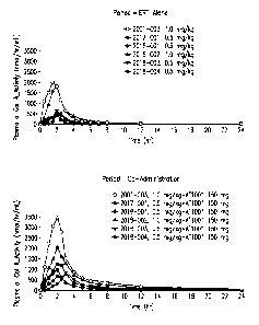
Plasma a-Gal A activity composites of the six patients for Periods 1
and 2 are shown in Figure 1. Plasma a-Gal A activity AIX increases for all
patients
following co-administration with migalastat is shown in Figure 2. Figure 3
shows the
29
CA 02829947 2013-09-11
WO 2012/125402
PCT/US2012/028260
partial AUC's for each sampling time which show increased activity plasma a-
Gal A
activity with co-administration of 0.5 mg,/kg or 1.0 mg/kg agalsidase beta and
150 mg
migalastat.
Skin a-Gal-A Activity Increases with Migalastat
For co-administration with migalastat (Period 2) relative to ERT alone
(Period 1), the following mean increases in skin a-Gal A activity were
observed:
Mean increases for the 0.5 mg/kg agalsidase beta infusion:
= 2.6-fold for 0.5 mg/kg agalsidase beta on Day 2 (N=3, 1 patient
had a lost sample)
* Individual patient increases: 2.8-, 3.9-, and 1.1-fold
* No change in skin activity from Period I on Day 7
Mean increases for the 1.0 mg/kg agalsidase beta infusion:
= 1.9- and 1.5-fold for 1.0 mg/kg agalsidase beta on Days 2 and 7,
respectively (N=2)
Individual patient increases: 1.6- and 2.1-fold on Day 2, 1.7-
and 1.2-fold on Day 7
The increase in skin a-Gal A activity following co-administration of
0.5 mg/kg or 1.0 mg/kg agalsidase beta and 150 mg migalastat are shown in
Figures
4-6. Figure 5A shows the increase in skin a-Gal A activity following co-
administration of 0.5 mg/kg agalsidase beta and 150 mg migalastat in the
patient who
received a 40 min. longer ERT infusion during Period 2 than the other
patients.
PBMC a-Gal-A Activity Increases with Migalastat
For co-administration with migalastat (Period 2) relative to ERT alone
(Period 1), the following mean increases in PBMC a-Gal A activity were
observed:
Mean increases for the 0.5 mg/kg agalsidase beta infusion:
= 2.3-, 2.0-, and 2.2-fold for 0.5 mg/kg agalsidase beta (N=4) on
Days 2, 7, and 14, respectively
= Individual patient increase ranges: 1.4-3.1-, 1.4-2.3-, and 1.7-
2.8-fold on Days 2, 7, and 14, respectively
* No change in skin activity from Period 1 on Day 7
Mean increases for the 1.0 mg/kg agalsidase beta infusion:
CA 02829947 2013-09-11
WO 2012/125402 PCT/US2012/028260
= 1.8-, 4.8- and 3.5-fold for 1.0 mg/kg agalsidase beta (N=2) on Days
2, 7 and 14, respectively
II Individual patient increases: 1.1- and 2.5-, 3.6- and 6.0-
, and
1.7- and 5.4-fold on Days 2, 7, and 14, respectively
The increase in PBMC a-Gal A activity following co-administration of
0.5 mg/kg or 1.0 mg/kg agalsidase beta and 150 mg migalastat are shown in
Figures
7-9.
Conclusion:
150 mg migalastat interaction with 0.5 mg/kg and 1.0 mg/kg agalsidase
beta resulted in a-Gal A activity increases for:
All patients' plasma a-Gal A AUC (N=6)
Al! patients' skin a-Gal A on Day 2 (N=5), but only 3 of 5 patients on Day
7
All patients' PBMC a-Gal A on Days 2, 7, and 14 (N=6)
150 mg migalastat interaction with 0.5 mg/kg or 1.0 mg/kg agalsidase
beta resulted in 2-to 4-fold increases in a-galactosidase A activity AUC, 1.1-
to 3.9-
fold increases in Day 2 skin a-galactosidase A activity, and 1.1- to 6.0-fold
increases
in PBMC a-galactosidase A activity for Days 2, 7, and 14 relative to
agalsidase beta
alone. On Day 7, four patients had increased a-galactosidase A activity in
skin
following co-administration.
The 150 mg migalastat dose increased enzyme activity of the half-dose
of agalsidase beta (0.5 mg/kg) better than the full dose (1.0 mg/kg) up to 24
his post
dose in plasma, skin, and PBMC's; however the reverse was true (1.0 mg/kg >
0.5
mg/kg) at 7 and 14 days post dose in skin and PBMC's. A table summarizing the
results is shown in Figure 10.
For agalsidase beta alone, all patients had increased PBMC a-Gal A
activity relative to baseline at all time points, however 2 patients had
decreased skin
a-Gal A activity on Day 2 relative to baseline following the 0.5 mg/kg
infusion.
EXAMPLE 3: Dosing Regimen for the Treatment of Fabry Disease using
Migalastat Hydrochloride and Agalsidase
The following Example is an update of the study described in Example
2. The present example includes an additional subject for a total of seven
subjects.
31
CA 02829947 2013-09-11
WO 2012/125402 PCT/US2012/028260
The objective of the present example is to evaluate the safety and PK
of two doses of inigalastat HC1 (150 mg and 450 mg) co-administered with ERT
(agalsidase) in males diagnosed with Fabry disease.
Methods This is an ongoing, open-label, non-randomized, 2-stage,
fixed-sequence study. Stage 1 is comprised of 3 periods.
= Period 1: IV infusion of ERT alone.
= Period 2: migalastat HC1 (150 mg) orally administered 2 hours prior to
IV infusion of the ERT (at the same dose as in period 1).
= Period 3: oral administration of migalastat HC1150 mg alone.
Eligible patients: Male, 18 to 65 years old with Fabry Disease.
Inclusion criteria:
= Body Mass Index (BMI) between 18-35.
= Initiated treatment with agalsidase at least 1 month before dosing.
= Estimated creatinine clearance (CLcr) > 50 mL/min at screening.
Exclusion criteria:
a A documented transient isehemic attack, ischemic stroke, unstable
myocardial infarction within 3 months before screening.
= Clinical significant unstable cardiac disease.
= Sensitivity to or concomitant therapy with irninosugars (e.g., miglustat,
miglitol).
Subjects receive their current dose and regimen of agalsidase beta
alone at one infusion (0.5 or 1.0 mg/kg for about 2 hrs) followed by oral
migalastat
HC1 150 mg administered two hours prior to agalsidase beta at their next
infusion.
Five of the current seven subjects received 0.5 mg/kg every two weeks
and two of the seven subjects received a dose of 1.0 mg/kg every four weeks.
In stage 2, a 450 mg dose of migalastat NCI will be studied.
Stages 1 and 2 will be repeated in unique subjects with an ERT
infusion of agalsidase alpha (0.2 mg/kg for about 40 min).
Samples:
Serial blood samples were taken to 24 hours post dose for plasma a-
Gal A activity and protein levels each period. Blood samples for a-Gal A
activity in
peripheral blood mononuclear cells (PBMCs) were taken at predose and on Days
1, 2,
32
CA 02829947 2013-09-11
WO 2012/125402 PCT/US2012/028260
7, and 14 of each period. Punch biopsies for skin a-Gal A activity were taken
at
prcdose Period 1 and on Days 2 and 7 during Periods 1 and 2. Plasma a-Gal A
activity PK parameters include CIllaX,Tmax,AUC04, AUCo-mr, and t4.
Phaffnacokinetic parameters are calculated using standard non-compartmental
procedures (WINNONLIN version 5.0 or higher).
a-Gal A activity in plasma skin, and PBMC lysates were measured by
a fluorescence enzyme assay using 4-methylumbelliferyl-a-D-galactomTanoside (4-
MUG). a-Gal A activity on 4 MUG was measured in vitro following serial
dilutions
to dissociate migalastat.
Western blot analysis of a-Gal A protein were performed on plasma
samples using anti-human a-Gal A antibody. An rha-Gal A (agalsidase) standard
curve was run to calculate the appropriate concentration of a-Gal protein in
each
sample.
The safety parameters included adverse events (AEs), vital signs,
clinical laboratory tests (hematology, serum chemistry, and urinalysis),
electrocardiograms (ECGs), physical examinations, and use of concomitant
medications.
Results: Preliminary results are available for Stage 1, Periods 1 and 2.
Patient Disposition and Demographics: Seven patients with plasma,
skin and PBMC a-Gal A activity from Stage 1, Periods 1 and 2 were evaluated.
= All 7 patients received agalsidase beta alone during Period 1; all
patients were co-administered 150 mg migalastat HC12 hours prior to
initiation of agalsidase beta during Period 2.
= Two patients (identified as Subjects A and B) received IV infusions of
agalsidase beta 1.0 mg/kg for 2-hr durations.
= Five patients (identified as Subjects C, D, E, F, and G) received IV
infusions of agalsidase beta 0.5 mg/kg for 2-hr durations with 1
exception: Subject E was infused 2 hrs and 40 min during Period 2, but
was infused for 2 hours during Period 1.
* All subjects were males with Fabry Disease aged 44-61 years, body
mass index (BMI) ranged from 20.9-29.1 kg/m2 and estimated CLcr
33
CA 02829947 2013-09-11
WO 2012/125402
PCT/US2012/028260
ranged from 54-88 mL/min. The genotype for each subject is
presented in Figure 11.
Safety:
To date, 12 adverse events (AEs) have been reported, one of which
was serious. The serious AE was a transient ischemic attack (TIA) which
occurred
after the screening visit, but prior to dosing, was moderate in severity,
required
hospitalization, and was considered unrelated to study drug by the
investigator. The
TIA resolved without sequalae. All other AEs were mild in severity, all
considered
unrelated to study drug, and most resolved without treatment. Three AEs in
three
different subjects are ongoing: premature atrial contractions, atrial flutter,
and lower
extremity edema, all unrelated to study drug.
Plasma a-Gal A Activity:
The plasma AUC (area under the curve) of a-Gal A activity versus
treatment with 1.0 mg/kg agalsidase beta alone, and treatment with 1.0 mg/kg
agalsidase beta in combination with 150 mg migalastat HCI (with inserted means
and
standard deviations) activity-time profiles is shown in Figure 12 for subjects
A and B.
The combined treatment of 1.0 mg/kg agalsidase beta and 150 mg migalastat HC1
resulted in 2.2 and 1.6 fold increases in a-Gal A activity for subjects A and
B,
respectively, compared to treatment with 1.0 mg/kg agalsidase beta
monotherapy.
The plasma AUC (area under the curve) of a-Gal A activity versus
treatment with 0.5 mg/kg agalsidase beta alone, and treatment with 0.5 mg/kg
agalsidase beta in combination with 150 mg migalastat HCI (with inserted means
and
standard deviations) activity-time profiles is shown in Figure 13 for subjects
C-G.
The combined treatment of 0.5 mg/kg agalsidase beta and 150 mg migalastat HCI
resulted in a 2.0 to 4.2 fold increase in a-Gal A activity compared to
treatment with
0.5 mg/kg agalsidase beta monotherapy.
Plasma a-Gal A activity increased at all time points for most subjects
for co-administration relative to enzyme replacement therapy (ERT) alone with
one
exception. Subject E received an unbalanced infusion, 40 minutes longer during
period 2 which caused relative decreased enzyme activity during the infusion
phase.
However, all Subject E time points post peak activity were increased relative
to ERT
alone. Additionally, consistent with Eng et al., Am. J. Hum. Genet. 68:711-722
(2001), exposures increased in a non-linear manner for a-Gal A activity.
34
CA 02829947 2013-09-11
WO 2012/125402
PCT/US2012/028260
For co-administration with migalastat HCI (Period 2) relative to ERT
alone (Period 1), all subjects had increased plasma a-Gal A activity AUC as
shown in
Figures 12 and 13. The mean increase in plasma a-Gal A activity AUC was 3.0-
fold
for 0.5 mg/kg agalsidase beta. Excluding the 2.0-fold patient with the
unbalanced
infusion duration, the mean increase was 3.2-fold. The mean increase in plasma
a-
Gal A activity AUC was 1.9-fold for 1.0 mg/kg agalsidase beta.
Skin a-Gal A activity
Figure 14 shows that treating subjects with 0.5 mg/kg or 1.0 mg/kg
agalsidase beta in combination with 150 ma migalastat HC1 increased a-Gal A
activity in Day 2 skin samples compared to enzyme monotherapy treatment alone.
Figure 15 shows a-Gal A activity in Day 7 skin samples following monotherapy
and
combined therapy with agalsidase beta and 150 mg migalastat HC1. The following
mean increases in skin a-Gal A activity were observed: 2.6-fold and 1.4-fold
on Days
2 and 7, respectively, for 0.5 mg/kg agalsidase beta (N=5), and 1.9-and 1.5-
fold on
Days 2 and 7, respectively, for 1.0 mg/kg agalsidase beta (N-2).
PMBC a-Gal A activity
The following mean increases in PBMC a-Gal A activity were
observed: 2.4-, 1.9-, and 2.1-fold on Days 2, 7, and 14, respectively, for 0.5
mg/kg
agalsidase beta (N=5), and 1.8-, 4.8- and 3.5-fold for 1.0 mg/kg (N=2)
agalsidase beta
on Days 2, 7 and 14, respectively.
Western Blot analysis
No change was observed in plasma a-Gal A protein by Western Blot
analysis of Period 2 (co-administration) / Period 1 (ERT alone) AUC ratio for
5 out of
7 subjects. However, 2 subjects, (Subject C who received 0.5 mg/kg agalsidase
beta
and Subject G who receive 1.0 mg/kg agalsidase beta) had 20% increases in
protein
amount following co-administration relative to ERT alone (data not shown),
Conclusion
The interaction of 150 mg migalastat with 0.5 mg/kg and 1.0 mg/kg
agalsidase beta resulted in a-Gal A activity increases in plasma, skin, and
PBMCs.
Co-administration of 150 mg migalastat HC1 with agalsidase beta was generally
safe
and well-tolerated.
WO 2012/125402 PCT/US2012/028260
The present application is not to be limited in scope by the specific
embodiments described herein. Indeed, various modifications of the application
in
addition to those described herein will become apparent to those skilled in
the art
from the foregoing description and the accompanying figures. Such
modifications are
intended to fall within the scope of the appended claims.
It is further to be understood that all values are approximate, and are
provided for description.
36
CA 2829947 2018-03-12